a gfp-based genetic screen reveals mutations that disrupt...
TRANSCRIPT

2955
IntroductionWork over the past decade has increased our understanding ofthe mechanisms involved in specifying neuronal connections.Long-range axon guidance in the vertebrate CNS is mediatedby a handful of families of axon guidance molecules, notablyephrins, semaphorins, Slit proteins and netrins (Dickson,2002). Moreover, molecules involved in close-range synapticrecognition have been identified, including cadherins andimmunoglobulin superfamily molecules (Biederer et al., 2002;Serafini, 1999; Yamagata et al., 2002). The visual system,particularly the projection from the retina to the optic tectum,has long served as a discovery vehicle for new mechanisms, aswell as a testing ground for candidate factors (Dingwell et al.,2000; McLaughlin et al., 2003). For example, netrin 1 isrequired to guide retinal axons into the optic nerve (Deiner etal., 1997), while Slit proteins channel RGC axons through theoptic tract (Fricke et al., 2001). Moreover, ephrin A proteinsdirect the mapping of temporal axons onto the anterior tectum(Drescher et al., 1997; Flanagan and Vanderhaeghen, 1998),while semaphorin 3D (Liu et al., 2004) and EphB proteins(Hindges et al., 2002; Mann et al., 2002) carry out theequivalent function along the dorsoventral axis.
Most of the molecular players listed above have beenidentified through biochemical purification or by candidategene approaches. A forward-genetic screen provides analternative strategy to reveal novel genes (or new functions for
known genes) in an unbiased manner. A previous anatomicalscreen for retinotectal projection defects in zebrafish usedfluorescent lipophilic dyes to trace RGC axons as theynavigated to the tectum. Two carbocyanine dyes, DiI and DiO,were injected at different locations into the retina of larvalzebrafish, thus labeling separate subpopulations of RGCsterminating in topographically distinct regions of the tectum(Baier et al., 1996; Karlstrom et al., 1996; Trowe et al., 1996).This large-scale screen uncovered 114 mutations, in about 35genes, disrupting either the pathfinding of axons from the eyeto the tectum or the retinotopic map. Although some of themore specific mutations, such as gna, woe and nev, still awaitmolecular identification, most of the genes have now beencloned (Culverwell and Karlstrom, 2002). Unsurprisingly,retinotectal pathfinding was found to depend on proper brainpatterning (Hedgehog signaling, syu, igu, con, dtr, uml and yot;Nodal signaling, cyc; homeodomain transcription factors, noi)and on components of the extracellular matrix (bal, gpy andsly). A small minority of mutations was found to disruptspecific signaling pathways within the retinal growth cones,such as those mediated by Slit/Robo (ast) (Fricke et al., 2001),heparan sulfate proteoglycans (box and dak) (Lee et al., 2004)or PAM/highwire (esr) (D’Souza et al., 2005).
Although productive with regard to isolating retinotectalmutants, this first screen was laborious and intrinsically limitedto finding only a subset of interesting phenotypes. Important
The retinotectal projection is a premier model system forthe investigation of molecular mechanisms that underlieaxon pathfinding and map formation. Other importantfeatures, such as the laminar targeting of retinal axons, thecontrol of axon fasciculation and the intrinsic organizationof the tectal neuropil, have been less accessible toinvestigation. In order to visualize these processes in vivo,we generated a transgenic zebrafish line expressingmembrane-targeted GFP under control of the brn3cpromoter/enhancer. The GFP reporter labels a distinctsubset of retinal ganglion cells (RGCs), which projectmainly into one of the four retinorecipient layers of thetectum and into a small subset of the extratectalarborization fields. In this transgenic line, we carried outan ENU-mutagenesis screen by scoring live zebrafish larvae
for anatomical phenotypes. Thirteen recessive mutations in12 genes were discovered. In one mutant, ddl, the majorityof RGCs fail to differentiate. Three of the mutations, vrt,late and tard, delay the orderly ingrowth of retinal axonsinto the tectum. Two alleles of drg disrupt the layer-specifictargeting of retinal axons. Three genes, fuzz, beyo and brek,are required for confinement of the tectal neuropil.Fasciculation within the optic tract and adhesion within thetectal neuropil are regulated by vrt, coma, bluk, clew andblin. The mutated genes are predicted to encode moleculesessential for building the intricate neural architecture ofthe visual system.
Key words: Retinal ganglion cell, Tectum, Transgenic, Mutant, Axonguidance, brn3c, pou4f3, Zebrafish
Summary
A GFP-based genetic screen reveals mutations that disrupt thearchitecture of the zebrafish retinotectal projectionTong Xiao, Tobias Roeser, Wendy Staub and Herwig Baier*
Department of Physiology, University of California, San Francisco, Programs in Neuroscience, Genetics, Human Genetics, andDevelopmental Biology, 1550 4th Street, San Francisco, CA 94158-2722, USA*Author for correspondence (e-mail: [email protected])
Accepted 14 April 2005
Development 132, 2955-2967Published by The Company of Biologists 2005doi:10.1242/dev.01861
Research article
Dev
elop
men
t
2956
organizing principles of retinotectal connectivity, such asneuropil assembly and laminar targeting of RGC axons couldnot be investigated with the screening method employed. As aconsequence, the factors that assemble the characteristicarchitecture of optic tract and tectum and coordinate thedevelopment of the visual system are still elusive. We expectthat disruption of some of these factors may lead to relativelysubtle anatomical alterations, whose detection and analysisrequire sensitive methods. As structural changes of the CNSwill ultimately influence its function, mutations in the humanhomologs of these genes might turn out to be responsible forneurological and psychiatric diseases.
We have developed a screening strategy aimed atdiscovering specific disruptions of retinotectal architecture.Enhancer sequences from the zebrafish brn3c gene were usedto drive membrane-targeted GFP in a distinct subset of RGCs.GFP-based forward-genetic screens have been employedextensively in Drosophila and C. elegans, and more recentlyin zebrafish (Lawson et al., 2003). The use of a geneticallyencoded label overcomes the limitations of dye injections.First, the screening assay is rapid, because fish do not need tobe aldehyde fixed and injected. Second, the labeling is robustand reproducible among fish, thus allowing the detection ofeven subtle abnormalities. Finally, the GFP label allows theobservation of the same fish at multiple stages of development.We tested the suitability of the Brn3c:mGFP transgenic line ina screen of 233 ENU-mutagenized F2 families. Our relativelysmall-scale effort (three investigators, 1 year) detected 13 novelphenotypes, including ones in which the retinotectal projectionis delayed or disorganized. These new mutants should add toa cellular and molecular understanding of the processes thatgenerate precise neuronal connections in the visual system andother parts of the nervous system.
Materials and methodsFish breedingZebrafish of the TL strain were raised and bred at 28.5°C on a 14 hlight/10 h dark cycle. Embryos were produced by natural crosses andstaged by hours or days post fertilization (hpf or dpf).
Cloning of zebrafish brn3b and brn3cA 168 bp brn3b fragment was cloned from zebrafish cDNA bydegenerate PCR. Further parts of the sequence were obtained usingdegenerate primers targeted to conserved regions at the 5′ and 3′ endsof the brn3b sequence. At least two clones from independent PCRreactions were sequenced. Primers to the resulting consensussequence of these fragments were used to identify genomic PACclones in a pooled PAC library available from RZPD (Berlin,Germany) (Amemiya and Zon, 1999). Three clones contained brn3b.The remaining brn3b sequence was obtained by direct sequencingfrom PAC clones BUSMP706A1597Q2 and BUSMP706N19174Q2from RZPD. The GenBank Accession Number for brn3b isAF395831.
Zebrafish full-length brn3c was identified as for brn3b, except thatprimers for nested PCR were designed based on a partial cDNAsequence published previously (Sampath and Stuart, 1996). TheGenBank Accession Numbers for full-length brn3c and its first intronare AY995217 and AY995218. The forward primers were 5′-GGCAAT ATA TTC AGC GGC TTT G-3′ and 5′-GCT AAA CTC CTCGTA TTG TTA C-3′. The reverse primers were 5′-GTA TCT TCAGGT TGG CGA GAG-3′ and 5′-GGA GGA AAT GTG GTC GAGTAG-3′. Three positive PAC clones were identified.
Construction of the Brn3c:mGFP transgenic vectorThe brn3c-containing PACs were characterized by restriction digestsand Southern hybridization. A 7.5 kb BspEI fragment was identifiedthat contained part of the brn3c-coding region and 6 kb of upstreamsequence. It was subcloned from PAC clone BUSMP706K02247Q2and partially sequenced. The PAC sequences, together with a 5′ RACEproduct, yielded the remaining parts of the zebrafish brn3c sequenceand identified the putative translation start. The 6 kb promoterfragment was cloned into pG1, a GFP expression vector (kindlyprovided by C.-B. Chien, MPI Tübingen). For membrane targeting,the sequence encoding the first 20 amino acids of zebrafish GAP43(Kay et al., 2004) was generated from two overlappingoligonucleotides, which also contained a Xenopus β-globin ribosomalbinding site. The sequences were 5′-G GAA TTC CAC GAA ACCATG CTG TGC TGC ATC AGA AGA ACT AAA CCG GTT GAGAAG-3′ and 5′-TCC CCC GGG CTG CAG CTG ATC GGA CTCTTC ATT CTT CTC AAC CGG TTT AGT-3′. The oligonucleotideswere fused, filled in with Klenow Polymerase, and cloned into thePstI and EcoRI site of the brn3c promoter. The resulting pTR56 vectorwas used to generate transgenic zebrafish (see below).
Generation of transgenic fishThe insert from vector pTR56 was excised by digestion with NotI andseparated from the vector backbone by agarose gel electrophoresis.The DNA was extracted and eluted in 10 mM triethanolamine (Tris,pH 7.5). Prior to injection, the DNA was diluted in water containing0.05% Phenol Red (Sigma) to 10-20 ng/µl. Injected embryos wereraised to sexual maturity and crossed to identify founder fish. Theembryos from these crosses were scored for their GFP expression andraised. This effort led to the production of several stable lines, one ofwhich was used in this study. The official designation of this line isTG(Brn3c:GAP43-GFP)s356t (http://www.zfin.org).
MutagenesisTo efficiently induce random point mutations in the zebrafish genome,we followed published protocols (van Eeden et al., 1999). Briefly,adult male TL fish were treated three to five times at weekly intervalswith ENU (3.0 mM, 1 hour). Four weeks after the last treatment, theywere outcrossed to produce up to 200 F1 fish per male. These F1 fishwere crossed to Brn3c:mGFPs356t carriers to generate F2 families. Sixor more pairs of random crosses were set up between siblings for eachF2 family. In total, 233 F2 families were screened.
Screening assayEmbryos (3 dpf) and larvae (6 dpf) were embedded in 2.5%methylcellulose in E3 medium (5 mM NaCl, 0.17 mM KCl, 0.33 mMCaCl2, 33 mM MgSO4) and screened under a Leica MZ25fluorescence-equipped dissecting microscope with 100�magnification. Embryos with obvious morphological defects prior to3 dpf were discarded. We scored the presence of retinal fibers in thetectum (particularly at 3 dpf), the trajectory of GFP-positive fibers inthe optic tract, the width and shape of the optic tract, the density oftectal innervation, the size and shape of the tectal neuropil, the layerstructure of the tectum, and the appearance of the RGC axonalmeshwork in the tectal neuropil. Crosses in which a quarter of the F3progeny showed a mutant phenotype were repeated. Two or three suchre-tests were carried out before a mutant was considered a realcandidate. In these cases, one or both of their parents were outcrossedto unrelated Brn3c:mGFP carriers to establish a mutant stock. Themutation was recovered in the next generation through randompairwise crosses. All mutants described in this report have beenpropagated as heterozygous stocks for at least three generations.
ImmunohistochemistryMutant embryos/larvae were obtained by crosses of heterozygouscarriers. Anaesthetized embryos/larvae were fixed in 4%paraformaldehyde (PFA) in phosphate-buffered saline. To enhance
Development 132 (13) Research article
Dev
elop
men
t

2957GFP screen for retinotectal mutants
permeability, PFA-fixed larvae older than 3 dpf were incubated in0.1% collagenase in PBS. Incubation times for 3 dpf and 5 dpf were45 minutes and 2 hours, respectively. For horizontal sections,embryos/larvae were cryoprotected in 30% sucrose/0.02% azide/PBS,embedded in OCT (Tissue-Tek, Sakura), and sectioned at 12 µmhorizontally on a cryostat. The following antibodies andconcentrations were used for whole-mount immunohistochemistry:anti-GFP, 1:40,000 (Molecular Probes); zn5, 1:1000 (AntibodyFacility, University of Oregon); anti-acetylated tubulin, 1:2000(Sigma); zpr-1, 1:200 (Developmental Studies Hybridoma Bank,University of Iowa); anti-phosphohistone H3, 1:500 (UpstateBiotechnology); and anti-parvalbumin, 1:1200 (Chemicon). TUNEL-based cell death assays were carried out according to manufacturer’sinstructions (In Situ Cell Death Detection Kit – Fluorescein, Roche).
Labeling of the retinotectal projection with carbocyaninedyesFish were deeply anesthetized in 0.016% tricaine (Sigma) and fixedin 4% PFA in PBS for 2-24 hours. They were embedded in 1% agarosein 0.5� PBS) and pressure injected with dye solution using aPV-820 pressure injector (World Precision Instruments, Sarasota,FL). The carbocyanine dye DiI (1,1′-dioctadecyl-3,3,3′,3′-tetramethylindocarbocyanine perchlorate, Molecular Probes D-282)was dissolved either in N,N-dimethylformamide (DMF) forfixed larvae or in ethanol for live larvae to a concentration of 2%(m/v). DiO (1,1′-dioctadecyl-3,3,3′,3′-tetramethyloxacarbocyanineperchlorate, Molecular Probes D-275) was dissolved in chloroform(2% m/v). To visualize the retinotectal map, DiI and/or DiO wereinjected into the eyes of larvae and imaged 12-24 hours later.
Confocal imagingLarvae were embedded in a 0.5�20 mm imaging chamber(CoverWell, Grace Bio-labs) in 1.2% low melting point agarosedissolved in E3 medium containing 0.8% norepinephrine and 0.016%tricaine. For immunohistochemistry, the left eye was removed andstained embryos/larvae were mounted laterally in 70% glycerol inPBS. Confocal image stacks were acquired using either a BioRad1024M or a Zeiss 510 META laser-scanning microscope. Longworking distance objectives, 20� (air, NA 0.5) and 40� (water, NA0.8), were used. To reconstruct axons and their arbors, a series of
optical planes were collected (z-stack) and collapsed into a singleimage (maximum intensity projection) or rendered in threedimensions to provide views of the image stack at different angles.The step size for each z-stack was chosen upon calculation of thetheoretical z-resolution of the objective used (typically 0.5-1 µm).
ResultsRegulatory regions of brn3c drive stable expressionof GFP in a subset of RGCsWe identified zebrafish brn3c (brn3.1, pou4f3) by PCRcloning. The putative amino acid sequence of Brn3c consistsof 331 amino acids. Compared to mouse Brn3c, 81% of theamino acid positions are identical within the total sequence and97% within the POU-domain. We confirmed the annotation byalso cloning a highly related gene, brn3b (brn3.2, pou4f2)(DeCarvalho et al., 2004). A phylogenetic tree based onClustalW sequence comparison (Higgins and Sharp, 1988)places the zebrafish Brn3b and Brn3c sequences into separateclades together with their mammalian orthologs (data notshown).
To generate a stable transgenic zebrafish line able to driveGFP expression in RGCs, we isolated about 6 kb of genomicsequence upstream of the brn3c-coding region and cloned itinto a GFP expression vector (Fig. 1A) (see Materials andmethods). Enhanced GFP, fused to the first 20 amino acids ofGAP43, served as the marker gene, as described previously(Kay et al., 2004). The GAP43 sequence provides a membrane-targeting signal and ensures complete labeling of neurites(Zuber et al., 1989). Injection of the construct at the one-cellstage resulted in transient expression in select populations ofneurons. Fish that showed bright expression in a large numberof cells were raised to adulthood. Eighty-four injected fishwere outcrossed against wild-type fish and analyzed forgermline transmission. A total of 10 germline transgenicfounder fish (12% of the injected embryos) were identified byvisual inspection of their progeny under a fluorescence-
Fig. 1. A large subset of RGCs express GFP in thestable Brn3c:mGFP transgenic line. (A) Schematicdrawing of the DNA construct used to generate theBrn3c:mGFP transgenic line. (B) Lateral view of56 hpf live Brn3c:mGFP transgenic embryoshowing GFP expression by RGCs andmechanosensory hair cells (neuromasts) of thelateral line and inner ear. The optic nerve is visible.(C-E) Fixed Brn3c:mGFP transgenic embryoslabeled by whole-mount immunohistochemistry.(C) Ventral view of a 42 hpf retina, labeled withanti-GFP. Anterior (nasal) is upwards. GFPexpression starts in a small cluster of cells in theventronasal retina, near the optic fissure. Thearrowhead indicates the first GFP-positive axonsexiting the eye through the optic stalk. (D) Lateralview of a 44 hpf retina, labeled with anti-GFP ingreen and zn5 in red. GFP has spread into centralretina. The onset of Brn3c:mGFP transgeneexpression follows that of zn5 by ~6 hours.(E) Lateral view of a 72 hpf retina, labeled withanti-GFP. GFP-positive RGCs are distributeduniformly throughout the ganglion cell layer. Scalebars: 200 µm in B; 20 µm in C-E.
Dev
elop
men
t
2958
equipped dissecting microscope. Offspring of these founderfish were raised to establish lines. The experiments describedhere were carried out in transgenic line TG(Brn3c:mGFP) s356t.
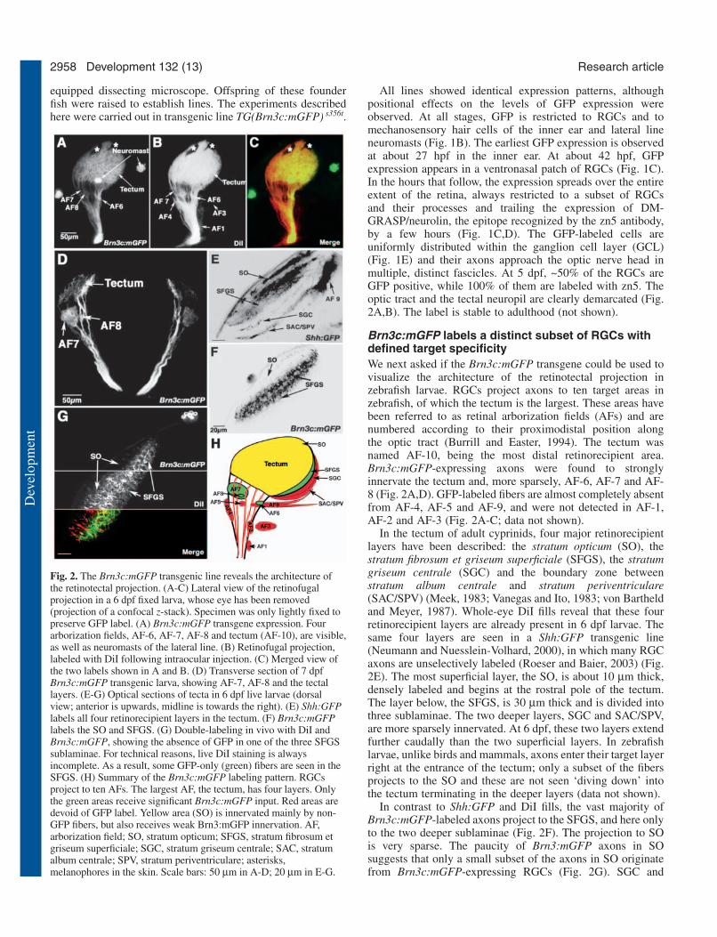
All lines showed identical expression patterns, althoughpositional effects on the levels of GFP expression wereobserved. At all stages, GFP is restricted to RGCs and tomechanosensory hair cells of the inner ear and lateral lineneuromasts (Fig. 1B). The earliest GFP expression is observedat about 27 hpf in the inner ear. At about 42 hpf, GFPexpression appears in a ventronasal patch of RGCs (Fig. 1C).In the hours that follow, the expression spreads over the entireextent of the retina, always restricted to a subset of RGCsand their processes and trailing the expression of DM-GRASP/neurolin, the epitope recognized by the zn5 antibody,by a few hours (Fig. 1C,D). The GFP-labeled cells areuniformly distributed within the ganglion cell layer (GCL)(Fig. 1E) and their axons approach the optic nerve head inmultiple, distinct fascicles. At 5 dpf, ~50% of the RGCs areGFP positive, while 100% of them are labeled with zn5. Theoptic tract and the tectal neuropil are clearly demarcated (Fig.2A,B). The label is stable to adulthood (not shown).
Brn3c:mGFP labels a distinct subset of RGCs withdefined target specificityWe next asked if the Brn3c:mGFP transgene could be used tovisualize the architecture of the retinotectal projection inzebrafish larvae. RGCs project axons to ten target areas inzebrafish, of which the tectum is the largest. These areas havebeen referred to as retinal arborization fields (AFs) and arenumbered according to their proximodistal position alongthe optic tract (Burrill and Easter, 1994). The tectum wasnamed AF-10, being the most distal retinorecipient area.Brn3c:mGFP-expressing axons were found to stronglyinnervate the tectum and, more sparsely, AF-6, AF-7 and AF-8 (Fig. 2A,D). GFP-labeled fibers are almost completely absentfrom AF-4, AF-5 and AF-9, and were not detected in AF-1,AF-2 and AF-3 (Fig. 2A-C; data not shown).
In the tectum of adult cyprinids, four major retinorecipientlayers have been described: the stratum opticum (SO), thestratum fibrosum et griseum superficiale (SFGS), the stratumgriseum centrale (SGC) and the boundary zone betweenstratum album centrale and stratum periventriculare(SAC/SPV) (Meek, 1983; Vanegas and Ito, 1983; von Bartheldand Meyer, 1987). Whole-eye DiI fills reveal that these fourretinorecipient layers are already present in 6 dpf larvae. Thesame four layers are seen in a Shh:GFP transgenic line(Neumann and Nuesslein-Volhard, 2000), in which many RGCaxons are unselectively labeled (Roeser and Baier, 2003) (Fig.2E). The most superficial layer, the SO, is about 10 µm thick,densely labeled and begins at the rostral pole of the tectum.The layer below, the SFGS, is 30 µm thick and is divided intothree sublaminae. The two deeper layers, SGC and SAC/SPV,are more sparsely innervated. At 6 dpf, these two layers extendfurther caudally than the two superficial layers. In zebrafishlarvae, unlike birds and mammals, axons enter their target layerright at the entrance of the tectum; only a subset of the fibersprojects to the SO and these are not seen ‘diving down’ intothe tectum terminating in the deeper layers (data not shown).
In contrast to Shh:GFP and DiI fills, the vast majority ofBrn3c:mGFP-labeled axons project to the SFGS, and here onlyto the two deeper sublaminae (Fig. 2F). The projection to SOis very sparse. The paucity of Brn3:mGFP axons in SOsuggests that only a small subset of the axons in SO originatefrom Brn3c:mGFP-expressing RGCs (Fig. 2G). SGC and
Development 132 (13) Research article
Fig. 2. The Brn3c:mGFP transgenic line reveals the architecture ofthe retinotectal projection. (A-C) Lateral view of the retinofugalprojection in a 6 dpf fixed larva, whose eye has been removed(projection of a confocal z-stack). Specimen was only lightly fixed topreserve GFP label. (A) Brn3c:mGFP transgene expression. Fourarborization fields, AF-6, AF-7, AF-8 and tectum (AF-10), are visible,as well as neuromasts of the lateral line. (B) Retinofugal projection,labeled with DiI following intraocular injection. (C) Merged view ofthe two labels shown in A and B. (D) Transverse section of 7 dpfBrn3c:mGFP transgenic larva, showing AF-7, AF-8 and the tectallayers. (E-G) Optical sections of tecta in 6 dpf live larvae (dorsalview; anterior is upwards, midline is towards the right). (E) Shh:GFPlabels all four retinorecipient layers in the tectum. (F) Brn3c:mGFPlabels the SO and SFGS. (G) Double-labeling in vivo with DiI andBrn3c:mGFP, showing the absence of GFP in one of the three SFGSsublaminae. For technical reasons, live DiI staining is alwaysincomplete. As a result, some GFP-only (green) fibers are seen in theSFGS. (H) Summary of the Brn3c:mGFP labeling pattern. RGCsproject to ten AFs. The largest AF, the tectum, has four layers. Onlythe green areas receive significant Brn3c:mGFP input. Red areas aredevoid of GFP label. Yellow area (SO) is innervated mainly by non-GFP fibers, but also receives weak Brn3:mGFP innervation. AF,arborization field; SO, stratum opticum; SFGS, stratum fibrosum etgriseum superficiale; SGC, stratum griseum centrale; SAC, stratumalbum centrale; SPV, stratum periventriculare; asterisks,melanophores in the skin. Scale bars: 50 µm in A-D; 20 µm in E-G.
Dev
elop
men
t

2959GFP screen for retinotectal mutants
SAC/SPV do not receive Brn3c:mGFP-labeled input. Insummary, these labeling studies show that the Brn3c:mGFPtransgene labels a distinct subpopulation of RGCs withrestricted target specificity (Fig. 2H). Therefore, Brn3c:mGFPreveals details of the retinotectal architecture that would bemasked by an unselective RGC stain. We employed this linenext in a small-scale screen in search of new retinotectalmutants.
Overview of the screenFor the screen, we mutagenized the spermatogonia of 17 adultmale zebrafish with ENU and outcrossed them to non-mutagenized females. The mutation rate was determined to beabout 0.3% per gene per haploid genome. We used thepigmentation gene sandy (tyrosinase) as the specific locus forits easily detectable phenotype (Page-McCaw et al., 2004). TheF1 fish were then crossed to Brn3c:mGFP carriers to generate233 F2 families. A total of 1303 crosses were screened byvisual inspection of the GFP-labeled retinotectal projection.Based on the varying number of crosses per individual F2family (average 5.6, corresponding to 0.8 genomes), wecalculated that our screen encompassed 168 genomes (Mullinset al., 1994).
The screen was carried out on F3 progeny at 3 dpf and againat 6 dpf. These two developmental stages are particularlyinformative for revealing perturbations in the retinotectalprojection. In wild type, RGC axons reach the anteroventralboundary of the tectum at 48 hpf. One day later, at 72 hpf (3dpf), RGC axon arbors reach the posterodorsal boundary of thetectum and completely cover the tectum (Stuermer, 1988).Thus, screening at 3 dpf enabled us to uncover mutationsdisrupting the initial stages of the retinotectal projection.Between 3 dpf and 6 dpf, RGC axonal arbors elaboratesynaptic connections with tectal neurons. Screening at 6 dpf
thus allowed us to discover mutations affecting the refinementand maintenance of the retinotectal projection.
Progeny of crosses from 65 F2 families initially showed aputative mutant phenotype and were re-screened by mating thesame pair and scoring their F3 progeny again. Less than onethird of these (21) were confirmed and outcrossed toBrn3c:mGFP transgenic wild type. All but two mutants wererecovered in the next generation. However, six were consideredunspecific upon closer inspection and discarded. The discardedmutants had smaller tecta or smaller eyes, owing todegeneration or early developmental problems. All mutantswere sectioned at 6 dpf and examined histologically usingDAPI to highlight cytoarchitecture of retina and tectum, inconjunction with anti-GFP. Two mutations with similarphenotypes were found to be alleles of the same gene bycomplementation analysis. As described below, we discovered13 mutations in 12 genes, which have been grouped into fiveclasses (Table 1). All mutants are recessive, completelypenetrant with respect to their axon phenotype and aretransmitted in Mendelian ratios. Their phenotypes uncoverlargely unexplored processes in the assembly of neuralarchitecture.
The daredevil (ddl) gene is important for thedifferentiation of most RGCsWe identified a mutant, ddl, showing a severe reduction ofretinal axons that innervate the tectum at 3 dpf (Fig. 3A,B).The optic tract is thin, and RGC axons do not cover the entiretectum. Closer investigation revealed that the primary defect ofthis mutant is in generating RGCs. Between 42 and 48 hpf,when newly differentiating RGCs begin to expressBrn3c:mGFP in wild type, GFP expression in the ddl retina issparse. This is in contrast to GFP expression in hair cells,which is comparable with wild type in strength and time of
Table 1. Summary of retinotectal mutants identified in the Brn3c:mGFP screenViability
Gene (abbreviation) Alleles Retina Background adaptation* Other phenotypes (maximal age)
Reduced number of retinal axonsdaredevil (ddl) s563 Fewer RGCs, wider Dark Heart conductivity defect, swimbladder 6 dpf
ciliary margin, fewer not inflatedPhR
Delayed innervation of the tectumvertigo (vrt) s1614 Normal Dark Balance deficit, swimbladder not inflated 10 dpflate bloomer (late) s551 Normal Normal None detected Adulttarde demais (tard) s587 Normal Mildly dark Swimbladder not inflated 14 dpf
Laminar specificity defectdragnet (drg) s510, s530 Normal Mildly dark, variable Lens covered by extra cells Adult
Deficient confinement of axons to tectal neuropilfuzz wuzzy (fuzz) s531 Normal Mildly dark Swimbladder not inflated 10 dpfbeyond borders (beyo) s578 ‘Smiling’ IPL at margin Mildly dark Reduced forebrain, swimbladder not inflated 10 dpfbreaking up (brek) s574 RGC axon degeneration Dark after 5 dpf Swimbladder not inflated 10 dpf
Disorganized fascicles in the tectumblue kite (bluk) s582 Normal Dark Swimbladder not inflated 10 dpfclewless (clew) s567 Normal Mildly dark Lens degeneration Adultblind date (blin) s573 Normal Mildly dark, recover None detected Adult
after 7 dpfcoming apart (coma) s532 Misplaced RGCs at Normal Swimbladder not inflated 10 dpf
ciliary margin
*Background adaptation is a crude indicator of visual function (Neuhauss et al., 1999). Visually impaired fish are often unable to measure ambient light levelsand are dark, owing to dispersal of melanin pigment in their skin.
IPL, inner plexiform layer.
Dev
elop
men
t

2960
onset (data not shown). After 60 hpf, when wild-type retina isuniformly filled with GFP-positive RGCs, the ddl retinacontains less than 10% of the GFP-positive population (rangingfrom three to a few dozen individual RGCs scattered over theretina) (Fig. 3C,D). The few remaining RGCs send out axons,projecting in a straight course to the tectum and reach the targetbefore and around 3 dpf, the same time as wild type (Fig.3A,B). We asked if the retinotopic map was intact, despite thisdramatic depletion of RGCs. Following injection of DiI intothe nasal retina and DiO into the temporal retina of analdehyde-fixed wild-type larva (3 dpf), we observed that nasalaxons invariably projected to the posterior tectum and temporalaxons to the anterior tectum (Fig. 3E), as reported before(Stuermer, 1988; Baier et al., 1996). Perhaps surprisingly,given the sparse filling of the tectum, this topographicrelationship is retained in ddl mutants (Fig. 3F).
To test whether only the Brn3c-expressing subpopulationwas reduced, we stained the 3 dpf retina with zn5 and HuCantibodies, which label all RGCs (zn5) or RGCs and amacrinecells (HuC). This experiment showed that most RGCs areabsent in ddl mutants (Fig. 3G-J). Amacrine cells are alsoreduced in number (data not shown). DAPI staining further
suggested that photoreceptors are undifferentiated,but overall retinal lamination is unaffected (Fig.3K,L). The ciliary margin of this mutant isenlarged (Fig. 3I-L) and anti-phospho-histone H3labeling demonstrated an increase in the numberof mitotically active progenitors in this region(Fig. 3M,N). At 3 dpf, ddl mutants have normalbody morphology and are indistinguishable fromtheir wild-type siblings, except for their retinaphenotype. After 3 dpf, the eye is visibly smaller,and the swimbladder fails to inflate. At about 3.5dpf, the hearts of ddl mutants begin to show
arrhythmia, suggestive of a conductivity defect. At 4 dpf,mutants can be sorted based on pericardial edema; they die ataround 6 dpf.
Innervation of the tectum is delayed in vertigo (vrt),tarde demais (tard), and late bloomer (late) mutantsAt 72 hpf, RGC axons already occupy the entire tectal neuropil.However, the tectum is immature at this stage andretinorecipient layers are not detectable. Large growth conesare seen, and axon arbors appear to spread out without thecharacteristic adhesion between branches, which is seen lateron (see below). Three mutants, vrts1614, tards587 and lates551,were found to have few if any arbors in the tectum at 3 dpf,although tectal architecture (as judged by DAPI histology)appears normal. Strikingly, the retinotectal projection in thesemutants recovers later on.
vrt mutants exhibit the most severe delay. Once reaching theboundary of the tectum, RGC axons stall (Fig. 4A,B). After 4dpf, the axons resume their growth into the tectum, which theyeventually cover completely (Fig. 4C-F). Prior to 3 dpf, RGCdifferentiation and axon pathfinding are normal and onschedule (Fig. 4G,H). The number of RGCs, expressing GFP
Development 132 (13) Research article
Fig. 3. daredevil (ddl) mutants have fewer RGCs.Analysis of cell-type markers in wild type(A,C,E,G,I,K,M) and ddl (B,D,F,H,J,L,N).(A,B) Lateral views of 78 hpf tecta, labeled withwhole-mount anti-GFP. Broken lines outlineboundaries of the tectal neuropil. The wild-type tectum(A) is covered by axons. Few axons can be detected inthe ddl tectum (B). (C,D) Confocal images of retinas inlive 60 hpf embryos. The number of GFP-positiveRGCs is greatly reduced in ddl (D). (E,F) Analysis ofthe retinotopic map in 72 hpf wild type (E) and ddl (F).DiI (red) and DiO (green) were pressure injected intonasal and temporal retina, respectively (see inserts forillustration of retinal injection sites). The grosstopography of axon targeting in ddl mutants is notaffected, with nasal axons still projecting to theposterior tectum and temporal axons to the anteriortectum (F). (G-J) Whole-mount Zn5 staining of 72 hpfretinas. (G,H) Lateral views. (I,J) Dorsal views. Thenumber of zn5-positive RGCs is greatly reduced in ddl.(K,L) Sections of 78 hpf retinae, labeled with anti-GFP(green) and DAPI (blue). The ciliary margin (betweenarrowheads) in the ddl retina is wider than in wild type.(M,N) Dorsal views of 72 hpf whole-mount retinae,labeled with anti-phosphohistone H3 (H3P). Thenumber of dividing cells (arrows) is greatly increasedat the ciliary margin in ddl. Scale bars: 50 µm in A andE (for tectum panels); 20 µm in G (for retina panels).
Dev
elop
men
t

2961GFP screen for retinotectal mutants
or stained with the zn5 antibody, is identical to wild type, andtheir axons follow the normal path across the midline andtowards the tectum. This suggests that the vrt mutationspecifically impairs one particular stage of axon growth: the
entry into the tectum. Interestingly, the optic tract is noticeablywider in vrt mutants. The thickening of the optic tract couldbe the result of reduced fasciculation or of excessivebackbranching by axons that are stalled at the anterior pole ofthe tectum. vrt mutants lack a swimbladder and die at around10 dpf.
tard and late mutants display a milder delay of theretinotectal projection than vrt. At 3 dpf, axon arbors can beobserved in anterior tectum, but not in posterior tectum (Fig.5A-C). The optic tracts are morphologically normal at alltimes. RGC axon growth recovers by 4 dpf, and at 6 dpf thetectum is completely covered by fibers (Fig. 5D-F). The tectalneuropil in tard mutants retains an abnormal shape and itsmargins are less delineated than in wild type (Fig. 5E). tardmutants fail to inflate their swim bladders and die at around 2weeks of age. By contrast, late mutants are morphologicallyinconspicuous, including their tectum (Fig. 5F), have swimbladders and are adult viable. In fact, they can only bedistinguished from their wild-type siblings by the delayedinnervation of the tectum around 3 dpf.
Lamination of retinal input is disrupted in dragnet(drg) mutantsWe identified two alleles, drgs510 and drgs530, of a geneimportant for targeting retinal axons to the appropriate tectallayer. drgs530 results in a somewhat weaker phenotype thandrgs510. In wild-type zebrafish larvae, most axons extenddirectly into a specific layer at the entrance of the tectum andremain confined to the chosen layer (data not shown).Brn3c:mGFP-labeled RGCs project mostly to the two deepsublaminae of the SFGS, and the SO is only lightly labeled(see Fig. 2F,G; Fig. 6A,C). In drg mutants, this pattern oflaminar targeting is perturbed (Fig. 6B,D). Ectopic fascicles,2-4 µm in diameter, are observed in the SO, apparentlydisplaced from the SFGS. GFP-labeled axons travel betweenthe two layers. The entire tectum is innervated in this mutant,and both tectal cytoarchitecture (by DAPI histology; data notshown) and retinotopic mapping are intact (Fig. 6E,F). Thelamination defects are detectable as early as 3.5 dpf and arespecific to the tectum. Sublaminar targeting of amacrineprocesses and of RGC dendrites in the inner plexiform layerof the retina is normal, as shown by anti-parvalbumin and anti-GFP double-labeling (Fig. 6G-N). The only other detectablephenotype of drg mutants is an opaque lens after 5 dpf, causedby overgrowth by epithelial cells (Fig. 6G,H). drg mutants arefully viable as adults.
Neuropil boundaries dissolve in fuzzy wuzzy (fuzz),beyond borders (beyo) and breaking up (brek)mutantsIn fuzzs531, beyos578 and breks574 mutants, the retinotectalprojection forms on time and is initially indistinguishable fromwild type, but the density of axon arbors is reduced after 5 dpf.Optic tracts are of normal width and show the characteristicbranching pattern, as the RGC fibers enter the tectum. Thissuggests that the normal complement of axons is present, butthat their arbors are smaller or have fewer branches. Theborders of the retinotectal fiber zone are less well demarcated(‘fuzzy’) in these mutants, with axons and growth conesfrequently straying outside the neuropil area (Fig. 7). Tectalcytoarchitecture is unaltered, as determined by DAPI staining.
Fig. 4. vertigo (vrt) mutants show severely delayed innervation of thetectum. Analysis of retinal axon projection in wild type (A,C,E,G)and vrt mutants (B,D,F,H). (A-F) Lateral views of the retinotectalprojection in Brn3c:mGFP transgenic fish, labeled with anti-GFP. At3 dpf, the wild-type tectum (A) is fully innervated, while the vrttectum (B) is devoid of axons. Broken lines outline the tectumboundaries. In 4 dpf wild-type larvae (C), the density of axon arborsis increased compared with 3 dpf (A), and dorsal and ventralbranches are clearly visible in the optic tract. In vrt (D), axons haveinvaded the anterior tectum, and the optic tract (OT) is abnormallywide (arrowheads). At 6 dpf, axons innervate the whole tectum in thevrt mutant (F) similar to wild type (E) The optic tract remains widerthan normal. (G,H) Dorsal views of 48 hpf retinas and optic nerves,labeled with zn5. The number of RGCs and their axons is similarbetween wild type (G) and vrt (H). Scale bars: 50 µm.
Dev
elop
men
t

2962 Development 132 (13) Research article
Fig. 5. tarde demais (tard) and late bloomer(late) mutants show mildly delayedinnervation of the tectum. (A-F) Confocalimages of Brn3c:mGFP labeled retinotectalprojections of 80 hpf tecta in live larvae (A-C). The wild-type tectum (A) is filled withretinal axons. The optic tract has branchedinto stereotyped fascicles, labeled withnumbers. The tard tectum (B) and the latetectum (C) are less than halfway innervated.(D-F) Lateral views of 6 dpf tecta. The tardtectum and the late tectum are now coveredwith axons. However, the tard tectum issmall and its boundary remains abnormal(E), compared with wild type (D) and incontrast to late (F). Asterisks indicatemelanophores on the skin. Broken linesoutline the tectal neuropil. Scale bars:20 µm.
Fig. 6. Laminar specificity is perturbed in dragnet(drg) mutants. (A-D) Analysis of retinorecipientlayers in 6 dpf wild type (A,C) and drg (B,D).(A,B) Z projections of confocal image stacks,labeled with Brn3c:mGFP. Larvae are mounted at aslightly oblique angle to better visualize the gapbetween SO and SFGS. (C,D) Optical sections ofBrn3c:mGFP tecta, stained with whole-mount anti-acetylated tubulin (red) and anti-GFP (green).Arrowheads indicate ectoptic axon fasciclestraveling between SO and SFGS in drg mutants.(E,F) Analysis of the retinotopic map in 6 dpf wildtype (E) and drg (F). DiI (red) and DiO (green) werepressure injected into ventronasal and dorsotemporalretina, respectively (see inserts for illustration ofinjection sites). Axon targeting in drg (F) iscomparable with wild type (E), suggesting thatpositional information along the retinotopic axes isintact. (G-N) Analysis of the inner plexiform layer(IPL) in sections of 6 dpf retina (see M,N for labelsof retinal layers). (G,H) DAPI labeling. (I,J) Anti-GFP labeling to visualize the four sublaminae towhich Brn3c:mGFP RGC dendrites project.(K,L) Anti-parvalbumin labeling, to highlight asubpopulation of amacrine cells that project to threesublaminae in the IPL. (M,N) Triple-labeling(merged image) showing DAPI (blue), anti-GFP(green) and anti-parvalbumin (red). Formation ofIPL sublaminae is not affected in drg. An apparentlyunrelated phenotype of drg can be seen using theDAPI stain: an abnormal aggregation of cells infront of the drg lens (H). Asterisks indicatemelanophores in the skin. Scale bars: 20 µm in A,G.
Dev
elop
men
t

2963GFP screen for retinotectal mutants
In brek mutants, the GFP label in the tectal neuropil becomespunctate after 4 dpf, sometimes as late as 6 dpf, indicating thataxons are disintegrating in this mutant (see Fig. S1 in thesupplementary material). In a substantial proportion of fish,axon degeneration is uneven, with one region of the tectuminitially more affected than another, although a time-courseanalysis showed that the temporal pattern is inconsistentbetween animals (n=6). We could not detect an excessivenumber of TUNEL-positive cells in the mutant retina at 5 dpf,suggesting that axon retraction is not secondary to RGC deathin the retina (data not shown). The axon degenerationphenotype is not seen in fuzz or beyo. In beyo mutants, the innerplexiform layer of the retina is misshaped at the margins,bending distally to the outer nuclear layer (see Fig. S2 in thesupplementary material), and the telencephalon is reduced(data not shown). All three mutants do not inflate theirswimbladders and die as young larvae.
Fiber organization is disrupted in clewless (clew),blue kite (bluk), coming apart (coma) and blind date(blin) mutantsIn wild type, axons are sorted in the optic tract according totheir topographic position and enter the tectum through a setof stereotyped fascicles (Stuermer, 1988) (see Fig. 5C). Afterentering the tectum, axon arbors do not spread uniformly, butrather form a characteristic grid within the neuropil, withregions of high fiber density evidently separated by gaps. Thickfascicles circle the perimeter of the tectum, from which singleaxons depart at various positions to navigate to their respective
targets in the center of the neuropil. Thin fascicles are alsoobserved traveling through the wild-type tectum. Thisorganization becomes evident in confocal images ofBrn3c:mGFP tecta at high magnification (Fig. 8A,B).
In a heterogeneous group of four mutants, clews567, comas532,bluks582 and blins573, the RGC fibers are more diffuse and lessmesh-like than in wild type (Fig. 8C,D), or appear to meanderin the neuropil (Fig. 8E,F). We interpret the ‘diffuse’phenotype as a lack of fiber-fiber adhesion, but the apparentdispersion may have other causes as well. Although tectalcytoarchitecture appears overall normal (by DAPI histology),the tectum is slightly smaller in coma and bluk. In comamutants, the optic tract is already disorganized upon enteringthe tectum: axons preferentially join the dorsal and ventralbranches and appear to avoid the more centrally positionedfascicles (Fig. 8F). The fascicles circling the tectum are alsothicker and more compact. However, DiI and DiO double-labeling of RGC axons originating from the nasoventral andtemporodorsal quadrants, respectively, demonstrated that theretinotopic map is intact (data not shown). The coma mutantalso has a specific retinal defect: a small number of thenewborn RGCs at the ciliary margin are mispositioned in thedistal retina near the inner nuclear layer. These cells expressGFP at the same intensity as regularly positioned RGCs andexhibit a neuronal morphology (see Fig. S2 in thesupplementary material). The coma and bluk mutations arelarval lethal, while blin and clew mutants are adult viable.
DiscussionWe generated a transgenic zebrafish line that expressesmembrane-targeted GFP under control of a brn3c enhancerfragment. Brn3c is a POU-domain transcription factor, whichis involved in RGC differentiation and axon outgrowth (Liu etal., 2000; Wang et al., 2002). The Brn3c:mGFP transgene isexpressed in a subset of RGCs and in mechanosensory haircells, similar to endogenous brn3c (DeCarvalho et al., 2004;Erkman et al., 1996; Xiang et al., 1995). The opticaltransparency of zebrafish embryos and larvae allowed us tovisualize the retinotectal projection in living fish, relying solelyon the crisp labeling of RGC axons by membrane-bound GFP.We could thus investigate, at unprecedented resolution, tectalneuropil organization, layer formation, fasciculation and thetime course of innervation in both wild-type and mutantzebrafish. A screen of 168 ENU-mutagenized genomes in theBrn3c:mGFP background revealed 13 mutations disrupting theorderly innervation of the tectum (Fig. 9). The affected genesmay be expressed in the RGCs or the tectum, or both. Thenewly discovered mutant phenotypes can be categorized intofive groups (Table 1), none of which has, to our knowledge,been described before.
We designed our new GFP-based screen to find phenotypesthat would have escaped discovery in the earlier screen(Karlstrom et al., 1996; Trowe et al., 1996) or would have beendifficult to analyze in sufficient detail using carbocyaninetracing alone (Burrill and Easter, 1994; Kaethner and Stuermer,1992; Stuermer, 1988). We concentrated on mutationsdisrupting the fine architecture and temporal coordination ofthe retinotectal projection, taking advantage of the highlyreproducible label afforded by a genetically encoded reporter,by its stability, and by its cell-type specific expression pattern.
Fig. 7. Tectal neuropil boundaries dissolve in fuzz wuzzy (fuzz),beyond borders (beyo) and breaking up (brek) mutants. (A-D) Z-projections of confocal image stacks showing lateral views of tecta in6 dpf live Brn3c:mGFP larvae. The boundary of the tectum issmooth and well defined in wild type (A). In fuzz (B), axon arborsare less dense in the tectal neuropil and often overshoot the tectalboundary (arrowheads). The overshooting phenotype is more severein beyo (C) and brek (D). Asterisks indicate melanophores. Scalebars: 20 µm.
Dev
elop
men
t

2964
Although the scale of our new screen was less than one-tenthof the original retinotectal screen, it was very productive indetecting specific phenotypes. The previous screen hadenriched for mutants with pleiotropic phenotypes. As a result,of the 114 mutants described, all but eight (7%) die asyoung larvae, indicating pervasive, non-visual defects. Forcomparison, of the 13 new mutants, five are adult viable (39%)and most of them show few if any phenotypes outside thevisual system (Table 1).
Our screen was preordained to find mutations disruptingRGC differentiation, as these are sure to prevent, or perturb,axonal projections to the tectum. In fact, one mutant (ddl) withsparse innervation of the tectum, was shown to produce only afraction (less than 10%) of the normal complement of RGCs.The transcription factor Brn3b is required for differentiation,pathfinding and survival of RGCs, and might therefore be acandidate gene for ddl (Erkman et al., 2000; Liu et al., 2000).However, the few remaining RGCs, which are spared in ddlmutants, are able to extend axons into the tectum in a straightcourse, suggesting that pathfinding is normal. Moreover, otherorgans, such as the heart, are also affected, arguing againstbrn3b as a candidate gene. ddl is also distinct from lakritz/ath5,whose mutation leads to a complete loss of RGCs, butotherwise milder retinal defects (Kay et al., 2001; Kay et al.,2004). The unique combination of phenotypes, together withits genetic map position (T.X., unpublished), suggests thatddl encodes a gene not previously implicated in RGCdevelopment.
In wild type, the first RGC axons enter the tectum at 48 hpfand reach its posterior end within the next 24 hours (Stuermer,1988). The larvae display their first visual reflexes at aroundthe same time, suggesting that synaptogenesis is rapid (Easterand Nicola, 1996). We discovered three genes, vrt, tard andlate, important for keeping innervation of the tectum on thistight schedule. In these mutants, axons stall at the anterior endof the tectum and invade it after a substantial delay. The
Development 132 (13) Research article
Fig. 8. Fasciculation is disorganized in blue kite (bluk) and comingapart (coma), blue kite (bluk), clewless (clew) and blind date (blin)mutants. (A-F) Surface-rendered 3D reconstructions of confocalimages taken from 6 dpf live Brn3c:mGFP larvae. Dorsal views(A,C,E,G,I) and mirror-image ventral views (B,D,F,H,J) of the sametecta are shown. On the dorsal surface, axon fascicles are visiblewithin the tectal neuropil of wild type (A) (arrowheads). The ventralview (B) demonstrates the characteristic dense grid of arbors.(C,D) In coma, fascicles both in the optic tract (hatched rectangle inD) and in the neuropil are disorganized. (E-J) In bluk, clew and blin,fascicles within the neuropil are greatly reduced (E-I) and axontermations appear diffuse. Asterisks indicate melanophores in theskin. Scale bars: 20 µm.
Fig. 9. Summary of retinotectal mutants. The 13 loci are predicted toaffect different aspects of tectal development and architecture. Theddl protein is required for differentiation of most RGCs. The vrt, tardand late gene products ensure timely innervation of the tectum.Products of vrt and coma organize the fascicles in the optic tract,while bluk, clew, blin and coma regulate fiber-fiber interaction in thetectum. Proteins encoded by fuzz, beyo and brek confine axons to theneuropil, while drg is required for targeting axons to the SFGS. (Thedeeper layers of the tectum, SGC and SAC/SPV, are not labeled byBrn3c:mGFP.)
Dev
elop
men
t

2965GFP screen for retinotectal mutants
mutations may disrupt extracellular signals that regulateinnervation of the tectum, or a component of the transductionmachinery that transmits these signals inside the growth coneand links them to the cytoskeleton. Blocking FGF signaling inthe Xenopus retinotectal projection results in a bypassphenotype, where RGC axons grow around the tectum(McFarlane et al., 1996; Walz et al., 1997). This is differentfrom the stalled growth we observed here in zebrafish vrtmutants. Alternatively, one or more of these genes may encodea factor required for axon elongation, such as a cytoskeletal ormotor protein. Strikingly, the defects are only transient – by 6dpf, the retinotectal projection has recovered – and appear tobe specific to the optic tract/tectum boundary along the visualpathway. Although a retinotectal map forms eventually, thesemutants remain completely (vrt) or partially blind (tard, late)(T.X., unpublished). It will be interesting to find out if thesepersistent visual impairments are secondary to the delayedinnervation or caused by a direct effect on visual functions bythe respective gene products.
Axon-axon interactions are important for assembling ahighly organized neural structure and for maintaining itsintegrity. As the optic tract merges with the anterior border ofthe tectal neuropil, retinal axons are ordered into a stereotypedset of fascicles (von Bartheld and Meyer, 1987). About ten thinfascicles emanate from two thicker branches, the dorsal(medial) and the ventral (lateral) brachia of the optic tract,which mainly contain axons of ventral and dorsal RGCs,respectively. These fascicles continue their course within thetectal neuropil and around its margins. Axons leaving thesefascicles branch to form arbors near their termination zone.Confocal analysis further demonstrates that fiber distributionin the neuropil is not homogeneous, suggesting that adhesionbetween branches of neighboring arbors produces a fine-meshed grid. Thus, Brn3c:mGFP labeling showed an intricateorganization of axons and arbors in the wild-type tectum,which, to our knowledge, has not been the subject of previousanalysis and whose functional significance is unknown.
Our screen discovered two mutants with optic tractphenotypes, clearly different from box and dak (Lee et al.,2004), and four with putative neuropil adhesion phenotypes. Invrt mutants, RGC axons appear diffuse and invade the tectumin several broad bundles. In coma mutants, the central-mostfascicles are depleted of axons, and the marginal fascicles, atthe dorsal and ventral edges of the optic tract, are expandedinstead. Neuropil organization is also disrupted in comamutants, where most axons are bundled around the margins ofthe neuropil and the few internal fascicles are seen meanderingwithin the neuropil. In bluk, blin and clew mutants, theregularly spaced axon meshwork in the tectum is diffuse. Thecorresponding genes may encode adhesion molecules orfactors that influence branching. For example, cadherins (Elulet al., 2003; Inoue and Sanes, 1997; Liu et al., 1999; Miskevichet al., 1998; Riehl et al., 1996; Stone and Sakaguchi, 1996;Treubert-Zimmermann et al., 2002) and immunoglobulinsuperfamily molecules, such as L1 and NCAM (Lyckman etal., 2000; Rathjen et al., 1987; Thanos et al., 1984; Vielmetteret al., 1991; Yamagata et al., 1995), are expressed by both RGCaxons and their targets in a variety of vertebrates and couldtherefore underlie some of the recognition events that underliefascicle formation, axon arbor organization or synapsestabilization.
Many areas in the vertebrate CNS are layered, and laminartargeting by axons is thought to be crucial for synapticspecificity (Sanes and Yamagata, 1999). Although a DiI tracingstudy in larval zebrafish made only cursory mention of tectallayers (Burrill and Easter, 1994), our confocal analysis of theBrn3c:mGFP and the Shh:GFP transgenic lines demonstratesthat exactly four well-defined retinorecipient layers exist at 6dpf. In adult teleost species, four major layers of retinal fibershave been described (Reperant and Lemire, 1976; Vanegas andIto, 1983; von Bartheld and Meyer, 1987), with very similarcharacteristics to the layers we saw in the larvae. We havetherefore adopted the nomenclature developed for the adultstructures. Only the SFGS, receives substantial input fromBrn3c:mGFP-expressing RGCs, and not all the axons thatinnervate these layers are GFP positive. Retinal axons in theSO and in the two deeper layers, SGC and SAC/SPV, arelabeled in Shh:GFP fish and by whole-eye DiI fills, but not atall (SGC, SAC/SPV) or very little (SO) in Brn3c:mGFP fish.Brn3c:mGFP provides one of the first examples of a markerdifferentially expressed among RGCs with different laminarspecificity.
The layer-specificity of retinal afferents is formed andmaintained by molecular cues in the tectum, such as N-cadherin (Inoue and Sanes, 1997; Miskevich et al., 1998;Yamagata et al., 1995), DM-GRASP (Yamagata et al., 1995),neuropilin (Feiner et al., 1997; Takagi et al., 1995) and EphrinB molecules (Braisted et al., 1997). Furthermore, patterninggenes such pax7 (Thompson et al., 2004) and grg4 (Nakamuraand Sugiyama, 2004) may have a role in patterning the layersof the mammalian superior colliculus. We identified anapparently novel gene, drg, which is important for specifyingSFGS and SO layer identities. In drg mutants, retinal axonstravel between the SO and SFGS, often in thick fascicles. TheSO appears more extensively innervated. These observationssuggest that axons that would normally project to the SFGS aremisrouted to the SO. The drg gene might encode an adhesionmolecule expressed in the SFGS, or a repellent factor presentin the SO. Alternatively, the mutation may disrupt transductionof layer-specific cues inside retinal growth cones. The drgmutant is viable and therefore offers the opportunity to studythe fate of the mistargeted axons in later life.
Although a great deal is known about molecules that guideaxons to the tectum and align their arbors retinotopically withinit, it is not clear what confines them to the tectal neuropil. Inwild type, many retinal axons grow around the tectum in thickbundles, from which individual axons or small fascicles departinto the center of the neuropil. The Brn3c:mGFP labelingallowed us to identify three mutants, fuzz, beyo and brek, inwhich this organization has broken down. The neuropil edgesare less well demarcated, and axons overshoot the borders. Thisdescription is superficially reminiscent of the zebrafishacerebellar/fgf8 (ace) mutant, which lacks the midbrain-hindbrain boundary and has an enlarged tectum (Jaszai et al.,2003; Picker et al., 1999), although the phenotypes of our newmutants do not match the comparatively severe defects of ace.Repellent factors, such as Ephrin A3 (Hirate et al., 2001),Ephrin B2a (Wagle et al., 2004) and Tenascin (Becker et al.,2003; Perez and Halfter, 1994; Yamagata et al., 1995), as wellas adhesion molecules (Demyanenko and Maness, 2003), mayplay a role in demarcating neuropil boundaries, so these genesrepresent plausible candidates for fuzz, beyo and brek.
Dev
elop
men
t
2966
Our study demonstrates the feasibility of a sensitive screenfor subtle anatomical disruptions in the vertebrate brain. Thegenetically encoded GFP reporter provides resolution superiorto carbocyanine dyes and pan-RGC markers. Our small-scaleeffort helped to discover larval- and adult-viable zebrafishmutants with changes in the patterning of retinotectalconnections. Using Brn3c:mGFP, or similar lines, it should bepossible to perform a saturated screen for genes assembling theoptic tectum and other areas of the vertebrate brain.
We thank Pamela Raymond, Ann Wehman, Matthew Smear, andLinda Nevin for comments on the manuscript and all members of ourlaboratory for advice. Chi-Bin Chien (MPI Tübingen/University ofUtah) provided pG1, the original GFP vector. This work wassupported by a fellowship from the UCSF Neuroscience Traininggrant (T.X.), by the David and Lucile Packard Foundation and bygrants from the NIH Eye Institute (H.B.).
Supplementary materialSupplementary material for this article is available athttp://dev.biologists.org/cgi/content/full/132/13/2955/DC1
ReferencesAmemiya, C. T. and Zon, L. I. (1999). Generation of a zebrafish P1 artificial
chromosome library. Genomics 58, 211-213.Baier, H., Klostermann, S., Trowe, T., Karlstrom, R. O., Nüsslein-Volhard,
C. and Bonhoeffer, F. (1996). Genetic dissection of the retinotectalprojection. Development 123, 415-425.
Becker, C. G., Schweitzer, J., Feldner, J., Becker, T. and Schachner, M.(2003). Tenascin-R as a repellent guidance molecule for developing opticaxons in zebrafish. J. Neurosci. 23, 6232-6237.
Biederer, T., Sara, Y., Mozhayeva, M., Atasoy, D., Liu, X., Kavalali, E. T.and Sudhof, T. C. (2002). SynCAM, a synaptic adhesion molecule thatdrives synapse assembly. Science 297, 1525-1531.
Braisted, J. E., McLaughlin, T., Wang, H. U., Friedman, G. C., Anderson,D. J. and O’Leary, D. D. (1997). Graded and lamina-specific distributionsof ligands of EphB receptor tyrosine kinases in the developing retinotectalsystem. Dev. Biol. 191, 14-28.
Burrill, J. D. and Easter, S. S., Jr (1994). Development of the retinofugalprojections in the embryonic and larval zebrafish (Brachydanio rerio). J.Comp. Neurol. 346, 583-600.
Culverwell, J. and Karlstrom, R. O. (2002). Making the connection: retinalaxon guidance in the zebrafish. Semin. Cell Dev. Biol. 13, 497-506.
DeCarvalho, A. C., Cappendijk, S. L. and Fadool, J. M. (2004).Developmental expression of the POU domain transcription factor Brn-3b(Pou4f2) in the lateral line and visual system of zebrafish. Dev. Dyn. 229,869-876.
Deiner, M. S., Kennedy, T. E., Fazeli, A., Serafini, T., Tessier-Lavigne, M.and Sretavan, D. W. (1997). Netrin-1 and DCC mediate axon guidancelocally at the optic disc: loss of function leads to optic nerve hypoplasia.Neuron 19, 575-589.
Demyanenko, G. P. and Maness, P. F. (2003). The L1 cell adhesion moleculeis essential for topographic mapping of retinal axons. J. Neurosci. 23, 530-538.
Dickson, B. J. (2002). Molecular mechanisms of axon guidance. Science 298,1959-1964.
Dingwell, K. S., Holt, C. E. and Harris, W. A. (2000). The multiple decisionsmade by growth cones of RGCs as they navigate from the retina to thetectum in xenopus embryos. J. Neurobiol. 44, 246-259.
Drescher, U., Bonhoeffer, F. and Muller, B. K. (1997). The Eph family inretinal axon guidance. Curr. Opin. Neurobiol. 7, 75-80.
D’Souza, J., Hendricks, M., Le Guyader, S., Subburaju, S., Grunewald,B., Scholich, K. and Jesuthasan, S. (2005). Formation of the retinotectalprojection requires Esrom, an ortholog of PAM (protein associated withMyc). Development 132, 247-256.
Easter, S. S., Jr and Nicola, G. N. (1996). The development of vision in thezebrafish (Danio rerio). Dev. Biol. 180, 646-663.
Elul, T. M., Kimes, N. E., Kohwi, M. and Reichardt, L. F. (2003). N- andC-terminal domains of beta-catenin, respectively, are required to initiate and
shape axon arbors of retinal ganglion cells in vivo. J. Neurosci. 23, 6567-6575.
Erkman, L., McEvilly, R. J., Luo, L., Ryan, A. K., Hooshmand, F.,O’Connell, S. M., Keithley, E. M., Rapaport, D. H., Ryan, A. F. andRosenfeld, M. G. (1996). Role of transcription factors Brn-3.1 and Brn-3.2in auditory and visual system development. Nature 381, 603-606.
Erkman, L., Yates, P. A., McLaughlin, T., McEvilly, R. J., Whisenhunt, T.,O’Connell, S. M., Krones, A. I., Kirby, M. A., Rapaport, D. H.,Bermingham, J. R. et al. (2000). A POU domain transcription factor-dependent program regulates axon pathfinding in the vertebrate visualsystem. Neuron 28, 779-792.
Feiner, L., Koppel, A. M., Kobayashi, H. and Raper, J. A. (1997). Secretedchick semaphorins bind recombinant neuropilin with similar affinities butbind different subsets of neurons in situ. Neuron 19, 539-545.
Flanagan, J. G. and Vanderhaeghen, P. (1998). The ephrins and Ephreceptors in neural development. Annu. Rev. Neurosci. 21, 309-345.
Fricke, C., Lee, J. S., Geiger-Rudolph, S., Bonhoeffer, F. and Chien, C. B.(2001). astray, a zebrafish roundabout homolog required for retinal axonguidance. Science 292, 507-510.
Higgins, D. G. and Sharp, P. M. (1988). CLUSTAL: a package forperforming multiple sequence alignment on a microcomputer. Gene 73, 237-244.
Hindges, R., McLaughlin, T., Genoud, N., Henkemeyer, M. and O’Leary,D. D. (2002). EphB forward signaling controls directional branch extensionand arborization required for dorsal-ventral retinotopic mapping. Neuron 35,475-487.
Hirate, Y., Mieda, M., Harada, T., Yamasu, K. and Okamoto, H. (2001).Identification of ephrin-A3 and novel genes specific to the midbrain-MHBin embryonic zebrafish by ordered differential display. Mech. Dev. 107, 83-96.
Inoue, A. and Sanes, J. R. (1997). Lamina-specific connectivity in the brain:regulation by N-cadherin, neurotrophins, and glycoconjugates. Science 276,1428-1431.
Jaszai, J., Reifers, F., Picker, A., Langenberg, T. and Brand, M. (2003).Isthmus-to-midbrain transformation in the absence of midbrain-hindbrainorganizer activity. Development 130, 6611-6623.
Kaethner, R. J. and Stuermer, C. A. (1992). Dynamics of terminal arborformation and target approach of retinotectal axons in living zebrafishembryos: a time-lapse study of single axons. J. Neurosci. 12, 3257-3271.
Karlstrom, R. O., Trowe, T., Klostermann, S., Baier, H., Brand, M.,Crawford, A. D., Grunewald, B., Haffter, P., Hoffmann, H., Meyer, S.U. et al. (1996). Zebrafish mutations affecting retinotectal axon pathfinding.Development 123, 427-438.
Kay, J. N., Finger-Baier, K. C., Roeser, T., Staub, W. and Baier, H. (2001).Retinal ganglion cell genesis requires lakritz, a Zebrafish atonal Homolog.Neuron 30, 725-736.
Kay, J. N., Roeser, T., Mumm, J. S., Godinho, L., Mrejeru, A., Wong, R.O. and Baier, H. (2004). Transient requirement for ganglion cells duringassembly of retinal synaptic layers. Development 131, 1331-1342.
Lawson, N. D., Mugford, J. W., Diamond, B. A. and Weinstein, B. M.(2003). Phospholipase C gamma-1 is required downstream of vascularendothelial growth factor during arterial development. Genes Dev. 17, 1346-1351.
Lee, J. S., von der Hardt, S., Rusch, M. A., Stringer, S. E., Stickney, H.L., Talbot, W. S., Geisler, R., Nüsslein-Volhard, C., Selleck, S. B., Chien,C. B. et al. (2004). Axon sorting in the optic tract requires HSPG synthesisby ext2 (dackel) and extl3 (boxer). Neuron 44, 947-960.
Liu, Q., Marrs, J. A. and Raymond, P. A. (1999). Spatial correspondencebetween R-cadherin expression domains and retinal ganglion cell axons indeveloping zebrafish. J. Comp. Neurol. 410, 290-302.
Liu, W., Khare, S. L., Liang, X., Peters, M. A., Liu, X., Cepko, C. L. andXiang, M. (2000). All Brn3 genes can promote retinal ganglion celldifferentiation in the chick. Development 127, 3237-3247.
Liu, Y., Berndt, J., Su, F., Tawarayama, H., Shoji, W., Kuwada, J. Y. andHalloran, M. C. (2004). Semaphorin3D guides retinal axons along thedorsoventral axis of the tectum. J. Neurosci. 24, 310-318.
Lyckman, A. W., Moya, K. L., Confaloni, A. and Jhaveri, S. (2000). Earlypostnatal expression of L1 by retinal fibers in the optic tract and synaptictargets of the Syrian hamster. J. Comp. Neurol. 423, 40-51.
Mann, F., Ray, S., Harris, W. and Holt, C. (2002). Topographic mapping indorsoventral axis of the Xenopus retinotectal system depends on signalingthrough ephrin-B ligands. Neuron 35, 461-473.
McFarlane, S., Cornel, E., Amaya, E. and Holt, C. E. (1996). Inhibition of
Development 132 (13) Research article
Dev
elop
men
t
2967GFP screen for retinotectal mutants
FGF receptor activity in retinal ganglion cell axons causes errors in targetrecognition. Neuron 17, 245-254.
McLaughlin, T., Hindges, R. and O’Leary, D. D. (2003). Regulation of axialpatterning of the retina and its topographic mapping in the brain. Curr OpinNeurobiol 13, 57-69.
Meek, J. (1983). Functional anatomy of the tectum mesencephali of thegoldfish. An explorative analysis of the functional implications of thelaminar structural organization of the tectum. Brain Res. 287, 247-297.
Miskevich, F., Zhu, Y., Ranscht, B. and Sanes, J. R. (1998). Expression ofmultiple cadherins and catenins in the chick optic tectum. Mol. Cell.Neurosci. 12, 240-255.
Mullins, M. C., Hammerschmidt, M., Haffter, P. and Nüsslein-Volhard, C.(1994). Large-scale mutagenesis in the zebrafish: in search of genescontrolling development in a vertebrate. Curr. Biol. 4, 189-202.
Nakamura, H. and Sugiyama, S. (2004). Polarity and laminar formation ofthe optic tectum in relation to retinal projection. J. Neurobiol. 59, 48-56.
Neuhauss, S. C., Biehlmaier, O., Seeliger, M. W., Das, T., Kohler, K.,Harris, W. A. and Baier, H. (1999). Genetic disorders of vision revealedby a behavioral screen of 400 essential loci in zebrafish. J. Neurosci. 19,8603-8615.
Neumann, C. J. and Nuesslein-Volhard, C. (2000). Patterning of thezebrafish retina by a wave of sonic hedgehog activity. Science 289, 2137-2139.
Page-McCaw, P. S., Chung, S. C., Muto, A., Roeser, T., Staub, W., Finger-Baier, K. C., Korenbrot, J. I. and Baier, H. (2004). Retinal networkadaptation to bright light requires tyrosinase. Nat. Neurosci. 7, 1329-1336.
Perez, R. G. and Halfter, W. (1994). Tenascin protein and mRNA in the avianvisual system: distribution and potential contribution to retinotectaldevelopment. Perspect. Dev. Neurobiol. 2, 75-87.
Picker, A., Brennan, C., Reifers, F., Clarke, J. D., Holder, N. and Brand,M. (1999). Requirement for the zebrafish mid-hindbrain boundary inmidbrain polarisation, mapping and confinement of the retinotectalprojection. Development 126, 2967-2978.
Rathjen, F. G., Wolff, J. M., Frank, R., Bonhoeffer, F. and Rutishauser, U.(1987). Membrane glycoproteins involved in neurite fasciculation. J. CellBiol. 104, 343-353.
Reperant, J. and Lemire, M. (1976). Retinal projections in cyprinid fishes:a degeneration and radioautographic study. Brain Behav. Evol. 13, 34-57.
Riehl, R., Johnson, K., Bradley, R., Grunwald, G. B., Cornel, E.,Lilienbaum, A. and Holt, C. E. (1996). Cadherin function is required foraxon outgrowth in retinal ganglion cells in vivo. Neuron 17, 837-848.
Roeser, T. and Baier, H. (2003). Visuomotor behaviors in larval zebrafish afterGFP-guided laser ablation of the optic tectum. J. Neurosci. 23, 3726-3734.
Sampath, K. and Stuart, G. W. (1996). Developmental expression of classIII and IV POU domain genes in the zebrafish. Biochem. Biophys. Res.Commun. 219, 565-571.
Sanes, J. R. and Yamagata, M. (1999). Formation of lamina-specific synapticconnections. Curr. Opin. Neurobiol. 9, 79-87.
Serafini, T. (1999). Finding a partner in a crowd: neuronal diversity andsynaptogenesis. Cell 98, 133-136.
Stone, K. E. and Sakaguchi, D. S. (1996). Perturbation of the developingXenopus retinotectal projection following injections of antibodies againstbeta1 integrin receptors and N-cadherin. Dev. Biol. 180, 297-310.
Stuermer, C. A. (1988). Retinotopic organization of the developingretinotectal projection in the zebrafish embryo. J. Neurosci. 8, 4513-4530.
Takagi, S., Kasuya, Y., Shimizu, M., Matsuura, T., Tsuboi, M., Kawakami,A. and Fujisawa, H. (1995). Expression of a cell adhesion molecule,neuropilin, in the developing chick nervous system. Dev. Biol. 170, 207-222.
Thanos, S., Bonhoeffer, F. and Rutishauser, U. (1984). Fiber-fiber interactionand tectal cues influence the development of the chicken retinotectalprojection. Proc. Natl. Acad. Sci. USA 81, 1906-1910.
Thompson, J., Lovicu, F. and Ziman, M. (2004). The role of Pax7 indetermining the cytoarchitecture of the superior colliculus. Dev. GrowthDiffer. 46, 213-218.
Treubert-Zimmermann, U., Heyers, D. and Redies, C. (2002). Targetingaxons to specific fiber tracts in vivo by altering cadherin expression. J.Neurosci. 22, 7617-7626.
Trowe, T., Klostermann, S., Baier, H., Granato, M., Crawford, A. D.,Grunewald, B., Hoffmann, H., Karlstrom, R. O., Meyer, S. U., Muller,B. et al. (1996). Mutations disrupting the ordering and topographic mappingof axons in the retinotectal projection of the zebrafish, Danio rerio.Development 123, 439-450.
van Eeden, F. J., Granato, M., Odenthal, J. and Haffter, P. (1999).
Developmental mutant screens in the zebrafish. Methods Cell Biol. 60, 21-41.
Vanegas, H. and Ito, H. (1983). Morphological aspects of the teleostean visualsystem: a review. Brain Res. 287, 117-137.
Vielmetter, J., Lottspeich, F. and Stuermer, C. A. (1991). The monoclonalantibody E587 recognizes growing (new and regenerating) retinal axons inthe goldfish retinotectal pathway. J. Neurosci. 11, 3581-3593.
von Bartheld, C. S. and Meyer, D. L. (1987). Comparative neurology of theoptic tectum in ray-finned fishes: patterns of lamination formed byretinotectal projections. Brain Res. 420, 277-288.
Wagle, M., Grunewald, B., Subburaju, S., Barzaghi, C., Le Guyader, S.,Chan, J. and Jesuthasan, S. (2004). EphrinB2a in the zebrafish retinotectalsystem. J. Neurobiol. 59, 57-65.
Walz, A., McFarlane, S., Brickman, Y. G., Nurcombe, V., Bartlett, P. F.and Holt, C. E. (1997). Essential role of heparan sulfates in axon navigationand targeting in the developing visual system. Development 124, 2421-2430.
Wang, S. W., Mu, X., Bowers, W. J., Kim, D. S., Plas, D. J., Crair, M. C.,Federoff, H. J., Gan, L. and Klein, W. H. (2002). Brn3b/Brn3c doubleknockout mice reveal an unsuspected role for Brn3c in retinal ganglion cellaxon outgrowth. Development 129, 467-477.
Xiang, M., Zhou, L., Macke, J. P., Yoshioka, T., Hendry, S. H., Eddy, R.L., Shows, T. B. and Nathans, J. (1995). The Brn-3 family of POU-domainfactors: primary structure, binding specificity, and expression in subsets ofretinal ganglion cells and somatosensory neurons. J. Neurosci. 15, 4762-4785.
Yamagata, M., Herman, J. P. and Sanes, J. R. (1995). Lamina-specificexpression of adhesion molecules in developing chick optic tectum. J.Neurosci. 15, 4556-4571.
Yamagata, M., Weiner, J. A. and Sanes, J. R. (2002). Sidekicks: synapticadhesion molecules that promote lamina-specific connectivity in the retina.Cell 110, 649-660.
Zuber, M. X., Strittmatter, S. M. and Fishman, M. C. (1989). A membrane-targeting signal in the amino terminus of the neuronal protein GAP-43.Nature 341, 345-348.
Dev
elop
men
t