a genome-wide transgenic resource for conditional
TRANSCRIPT

TECHNICAL PAPER RESEARCH ARTICLE 2821
Development 139, 2821-2831 (2012) doi:10.1242/dev.079939© 2012. Published by The Company of Biologists Ltd
INTRODUCTIONmicroRNAs (miRNAs) are ~22 nucleotide RNAs derived fromtranscripts bearing short inverted repeats, and are broadlydistributed across eukaryotes (Axtell et al., 2011). Hundreds ofmiRNAs have been recorded in higher animals, and theirbiogenesis via canonical and alternative pathways has beenextensively characterized (Yang and Lai, 2011). Computationalstrategies have been developed to identify target sites that are underevolutionary constraint, or that are species specific yet exhibitfeatures of functional sites (Bartel, 2009; Garcia et al., 2011). Theseapproaches yield predictions of hundreds to thousands of directtargets per animal miRNA. Evidence for such extensive targetnetworks have been corroborated by systematic profiling of thetranscriptome (Lim et al., 2005; Giraldez et al., 2006) and proteome(Baek et al., 2008; Selbach et al., 2008). Such quantitative studiesindicate that most miRNA targets are influenced only subtly, evenwhen measured using ectopic expression assays. This has beenrationalized by the notion that animal miRNAs are often used forbroad ‘fine-tuning’ of the transcriptome, or perhaps to silencespurious transcription.
A contrasting perspective emerges from genetic studies (Flyntand Lai, 2008; Smibert and Lai, 2010). In C. elegans, the firstmiRNAs and their targets emerged from the cloning of mutants thatdisrupted developmental progression and from knowledge ofepistatic genetic interactions (Lee et al., 1993; Wightman et al.,1993; Reinhart et al., 2000). Contemporary studies in D.melanogaster demonstrated essential control of neural patterningby miRNA-binding sites within individual Notch target genes (Laiand Posakony, 1997; Lai et al., 1998; Lai and Posakony, 1998) andrevealed the basic principle of seed-matched targeting (Lai, 2002).One may wonder if the first few miRNAs were somehow atypical,having been selected on the basis of visible morphological defects.A nearly genome-wide set of C. elegans miRNA deletions revealedsurprisingly little in the way of obvious developmental orbehavioral phenotypes (Miska et al., 2007), even when assayed asdeletions of entire miRNA families (Alvarez-Saavedra and Horvitz,2010). Similarly, most published deletions of Drosophila or mousemiRNAs are viable and of relatively normal exterior appearance(Smibert and Lai, 2008). Nevertheless, there is no shortage ofcompelling miRNA phenotypes in Drosophila (Dai et al., 2012),mouse (Xiao and Rajewsky, 2009; Small and Olson, 2011) andeven C. elegans (Brenner et al., 2010), once the appropriatebiological setting and the appropriate genetic background havebeen examined. However, such information is not easy to glean.
A complementary approach is the study of gain-of-functionphenotypes. Although one must be cautious in divining theendogenous function of genes from elevating their activity, over-and mis-expression assays have long been crucial tools in thegenetic arsenal. In several cases, miRNA gain-of-functionphenotypes proved relevant to their loss-of-function phenotypes.One of the most overtly important Drosophila miRNAs, bantam,was isolated by gain-of-function assays that revealed its capacityto promote tissue growth and inhibit apoptosis (Brennecke et al.,
1Sloan-Kettering Institute, Department of Developmental Biology, 1275 YorkAvenue, Box 252, New York, NY 10065, USA. 2University of Chicago, Departmentof Pathology, 5841 South Maryland, MC 1089, Chicago, IL 60637, USA. 3HarvardMedical School, Department of Genetics, 77 Avenue Louis Pasteur, Boston, MA 02115, USA. 4Harvard Medical School, Department of Cell Biology, Boston, MA 02115, USA.
*These authors contributed equally to this work‡Present address: Gene Regulation Laboratory and Tsinghua Fly Center, School ofLife Sciences and School of Medicine, Tsinghua University, Beijing, China§Present address: The Weatherall Institute of Molecular Medicine, University ofOxford, John Radcliffe Hospital, Headington, Oxford OX3 9DS, UK¶Author for correspondence ([email protected])
Accepted 14 May 2012
SUMMARYmicroRNAs (miRNAs) are endogenous short RNAs that mediate vast networks of post-transcriptional gene regulation. Althoughcomputational searches and experimental profiling provide evidence for hundreds of functional targets for individual miRNAs, suchdata rarely provide clear insight into the phenotypic consequences of manipulating miRNAs in vivo. We describe a genome-widecollection of 165 Drosophila miRNA transgenes and find that a majority induced specific developmental defects, includingphenocopies of mutants in myriad cell-signaling and patterning genes. Such connections allowed us to validate several likely targetsfor miRNA-induced phenotypes. Importantly, few of these phenotypes could be predicted from computationally predicted targetlists, thus highlighting the value of whole-animal readouts of miRNA activities. Finally, we provide an example of the relevance ofthese data to miRNA loss-of-function conditions. Whereas misexpression of several K box miRNAs inhibited Notch pathway activity,reciprocal genetic interaction tests with miRNA sponges demonstrated endogenous roles of the K box miRNA family in restrictingNotch signaling. In summary, we provide extensive evidence that misexpression of individual miRNAs often induces specific mutantphenotypes that can guide their functional study. By extension, these data suggest that the deregulation of individual miRNAs inother animals may frequently yield relatively specific phenotypes during disease conditions.
KEY WORDS: Drosophila, Notch, Cell signaling, MicroRNA
A genome-wide transgenic resource for conditionalexpression of Drosophila microRNAsFernando Bejarano1,*, Diane Bortolamiol-Becet1,*, Qi Dai1, Kailiang Sun1, Abil Saj1, Yu-Ting Chou1, David R. Raleigh2, Kevin Kim3, Jian-Quan Ni3,‡, Hong Duan1, Jr-Shiuan Yang1, Tudor A. Fulga4,§, David Van Vactor4, Norbert Perrimon3 and Eric C. Lai1,¶
DEVELO
PMENT

2822
2003). Bantam proved to mediate these functions as a directtranscriptional target of the Hippo and BMP signaling pathways,which have deeply conserved activities in controlling tissuepatterning and organ size (Nolo et al., 2006; Thompson and Cohen,2006; Oh and Irvine, 2011), and bantam loss severely impairstissue growth and confers susceptibility to apoptosis (Hipfner et al.,2002; Brennecke et al., 2003; Jaklevic et al., 2008). As anotherexample, deletion of Drosophila Hox locus mir-iab-4/8 exhibitsspatial broadening of the Hox protein Ubx in the embryo (Bender,2008), consistent with the striking Ubx phenocopies of haltere-to-wing transformation induced by ectopic miR-iab-4 and miR-iab-8(Ronshaugen et al., 2005; Stark et al., 2008; Tyler et al., 2008).
We created a genome-wide resource for conditional expressionof Drosophila miRNAs using the Gal4-UAS system. For maximalutility, we generated collections of random insertions using Ptransgenesis, as well as site-directed insertions using øC31integrase. These miRNA transgenes collectively induced hundredsof dominant morphological phenotypes, many of which closelyresemble specific alterations in core cell signaling pathways thatmediate tissue patterning. The specificities of these phenotypeswere not predictable from computational studies, highlighting theutility of in vivo phenotypic assays of miRNA function. Weprovide case illustrations of how these data can be used todetermine relevant direct miRNA targets, and how they can informthe analysis of miRNA loss-of-function phenotypes inappropriately sensitized genetic backgrounds. Altogether, thesetransgenes comprise a genetic resource to study miRNA biology,and reveal unexpected capacity of different miRNAs to generatespecific dominant phenotypes in the intact animal.
MATERIALS AND METHODSDrosophila stocksWe obtained the following Gal4 driver stocks from the Bloomington StockCenter: sd-Gal4, bx-Gal4, ptc-Gal4, dpp-Gal4, GMR-Gal4 and da-Gal4.Stocks for generating mutant wings included UAS-neur (Lai and Rubin,2001), UAS-E(spl)mgamma (Ligoxygakis et al., 1999), UAS-rolled[Sem](Martin-Blanco, 1998), FRT40-Su(H)�47, P(B) (Morel and Schweisguth,2000) and UAS-tkv[QD] (Gilboa and Lehmann, 2004). UAS-RNAitransgenes against dpp, hedgehog, Su(H), expanded and UAS-Dicer-2 forenhancing RNAi effects, were from the Vienna Drosophila RNAi Center.We also used the miRNA sensor tub-GFP-abrupt 3�UTR (Okamura et al.,2008).
UAS-miRNA transgenesP element collectionWe used these published UAS-DsRed-miRNA transgenes: mir-2 cluster,mir-4/5/286, mir-7 and mir-11 (Stark et al., 2003), as well as mir-6-1/6-2/6-3 and mir-79 (Lai et al., 2005). The remaining transgenes were preparedfor this study. About 70 of these derived from our UAS-DsRed-miRNAplasmid collection (Silver et al., 2007). The rest were cloned using a similarstrategy, placed into the 3� UTR position of UAS-DsRed (Stark et al.,2003) using 5� NotI and 3� XbaI or 3� XhoI sites. The miRNA insertsincluded ~200-250 nucleotides flanking each side of the pre-miRNAhairpin and were amplified from w[1118] or Canton S genomic DNA. Incases of clusters, we included a similar amount of genomic sequenceflanking the 5�-most and 3�-most miRNA hairpins. When subdividingclusters so as to express only single miRNAs, we were not always able toinclude >200 nucleotide flanks; we maximized the endogenous flankingsequence as much as possible. These amplicons were either cloned directlyinto UAS-DsRed or were first cloned using TOPO-D-entr (Invitrogen) andthen subcloned. In some cases, we used a version of UAS-DsRed in whichwe had inserted a Gateway compatible cassette for direct transferal ofTOPO-D-entr inserts. The primers used for cloning are available upon
request. P element lines were injected in house (with great help from ToddLaverty) or by BestGene (Chino Hills, CA, USA) using �2-3 helpertransposase.
attP collectionTo construct pWALIUM10-Luc, pWALIUM10-moe (http://www.flyrnai.org/TRiP-HOME.html) was digested with XbaI. Luciferase DNA fragment wasamplified by PCR from pVALIUM10-Luciferase using specific primers (F, 5�-GGTCTAGAACCATGGAAGACGCCAAAAAC-3�; R, 5�-GGACTAGT TTACACGGCGATCTTTCCGC-3�). Luciferase fragmentwas digested with XbaI and SpeI, and was ligated into the linearizedpWALIUM10-moe vector resulting in pWALIUM10-Luc. We amplifiedthe miRNA inserts from the P element transgene collection using specificprimers (XbaI-F, 5�-GCTCTAGAATCGTGGAGCAGTACGAGCG-3�;XbaI-R, 5�-GCTCTAGAGTAAGGTTCCTTCACAAAGATC-3�). PCRproducts were digested with XbaI and were cloned into the XbaI sites ofthe pWALIUM10-Luc vector. The sequence and the orientation of theindividual miRNA fragments were confirmed by DNA sequencing.Landing site transformants were established at Genetic Services (GSI,http://www.geneticservices.com) by injecting miRNA constructs into fliescarrying attP docking site, attP2, located on the third chromosome.
UAS-miRNA sponge transgenesTo generate the second generation miR-SP constructs used in this study, wemodified the original miR-SP design (Loya et al., 2009) as follows: asilencing cassette of 20 repetitive microRNA complementary sequencesseparated by variable four-nucleotide linker sequences was introduced intothe 3�UTR of mCherry. To avoid off-target effects, a sliding window ofseven or eight nucleotides long between each linker and the adjacentsponge subunits was checked against every mature microRNA sequence inthe Drosophila genome. The entire cassette was then cloned between NotIand XbaI sites into a modified pVALIUM10 vector (Ni et al., 2009)carrying the white+ selectable eye color marker instead of Vermilion(pWALIUM10-moe, http://www.flyrnai.org/TRiP-HOME.html) accordingto the manufacturer’s protocols (BioBasic). To avoid epigenetic positionaleffects and obtain lines with equal expression levels, transgenic fliescarrying one or two copies of the each miR-SP cassette were generatedusing phiC31 site-specific genomic integration (Genetic Services). Thesequences of all K box miR-SP (miR-2bSP, miR-2cSP, miR-13aSP, miR-13bSP and miR-6SP), miR-7SP and SCRAMBLE-SP constructs are listedin supplementary material Table S3.
Immunostaining of imaginal discsWe used rabbit anti-GFP (Molecular Probes, 1:500), mouse anti--Gal(Developmental Studies Hybridoma Bank, 1:2000) and a previouslydescribed immunostaining protocol for imaginal disc histology (Lai andRubin, 2001).
Luciferase sensor assays3� UTRs of predicted miRNA targets were cloned into the psiCHECK-2vector (Promega) using the cold fusion cloning kit (System Biosciences).Sensors contained the entire annotated 3� UTR, as well as a more than 150nucleotide downstream sequence to ensure normal 3� end formation;primers for cloning sensors are listed in supplementary material Table S3.Luciferase assays were performed as previously described (Okamura et al.,2007).
RESULTSGenome-wide collections of conditionallyactivatable Drosophila miRNA transgenesPrior to the general recognition of miRNA genes in Drosophila,functional screening of large collections of randomly inserted Pelements bearing UAS sites (i.e. ‘EP’ lines) identified certain locithat readily generated dominant phenotypes, but were notassociated with obvious protein-coding genes (Abdelilah-Seyfriedet al., 2000; Kraut et al., 2001; Hipfner et al., 2002). With thesubsequent identification of scores of miRNA genes in flies(Lagos-Quintana et al., 2001; Aravin et al., 2003; Lai et al., 2003),
RESEARCH ARTICLE Development 139 (15)
DEVELO
PMENT

some EP lines were recognized to hit miRNA loci, implying thatmiRNA genes could be conditionally regulated using the Gal4-UAS system.
These observations prompted us to construct a large-scale set ofUAS-miRNA transgenes. We initially created a dozen of these asstraight UAS constructs bearing 400-500 bp fragments, includingthe miRNA hairpin. However, following the demonstration thatUAS-DsRed-miRNA transgenes effectively produce active miRNAsand functional DsRed protein as a cell-autonomous marker (Starket al., 2003), we switched to this format (Fig. 1). We remade theinitial transgenes in the UAS-DsRed-miRNA layout and found thatthey induced identical phenotypes (supplementary material TableS1, S2). We then made transgenes covering the initiallycharacterized miRNAs (Lagos-Quintana et al., 2001; Aravin et al.,2003; Lai et al., 2003), then updated the collection as additionalcanonical miRNAs (Ruby et al., 2007b; Berezikov et al., 2010) andnon-canonical miRNAs (e.g. ‘mirtrons’) were annotated (Okamuraet al., 2007; Ruby et al., 2007a). In many cases, miRNAs reside inlocal clusters that are co-expressed as operons from longer primarymiRNA transcripts. Where practical, we generated transgenes ofthe entire cluster (usually for operons of less than 4 kb), as well asfor the individual members of the cluster; occasionally, weconstructed additional ‘sub-cluster’ transgenes where certaingroups of miRNAs happened to be in close proximity. In total, thiscollection includes 665 lines, comprising 165 different transgenesthat cover 149 distinct miRNA hairpins.
Given the random nature of P insertions, occasional positioneffects can occur. We generally analyzed three to five P insert linesfor each miRNA construct to verify that phenotypes were due tothe transgene sequence, as opposed to genomic locale. The vastmajority of phenotypes (described below) were qualitativelysimilar across each insertion panel, although their strength could
vary to a certain degree. The ability to recover allelic series oftransgene strengths is an advantage of random P insertions (Fig. 1).In addition, the fact that independent insertions are distributedthroughout the genome facilitates downstream efforts to makerecombinant stocks. Nevertheless, we recognized that it might bebeneficial to have inducible miRNA transgenes inserted in acommon genomic location, to provide uniformity of expression andto permit direct comparisons between different constructs. Wetherefore constructed a second set of 107 UAS-luciferase-miRNAinserts using an attP vector (Groth et al., 2004); these transgenesoffer the ability to express miRNAs without accompanying DsRed(Fig. 1). These comprise the miRNAs that are well-conservedacross the Drosophilids, as well as some less-conserved butreasonably expressed loci. We summarize the P and attP collectionsof miRNA transgenes in supplementary material Table S1.
A diversity of defects induced by miRNAs in thedeveloping wing resembles alteration of knownsignaling and patterning genesThe binary Gal4-UAS system constitutes a flexible screeningplatform (Brand and Perrimon, 1993; Rorth et al., 1998), and theUAS-miRNA transgenes can be deployed with numerous Gal4drivers towards diverse assessments of miRNA gain-of-function invivo. Initial screening of the P lines against the ubiquitous driverda-Gal4 revealed that 108/165 miRNA transgenes were lethal atembryonic or larval stages (supplementary material Table S2).Although this reported on the adverse consequences of miRNAmisexpression, it did not provide substantial phenotypicinformation. We therefore turned to the adult wing, as wingdevelopment requires the coordinate function of multiple signalingpathways (Molnar et al., 2011), and even minor alterations in wingdevelopment are visible under the dissecting microscope.
2823RESEARCH ARTICLEFly miRNA transgene library
Fig. 1. Use of the Gal4-UAS binary system for systematic in vivo screens of activated miRNA transgenes. We made two collections ofUAS-miRNA transgenes, one consisting of random insertions in P-element backbones (165 different transgenes covering 149 different miRNAhairpins) and another consisting of defined insertions in attP landing sites (106 different transgenes covering 108 different miRNA hairpins),including stand-alone loci, miRNA clusters and subdivided clusters. The collections have complementary experimental strengths. We crossed thesewith a variety of ubiquitous and tissue-specific Gal4 drivers to assess systematically the consequences of ectopic miRNA activity. Examples of Gal4driver patterns are shown on the left in wing imaginal discs (A-C) in which a lacZ or GFP reporter is driven by representative Gal4 insert; A�-C� showreporter accumulation counterstained with DAPI. (A,A�) ptc-Gal4 is active at the border of the anterior (a) and posterior (p) compartments. (B,B�)bx-Gal4 is active throughout the wing pouch, but is elevated in the dorsal (d) relative to the ventral (v) compartment, and is not active along thepresumptive wing margin (wm). (C,C�) sd-gal4 is active more uniformly throughout the wing pouch. D
EVELO
PMENT
2824
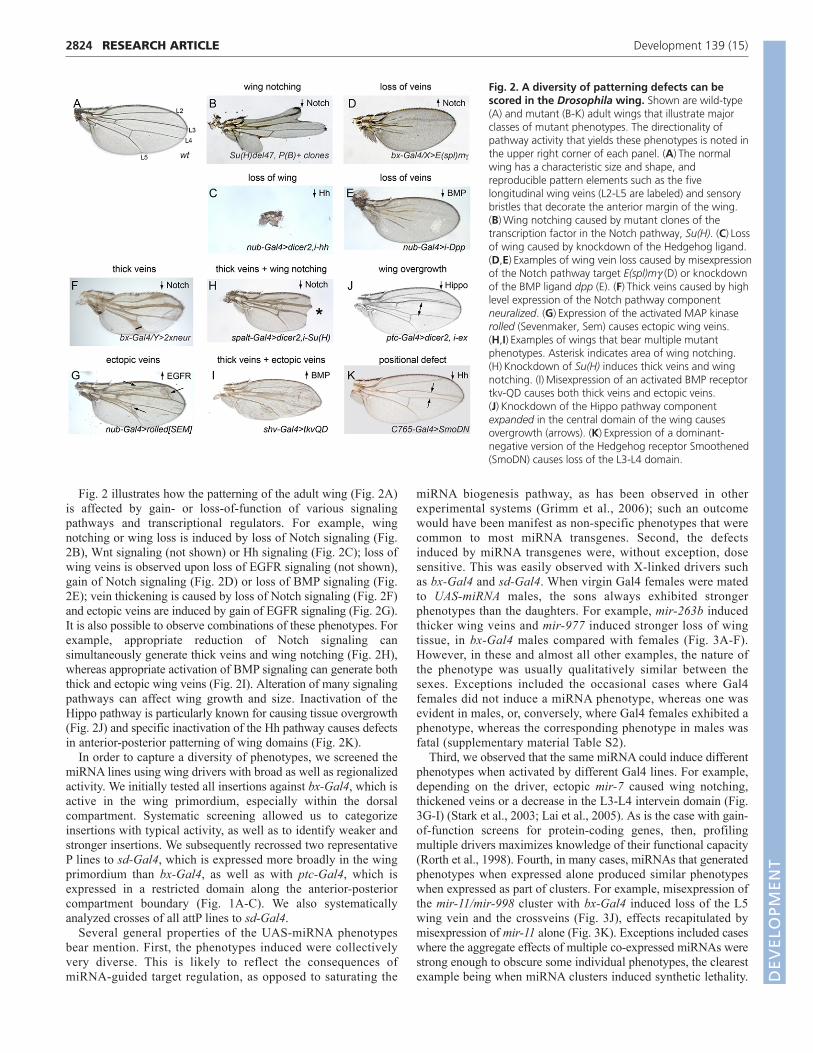
Fig. 2 illustrates how the patterning of the adult wing (Fig. 2A)is affected by gain- or loss-of-function of various signalingpathways and transcriptional regulators. For example, wingnotching or wing loss is induced by loss of Notch signaling (Fig.2B), Wnt signaling (not shown) or Hh signaling (Fig. 2C); loss ofwing veins is observed upon loss of EGFR signaling (not shown),gain of Notch signaling (Fig. 2D) or loss of BMP signaling (Fig.2E); vein thickening is caused by loss of Notch signaling (Fig. 2F)and ectopic veins are induced by gain of EGFR signaling (Fig. 2G).It is also possible to observe combinations of these phenotypes. Forexample, appropriate reduction of Notch signaling cansimultaneously generate thick veins and wing notching (Fig. 2H),whereas appropriate activation of BMP signaling can generate boththick and ectopic wing veins (Fig. 2I). Alteration of many signalingpathways can affect wing growth and size. Inactivation of theHippo pathway is particularly known for causing tissue overgrowth(Fig. 2J) and specific inactivation of the Hh pathway causes defectsin anterior-posterior patterning of wing domains (Fig. 2K).
In order to capture a diversity of phenotypes, we screened themiRNA lines using wing drivers with broad as well as regionalizedactivity. We initially tested all insertions against bx-Gal4, which isactive in the wing primordium, especially within the dorsalcompartment. Systematic screening allowed us to categorizeinsertions with typical activity, as well as to identify weaker andstronger insertions. We subsequently recrossed two representativeP lines to sd-Gal4, which is expressed more broadly in the wingprimordium than bx-Gal4, as well as with ptc-Gal4, which isexpressed in a restricted domain along the anterior-posteriorcompartment boundary (Fig. 1A-C). We also systematicallyanalyzed crosses of all attP lines to sd-Gal4.
Several general properties of the UAS-miRNA phenotypesbear mention. First, the phenotypes induced were collectivelyvery diverse. This is likely to reflect the consequences ofmiRNA-guided target regulation, as opposed to saturating the
miRNA biogenesis pathway, as has been observed in otherexperimental systems (Grimm et al., 2006); such an outcomewould have been manifest as non-specific phenotypes that werecommon to most miRNA transgenes. Second, the defectsinduced by miRNA transgenes were, without exception, dosesensitive. This was easily observed with X-linked drivers suchas bx-Gal4 and sd-Gal4. When virgin Gal4 females were matedto UAS-miRNA males, the sons always exhibited strongerphenotypes than the daughters. For example, mir-263b inducedthicker wing veins and mir-977 induced stronger loss of wingtissue, in bx-Gal4 males compared with females (Fig. 3A-F).However, in these and almost all other examples, the nature ofthe phenotype was usually qualitatively similar between thesexes. Exceptions included the occasional cases where Gal4females did not induce a miRNA phenotype, whereas one wasevident in males, or, conversely, where Gal4 females exhibited aphenotype, whereas the corresponding phenotype in males wasfatal (supplementary material Table S2).
Third, we observed that the same miRNA could induce differentphenotypes when activated by different Gal4 lines. For example,depending on the driver, ectopic mir-7 caused wing notching,thickened veins or a decrease in the L3-L4 intervein domain (Fig.3G-I) (Stark et al., 2003; Lai et al., 2005). As is the case with gain-of-function screens for protein-coding genes, then, profilingmultiple drivers maximizes knowledge of their functional capacity(Rorth et al., 1998). Fourth, in many cases, miRNAs that generatedphenotypes when expressed alone produced similar phenotypeswhen expressed as part of clusters. For example, misexpression ofthe mir-11/mir-998 cluster with bx-Gal4 induced loss of the L5wing vein and the crossveins (Fig. 3J), effects recapitulated bymisexpression of mir-11 alone (Fig. 3K). Exceptions included caseswhere the aggregate effects of multiple co-expressed miRNAs werestrong enough to obscure some individual phenotypes, the clearestexample being when miRNA clusters induced synthetic lethality.
RESEARCH ARTICLE Development 139 (15)
Fig. 2. A diversity of patterning defects can bescored in the Drosophila wing. Shown are wild-type(A) and mutant (B-K) adult wings that illustrate majorclasses of mutant phenotypes. The directionality ofpathway activity that yields these phenotypes is noted inthe upper right corner of each panel. (A)The normalwing has a characteristic size and shape, andreproducible pattern elements such as the fivelongitudinal wing veins (L2-L5 are labeled) and sensorybristles that decorate the anterior margin of the wing.(B)Wing notching caused by mutant clones of thetranscription factor in the Notch pathway, Su(H). (C)Lossof wing caused by knockdown of the Hedgehog ligand.(D,E)Examples of wing vein loss caused by misexpressionof the Notch pathway target E(spl)m (D) or knockdownof the BMP ligand dpp (E). (F)Thick veins caused by highlevel expression of the Notch pathway componentneuralized. (G)Expression of the activated MAP kinaserolled (Sevenmaker, Sem) causes ectopic wing veins.(H,I)Examples of wings that bear multiple mutantphenotypes. Asterisk indicates area of wing notching.(H)Knockdown of Su(H) induces thick veins and wingnotching. (I)Misexpression of an activated BMP receptortkv-QD causes both thick veins and ectopic veins.(J)Knockdown of the Hippo pathway componentexpanded in the central domain of the wing causesovergrowth (arrows). (K)Expression of a dominant-negative version of the Hedgehog receptor Smoothened(SmoDN) causes loss of the L3-L4 domain.
DEVELO
PMENT

Fifth, we frequently observed that members of miRNA seedfamilies often induced similar phenotypes. For example,misexpression of mir-2, which shares a ‘K box’ seed with mir-11,also induced L5 vein loss (Fig. 3L). Phenotypic overlap amongmiRNA seed families provided further evidence that the observedeffects were driven by misregulation of specific targets, as opposedto non-specific titration of miRNA pathway components. The Kbox miRNAs also illustrate that distinct phenotypes are observedwith different drivers, as mir-2 and mir-11, along with other familymembers mir-6 and mir-13, can all induce wing notching(supplementary material Fig. S1).
Of the 165 miRNA transgenes, 96 induced fully penetrantphenotypes with bx-Gal4, 98 with sd-Gal4 and 73 with ptc-Gal4(supplementary material Table S2). Interestingly, although many ofthese could be grouped into qualitatively similar cohorts, themajority of miRNAs that generated similar phenotypes were notrelated in sequence. This was not a trivial outcome, given thatdifferent miRNA seed families typically exhibit little overlap inpredicted target sets. Some of the prominent phenotypic classes thatwe observed included loss of wing veins (Fig. 4F-J), thickenedwing veins (Fig. 4K-T), notched wings (Fig. 4U-AA), ectopic wingveins (Fig. 4BB-DD), specific loss of wing margin bristles (Fig.4EE-OO), wing overgrowth (Fig. 4PP-UU), wing undergrowth/lossof L3-L4 domain (Fig. 4VV-ZZ), wing blistering (Fig. 4AAA-EEE), compression of the anterior-posterior axis (Fig. 4FFF) andshortening of the proximal-distal axis (Fig. 4GGG).
We also recorded a number of examples of curly wing, smallcrumpled wings or wing vestiges (Fig. 4HHH-OOO), thedevelopmental bases of which were less obviously attributable toany specific process. Nevertheless, these phenotypes were areminder of the detrimental consequences of miRNA deregulation.Altogether, the gain-of-function of different miRNAs collectivelyyields a surprising variety of characteristic phenotypes. Acomprehensive compilation of phenotypes is presented in
supplementary material Fig. S1, and we catalog these line by lineusing a controlled vocabulary in supplementary material Table S2.These resources permit visual browsing as well as text searchingfor phenotypes of interest. Reassuringly, systematic tests of thephenotypes induced using the attP insertion lines when crossed tosd-Gal4 showed that they were qualitatively similar to thecompanion UAS-miRNA P insertion lines. There was not aconsistent trend for the attP lines to be stronger or weaker,highlighting the advantage of the allelic diversity possible withrandom insertions.
Association of miRNA gain-of-functionphenotypes with relevant target genesPerhaps the simplest explanation for the ability of sequence-unrelated miRNAs to induce similar phenotypes is that they maytarget common pathways. Indeed, the phenotypic similarity ofgroups of mutants is the fundamental basis of using genetics toassemble molecular pathways. The sundry miRNA-inducedphenotypes were strikingly reminiscent of phenotypes caused bydysfunction of cell signaling components and transcription factorsthat control wing development (Fig. 2). Although it cannot be ruledout that these miRNA-induced phenotypes were the outcome ofcoordinately mild reduction in the activity of tens or hundreds oftargets, it seems more parsimonious to infer that they might be dueto the suppression of specific wing patterning genes. The similarityof miRNA gain-of-function phenotypes to known mutants providesa way to focus target searches from among lists of hundreds ofconserved targets.
For example, miR-8 was reported as an inhibitor of Wntsignaling, and shown to directly repress wntless and CG32767(Kennell et al., 2008). These findings are consistent with theobservation that ectopic miR-8 could generate wing notching (Fig.2B), but did not induce vein thickening (which might have beenconsistent with a reduction of Notch signaling). The Wnt pathway
2825RESEARCH ARTICLEFly miRNA transgene library
Fig. 3. Selected examples of miRNA-induced wing phenotypes illustrategeneral properties of the UAS-DsRed-miRNA transgenes. (A,B)Wings of aheterozygous bx-Gal4 (‘bx’) female (A) andhemizygous male (B); males are smaller thanfemales, which accounts for size difference.(C-F)Examples of dose effects. Vein thickening(arrows) induced by mir-263b is weaker in bx-Gal4 females (C) than males (D). Wingnotching (asterisks) induced by mir-977 isweaker in sd-Gal4 (sd) females (E) than males(F). (G-I)Different miRNA misexpressionphenotypes are evident in different Gal4backgrounds. (G)bx/Y>mir-7 exhibits massivevein thickening (arrows), but the margin iscontinuous. (H)sd/Y>mir-7 exhibits massivewing notching (asterisks), and only mild veinthickening. (I)ptc-Gal4>mir-7 exhibits distalwing notching and reduction in the L3-L4domain (arrows). (J,K)Dissection of a miRNAcluster. (J)Activation of the mir-11/mir-998operon induces vein (arrow) and crossvein(arrowhead) loss; these phenotypes arerecapitulated by ectopic mir-11 (K).(L)Similarity of seed families; mir-2 is in thesame family as mir-11 and also induces veinloss (arrows).
DEVELO
PMENT

2826 RESEARCH ARTICLE Development 139 (15)
Fig. 4. Summary of wing phenotypes caused by misexpression of different Drosophila miRNAs. Shown are adult female wings, except asnoted for X-linked Gal4 drivers (/X female; /Ymale). (A-E)Except for the wild-type [w1118] wing (A), all other flies contain a single copy of Gal4and UAS-DsRed-miRNA transgene. (B)bx-Gal4/X heterozygous females exhibit a normal wing, as do sd-Gal4/X females (not shown). (C)Gal4activity in sd-Gal4/Y males results in a minor loss of posterior wing margin, especially near the wing hinge (boxed regions in A and C are enlarged inD and E, respectively). (F-J)Examples of vein loss induced by different miRNAs. (K-T)Examples of vein thickening induced by different miRNAs. (U-AA) Examples of wing notching induced by different miRNAs. Note that P-S and U-X highlight miRNAs that induce vein thickening or marginloss, respectively, depending on the driver. Other combinations of phenotypes are evident by inspection. (BB-DD) Examples of ectopic wing veins(arrows) induced by different miRNAs. (EE-OO) Examples of miRNAs that have a selective effect on wing margin bristles; close-ups of the anteriormargin are shown in JJ-OO. (PP)ptc-Gal4 heterozygous female; the ptc+ domain includes the L3-L4 region marked by the double arrow. (QQ-UU) Examples of miRNAs that induced overgrowth of the L3-L4 domain. (VV-ZZ) Examples of miRNAs that induced undergrowth or loss ofthe L3-L4 domain. (AAA-EEE) Examples of miRNAs that induced wing blisters. (FFF) miRNA that induces a potential defect along the anterior-posterior axis. (GGG) miRNA that induces a potential proximal-distal defect. (HHH-OOO) Examples of other severe wing deformities or wing lossinduced by different miRNAs. D
EVELO
PMENT

ligand Wingless (Wg) is required for wing formation, and its 3�UTR bears a highly conserved 8-mer target site for miR-8(supplementary material Fig. S2B). The wg 3� UTR was responsiveto ectopic miR-8 in luciferase sensor assays in S2 cells(supplementary material Fig. S2C). Therefore, targeting of wg maybe relevant to miR-8 activity.
Studies of let-7 deletion indicated that the transcription factorencoded by abrupt is a key direct target (Fig. 5A), as Abrupt ismisexpressed in let-7 clones and abrupt heterozygosity cansuppress certain let-7 mutant phenotypes (Caygill and Johnston,2008). Although ectopic let-7 strongly perturbed wingdevelopment, these wings also exhibited shortening of L5 wingvein, corresponding to the phenotype of viable abrupt mutants(Diaz-Benjumea and Garcia-Bellido, 1990) (Fig. 5B,C). We useda genetic assay to confirm that the 3� UTR of abrupt can bedirectly repressed by let-7. We stained for the expression of aubiquitously active tub-GFP-abrupt 3� UTR sensor transgene(Okamura et al., 2008) in the presence of spatially restricted let-7
driven along the AP compartment boundary using dpp-Gal4. Weobserved strong cell-autonomous reduction of the abrupt GFPsensor in the dpp+ domain (Fig. 5E,F). Browsing our collection ofwing phenotypes, we observed that ectopic expression of mir-275was among the miRNAs that also led to loss of wing veins,particularly of L5 (Fig. 5D). We observed the abrupt 3� UTRcontains a highly conserved seed match for miR-275 (Fig. 5A), andthat ectopic mir-275 suppressed the abrupt sensor in vivo (Fig.5G). The stronger repression of the abrupt sensor by let-7compared with miR-275 was recapitulated in luciferase assays inS2 cells (Fig. 5H). Together with our previous observation that mir-iab-4 is another strong in vivo repressor of abrupt (Okamura et al.,2008) (Fig. 5H), it appears that abrupt is substantially targeted bymultiple miRNAs in Drosophila.
We suggest that the forward phenotypic screening is likely toserve as a rich resource for pairing many miRNAs to biologicalpathways. Strikingly, the in vivo work revealed many cases ofmiRNAs whose misexpression phenocopied strong or nearly null
2827RESEARCH ARTICLEFly miRNA transgene library
Fig. 5. Validation of miRNAs that directly target abrupt. (A)Targetscan predictions of conserved miRNA-binding sites in the abrupt 3� UTR. (B-D)Adult female wings. (B)The viable abrupt[1] mutant exhibits loss of distal L5 wing vein (arrow). (C)Misexpression of let-7 induces wing deformityeven when cultured at low temperature to limit Gal4 activity; in addition, loss of the distal region of L5 is seen (arrow). (D)Misexpression of mir-275/mir-305 also induces loss of L5. (E-G�) Transgenic sensor assays in wing imaginal discs that carry tub-GFP-abrupt 3� UTR, dpp-Gal4 and UAS-DsRed (linked to a miRNA in F-G�); the central domain of the wing pouch is shown. (E-E�) Control staining shows that expression of DsRed does notrepress the abrupt sensor. (F-F�) Ectopic let-7 strongly represses the abrupt sensor. (G-G�) Ectopic miR-275 mildly represses the abrupt sensor.(H)Renilla-abrupt 3� UTR sensor assays in S2 cells. Consistent with the in vivo results, mir-275 weakly repressed the abrupt sensor, while let-7strongly repressed it; mir-iab-4 has previously been validated to repress the abrupt 3� UTR (Okamura et al., 2008). Data are mean±s.e.m. D
EVELO
PMENT

2828
situations of known wing patterning genes and pathways (Figs 2-4), for which perusal of available computational predictions ofmiRNA binding sites (e.g. http://www.targetscan.org/) did notreveal any candidate hits. It is also worth stating the obvious thatthe mere presence of a conserved miRNA binding site in a givengene does not guarantee that misexpression of the correspondingmiRNA will necessarily induce a corresponding mutant phenotype.
The connections of a given miRNA to a specific pathway werebolstered in some instances by independent phenotypes. Forexample, ectopic miR-7 can induce both vein thickening and wingnotching, both of which are associated with Notch pathway loss-of-function (Fig. 2H). We observed that mir-980, mir-12, mir-190and mir-982 similarly induced both strong vein thickening (Fig. 4P-S) and strong notching (Fig. 4U-X), suggesting their potentialconnection to Notch signaling. As another example, amongDrosophila miRNAs that regulate neurogenesis, mir-9a mutantsexhibit ectopic notum sensory bristles (Li et al., 2006; Bejarano etal., 2010), while mir-279 mutants exhibit ectopic CO2-sensingneurons (Cayirlioglu et al., 2008). These phenotypes broadly placethese miRNAs as having anti-neural properties. Consistent withthis, we observed that ectopic expression of miR-279 and membersof the miR-9a/b/c family specifically disrupted wing marginsensory bristles while retaining relatively normal patterning of thewing proper (Fig. 4EE-HH,KK-NN). These observations areconsistent with anti-neural activity of these miRNAs. Interestingly,mir-252 similarly induced strong loss of wing margin bristles withrelatively little effect on the wing margin (Fig. 4II,OO), suggestingit may also have a role in regulating neurogenesis.
Endogenous functional relevance of gain-of-function phenotypes: K box miRNAsOur systematic phenotypic profiling tests provide the first in vivoevidence of the biological activities of the majority of DrosophilamiRNAs. Certainly, many of these may be misexpression effectscaused by introduction of the miRNA into an ectopic setting. Suchphenotypes still provide useful insights on the in vivo functionalcapacities of deregulated miRNAs, and offer insights into thepossible roles of these miRNAs in their normal locations.Nevertheless, it is pertinent to consider whether any of thesephenotypes are relevant to the endogenous function of the miRNA.
We previously showed that multiple members of the K box family(sharing the UGUGAU seed) directly inhibit Notch target genes, andinduce phenotypes reminiscent of Notch loss of function whenmisexpressed (Lai et al., 1998a; Lai, 2002; Lai et al., 2005). Forexample, miR-2 and miR-6 induce wing notching and loss of wingmargin when ectopically expressed, and we extend these activities toother members of the family, including miR-11 and miR-13(supplementary material Fig. S1). The pervasive capacity of differentK box miRNAs to suppress Notch signaling motivated us to testwhether their loss of function could promote Notch signaling.
Recently, we showed that transgenes bearing multimers of bulgedtarget sites (‘sponges’) could induce miRNA loss-of-functionphenotypes in Drosophila (Loya et al., 2009). We generated spongesfor several K box miRNAs, including miR-2b, miR-2c, miR-13a andmiR-13b. These did not induce obvious dominant phenotypes, evenin animals carrying two copies of the sponge transgene (Fig. 6B anddata not shown). However, several Notch pathway components lacksubstantial phenotypes in certain settings unless the geneticbackground is sensitized (Schrons et al., 1992; Zeng et al., 1998;Duan et al., 2011). We therefore analyzed females heterozygous forNotch, which normally exhibit notching of the distal wing (Fig. 6A).Two-thirds of N/+ flies expressing control sponges exhibited
RESEARCH ARTICLE Development 139 (15)
Fig. 6. K box miRNA sponges enhance Notch signaling duringwing development. (A,C-H) Notch[55e11]/+ (N/+) heterozygousfemales that carry ptc-Gal4 and two copies of the indicated miRNAsponges (SP); scr, scrambled sponge control. (A)N/+ females expressingcontrol sponges exhibit a notch (asterisk) at the distal tip and mild veinthickening; the regions outlined are magnified to highlight thesephenotypes. (B)Misexpression of any of the sponges used in this figuredid not alter wing development; ptc-Gal4>2xmir-13aSP is shown as anexample. The magnified insets exhibit normal vein thickness and can beused to judge N/+ haploinsufficiency. (C)Misexpression of the miR-13aSP rescued N/+ notching, but not vein thickening. (D)Misexpressionof miR-7SP did not rescue either N/+ phenotype. Asterisk indicates areaof wing notching. (E-H)Magnifications of the distal wing tips tohighlight the status of wing notching in other sponge backgrounds.Asterisk indicates area of wing notching. (E)miR-2bSP, (F) miR-2cSP and(G) miR-13bSP all rescued N/+ notching, but (H) miR-6SP could not(asterisk). (I)Quantification of rescue of wing notching in variousgenotypes. N/+ in various ptc-Gal4>UAS-mir-SP backgrounds exhibitnotching in about two-thirds of animals, this is reduced to less than20% in the presence of miR-2cSP, to 10% in miR-2bSP and miR-13aSP,and to less than 2% in miR-13bSP. Inset shows the sequencerelationship of these K box miRNAs. D
EVELO
PMENT

notching. However, four independent K box miRNA sponges (miR-13a, miR-13b, miR-2b and miR-2c) rescued the haploinsufficientNotch phenotype (Fig. 6C,E-G), indicating that one or moremiRNAs of this family has an endogenous function to limit Notchsignaling during wing margin specification. Fig. 6I quantifies thedegree of rescue provided by these sponges. The miR-13b spongenearly completely suppressed wing notching, whereas the othersreduced notching to only 10% of flies with either miR-2b/13asponges and to 20% of flies in the case of miR-2c sponge. We notemiR-2c is slightly offset in its seed region from canonical K boxmiRNAs, providing a rationale for its sponge being less effectivethan the other K box sponges in suppressing wing notching.
Expression of K box sponges using ptc-Gal4 only rescuednotching and did not suppress vein thickening in N/+ (Fig. 6A-C,inset panels), providing evidence for the specificity of their activity.As a further check, we assayed sponges for the K box familymember miR-6 and the GY box family member miR-7 (Lai, 2002).Both of these miRNAs induce wing notching when misexpressed(Stark et al., 2003; Lai et al., 2005), but neither of their spongessuppressed N/+ (Fig. 6D,H). This can be rationalized by the factthat these miRNAs are not endogenously expressed in thedeveloping wing margin: miR-6 is a member of a miRNA clusterthat is exclusively expressed in the early embryo (Aboobaker et al.,2005; Bushati et al., 2008), whereas miR-7 is specifically expressedin the eye and in proneural domains of imaginal discs (Li andCarthew, 2005; Li et al., 2009).
K box miRNAs comprise the largest family in Drosophila (Laiet al., 2003), and both miR-2 and miR-13 families are well-expressed in imaginal discs (Ruby et al., 2007b). As genetic 3�UTR sensors bearing K boxes that exhibit only seed pairing to anyfamily member direct potent suppression in imaginal discs (Lai etal., 1998) and are sensitive to multiple K box miRNAs (Lai et al.,2005), multiple members of this family probably contribute torestricting Notch signaling. Given such likely functional overlap,single K box miRNA mutants may not suffice to reveal theseeffects. These tests therefore provide a proof of principle of howmiRNA gain-of-function phenotypes can be used to directexperimental approaches that reveal the endogenous contributionof miRNAs to tissue patterning, especially in cases that mayrequire appropriate genetic sensitization.
DISCUSSIONA plethora of specific phenotypes induced bymiRNA gain-of-function in vivoThere is abundant evidence from cell-based studies that animalmiRNAs directly but mildly repress hundreds of targets (Lim et al.,2005; Hendrickson et al., 2009; Guo et al., 2010), even whenmeasured in contexts of ectopic activity. Given this, the in vivoconsequences of miRNA deregulation might reasonably have beensupposed to often be subtle (with the view that few targets can besufficiently suppressed to reveal loss-of-function phenotypes), ormight often compromise general cell viability (with the view thatthe coordinate downregulation of hundreds of targets might causecells to simply become unhealthy).
We describe a systematic in vivo examination of theconsequences of targeted miRNA misexpression within the intactanimal. In contrast to the aforementioned possibilities, we find thatthe majority of miRNAs tested generated diverse and relativelydistinct mutant phenotypes, most of which could not be anticipatedfrom target predictions or from the general fine-tuning model ofmiRNA function. Although a number of miRNAs have profoundlyadverse consequences that might be due to cellular toxicity, many
miRNA-induced phenotypes closely resemble those exhibited bymutants of genes in signaling/proliferation/apoptosis pathways thatare crucial to tissue development and patterning. The presentstudies extend our earlier functional screens in cultured cells thatlinked miR-315 to activation of the Wnt pathway (Silver et al.,2007), and now permit diverse functional screening in the animal.Indeed, many miRNAs of unrelated sequences generated similarphenotypes in vivo, which might be explained if they hit differentnodal points in the same pathways.
Many, if not most, genes contain conserved binding sites formultiple miRNAs. But it is clear that the simple presence ofconserved miRNA-binding sites does not guarantee responsivenessin directed sensor assays. It is even more so the case that presenceof cognate binding sites does not render a miRNA likely to be ableto induce a corresponding loss-of-function phenotype in the animal,even when misexpressed (Silver et al., 2007). Even when usingartificial shRNA constructs designed to have perfectcomplementarity for maximal effect, it is typical for them to elicitonly partial knockdown or sometimes to not work at all. Theknown dose sensitivity of the core cell signaling pathways andpatterning genes provides a genetic rationale for why they may beespecially prone to be affected by miRNAs in a way that translatesinto overt mutant phenotypes (Hagen and Lai, 2008; Smibert andLai, 2010). Such genetic connections can guide functional studiesand point to likely target pathways, even when knowledge ofrelevant computationally predicted targets is lacking.
Beyond understanding the underlying genetic circuitry ofinsects, our studies highlight that ectopic miRNAs can generatespecific developmental phenotypes, often as a result of alteringtissue patterning, proliferation of apoptosis. This has substantialconsequences for interpreting the etiology of disease and cancer.For example, the overexpression of a growing number ofmammalian miRNAs can generate cell specification or metabolicdefects, and miRNAs such as mir-21 (Medina et al., 2010) and mir-17-92 (He et al., 2005) are overt oncogenes. Our systematicscreening in Drosophila strongly suggests that scores of vertebratemiRNAs may prove to induce relatively specific phenotypes in theanimal, but that these may only rarely be predicted on the basis ofcomputationally derived target associations.
A genetic resource for miRNA screening in vivoA great deal of effort has been devoted to expanding collections ofconditionally activated transgene insertions in Drosophila (Brandand Perrimon, 1993; Rorth et al., 1998). Over the past 15 years,these have been of tremendous use in revealing the biologicalactivity and function of protein-coding genes. Here, we describegenome-wide collections of miRNA transgenes, and demonstratetheir collectively diverse activities during wing development. Thesecollections include both P insertion and attP insertion lines,providing a great deal of flexibility for their subsequent screening.The latter permits the activity of different miRNAs to be compareddirectly, whereas the former provides in many cases allelic seriesof transgene strengths. The availability of these lines permits awide variety of screens using tissue- or cell-specific drivers, toevaluate the consequences of miRNA deregulation ondevelopment, as well as adult roles in physiology or behavior.Knowledge of their functional capacities can then inform the studyof different miRNAs within their endogenous expression domains(Aboobaker et al., 2005; Berezikov et al., 2011).
While this work was under review, Cohen and colleaguesdescribed a smaller set of UAS-miRNA transgenes and theirapplication towards searching for modifiers of a bristle phenotype
2829RESEARCH ARTICLEFly miRNA transgene library
DEVELO
PMENT
2830
of the cell cycle regulator minus (Szuplewski et al., 2012). Beyonda limited set of bristle modifiers, however, their analysis primarilyrevealed lethality as the outcome of miRNA expression(Szuplewski et al., 2012). We find over 100 of our miRNAtransgenes induced lethality when broadly expressed with da-Gal4.However, our detailed analysis using a panel of wing driversrevealed a cornucopia of distinct phenotypes, many of whichphenocopy the modulation of fundamental signaling pathways andpatterning factors (Figs 2-6). Our UAS-miRNA collectionscomplement and substantially extend their transgenes, and togetherthey constitute a formidable resource for in vivo analysis ofmiRNA activity. Many miRNAs have subtle if not undetectableloss-of-function phenotypes (Miska et al., 2007; Alvarez-Saavedraand Horvitz, 2010), but it is also the case that many miRNAmutants produce synthetic phenotypes in combination with othergenetic insults (Brenner et al., 2010). Data such as ours provide agenetic basis for pursuing more than 100 demonstrable miRNAactivities and many tens of compelling miRNA-target/pathwaylinkages, and can inform more complex interaction studies withmiRNA sponges (Loya et al., 2009). Indeed, we provide proof ofprinciple for how K box miRNA gain of function, which inhibitedNotch signaling, informed sensitized genetic assays that revealedthe endogenous activity of a likely highly redundant set ofendogenous K box miRNAs in restricting Notch signaling duringwing development. Our extensive assays provide compellingevidence of the usefulness of these genome-wide collections ofconditionally activatable miRNA transgenes, and suggest that thesemay be well complemented by similar collections of miRNAsponge transgenes.
AcknowledgementsThis work was initiated in the laboratory of Gerald Rubin and we are gratefulfor his support. We thank Todd Laverty for performing many of the embryoinjections, and Stephen Cohen and Julius Brennecke for reagents. KieranHarvey, Carole Poon, Ethan Bier and Jose de Celis provided enlighteningdiscussion and kindly provided some images of mutant wings.
FundingQ.D. was supported by the Swedish Research Council. Work in N.P.’slaboratory was supported by the Howard Hughes Medical Institute, StarrCancer Consortium and National Institutes of Health (NIH) [R01-GM084947].T.A.F. and D.V.V. were supported by the NIH [R01-NS069695]. Work inE.C.L.’s group was supported by the Burroughs Wellcome Fund [1004721],the Starr Cancer Consortium [I3-A139] and the NIH [R01-GM083300 andU01-HG004261]. Deposited in PMC for release after 12 months.
Competing interests statementThe authors declare no competing financial interests.
Supplementary materialSupplementary material available online athttp://dev.biologists.org/lookup/suppl/doi:10.1242/dev.079939/-/DC1
ReferencesAbdelilah-Seyfried, S., Chan, Y. M., Zeng, C., Justice, N. J., Younger-
Shepherd, S., Sharp, L. E., Barbel, S., Meadows, S. A., Jan, L. Y. and Jan, Y.N. (2000). A gain-of-function screen for genes that affect the development ofthe Drosophila adult external sensory organ. Genetics 155, 733-752.
Aboobaker, A. A., Tomancak, P., Patel, N., Rubin, G. M. and Lai, E. C. (2005).Drosophila microRNAs exhibit diverse spatial expression patterns duringembryonic development. Proc. Natl. Acad. Sci. USA 102, 18017-18022.
Alvarez-Saavedra, E. and Horvitz, H. R. (2010). Many families of C. elegansmicroRNAs are not essential for development or viability. Curr. Biol. 20, 367-373.
Aravin, A., Lagos-Quintana, M., Yalcin, A., Zavolan, M., Marks, D., Snyder,B., Gaasterland, T., Meyer, J. and Tuschl, T. (2003). The small RNA profileduring Drosophila melanogaster development. Dev. Cell 5, 337-350.
Axtell, M. J., Westholm, J. O. and Lai, E. C. (2011). Vive la différence:biogenesis and evolution of microRNAs in plants and animals. Genome Biol. 12,221.
Baek, D., Villen, J., Shin, C., Camargo, F. D., Gygi, S. P. and Bartel, D. P.(2008). The impact of microRNAs on protein output. Nature 455, 64-71.
Bartel, D. P. (2009). MicroRNAs: target recognition and regulatory functions. Cell136, 215-233.
Bejarano, F., Smibert, P. and Lai, E. C. (2010). miR-9a prevents apoptosis duringwing development by repressing Drosophila LIM-only. Dev. Biol. 338, 63-73.
Bender, W. (2008). MicroRNAs in the Drosophila bithorax complex. Genes Dev.22, 14-19.
Berezikov, E., Liu, N., Flynt, A. S., Hodges, E., Rooks, M., Hannon, G. J. andLai, E. C. (2010). Evolutionary flux of canonical microRNAs and mirtrons inDrosophila. Nat. Genet. 42, 6-9.
Berezikov, E., Robine, N., Samsonova, A., Westholm, J. O., Naqvi, A., Hung,J. H., Okamura, K., Dai, Q., Bortolamiol-Becet, D., Martin, R. et al. (2011).Deep annotation of Drosophila melanogaster microRNAs yields insights intotheir processing, modification, and emergence. Genome Res. 21, 203-215.
Brand, A. H. and Perrimon, N. (1993). Targeted gene expression as a means ofaltering cell fates and generating dominant phenotypes. Development 118, 401-415.
Brennecke, J., Hipfner, D. R., Stark, A., Russell, R. B. and Cohen, S. M. (2003).bantam Encodes a developmentally regulated microRNA that controls cellproliferation and regulates the proapoptotic gene hid in Drosophila. Cell 113,25-36.
Brenner, J. L., Jasiewicz, K. L., Fahley, A. F., Kemp, B. J. and Abbott, A. L.(2010). Loss of individual microRNAs causes mutant phenotypes in sensitizedgenetic backgrounds in C. elegans. Curr. Biol. 20, 1321-1325.
Bushati, N., Stark, A., Brennecke, J. and Cohen, S. M. (2008). Temporalreciprocity of miRNAs and their targets during the maternal-to-zygotic transitionin Drosophila. Curr. Biol. 18, 501-506.
Caygill, E. E. and Johnston, L. A. (2008). Temporal regulation of metamorphicprocesses in Drosophila by the let-7 and miR-125 heterochronic microRNAs.Curr. Biol. 18, 943-950.
Cayirlioglu, P., Kadow, I. G., Zhan, X., Okamura, K., Suh, G. S., Gunning, D.,Lai, E. C. and Zipursky, S. L. (2008). Hybrid neurons in a microRNA mutant areputative evolutionary intermediates in insect CO2 sensory systems. Science 319,1256-1260.
Dai, Q., Smibert, P. and Lai, E. C. (2012). Exploiting Drosophila genetics tounderstand microRNA function and regulation. Curr. Top. Genes Dev. 99, 201-235.
Diaz-Benjumea, F. J. and Garcia-Bellido, A. (1990). Genetic analysis of the wingvein pattern of Drosophila. Roux’s Arch. Dev. Biol. 198, 336-354.
Duan, H., Dai, Q., Kavaler, J., Bejarano, F., Medranda, G., Negre, N. and Lai,E. C. (2011). Insensitive is a novel corepressor of Suppressor of Hairless andregulates Notch-mediated cell fate decisions in the peripheral nervous system.EMBO J. 30, 3120-3133.
Flynt, A. S. and Lai, E. C. (2008). Biological principles of microRNA-mediatedregulation: shared themes amid diversity. Nat. Rev. Genet. 9, 831-842.
Garcia, D. M., Baek, D., Shin, C., Bell, G. W., Grimson, A. and Bartel, D. P.(2011). Weak seed-pairing stability and high target-site abundance decrease theproficiency of lsy-6 and other microRNAs. Nat. Struct. Mol. Biol. 18, 1139-1146.
Gilboa, L. and Lehmann, R. (2004). Repression of primordial germ celldifferentiation parallels germ line stem cell maintenance. Curr. Biol. 14, 981-986.
Giraldez, A. J., Mishima, Y., Rihel, J., Grocock, R. J., Van Dongen, S., Inoue,K., Enright, A. J. and Schier, A. F. (2006). Zebrafish MiR-430 promotesdeadenylation and clearance of maternal mRNAs. Science 312, 75-79.
Grimm, D., Streetz, K. L., Jopling, C. L., Storm, T. A., Pandey, K., Davis, C. R.,Marion, P., Salazar, F. and Kay, M. A. (2006). Fatality in mice due tooversaturation of cellular microRNA/short hairpin RNA pathways. Nature 441,537-541.
Groth, A. C., Fish, M., Nusse, R. and Calos, M. P. (2004). Construction oftransgenic Drosophila by using the site-specific integrase from phage phiC31.Genetics 166, 1775-1782.
Guo, H., Ingolia, N. T., Weissman, J. S. and Bartel, D. P. (2010). MammalianmicroRNAs predominantly act to decrease target mRNA levels. Nature 466, 835-840.
Hagen, J. W. and Lai, E. C. (2008). microRNA control of cell-cell signaling duringdevelopment and disease. Cell Cycle 7, 2327-2332.
He, L., Thomson, J. M., Hemann, M. T., Hernando-Monge, E., Mu, D.,Goodson, S., Powers, S., Cordon-Cardo, C., Lowe, S. W., Hannon, G. J. etal. (2005). A microRNA polycistron as a potential human oncogene. Nature 435,828-833.
Hendrickson, D. G., Hogan, D. J., McCullough, H. L., Myers, J. W., Herschlag,D., Ferrell, J. E. and Brown, P. O. (2009). Concordant regulation of translationand mRNA abundance for hundreds of targets of a human microRNA. PLoS Biol.7, e1000238.
Hipfner, D. R., Weigmann, K. and Cohen, S. M. (2002). The bantam generegulates Drosophila growth. Genetics 161, 1527-1537.
Jaklevic, B., Uyetake, L., Wichmann, A., Bilak, A., English, C. N. and Su, T. T.(2008). Modulation of ionizing radiation-induced apoptosis by bantammicroRNA in Drosophila. Dev. Biol. 320, 122-130.
RESEARCH ARTICLE Development 139 (15)
DEVELO
PMENT

Kennell, J. A., Gerin, I., MacDougald, O. A. and Cadigan, K. M. (2008). ThemicroRNA miR-8 is a conserved negative regulator of Wnt signaling. Proc. Natl.Acad. Sci. USA 105, 15417-15422.
Kraut, R., Menon, K. and Zinn, K. (2001). A gain-of-function screen for genescontrolling motor axon guidance and synaptogenesis in Drosophila. Curr. Biol.11, 417-430.
Lagos-Quintana, M., Rauhut, R., Lendeckel, W. and Tuschl, T. (2001).Identification of novel genes coding for small expressed RNAs. Science 294, 853-858.
Lai, E. C. (2002). microRNAs are complementary to 3� UTR sequence motifs thatmediate negative post-transcriptional regulation. Nat. Genet. 30, 363-364.
Lai, E. C. and Posakony, J. W. (1997). The Bearded box, a novel 3� UTR sequencemotif, mediates negative post-transcriptional regulation of Bearded andEnhancer of split Complex gene expression. Development 124, 4847-4856.
Lai, E. C. and Posakony, J. W. (1998). Regulation of Drosophila neurogenesis byRNA:RNA duplexes? Cell 93, 1103-1104.
Lai, E. C. and Rubin, G. M. (2001). neuralized functions cell-autonomously toregulate a subset of Notch-dependent processes during adult Drosophiladevelopment. Dev. Biol. 231, 217-233.
Lai, E. C., Burks, C. and Posakony, J. W. (1998). The K box, a conserved 3� UTRsequence motif, negatively regulates accumulation of Enhancer of split Complextranscripts. Development 125, 4077-4088.
Lai, E. C., Tomancak, P., Williams, R. W. and Rubin, G. M. (2003).Computational identification of Drosophila microRNA genes. Genome Biol. 4,R42.1-R42.20.
Lai, E. C., Tam, B. and Rubin, G. M. (2005). Pervasive regulation of DrosophilaNotch target genes by GY-box-, Brd-box-, and K-box-class microRNAs. GenesDev. 19, 1067-1080.
Lee, R. C., Feinbaum, R. L. and Ambros, V. (1993). The C. elegans heterochronicgene lin-4 encodes small RNAs with antisense complementarity to lin-14. Cell75, 843-854.
Li, X. and Carthew, R. W. (2005). A microRNA mediates EGF receptor signalingand promotes photoreceptor differentiation in the Drosophila eye. Cell 123,1267-77.
Li, X., Cassidy, J. J., Reinke, C. A., Fischboeck, S. and Carthew, R. W. (2009). AmicroRNA imparts robustness against environmental fluctuation duringdevelopment. Cell 137, 273-282.
Li, Y., Wang, F., Lee, J. A. and Gao, F. B. (2006). MicroRNA-9a ensures theprecise specification of sensory organ precursors in Drosophila. Genes Dev. 20,2793-2805.
Ligoxygakis, P., Bray, S. J., Apidianakis, Y. and Delidakis, C. (1999). Ectopicexpression of individual E(spl) genes has differential effects on different cell fatedecisions and underscores the biphasic requirement for notch activity in wingmargin establishment in Drosophila. Development 126, 2205-2214.
Lim, L. P., Lau, N. C., Garrett-Engele, P., Grimson, A., Schelter, J. M., Castle, J.,Bartel, D. P., Linsley, P. S. and Johnson, J. M. (2005). Microarray analysisshows that some microRNAs downregulate large numbers of target mRNAs.Nature 433, 769-773.
Loya, C. M., Lu, C. S., Van Vactor, D. and Fulga, T. A. (2009). TransgenicmicroRNA inhibition with spatiotemporal specificity in intact organisms. Nat.Methods 6, 897-903.
Martin-Blanco, E. (1998). Regulatory control of signal transduction duringmorphogenesis in Drosophila. Int. J. Dev. Biol. 42, 363-368.
Medina, P. P., Nolde, M. and Slack, F. J. (2010). OncomiR addiction in an in vivomodel of microRNA-21-induced pre-B-cell lymphoma. Nature 467, 86-90.
Miska, E. A., Alvarez-Saavedra, E., Abbott, A. L., Lau, N. C., Hellman, A. B.,McGonagle, S. M., Bartel, D. P., Ambros, V. R. and Horvitz, H. R. (2007).Most Caenorhabditis elegans microRNAs are individually not essential fordevelopment or viability. PLoS Genet. 3, e215.
Molnar, C., Resnik-Docampo, M., Organista, M., Martin, M., Hevia, C. F. andde Celis, J. F. (2011). Signalling pathways in development and human disease: aDrosophila wing perspective. In Human Genetic Diseases (ed. Dr. DijanaPlaseska-Karanfilska). Croatia: InTech.
Morel, V. and Schweisguth, F. (2000). Repression by Suppressor of Hairless andactivation by Notch are required to define a single row of single-mindedexpressing cells in the Drosophila embryo. Genes Dev. 14, 377-388.
Ni, J. Q., Liu, L. P., Binari, R., Hardy, R., Shim, H. S., Cavallaro, A., Booker, M.,Pfeiffer, B. D., Markstein, M., Wang, H. et al. (2009). A Drosophila resourceof transgenic RNAi lines for neurogenetics. Genetics 182, 1089-1100.
Nolo, R., Morrison, C. M., Tao, C., Zhang, X. and Halder, G. (2006). Thebantam microRNA is a target of the hippo tumor-suppressor pathway. Curr. Biol.16, 1895-1904.
Oh, H. and Irvine, K. D. (2011). Cooperative regulation of growth by Yorkie andMad through bantam. Dev. Cell 20, 109-122.
Okamura, K., Hagen, J. W., Duan, H., Tyler, D. M. and Lai, E. C. (2007). Themirtron pathway generates microRNA-class regulatory RNAs in Drosophila. Cell130, 89-100.
Okamura, K., Phillips, M. D., Tyler, D. M., Duan, H., Chou, Y. T. and Lai, E. C.(2008). The regulatory activity of microRNA* species has substantial influence onmicroRNA and 3� UTR evolution. Nat. Struct. Mol. Biol. 15, 354-363.
Reinhart, B. J., Slack, F., Basson, M., Pasquinelli, A., Bettinger, J., Rougvie,A., Horvitz, H. R. and Ruvkun, G. (2000). The 21-nucleotide let-7 RNAregulates developmental timing in Caenorhabditis elegans. Nature 403, 901-906.
Ronshaugen, M., Biemar, F., Piel, J., Levine, M. and Lai, E. C. (2005). TheDrosophila microRNA iab-4 causes a dominant homeotic transformation ofhalteres to wings. Genes Dev. 19, 2947-2952.
Rorth, P., Szabo, K., Bailey, A., Laverty, T., Rehm, J., Rubin, G. M.,Weigmann, K., Milan, M., Benes, V., Ansorge, W. et al. (1998). Systematicgain-of-function genetics in Drosophila. Development 125, 1049-1057.
Ruby, J. G., Jan, C. H. and Bartel, D. P. (2007a). Intronic microRNA precursorsthat bypass Drosha processing. Nature 448, 83-86.
Ruby, J. G., Stark, A., Johnston, W. K., Kellis, M., Bartel, D. P. and Lai, E. C.(2007b). Evolution, biogenesis, expression, and target predictions of asubstantially expanded set of Drosophila microRNAs. Genome Res. 17, 1850-1864.
Schrons, H., Knust, E. and Campos-Ortega, J. A. (1992). The Enhancer of splitcomplex and adjacent genes in the 96F region of Drosophila melanogaster arerequired for segregation of neural and epidermal progenitor cells. Genetics 132,481-503.
Selbach, M., Schwanhausser, B., Thierfelder, N., Fang, Z., Khanin, R. andRajewsky, N. (2008). Widespread changes in protein synthesis induced bymicroRNAs. Nature 455, 58-63.
Silver, S. J., Hagen, J. W., Okamura, K., Perrimon, N. and Lai, E. C. (2007).Functional screening identifies miR-315 as a potent activator of Winglesssignaling. Proc. Natl. Acad. Sci. USA 104, 18151-18156.
Small, E. M. and Olson, E. N. (2011). Pervasive roles of microRNAs incardiovascular biology. Nature 469, 336-342.
Smibert, P. and Lai, E. C. (2008). Lessons from microRNA mutants in worms, fliesand mice. Cell Cycle 7, 2500-2508
Smibert, P. and Lai, E. C. (2010). A view from Drosophila: multiple biologicalfunctions for individual microRNAs. Semin. Cell Dev. Biol. 21, 745-753.
Stark, A., Brennecke, J., Russell, R. B. and Cohen, S. M. (2003). Identificationof Drosophila microRNA targets. PLoS Biol. 1, E60.
Stark, A., Bushati, N., Jan, C. H., Kheradpour, P., Hodges, E., Brennecke, J.,Bartel, D. P., Cohen, S. M. and Kellis, M. (2008). A single Hox locus inDrosophila produces functional microRNAs from opposite DNA strands. GenesDev. 22, 8-13.
Szuplewski, S., Kugler, J. M., Lim, S. F., Verma, P., Chen, Y. W. and Cohen, S.M. (2012). MicroRNA transgene overexpression complements deficiency-basedmodifier screens in Drosophila. Genetics 190, 617-626.
Thompson, B. J. and Cohen, S. M. (2006). The Hippo pathway regulates thebantam microRNA to control cell proliferation and apoptosis in Drosophila. Cell126, 767-774.
Tyler, D. M., Okamura, K., Chung, W. J., Hagen, J. W., Berezikov, E., Hannon,G. J. and Lai, E. C. (2008). Functionally distinct regulatory RNAs generated bybidirectional transcription and processing of microRNA loci. Genes Dev. 22, 26-36.
Wightman, B., Ha, I. and Ruvkun, G. (1993). Posttranscriptional regulation ofthe heterochronic gene lin-14 by lin-4 mediates temporal pattern formation inC. elegans. Cell 75, 855-862.
Xiao, C. and Rajewsky, K. (2009). MicroRNA control in the immune system: basicprinciples. Cell 136, 26-36.
Yang, J. S. and Lai, E. C. (2011). Alternative miRNA biogenesis pathways and theinterpretation of core miRNA pathway mutants. Mol. Cell 43, 892-903.
Zeng, C., Younger-Shepherd, S., Jan, L. Y. and Jan, Y. N. (1998). Delta andSerrate are redundant Notch ligands required for asymmetric cell divisions withinthe Drosophila sensory organ lineage. Genes Dev. 12, 1086-1091.
2831RESEARCH ARTICLEFly miRNA transgene library
DEVELO
PMENT