a fermi national accelerator laboratory - fermilab
TRANSCRIPT

a Fermi National Accelerator Laboratory TM-60 8 1107.000
PERSONNEL DOSIMETRY AT ACCELERATOR LABORATORIES
Peter J. Gollon
September, 19 75
I. INTRODUCTION
In the present paper I will attempt to cover a number of more or less complementary topics:
First, I will discuss the various rationales for personnel moni- toring practices at accelerator laboratories, including operational con- siderations which are not directly relevant to dosimetry; second, I will discuss current practices at various laboratories, comparing current realities with our idealized goals, and third, I hope to indicate some possible paths for the future.
Most of what I say1 hzre is not.original, cosml;ng from a number of conference proceedings - and review articles. - Many of the points I make here apply equally well to other types of installations; accelerator laboratories are unique primarily in regard to their openness, their large transient populations and the types of radiation present.
II. ADMINISTRATIVE AND RADIOBIOLOGICAL CONSIDERATIONS
First, let us consider the rationale for monitoring. We know that exposure to large amounts of radiation is known to be harmful, and ex- posure to lesser quantities might also be deleterious. It is therefore prudent to keep track of what individual exposures are. Thus, we have the first function of radiation monitoring, namely, the measurement and documentation of exposure to potentially hazardous conditions.
Second, if we are even moderately clever and responsible, we also use information on past exposures in order to control future exposures. For example, individuals with high exposures may be assigned different duties to limit their total exposures; jobs which result in large exposures are studied in order to reduce resulting exposures.
Finally, the third function that I see for personnel dosimetry is to tell us what the exposures aren’t. That is, a report of a low or minimal exposure for people who should receive no exposure tells us that nothing unusual is happening.

-2-
1 find it interesting and disturbing that many individuals and groups involved in radiation protection recognize only the first of these reasons for monitoring, namely, record-keeping.
Various scientific bodies, such as the International Commission on Radiological Protection (ICRP) and National Council on Radiation Protection and Measurements (N,Cl$P have established “maximum per- missible occupational exposures . ’ d These bodies further recommend that individuals be monitored for exposure if it is “likely” that their doses will exceed some fraction-- such as one-thi d--of the occupational limit. Other governmental and regulatory bodies f0 have adopted similar guidelines. Thus, if we were to follow their guidelines, most of the people monitored would have exposures above 50 to 100 mrem per month.
In actual fact, most of the people monitored have monthly exposures close to zero. (At Fermilab, for example, 85% of the people regularly monitored receive a “minimal” exposure each month, and the mean positive exposure is about 60 mrem per month. ) I/believe this happens for several reasons. First, it is far easier to issue dosimeters to everybody (or everybody entering certain areas) than it is to determine who is “likely” to receive more than a certain exposure. Second, one is protected against errors of judgment if everybody is monitored. And finally, it placates inspectors who become unhappy if they see an unmoni- tored person even passing through an area of a few mrem/hr. Thus, because of practical considerations, governmental pressures, and public- relation questions, we are monitoring many people who would not be monitored on health physics grounds alone.
The current practice at all major United States accelerators is to monitor anybody who goes anywhere near the accelerator or experi- mental areas, even though there is no reasonable expectation of his receiving any significant dose under “normal” circumstances.
“Why”, asks Klaus Becker, “do we need to monitor to a large ex- tent, and at a relatively high cost people who, as experience has shown, are extremely unlikely ever to receive a significant radiation dose, when for the same price we could indeed increase substantially their average life span by providin$,$hem with safety belts for their cars or convince them to smoke less?
A logical way of dealing with this pressure to monitor everybody would be to use different types of dosimeters for personnel working in “high-risk” and “low-risk” jobs, as suggested by Becker. The former would be sophisticated and read fairly frequently; the latter, inexpensive and read only once or twice a year. In this context, “high risk” could mean either subject to the possibility of a serious radiation accident,

-3-
or likely to be exposed to a significant fraction of the “maximum per- missible dose”. Becker intends the former meaning. However, the latter criterion is used as a basis for assigning monitoring devices by one United States laboratory. Recognizing that most of the monitoring they do is for reassurance purposes, the Stanford Linear Accelerator Center (SLAC) designates such monitors as “accident dosimeters”, reads them out once per year, and doe
h- not normally enter the results into the person’s radiation history file. This saves them a sizable reporting effort under current United States regulations. The employees who are routinely exposed have their dosimeters read quarterly. SLAC is also the only accelerator laboratory to use thermoluminescent dosi- meters (TLD’s) for routine personnel monitoring. All the other accel- erator laboratories treat the casually exposed and the routinely exposed people the same way, and all but one use film for both gamma and neu- tron dosimetry.
When we examine the sensitivity and accuracy desired (or attain- able) in personnel monitoring, we find another contradiction between what logic might tell us should y done, and what is actually being done. In his thought-provoking paper, Becker points out the many logical steps between reading a dosimeter and assigning a dose to a critical organ, or making a meaningful estimate of possible somatic or genetic damage. I will just touch briefly on a few of the questions he raises. For example, how the dosimeter reading relates to the dose received by the critical organ( s) depends on a variety of factors, including where the dosimeter is worn, type, energy and direction of radiation, and which organ is being considered. We may also ask about the various factors needed to make radiobiological sense out of dose measurements. More simply --how well do we know quality (or other modifying) factors and how meaningful are they ? Figure I shows the RBE for different mammalian systems as a function of LET, and compares an “average” mammalian response with the ICRP Quality Factor recommendations. I would characterize these as conservative educated guesses. To go from an incident neutron flux to a calculation of a quality factor intro- duces the uncertainties of body composition, and n-p and n-nucleus interactions. Introduction of continuous neutron spectra and bilateral or isotropic irradiations only makes matters even more complicated. (One sometimes wonders at the wisdom or utility of accumulating large collections of numbers which may only be of dubious value. )
Becker then suggests that in normal monitoring situations we recognize and learn to live with the tenuous connection between dosi- meter readings and somatic damage, and content ourselves with dosi- meters which are stable with time and reasonably reproducible--say GO%. (We will see shortly whether such dosimeters in fact exist. ) Only in cases of very high and potentially lethal exposures does he feel

-4-
it is necessary to have the ultimate in dosimeter sophistication, so that it might be possible to determine the do?2 to the critical organs to per- haps 25 to 40%. The ICRP recommends that “the uncertainty in assessing the annual dose should not exceed 50% of the recorded dose or 1 rem, whichever is larger”; these two sets of recommendations are generally consistent.
III. DOSIMETRY TECHNIQUES
A. Introduction
Let me start by reviewing the criteria that have been formulatedi3’ i4 for personnel dosimetry systems. From a technical standpoint, each monitoring element:
(a) should be sensitive predominately to the type of radiation it is meant to monitor, and insensitive to other types which might be simultaneously present,
(b) should be stable and its response unaffected by environmental factors,
(c) should cover the entire dose range desired, preferably linearly, (d) should respond well over the entire range of particle energies
which will be encountered, (e) should provide adequate accuracy.
From an administrative standpoint, the system
(a) should be easy to use in the field, (b) should have a straightforward readout and dose interpretation,
and (c) should be inexpensive and capable of handling large numbers
of people.
With these standards, let us look first at gamma and then at fast neutron dosimetry.
B. Gamma Dosimetry
While gamma dosimetry may not be very exciting, it should be remembered that perhaps 90% of the total exposure at accelerator lab- oratories is due to maintenance of radioactivated machine components. 15
The most widely used method of gamma monitoring is still photographic film. This method has a number of advantages which continue to make it attractive:

-5-
1. it has reasonable accuracy and reproducibility (*15%/o), 2. it has reasonable sensitivity, -20 mrad, 3. it is not expensive-- about United States $i per unit per
reading, 4. it provides a permanent record, 5. examination of the film can provide information concerning
conditions of exposure: x or y rays, frontal or backwards irradiation.
Those who continue to use film may or may not be aware that film is inferior to other means of dosimetry with regard to5
1. accuracy and reproducibility, 2. sensitivity, 3. long-term stability under conditions of heat and humidity.
The latter effect,’ which is widely known for fast neutron dosimetry, also occurs in gamma dosimetry; different authors have had widely vary- ing degrees of success trying to eliminate it with hermetically sealed packaging. ‘6-‘9 The systems which are superior to film in these respects are solid-state systems: Thermoluminescent dosimetry (TLD) and Radio-Photo-Luminescent glass (RPL). The former is quite widely used for personnel dosimetry of a number of large facilities. TLD materials (CaSO4:Dy) are available which provide sensitivities of a frac- tion of a mrad. Commercial automated TLD systems exist, although their reliability still leaves something to be desired. Use of a phosphor (such as LiF) with a mean atomic number approximately that of tissue eliminates the need for the various filters needed with film dosimetry. With the small size of TLD materials comes the possibility of dosimeters which are more convenient to wear, and thus less susceptible to loss.
Progress with RPL glass has come more slowly, but recent ad- vances2’ . m the understanding and formulation of such glass have made its performance comparable to that of the TLD systems in current use. RPL systems enjoy a greater popularity in Japan than they do in Europe or the United States. For those not familiar with the RPL concept, the dosimeter material is a piece of silver-activated phosphate glass, Under the action of radiation, 21
(a) the optical density (opacity) increases over most of the visible and UV spectrum; and
(b) stable fluorescing color c$nters are excited in the glass. o These emit orange light (5000-7000A) upon excitation by UV at 3650A.

-6-
Readout is accomplished by stimulating the glass with UV light and measuring the light emitted in the orange region of the spectrum.
As an example of what can be done with this technique, Yokota and others have been able to obtain IS% standard deviation for a 10 mrad y exposure, and a 2% standard $eviation for a 50-mrad exposure, using blocks of glass 8 X 8 X 4.7 mm . Unfortunately, these glasses are quite sensitive to thermal neutrons because of their Li and B content.
The cost of operating a gamma dosimetry service based on either of these two techniques should be roughly comparable to that for a film service. Operating costs might even be lower, since all the materials are reusable. The crossover point may well depend on the rate at which badges are lost, since their initial cost may be significantly higher than for film badges.
Why, then, have accelerator laboratories lagged behind other institutions in adopting newer and presumably better types of photon dosimetry ? I do not believe it is because their health physics per- sonnel have lacked either imagination or the desire to do a better job. Rather, I think it is a combination of two reasons--film has performed sufficiently well under their relatively controlled circumstances to make them reluctant to change, and their neutron dosimetry has kept them tied to film anyway. This brings us to the question of neutron dosimetry.
C. Neutron Dosimetry
By “neutrons” at an accelerator laboratory I mean fast neutrons, with energies in the multi-MeV range. Figure 2 shows experimentally unfolded neutron spectra from the CERN-LBL-RHEL shielding experi- ment. 22 Also shown is a calculated spectrum produced by 200-GeV protons under a thick earth shield. 23 The relevant feature of all these spectra is that they are roughly 1 /E (up to a sharp drop between 100 MeV and 1 GeV) when expressed in fluence/MeV. This means that they are roughly flat --up to the same break point --when plotted as neutrons per lethargy interval. The dose-equivalent per neutron is a rising function of energy. These factors combine to put most of the dose-equivalent in the multi-MeV range. Rather roughly, for all the spectra shown in the figure, 80% of the dose-equivalent is due to neutrons between 5 MeV and 1 GeV. (Low-energy neutrons diffusing through penetrations in the shielding are an exception to this rule. )
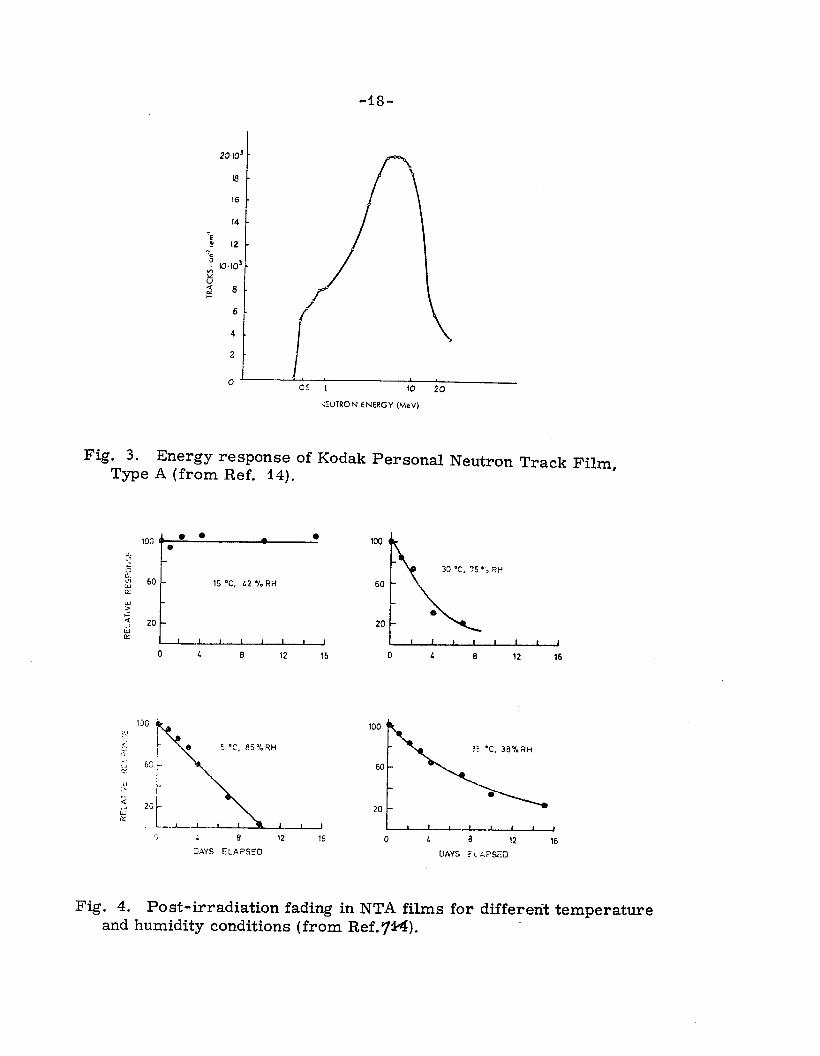
NEUTRON TRACK FILM. Figure 3 shows a measured response curve for NTA film. I4 The low-energy cutoff (roughly 0.5 to 1 MeV) is determined by the number of emulsion grams desired in the shortest acceptable proton recoil track. The film is sensitive to only about 20%
-7-
of the dose-equivalent carried by the neutron spectrum because of the high-energy falloff in response. However, interactions of higher energy neutrons with C and N nuclei in the film do give it some response past 15 or 20 MeV.
Figure 4 shows the “fading” of latent proton recoil tracks in NTA film. This fading is caused by the oxidation of the silver grains by atmospheric oxygen in the pres’ence of water vapor. The rate of fading increases rapidly with increasing temperature and humidity.
There is still no uniformity of results concerning the effective- ness hermetically sealing the film prior to use. Best s ccess has re- sulted from dessicating the film prior to sealing it. 14, lx-l8 The best packaging material for this purpose is an aluminum foil--plastic--paper combination ( “pipe tobacco pouch paper”) in which the alumin m foil is the actual vapor barrier and the plastic is us%$ for sealing. lb” At this time only one commercial film badge service in the United States offers this type of packaging, and no test data is yet available from them. Thus, NTA film is not an intrinsically stable dosimetry medium, and antifading measures=e not yet being taken on a large scale.
Further, neutron track film is easily fogged by several rads of gamma radiation, wl+ich reduces its sensitivity and ultimately makes it entirely unreadable. Finally, the inherent precision of track film is limited by track-counting statistics, among other things. Current scanning practices yield a precision of perhaps 50% for a 100-mrem dose.
What, then, are the fast-neutron dosimetry systems of the future?
First of all, there is FILM. Two improvements could make this a reasonably satisfactory, although still not ideal system. First, the widespread adoption of a proven system for hermetitially sealing the film would eliminate fading in all but tropicalzc;5imates. Secondly, development of automated scanning equipment might allow a larger field to be scanned easily and thus allow better statistics to be accumu- lated. Further, automatic measurement of the distribution of proton track lengths could routinely be used to provide information on the in- cident neutron spectrum, and thus yield a better dose estimate.
Thick emulsions (100-200-pm thick) do not suffer from track fading the way 30-pm thick NTA film does. Use of such emulsions would also allow higher energy recoil tracks to be recognized. How- ever, they are more difficul$+and require a much longer time to develop than does conventional film.
-8-
SOLID-STATE SYSTEMS-:-the established solid-state systems are TLD and RPL. The thermally stimulated exo-electron (TSEE) con- cept is still being developed. All of these detectors have what NTA film lacks: sensitivity, precision, and stability. Unfortunately, none of these systems has an inhere&sensitivity to fast neutrons. One there- fore has two options: to measure only the low-energy tail of a hard- neutron spectrum and make an educated guess about the shape of the spectrum; or to enhance the sensitivity of these detectors to fast neu- trons. The rocedure us d at SLAC involves measurement of the low- energy tail. fl They use LiF and ‘LiF TLD dosimeters incorporated %
into a wallet identification card. The system is sensitive primarily to thermal neutrons via the reaction 6Li{n, a)T. It is also sensitive to higher-energy neutrons which are moderated in the wearer’s body; the efficiency of this process decreases with increasing neutron energy. This concept will be discussed later.
They assume that, because of their shield thickness, the higher- energy neutrons have reached an equilibrium spectrum. They further assume, for dosimetry purposes, that the ratio of fast to “thermal” neutron flux is constant wherever neutron doses can be received. They measured an average flux ratio of 3.4, but use a factor of 6 for safety. This ratio, when combined with the appropriate flux to dose conversion factors for fast and thermal energies, yields an assigned fast-neutron dose of 200 mrem for every mrem of measured thermal neutron dose,
This scheme works only because the actual neutron spectra are all quite similar, and because the actual neutron doses are small com- pared to the legal limits. Its pitfalls are illustrated by what happened when a SLAC experimenter worked at BNL and was exposed to a much softer neutron spectrum than was assumed for the SLAC calibration, His dose, as interpreted by the SLAC ID card dosimeter, was some ten times the dose that BNL assigned on the basic of film measurements! Spectral measurements indicated the correctness of the BNL results. 28
ALBEDO DOSIMETERS. It is possible to reduce the response of a 6LiF chip to thermal neutrons and simultaneously enhance its response to higher energy ones by putting it, sometimes along with a small amount of hydrogenous moderator, into a partial or complete cadmium shield. The cadmium blocks the neutrons which were thermalized in the environ- ment or in the userrs body. High-energy neutrons which are strongly moderated in the wearer’s body ( “albedo neutrons”), but are still above thermal energies, are not absorbed by the cadmium. If they are then thermalized tured by the %
y the moderator inside the dosimeter, they may be cap- Li. A companion ‘LiF permits a gamma subtraction. A
number of designs for these so-called “albedo” dosimeters exist, and

-9-
systemf9 -32 h ve been used rather successfully in reactor or fuel-processing plants.
Unfortunately, measurements made by Hankins and calculations made by Alsmiller and BarishS4 both show that the response of such systems is still strongly dependent on the incident spectrum. As an example, Fig. 5 shows the relative response of a particular albedo dosimeter design as a function of neutron energy. The relative response to a number of typical spectra is shown in Fig. 6. The strong falloff of response with neutron energy above about 100 keV makes this unsuit- able for use whenever the neutron spectrum is not uniform. Thus, the albedo dosimeter system now being implemented at LASL will not be used for fast neutron dosimetrystround LAMPF; they will continue to rely on NTA film for that area.
There are, however, solid-state systems which are either inher- ently sensitive to fast neutrons or which can be made to be sensitive. Some of these systems appear to have promise, but I am cautioned by some of the overly optimistic statements made in the past.
In the field of TLD, work at Battelle-Northwest has shown that both ‘LiF (TLD-700) and CaF2:Tm (TLD-300) have an intrinsic response to fast neutrons; this occurs via scatt ring interactions which transfer energy to higher temperature traps. 3% These peaks are unfortunately also about ten times as gamma-sensitive as they are neutron-sensitive, on a rad basis. In the CaF2:Tm material, the normal glow peak is at 150’ C, the neutron peak is 240” C; thus, the
J are easy to separate.
The,present detection limit is 100 mrads of 52Cf neutrons using small chips of thermoluminescent material. If the same phosphor is put into a glass bulb, the indications are that it will be ten or more times as sensitive. This system is still in the early development stage, and all of its problems are not yet known. One known problem is reducing the gamma response. If this type of dosimeter turns out to be successful, it should be very easy to implement, since the automatic readout systems are already available.
Another promising prospect for fast neutron dosimetry involves the measurement of neutron damage in specially prepared silicon diodes. The forward voltage drop across the diode increases with increasing fast neutron dose. The diodes are not sensitive to gamma radiation. Fractional fading effects are easily circumvented. The observed effect is independent of neutron energy over the range tested. The main pro- blem right now is sensitivity, present sensitivities being a major fraction of a rad. This is adequate for use as an accident dosimeter, but it Will require some substantial advances in sensitivity before this is a useful system for routine monitoring,

-lO-
Finally, we come to TSEE in BeO. This is not inherently sensi- tive to fast neutrons, but can be given an adequate response for many neutron spectra. The sensitive volume in a TSEE detector is right at the surface. If a hydrogenous radiator is placed in front of the Be0 disk, recoil protons will deposit energy in the disk. This will give it an enhanced respo graphite radiator. 8
se compared to a similar disk with a teflon or The TSEE material is also gamma sensitive, thus,
is mixed fields, when the neutron dose is obtained as a difference of two dosimeter readings, the neutron dosimetry becomes harder as the gamma component increases. TSEE systems for neutrons are still in the development stage, altgugh a system for gammas is reported to be in use in the Soviet Union.
Before leaving solid-state systems, I would like to talk about the supposed advantages of “tissue equivalence”. I have the feeling that the underlying physics of radiation interactions has gotten lost, and that all that remains is a magic phrase, whose application is thought sufficient for success. Consider the following statement:
“The relationship between absorbed dose in silicon and in tissue is a rapidly varying function of neutron energy. Thus, the applicability as a personnel dosimeter will be limited to cases where the neutron spectrum is known and where calibra4l;ons have been carried out with neutrons having a similar spectrum. The implied conclusion is that silicon diodes would not make good personnel dosimeters; this is not necessarily true at accelerators.
On the other hand a Be0 TSEE dosimeter consisting of a poly- ethylene radiator and Be0 detector is similar in mean atomic number to tissue; but this is not relevant at all neutron energies. At high enough energies it may not work at all.
Consider a typical accelerator neutron spectrum, such as was discussed earlier. Half the dose is delivered by neutrons with energies above about 60 MeV. At these energies, neutron interactions in matter consist of inelastic nuclear events as well as elastic scattering from hydrogen nuclei. Thus, the effect of hydrogen content is partially masked by other interactions. The nuclear effects such as thresholds, etc. , in light nuclei reactions become less important at higher energies. At high neutron energies, all shielding materials tend to look rather simi- lar. After going through many interaction lengths, the neutrons have reached an equilibrium spectrum; adding further shielding material simply attenuates the spectrum uniformly at all energies. At these high energies, energy deposition by the neutrons is essentially material- independent; all materials absorb energy about equally well on a per gram basis. It is then not serious that a TLD or Si diode material is
-ll-
not “tissue equivalent”. The only neutrons for which “tissue equiva- lence” is important are those whose energies are so low that their most important interaction is elastic scattering from hydrogen.
Consider the TSEE system, on the other hand. Its functioning depends on a difference in neutron interactions in carbon and poly- ethylene, for example. In the high-energy limit, this difference dis- appears, and with it, the proper functioning of the system. Further, as the neutron energy increases, so does the energy and range of the resulting recoil protons or other secondary particles. This range, which is 1 or more gm/cm2 at 60 MeV, determines the thickness of the “effective radiator” in front of the Be0 disk. At higher energies an even deeper volume provides charged recoils, further diluting the n-y discrimination of the dosimeter.
TRACK DAMAGE DOSIMETRY. We now come to a type of dosi- meter which has reasonable sensitivity, does not fade, is neutron- sensitive but gamma insensitive, is easy to read out, and is in practical use in at least one accelerator. This is the track damage dosimeter. Over 15 years ago it was discovered that highly ionizing particles -- such as cr or fission fragments --cause local damage along their tracks when they pass through certain materials. This damaged area may be rendered visible by being etched away at a rate faster than the undam- aged areas. Materials which have been found to be sensitive include minerals such as mica, glasses, and organic polymers. This method of registe$ng tracks is now used in a wide variety of scientific appli- cations.
All dosimeter schemes in current use consist of two parts: a fissionable radiator and a track-recording foil. The radiator foil pro- vides highly ionizing fission fragments which cause ionization damage tracks in the recording foil. Different nuclides are used for the radi- ator foils, depending on the neutron energy range to be covered. For example, 239Pu and 235 U can be used for thermal neutrons; 238U, and 232
2 37Np, Th can be used for neutron energies above 1 MeV. Bis-
muth and gold have fission thresholds at 50 to 100 MeV. Other factors influencing the device of a radiator material include the fission cross section, spontaneous fission rate, associated gamma activity, radio - toxicity of the nuclide, and onels ability to prepare a chemically and mechanically stable foil.
The detector foil is usually mica, polycarbonate (Lexan, Makrofol, Kimfol) or cellulose nitrate, in order of increasing sensitivity. It is “developed” by etching in KOH or NaOH for about one hour. The etching produces pits and eventually holes at the location of each fission track. These holes can then be counted in any of a number of ways. First,
-12-
they can be counted visually, using a microscope, as in NTA film scanning. Secondly, they can be counted electronically by
iif e method of spark counting, as developed by Cross and Tommasino. In this method the etched foil is placed between two electrodes, one of solid metal, the other an aluminized plastic film. A high voltage is applied between the electrodes, causing sparks to jump through the holes in the etched detector foil. These sparks are counted electronically. Each spark vaporizes sufficient aluminum from the foil electrode to prevent a second spark through that hole. The method is quite reproducible and is the preferred method of counting.
One can also measure the light transmitted through the holes at a frequency for which the foil is opaque. Finally, in case of a large num- ber of etched hole it is possible to measure the electrolytic conduc- tance of the foil. 42’
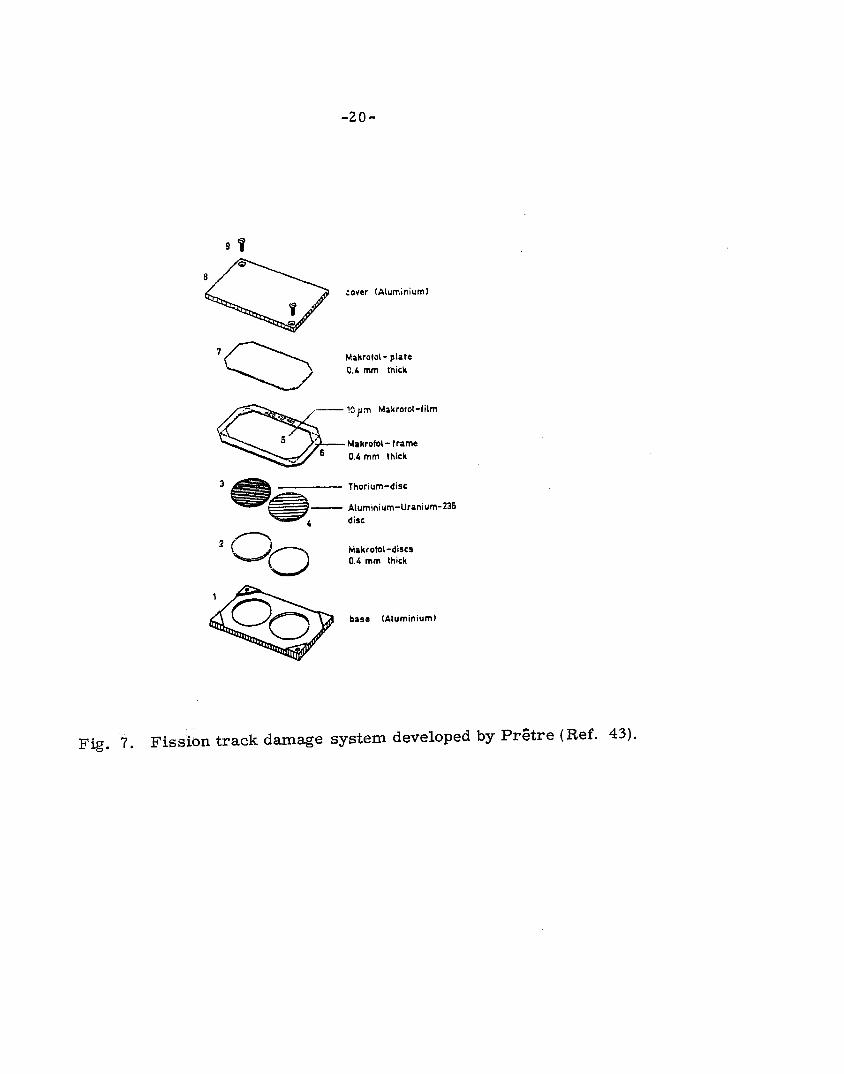
A fission track system has been in u%~ at the Swiss Federal Institute for Reactor Research since 1968. (More recently its use has been inf&tuted at the SIN cyclotron in Switzerland. ) Two radiators are used: U for thermal neutrons and Th (2 -cm diameter, 20 -pm thick) for the fast neutrons (Fig. 7). Makrofol disks are used as acci- dent dosimeters. A calibration of 4-mrad/ spark for fast neutrons is used, so there is no lack of sensitivity. (A slightly different calibration would be used at accelerators. ) The foil can be spark-counted for doses up to 7 rads; microscope counting of the accident dosimeter extends its range up to 20 krad.
A possible problem with this dosimeter is the dose delivered to the wearer by the natural radioactivity of the dosimeter. Despite some 3-mm of aluminum shielding in the badge, is received by the area under the badge. u~,“06s,ezs~~~~i~s~~a~~~~‘~~h
would considerably reduce the gamma dose of the wearer. 4o Other potential problems involve the question of ownership or distribution of fissionable material, albeit in small quantities. Finally, the authors caution that such a system should not be used at electron accelerators, where high energy bremsstrahlung produces photofission in competetion with the neutron fissions used for monitoring. I wish to emphasize that this is an established, functioning system. Costs quoted by the institute to outside organizations for this service seem to be two or three times what a commercial service would charge for film service.
Carl Distenfeld of BNL has combined a Th-fission track detector with ‘LiF and ‘LiF TLDjs which form a crude albedo dosimeter to get some indications of the hardness or softness of the 4% eutron spectrum, and thereby gain additional dosimetry information. This system is still in the developmental stage.

-13-
There is also the possibility of dispensing with the fissionable foil and simply looking for holes due to C, N, or 0 recoils or LY particles from (n, ar) reactions from these nuclei in a very thin and4;e@tive cellu- lose nitrate foil. This is still in the development stage. J
In conclusion, besides NTA film, there is only one proven, opera- tional method of fast neutron dosimetry suitable for use at accelerators. That is fission-track dosimetry. If I had to abandon film today, that is what I would choose. It is remarkable that only one accelerator labora- tory, and less than half a dozen installations in the world, have made that change so far.

-14-
REFERENCES
1 Neutron Monitoring, (P roceedings of Vienna Symposium) IAEA, Vienna, 19 67.
2 Neutron Monitoring for Radiation Protection Purposes, 2 Vols. IAEA, Vienna, 1973.
3 Fourth AEC Workshop on Personnel Neutron Dosimetry, BNWL-1777, $973.
4 Fifth ERDA Workshop on Personnel Neutron Dosimetry, to appear as BNWL report, 1975.
5 K. Becker, Health Physics 23, 729 (1972). 6 K. Becker, Vol. II, p. 145 of ref. 2. 7 P. N. Krishnamoorthy, G. Venkataraman, D. Singh, Dayashankar,
Vol. II, 343 of ref. 2. 8 Recommendations of the International Commission on Radiological
Protection, ICRP # 9, Pergammon Press, 19 66. 9 Basic Radiation Protection Criteria, NCRP # 39, NCRP, Washington,
D. C., 1971. 10 United States Governmental authorities require monitoring for those
who are likely to exceed one-tenth the maximum dose. 11 T. M. Jenkins and K. R. Kase, “Personnel Neutron Dosimetry at
SLAC “, in ref. 3; D. D. Busick, T. M. Jenkins, and W. P. Swanson, “Personnel Neutron Dosimetry at SLAC”, in ref. 4.
12 ICRP #12, paragraph 101. 13 International Atomic Energy Agency, Personnel Dosimetry Systems
for External Radiation Exposures, Technical Reports Series No. 109, IAEA, Vienna (19 70).
14 J. Jasiak and T. Musialowicz, Vol. II, p. 19l of ref. 2. 15 For most laboratories, the gamma fraction is even higher; but at the
ANL ZGS, about one-fourth of the total exposure is due to neutrons. R. Mundis, private communication.
16 K. Becker, ORNL-TM-429 7 ( 19 73). 17 E. Piesch and A. M. Sayed, Nucl. Inst. & Meth. 123, 397 (1975). 18 W. Schimmerling and R. E. Fass, Health Physics 15, 73 (1968). 19 A. Knight, Health Physics 27, 606 (1964); R. S. Landauer, Jr. ,
A. Avni, R. V. Wheeler, (zivate communication).

-15-
20 Proceedings Third International Conference Luminescent DosimetrJ, Riso (1971).
21 F. H. Attix and W. C. Roesch, editors, Radiation Dosimetrx, Second Ed., Vol. II, pp 257ff.
22 W. S. Gilbert et al. , UCRL-17941 (1968).
23M. Awschalom, T. Borak, and H. Howe, NAL TM-266 (1970). 24 G. Portal, in Personnel. Dosimetrg Techniques for External Radiation
(Proceedings Symposium, Madrid, 1963) p. 219, OECD Paris (1963). 25 R. S. Landauer, Jr. , Inc. , Glenwood, Illinois. 26 C. Paretti and A. Ricci, Nucl. Instrum. Methods 122, 389 (1974). 27 W. H. Barkas, Nuclear Research Emulsions, Vol. I, Academic
Press (19 63). 28 R. Casey, private communication. 29 E. Piesch and B. Burgkhardt, ref. 2, Vol. II, p. 31. 30 R. B. Falk, ref. 3, p* 30. 31 J. R. Harvey, W. H. R. Hudd, S. Townsend, ref. 3, Vol. II, p. 199. 32 J. E. Hoy, USAEC report DP-1277 and J. E. Hoy and R. M. Hall,
ref. 4. 33 D. F. Hankins, ref. 3, Vol. II, p. 15; and LA-4832 (1972). 34 R. G. Alsmiller, Jr. and J. Barish, Health Physics 26 13 (1974). J 35 A. W. Blackstock, private communication. 36 G. W. R. Endres, ref. 4 and private communication. 37 C. 0. Widell, Radioprotection 7, 253 (1972). 38 A. I. Beskorskii et al. , paper SM-143-69, IAEA Symp. Advances
Rad. Detect., Vienna (1970). 39 J. F. Fowler, ref. 21, p. 308. 40 K. Becker, in Topics in Radiation Dosimetrx, Suppl. 1, F. H. Attix,
editor; Academic Press, New York (1972). 41 W. G. Cross and L. Tommasini, Health Physics 15, $96 (1968). 42 W. R. Cross and H. Ing, ref. 4. 43 S. Pr&tre, ref. 2, Vol. II, p. 99; and S. Pr,Ftre, K. Heusi, EIR-TM-
SU-149 (1972). 44 C. Distenfeld, BNL-17452 ( 19 72). 45 B. J. Tymons, J. W. N. Tvyn, J. Baarli, ref. 2, Vol. II, p. 63;
M. Sohrabi, Health Physics 27, 598 (1974). -

-16-
IO IOQ
LET, keV/pm
Fig. 1. Schematic curve summarizing the response of mammalian cells to radiation as a function of LET. Some data points are shown. The stepped line represents current values of Quality Factors as recommended by ICRP. Fropa “Dose-Effect Modi- fying Factors in Radiation Protection”, Report of Subcommittee M-4 of NCRP, BNL 50073 (1967).

-17-
10
10
20
10
30
10’
7i 7 10’
ii I
NE 10~ 0 .-. = 10:
1oc
IO-’
10-g
m3
1O-4
--- ,
--4-
Neutron energy in MeV
Fig. 2. Typical neutron spectra obtained from CERN-LBL-RHEL shielding experiment and other work [Ref. 22). The curve labelled “200 GeV” is a calculated spectrum due to protons lost on iron mag- nets behind a thick earth shield (Ref. 2 3). Relative intensities of spectra are arbitrary.
\ \ \ ion,-. -=c 0
- _.- “_-
t
-ig-
J.
I.
\
/ i I \
1. , 05 1 10 20
.EUTRON ENERGY (MeV)
Fig. 3. Energy response of Kodak Personal Neutron Track Film, Type A (from Ref. 14).
15 ‘C, L2 % RH
; L 8 12 16
LXYS ELAPSED
0 t 8 12 16
Fig. 4. Post-irradiation fading in NTA films for differerit temperature and humidity conditions (from Ref. rti).

Fig. 5. Relative response of an albedo neutron dosimeter due to J. Hoy [ Health Physics 23, 385 (1972)] for monoenergetic neutrons (from - Ref. 34).
o-normal incidence o-isotropic incidence
Fig, 6. Relative response of the Hoy dosimeter to various neutron spectra (from Ref. 34).
-2o-
corer (Aluminiuml
Makrotol- plate C.l, mm thick
Ihorium-disc
- Aluminium-Uranium-235
2 0 0
diet
Mekrofol-discs 0.4 mm thick
base (Aluminium)
Fig. 7. Fission track damage system developed by Pr6tre (Ref. 43).