a case of ameloblastoma recurred 25 years after surgery · recurrent ameloblastoma developing in an...
TRANSCRIPT
Remedy Publications LLC.
Journal of Dentistry and Oral Biology
2017 | Volume 2 | Issue 14 | Article 10851
Case PresentationThe patient was a 77-year-old woman. She was examined by our department in March 2009
for a painless tumor in the gingiva of the anterior mandibular ridge. She had a history of surgery to remove a left-mandibular Ameloblastoma at the department of dental and oral surgery at X general hospital in 1985. Follow-up was conducted for 5 years, but no abnormalities were observed so the examinations were terminated. The information was asked this hospital for her medical records, imaging data, and other information, but it had been discarded. About 25 years after the initial surgery, in February 2009, she was referred to our department after her primary care dentist found a tumor in the anterior alveolar ridge of the mandible.
Medical historyHypertension and osteoporosis; No allergies.
Present illnessShe exhibited no facial morphological abnormalities, no sensory abnormalities, and no
enlargement of local lymph nodes. In the oral cavity, a painless, slightly hard tumor with surface uniformity, a well-defined boundary, and elasticity was observed beneath the alveolar mucosa near the left-mandibular incisor and premolar area, covered by a normal lining mucosa. The tumor was 20 mm × 20 mm in size and 5 mm in height. The lining mucosa exhibited no abnormalities.
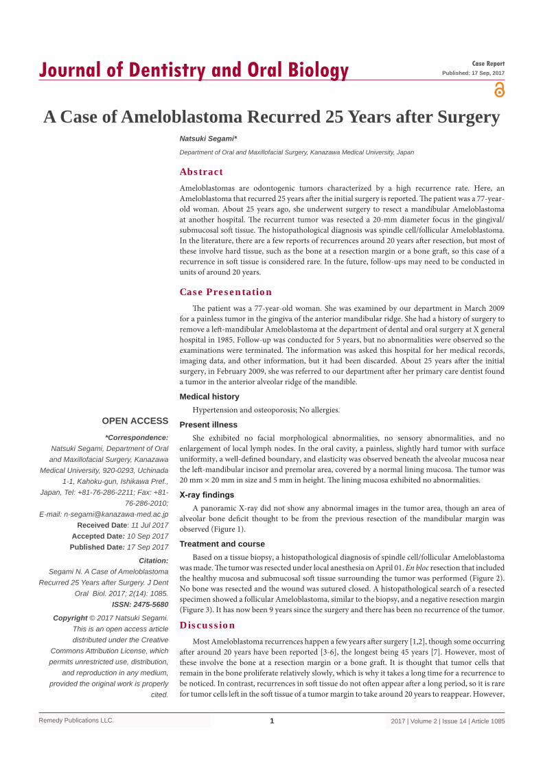
X-ray findingsA panoramic X-ray did not show any abnormal images in the tumor area, though an area of
alveolar bone deficit thought to be from the previous resection of the mandibular margin was observed (Figure 1).
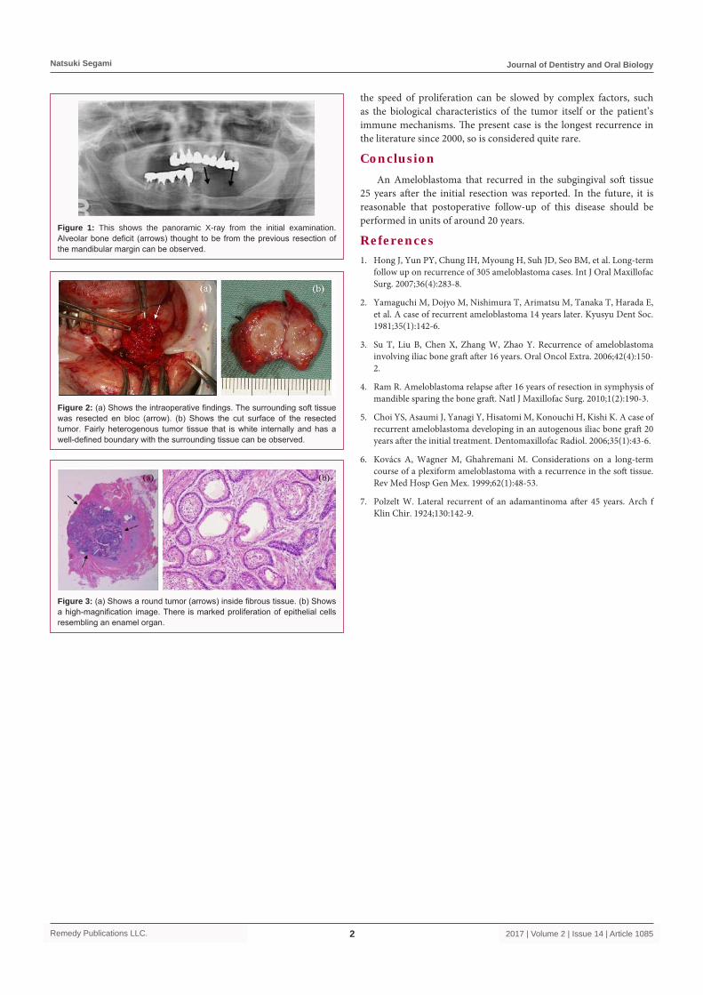
Treatment and courseBased on a tissue biopsy, a histopathological diagnosis of spindle cell/follicular Ameloblastoma
was made. The tumor was resected under local anesthesia on April 01. En bloc resection that included the healthy mucosa and submucosal soft tissue surrounding the tumor was performed (Figure 2). No bone was resected and the wound was sutured closed. A histopathological search of a resected specimen showed a follicular Ameloblastoma, similar to the biopsy, and a negative resection margin (Figure 3). It has now been 9 years since the surgery and there has been no recurrence of the tumor.
DiscussionMost Ameloblastoma recurrences happen a few years after surgery [1,2], though some occurring
after around 20 years have been reported [3-6], the longest being 45 years [7]. However, most of these involve the bone at a resection margin or a bone graft. It is thought that tumor cells that remain in the bone proliferate relatively slowly, which is why it takes a long time for a recurrence to be noticed. In contrast, recurrences in soft tissue do not often appear after a long period, so it is rare for tumor cells left in the soft tissue of a tumor margin to take around 20 years to reappear. However,
A Case of Ameloblastoma Recurred 25 Years after Surgery
OPEN ACCESS
*Correspondence:Natsuki Segami, Department of Oral and Maxillofacial Surgery, Kanazawa
Medical University, 920-0293, Uchinada 1-1, Kahoku-gun, Ishikawa Pref.,
Japan, Tel: +81-76-286-2211; Fax: +81-76-286-2010;
E-mail: [email protected] Date: 11 Jul 2017
Accepted Date: 10 Sep 2017Published Date: 17 Sep 2017
Citation: Segami N. A Case of Ameloblastoma
Recurred 25 Years after Surgery. J Dent Oral Biol. 2017; 2(14): 1085.
ISSN: 2475-5680
Copyright © 2017 Natsuki Segami. This is an open access article distributed under the Creative
Commons Attribution License, which permits unrestricted use, distribution,
and reproduction in any medium, provided the original work is properly
cited.
Case ReportPublished: 17 Sep, 2017
AbstractAmeloblastomas are odontogenic tumors characterized by a high recurrence rate. Here, an Ameloblastoma that recurred 25 years after the initial surgery is reported. The patient was a 77-year-old woman. About 25 years ago, she underwent surgery to resect a mandibular Ameloblastoma at another hospital. The recurrent tumor was resected a 20-mm diameter focus in the gingival/submucosal soft tissue. The histopathological diagnosis was spindle cell/follicular Ameloblastoma. In the literature, there are a few reports of recurrences around 20 years after resection, but most of these involve hard tissue, such as the bone at a resection margin or a bone graft, so this case of a recurrence in soft tissue is considered rare. In the future, follow-ups may need to be conducted in units of around 20 years.
Natsuki Segami*
Department of Oral and Maxillofacial Surgery, Kanazawa Medical University, Japan
Natsuki Segami Journal of Dentistry and Oral Biology
Remedy Publications LLC. 2017 | Volume 2 | Issue 14 | Article 10852
Figure 1: This shows the panoramic X-ray from the initial examination. Alveolar bone deficit (arrows) thought to be from the previous resection of the mandibular margin can be observed.
Figure 2: (a) Shows the intraoperative findings. The surrounding soft tissue was resected en bloc (arrow). (b) Shows the cut surface of the resected tumor. Fairly heterogenous tumor tissue that is white internally and has a well-defined boundary with the surrounding tissue can be observed.
Figure 3: (a) Shows a round tumor (arrows) inside fibrous tissue. (b) Shows a high-magnification image. There is marked proliferation of epithelial cells resembling an enamel organ.
the speed of proliferation can be slowed by complex factors, such as the biological characteristics of the tumor itself or the patient’s immune mechanisms. The present case is the longest recurrence in the literature since 2000, so is considered quite rare.
ConclusionAn Ameloblastoma that recurred in the subgingival soft tissue
25 years after the initial resection was reported. In the future, it is reasonable that postoperative follow-up of this disease should be performed in units of around 20 years.
References1. Hong J, Yun PY, Chung IH, Myoung H, Suh JD, Seo BM, et al. Long-term
follow up on recurrence of 305 ameloblastoma cases. Int J Oral Maxillofac Surg. 2007;36(4):283-8.
2. Yamaguchi M, Dojyo M, Nishimura T, Arimatsu M, Tanaka T, Harada E, et al. A case of recurrent ameloblastoma 14 years later. Kyusyu Dent Soc. 1981;35(1):142-6.
3. Su T, Liu B, Chen X, Zhang W, Zhao Y. Recurrence of ameloblastoma involving iliac bone graft after 16 years. Oral Oncol Extra. 2006;42(4):150-2.
4. Ram R. Ameloblastoma relapse after 16 years of resection in symphysis of mandible sparing the bone graft. Natl J Maxillofac Surg. 2010;1(2):190-3.
5. Choi YS, Asaumi J, Yanagi Y, Hisatomi M, Konouchi H, Kishi K. A case of recurrent ameloblastoma developing in an autogenous iliac bone graft 20 years after the initial treatment. Dentomaxillofac Radiol. 2006;35(1):43-6.
6. Kovács A, Wagner M, Ghahremani M. Considerations on a long-term course of a plexiform ameloblastoma with a recurrence in the soft tissue. Rev Med Hosp Gen Mex. 1999;62(1):48-53.
7. Polzelt W. Lateral recurrent of an adamantinoma after 45 years. Arch f Klin Chir. 1924;130:142-9.