9/11/2012 chapter 12ems.jbpub.com/chapleau/firstresponder/docs/ppt_lectures/chapter... · 9/11/2012...
TRANSCRIPT

9/11/2012
1
Chapter 12
Musculoskeletal Injuries
2
Learning Objectives
Describe the anatomy and function of the musculoskeletal system
Demonstrate the assessment and management of a patient with a suspected musculoskeletal injury
Differentiate between an open and closed musculoskeletal injury
3
Learning Objectives
Perform manual stabilization of a suspected injury to the upper extremity, lower extremity, and spine
Identify concerning mechanisms of injury for potential spinal injury
Describe signs and symptoms of a patient with a suspected spinal injury
Copyright © 2013 by Jones & Bartlett Learning, LLC, an Ascend Learning Company

9/11/2012
2
4
Learning Objectives
Demonstrate the assessment and management of a patient with a suspected spinal injury
5
Introduction
Musculoskeletal system involves all bones, muscles, and connective tissue of the human body Gives the body shape, protects vital organs, and
provides movement
Injuries can alter the normal structure and function of the body
6
Musculoskeletal System
Adult body has >600 individual muscles (muscular system) and 206 skeletal bones (skeletal system)
As you assess a patient suspected of having a musculoskeletal injury rely on your knowledge of normal structure and function
Copyright © 2013 by Jones & Bartlett Learning, LLC, an Ascend Learning Company

9/11/2012
3
7
Musculoskeletal System
Involves body’s intricate network of nerves and blood vessels
When two or more bones come together, their union creates a joint
8
Musculoskeletal System
Skeletal system Skeleton is divided into two main sections
• Axial skeleton Central part of body
• Appendicular skeleton Extremities of the body
9
Musculoskeletal System
Copyright © 2013 by Jones & Bartlett Learning, LLC, an Ascend Learning Company

9/11/2012
4
10
Musculoskeletal System
Skeletal system Axial skeleton
• Central part of the skeleton carries most of the weight of the body and is composed of bones that make up the skull, spinal column, and thorax
• Skull comprises the cranium
11
Musculoskeletal System
Skeletal system Axial skeleton
• Spinal column is made up of 33 individual bones Each bone is called a vertebra
Runs from the base of the skull to the bottom of the pelvis
Houses and protects the spinal cord
Nerves of the spinal cord exit the spinal column between each vertebra and continue to each muscle and organ
12
Musculoskeletal System
Skeletal system Axial skeleton
• Injury to the spinal column can cause: Feeling of “pins and needles”
Sharp pain with movement
Total lack of sensation or ability to move the extremities
Paralysis of the muscles
Copyright © 2013 by Jones & Bartlett Learning, LLC, an Ascend Learning Company

9/11/2012
5
13
Musculoskeletal System
Skeletal system Axial Skeleton
• Spinal column is divided into five specific areas Cervical area is composed of the first seven vertebrae and
is associated with the patient’s neck
Thoracic vertebrae are the next 12 vertebra that make up the back (posterior) of the chest
Lumbar portion is the patient’s lower back and contains the next five vertebrae
Sacrum makes up the posterior portion of the pelvis and is composed of five fused vertebrae
Coccyx (tailbone) is composed of four fused vertebrae
14
Musculoskeletal System
15
Musculoskeletal System
Skeletal system Axial Skeleton
• Thorax (chest) is composed of: Clavicles (collarbones)
Scapula (shoulder blades)
12 pairs of ribs
Sternum (breastbone)
Copyright © 2013 by Jones & Bartlett Learning, LLC, an Ascend Learning Company

9/11/2012
6
16
Musculoskeletal System
17
Musculoskeletal System
Skeletal system
Appendicular skeleton• Composed of the pelvis and upper and lower extremities
• Pelvis is made up of two larger bones, which combine with the sacrum and coccyx to form the pelvic girdle Protects lower internal organs of the digestive system,
urinary systems, and internal female reproductive organs
Highly vascular area
Injury can cause significant blood loss
18
Musculoskeletal System
Skeletal system Appendicular skeleton
• Bones of the upper extremities (arms): Humerus
Radius
Ulna
Carpals
Metacarpals and phalanges
Lower arm/forearm
Copyright © 2013 by Jones & Bartlett Learning, LLC, an Ascend Learning Company

9/11/2012
7
19
Musculoskeletal System
Skeletal system Appendicular skeleton
• Bones of the lower extremities (legs): Femurs
Patellae
Tibias
Fibulas
Tarsals
Metatarsals and phalanges of the feet
20
Musculoskeletal System
Muscular system Responsible for movement of the body and is
composed of three different types of muscles:• Skeletal
• Smooth
• Cardiac
21
Musculoskeletal System
Muscular system All muscles work by contracting and relaxing
• Coordinated effort allows for muscle activity such as: Skeletal movement
Movement of food through the digestive system
Beating heart
Many other tasks
Copyright © 2013 by Jones & Bartlett Learning, LLC, an Ascend Learning Company
9/11/2012
8
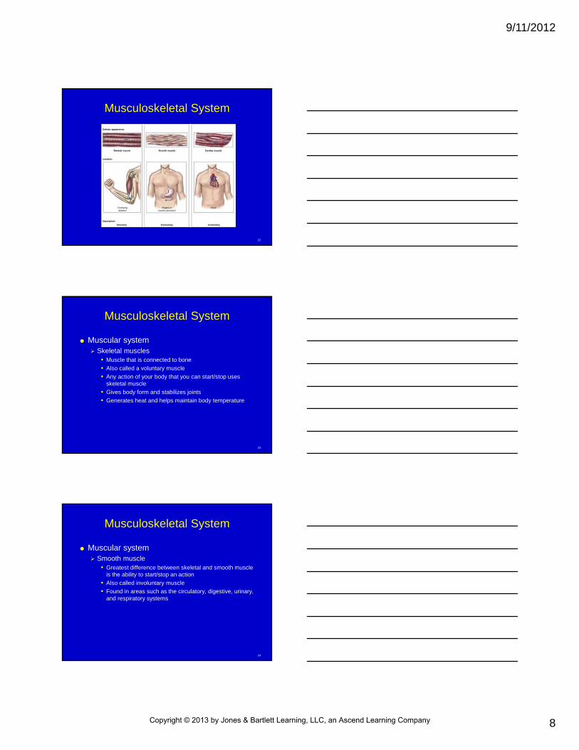
22
Musculoskeletal System
23
Musculoskeletal System
Muscular system Skeletal muscles
• Muscle that is connected to bone
• Also called a voluntary muscle
• Any action of your body that you can start/stop uses skeletal muscle
• Gives body form and stabilizes joints
• Generates heat and helps maintain body temperature
24
Musculoskeletal System
Muscular system Smooth muscle
• Greatest difference between skeletal and smooth muscle is the ability to start/stop an action
• Also called involuntary muscle
• Found in areas such as the circulatory, digestive, urinary, and respiratory systems
Copyright © 2013 by Jones & Bartlett Learning, LLC, an Ascend Learning Company

9/11/2012
9
25
Musculoskeletal System
Muscular system Cardiac muscle
• Hearts are composed of cardiac muscle
• Has unique ability to generate its own electrical impulse independent of the nervous system
• Not under voluntary control and is, therefore, a second type of involuntary muscle
• Sensitive to any decrease in O2/blood supply
• Can tolerate an interruption of an inadequate O2 or blood supply for only short time before suffering damage
26
Mechanisms of Injury
MOI Most musculoskeletal injuries are the result of
some type of trauma• Trauma is the result of an outside force that has a
negative effect on the body Direct injury is a result of force applied directly to injured
part of the body
Indirect injury is caused by a force applied to a different area of the body that is then transmitted to the injured part
Twisting injury results from an extremity being twisted/pulled
27
Mechanisms of Injury
Copyright © 2013 by Jones & Bartlett Learning, LLC, an Ascend Learning Company

9/11/2012
10
28
Mechanisms of Injury
When approaching a scene in which a musculoskeletal injury may have occurred, pay attention to the surrounding environment and consider forces involved
Certain injuries can be predicted based on the MOI
29
Mechanisms of Injury
Certain medical conditions/process of aging may also have a role in musculoskeletal injuries Bones become more fragile and brittle with age
Certain conditions such as osteoporosis weaken bone structure
Understanding how an injury has occurred will allow you to better assess and manage the patient
30
Types of Musculoskeletal Injuries
Can be classified into many different types Prehospital emergency care is the same
regardless of type
As an EFR, you are not responsible for distinguishing between types of injury
Copyright © 2013 by Jones & Bartlett Learning, LLC, an Ascend Learning Company

9/11/2012
11
31
Types of Musculoskeletal Injuries
Classified by defining musculoskeletal injury as open or closed Injury in which the skin is open is called an open
injury
If skin is not open, referred to as a closed injury
32
Types of Musculoskeletal Injuries
Other classifications include: Fracture
• Another name for a broken bone
• Often involves injury to nearby soft tissue, nerves, blood vessels; results in bleeding and potential nerve damage
33
Types of Musculoskeletal Injuries
Other classifications include: Sprain
• Injury in which ligaments (connect bone to bone) are stretched or torn
Copyright © 2013 by Jones & Bartlett Learning, LLC, an Ascend Learning Company

9/11/2012
12
34
Types of Musculoskeletal Injuries
Other classifications include: Strain
• Muscle pull around a joint
• Does not involve ligament and is characterized by pain with movement
• Little to no swelling of the joint
35
Types of Musculoskeletal Injuries
Other classifications include: Dislocation
• Separation of a bone from its normal position in a joint
• Damage blood vessels, nerves, soft tissue, and ligaments, very painful
• Can also be associated with fractures
36
General Assessment of Musculoskeletal Injuries
General assessment of musculoskeletal injuries Before attempting assessment for injury, ensure
that scene is safe and all life threatening conditions have been identified and treated
• Musculoskeletal injuries are often painful and visually dramatic but rarely life threatening
Copyright © 2013 by Jones & Bartlett Learning, LLC, an Ascend Learning Company
9/11/2012
13
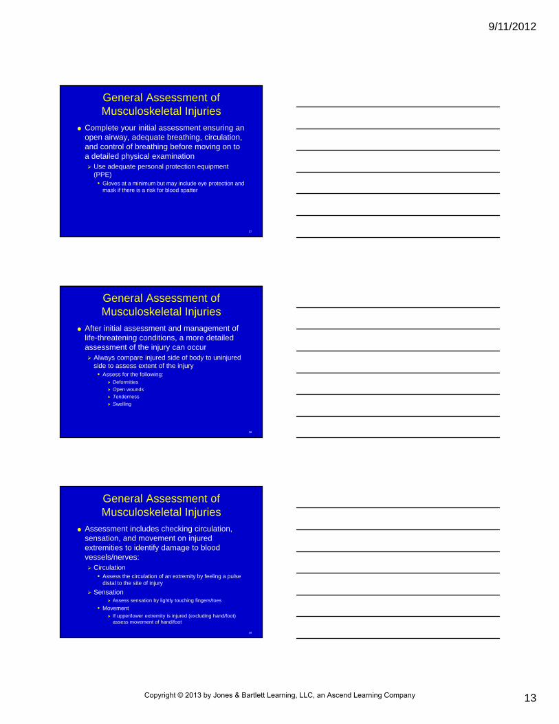
37
General Assessment of Musculoskeletal Injuries
Complete your initial assessment ensuring an open airway, adequate breathing, circulation, and control of breathing before moving on to a detailed physical examination Use adequate personal protection equipment
(PPE)• Gloves at a minimum but may include eye protection and
mask if there is a risk for blood spatter
38
General Assessment of Musculoskeletal Injuries
After initial assessment and management of life-threatening conditions, a more detailed assessment of the injury can occur Always compare injured side of body to uninjured
side to assess extent of the injury• Assess for the following:
Deformities
Open wounds
Tenderness
Swelling
39
General Assessment of Musculoskeletal Injuries
Assessment includes checking circulation, sensation, and movement on injured extremities to identify damage to blood vessels/nerves: Circulation
• Assess the circulation of an extremity by feeling a pulse distal to the site of injury
Sensation Assess sensation by lightly touching fingers/toes
• Movement If upper/lower extremity is injured (excluding hand/foot)
assess movement of hand/foot
Copyright © 2013 by Jones & Bartlett Learning, LLC, an Ascend Learning Company

9/11/2012
14
40
Skill 12-1Assessing Circulation, Sensation,
and Movement Assess radial pulse for
and upper extremity injury
Assess sensation of upper extremity injury by lightly pressing on a finger and asking patient if he can feel it
41
Skill 12-1Assessing Circulation, Sensation,
and Movement Assess movement of
upper extremity injury by asking patient to move his hand/grasp your hand
Assess pedal pulse for lower extremity injury
42
Skill 12-1Assessing Circulation, Sensation,
and Movement Assess lower extremity
injury by pressing on toe and asking patient if he can feel it
Assess movement of lower extremity injury by asking patient to point/flex foot/move foot against your hand
Copyright © 2013 by Jones & Bartlett Learning, LLC, an Ascend Learning Company

9/11/2012
15
43
Management of Musculoskeletal Injuries
Goal of management of musculoskeletal injuries despite type or cause is: Manage patient’s pain
Prevent further injury
Minimize risk of permanent injury
44
Management of Musculoskeletal Injuries
General management steps in managing musculoskeletal injury include the following: Manually stabilize the injury
Allow patient to remain in position of comfort
Control any bleeding unless it is coming from patient’s ears
Never attempt to straighten any musculoskeletal injury that is angled or misshapen
Check and compare circulation, sensation, and movement both above and below the injury site and continue to monitor
45
Management of Musculoskeletal Injuries
General management steps in managing a musculoskeletal injury include the following: Dress any open wounds
Do not move patient until injury is appropriately splinted unless it is absolutely necessary
Consider application of cold to injury site to help control
swelling and pain
If bone ends are visible, do not try to reposition or replace
Calm, comfort, and reassure the patient
Splint the injury as required
Copyright © 2013 by Jones & Bartlett Learning, LLC, an Ascend Learning Company

9/11/2012
16
46
Management of Musculoskeletal Injuries
Splinting of a musculoskeletal injury Splint if possible
• Splint is a device used to immobilize/prevent movement of injured bones/joints and to prevent further damage
47
Management of Musculoskeletal Injuries
Splinting of musculoskeletal injuries General principles for splinting a musculoskeletal
injury are as follows:• Manually stabilize the injury
• Remove/cut away clothing from injured site and dress any open wounds
• Assess circulation, sensation, movement distal to injury
• Immobilize the joint above and the joint below injured site with a splint
• Splint injury in position found
• After splinting, reassess circulation, sensation, and movement distal to the injury
• Pad the splint to prevent pressure points on patient
48
Skill 12-2Splinting an Upper Extremity
Provide manual stabilization of joint above and joint below the injury
Assess circulation distal to the injury
Copyright © 2013 by Jones & Bartlett Learning, LLC, an Ascend Learning Company
9/11/2012
17
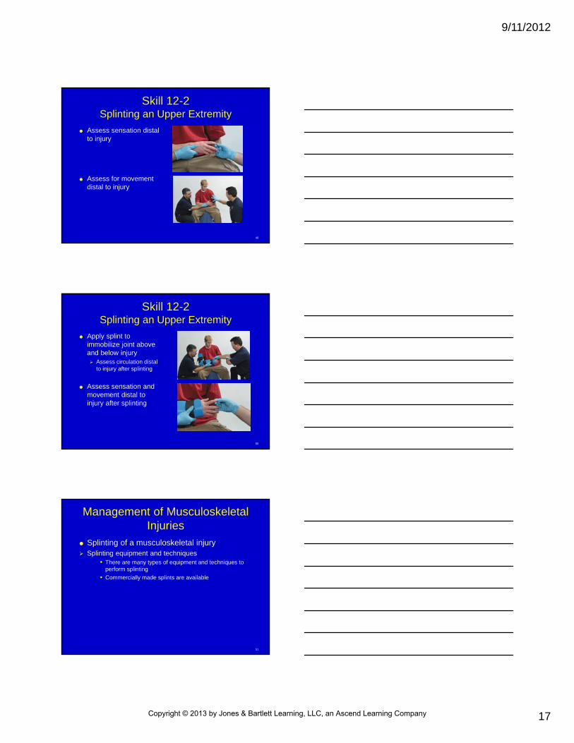
49
Skill 12-2Splinting an Upper Extremity
Assess sensation distal to injury
Assess for movement distal to injury
50
Skill 12-2Splinting an Upper Extremity
Apply splint to immobilize joint above and below injury Assess circulation distal
to injury after splinting
Assess sensation and movement distal to injury after splinting
51
Management of Musculoskeletal Injuries
Splinting of a musculoskeletal injury Splinting equipment and techniques
• There are many types of equipment and techniques to perform splinting
• Commercially made splints are available
Copyright © 2013 by Jones & Bartlett Learning, LLC, an Ascend Learning Company

9/11/2012
18
52
Management of Musculoskeletal Injuries
Splinting of a musculoskeletal injury Splints can be improvised from things such as towels,
pillows, rolled magazines, wood, or cardboard:• Rigid splint
Made of firm, nonformable material
• Soft splint Flexible, formable, provides gentle support from an injury
Air splint is a special type of commercially available soft splint
53
Management of Musculoskeletal Injuries
54
Skill 12-3Techniques of Splinting
Splinting of an injury to a single finger
Splinting of an injury to two fingers
Copyright © 2013 by Jones & Bartlett Learning, LLC, an Ascend Learning Company

9/11/2012
19
55
Skill 12-3Techniques of Splinting
Rigid splint applied to an elbow injury
Rigid splint to a forearm injury
56
Skill 12-3Techniques of Splinting
Rigid splint applied to a knee injury
Soft splint applied to an ankle injury
57
Skill 12-3Techniques of Splinting
Semirigid splint applied to a lower leg injury
Copyright © 2013 by Jones & Bartlett Learning, LLC, an Ascend Learning Company

9/11/2012
20
58
Management of Musculoskeletal Injuries
Splinting of a musculoskeletal injury Splints can be improvised from things such as towels,
pillows, rolled magazines, wood, or cardboard• Sling and swathe
Injuries to the shoulder, clavicle, or humerus are best splinted using a sling and swathe technique
59
Management of Musculoskeletal Injuries
60
Skill 12-4Applying the Swathe
Check circulation, sensation, movement distal to injured site Apply padding
underneath extremity on side of the injury
• Material for sling should be made into a triangle
Copyright © 2013 by Jones & Bartlett Learning, LLC, an Ascend Learning Company

9/11/2012
21
61
Skill 12-4Applying the Swathe
Apply sling to support weight of extremity Point of triangle should
be toward patient’s elbow• Bring two long ends
around patient’s neck and secure in a knot behind shoulder
62
Skill 12-4Applying the Swathe
Secure point of triangle into a knot, or point to the rest of the material to maintain support of the extremity
63
Skill 12-4Applying the Swathe
Apply swathe to secure extremity to chest without restricting breathing Recheck circulation,
sensation, and movement
Pad behind both knots
Copyright © 2013 by Jones & Bartlett Learning, LLC, an Ascend Learning Company

9/11/2012
22
64
Special Considerations
Any patient with suspected spinal or head/chest injury will require special considerations
Initial assessment will always remain the same: Identify and manage any life-threatening
conditions
65
Special Considerations
Suspected spinal injury Manage aggressively
• Injuries to the spine can result in permanent paralysis if not recognized and treated
• As an EFR, you should be able to identify a suspected spinal injury and provide appropriate and rapid management
66
Special Considerations
Suspected spinal injury MOI
• Can present with/without initial signs and symptoms
• Identifying MOI is an important part of the assessment
• According to PHTS committee, MOIs that lead to an assumption of a spinal injury include the following: Any mechanism that produces a violent impact on head,
neck, torso/pelvis
Incidents that produce sudden forces to neck/torso
Any fall, especially in the elderly
Ejection/fall from a motorized device
Shallow-water diving incidents
Copyright © 2013 by Jones & Bartlett Learning, LLC, an Ascend Learning Company

9/11/2012
23
67
Special Considerations
Suspected spinal injury• Assessment and management
Initial assessment of suspected spinal-injured patient is the same as with any other patient
Wear appropriate PPE
Ensure scene is safe
Identify and manage any life-threatening conditions
Special attention should be given to control the airway
68
Special Considerations
Suspected spinal injury Assessment and management
• Patient should not be moved until full spinal immobilization has been applied
• If unresponsive patient is having difficulty breathing, assist his respirations
• After initial assessment, assess circulation and movement in all four extremities
• Complete detailed assessment when needed
69
Special Considerations
Suspected spinal injury Manual stabilization of head and neck
• Once you recognize the potential for spinal injury, the first step will be to manually stabilize patient’s head and neck in a neutral position Position that maintains normal curvature of cervical spine
with eyes facing forward and parallel to the ground if patient is standing
Copyright © 2013 by Jones & Bartlett Learning, LLC, an Ascend Learning Company

9/11/2012
24
70
Special Considerations
Suspected spinal injury Manual stabilization of head and neck
• Can be provided for a patient lying on the ground, sitting upright, or found standing
• Patients found lying face down should be log rolled onto their backs to maintain spinal stabilization whenever possible
71
Special Considerations
72
Special Considerations
Suspected head injury Need special attention and management
Injuries to head can be either open/closed
Early recognition, management, and transportation of a patient with suspected head injury to appropriate facility are imperative
Copyright © 2013 by Jones & Bartlett Learning, LLC, an Ascend Learning Company

9/11/2012
25
73
Special Considerations
Suspected head injury Assessment and management
• Any patient who has altered mental status or is unresponsive should be assumed to have a head injury and should be managed accordingly
• During initial assessment, in addition to identifying and managing any life-threatening conditions, calculate a GCS score and repeat in ongoing assessment
• When performing a physical examination of a suspected head injury patient, avoid movement of head and spine when palpating the skull
74
Special Considerations
Suspected head injury Assessment and management
• Management includes the following: Do not move patient unless it is absolutely necessary
Manually stabilize head and neck
Use trauma jaw-thrust to open and maintain airway
Assist patient’s ventilations if necessary
Apply supplemental O2 if possible
Control minor external bleeding
Cover all open wounds
Do not stop any bleeding/fluid loss from ears
Continue to monitor patient’s vital signs
75
Special Considerations
Suspected chest injuries Patients with suspected chest trauma may have
serious internal injuries
Most common are rib fractures• Patients will almost always have chest pain and may
experience significant difficulty with breathing In these cases, you may need to assist ventilations
Copyright © 2013 by Jones & Bartlett Learning, LLC, an Ascend Learning Company

9/11/2012
26
76
Management
Management of a patient with suspected chest injury includes: Assisting ventilations as needed
Providing supplemental O2, if available• Traumatic injuries to the chest may also have caused
spinal injuries
77
Management
As an EFR, you may be asked to assist in further management of a patient with suspected musculoskeletal injury May include:
• Applying a traction splint
• Applying a cervical collar
• Performing spinal immobilization
78
Management
Traction splint Specialty commercial splint that is used to splint a
fracture of the femur (thigh)
As an EFR, you may be asked to assist with placement
It is important for you to be able to recognize the equipment and have an understanding of how the device is applied
Copyright © 2013 by Jones & Bartlett Learning, LLC, an Ascend Learning Company

9/11/2012
27
79
Management
80
Management
Traction splint General steps taken when applying a traction
splint to a patient:• Manually stabilize the joint above and below the injury
site Remove/cut away clothing from the injury site
Control any bleeding and apply dressings as needed
Check circulation, sensation, and movement above and below the injury site
Attach ankle hitch and apply manual traction
Place traction device along uninjured leg
If using a sling-type device, elevate injured leg just enough to place traction device under it
81
Management
Traction splint General steps taken when applying a traction
splint to a patient: If using a single pole device, place device between
patient’s legs
Apply proper straps and tighten
Apply securing device for ankle
Apply traction according to manufacturer’s recommendations
Recheck circulation, sensation, and movement above and below the injury site
Secure patient to a long backboard
Copyright © 2013 by Jones & Bartlett Learning, LLC, an Ascend Learning Company

9/11/2012
28
82
Management
Cervical collars Rigid devices that help support the head and neck
and keep it from moving
On its own does not completely immobilize a patient’s head and neck
As an EFR, you may have to assist with placement for a patient with a suspected spinal injury
83
Skill 12-5Sizing and Placing a Cervical Collar
Apply and maintain manual stabilization of the head and neck in a neutral inline position
Using your fingers, measure distance between patient’s lower jaw and shoulders Make sure your fingers
are placed parallel to patient’s jaw
84
Skill 12-5Sizing and Placing a Cervical Collar
Find a cervical collar that matches the patient’s measurements or adjust collar size to fit the measurement
Apply the cervical collar and secure
Copyright © 2013 by Jones & Bartlett Learning, LLC, an Ascend Learning Company

9/11/2012
29
85
Skill 12-5Sizing and Placing a Cervical Collar
Maintain manual stabilization until the patient is fully immobilized to a long backboard
86
Management
Spinal immobilization Any patient suspected of having a spinal injury
should be fully immobilized
Full immobilization of the spine requires stabilization of the joint above and below injured head
Full immobilization is done by moving patient onto a long backboard
As an EFR, you will probably be asked to assist with immobilization of a patient with a suspected spinal injury because it requires at least three or four providers
87
Management
Copyright © 2013 by Jones & Bartlett Learning, LLC, an Ascend Learning Company

9/11/2012
30
88
Management
Steps involved in immobilizing a patient to a long backboard: Apply and maintain manual stabilization of
patient’s head and neck in a neutral inline position
Measure and apply a cervical collar
Place a long backboard along the side of the patient, with foot end of the board at patient’s knees
While maintaining manual stabilization, place two or three providers on the side of the patient without the long backboard
89
Management
Steps involved in immobilizing a patient to a long backboard: Providers at the side of the patient place their
hands on opposite sides of the patient under the patient’s shoulders and hips (and lower legs if using three providers)
On command of provider maintaining manual stabilization of head and neck, patient is rolled just enough to slide backboard under the patient
Providers now place their hands between patient’s arms and chest and along the pelvis
90
Management
Steps involved in immobilizing a patient to a long backboard: At the command of the provider maintaining
manual stabilization of head and neck, patient is moved in a straight line upward and centered onto the backboard
Patient can now be immobilized to the backboard with straps
• Torso and legs are immobilized first with the head being immobilized last
Copyright © 2013 by Jones & Bartlett Learning, LLC, an Ascend Learning Company

9/11/2012
31
91
Management
Steps involved in immobilizing a patient to a long backboard: After patient’s head is secured to the board and
full immobilization has been achieved, manual stabilization may be released
Padding may be used to fill any voids/holes • Such as behind the patient’s neck or between the
patient’s legs
92
Management
93
Questions?
Copyright © 2013 by Jones & Bartlett Learning, LLC, an Ascend Learning Company