6 the biology and role of cryosurgery in the treatment of...
TRANSCRIPT

6
The Biology and Role ofCryosurgery in the Treatment of Bone Tumors
Jacob Bickels, Isaac Meller and Martin Malawer
OVERVIEW
The application of liquid nitrogen as a local adjuvant to curettage in the treatment of bone tumors was firstintroduced three decades ago. This technique, termed cryosurgery, was shown to achieve excellent local controlin a variety of benign-aggressive and malignant bone tumors. However, early reports showed that cryosurgeryhas been associated with a significant injury to the adjacent rim of bone and soft tissue, resulting in high rates offractures and infections. These results reflected an initial failure to appreciate the potentially destructive effects ofliquid nitrogen and establish appropriate guidelines for its use. This chapter reviews the biological effect ofcryosurgery on bone, surgical technique, and current indications for its use.
Malawer Chapter 06 21/02/2001 15:16 Page 135
INTRODUCTION
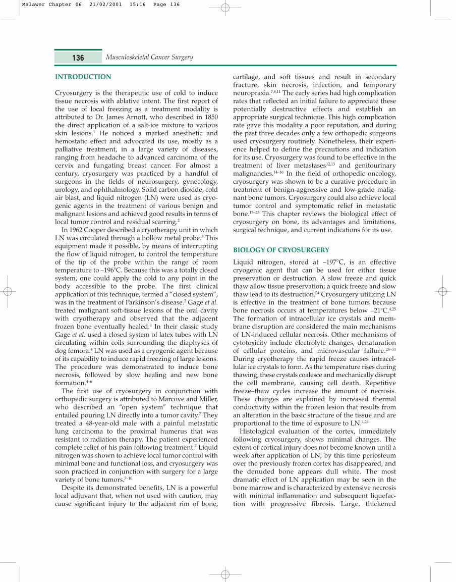
Cryosurgery is the therapeutic use of cold to inducetissue necrosis with ablative intent. The first report ofthe use of local freezing as a treatment modality isattributed to Dr. James Arnott, who described in 1850the direct application of a salt-ice mixture to variousskin lesions.1 He noticed a marked anesthetic andhemostatic effect and advocated its use, mostly as apalliative treatment, in a large variety of diseases,ranging from headache to advanced carcinoma of thecervix and fungating breast cancer. For almost acentury, cryosurgery was practiced by a handful ofsurgeons in the fields of neurosurgery, gynecology,urology, and ophthalmology. Solid carbon dioxide, coldair blast, and liquid nitrogen (LN) were used as cryo-genic agents in the treatment of various benign andmalignant lesions and achieved good results in terms oflocal tumor control and residual scarring.2
In 1962 Cooper described a cryotherapy unit in whichLN was circulated through a hollow metal probe.3 Thisequipment made it possible, by means of interruptingthe flow of liquid nitrogen, to control the temperatureof the tip of the probe within the range of roomtemperature to –196°C. Because this was a totally closedsystem, one could apply the cold to any point in thebody accessible to the probe. The first clinicalapplication of this technique, termed a “closed system”,was in the treatment of Parkinson’s disease.2 Gage et al.treated malignant soft-tissue lesions of the oral cavitywith cryotherapy and observed that the adjacentfrozen bone eventually healed.4 In their classic studyGage et al. used a closed system of latex tubes with LNcirculating within coils surrounding the diaphyses ofdog femora.4 LN was used as a cryogenic agent becauseof its capability to induce rapid freezing of large lesions.The procedure was demonstrated to induce bonenecrosis, followed by slow healing and new boneformation.4–6
The first use of cryosurgery in conjunction withorthopedic surgery is attributed to Marcove and Miller,who described an “open system” technique thatentailed pouring LN directly into a tumor cavity.7 Theytreated a 48-year-old male with a painful metastaticlung carcinoma to the proximal humerus that wasresistant to radiation therapy. The patient experiencedcomplete relief of his pain following treatment.7 Liquidnitrogen was shown to achieve local tumor control withminimal bone and functional loss, and cryosurgery wassoon practiced in conjunction with surgery for a largevariety of bone tumors.7–10
Despite its demonstrated benefits, LN is a powerfullocal adjuvant that, when not used with caution, maycause significant injury to the adjacent rim of bone,
cartilage, and soft tissues and result in secondaryfracture, skin necrosis, infection, and temporaryneuropraxia.7,8,11 The early series had high complicationrates that reflected an initial failure to appreciate thesepotentially destructive effects and establish anappropriate surgical technique. This high complicationrate gave this modality a poor reputation, and duringthe past three decades only a few orthopedic surgeonsused cryosurgery routinely. Nonetheless, their experi-ence helped to define the precautions and indicationfor its use. Cryosurgery was found to be effective in thetreatment of liver metastases12,13 and genitourinarymalignancies.14–16 In the field of orthopedic oncology,cryosurgery was shown to be a curative procedure intreatment of benign-aggressive and low-grade malig-nant bone tumors. Cryosurgery could also achieve localtumor control and symptomatic relief in metastaticbone.17–23 This chapter reviews the biological effect ofcryosurgery on bone, its advantages and limitations,surgical technique, and current indications for its use.
BIOLOGY OF CRYOSURGERY
Liquid nitrogen, stored at –197°C, is an effectivecryogenic agent that can be used for either tissuepreservation or destruction. A slow freeze and quickthaw allow tissue preservation; a quick freeze and slowthaw lead to its destruction.24 Cryosurgery utilizing LNis effective in the treatment of bone tumors becausebone necrosis occurs at temperatures below –21°C.4,25
The formation of intracellular ice crystals and mem-brane disruption are considered the main mechanismsof LN-induced cellular necrosis. Other mechanisms ofcytotoxicity include electrolyte changes, denaturationof cellular proteins, and microvascular failure.26–31
During cryotherapy the rapid freeze causes intracel-lular ice crystals to form. As the temperature rises duringthawing, these crystals coalesce and mechanically disruptthe cell membrane, causing cell death. Repetitivefreeze–thaw cycles increase the amount of necrosis.These changes are explained by increased thermalconductivity within the frozen lesion that results froman alteration in the basic structure of the tissue and areproportional to the time of exposure to LN.4,24
Histological evaluation of the cortex, immediatelyfollowing cryosurgery, shows minimal changes. Theextent of cortical injury does not become known until aweek after application of LN; by this time periosteumover the previously frozen cortex has disappeared, andthe denuded bone appears dull white. The mostdramatic effect of LN application may be seen in thebone marrow and is characterized by extensive necrosiswith minimal inflammation and subsequent liquefac-tion with progressive fibrosis. Large, thickened
Musculoskeletal Cancer Surgery136
Malawer Chapter 06 21/02/2001 15:16 Page 136

thrombosed vessels are occasionally seen.4–6,28 Bonerepair, beginning in the periphery of the bone cavity,occurs slowly and is first evident at the seventh toeighth week. Only after 5–6 months is the new boneformation sufficient to prevent pathologic fracture. Thehistological features after repeated freeze–thaw cyclesare almost identical to those described for a singleepisode of freezing.4
Malawer et al. demonstrated a 7–12-mm rim of bonenecrosis with no effect on articular cartilage when liquidnitrogen was utilized in a dog model (Figure 6.1).28
Marcove et al. stated that three freeze–thaw cyclesproduce tumor cell death up to 2 cm from the cavitymargin.10 This extent of bone destruction makescryosurgery, which is by definition an intralesionalsurgical modality, as effective as a wide resection in thetreatment of benign-aggressive, low-grade primarybone sarcomas, or metastatic lesions.8,9,17,22 High-gradebone sarcomas, on the other hand, usually have asignificant soft-tissue extension that is not affected byLN, unless poured directly on it. This is notrecommended due to the expected damage to thesurrounding muscles and neurovascular bundle.Articular cartilage seems to be resistant to cryotherapyand remains intact, even when freezing extends to thesubchondral bone or crosses the joint.17,32
SURGICAL TECHNIQUE
Although a simple procedure, cryosurgery can causesignificant morbidity if performed inappropriately. Aneffective and safe procedure must follow theseconsecutive steps: (1) adequate exposure of the tumorcavity; (2) meticulous curettage and burr drilling; (3)soft-tissue mobilization and protection prior tointroduction of LN to the tumor cavity; (4) internalfixation of the cavity after cryotherapy; and (5) protec-tion of the operated bone throughout the healing period.
Exposure
When possible, a pneumatic tourniquet is used duringthe procedure to decrease local bleeding and preventblood from acting as a heat sink and a thermal barrierfor the cryotherapy. Benign-aggressive, low-gradeprimary bone sarcomas, and metastatic tumors, rarelyinvade the articular cartilage, and an extracapsularapproach is therefore possible in most cases. Violationof the joint cavity must be avoided because of thepossibility that it may be contaminated by tumor cellsand the risk of injury to the cartilage following directexposure to LN. After exposure of the involved boneand soft tissues, a cortical window the size of thelongest longitudinal dimension of the tumor is made.To minimize additional bone loss the tumor isapproached through the retained thinned or destroyedcortex. A large cortical window is essential to expose theentire tumor and avoid inadequate curettage. Thewindow must be elliptical, and its axis must be parallelto the long axis of bone in order to reduce the stressrising effect (Figure 6.2).
Cryosurgery in Treatment of Bone Tumors 137
Figure 6.1 Tetracycline fluorescence of a dog femorafollowing cryosurgery demonstrates a circumferential rim ofno fluorescence around the cavity, which is the result ofbone necrosis. The outer rim of fluorescence (Fl) corres-ponds to attempted bony repair in the adjacent viable tissue.
Figure 6.2 A large cortical window, the size of the largestdiameter of the lesion, is essential for adequate exposure. Asmaller window is not sufficient for complete curettage andburr drilling of the tumor.
CORRECT INCORRECT
Malawer Chapter 06 21/02/2001 15:16 Page 137

Curettage
All gross tumor is removed with hand curettes. Afterthe neoplastic tissue is curetted away from the innerwall of the lesion the reactive wall reveals an irregularcontour. This irregularity makes it virtually impossibleto remove all the tissue from the inner reactive shellwith a curette. Therefore, curettage is followed by high-speed burr drilling with Midas Rex® (Midas Rex, ForthWorth, TX) or Black Max® (Anspach, Lake Park, FL;Figures 6.3, 6.4).
Cryosurgery
Before introduction of the LN, bony perforations areidentified and sealed, and the surrounding skin, softtissues, and neurovascular bundle are protected bymobilization and shielding with Gelfoam® (Upjohn,Kalamazoo, MI). Large skin flaps are retracted toprotect them from possible spillage of the LN.
Liquid nitrogen can be applied to a bony cavity bydirect pour (open system) or by perfusing it throughmetal probes.2,3,33 Cryosurgery is not effective unless aclose contact is achieved between the LN and theoutermost layer of the tumor cavity. Therefore, size andconfiguration of the tumoral cavity are determinatorsof the chosen technique. Most bone lesions have alarge, irregular inner wall, and use of the open systemmakes it possible to homogeneously spread the LNthroughout the cavity. Liquid nitrogen spray is an “opensystem” modality, used for unique anatomic locationsin which pouring of LN is not technically feasible (forexample, deep-seated pelvic lesions). The closed system,on the other hand, can be effectively used in small,regular cavities, such as those remaining after curettageof a small lesion in the digits or the distal radius.
Using the open system, LN is poured through astainless-steel funnel into the tumor cavity. Care istaken to fill the entire cavity. The Gelfoam® blocksimmediately freeze, forming a tight seal around the
funnel. Thermocouples are used to monitor thefreezing effect within the cavity, cavity wall, andadjacent soft tissue, as well as in the area 1–2 cm fromthe periphery of the cavity. The surrounding softtissues are continuously irrigated with warm salinesolution to decrease the possibility of thermal injury(Figures 6.5–6.8). In each cycle, LN is left in the cavityuntil it has completely evaporated. Each cycle lasts1–2 min and is proportional to the volume of pouredLN. Spontaneous thaw is then allowed to occur over aperiod of 3–5 min. Once the temperature of the cavityrises above 0°C the cycle is considered complete. Twofreeze–thaw cycles are administered, at the end of eachof which the cavity is irrigated with saline solution.
Reconstruction
Reconstruction is performed using polymethyl-methacrylate (PMMA), internal fixation, and sub-chondral bone graft (Figures 6.9–6.15). The subchondralsurface is reconstructed with bone graft prior tocementation. Internal fixation is strongly recommended,as indicated by recently reported long-term follow-upresults in a large series of patients with giant-cell tumorof bone who were treated with cryosurgery.17 In thatseries, fractures occurred only when internal fixationwas not used as part of reconstruction. The combinationof PMMA and internal fixation provides immediatestability and structural support for large defects andallows early rehabilitation of the adjacent joint.
Postoperative Management
Routine perioperative prophylactic antibiotics areadministered for 3–5 days. The wound is examined onthe third day after surgery. If the skin is intact, passiveand active motion of the adjacent joint are performed.
Musculoskeletal Cancer Surgery138
Figure 6.3 Curettage of the tumor cavity is followed bymeticulous burr drilling until no tumor matrix is seen.
Figure 6.4 Burr drilling of a tumor cavity using the MidasRex® system (Midas Rex, Forth Worth, TX).
Curettage Mechanical curettage
Malawer Chapter 06 21/02/2001 15:16 Page 138

Patients with lesions of the lower extremities are keptpartial-weight-bearing for 6 weeks. Plain radiographyis then performed to rule out fracture and establishbone graft incorporation. If healing is progressingsatisfactorily, weight-bearing is allowed. Patients areinstructed to avoid high-impact activities for 6additional months.
Complications
The exposure of normal bone and soft tissues (skin,muscles, nerves, and blood vessels) to the freezing effectof LN can result in significant morbidity. Early studiesof the use of cryosurgery in the treatment of bone
Cryosurgery in Treatment of Bone Tumors 139
Figure 6.5 Liquid nitrogen is poured through a stainless-steel funnel. Temperature within the cavity, as well as in thesurrounding bone and soft tissues, is monitored withthermocouples. Soft tissues are protected with Gelfoam®
and irrigated continuously with warm saline solution.
Figure 6.6 Cryosurgery of the proximal phalanx of thefourth finger.
Figure 6.7 Cryosurgery of the proximal phalanx of thethird toe. Mobilization and protection of the soft tissues ispracticed in all anatomic locations.
Figure 6.8 Illustration of the direct pour (open system)technique of cryosurgery.
Gelfoam
Media gel/saline
Metacarpalopen vsclosed technique
Malawer Chapter 06 21/02/2001 15:16 Page 139

Musculoskeletal Cancer Surgery140
Figure 6.9 Reconstruction of the tumor cavity, usingsubchondral bone graft, followed by intramedullaryhardware and polymethylmethacrylate. That type ofcomposite reconstruction is used in all anatomic locations.
Figure 6.10 Reconstruction of the proximal tibialmetaphysis with Ender rods.
Figure 6.11 Following cementation, the cortical window iscovered with autologous corticocancellous iliac bone graft.
Figure 6.12 (A) Giant cell tumor of the proximal femur(marked with arrows) in a 31-year-old patient. (B) Followingcryosurgery the tumor cavity was reconstructed withsubchondral bone graft, PMMA, and supported with a sideplate, sliding nail, and a screw. The cortical window wascovered with a corticocancellous bone graft.
A
B
PMMA Reinforced withIM rods
Autograft(on subchondralbone)
Malawer Chapter 06 21/02/2001 15:16 Page 140
Cryosurgery in Treatment of Bone Tumors 141
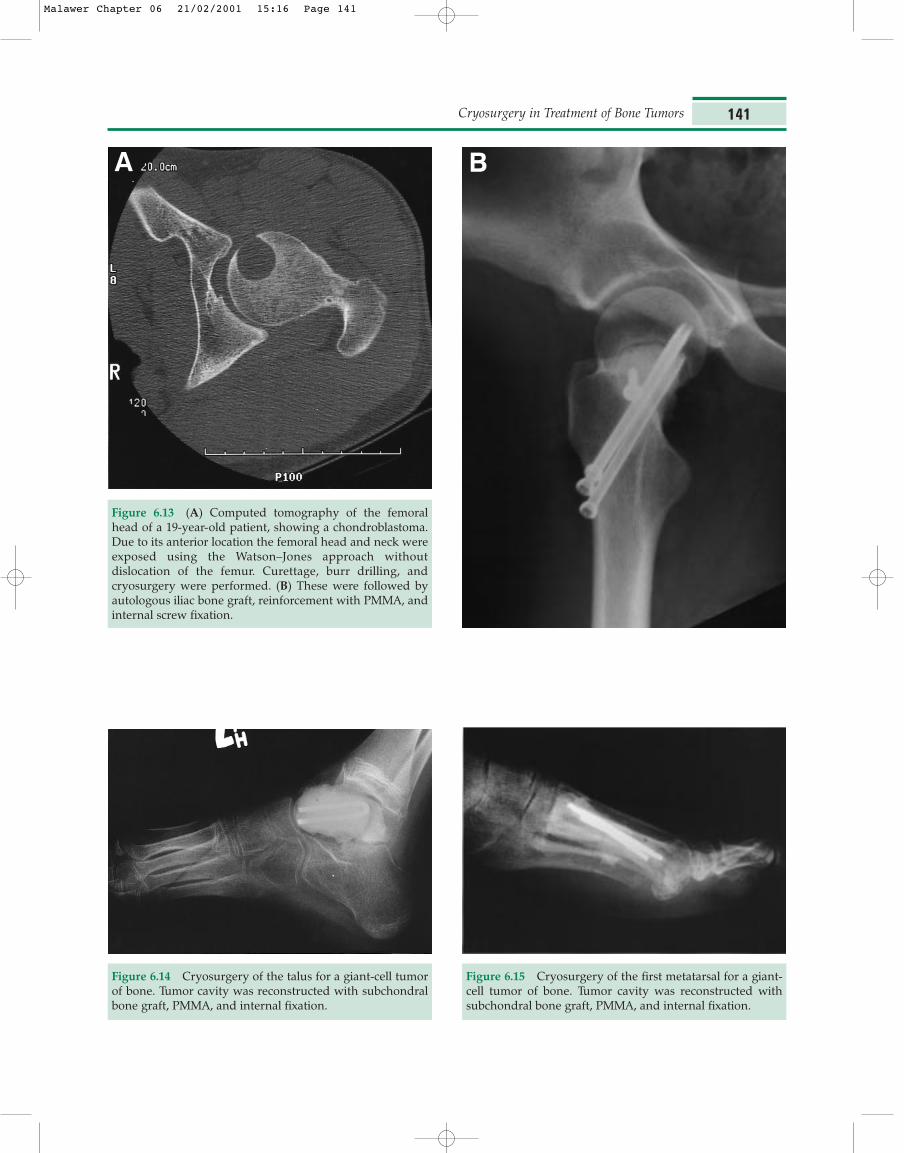
Figure 6.13 (A) Computed tomography of the femoralhead of a 19-year-old patient, showing a chondroblastoma.Due to its anterior location the femoral head and neck wereexposed using the Watson–Jones approach withoutdislocation of the femur. Curettage, burr drilling, andcryosurgery were performed. (B) These were followed byautologous iliac bone graft, reinforcement with PMMA, andinternal screw fixation.
A B
Figure 6.14 Cryosurgery of the talus for a giant-cell tumorof bone. Tumor cavity was reconstructed with subchondralbone graft, PMMA, and internal fixation.
Figure 6.15 Cryosurgery of the first metatarsal for a giant-cell tumor of bone. Tumor cavity was reconstructed withsubchondral bone graft, PMMA, and internal fixation.
Malawer Chapter 06 21/02/2001 15:16 Page 141

tumors reported high complication rates, mostlypathological fractures and infection (Table 6.1). Many ofthese early investigators did not recognize theimportance of soft-tissue protection, use of PMMA andinternal fixation for reconstruction, and avoidance ofsignificant impact on the operated extremity until heal-ing was complete. Gage et al. performed cryosurgeryon 34 dog femora and documented a 32.3% pathologicfracture rate when activity was not restricted and theoperated bone was not protected.4 Marcove et al.practiced cryosurgery prior to the use of PMMA com-bined with onlay bone graft and/or internal fixation.10
They made only a minimal attempt to reconstruct thebone defect remaining after curettage and cryosurgeryand reported a postoperative fracture rate of 25% in theearly series (Figure 6.16). Malawer et al. reported a 5.9%rate of pathologic fractures following cryosurgeryamong 102 patients, treated with cryosurgery forgiant-cell tumor of bone.17 As mentioned, all thesefractures occurred when internal fixation was not usedfor reconstruction.
Prophylactic antibiotics, wide exposure, and adequatemobilization of skin flaps and adjacent neurovascularbundle, along with continuous irrigation of tissues withwarm saline solution, reduce the incidence of skinnecrosis. Malawer et al., who used this technique,reported no cases of infection and only three cases(2.9%) of partial skin necrosis.17 The latter were theresult of contact with leaking LN and were satis-factorily managed with nonsurgical treatment. Nervepalsy after exposure to LN occurs in less than 1% ofpatients and is usually transient.17,20,33
CLINICAL APPLICATION
Table 6.2 summarizes the reported series on bonetumors treated with cryosurgery. The most extensiveorthopedic surgical experience with cryosurgery, by far,has involved giant-cell tumor (GCT) of bone, a benign-aggressive primary bone tumor. Seventy percent ofthese lesions occur in the third or fourth decades of life,and in most cases they are located in the metaphyseal–epiphyseal region of long bones.34 Because wideexcision of these tumors would cause significantfunctional limitation, due to their proximity to the joint,intralesional procedures have been common practice;however, the rate of local recurrence, mainly aftercurettage, has been unacceptably high (i.e. 40–55%).35–38
The introduction of LN as an adjuvant to meticulouscurettage and burr drilling significantly lowered therecurrence rate; Malawer et al. used cryosurgery in thetreatment of GCT of bone and reported a 2.3%recurrence rate among patients treated primarily withcryosurgery.17
Marcove and Miller used cryosurgery to treat a varietyof benign and malignant bone tumors and concludedthat it should be reserved for benign-aggressive bonetumors.7 Surgical treatment of high-grade primarybone sarcoma necessitates wide excision of the tumorwith its soft-tissue component; any violation of thetumor margins is associated with a high risk of localtumor recurrence.39 Cryosurgery is not an appropriatesurgical modality for high-grade primary bonesarcomas for its being an intraosseous procedure withminimal effect on the soft-tissue component of thetumor. Bone tumors that have minimal or no soft-tissue
Musculoskeletal Cancer Surgery142
Table 6.1 Summary of literature review on complication rates following cryosurgery
Complications
Author/year Cases Fracture Infection Skin necrosis Join degeneration Nerve palsy Other
Marcove 1969 57 4 – 6 – – –Marcove 1973 52 13 8 – 2 4 –Marcove 1977 18 7 – – 3 4 –Jacobs 1985 12 6 – – – – –Malawer 1991 25 2 – 1 – – Synovial fistula (1)Aboulafia 1994 9 – – – – – –Marcove 1994 7 – 2 – – – Rectal fistula (1)Marcove 1995 51 5 – – – 1 –Schreuder 1997 26 1 2 – – 1 –Schreuder 1998 26 2 1 – – – Venous gas embolismMalawer 1999 102 6 – 3 2 1 –
Malawer Chapter 06 21/02/2001 15:16 Page 142

component, such as low-grade, unicompartmentalchondrosarcomas and metastatic tumors of bone, canbe treated with cryosurgery.7,9,22
SUMMARY
Cryosurgery is an effective adjuvant to curettage for avariety of benign and malignant bone tumors. It is acurative procedure in the treatment of benign-
aggressive bone lesions as well as of low-grade primarybone sarcomas, and can achieve local control inmetastatic bone disease. The previously reported highrates of fracture and infection can be avoided by carefulattention to surgical details. Adequate exposure,meticulous curettage and burr drilling, soft-tissuemobilization and protection, and routine use of internalfixation with PMMA for reconstruction are essential.
Cryosurgery in Treatment of Bone Tumors 143
Figure 6.16 Plain radiographs of the distal femur: (A) anteroposterior, and (B) lateral views, showing a pathologic fracture ofthe lateral femoral condyle. The fracture occurred 18 months following cryosurgery, in which internal fixation was not used forreconstruction. Reinforcement of the tumor cavity with PMMA and internal fixation is strongly recommended in all cases.
A B
Malawer Chapter 06 21/02/2001 15:16 Page 143

Musculoskeletal Cancer Surgery144
Table 6.2 Summary of literature review on bone tumors treated with cyrosurgery
Tumor type Author/year
Benign aggressive lesionsGiant-cell tumor Aboulafia 1994; Jacobs 1985; Malawer 1991, 1999; Marcove 1969, 1973, 1978, 1994Echondroma Schreuder 1998Chondroblastoma Schreuder 1998Unicameral bone cyst Schreuder 1997Aneurysmal bone cyst Marcove 1969, 1995; Oeseburg, 1978; Schreuder 1997Fibrous dysplasia Marcove 1969Hemangioma of bone Marcove 1969Sacral chordoma Marcove 1969; Vries 1986Eosinophilic granuloma Marcove 1969
Metastatic lesionsCarinoma of lung Marcove 1969Carcinoma of breast Marcove 1969Carcinoma of prostate Marcove 1969Hypernephroma Marcove 1969, 1977Carcinoma of bladder Marcove 1969Soft-tissue sarcoma Marcove 1969Adenocarcinoma of uterus Marcove 1969
Primary bone sarcomasOsteosarcoma Marcove 1969, 1984Chondrosarcoma Marcove 1969; Marcove 1977, Schreuder 1998
OtherMultiple myeloma Marcove 1969
1. Arnott JM. Practical illustrations of the remedial efficiencyof a very low or anaesthetic temperature in cancer. Lancet.1850;2:257–316.
2. Holden HB. History and development of cryosurgery. In:Holden HB, editor. Practical Cryosurgery. Chicago:Pitman Medical Publication, 1975:1–9.
3. Cooper IS. Cryogenic surgery of the basal ganglia. J AmMed Assoc. 1962;181:600–4.
4. Gage AA, Greene GW, Neiders ME, Emmings FG.Freezing bone without excision. An experimental study ofbone-cell destruction and manner of regrowth in dogs. JAm Med Assoc. 1966;196:770–4.
5. Kuylenstierna R, Lundquist PG, Nathanson A. Destructionand regeneration of jaw bone after cryogenic application.An experimental study. Ann Otol. 1980;89:582–9.
6. Schargus G, Winckler J, Schröder F, Schöfer B. Cryosurgicaldevitalization of bone and its regeneration. An experi-mental study with animals. J Maxillofac Surg. 1975;3:128–31.
7. Marcove RC, Miller TR. The treatment of primary andmetastatic localized bone tumors by cryosurgery. SurgClin N Am. 1969;49:421–30.
8. Marcove RC, Searfoss RC, Whitmore WF, Grabstald H.Cryosurgery in the treatment of bone metastases fromrenal cell carcinoma. Clin Orthop. 1977;127:220–7.
9. Marcove RC, Stovell PB, Huvos AG, Bullough PG. The useof cryosurgery in the treatment of low and medium gradechondrosarcoma. Clin Orthop. 1977;122:147–56.
10. Marcove RC, Weis LD, Vaghaiwalla MR. Cryosurgery inthe treatment of giant cell tumor of bone. A report of 52consecutive cases. Cancer. 1978;41:957–69.
11. Marcove RC, Lyden JP, Huvos AG, Bullough PG. Giant celltumors treated by cryosurgery. J Bone Joint Surg. 1973;55:1633–44.
12. Korpan NN. Hepatic cryosurgery for liver metastases.Ann Surg. 1997;225:193–201.
13. Weaver ML, Atkinson D, Zemel R. Hepatic cryosurgery in treating colorectal metastases. Cancer. 1995;76:210–14.
14. Creasman WT, Hinshaw WM, Clarke-Pearson DL.Cryosurgery in the management of cervical intraep-ithelial neoplasia. Obstet Gynecol. 1984;63:145–9.
15. Miller RJ Jr, Cohen JK, Shuman B, Merlotti LA.Percutaneous, transperineal cryosurgery of the prostate
References
Malawer Chapter 06 21/02/2001 15:16 Page 144
as salvage therapy for post radiation recurrence ofadenocarcinoma. Cancer. 1996;77:1510–14.
16. Wong WS, Chinn DO, Chinn M, Chinn J, Tom WL, TomWL. Cryosurgery as a treatment for prostate carcinoma:results and complications. Cancer. 1997;79:963–74.
17. Malawer MM, Bickels J, Meller I, Buch R, Kollender Y.Cryosurgery in the treatment of giant cell tumor. A longterm follow-up study. Clin Orthop. 1999;359:176–88.
18. Malawer MM, Dunham W. Cryosurgery and acryliccementation as surgical adjuncts in the treatment ofaggressive (benign) bone tumors. Analysis of 25 patientsbelow the age of 21. Clin Orthop. 1991;262:42–57.
19. Marcove RC, Sheth DS, Brien EW, Huvos AG, Healey JH.Conservative surgery for giant cell tumors of the sacrum.The role of cryosurgery as a supplement to curettage andpartial excision. Cancer. 1994;74:1253–60.
20. Marcove RC, Sheth DS, Takemoto S, Healey JS. The treat-ment of aneurysmal bone cyst. Clin Orthop. 1995;311:157–63.
21. Schreuder HW, Conrad EU 3rd, Bruckner JD, Howlett AT,Sorensen LS. Treatment of simple bone cysts in childrenwith curettage and cryosurgery. J Pediatr Orthop. 1997;17:814–20.
22. Schreuder HW, Pruszczynski M, Veth RP, Lemmens JA.Treatment of benign and low-grade malignant intra-medullary chondroid tumours with curettage andcryosurgery. Eur J Surg Oncol. 1998;24:120–6.
23. Schreuder HW, Veth RPH, Pruszczynski M, LemmensJAM, Koops HS, Molenaar WM. Aneurysmal bone cyststreated by curettage, cryotherapy and bone grafting. JBone Joint Surg. 1997;79:20–5.
24. Gill W, Fraser J, Carter DC. Repeated freeze–thaw cyclesin cryosurgery. Nature. 1968;219:410–13.
25. Schreuder HW, van Egmond J, van Beem HB, Veth RP.Monitoring during cryosurgery of bone tumors. J SurgOncol. 1997;65:40–5.
26. Harris L, Griffiths J. Relative effects of cooling and warm-ing rates on mammalian cells during the freeze–thawcycle. Cryobiology. 1977;14:662–9.
27. Karow AR, Webb WR. Tissue freezing, a theory for injuryand survival. Cryobiology. 1965;2:99–108.
28. Malawer MM, Marks MR, McChesney D, Piasio M,Gunther SF, Shmookler BM. The effect of cryosurgery andpolymethylmethacrylate in dogs with experimental bonedefects comparable to tumor defect. Clin Orthop. 1988;226:299–310.
29. Mazur P. Cryobiology: the freezing of biological systems.Science. 1970;168:939–49.
30. McGann LE, Kruuv J, Frim J, Frey HE. Factors affectingthe repair of sublethal freeze–thaw damage in mam-malian cells. Suboptimal temperature and hypoxia.Cryobiology. 1975;12:530–9.
31. Miller RH, Mazur P. Survival of frozen-thawed human redcells as a function of cooling and warming velocities.Cryobiology. 1976;13:404–14.
32. Aboulafia AJ, Rosenbaum DH, Sicard-Rosenbaum L,Jelinek JS, Malawer MM. Treatment of large subchondraltumors of the knee with cryosurgery and compositereconstruction. Clin Orthop. 1999;307:189–99.
33. Jacobs PA, Clemency RE. The closed cryosurgical treat-ment of giant cell tumor. Clin Orthop. 1985;192:149–58.
34. Huvos GA. Giant-cell tumor of bone. In: Huvos GA,editor. Bone Tumors: Diagnosis, Treatment and Prognosis,2nd edn. Baltimore: W.B. Saunders Company; 1991:429–67.
35. Campanacci M, Baldini N, Boriani S, Sudanese A. Giantcell tumor of bone. J Bone Joint Surg. 1987;69:106–14.
36. Goldenberg R, Campbell C, Bonfiglio M. Giant cell tumor:an analysis of 218 cases. J Bone Joint Surg. 1970;52:619–64.
37. Johnson EW, Dahlin DC. Treatment of giant cell tumor ofbone. J Bone Joint Surg. 1959;41:895–904.
38. McDonald DJ, Sim FH, McLeod RA, Dahlin DL. Giant celltumor of bone. J Bone Joint Surg. 1986;68:235–42.
39. Petersson H, Springfield DS, Enneking WF. Surgicalprinciples. In: Petersson H, Springfield DS, Enneking WF,editors. Radiologic Management of MuskuloskeletalTumors. Berlin: Springer-Verlag; 1987:9–13.
Cryosurgery in Treatment of Bone Tumors 145
Malawer Chapter 06 21/02/2001 15:16 Page 145

Malawer Chapter 06 21/02/2001 15:16 Page 146