5fu encapsulated polyglycerol sebacate nanoparticles as
TRANSCRIPT

RSC Advances
PAPER
Ope
n A
cces
s A
rtic
le. P
ublis
hed
on 2
5 M
ay 2
021.
Dow
nloa
ded
on 2
/15/
2022
1:5
5:54
AM
. T
his
artic
le is
lice
nsed
und
er a
Cre
ativ
e C
omm
ons
Attr
ibut
ion-
Non
Com
mer
cial
3.0
Unp
orte
d L
icen
ce.
View Article OnlineView Journal | View Issue
5FU encapsulate
aDepartment of Biotechnology, Indian
Chennai-600036, India. E-mail: vermars@iibDepartment of Chemistry, Indian Institute
India. E-mail: [email protected]
† Electronic supplementary informa10.1039/d1ra01722e
Cite this: RSC Adv., 2021, 11, 18984
Received 4th March 2021Accepted 10th May 2021
DOI: 10.1039/d1ra01722e
rsc.li/rsc-advances
18984 | RSC Adv., 2021, 11, 18984–18
d polyglycerol sebacatenanoparticles as anti-cancer drug carriers†
Divya Sivanesan,a Rama S. Verma *a and Edamana Prasad *b
Themajority of anti-cancer drugs fail to reach clinical trials due to their lowwater solubility. A biocompatible
drug delivery system that encapsulates and efficiently delivers hydrophobic drugs to the target site is the
need of the hour. This study addresses the issue by focusing on a polymeric polyglycerol sebacate (PGS)
nanoparticles loaded with 5-fluorouracil (5FU), a primary line chemotherapy drug for many types of
cancers. The generated nanoparticle (PGS-NP) was biocompatible and had minimal cytotoxicity against
the MDA-MB-231 and A549 cell lines, even at a high concentration of 100 mg mL�1. The cell viability post
treatment with PGS nanoparticles encapsulated with 5FU (PGS-5FU) decreased to as low as around 40%
whereas, in the case of treatment with 5FU, the viability percentage increased. The nanoparticles also
showed controlled drug release when encapsulated with 5FU. This striking observation suggested that
these nanoparticles can improve the efficacy of drug delivery to tumor sites. Apoptosis assay and
caspase-3 activity quantification supported these data wherein PGS-5FU treatment showed almost three
times caspase-3 activity as compared to control cells. Additionally, throughout all the experiments,
MDA-MB-231 cells were more sensitive to PGS-5FU than A549 cells, indicating that these nanoparticles
are ideal for breast cancer treatment. In summary, 5FU encapsulated PGS nanoparticles are a potential
drug carrier to deliver 5FU efficiently to cancer cells.
1. Introduction
Cancer is the second leading death-causing disease according tothe WHO.1 The different types of cancer account for almost 10million deaths per year.2 Breast cancer is currently diagnosed in 1out of 4 women globally,3 and metastasis of this cancer is as highas 30%.4 Similarly, the number of lung cancer cases reported in2020 alone was 2.2 million5 worldwide with the highest death rateof all cancer types. Whichever type of cancer is detected, the careand treatment given to patients include chemotherapy, radio-therapy or surgery or a combination.6 Depending upon the stageand location of the tumor, the chemotherapy regime can includeanti-cancer drugs such as 5-uorouracil (5FU)7–10 and paclitaxel.11
Many of these drugs are hydrophobic with solubility andbiocompatibility issues. This has hindered the drug developmentprocess over many decades. One of the solutions is to discovera vehicle for delivery of the drugs precisely to the site with highbiocompatibility and bioavailability. This could also avoid relapseby improving the efficiency of the current therapy given to patients.
The recent trend in drug delivery systems has shiedtowards nanomedicines like polymeric nanoparticles,
Institute of Technology Madras,
tm.ac.in
of Technology Madras, Chennai-600036,
tion (ESI) available. See DOI:
993
liposomes, and micelles.12 Organic and inorganic nanoparticlesare gaining interest and as many as 25 have been approved bythe FDA, and nearly 75 nanomedicines are currently in clinicaltrials.13 The major advantages of nanoparticles are an improvedbioavailability owing to their enhanced aqueous solubility andincreased half-life.14,15 As most cancer drugs have poor solu-bility, giving them as injections is challenging and tabletformulation can also be difficult.16,17 By encapsulating thesedrugs within nanoparticles, a topical therapy can be rened.
Polymeric nanoparticles have been gaining attention due totheir small particle size and their ability to protect encapsulateddrugs from the biological environment with improved thera-peutic indices. Regarding polymers used to synthesize nano-particles, polyglycerol sebacate (PGS)18–20 that is widely used asa scaffold in tissue engineering is less studied in the eld ofnanomedicines. The monomers that produce this polymer viapolycondensation, glycerol and sebacic acid, are both FDAapproved and biocompatible. This polymer has utility asa scaffold or patch due to its high tensile strength and elasto-meric property.21 By tweaking the molar ratios of the mono-mers, we can synthesize polymeric PGS as a gel that can then bemade into nanoparticles (NPs)22,23 using the solvent displace-ment method.24 This has not been particularly well exploredand very few studies have reported the application of PGS asa drug delivery system to treat cancer.
Here, in this study, PGS nanoparticles (PGS-NPs) aresynthesized, characterized, and encapsulated with 5FU (PGS-
© 2021 The Author(s). Published by the Royal Society of Chemistry

Paper RSC Advances
Ope
n A
cces
s A
rtic
le. P
ublis
hed
on 2
5 M
ay 2
021.
Dow
nloa
ded
on 2
/15/
2022
1:5
5:54
AM
. T
his
artic
le is
lice
nsed
und
er a
Cre
ativ
e C
omm
ons
Attr
ibut
ion-
Non
Com
mer
cial
3.0
Unp
orte
d L
icen
ce.
View Article Online
5FU), a common drug used in the treatment of breast cancerand whose derivates are currently being studied for targetedtherapy against lung cancer. PGS-NPs had sizes within the range of200–300 nm, and 5FU encapsulation did not vary the size much.These PGS nanoparticles also showed minimal cytotoxicity towardcancer cells, indicating their high biocompatibility. When loadedwith 5FU, the cytotoxicity of these NPs increased signicantly andover a long period of 48 h, the percentage of viable cells continuedto decrease, showing a controlled drug release. This is one of therst studies reporting PGS nanoparticles encapsulated with 5FU asa drug delivery system for the treatment of breast cancer and lungcancer. When studying the effects of these drug-loaded NPs, it isevident that they provide an efficient way of delivering hydro-phobic anti-cancer drugs to tumor sites.
2. Experimental section2.1. Materials and methods
The chemicals and reagents were purchased from Sigma-AldrichChemical Co. Sebacic acid (cat. no. 283258) and glycerol (cat. no.G5516) were purchased from Sigma-Aldrich Chemical Co. 5-Fluo-rouracil (5FU), Tween 80, MTT (3-(4,5-dimethylthiazol-2-yl)-2,5-diphenyltetrazolium bromide), rhodamine B and other chem-icals used for this study were obtained from Sisco ResearchLaboratories Pvt. Ltd. NMR spectra were recorded using a BrukerADVANCE III 500 MHz spectrometer with DMSO-d6 as the solvent.FTIR spectra were recorded using a Jasco FTIR-4100 instrumentequipped with an ATR accessory. UV-visible spectra were collectedusing a PerkinElmer spectrophotometer. Thermogravimetricanalysis (TGA) was performed under a nitrogen atmosphere witha heating rate of 10 �Cmin�1 using a TGA Q-50 thermogravimetricanalyzer. Dynamic light scattering (DLS) was performed usinga Malvern Instruments Zetasizer nano ZS90. Powder X-raydiffraction (XRD) was performed using a D8 Advance (Bruker).Scanning electron microscopy (SEM) was used to take images ofthe nanoparticles and the SEM instrument was bought fromThermo Fisher Scientic (Apreo S). The colorimetric caspase-3assay was purchased from Takara Bio. Annexin V-FITC and pro-pidium iodide were obtained from BD Biosciences. The primaryand secondary-HRP conjugated antibodies used for western blotanalysis were obtained from Cell Signaling Technology.
2.2. Maintenance of cell lines
Human non-small-cell lung cancer cell line A549 and humanbreast cancer cell line MDA-MB-231 were procured from the
Loading efficiency% ¼ ðTotal amount of 5FUÞ � ðamount of 5FU washedÞ � 100
Total amount of 5FU
National Centre for Cell Sciences (NCCS), Pune, Maharashtra,India. They were cultured in DMEM (Gibco, Grand Island, U.S.A.)supplemented with 10% fetal bovine serum (FBS) (Gibco, GrandIsland, U.S.A.) with 1� penicillin–streptomycin (Gibco, GrandIsland, U.S.A.) with 5% CO2 at 37 �C in a humied incubator.
© 2021 The Author(s). Published by the Royal Society of Chemistry
2.3. Synthesis of the polyglycerol sebacate pre-polymer
The polyglycerol sebacate polymer was synthesized using a previ-ously reported procedure.25 Equimolar amounts of glycerol andsebacic acid were taken and kept in an oil bath at 120 �C for 24 hunder a nitrogen atmosphere with constant magnetic stirring.Aer 24 h, a sticky-gel polymer formed, which was dissolved inethanol and puried using distilled water and a 10 000 Da dialysisbag. This solution was freeze-dried and stored at room tempera-ture. FTIR (PGS) (cm�1): 3443, 2926, 2854, 1733, 1410, 1180, 936;1H NMR (PGS) (500 MHz) d/ppm: 1.24 (s, –CH2–), 1.55 (d, –CH2-CH2O(CO)–), 2.27 (m, –CH2O(CO)–), 3.6–4.1 (m, OHCH2CHO–),4.2–4.35 (m, –OCH2CHO–), 4.95 (2-acylglyceride), 5.09 (1,2-diac-ylglyceride), 5.26 (1,2,3-triacylglyceride).
2.4. Preparation of PGS nanoparticles
The nanoparticles were generated using the solvent displace-ment method. Different amounts of PGS were taken and dis-solved in 1 mL of ethanol to obtain different concentrations ofthe nanoparticles (5 mg mL�1, 10 mg mL�1, 20 mg mL�1, and25 mg mL�1).22 This solution was added dropwise to 10 mL ofdistilled water slowly and 100 mL of Tween 80 was also added.The solution was kept under constant stirring with sonication at60 �C. It was then centrifuged at a high speed of 12 000g for10 min and the supernatant was discarded. The pellet waswashed with phosphate buffer saline (PBS) twice at high speedfor 5 min. This pellet was then freeze-dried and weighed forfurther experiments.
2.5. Encapsulation of 5FU within the nanoparticles
The 5FU drug and PGS nanoparticles were taken in differentratios (1 : 1, 1 : 2, 1 : 2.5, 1 : 4, and 1 : 5) to optimize the loadingefficiency of the 5FU loaded nanoparticles. 5FU was dissolved in1 mL of DMSO and this solution was added to 9 mL of distilledwater (1 : 10) containing 250 mg of PGS (25 mg mL�1).26 Thismixture was le stirring overnight. Then, the solution wascentrifuged at a high speed of 12 000g for 10 min and thesupernatant was discarded. The pellet was washed with phos-phate buffer saline (PBS) at high speed for 5 min and thesupernatant was taken for UV-vis spectral measurement at508 nm to calculate the amount of encapsulated 5FU. Aerwashing with PBS, the pellet was lyophilized and weighed. Theloading efficiency27 was calculated using the following formula:
2.6. Characterization of 5FU loaded PGS nanoparticles
2.6.1. Physical characterization. The zeta potential wasrecorded using a Zetaplus instrument and dynamic light scat-tering (DLS) was used to study the particle size and the
RSC Adv., 2021, 11, 18984–18993 | 18985

RSC Advances Paper
Ope
n A
cces
s A
rtic
le. P
ublis
hed
on 2
5 M
ay 2
021.
Dow
nloa
ded
on 2
/15/
2022
1:5
5:54
AM
. T
his
artic
le is
lice
nsed
und
er a
Cre
ativ
e C
omm
ons
Attr
ibut
ion-
Non
Com
mer
cial
3.0
Unp
orte
d L
icen
ce.
View Article Online
polydispersity index (PI). The stability of the particles wasanalyzed by noting the size and PI aer 20 days. The surfacemorphology and structure of the nanoparticles were analyzedusing SEM images recorded at 5 kV. The nanoparticles weredispersed in distilled water and dropped on a glass cover slip.The cover slip was dried in a desiccator for 3 days to evaporatethe solvent, then it was sputter-coated with gold beforerecording images using the microscope.
2.6.2. Chemical characterization. 1H NMR and 13C NMRspectroscopies were performed to conrm the structure of PGS.FTIR spectroscopy28 was employed to determine the functionalgroups present in the nanoparticle and polymer. The lyophi-lized samples were mixed with potassium bromide (KBr) andpellets were formed using a hydraulic press. Then, the pellets
Cell viability% ¼ ðMean absorbance of sample� absorbance of the blankÞ � 100
Mean of absorbance of the control sample
were placed in the FTIR instrument and the spectra wererecorded and analyzed. XRD was performed to ensure encap-sulation of 5FU within PGS-NP.22 The thermal stability of PGSwas assessed using TGA, and DSC was used to characterize thethermal phase transitions.29 The averaged molecular weight(Mw) and the number average molecular weight (Mn) of the PGSpolymer were calculated using gel permeation chromatography(GPC) analysis in a styragel column using polystyrene standardsas reference controls for calculating Mw.
2.7. Drug release study
The dialysis method was used to determine the concentration of5FU in PBS and, thus, the drug release pattern of the nano-particles.30 PGS-5FU NPs were dispersed in PBS and put ina dialysis tube in a beaker with 200 mL of PBS. Every hour, 3 mLof PBS was replaced with fresh PBS in the beaker with stirring at50 rpm and this was done for 24 h. The drug release rate wasobtained by taking readings of the 3 mL of extracted PBS usingUV/visible spectroscopy. DD Solver soware was used to simu-late the drug release kinetics.
2.8. Cellular uptake of the NPs
The nanoparticles were loaded with rhodamine B (0.5 mg mg�1
w/w)31 and the uptake of the NPs was observed using a uores-cence microscope. Around 25 000 cells (MDA-MB-231 and A549)were seeded in separate 60 mm dishes then treated with therhodamine B loaded nanoparticles. Aer 12 h of incubation, thecells were washed with PBS and the nucleus was stained with 2mL of live Hoescht (25 mg mL�1) for 15 min. Images were thentaken using a uorescence microscope (Ti Eclipse Nikon,U.S.A.).
18986 | RSC Adv., 2021, 11, 18984–18993
2.9. In vitro cytotoxicity study
Cells were seeded in a 96 well plate (5000 cells per well) and,24 h later, the free drug, PGS nanoparticles and the drug loadednanoparticles were added to the wells in triplicate at differentconcentrations; untreated cells were also used in the experi-ment.32 Readings were taken at different time points such as12 h, 24 h, 36 h and 48 h. An MTT stock solution was prepared(5 mgmL�1) and 10 mL was added to each well along with blanksat each time point. 2 h later, the stop solution (DMSO) wasadded to solubilize the crystals formed. The reading of thesamples was performed at 560 nm using an ELISA plate reader(PerkinElmer). The cell viability percentage33 was calculatedusing the following formula:
2.10. Apoptosis analysis using ow cytometry
The cells were treated for 24 h with free 5FU, PGS-NP, and PGS-5FU and 2 � 105 cells were taken along with the untreatedcontrol cells. 50 mL of binding buffer was added to each tubeand then annexin V-FITC (5 mL) was added with 10 mL of pro-pidium iodide (PI).34 Aer 10 min of incubation at 37 �C, thecells were run through a FACS Calibur instrument to calculatethe number of cells undergoing apoptosis. The gating was doneusing unstained control cells.
2.11. Evaluation of apoptosis
The intrinsic apoptosis pathway is initiated by cleavage ofcaspase-9, and the cleaved caspase-9 cleaves caspase-3. Thiscleaved caspase-3 then translocates into the nucleus toinduce apoptosis. To understand the mechanism of the NPinduced apoptosis, a caspase-3 colorimetric assay kit wasused.35 The protocol given by Takara Bio was followedwhereby 2 � 106 treated and control cells were centrifuged at400g for 5 min. Then, 50 mL of cold lysis buffer was added andthe solution was kept in ice for 10 min. The tubes were thencentrifuged at 12 000g for 10 min at 4 �C and the supernatantwas transferred to new tubes. One set of positive control cellswas treated with 1 mL of caspase-3 inhibitor and 50 mL ofreaction buffer containing DTT. The rest of the samples weretreated only with reaction buffer having DTT and the solutionwas incubated for 30 min in ice. Then, 5 mL of the 1 mMcaspase-3 substrate was added and kept for 1 h in a waterbath at 37 �C. A standard curve was plotted using pNAprovided in the kit dissolved in DMSO at different concen-trations ranging from 0 to 200 mM.
2.12. Expression levels of apoptosis proteins
MDA-MB-231 cells and A549 cells were treated with 5FU, PGSnanoparticles and PGS-5FU, the cells were trypsinized, and the
© 2021 The Author(s). Published by the Royal Society of Chemistry

Scheme 1
Fig. 1 (A) FTIR spectra of the polyglycerol sebacate prepolymer, PGS nanoparticle and 5FU loaded PGS nanoparticle. (B) Powder XRD analysis ofthe PGS polymer, PGS nanoparticle and 5FU encapsulated nanoparticle. (C) Dynamic light scattering (DLS) histogram showing a unimodal peakindicating uniformly sized particles; the same was validated using scanning electron microscopy (SEM) (D).
© 2021 The Author(s). Published by the Royal Society of Chemistry RSC Adv., 2021, 11, 18984–18993 | 18987
Paper RSC Advances
Ope
n A
cces
s A
rtic
le. P
ublis
hed
on 2
5 M
ay 2
021.
Dow
nloa
ded
on 2
/15/
2022
1:5
5:54
AM
. T
his
artic
le is
lice
nsed
und
er a
Cre
ativ
e C
omm
ons
Attr
ibut
ion-
Non
Com
mer
cial
3.0
Unp
orte
d L
icen
ce.
View Article Online

RSC Advances Paper
Ope
n A
cces
s A
rtic
le. P
ublis
hed
on 2
5 M
ay 2
021.
Dow
nloa
ded
on 2
/15/
2022
1:5
5:54
AM
. T
his
artic
le is
lice
nsed
und
er a
Cre
ativ
e C
omm
ons
Attr
ibut
ion-
Non
Com
mer
cial
3.0
Unp
orte
d L
icen
ce.
View Article Online
pellets were washed with PBS. The supernatant was discarded,and the total protein was extracted using a radio-immunoprecipitation buffer (RIPA) with a 1� protein inhibitioncocktail (PIC).36 30 mg of protein of each sample was run usingSDS-PAGE and then transferred onto a polyvinylidene diuoridemembrane aer activation. This membrane was blocked with5% skimmed milk and then incubated overnight at 4 �C withprimary antibodies against procaspase-3, procaspase-9, andvinculin. The membrane was washed with TBS—T thrice andincubated with the respective alkaline phosphatase-conjugatedsecondary antibody. The blots were developed using anenhanced chemiluminescence (ECL) buffer, imaged usinga BioRad ChemiDoc system and analysed by normalizing withan endogenous control vinculin protein.
2.13. Statistical analysis
The statistical data analysis was performed using Student's t-test to compare the different samples where p < 0.05 was takento be signicant. All the experiments were done in triplicate, theaverage was noted, and the standard error was plotted usingGraphpad Prism 8. Origin Pro 8.5 soware was used to plotgraphs of FTIR, XRD, and TGA data. Flowjo v8 was used to plotthe dot plot of ow cytometry experiments. Image Lab and FijiImageJ soware were used to analyse western blot and uo-rescence microscope images, respectively. To calculate theloading efficiency and the drug release rate, the supply of 5FUwas measured over a range of different concentrations and lmax
was determined by UV-vis spectroscopy.
Fig. 2 (A) Cumulative release rate of 5FU from the drug loaded NPs. (B aand PGS-5FU done with A549 and MDA-MB-231 cells, respectively (*p <
18988 | RSC Adv., 2021, 11, 18984–18993
3. Results and discussion3.1. Synthesis and properties of 5FU-NPs
The synthesis of the polyglycerol polymer was done accordingto a previously reported protocol by taking equimolar amountsof glycerol and sebacic acid, as shown in Scheme 1. Thecharacterization of the monomers and the pure polymer wasperformed using 1H NMR spectroscopy and data were analyzedusing MestReNova soware (Fig. S1†). The proton peaks atd 1.30, 1.60, and 2.3 ppm in the polymer data corresponded tosebacic acid and the peaks at 3.43–3.96, 4.08–4.35, and 5.09,5.26 ppm are related to glycerol. These data showed a linearstructure of the PGS polymer with the molar ratio of mono-mers of 1. Additionally, this result was corroborated by FTIRanalysis (Fig. 1A), which exhibited a broad peak at 3443 cm�1
attributed to the stretch vibration absorption of O–H endgroups in the molecules while their bending gave a peak at1410 cm�1. The sharp peaks at 1180 cm�1 and 1733 cm�1 arerelated to C–O whereby the 1733 cm�1 peak indicated an estercarbonyl linkage. C]O bonds and methylene groups wereindicated by peaks at 2854 cm�1 and 2926 cm�1, respectively.The peak at 1691 cm�1 indicates unreacted sebacic acid andthis peak was absent in the spectrum of the polymer owing tothe purication step. These data demonstrate the successfulgeneration of the PGS polymer and the active groups on thesurface can be functionalized further to attach compoundsonto the surface of the nanoparticles. XRD analysis (Fig. 1B)showed a sharp peak in the pattern of the PGS-5FU nano-particles at 30 2q and a peak at 20 2q present in the pattern of
nd C) Cytotoxicity assay with different concentrations of PGS-NP, 5FU0.05 vs. PGS-NP, $p < 0.05 vs. 5FU, &p < 0.05 vs. PGS-5FU).
© 2021 The Author(s). Published by the Royal Society of Chemistry

Fig. 3 (A) Increasing uptake of Rho-NPs by A549 as well as MDA-MB-231 cells with time and (B) its quantified representation.
Paper RSC Advances
Ope
n A
cces
s A
rtic
le. P
ublis
hed
on 2
5 M
ay 2
021.
Dow
nloa
ded
on 2
/15/
2022
1:5
5:54
AM
. T
his
artic
le is
lice
nsed
und
er a
Cre
ativ
e C
omm
ons
Attr
ibut
ion-
Non
Com
mer
cial
3.0
Unp
orte
d L
icen
ce.
View Article Online
PGS-NP that had almost completely disappeared in the patternof PGS-5FU, showing the encapsulation of 5FU within PGS-5FU. These data conrm the crystalline nature of the nano-particles as compared to the semi-crystalline nature of the PGSgel and, therefore, the increased stability of PGS-5FU. Thethermostability of PGS-NP and PGS-5FU was assessed usinga thermogravimetric analyzer (TGA-Q50) and a representativethermogram is plotted in Fig. S2.† The results showed a rstweight loss of about 30% at 250 �C for the PGS nanoparticlesand a second thermal decomposition at 400 �C of 40% weightloss. The rst weight loss is due to absorbed solvent evapora-tion and the second one is due to polymer degradation. In thecase of the 5FU loaded PGS nanoparticles, there was a weightloss of up to 90% in the range from 300 �C to 400 �C, indicatingan increase in stability with the encapsulation of 5FU. Theheat ow through the PGS prepolymer, PGS-NP and PGS-5FUwas measured by differential scanning calorimetry (DSCQ200, TA Instruments) (Fig. S3†). The results showed a glasstransition temperature (Tg) of the PGS prepolymer of �7.52 �Cand that of PGS-NP of 104.87 �C. The shi to positivetemperature is due to the increased crosslinking within thenanoparticle. Additionally, the melting temperature (Tm) was5.56 �C for the PGS prepolymer, which increased to 105.33 �C
© 2021 The Author(s). Published by the Royal Society of Chemistry
for PGS-NP, indicating that PGS is miscible with the sol–gelcomposite and that the nanoparticle has partial crystallinity.Interestingly, aer loading 5FU into the nanoparticle, itbecame completely amorphous, and this suggests thesuccessful entrapment of the drug within the nanoparticlematrix. Furthermore, GPC analysis (GPC, Waters, U.S.A.)showed a PDI of the PGS polymer of 1.0054, a calculated Mn of93.9 kDa and aMw of 94.4 kDa using polystyrene standards forgenerating the calibration curve.
3.2. Morphological characteristics of the nanoparticle
The size and zeta potential of the nanoparticles were assessedusing a Malvern Instruments Zetasizer nano ZS90. The sizedistribution histogram (Fig. S5†) showed varied sizes fordifferent concentrations of PGS used to make the nanoparticles.Interestingly, the size and PDI data for the 25 mg mL�1
concentration of PGS measured 30 days post synthesis showedhomogenous particles in the range of 200–350 nm and havinglong shelf-life stability (Fig. 1C). The observed zeta potential wasbetween �35 mV and �20 mV. The zeta potential magnitude ofPGS-NPs indicates high colloidal stability. These nanoparticlesare also small-sized, within which the drug can be easily loaded.To corroborate the results, scanning electron microscopy (SEM)
RSC Adv., 2021, 11, 18984–18993 | 18989
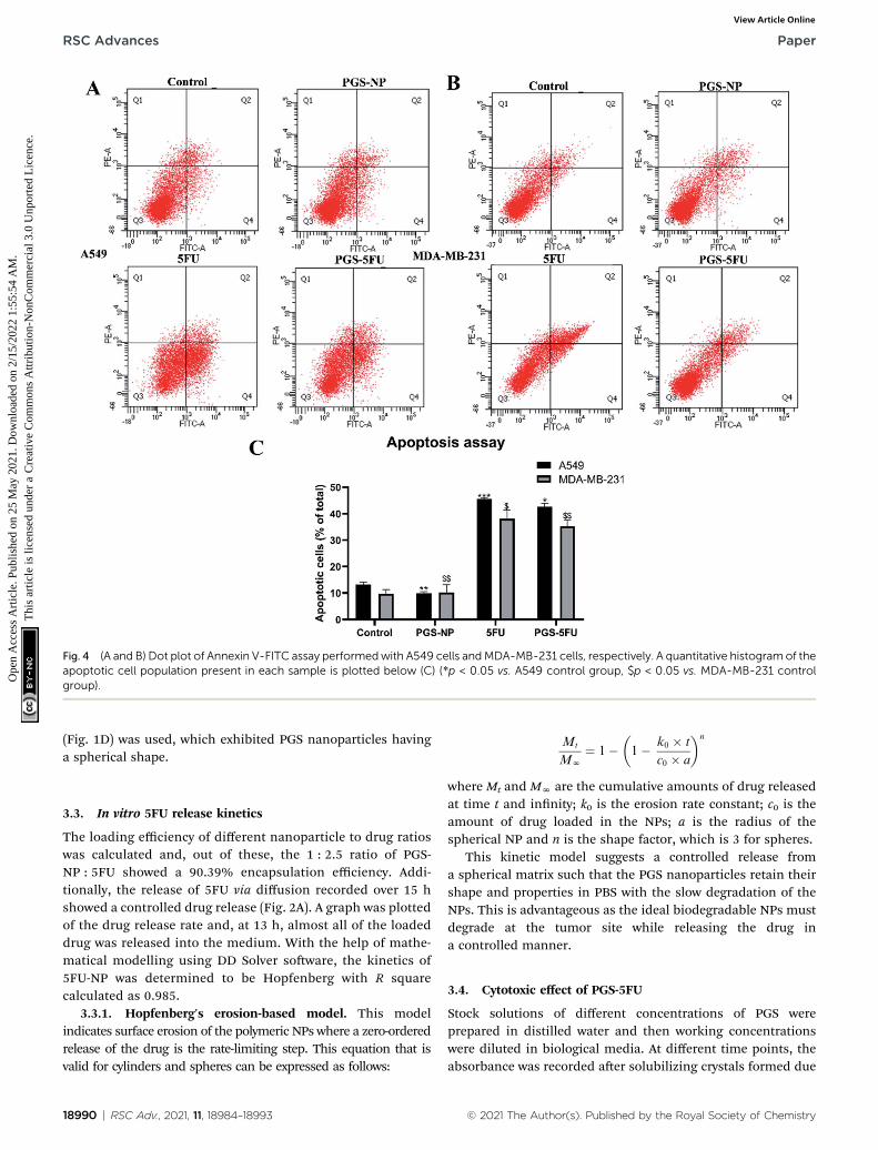
Fig. 4 (A and B) Dot plot of Annexin V-FITC assay performedwith A549 cells andMDA-MB-231 cells, respectively. A quantitative histogram of theapoptotic cell population present in each sample is plotted below (C) (*p < 0.05 vs. A549 control group, $p < 0.05 vs. MDA-MB-231 controlgroup).
RSC Advances Paper
Ope
n A
cces
s A
rtic
le. P
ublis
hed
on 2
5 M
ay 2
021.
Dow
nloa
ded
on 2
/15/
2022
1:5
5:54
AM
. T
his
artic
le is
lice
nsed
und
er a
Cre
ativ
e C
omm
ons
Attr
ibut
ion-
Non
Com
mer
cial
3.0
Unp
orte
d L
icen
ce.
View Article Online
(Fig. 1D) was used, which exhibited PGS nanoparticles havinga spherical shape.
3.3. In vitro 5FU release kinetics
The loading efficiency of different nanoparticle to drug ratioswas calculated and, out of these, the 1 : 2.5 ratio of PGS-NP : 5FU showed a 90.39% encapsulation efficiency. Addi-tionally, the release of 5FU via diffusion recorded over 15 hshowed a controlled drug release (Fig. 2A). A graph was plottedof the drug release rate and, at 13 h, almost all of the loadeddrug was released into the medium. With the help of mathe-matical modelling using DD Solver soware, the kinetics of5FU-NP was determined to be Hopfenberg with R squarecalculated as 0.985.
3.3.1. Hopfenberg's erosion-based model. This modelindicates surface erosion of the polymeric NPs where a zero-orderedrelease of the drug is the rate-limiting step. This equation that isvalid for cylinders and spheres can be expressed as follows:
18990 | RSC Adv., 2021, 11, 18984–18993
Mt
MN
¼ 1��1� k0 � t
c0 � a
�n
where Mt and MN are the cumulative amounts of drug releasedat time t and innity; k0 is the erosion rate constant; c0 is theamount of drug loaded in the NPs; a is the radius of thespherical NP and n is the shape factor, which is 3 for spheres.
This kinetic model suggests a controlled release froma spherical matrix such that the PGS nanoparticles retain theirshape and properties in PBS with the slow degradation of theNPs. This is advantageous as the ideal biodegradable NPs mustdegrade at the tumor site while releasing the drug ina controlled manner.
3.4. Cytotoxic effect of PGS-5FU
Stock solutions of different concentrations of PGS wereprepared in distilled water and then working concentrationswere diluted in biological media. At different time points, theabsorbance was recorded aer solubilizing crystals formed due
© 2021 The Author(s). Published by the Royal Society of Chemistry

Fig. 5 (A) Western blots showing expression levels of inactive prote-ases caspase-3 and caspase-9 when A549 and MDA-MB-231 cellswere treated with PGS-NP, 5FU and PGS-5FU, respectively. (B)Histogram representation with quantitative analysis of the blots.
Fig. 6 Increasing activity of caspase-3 protease when cells weretreated with PGS-5FU in both cell lines (*p < 0.05 vs. A549 controlgroup, $p < 0.05 vs. MDA-MB-231 control group).
Paper RSC Advances
Ope
n A
cces
s A
rtic
le. P
ublis
hed
on 2
5 M
ay 2
021.
Dow
nloa
ded
on 2
/15/
2022
1:5
5:54
AM
. T
his
artic
le is
lice
nsed
und
er a
Cre
ativ
e C
omm
ons
Attr
ibut
ion-
Non
Com
mer
cial
3.0
Unp
orte
d L
icen
ce.
View Article Online
to the addition of MTT, and the cell viability percentage wascalculated (Fig. 2B and C). Interestingly, even at higherconcentrations of PGS-NPs, there was no signicant decrease incell viability up to 48 h. This could illustrate the biocompati-bility of the polymeric PGS nanoparticles. The 50% inhibitoryconcentration (IC50) of 5FU was calculated as 10.86 � 0.34 mgmL�1 for A549 cells and 11.18 � 0.42 mg mL�1 for MDA-MB-231cells. Furthermore, PGS-5FU had IC50 values in similar ranges,calculated as 10.49 + 0.22 mg mL�1 (A549) and 10.88 + 0.17 mgmL�1 (MDA-MB-231). But, aer 24 h, 36 h, and 48 h, theviability% increased when cells were treated with the drugalone, demonstrating that the cells became resistant andsurvived. Interestingly, the cell viability decreased with timeupon treatment with PGS-5FU nanoparticles. This clearly showsthe improved efficiency of PGS-5FU nanoparticles in thecontrolled release of 5FU as compared to 5FU alone. Therefore,this drug delivery system can enhance the competence ofexisting conventional chemotherapy and help avoid resistancedevelopment and metastasis of the disease.
3.5. Cellular uptake of PGS-5FU nanoparticles
The uorescent dye rhodamine Bwas encapsulated in the PGS-NPs(Rho-NPs) to understand the efficiency of uptake of the nano-particles by cancer cells. There was a gradual increase in theuorescence with time (Fig. 3). At 6 h, almost all the cells showedthe intracellular uptake of Rho-NPs. The images showed that theNPs were transported efficiently into the cells. One of the keyrequisites of a drug delivery system is the ability of the nano-particle to penetrate the cell membrane; here, it can be seen thatthese PGS-NPs loaded with rhodamine were easily taken up by the
© 2021 The Author(s). Published by the Royal Society of Chemistry
cancer cells. Within a few hours, the nanoparticles were internal-ized in the cancer cells and, therefore, PGS-NPs could deliver theanti-cancer drugs enclosed within them inside the cells.
3.6. Apoptosis assay using ow cytometry
To estimate the induction of apoptosis upon treatment with thedrug loaded nanoparticles, Annexin V-FITC assay was performedwherein the treated cells along with control cells were stained withpropidium iodide and Annexin V-FITC dyes. Calibration gating wasperformed using unstained cells, and results were obtained usingow cytometry (Fig. 4). The dot plots validated that PGS-5FU treat-ment induced apoptosis in the cancer cells as there was a shi seentowards the Q2 and Q4 quadrants, as shown in Fig. 4A and B,indicating late and early apoptosis, respectively. A549, as well asMDA-MB-231 cells treated with PGS-NPs had lower populations inthese quadrants, which conrms the previous toxicity ndings.These outcomes provide insight into the molecular mechanism ofPGS-5FU nanoparticles and conrm that the cell death is caused byapoptosis induction.
3.7. Immunoprecipitation analysis of apoptotic proteins
The western blot analysis (Fig. 5) also indicated cell death viaapoptosis for the cells treated with PGS-5FU nanoparticles. Toprove the same, the expression levels of inactive caspase-3 andcaspase-9 proteins were recorded, and they remained almostthe same when normalized with vinculin protein in the case ofPGS-NP and 5FU treatment in A549 cells. This means thatneither the PGS nanoparticles nor 5FU alone could initiateapoptosis in the cancer cells. However, there was a signicantdecrease in the protease levels upon treatment with PGS-5FU,evidently showing initiation. In MDA-MB-231 cells as well,a similar pattern of expression levels was observed. The signif-icant downregulation of procaspases in both cell lines whentreated with the drug loaded nanoparticles proves theaugmented ability of PGS-5FU nanoparticles in initiatingapoptosis in cancer cells.
3.8. Caspase-3 assay
When initiating apoptosis, caspase proteases play a very crucialrole. The activity of caspase-3 indicates the process of apoptosisand, therefore, to assess the effect of PGS-5FU, colorimetric
RSC Adv., 2021, 11, 18984–18993 | 18991

RSC Advances Paper
Ope
n A
cces
s A
rtic
le. P
ublis
hed
on 2
5 M
ay 2
021.
Dow
nloa
ded
on 2
/15/
2022
1:5
5:54
AM
. T
his
artic
le is
lice
nsed
und
er a
Cre
ativ
e C
omm
ons
Attr
ibut
ion-
Non
Com
mer
cial
3.0
Unp
orte
d L
icen
ce.
View Article Online
caspase-3 assay was performed. The protease levels of the A549lung cancer and MDA-MB-231 breast cancer cells treated withPGS-5FU at the IC50 concentration were different (Fig. 6). Thebreast cancer cells showed a 4.15-fold change in caspase-3 levelsand the A549 cells showed a 4-fold change in levels, but the 5FUtreated MDA-MB-231 cells, and A549 cells showed 2.9- and 3.29-fold changes of caspase-3 levels, respectively. This is due to thedifferent cellular uptake rates of PGS-5FU within these two celllines in the given period and also indicates that breast cancercells are more sensitive to PGS-5FU treatment. As mentionedearlier, PGS-NPs did not initiate apoptosis as the caspase-3levels were signicantly low as compared to the untreatedcontrol cells.
4. Conclusion
A polymeric polyglycerol sebacate nanoparticle was synthesizedand successfully loaded with 5FU. The hydroxyl functionalgroups present on the PGS polymer make it hydrophilic,37 andthe MTT assay showed that PGS-NPs, even at higher concen-trations like 100 mg mL�1, did not have a toxic effect on cancercells, thus making them biocompatible. Characterization of thegenerated PGS-NPs illustrated uniformly sized, spherical parti-cles (200–350 nm) that were highly stable over an extendedperiod of 30 days. Additionally, this PGS nanoparticle was easilytaken up by the cancer cells within 6 h, proving it to be anefficient vehicle for internalization of the drug within cancercells. The PGS-5FU nanoparticles showed a sustained low cellviability percentage for over 48 h whereas the free drug showedan increased cell viability post 24 h in both the lung cancer andbreast cancer cells with calculated IC50 values of 10.49� 0.22 mgmL�1 and 10.88 � 0.18 mg mL�1, respectively. The PGS-5FUnanoparticles exhibited a controlled drug release property thatprevented resistance development and eliminated cancer cells.To corroborate this, apoptosis assay was performed using owcytometry, and the data indicated the ameliorated potential ofthe PGS-5FU nanoparticles to cause apoptosis in A549 andMDA-MB-231 cells when compared to PGS-NP or 5FU treatment.Also, the PGS-5FU nanoparticles showed an almost 3 timesincrease in caspase-3 activity upon treatment as compared tocontrol cells. These PGS-5FU nanoparticles exhibited improvedproperties over some popular polymeric nanoparticles likePEGylated PLGA nanoparticles38 and chitosan nanoparticles,39
which have reported 5FU encapsulation efficiencies of 80.37and 69.69% whereas, in the current study, PGS-5FU nano-particles had an efficiency of 90.39%. In another article, PGSwas chemically conjugated with a derivative of 5FU;40 however,in the present case, 5FU is encapsulated within PGS nano-particles, making their synthesis very easy. Thus, this deliverysystem is muchmore efficient and canmore easily generate 5FUloaded nanoparticles. In conclusion, this study reports a novelpolymeric PGS nanoparticle as an excellent drug delivery systemto improve the solubility of anti-cancer drugs with enhancedbiocompatibility. It can also increase the efficiency of thetraditional chemotherapy to provide a better quality of life forcancer patients.
18992 | RSC Adv., 2021, 11, 18984–18993
Conflicts of interest
There is no conict of interests to declare.
Acknowledgements
The authors are thankful for the timely and crucial suggestionsgiven by Dr Ramya Kannan and the help with the design of thework. Ms Alphy Sebastian and Ms Swatilekha were supportiveand patient in helping with the working of instruments. Theauthors heartfully thank Mr Rakiraj from the Metallurgy andMechanical Engineering Department, IIT Madras for takingprecise SEM images. D. S. wants to thank Mr Bamadeb forcarrying out ow cytometry experiments and helping with theanalysis.
References
1 World Health Organization, WHO reports, 2018, pp. 6–13.2 World Health Organization, Key facts, https://www.who.int/news-room/fact-sheets/detail/cancer.
3 R. L. Siegel, K. D. Miller and A. Jemal, Ca-Cancer J. Clin.,2020, 70, 7–30.
4 A. S. B. Primeau, Cancer Recurrence Statistics, Cancer TherapyAdvisor, 2018, 1–9.
5 H. Sung, J. Ferlay, R. L. Siegel, M. Laversanne,I. Soerjomataram, A. Jemal and F. Bray, Ca-Cancer J. Clin.,2021, 1–41.
6 National Cancer Institute, Types of Cancer Treatment, NIH,2015, pp. 1–4.
7 D. B. Longley, D. P. Harkin and P. G. Johnston, Nat. Rev.Cancer, 2003, 3, 330–338.
8 H. Kato, Y. Ichinose, M. Ohta, E. Hata, N. Tsubota, H. Tada,Y. Watanabe, H. Wada, M. Tsuboi, N. Hamajima andM. Ohta, N. Engl. J. Med., 2004, 1713–1721.
9 J. Nakano, C. Huang, D. Liu, D. Masuya, T. Nakashima,H. Yokomise, M. Ueno, H. Wada and M. Fukushima, Br. J.Cancer, 2006, 95, 607–615.
10 D. A. Cameron, H. Gabra and R. C. F. Leonard, Br. J. Cancer,1994, 70, 120–124.
11 C. J. Creel, M. A. Lovich and E. R. Edelman, Circ. Res., 2000,86, 879–884.
12 D. Bobo, K. J. Robinson, J. Islam, K. J. Thurecht andS. R. Corrie, Pharm. Res., 2016, 33, 2373–2387.
13 A. C. Anselmo and S. Mitragotri, Bioeng. Transl. Med., 2019,4, 1–16.
14 G. Pillai, SOJ Pharm. Pharm. Sci., 2014, 1(2), 13.15 S. Sawant and R. Shegokar, Int. J. Canc. Ther. Oncol., 2014, 2,
020408.16 D. J. Adams, Trends Pharmacol. Sci., 2012, 33, 173–180.17 L. Di, P. V. Fish and T. Mano, Drug Discovery Today, 2012, 17,
486–495.18 K. W. Lee and Y. Wang, J. Visualized Exp., 2011, 1–6.19 X. Zhang, C. Jia, X. Qiao, T. Liu and K. Sun, Polym. Test.,
2016, 54, 118–125.20 B. Xiao, W. Yang, D. Lei, J. Huang, Y. Yin, Y. Zhu, Z. You,
F. Wang and S. Sun, Adv. Healthcare Mater., 2019, 8, 1–14.
© 2021 The Author(s). Published by the Royal Society of Chemistry

Paper RSC Advances
Ope
n A
cces
s A
rtic
le. P
ublis
hed
on 2
5 M
ay 2
021.
Dow
nloa
ded
on 2
/15/
2022
1:5
5:54
AM
. T
his
artic
le is
lice
nsed
und
er a
Cre
ativ
e C
omm
ons
Attr
ibut
ion-
Non
Com
mer
cial
3.0
Unp
orte
d L
icen
ce.
View Article Online
21 Y. Wang, H. Wu, Z. Wang, J. Zhang, J. Zhu, Y. Ma, Z. Yangand Y. Yuan, Polymers, 2019, 11(6), 965, DOI: 10.3390/polym11060965.
22 B. Louage, L. Tack, Y. Wang and B. G. De Geest, Polym.Chem., 2017, 8, 5033–5038.
23 N. Zanjanizadeh Ezazi, R. Ajdary, A. Correia, E. Makila,J. Salonen, M. Kemell, J. Hirvonen, O. J. Rojas,H. J. Ruskoaho and H. A. Santos, ACS Appl. Mater.Interfaces, 2020, 12, 6899–6909.
24 C. P. Dora, S. K. Singh, S. Kumar, A. K. Datusalia andA. Deep, Acta Pol. Pharm., 2010, 67, 283–290.
25 Y. Jia, W. Wang, X. Zhou, W. Nie, L. Chen and C. He, Polym.Chem., 2016, 7, 2553–2564.
26 A. Pourjavadi, S. S. Amin and S. H. Hosseini, Ind. Eng. Chem.Res., 2018, 57, 822–832.
27 D. Press, Int. J. Nanomed., 2017, 4085–4109.28 A. Saudi, M. Raenia, A. Zargar Kharazi, H. Salehi, A. Zarrabi
and M. Karevan, Polym. Adv. Technol., 2019, 30, 1427–1440.29 B. C. Tang, C. L. Yao, K. Y. Xieh and S. G. Hong, J. Polym. Res.,
2017, 24, 1–8.30 X. Li, L. Li, Y. Huang, B. Liu, H. Chi, L. Shi, W. Zhang, G. Li,
Y. Niu and X. Zhu, Biomater. Sci., 2017, 5, 2068–2078.
© 2021 The Author(s). Published by the Royal Society of Chemistry
31 A. Jonderian and R. Maalouf, Front. Pharmacol., 2016, 7, 1–7.32 E. Lee, H. Jeon, M. Lee, J. Ryu, C. Kang, S. Kim, J. Jung and
Y. Kwon, Sci. Rep., 2019, 9, 1–13.33 C. M. Park and M. Xian, Methods Enzymol., 2015, 554, 127–
142.34 X. Lu, J. Qian, H. Zhou, Q. Gan, W. Tang, J. Lu, Y. Yuan and
C. Liu, Int. J. Nanomed., 2011, 6, 1889–1901.35 Y. Pan, M. Guo, Z. Nie, Y. Huang, Y. Peng, A. Liu, M. Qing
and S. Yao, Chem. Commun., 2012, 48, 997–999.36 R. P. Arun, D. Sivanesan, B. Patra, S. Varadaraj and
R. S. Verma, Sci. Rep., 2019, 9, 1–12.37 P. Denis, M. Wrzecionek, A. Gadomska-Gajadhur and
P. Sajkiewicz, Polymers, 2019, 11(12), 2113.38 Y. A. Haggag, M. A. Osman, S. A. El-Gizawy, A. E. Goda,
M. M. Shamloula, A. M. Faheem and P. A. McCarron,Biomed. Pharmacother., 2018, 105, 215–224.
39 R. S. Tigli Aydin and M. Pulat, J. Nanomater., 2012, 42.40 Z. J. Sun, B. Sun, C. W. Sun, L. B. Wang, X. Xie, W. C. Ma,
X. L. Lu and D. L. Dong, J. Bioact. Compat. Polym., 2012,27, 18–30.
RSC Adv., 2021, 11, 18984–18993 | 18993