39: ' # '1& *#3 & 8 - intechcdn.intechopen.com/pdfs-wm/25114.pdf ·...
TRANSCRIPT

3,350+OPEN ACCESS BOOKS
108,000+INTERNATIONAL
AUTHORS AND EDITORS114+ MILLION
DOWNLOADS
BOOKSDELIVERED TO
151 COUNTRIES
AUTHORS AMONG
TOP 1%MOST CITED SCIENTIST
12.2%AUTHORS AND EDITORS
FROM TOP 500 UNIVERSITIES
Selection of our books indexed in theBook Citation Index in Web of Science™
Core Collection (BKCI)
Chapter from the book Acute Leukemia - The Scientist's Perspective and ChallengeDownloaded from: http://www.intechopen.com/books/acute-leukemia-the-scientist-s-perspective-and-challenge
PUBLISHED BY
World's largest Science,Technology & Medicine
Open Access book publisher
Interested in publishing with IntechOpen?Contact us at [email protected]

1
Classification of Acute Leukemia
Gamal Abdul-Hamid University of Aden/Hematology unit
Yemen
1. Introduction
Acute leukemia is a proliferation of immature bone marrow-derived cells (blasts) that may
also involve peripheral blood or solid organs. The percentage of bone marrow blast cells
required for a diagnosis of acute leukemia has traditionally been set arbitrarily at 30% or more.
However, more recently proposed classification systems have lowered the blast cell count to
20% for many leukemia types, and do not require any minimum blast cell percentage when
certain morphologic and cytogenetic features are present.
2. Acute leukemia can be classified in many ways
(1) by morphology and cytochemistry supplemented by immunophenotyping, as proposed
by French-American-British (FAB) group (Bennet et al 1976) ; (2) Proposed World Health
Organization Classification of Acute Leukemia (Harris et al 1999); (3) by immunophenotyping
alone, as proposed by the European Group for the immunological classification of leukemias
(EGIL) (Bene et al 1995 & Hayhoe FG 1988).
The traditional classification of acute leukemia used criteria proposed by the French–
American–British Cooperative Group (FAB) , using the 30% bone marrow blast cell cutoff
(Bennett et al, 1985). This classification system originally distinguished different leukemia
types by morphologic features and cytochemical studies, particularly myeloperoxidase (or
Sudan black B) and non-specific esterase staining. It was revised to include leukemia types
that could only be accurately identified with the addition of immunophenotyping or
electron microscopic studies (Bennett et al., 1991). Although the FAB classification failed to
distinguish immunophenotypic groups of acute lymphoblastic leukemias, did not recognize
the significance of myelodysplastic changes in acute myeloid leukemias or cytogenetic
abnormalities in either leukemia type, and resulted in some subcategories of little clinical
significance, this system provided very clear guidelines for classification. In addition, some
distinct leukemia subtypes, particularly acute promyelocytic leukemia and acute myeloid
leukemia with abnormal eosinophils, were found to correlate with specific cytogenetic
aberrations and had unique clinical features, and those remain in recently proposed
classification systems.
Acute myelogenous leukemia (AML) was based on how leukemic blasts, the predominant
cell in the disease process, recapitulate normal hematopoiesis. Are blasts in a given case
myeloblasts, monoblasts, megakaryoblasts, etc., and are they un-, minimally, or moderately
differentiated.
www.intechopen.com
Acute Leukemia – The Scientist's Perspective and Challenge
4
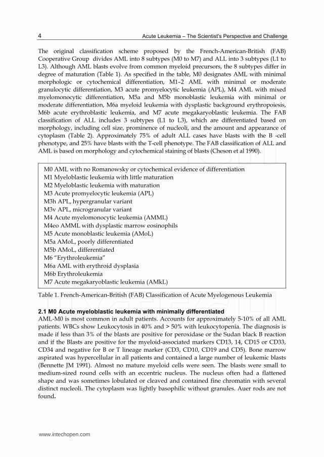
The original classification scheme proposed by the French-American-British (FAB)
Cooperative Group divides AML into 8 subtypes (M0 to M7) and ALL into 3 subtypes (L1 to
L3). Although AML blasts evolve from common myeloid precursors, the 8 subtypes differ in
degree of maturation (Table 1). As specified in the table, M0 designates AML with minimal
morphologic or cytochemical differentiation, M1–2 AML with minimal or moderate
granulocytic differentiation, M3 acute promyelocytic leukemia (APL), M4 AML with mixed
myelomonocytic differentiation, M5a and M5b monoblastic leukemia with minimal or
moderate differentiation, M6a myeloid leukemia with dysplastic background erythropoiesis,
M6b acute erythroblastic leukemia, and M7 acute megakaryoblastic leukemia. The FAB
classification of ALL includes 3 subtypes (L1 to L3), which are differentiated based on
morphology, including cell size, prominence of nucleoli, and the amount and appearance of
cytoplasm (Table 2). Approximately 75% of adult ALL cases have blasts with the B -cell
phenotype, and 25% have blasts with the T-cell phenotype. The FAB classification of ALL and
AML is based on morphology and cytochemical staining of blasts (Cheson et al 1990).
M0 AML with no Romanowsky or cytochemical evidence of differentiation
M1 Myeloblastic leukemia with little maturation
M2 Myeloblastic leukemia with maturation
M3 Acute promyelocytic leukemia (APL)
M3h APL, hypergranular variant
M3v APL, microgranular variant
M4 Acute myelomonocytic leukemia (AMML)
M4eo AMML with dysplastic marrow eosinophils
M5 Acute monoblastic leukemia (AMoL)
M5a AMoL, poorly differentiated
M5b AMoL, differentiated
M6 “Erythroleukemia”
M6a AML with erythroid dysplasia
M6b Erythroleukemia
M7 Acute megakaryoblastic leukemia (AMkL)
Table 1. French-American-British (FAB) Classification of Acute Myelogenous Leukemia
2.1 M0 Acute myeloblastic leukemia with minimally differentiated AML-M0 is most common in adult patients. Accounts for approximately 5-10% of all AML
patients. WBCs show Leukocytosis in 40% and > 50% with leukocytopenia. The diagnosis is
made if less than 3% of the blasts are positive for peroxidase or the Sudan black B reaction
and if the Blasts are positive for the myeloid-associated markers CD13, 14, CD15 or CD33,
CD34 and negative for B or T lineage marker (CD3, CD10, CD19 and CD5). Bone marrow
aspirated was hypercellular in all patients and contained a large number of leukemic blasts
(Bennette JM 1991). Almost no mature myeloid cells were seen. The blasts were small to
medium-sized round cells with an eccentric nucleus. The nucleus often had a flattened
shape and was sometimes lobulated or cleaved and contained fine chromatin with several
distinct nucleoli. The cytoplasm was lightly basophilic without granules. Auer rods are not
found.
www.intechopen.com

Classification of Acute Leukemia
5
Fig. 1. Acute myeloblastic leukemia AML –M0
2.2 M1 Acute myeloblastic leukemia without maturation
It is found in all aged groups with highest incidence seen in adult and in infants less than a year old. Leukocytes in about 50% of patients at the time of diagnosis was increased. The predominant cell in the peripheral blood is usually a poorly differentiated myeloblast with finely reticulated chromatin and prominent nucleoli. Auer rods are found in the blast of 50% of the M1. If no evidence of granules or Auer rods is present, the blasts may resemble L2 lymphoblast. The myeloperoxidase or Sudan black B stains are positive in more than 3% of the blasts indicating granulocytes differentiation, the diagnosis is more likely AML-M1 than ALL (Bennett et al, 1976). PAS and alpha-naphthyl acetate esterase and naphthol AS-D-esterase are negative. About 50% of the patients will have acquired clonal chromosome aberrations in the leukemic cells. CD13, 14, 15, 33 and CD34 myeloid antigens are frequently positive in M1 leukemia. The most common cytogenetic abnormalities are: t (9; 22) (q34; q11)
Fig. 2. Acute myeloblastic leukemia AML-M1
2.3 M2 Acute myeloblastic leukemia with maturation The presenting symptoms for M2 AML are similar to those of the M1 type. Leukocytes increased in 50% of patients. Myeloblast can usually be found in the blood smears and may be the predominant cell type. Pseudopelger–Huet and hypogranular neutrophils being most common cells are seen in M2.
www.intechopen.com

Acute Leukemia – The Scientist's Perspective and Challenge
6
The bone marrow is hypercellular and types I and II myeloblasts make up from 30-83% of the promyelocytes to mature segmented cells. The monocytic component is less than 20%, differentiating M2 from M4. Basophils in some patient (M2 baso) was increased. Eosinophils and their precursors may be abundant, and in some cases accounts for up to 15% of myelogram (Berger and Flandrin, 1984). The characteristic that distinguishes AML-M2 from AML-M1 is the presence of maturation at or beyond the promyelocyte stage. Abnormal neutrophil maturation appears to be an integral part of AML-M2 with t(8;21) translocation. The neutrophils may show many abnormal nuclear segmentations and Auer rods. Cytochemistry; Myeloperoxidase (MPO) reaction in blast cells gives the same result as in AML-M1, but the reaction is often of little practical value because the granulocytic nature of AML-M2 is usually demonstrated clearly by the presence of maturing cells in the granulocytic series. Sodium fluoride does not inhibit esterase. PAS and nonspecific esterase are negative. Positive reaction with CD13 and CD15 antigens are frequently seen in cases of M2.
Fig. 3. Acute myeloblastic leukemia AML-M2
2.4 M3 Acute promyelocytic leukemia (APL) The median age and survival average of APL is about 18 months and occurred in younger adult. M3 is of particular interest because it results in the fusion of a truncated retinoic acid receptor alpha (RAR-alpha) gene on chromosome 17 to a transcription unit called PML (for promyelocytic leukemia) on chromosome 15. It is interesting to note that high doses of the vitamin A derivative all-trans-retinoic acid are able to overcome thus block in differentiation both in vitro and in vivo and this agent has been successfully used to induce remission in patients. A "variant" form of M3 (Bennett et al, 1980) is characterized by paucity of granules within the promyelocytic blasts and should not be confused with monocytic leukemia. The blasts are large with abundant cytoplasm, and the nucleus is usually irregular. The nucleus is often bilobed or markedly indented and a nucleolus can be seen in each lobe. The cytoplasm is completely occupied by closely packed large granules, staining bright pink, red or purple. Cells containing bundles of Auer rods "faggots" randomly distributed in the cytoplasm are characteristic, but are not present in all cases. It is believed that the release of large numbers of promyelocytic granules containing a procoagulant initiate disseminated intravascular clotting (DIC). This is the most serious complication of M3 AML occurs frequently in both AML-M3 as well as AML-M3 variant (McKenna et al, 1982) . Initial therapy with the differentiating agent all-trans-retinoic acid
www.intechopen.com

Classification of Acute Leukemia
7
(ATRA) has improved significantly the treatment of AML-M3 in this regard; early mortality as a result of DIC is substantially reduced. Cytochemistry: Peroxidase (MPO) and Sudan black B are strong positive. The periodic acid Schiff (PAS) is negative and Nonspecific esterase is also weak positive . The MPO reaction is also strong positive in the AML-M3 variant. Immunological studies demonstrate positivity with CD13, CD15, CD1 and CD33 myeloid antigens. Cytogenetic studies have revealed a high prevalence (almost 50%) of the chromosomal translocation t(15; 17) associated with both AML M3 and M3 variant . M3 AML with t(15;17) is usually characterized by the association of the lymphoid marker, CD2 and CD19, with myeloid markers and the negativity of HLA-DR and CD34.
Fig. 4. Promyelocytic leukemia AML-M3
Fig. 5. Acute myelomonocytic leukemia M4
2.5 M4 Acute myelomonocytic leukemia (AMML)
It is distinguished from M1, M2, and M3 by an increased proportion of leukemia monocytic
cells in the bone marrow or blood or both. Gingival hyperplasia with gingival bleeding is
present. Serum and urine levels of muramidase (lysozyme) are usually elevated because of
the monocytic proliferation. The leukocyte count is usually increased monocytic cells
www.intechopen.com

Acute Leukemia – The Scientist's Perspective and Challenge
8
(monoblast, promoncytes, monocytes), are increased to 5000/L or more. Anemia and
thrombocytopenia are present in almost all cases. The marrow differs from M1, M2 and M3
in those monocytic cells exceed 20% of the nonerythroid nucleated cells. The sum of the
myelocytic cells including myeloblasts, promyelocytes and later granulocytes is >20% and
<80% of nonerythroid cells. This bone marrow picture together with a peripheral blood
monocyte count of 5000/L or more is compatible with a diagnosis of M4.
Confirmation of the monocytic component of this subgroup requires cytochemistry. The
profile includes positive reactions for sudan black B or peroxidase and both specific and
non-specific esterase. A few cases of M4 AML are characterized by increased marrow
eosinophils and classified as M4e (Berger et al 1985) . Immunological studies demonstrate
positivity with CD13, CD33, CD11b and CD14. Cytogenetic: inv(16) (p13; q22) and del
(16)(q22) .
2.6 M5 Acute monoblastic leukemia (AMoL)
Common findings are weakness, bleeding and a diffuse erythematous skin rash. There is a
high frequency of extramedulary infiltration of the lungs, colon, meninges, lymphnodes,
bladder and larynx and gingival hyperplasia. Serum and urinary muramidase levels are
often extremely high. The one criterion for a diagnosis of M5 is that 80% or more of all
nonerythroid cells in the bone marrow are monocytic cells. There are two distinct forms 5a
(maturation index <4%) and 5b (maturation index > 4%).M5a: Granulocyte <20% and
Monocyte >80% >80% monoblast. M5b: Granulocyte <20% and Monocyte >80% <80%
monoblast (Characterized by the presence of all developmental stages of monocytes;
monoblast, promonocyte, monocyte)
Cytochemistry: Non-specific esterase stains and alpha-naphthyl esterase are positive and
PAS is negative. Myeloperoxidase and Sudan black are weak diffuse activity in the
monoblast. The use of alph-naphthyl butyrate esterase (ANBE) is advantageous because of
its greater degree of specificity and stronger reaction, and also because sodium fluoride
inhibition is not required (Shibata et al, 1985). Immunological studies demonstrate positivity
with CD11b and CD14. There is a strong association between AML M5/M4 and deletion
and translocations involving band 11q23.
Fig. 6. M5 Acute monoblastic leukemia (AMoL)
www.intechopen.com

Classification of Acute Leukemia
9
2.7 M6 “Erythroleukemia"
M6 is a rare form of leukemia that primarily affects the peripheral cells. It is nonsexist in children. The clinical manifestations are similar to other types of AML. The most frequent presentation is bleeding. The most dominant changes in the peripheral blood are anemia with sticking poikilocytosis and anisocytosis. Nucleated red cells demonstrate abnormal nuclear configuration. The leukocytes and platelets are usually decreased. The diagnosis of erythroleukemia can be made when more than 50% of all nucleated bone marrow cells are erythroid and 30% or more of all remaining nonerythroid cells are type I or type II blast cells (Bennett et al, 1985). The erythroblast is abnormal with bizarre morphologic features. Giant multilobular or multinucleated forms are common. Other features are; fragmentation, Howell-Jolly bodies, ring sideroblast, megaloblastic and dyserythropoiesis changes are common. The cytochemistry of erythroblasts are normally PAS negative but in AML-M6, erythroblasts especially pronormoblast demonstrates coarse positivity of PAS. Blast cells express a variety of myeloid associated antigens such as CD13, CD33, anti-MPO with or without expression of precursor-cell markers as CD34, HLA-Dr determinants as for blast cells from other AML subtypes. In M6-variant forms, the more differentiated cells can be detected by the expression of glycophorin A and the absence of myeloid markers.
Fig. 7. M6 “Erythroleukemia”
2.8 M7 Acute megakaryoblastic leukemia (AMkL)
M7 is rare. It occurs as a leukemia transformation of chronic granulocytic leukemia (CGL) and myelodysplastic syndrome (MDS). Pancytopenia is characteristic at initial diagnosis. Peripheral blood shows micromegakaryocytes and undifferentiated blasts. Bone marrow dry tap is common. Bone marrow biopsy show increased fibroblasts and/or increased reticulin and presence of greater than 30% blast cells. The diagnosis of M7 should be suspected when the blast cells show cytoplasmic protrusion or budding. As bone marrow smears obtained by aspiration may not be adequate to make a diagnosis, the peripheral blood films must be examined carefully for the presence of micromegakaryoblasts. Bone marrow biopsy sections are usually necessary and show a prominent reticulin fibrosis and excessive numbers of small blasts. Cytochemistry: Peroxidase is negative, PAS +/-, Esterase +/- and positive acid phospatase.
Cytochemical positivity for -naphthyl acetate esterase reaction and negative reaction with -naphthyl butyrate esterase is unique to megakaryoblast. (Monocytes react positively with both esterase substrates).
www.intechopen.com

Acute Leukemia – The Scientist's Perspective and Challenge
10
The monoclonal antibodies that reacts with platelet glycoprotein Ib, IIb/IIIa and IIIb, using
immunologic technique as well as CD41, CD42 and CD61 positivity. There is no unique chromosomal abnormality associated with acute megakaryoblastic leukemia, with the exception of t(1;22)(p13;q13), which has been found almost exclusively in young children, less than 18 months old who do not have Downís syndrome.
Fig. 8. M7 Acute megakaryoblastic leukemia (AMkL)
Morphologic Classification
FAB Type Features of Blasts
L1 Small cells with scant cytoplasm; nucleoli indistinct and not visible
L2 Large, heterogeneous cells with moderately abundant cytoplasm; clefting and indentation of nucleus; large and prominent nucleoli
L3 Large cells with moderately abundant cytoplasm; regular, oval-to-round nucleus; prominent nucleoli; prominent cytoplasmic basophilia and cytoplasmic vacuoles
Table 2. Morphologic Classification of Acute Lymphocytic Leukemia
Acute lymphoblastic leukemia (ALL) is divided in FAB L1 (children), L2 (older children
and adult), and L3 (patients with leukemia secondary to Burkitt's lymphoma. These types
are defined according to two criteria (1) the occurrence of individual cytologic features and
(2) the degree of heterogeneity among the leukemic cells. These features considered are cell
size, chromatin, nuclear shape, nucleoli, and degree of basophilia in the cytoplasm and the
presence of cytoplasmic vacuolation (Bennett et al 1976).
ALL-L1: Homogenous cells (Small cell): One population of cells within the case. Small cells
predominant, nuclear shape is regular with occasional cleft. Nuclear contents are rarely
visible. Cytoplasm is moderately basophilic. L1 accounts 70% of patients. The L1 type is the
acute leukemia that is common in childhood, with 74% of these cases occurring in children
15 years of age or younger.
ALL-L2: Heterogeneous cells: Large cells with an irregular nuclear shape, cleft in the
nucleus are common. One or more large nucleoli are visible. Cytoplasm varies in colour and
nuclear membrane irregularities. L2 accounts 27% of ALL patients. The FAB-L2 blast may be
www.intechopen.com

Classification of Acute Leukemia
11
confused with the blasts of acute myeloid leukemia. Approximately 66% of these cases of
ALL in patients older than 15 years are of type 2.
Fig. 9. Acute lymphoblastic leukemia L1
Fig. 10. Acute lymphoblastic leukemia L2
ALL-L3: Burkitt's lymphoma type: Cells are large and homogenous in size, nuclear shape is round or oval. One to three prominent nucleoli and sometimes to 5 nuleoli are visible. Cytoplasm is deeply basophilic with vacuoles often prominent. Intense cytoplasmic basophilia is present in every cell, with prominent vacuolation in most. A high mitotic index is characteristic with presence of varying degrees of macrophage activity. Mature B-lymphoid markers are expressed by most cases.
Fig. 11. Acute lymphoblastic leukemia L3
www.intechopen.com

Acute Leukemia – The Scientist's Perspective and Challenge
12
Acute Myeloid Leukemia (AML) and Related Precursor Neoplasm AML with recurrent genetic abnormalities AML with t(8;21)(q22;q22); RUNX1-RUNX1T1 AML with inv(16)(p13.1q22) or t(16;16)(p13.1;q22); CBFB-MYH11 Acute promyelocytic leukemia with t(15;17)(q22;q12); PML-RARA AML with t(9 ;11)(p22;q23); MLLT3-MLL AML with t(6;9)(p23;q34); DEK-NUP214 AML with inv(3)(q21q26.2) or t(3;3)(q21;q26.2); RPN1-EVI1 AML with mutated NPM1 AML with mutated CEBPA AML with myelodysplasia-related changes Therapy-related myeloid neoplasms Myeloid sarcoma Myeloid proliferations related to Down syndrome Transient abnormal myelopoiesis Myeloid leukemia associated with Down syndrome Blastic plasmacytoid denderitic cell neoplasm
Table 3. World Health Organization Classification of Acute Myelogenous Leukemia (2008)
AML defined as ≥20% blasts in blood or bone marrow; however, clonal, recurring
cytogenetic abnormalities should be considered AML regardless of blast percentage.
Ongoing clinical trials may continue to use French-American-British (FAB) criteria of ≥30%
blasts until completion of trial. FAB classification identified as M0 through M7.
The classification schemes by the World Health Organization (WHO) require the additional
evaluation of the leukemic blasts by molecular analysis and flow cytometry (Harris NL 1997
& Brunangelo Falini 2010, Sachdeva et al 2006). The results of these 4 methods of evaluation
(i.e, morphology, staining, molecular analysis, flow cytometry) not only differentiate ALL
from AML, but also categorize the subtypes of acute leukemia. Table 3 summarizes the new
classification of AML as proposed by WHO, Knowing the subtype of a patient’s leukemia
helps in predicting the clinical behavior of the disease and the prognosis, and in making
treatment recommendations. This classification also improves the reproducibility of
diagnoses and stresses the heterogeneity of the subtypes of AML and ALL (Vardiman 2009).
Recent advances in molecular biology have shown that various subtypes of AML and ALL
behave differently and should not be treated similarly. For example, the identification of M3
AML (acute promyelocytic leukemia) is crucial because it is associated with disseminated
intravascular coagulation (DIC), and retinoic acid, in addition to chemotherapy, is the
treatment of choice.
The two most significant differences between the FAB and the WHO classifications are: (a) A lower blast threshold for the diagnosis of AML: The WHO defines AML when the
blast percentage reaches 20% in the bone marrow.
(b) Patients with recurring clonal cytogenetic abnormalities should be considered to have
AML regardless of the blast percentage (8;21)(q22;q22), t(16;16)(p13;q22), inv(16)(p13;q22),
or t(15;17)(q22;q12) (Arber DA et al 2008abc, Weinberg OK et al 2009).
The world Health Organization (WHO) classification has changed the grouping of ALL to
reflect increased understanding of the biology and molecular pathogenesis of the diseases. In
www.intechopen.com
Classification of Acute Leukemia
13
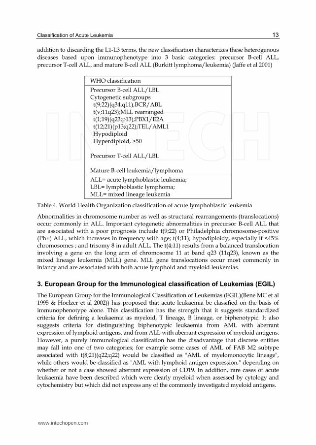
addition to discarding the L1-L3 terms, the new classification characterizes these heterogenous
diseases based upon immunophenotype into 3 basic categories: precursor B-cell ALL,
precursor T-cell ALL, and mature B-cell ALL (Burkitt lymphoma/leukemia) (Jaffe et al 2001)
WHO classification
Precursor B-cell ALL/LBL Cytogenetic subgroups t(9;22)(q34,q11),BCR/ABL t(v;11q23);MLL rearranged t(1;19)(q23;p13);PBX1/E2A t(12;21)(p13;q22);TEL/AML1 Hypodiploid Hyperdiploid, >50 Precursor T-cell ALL/LBL Mature B-cell leukemia/lymphoma
ALL= acute lymphoblastic leukemia; LBL= lymphoblastic lymphoma; MLL= mixed lineage leukemia
Table 4. World Health Organization classification of acute lymphoblastic leukemia
Abnormalities in chromosome number as well as structural rearrangements (translocations) occur commonly in ALL. Important cytogenetic abnormalities in precursor B-cell ALL that are associated with a poor prognosis include t(9;22) or Philadelphia chromosome-positive (Ph+) ALL, which increases in frequency with age; t(4;11); hypodiploidy, especially if <45% chromosomes ; and trisomy 8 in adult ALL. The t(4;11) results from a balanced translocation involving a gene on the long arm of chromosome 11 at band q23 (11q23), known as the mixed lineage leukemia (MLL) gene. MLL gene translocations occur most commonly in infancy and are associated with both acute lymphoid and myeloid leukemias.
3. European Group for the Immunological classification of Leukemias (EGIL)
The European Group for the Immunological Classification of Leukemias (EGIL)(Bene MC et al
1995 & Hoelzer et al 2002)) has proposed that acute leukaemia be classified on the basis of
immunophenotype alone. This classification has the strength that it suggests standardized
criteria for defining a leukaemia as myeloid, T lineage, B lineage, or biphenotypic. It also
suggests criteria for distinguishing biphenotypic leukaemia from AML with aberrant
expression of lymphoid antigens, and from ALL with aberrant expression of myeloid antigens.
However, a purely immunological classification has the disadvantage that discrete entities
may fall into one of two categories; for example some cases of AML of FAB M2 subtype
associated with t(8;21)(q22;q22) would be classified as "AML of myelomonocytic lineage",
while others would be classified as "AML with lymphoid antigen expression," depending on
whether or not a case showed aberrant expression of CD19. In addition, rare cases of acute
leukaemia have been described which were clearly myeloid when assessed by cytology and
cytochemistry but which did not express any of the commonly investigated myeloid antigens.
www.intechopen.com

Acute Leukemia – The Scientist's Perspective and Challenge
14
Precursor B-lymphoblastic leukemia ( HLA-DR+, TdT+, CD19+, and/or CD79a+, and/or CD22+, and/or CD34+). This type of ALL accounts for around 75% of adult cases and is subdivided into the following groups:
a. Pro B-ALL expresses HLA-DR, TdT, and CD19. CD10-, cytoplasmic immunoglobulin negative; represents approximately 10% of adult ALL.
b. Common ALL is characterized by the presence of CD10, cytoplasmic immunoglobulin negative; comprises greater than 50% of adult cases of ALL.
c. Pre B-ALL is characterised by the expression of cytoplasmic immunoglobulin and CD10; this subtype of ALL is identified in nearly 10% of adult cases.
d. Mature B-ALL is found in approximately 4% of adult ALL patients. The blast cells express surface antigens of mature B cells, including surface membrane immunoglobulin (SmIg+). They are typically TdT and CD34 negative and have L3 morphology. This category overlaps with Burkitt lymphoma, which is included under the mature B-cell neoplasms.
Precursor T-lymphoblastic leukemia
Cells are TdT+ in addition to cytoplasmic CD3+ and CD34+. This type of ALL accounts for around 25% of adult cases and is subdivided into:
a. Pro T-ALL CD2-, CD7+, CD4-, CD8- seen in around 7% of adult ALL. b. Pre T-ALL CD2+, CD7+, CD4-, CD8-. c. Cortical T-ALL or Thymic ALL (Thy ALL) is CD1a+ and accounts for 17% of
adult ALL CD7+, CD2+, CD5+, CD4+, CD8+ d. Mature T-ALL are surface CD3+, CD2+, CD7+, CD4 or 8, and
TdT/CD34/CD1a- and make up approximately 1% of adult ALL.
Table 5. European Group for the Immunological Characterization of Leukemias (EGIL) classification of acute lymphocytic leukemia (Hoelzer 2002)
The consensus considers a 20% minimum threshold to define a positive reaction of blast
cells to a given monoclonal antibody. Roughly 75% of cases of adult ALL are of B-cell
lineage. B-lineage markers are CD19, CD20, CD22, CD24, and CD79a (Huh 2000).
The earliest B-lineage markers are CD19, CD22 (membrane and cytoplasm) and CD79a
(Campana 1988). A positive reaction for any two of these three markers, without further
differentiation markers, identifies pro-B ALL. The presence of CD10 antigen (CALLA)
defines the "common" ALL subgroup. Cases with additional identification of cytoplasmatic
IgM constitute the pre-B group, whereas the presence of surface immunglobulin light chains
defines mature B-ALL. T-cell ALL constitutes approximately 25% of all adult cases of ALL. T-cell markers are CD1a, CD2, CD3 (membrane and cytoplasm), CD4, CD5, CD7 and CD8. CD2, CD5 and CD7 antigens are the most immature T-cell markers, but none of them is absolutely lineage-specific, so that the unequivocal diagnosis of T-ALL rests on the demonstration of surface/cytoplasmic CD3. ALL of B or T lineage can additionally express myeloid antigens or stem-cell antigen CD34. The latter has little diagnostic relevance but can be prognostically important (De Waele 2001) The scoring system recently proposed by the EGIL group addressed the characterization of the acute leukemia as B or T lineage ALL, or AML by including the most specific markers for the
www.intechopen.com

Classification of Acute Leukemia
15
lymphoid and myeloid lineages among those of earlier stages of cell differentiation, plus some non-specific but stem-cell markers. The system introduced a modified terminology specific to each 'maturation' step within the B- or T-cell lineage (EGIL 1995) and was confirmed as adequate for both diagnosis and subclassification of ALL (Thalhammer-Scherrer 2002).
4. European Group for the Immunological characterization of Leukemias (EGIL) classification for biphenotypic acute leukemia
Biphenotypic acute leukemia (BAL), or acute leukemia with a single population of blasts coexpressing markers of two different lineages, is a rare clinical entity. The scoring systems proposed by Catovsky et al. and by the EGIL (Bene 1995) allowed for a better definition of biphenotypic acute leukemia (BAL), clearly distinguishing them from classical AL expressing aberrantly one or two markers of another lineage. However, increasing evidence suggests that this system has limitations, as acknowledged by the 2008 World Health Organization (WHO) Classification of Tumors of Hematopoietic and Lymphoid Tissues. Although substantially improved in relation to the EGIL, the new WHO Classification is still not optimal for guiding the clinical management of patients with BAL. Typical examples of such aberrations are the expression of CD15 on B-ALL(Maynadie et al 1997), or of CD2 on acute promyelocytic –AML( Albano 2006). In 1998, the EGIL further refined this scoring system by attributing one point for the expression of CD117, after showing the strong relationship of this marker with engagement in the myeloid lineage (Bene MC et al 1998) . To identify BAL, it is therefore necessary to consider aberrant co-expression of markers usually associated to different lineages, with a score higher than 2 in more than one lineage (Zhao XF et al 2009)
Myeloid lineage T-lineage B-lineage
MPO CD3 CD79 2 point
Lisozyme Anti TCR IgM
CD22
CD13 CD2 CD19 1 point
CD33 CD5 CD10
CD65 CD8 CD20
CD10
CD14 TdT TdT 0.5 point
CD15 CD17 CD24
CD64, CD117 CD10
Table 6. EGIL Scoring system for biphenotypic acute leukemia
Biphenotypic acute leukemia is defined when scores are >2 for the myeloid lineage and >1 for the lymphoid lineage. In some T-ALL cases, clonality of TCR alphabeta rearrangements can now be assessed cytoflourmetrically (Langerak 2001, Xu XQ et al 2009). The prognosis of biphenotypic acute leukemia patients is poor when compared with de
novo acute myeloid leukemia or acute lymphoblastic leukemia. Biphenotypic acute
leukemia patients showed a much higher incidence of CD34 antigen expression, complex
abnormal karyotype, extramedullary infiltration, relapse, and resistance to therapy after
relapse (Xu XQ et al 2009).
www.intechopen.com

Acute Leukemia – The Scientist's Perspective and Challenge
16
5. Conclusion
Acute leukaemia can be classified in many ways. An ideal classification is one which recognizes real entities with fundamental biological differences. The FAB classification of ALL and AML is based on morphology and cytochemical staining of blasts. However, the recent classification schemes proposed by the World Health Organization (WHO) require the additional evaluation of the leukemic blasts by molecular analysis and flow cytometry. The results of these 4 methods of evaluation (ie, morphology, staining, molecular analysis, flow cytometry) not only differentiate ALL from AML, but also categorize the subtypes of acute leukemia . Recent advances in molecular biology have shown that various subtypes of AML and ALL behave differently and should not be treated similarly. For example, the identification of M3 AML (acute promyelocytic leukemia) is crucial because it is associated with disseminated intravascular coagulation (DIC), and retinoic acid, in addition to chemotherapy, is the treatment of choice. The European Group for the Immunological Classification of Leukemias (EGIL) has proposed that acute leukaemia be classified on the basis of immunophenotype alone. This classification has the strength that it suggests standardized criteria for defining a leukaemia as myeloid, T lineage, B lineage, or biphenotypic. It also suggests criteria for distinguishing biphenotypic leukaemia from AML with aberrant expression of lymphoid antigens, and from ALL with aberrant expression of myeloid antigens.
6. References
Albano F, Mestice A, Pannunzio A, Lanza F, Martino B, Pastore D, et al. The biological characteristics of CD34+ CD2+ adult acute promyelocytic leukemia and the CD34 CD2 hypergranular (M3) and microgranular (M3v) phenotypes. Haematologica. 2006;91:311–6.
Arber DA, Brunning RD, Le Beau MM, Falini B, Vardiman JW, Porwit A, Thiele J, Bloomfield CD. Acute myeloid leukaemia with recurrent genetic abnormalities. In: WHO Classification of Tumours of Haematopoietic and Lymphoid Tissues. 4th edition. pp. 110-123. Swerdlow SH, Campo E, Harris NL et al. (eds.). International Agency for Research on Cancer (IARC), Lyon, France, 2008a.
Arber DA, Brunning RD, Orazi A, Bain BJ, Porwit A, Vardiman JW, Le Beau MM, Greenberg PL. Acute myeloid leukaemia with myelodysplasia-related changes. In: WHO Classification of Tumours of Haematopoietic and Lymphoid Tissues. 4th edition. pp. 124-126. Swerdlow SH, Campo E, Harris NL et al. (eds.). International Agency for Research on Cancer (IARC), Lyon, France, 2008b.
Arber DA, Brunning RD, Orazi A, Porwit A, Peterson L, Thiele J, Le Beau MM. Acute myeloid leukemia, not otherwise specified. In: WHO Classification of Tumours of Haematopoietic and Lymphoid Tissues. 4th edition. pp. 130-139. Swerdlow SH, Campo E, Harris NL et al. (eds.). International Agency for Research on Cancer (IARC), Lyon, France, 2008c.
Bene MC, Castoldi G, Knapp W, et al. Proposals for the immunological classification of acute leukemias. Leukemia 1995;9:1783-6
Bennett JM, Catovsky D, Daniel MT, et al. Proposals for the classification of the acute leukaemias (FAB cooperative group). Brj Haematol 1976;33:451-8.
www.intechopen.com

Classification of Acute Leukemia
17
Benett JM, Catovsky D, Daniel MT et al(1980) The French-American-British (FAB) Cooperative Group. Corresponence; a variant form of hypergranular promyelocytic leukemia (M3). Br J Haematol 44: 169-70 Benette JM, CatoSky D, Daniel MT et al(1982) Proposals for the classification of
myelodsplastic syndromes. Br J Haematol 51; 189-99 Bennett JM, Catovsky D, Daniel M-T, et al. Criteria for the diagnosis of acute leukemia of
megakaryocytic lineage (M7): a report of the French-American-British cooperative group. Ann Intern Med 1985;103:460-2.
Bennett JM, Catovsky D, Daniel MT, et al. Proposal for the recognition of minimally differentiated acute myeloid leukaemia (AML MO). Brj Haematol 1991;78:325-9.
Berger R, Flandrin G (1984) Determining the nature of cells studied cytogenetically. Cancer Surveys 3:423-38
Berger R, Bernheim A, Daniel MT, et al(1985) Cytogenetic studies on acute myelomonocytic leukemia (M4) with eosinophilia. Leuk Res 9: 279-88
Campana D, Janossy G. Proliferation of normal and malignant human immature lymphoid cells. Blood 1988; 71: 1201-1210
Catovsky D, Matutes E, Buccheri V, Shetty V, Hanslip J, Yoshida N, Morilla R. A classification of acute leukaemia for the 1990s. Ann Hematol. 1991;62:16–21.
Cheson BD, Cassileth PA, Head DR, et al. Report of the National Cancer Institute-sponsored workshop on definitions of diagnosis and response in acute myeloid leukemia. J Clin Oncol 1990;8:813–9.
De Waele M, Renmans W, Vander GK, Jochmans K, Schots R, Otten J, et al. Growth factor receptor profile of CD34+ cells in AML and B-lineage ALL and in their normal bone marrow counterparts. Eur J Haematol 2001; 66: 178-187
European Group for the Immunological Characterization of leukemia (EGIL), Bene MC, Castoldi G, Knapp W, et al. Proposals for the immunological classification of acute leukemias. Leukemia 1995;9:1783-6.
Falini B, Tiacci E, Martelli MP, Ascni S, Pileri SA. New classification of acute myeloid leukemia and precursor-related neoplasms: changes and unsolved issues. Discov Med. 2010 Oct;10(53):281-92.
First MIC Cooperative Study Group. Morphologic, immunologic, and cytogenetic (MIC) working classification of acute lymphoblastic leukaemias. Cancer Genet Cytogenetic 1986;23:189-97.
Harris NL, Jaffe ES, Diebold J, et al. World Health Organization classification of neoplastic diseases of the hematopoietic and lymphoid tissues: report of the ClinicalAdvisory Committee meeting—Airlie House, Virginia, November 1997. J Clin Oncol 1999;17:3835–49.
Hayhoe FG. The classification of acute leukaemia. Blood Rev 1988;2:186-93. Hoelzer D., GokbugerN: Diagnostik und Therapie der akuten lymphatischen Leukaemia des
Erwachsenen. Onkologie 8 (7) (2002) 672-85 Huh YO, Ibrahim S. Immunophenotypes in adult acute lymphocytic leukemia. Role of flow
cytometry in diagnosis and monitoring of disease. Hematol Oncol Clin North Am 2000; 14: 1251-1265
Jaffe ES, Harris NL, Stein H et al: World Health Organization Classification of Tumours of haematopoietic and lymphoid tissue f. Lyon, IARC Press, 2001.
www.intechopen.com

Acute Leukemia – The Scientist's Perspective and Challenge
18
Langerak AW, van Den BR, I.L., Boor PP, van Lochem EG, Hooijkaas H, et al. Molecular and flow cytometric analysis of the Vbeta repertoire for clonality assessment in mature TCRalphabeta T-cell proliferations. Blood 2001; 98: 165-173
Maynadié M, Campos L, Moskovtchenko P, Sabido O, Aho S, Lenormand B, et al. Heterogenous expression of CD15 in acute lymphoblastic leukemia: a study of ten anti-CD15 monoclonal antibodies in 158 patients. Leuk Lymphoma. 1997;25:135–43.
McKenna RW, Parkin J, Bllomfield CD et al (1982)Acute promyelocytic leukemia: a study of 39 caes with identification of a hyperbasophilic microgranular variant. Br J Haematol 50:201-14
Sachdeva MU, Ahluwalia J, Das R, et al (2006) Role of FAB classification of acute leukemias in era of immunophenotyping.Indian J Pathol Microbiol: 2006 Oct;49(4):524-7.
Shibata A, Bennett JM, Castoldi GL et al (1985) Recommended methods for cytological procedures in haematology. Clin Lab Haematol 7:55-74
Thalhammer-Scherrer R, Mitterbauer G, Simonitsch I, Jaeger U, Lechner K, Schneider B, et al. The immunophenotype of 325 adult acute leukemias: relationship to morphologic and molecular classification and proposal for a minimal screening program highly predictive for lineage discrimination. Am J Clin Pathol 2002; 117: 380-389
Vardiman JW. The World Health Organization (WHO) classification of tumors of the hematopoietic and lymphoid tissues: an overview with emphasis on the myeloid neoplasms. Chem Biol Interact 2010 Mar 19;184(1-2):16-20.
Weinberg OK, Seetharam M, Ren K, Seo K, Merker JD, Gotlib J, Zehnder JL, Arber DA. Clinical characterization of acute myeloid leukemia with myelodysplasia-related changes as defined by the 2008 WHO classification system. Blood. 2009 Feb 26;113(9):1906-8.
Xu XQ, Wang JM, Lu SQ, Chen L, Yang JM, Zhang WP, Song XM, Hou J, Ni X, Qiu HY. Clinical and biological characteristics of adult biphenotypic acute leukemia in comparison with that of acute myeloid leukemia and acute lymphoblastic leukemia: a case series of a Chinese population. Haematologica. 2009 Jul;94(7):919-27.
www.intechopen.com

Acute Leukemia - The Scientist's Perspective and ChallengeEdited by Prof. Mariastefania Antica
ISBN 978-953-307-553-2Hard cover, 428 pagesPublisher InTechPublished online 22, December, 2011Published in print edition December, 2011
InTech EuropeUniversity Campus STeP Ri Slavka Krautzeka 83/A 51000 Rijeka, Croatia Phone: +385 (51) 770 447 Fax: +385 (51) 686 166www.intechopen.com
InTech ChinaUnit 405, Office Block, Hotel Equatorial Shanghai No.65, Yan An Road (West), Shanghai, 200040, China
Phone: +86-21-62489820 Fax: +86-21-62489821
This book provides a comprehensive overview of he basic mechanisms underlying areas of acute leukemia,current advances, and future directions in management of this disease. The first section discusses theclassification of acute leukemia, taking into account diagnoses dependent on techniques that are essential,and thankfully readily available, in the laboratory. The second section concerns recent advances in molecularbiology, markers, receptors, and signaling molecules responsible for disease progression, diagnostics basedon biochips and other molecular genetic analysis. These advances provide clinicians with importantunderstanding and improved decision making towards the most suitable therapy for acute leukemia.Biochemical, structural, and genetic studies may bring a new era of epigenetic based drugs along withadditional molecular targets that will form the basis for novel treatment strategies. Later in the book, pediatricacute leukemia is covered, emphasizing that children are not small adults when it comes to drug development.The last section is a collection of chapters about treatment, as chemotherapy-induced toxicity is still asignificant clinical concern. The present challenge lies in reducing the frequency and seriousness of adverseeffects while maintaining efficacy and avoiding over-treatment of patients.
How to referenceIn order to correctly reference this scholarly work, feel free to copy and paste the following:
Gamal Abdul-Hamid (2011). Classification of Acute Leukemia, Acute Leukemia - The Scientist's Perspectiveand Challenge, Prof. Mariastefania Antica (Ed.), ISBN: 978-953-307-553-2, InTech, Available from:http://www.intechopen.com/books/acute-leukemia-the-scientist-s-perspective-and-challenge/classification-of-acute-leukemia