3 ftfbsdi sujdmf $pousjcvujpopg-bshf ...downloads.hindawi.com/journals/bmri/2013/219897.pdf ·...
TRANSCRIPT

Hindawi Publishing CorporationBioMed Research InternationalVolume 2013, Article ID 219897, 7 pageshttp://dx.doi.org/10.1155/2013/219897
Research ArticleContribution of Large Genomic Rearrangements inItalian Lynch Syndrome Patients: Characterization of a NovelAlu-Mediated Deletion
Francesca Duraturo,1 Angela Cavallo,1 Raffaella Liccardo,1 Bianca Cudia,2
Marina De Rosa,1 Giuseppe Diana,2 and Paola Izzo1, 3
1 Dipartmento di Biochimica e Biotecnologie Mediche, Università degli Studi di Napoli Federico II, Via Pansini, 5 80131 Napoli, Italy2Dipartimento di Discipline Chirurgiche ed Oncologiche, Università degli Studi di Palermo, Via Liborio Giuffrè, 90127 Palermo, Italy3Department of Biochemistry and Medical Biotechnology and CEINGE-Biotecnologie Avanzate,Università degli Studi di Napoli Federico II, Via Pansini, 5 80131 Napoli, Italy
Correspondence should be addressed to Paola Izzo; [email protected]
Received 10 October 2012; Accepted 20 November 2012
Academic Editor: Francesco Baudi
Copyright © 2013 Francesca Duraturo et al. is is an open access article distributed under the Creative Commons AttributionLicense, which permits unrestricted use, distribution, and reproduction in any medium, provided the original work is properlycited.
Lynch syndrome is associatedwith germ-linemutations in theDNAmismatch repair (MMR) genes,mainlyMLH1 andMSH2.Mostof themutations reported in these genes to date are pointmutations, small deletions, and insertions. Large genomic rearrangementsin the MMR genes predisposing to Lynch syndrome also occur, but the frequency varies depending on the population studied onaverage from 5 to 20%. e aim of this study was to examine the contribution of large rearrangements in the MLH1 and MSH2genes in a well-characterised series of 63 unrelated Southern Italian Lynch syndrome patients who were negative for pathogenicpoint mutations in the MLH1, MSH2, and MSH6 genes. �e identi�ed a large novel deletion in the MSH2 gene, including exon 6in one of the patients analysed (1.6% frequency). is deletion was con�rmed and localised by long-range PCR. e breakpointsof this rearrangement were characterised by sequencing. Further analysis of the breakpoints revealed that this rearrangement wasa product of Alu-mediated recombination. Our �ndings identi�ed a novel Alu-mediated rearrangement within MSH2 gene andshowed that large deletions or duplications in MLH1 and MSH2 genes are low-frequency mutational events in Southern Italianpatients with an inherited predisposition to colon cancer.
1. Introduction
Hereditary nonpolyposis colorectal cancer (HNPCC; alsoknown as Lynch syndrome) is an autosomal dominantdisorder characterised by colorectal cancer [1] that accountsfor 3–5% of all colorectal cancers. Affected individuals haveapproximately 60–80% lifetime risk of developing colorectalcancer and women with Lynch syndrome have 54% risk ofdeveloping endometrial cancer [2]. It is associatedwith germ-line mutations in the DNA mismatch repair (MMR) genes,mainlyMLH1 andMSH2 [3]. Mutations inMSH6 [4], PMS2[5], and MLH3 [6] are less common. Recently, a germ-linepoint mutation in MSH3 was found to be associated withthe Lynch syndrome phenotype [7]. Inactivation of theMMR
complex manifests microsatellite instability (MSI), which isdetected in tumour tissue [8].
e majority of mutations in the MMR genes sofar identi�ed are missense, nonsense, or small inser-tions/deletions [http://www.insight-group.org/mutationsmutations]. Depending on the population studied, largegenomic rearrangements of the MMR genes constitutevarious proportions of the germ-line mutations thatpredispose to HNPCC [9–11]. Moreover, it seems that largegenomic rearrangements occur more frequently in somepopulations than in others [11, 12]. e relative incidence ofgenomic rearrangements among Lynch Syndrome familiesappears to vary from 5–20% [13]. A systematic studyon genomic rearrangement in Lynch Syndrome showed

2 BioMed Research International
that MLH1 and MSH2 are the most frequently targetedMMR genes for this type of mutation [14]. Furthermore,molecular characterisation of the breakpoints involved inlarge rearrangements withinMLH1 andMSH2 genes showedthat the majority are caused by homologous recombinationbetween Alu repeats [15–17]. ese mutations are notusually detected by conventional methods of mutationanalysis, such as denaturing high-performance liquidchromatography (DHPLC) and direct DNA sequencing, butthey are detectable by a simple and robust technique suchas the Multiplex Ligation-Probe Dependent Ampli�cation(MLPA) [18, 19] assay.
As little is known about the frequency of large rearrange-ments in the MLH1 and MSH2 genes to Lynch syndromein Italian population, the aim of our study was to assess thecontribution of large genomic rearrangements in these twogenes in a well-characterised series of 63 Southern Italianpatients affected by Lynch Syndrome.
2. Materials andMethods
2.1. Patients. Sixty-three families of Italian origin, 56 familiesclassi�ed according to the Amsterdam criteria [20] and 7atypical Lynch families selected according to MSI high status(MSI-H) [20], without germ-line pathogenic point mutationsin the MLH1, MSH2, or MSH6 genes, were recruited fromseveral health centres in Campania (Southern Italy).
All patients received genetic counselling and gave theirwritten informed consent to participate in this study.
2.2. Isolation of Genomic DNA. Total genomic DNA wasextracted from 4mL peripheral blood lymphocytes usinga Nucleon BACC2 Kit (Amersham Life Science) and fromtumour tissues and surgical margins by standard methods[21].
2.3. Multiple� Ligation-Dependent Probe Ampli�cation(MLPA). MLPA was performed using the SALSA MLPAP003-B1MLH1/MSH2kit (MRC-Holland,eNetherlands)according to the manufacturer’s instructions. Fragmentanalysis was conducted on an ABI Prism 3130 GeneticAnalyser using GeneMapper soware (Applied Biosystems,Foster City, CA, USA). Migration of fragments wascalculated by comparison to the GeneScan LIZ-500 sizestandard (Applied Biosystems, Foster City, CA, USA).Peak areas were then exported to a Microso spreadsheet(www.MLPA.com) and calculations were done accordingto the method described by Taylor and colleagues [22]. A30–50% decrease in the peak area(s) indicated a deletion ofthe corresponding exon(s), while a 30–50% increase in thepeak area(s) indicated a duplication of the correspondingexon(s). MLPA results were con�rmed in at least twoindependent experiments.
2.�. DNA Ampli�cation and Microsatellite Analysis. eMSIstatus was con�rmedwith a �uorescentmultiplex system [23]comprising six mononucleotide repeats (BAT-25, BAT-26,BAT-40, NR-21, NR-24, and TGF𝛽𝛽RII) and four dinucleotide
repeats (D2S123, D5S346, D17S250, and D18S58). 20 ngof DNA extracted by tumor tissue and peripheral bloodlymphocytes were ampli�ed in 25𝜇𝜇L reaction volume usingthe CC-MSI Kit (Ab Analitica, Padova, Italy), in according tomanufacture instructions. e PCR products were analysedby capillary electrophoresis analysis using an ABI Prism3130 Genetic Analyser (Applied Biosystems, Foster City, CA,USA).
2.5. RNA Analysis of MSH2 Gene. RNA was extracted from4mL peripheral blood lymphocytes using a Trizol reagentby standard methods (Quiagen). cDNA was synthesisedusing SuperScript II RT (Invitrogen by Life Technologies)and ampli�ed with primers that produced a 598-bp frag-ment (2cFP 5�-GGCTCTCCTCATCCAGATTG and 2cRP5�-AAGATCTGGGAATCGACGAA) spanning exons 4–7 ofthe messenger RNA. e PCR products were analysed on a2% agarose gel and visualised by ethidium bromide staining.
2.6. Long-Range Polymerase Chain Reaction and BreakpointAnalysis. 500 ng of genomic DNA was ampli�ed in a 50 𝜇𝜇L-reaction volume using 2.75mMMg2+, 500 𝜇𝜇Mof each dNTP,2U of Expand Long Template PCR System (Expand LongTemplate Buffer 2; Roche Diagnostics), and 300 nM of eachprimer. Primers were designed between exon 5 and intron7 of the MSH2 gene. is region was ampli�ed in four PCRfragments.e same forward oligonucleotide (5FP) was usedin each reaction with a different reverse oligonucleotide, eachapproximately 1000 bp apart (Table 1). Cycling conditionswere as follows: 94∘C for 2min, followed by 10 cycles con-sisting of 94∘C for 10 sec, 60∘C for 30 sec (−0.5∘C/cycle) and68∘C for 15min, followed by 25 cycles consisting of 94∘C for15 sec, 57∘C for 30 sec, and 68∘C for 15min (+20 sec/cycle),and �nishing with one cycle at 68∘C for 7min.
All oligonucleotides were designed using Primer3 So-ware (http://frodo.wi.mit.edu/primer3/) and checked usingthe Basic Local Alignment Search Tool program (BLAST,http://blast.ncbi.nlm.nih.gov/Blast.cgi).
2.7. Sequencing Analysis. e PCR products were sequencedin both the forward and reverse directions using anABIPrism3100 Genetic Analyser (Applied Biosystems, Foster City, CA,USA).
2.8. In Silico Analysis. e nucleotide sequences of thegenomic MSH2 region (NG_007110.1) were analysed withthe RepeatMasker program (http://repeatmasker.org/) usingthe default settings. Sequence comparisons in RepeatMaskerwere performed by the program cross_match [24].
3. Results
3.1. Detection of Large Genomic Rearrangements in the MSH2and MLH1 Genes by MLPA. MLPA analysis on 63 unrelatedpatients identi�ed a deletion in theMSH2 gene in one patientonly (1.6%) (Figure 1). is deletion removed exon 6, whichis located between the small intron 5 and the large intron6. e exon 6 deletion was con�rmed at the RNA level by

BioMed Research International 3
T 1: Primer sequences used for long-range PCR to characterise the breakpoints of the MSH2 exon 6 deletion.
Primer Sequence Nucleotide position (NG_007110.1) Amplicon size (bp)5FP GGATATTGCAGCAGTCAGAGCCC 11258–112807RP AGAGTGAGTCACCACCACCAACT 26890–26913 15657 bp6RPl AGCTTTTCTGGAGGCCATAGGCA 24694–24717 13459 bp6RPi AGTCTGGTCCAAGGATCACCAGCA 23620–23644 12386 bp6RPh TCGTCGGTGGAAGAGGTGGCT 22565–22586 11328 bp6RPg AGCCCATGAAGAGAGCTGACACC 21580–21603 10345 bp
RT/PCR sequencing of a fragment with a lower molecularweight.e deletion was identi�ed in a 39-year-old man witha family history of colorectal cancer, who had developed atubulovillous adenoma with small fragments of mucinousadenocarcinoma in the rectum, approximately 75 cm fromthe anus. e same deletion was also detected in his 33-year-old brother. Although the brother was asymptomatic,endoscopy revealed an adenocarcinoma located proximal tothe hepatic �exure (Figure 2).
3.2. Microsatellite Analysis. MSI analysis was performed onDNA extracted from tumour tissues (adenocarcinoma), andsurgical margins of both patients (the proband and hisbrother) carrying the MSH2 exon 6 deletion. Both patientswere found to have an MSI-H status, with instability at allmarkers analysed (data not shown).
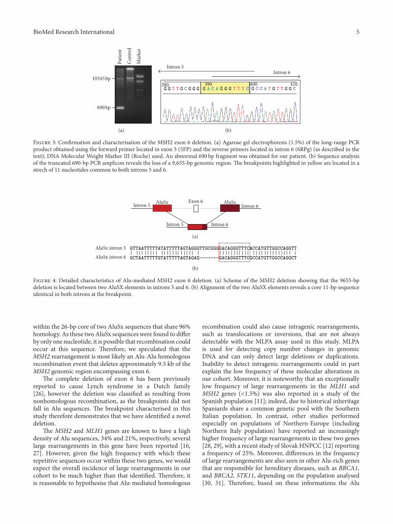
3.3. Breakpoint Characterisation of theMSH2Exon 6Deletion.e breakpoints of the exon 6 deletion within the MSH2gene were characterised by analysing the intragenic regionsbetween exon 5 and exon 7. is region was ampli�edusing region-speci�c oligonucleotides, as described in theMaterials and Methods section. One forward primer locatedin exon 5, and different reverse primers starting in exon 7were used. Abnormal fragment products of 3804, 2731, 1673and 690 bp were ampli�ed from the patient�s DNA but notfrom the DNA of the healthy control using the primer pairs5FP/6RPl, 5FP/6RPi, 5FP/6RPh, and 5FP/6RPg, respectively.No ampli�cation products were obtained using the primerpair 5FP/7 RP.
As shown in Figure 3, sequence analysis of the 690-bp ampli�cation product obtained using the primer pair5FP/6RPg revealed the loss of a 9655-bp genomic region.e 5� breakpoint is located in intron 5, in a strechof 11 nucleotides located 1,535–1,525 nt before the �rstnucleotide of exon 6. e 3� breakpoint is located inintron 6, in an identical sequence of 11 nucleotides located5,325–5,315 nt before the �rst nucleotide of exon 7. eexact breakpoints could not be ascertained because of thepresence of an identical 11-bp sequences at both ends.is deletion c.942+(346–356)_1077-(5323–5313)del, alter-natively NC_000002.11:g.47641903_47651558del, is namedin accordance with the mutation nomenclature instructionsprovided by the HGVS (http://www.hgvs.org/); it creates apremature stop codon and the formation of a truncatedprotein.
3.4. In Silico Analysis. Using the RepeatMasker program, the5� and 3� breakpoints of the 9655-bp deletion were foundto lie within the 26-bp core sequence of two Alu elements,which share 96% homology and differ by only one nucleotide.Both Alu elements belong to the AluSx subfamily and were269 bp and 310 bp, respectively. Homology analysis of theAluSx sequences included in the deletion was performedusing BLAST analysis (Figure 4).
e entireMSH2 genewas also analysed byRepeatMaskerprogram, as already described in the literature [25], to verifythe presence of repeat sequences. In this study, a total of 190repeat sequences, including 106 Alu-type SINE sequences, 19L1-type LINE sequences, 12 simple repeat sequences, and 12LTR sequences were identi�ed, and their positions on thegene de�ned. Of these, 32 Alu-type SINE sequences, oneL1-type LINE sequence, one LTR, and three simple repeatsequences were located in the genomic region between exons5 and 7.
4. Discussion
e Lynch syndrome, caused primarily by germ-line pointmutations within MMR genes, is also associated with largerearrangements that account for 5–20% of all mutations.Here, we report the results of our screening for large rear-rangements in the MLH1 and MSH2 genes in a cohort of 63Southern Italian patients who were negative for pathogenicpoint mutations in the MLH1, MSH2, and MSH6 genes. Weidenti�ed one large rearrangement in the MSH2 gene andnone in the MLH1 gene. erefore, large rearrangements inthe MLH1 and MSH2 genes occur at a low frequency in ourpatient cohort (1.6%).
e rearrangement in MSH2 identi�ed in this studycaused a large deletion that removed exon 6 and wasdetected in two patients from the same family who met theAmsterdam-1 criteria. e two affected brothers presentedcolorectal cancer with early-onset, before 40 years of age.Other family members were also affected (not tested inthis study) and presented with the same phenotype (Figure2). DNA extracted from the tumour tissues of the twopatients showed an MSI-H status, with instability at allmarkers analysed. e novel deletion is 9,655 bp long andextends from a region 346 bp downstream of exon 5 to5323 bp upstream of exon 7. e exact breakpoints couldnot be ascertained because of the presence of identical 11-bp sequences at both ends; in fact using the RepeatMaskerprogram, the breakpoints of this deletion were found to lie

4 BioMed Research International
P003-B1 MLH1/MSH2
490450410330290250210170130
4800
370
4000
3200
2400
1600
800
0
4400
3600
2800
2000
1200
400
(a)
Ctrl1
Ctrl2
MLH1-Ex1
MSH2-Ex1
MLH1-Ex2
MSH2-Ex2
MLH1-Ex3
MSH2-Ex3
MLH1-Ex4
MSH2-Ex4
MSH2-Ex1
Ctrl3
MLH1-Ex5
MSH2-Ex5
MSH2-Ex1
MLH1-Ex6
MSH2-Ex6
MLH1-Ex7
MSH2-Ex7
MLH1-Ex8
MSH2-Ex8
MLH1-Ex9
Ctrl4
MSH2-Ex9
MLH1-Ex10
MSH2-Ex10
MLH1-Ex11
MSH2-Ex11
MLH1-Ex12
MSH2-Ex12
MLH1-Ex13
MSH2-Ex13
Ctrl5
MLH1-Ex14
MSH2-Ex14
MLH1-Ex15
MSH2-Ex15
MLH1-Ex16
MSH2-Ex16
MLH1-Ex17
MLH1-Ex18
MLH1-Ex19
Ctrl6
EPCAM
EPCAM
Ctrl7
4800
4000
3200
2400
1600
800
0
4400
3600
2800
2000
1200
400
490450410330290250210170130 370
(b)
F 1: MLPA analysis reveals a candidate genomic rearrangement in the MSH2 gene. (a)e electropherogram of the DNA patient: thearrow shows half the level of ampli�cation of exon � in the carrier sub�ect. (b) e electropherogram of the DNA healthy control: the arrowshows normal level of ampli�cation of exon �.
CRC 39
CRC 40
CRC 38CRC 34
F 2: Family pedigree of the patient with the large MSH2 gene deletion. Symbols and abbreviations used are denoted as follows. Arrows:analysed members of family; black symbol: colorectal cancer; CRC, colorectal cancer. Number next to diagnosis denote age at oneset; •: notdetected.
BioMed Research International 5
10345 bp
690 bp
Pat
ien
t
Co
ntr
ol
Mar
ker
(a)
C C C C CCG G G G G G G G G G G G GGT T T T T T T TA A A380 390 400 410
Intron 5
Intron 6
(b)
F 3: Con�rmation and characterisation of the MSH2 exon 6 deletion. (a) Agarose gel electrophoresis (1.5%) of the long-range PCRproduct obtained using the forward primer located in exon 5 (5FP) and the reverse primers located in intron 6 (6RPg) (as described in thetext); DNA Molecular Weight Marker III (Roche) used. An abnormal 690 bp fragment was obtained for our patient. (b) Sequence analysisof the truncated 690-bp PCR amplicon reveals the loss of a 9,655-bp genomic region. e breakpoints highlighted in yellow are located in astrech of 11 nucleotides common to both introns 5 and 6.
Intron 5
Intron 5
Intron 6
Intron 6
Exon 6 AluSxAluSx
(a)
GTTAATTTTTATATTTTTAGTAGGGTTGCGGGGACAGGGTTTCACCATGTTGGCCAGGTT
GCTAATTTTTGTATTTTTAGTAGAG ------- GACAGGGTTTCGCCATGTTGGCCAGGCT
AluSx intron 5
AluSx intron 6
(b)
F 4: Detailed characteristics of Alu-mediated MSH2 exon 6 deletion. (a) Scheme of the MSH2 deletion showing that the 9655-bpdeletion is located between two AluSX elements in introns 5 and 6. (b) Alignment of the two AluSX elements reveals a core 11-bp sequenceidentical in both introns at the breakpoint.
within the 26-bp core of two AluSx sequences that share 96%homology. As these twoAluSx sequences were found to differby only one nucleotide, it is possible that recombination couldoccur at this sequence. erefore, we speculated that theMSH2 rearrangement is most likely an Alu-Alu homologousrecombination event that deletes approximately 9.5 kb of theMSH2 genomic region encompassing exon 6.
e complete deletion of exon 6 has been previouslyreported to cause Lynch syndrome in a Dutch family[26], however the deletion was classi�ed as resulting fromnonhomologous recombination, as the breakpoints did notfall in Alu sequences. e breakpoint characterised in thisstudy therefore demonstrates that we have identi�ed a noveldeletion.
e MSH2 and MLH1 genes are known to have a highdensity of Alu sequences, 34% and 21%, respectively, severallarge rearrangements in this gene have been reported [16,27]. However, given the high frequency with which theserepetitive sequences occur within these two genes, we wouldexpect the overall incidence of large rearrangements in ourcohort to be much higher than that identi�ed. erefore, itis reasonable to hypothesise that Alu-mediated homologous
recombination could also cause intragenic rearrangements,such as translocations or inversions, that are not alwaysdetectable with the MLPA assay used in this study. MLPAis used for detecting copy number changes in genomicDNA and can only detect large deletions or duplications.Inability to detect intragenic rearrangements could in partexplain the low frequency of these molecular alterations inour cohort. Moreover, it is noteworthy that an exceptionallylow frequency of large rearrangements in the MLH1 andMSH2 genes (<1.5%) was also reported in a study of theSpanish population [11]; indeed, due to historical inheritageSpaniards share a common genetic pool with the SouthernItalian population. In contrast, other studies performedespecially on populations of Northern-Europe (includingNorthern Italy population) have reported an increasinglyhigher frequency of large rearrangements in these two genes[28, 29], with a recent study of Slovak HNPCC [12] reportinga frequency of 25%. Moreover, differences in the frequencyof large rearrangements are also seen in other Alu-rich genesthat are responsible for hereditary diseases, such as BRCA1,and BRCA2, STK11, depending on the population analysed[30, 31]. erefore, based on these informations the Alu

6 BioMed Research International
sequences may be regarded as passive elements that serve asfavourable substrates for recombination and the molecularmechanism that promotes recombination events remains tobe clari�ed.
Beyond possible explanations about the low frequencyof large rearrangements in our population, it should behighlighted that the majority of patients with Lynch syn-drome tested in this study do not have a mutation in theMMR genes most frequently mutated. It is also important toemphasize that our families were selected on the basis of theAmsterdam clinical criteria and MSI-H, thus there is goodevidence that all affected have a strong genetic componentto early development of cancer. We therefore suggest thatsome undiscovered genetic mechanism in Lynch syndromepatients is yet to be investigated. Recently, it has been shownthat unclassi�ed genetic variants in MMR genes can behaveas low-risk alleles that contribute to the risk of colon cancerin Lynch syndrome families when interacting together orwith other low-risk alleles in other MMR genes [7, 32].Furthermore, it is also possible that the existence of otheras yet undiscovered genes may confer susceptibility to coloncancer in Lynch syndrome families. e EPCAM gene inaddition toMMR genes has already been associated HNPCCphenotype [33] as well asMYH in addition to APC gene hasbeen associated FAP phenotype [34]. Recently, associationstudies have identi�ed a number of loci that appear confermore increases in colon cancer risk [35, 36]. Further studiesare needed to better identify the underlying genetic riskfactors associated with disease in these families.
5. Conclusions
is paper is the �rst signi�cant study on contribution oflargeMLH1 andMSH2 genomic rearrangements in SouthernItalian Lynch syndrome patients, negative for point mutationin MMR genes. Our results enlarge the spectrum of largerearrangements inMSH2 genes and at the same time indicatethat these genomic rearrangements seem to be a less frequentmutational event in our population. Nonetheless, we believethat the detection of large rearrangements in the MLH1 andMSH2 genes should be included in the routine testing forLynch syndrome, especially considering the simplicity of theMLPA assay.
Acknowledgments
is work was supported by RECAM-2006-353005—Mini-stero Salute—Ricerca Oncologica; Prin 2007-prot. 2007EN8F7T-004; Regione Campania, DGRC 1901/2009.
References
[1] N. Carlomagno, L. Pelosio, A. Jamshidi et al., “e hereditarysyndrome,” in ANDREA RENDA, pp. 107–128, Springe, Milan,Italy, 2009.
[2] N. Carlomagno, F. Duraturo, G. Rizzo, C. Cremone, P. Izzo, andA. Renda, “Carcinogenesis,” in Multiple Primary Malignancies,A. Renda, Ed., pp. 51–61, Springer, Milan, Italy, 2009.
[3] P. M. Lynch, “e hMSH2 and hMLH1 genes in hereditarynonpolyposis colorectal cancer,” Surgical Oncology Clinics ofNorth America, vol. 18, no. 4, pp. 611–624, 2009.
[4] Y.M. C. Hendriks, A.Wagner, H.Morreau et al., “Cancer risk inhereditary nonpolyposis colorectal cancer due to MSH6 muta-tions: impact on counseling and surveillance,”Gastroenterology,vol. 127, no. 1, pp. 17–25, 2004.
[5] L. Senter, M. Clendenning, K. Sotamaa et al., “e clinical phe-notype of Lynch Syndrome due to germ-line PMS2 mutations,”Gastroenterology, vol. 135, no. 2, pp. 419–428, 2008.
[6] Y. Wu, M. J. W. Berends, R. H. Sijmons et al., “A role for MLH3in hereditary nonpolyposis colorectal cancer,” Nature Genetics,vol. 29, no. 2, pp. 137–138, 2001.
[7] F. Duraturo, R. Liccardo, A. Cavallo, M. D. Rosa, M. Grosso,and P. Izzo, “Association of low-risk MSH3 and MSH2 variantalleles with Lynch syndrome: probability of synergistic effects,”International Journal of Cancer, vol. 129, no. 7, pp. 1643–1650,2011.
[8] S. N. Shah, S. E. Hile, and K. A. Eckert, “Defective mismatchrepair, microsatellite mutation bias, and variability in clini-cal cancer phenotypes,” Cancer Research, vol. 70, no. 2, pp.431–435, 2010.
[9] G. Chong, J. Jarry, V. Marcus et al., “High frequency of exondeletions and putative founder effects in French CanadianLynch Syndrome Families,”HumanMutation, vol. 30, no. 8, pp.E797–E812, 2009.
[10] C. Martínez-Bouzas, E. Ojembarrena, E. Beristain, J. Errasti, N.Viguera, and M. I. Tejada Minguéz, “High proportion of largegenomic rearrangements in hMSH2 in hereditary nonpolyposiscolorectal cancer (HNPCC) families of the Basque Country,”Cancer Letters, vol. 255, no. 2, pp. 295–299, 2007.
[11] S. Castellví-Bel, A. Castells, M. Strunk et al., “Genomic rear-rangements in MSH2 and MLH1 are rare mutational eventsin Spanish patients with hereditary nonpolyposis colorectalcancer,” Cancer Letters, vol. 225, no. 1, pp. 93–98, 2005.
[12] K. Zavodna, T. Krivulcik, M. G. Bujalkova et al., “Partial lossof heterozygosity events at the mutated gene in tumors fromMLH1/MSH2 large genomic rearrangement carriers,” BMCCancer, vol. 9, article 405, 2009.
[13] F. Di Fiore, F. Charbonnier, C. Martin et al., “Screening forgenomic rearrangements of theMMRgenesmust be included inthe routine diagnosis of HNPCC,” Journal of Medical Genetics,vol. 41, no. 1, pp. 18–20, 2004.
[14] H. van der Kli, J. Wijnen, A. Wagner et al., “Molecular charac-terization of the spectrumof genomic deletions in themismatchrepair genes MSH2, MLH1, MSH6, and PMS2 responsible forhereditary nonpolyposis colorectal cancer (HNPCC),” GenesChromosomes and Cancer, vol. 44, no. 2, pp. 123–138, 2005.
[15] M. Kloor, C. Sutter, N. Wentzensen et al., “A large MHS2Alu insertion mutation causes HNPCC in a German kindred,”Human Genetics, vol. 115, no. 5, pp. 432–438, 2004.
[16] L. Li, S.McVety, R. Younan et al., “Distinct patterns of germ-linedeletions inMLH1 andMSH2: the implication of Alu repetitiveelement in the genetic etiology of Lynch syndrome (HNPCC),”Human Mutation, vol. 27, no. 4, p. 388, 2006.
[17] S. Aissi-BenMoussa, A.Moussa, T. Lovecchio et al., “Identi�ca-tion and characterization of a novel MLH1 genomic rearrange-ment as the cause of HNPCC in a Tunisian family: evidence fora homologous Alu-mediated recombination,” Familial Cancer,vol. 8, no. 2, pp. 119–126, 2009.
[18] J. P. Schouten, C. J. McElgunn, R. Waaijer, D. Zwijnenburg, F.Diepvens, and G. Pals, “Relative quanti�cation of 40 nucleic

BioMed Research International 7
acid sequences by multiplex ligation-dependent probe ampli�-cation,”Nucleic Acids Research, vol. 30, no. 12, article e57, 2002.
[19] D. J. Bunyan, D. M. Eccles, J. Sillibourne et al., “Dosage analysisof cancer predisposition genes by multiplex ligation-dependentprobe ampli�cation,” British Journal of Cancer, vol. 91, no. 6, pp.1155–1159, 2004.
[20] J. G. Monzon, C. Cremin, L. Armstrong et al., “Validation ofpredictive models for germline mutations in DNA mismatchrepair genes in colorectal cancer,” International Journal ofCancer, vol. 126, no. 4, pp. 930–939, 2010.
[21] J. Schlegel, T. Bocker, H. Zirngabel, F. Hofstadter, and J.Ruschoff, “Detection of microsatellite instability in human col-orectal carcinomas using a non-radioactive PCR-based screen-ing technique,” Virchows Archiv, vol. 426, no. 3, pp. 223–227,1995.
[22] C. F. Taylor, R. S. Charlton, J. Burn, E. Sheridan, and G. R. Tay-lor, “Genomic deletions inMSH2 orMLH1 are a frequent causeof hereditary non-polyposis colorectal cancer: identi�cation ofnovel and recurrent deletions byMLPA,”HumanMutation, vol.22, no. 6, pp. 428–433, 2003.
[23] K. M. Murphy, S. Zhang, T. Geiger et al., “Comparison of themicrosatellite instability analysis system and the Bethesda panelfor the determination of microsatellite instability in colorectalcancers,” Journal of Molecular Diagnostics, vol. 8, no. 3, pp.305–311, 2006.
[24] N. Juretic, T. E. Bureau, and R. M. Bruskiewich, “Transposableelement annotation of the rice genome,” Bioinformatics, vol. 20,no. 2, pp. 155–160, 2004.
[25] F. Charbonnier, S. Olschwang, Q. Wang et al., “MSH2 incontrast toMLH1 andMSH6 is frequently inactivated by exonicand promoter rearrangements in hereditary nonpolyposis col-orectal cancer,” Cancer Research, vol. 62, no. 3, pp. 848–853,2002.
[26] J.Wijnen, H. Van der Kli, H. F. A. Vasen et al., “MSH2 genomicdeletions are a frequent cause of HNPCC,”Nature Genetics, vol.20, no. 4, pp. 326–328, 1998.
[27] F. Charbonnier, S. Baert-Desurmont, P. Liang et al., “e 5region of the MSH2 gene involved in hereditary non-polyposiscolorectal cancer contains a high density of recombinogenicsequences,”Human Mutation, vol. 26, no. 3, pp. 255–261, 2005.
[28] Y. Wang, W. Friedl, C. Lamberti et al., “Hereditary nonpoly-posis colorectal cancer: frequent occurrence of large genomicdeletions in MSH2 and MLH1 genes,” International Journal ofCancer, vol. 103, no. 5, pp. 636–641, 2003.
[29] A. Gylling, M. Ridanpää, O. Vierimaa et al., “Large genomicrearrangements and germline epimutations in Lynch syn-drome,” International Journal of Cancer, vol. 124, no. 10, pp.2333–2340, 2009.
[30] P. Kang, S. Mariapun, S. Y. Phuah et al., “Large BRCA1 andBRCA2 genomic rearrangements inMalaysian high risk breast-ovarian cancer families,” Breast Cancer Research and Treatment,vol. 124, no. 2, pp. 579–584, 2010.
[31] M. De Rosa, M. Galatola, L. Quaglietta et al., “Alu-mediatedgenomic deletion of the serine/threonine protein kinase 11(STK11) gene in peutz-jeghers syndrome,” Gastroenterology,vol. 138, no. 7, pp. 2558–2560, 2010.
[32] S. L. Martinez and R. D. Kolodner, “Functional analysis ofhuman mismatch repair gene mutations identi�es weak allelesand polymorphisms capable of polygenic interactions,”Proceed-ings of the National Academy of Sciences of the United States ofAmerica, vol. 107, no. 11, pp. 5070–5075, 2010.
[33] H. T. Lynch, J. F. Lynch, C. L. Snyder, and D. Riegert-Johnson,“EPCAM deletions, Lynch syndrome, and cancer risk,” eLancet Oncology, vol. 12, no. 1, pp. 5–6, 2011.
[34] M. De Rosa, M. Galatola, S. Borriello, F. Duraturo, S. Masone,and P. Izzo, “Implication of adenomatous polyposis coli andMUTYH mutations in familial colorectal polyposis,” Diseasesof the Colon and Rectum, vol. 52, no. 2, pp. 268–274, 2009.
[35] H. Bläker, G. Mechtersheimer, C. Sutter et al., “Recurrentdeletions at 6q in early age of onset non-HNPCC- and non-FAP-associated intestinal carcinomas. Evidence for a novelcancer susceptibility locus at 6q14-q22,” Genes Chromosomesand Cancer, vol. 47, no. 2, pp. 159–164, 2008.
[36] L.M. Fitzgerald, S. K.McDonnell, E. E. Carlson et al., “Genome-wide linkage analyses of hereditary prostate cancer familieswithcolon cancer provide further evidence for a susceptibility locuson 15q11-q14,”European Journal ofHumanGenetics, vol. 18, no.10, pp. 1141–1147, 2010.

Submit your manuscripts athttp://www.hindawi.com
Stem CellsInternational
Hindawi Publishing Corporationhttp://www.hindawi.com Volume 2014
Hindawi Publishing Corporationhttp://www.hindawi.com Volume 2014
MEDIATORSINFLAMMATION
of
Hindawi Publishing Corporationhttp://www.hindawi.com Volume 2014
Behavioural Neurology
EndocrinologyInternational Journal of
Hindawi Publishing Corporationhttp://www.hindawi.com Volume 2014
Hindawi Publishing Corporationhttp://www.hindawi.com Volume 2014
Disease Markers
Hindawi Publishing Corporationhttp://www.hindawi.com Volume 2014
BioMed Research International
OncologyJournal of
Hindawi Publishing Corporationhttp://www.hindawi.com Volume 2014
Hindawi Publishing Corporationhttp://www.hindawi.com Volume 2014
Oxidative Medicine and Cellular Longevity
Hindawi Publishing Corporationhttp://www.hindawi.com Volume 2014
PPAR Research
The Scientific World JournalHindawi Publishing Corporation http://www.hindawi.com Volume 2014
Immunology ResearchHindawi Publishing Corporationhttp://www.hindawi.com Volume 2014
Journal of
ObesityJournal of
Hindawi Publishing Corporationhttp://www.hindawi.com Volume 2014
Hindawi Publishing Corporationhttp://www.hindawi.com Volume 2014
Computational and Mathematical Methods in Medicine
OphthalmologyJournal of
Hindawi Publishing Corporationhttp://www.hindawi.com Volume 2014
Diabetes ResearchJournal of
Hindawi Publishing Corporationhttp://www.hindawi.com Volume 2014
Hindawi Publishing Corporationhttp://www.hindawi.com Volume 2014
Research and TreatmentAIDS
Hindawi Publishing Corporationhttp://www.hindawi.com Volume 2014
Gastroenterology Research and Practice
Hindawi Publishing Corporationhttp://www.hindawi.com Volume 2014
Parkinson’s Disease
Evidence-Based Complementary and Alternative Medicine
Volume 2014Hindawi Publishing Corporationhttp://www.hindawi.com