25 distal femoral resections with endoprosthetic...
TRANSCRIPT
1
BACKGROUND■ Ralph C. Marcove (Memorial Sloan Kettering CancerCenter) and Kenneth C. Francis (New York University MedicalCenter) introduced limb-sparing resection in the early 1970sfor the management of malignant bone tumors, initially forosteosarcoma of the distal femur. The introduction of effec-tive chemotherapy agents (doxorubicin [Adriamycin] andmethotrexate) at the same time was a major impetus to the de-velopment of these procedures. These surgeons hoped bycombining surgery with chemotherapy, either preoperativelyor postoperatively (termed adjuvant chemotherapy), limb-sparing surgery would be safe for the patient and would per-mit a limb-sparing resection.■ Distal femoral endoprosthetic reconstruction has undergonean evolution of surgical techniques and manufacturing changes(initially by Howmedica, Inc., Rutherford, NJ), making it oneof the most satisfying orthopaedic oncology procedures avail-able today. Forging of components has greatly diminished me-chanical failure problems, and modularity has increased the in-dications for its use. Muscle-sparing and soft tissue coveragetechniques have minimized wound healing problems.■ The three major steps in limb-sparing surgery—wide exci-sion with good oncologic margins, reliable reconstruction ofthe skeletal defect, and adequate muscle transfer and goodprosthetic coverage—have formed the basis for reliable andsafe limb-sparing resections and reconstruction for both low-and high-grade bone sarcomas. Most clinical experience hasbeen gained in treating osteosarcoma of the bone. The mostcommon site is the distal femur and the proximal tibia. Thesetechniques have subsequently been used for other bony sarco-mas and recurrent benign tumors and more recently in thetreatment of failed allograft and complicated, multifailed totalknee arthroplasties.■ The goal is to have an adequate oncologic resection whilemaintaining enough muscle to permit a painless functionalresult. The techniques outlined in this chapter are based on thesenior authors’ (MM, JJE) 51 years of combined surgical ex-perience, with approximately 440 distal femoral reconstruc-tions since 1979.
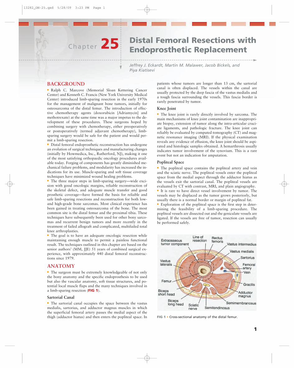
ANATOMY■ The surgeon must be extremely knowledgeable of not onlythe bony anatomy and the specific endoprosthesis to be usedbut also the vascular anatomy, soft tissue structures, and po-tential local muscle flaps and the many techniques involved ina limb-sparing resection (FIG 1).
Sartorial Canal■ The sartorial canal occupies the space between the vastusmedialis, sartorius, and adductor magnus muscles in whichthe superficial femoral artery passes the medial aspect of thethigh (adductor hiatus) and then enters the popliteal space. In
patients whose tumors are longer than 13 cm, the sartorialcanal is often displaced. The vessels within the canal areusually protected by the deep fascia of the vastus medialis anda tough fascia surrounding the vessels. This fascia border israrely penetrated by tumor.
Knee Joint■ The knee joint is rarely directly involved by sarcoma. Themain mechanisms of knee joint contamination are inappropri-ate biopsy, extension of tumor along the intra-articular cruci-ate ligaments, and pathologic fracture. The knee joint canreliably be evaluated by computed tomography (CT) and mag-netic resonance imaging (MRI). If the physical examinationreveals any evidence of effusion, the knee joint should be aspi-rated and histologic samples obtained. A hemarthrosis usuallyindicates tumor involvement of the synovium. This is a rareevent but not an indication for amputation.
Popliteal Space■ The popliteal space contains the popliteal artery and veinand the sciatic nerve. The popliteal vessels enter the poplitealspace from the medial aspect through the adductor hiatus asthe vessels exit the sartorial canal. The popliteal vessels areevaluated by CT with contrast, MRI, and plain angiography.■ It is rare to have direct vessel involvement by tumor. Thevessels may be displaced as the tumor grows posteriorly, butusually there is a normal border or margin of popliteal fat.■ Exploration of the popliteal space is the first step in deter-mining the feasibility of a limb-sparing procedure. Thepopliteal vessels are dissected out and the geniculate vessels areligated. If the vessels are free of tumor, resection can usuallybe performed safely.
Chapter 25Jeffrey J. Eckardt, Martin M. Malawer, Jacob Bickels, and Piya Kiatisevi
Distal Femoral Resections withEndoprosthetic Replacement
FIG 1 • Cross-sectional anatomy of the distal femur.
13282_ON-25.qxd 5/28/09 3:23 PM Page 1
2 Part 4 ONCOLOGY • Section IV LOWER EXTREMITIES
A B C
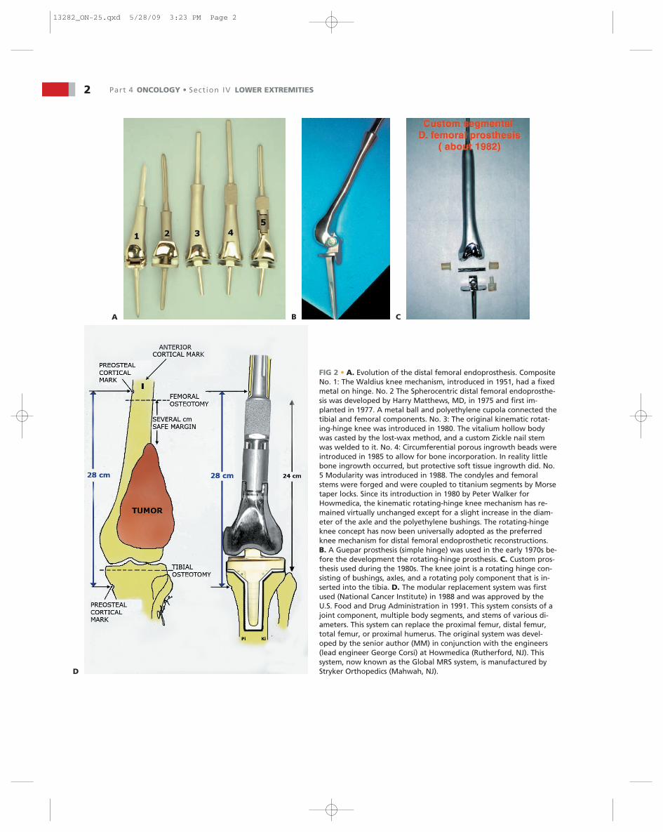
FIG 2 • A. Evolution of the distal femoral endoprosthesis. CompositeNo. 1: The Waldius knee mechanism, introduced in 1951, had a fixedmetal on hinge. No. 2 The Spherocentric distal femoral endoprosthe-sis was developed by Harry Matthews, MD, in 1975 and first im-planted in 1977. A metal ball and polyethylene cupola connected thetibial and femoral components. No. 3: The original kinematic rotat-ing-hinge knee was introduced in 1980. The vitalium hollow bodywas casted by the lost-wax method, and a custom Zickle nail stemwas welded to it. No. 4: Circumferential porous ingrowth beads wereintroduced in 1985 to allow for bone incorporation. In reality littlebone ingrowth occurred, but protective soft tissue ingrowth did. No.5 Modularity was introduced in 1988. The condyles and femoralstems were forged and were coupled to titanium segments by Morsetaper locks. Since its introduction in 1980 by Peter Walker forHowmedica, the kinematic rotating-hinge knee mechanism has re-mained virtually unchanged except for a slight increase in the diam-eter of the axle and the polyethylene bushings. The rotating-hingeknee concept has now been universally adopted as the preferredknee mechanism for distal femoral endoprosthetic reconstructions.B. A Guepar prosthesis (simple hinge) was used in the early 1970s be-fore the development the rotating-hinge prosthesis. C. Custom pros-thesis used during the 1980s. The knee joint is a rotating hinge con-sisting of bushings, axles, and a rotating poly component that is in-serted into the tibia. D. The modular replacement system was firstused (National Cancer Institute) in 1988 and was approved by theU.S. Food and Drug Administration in 1991. This system consists of ajoint component, multiple body segments, and stems of various di-ameters. This system can replace the proximal femur, distal femur,total femur, or proximal humerus. The original system was devel-oped by the senior author (MM) in conjunction with the engineers(lead engineer George Corsi) at Howmedica (Rutherford, NJ). Thissystem, now known as the Global MRS system, is manufactured byStryker Orthopedics (Mahwah, NJ).D
13282_ON-25.qxd 5/28/09 3:23 PM Page 2
■ A frozen section of the popliteal fat or adventitia of thepopliteal vessels should be obtained intraoperatively. If there isobvious vascular involvement, the vessels can be replaced byvascular graft.■ The popliteal vein is usually not repaired because it rarelystays patent after surgery.
Anterior and Posterior Cruciate Ligaments■ The cruciate ligaments are occasionally involved by directtumor extension from the distal femur. This occurs throughthe bone–tendinous junction of the intercondylar notch of thedistal femur. There is no cartilage in this area to act as a bar-rier to tumor growth.■ MRI is occasionally helpful in determining cruciate ligamentinvolvement.■ Tumor nodules of the anterior and posterior cruciates occa-sionally present with a hemarthrosis. The most common find-ing at resection is tumor nodule involvement of the cruciates.This does not rule out a limb-sparing procedure. The cruciateligaments as they attach to the proximal tibial plateau can beresected en bloc with the proximal tibial cut. This is a safe pro-cedure that avoids the need for a true extra-articular resection.
INDICATIONS■ Endoprostheses were initially used solely for reconstructionafter resection of malignant bone tumors. Manufacturingtime could be as long as 3 months, an interval that permittedinduction chemotherapy. Endoprosthetic reconstructionsproved to be enduring, and the designs have evolved (FIG 2).Modularity, which made for immediate availability, permittedthe expansion of the indications for distal femoral endopros-thetic reconstruction to some stage 3 giant cell tumors of bone;metastatic disease where conventional intralesional procedurescannot reasonably be done, possibly 10% of metastatic cases;complex supracondylar fractures in elderly osteoporotic pa-tients; failed internal fixation of distal femoral fractures; failedallograft or total knee reconstructions; and as a primary kneereplacement system in patients with a severe flexion contrac-ture where bone and ligament resection would lead to instabil-ity with conventional knee replacement systems.
PATIENT HISTORY AND PHYSICALFINDINGS■ The average age of patients with high-grade osteosarcomasis 5 to 30 years; the median is 16 to 21 years. Surface osteosar-comas occur in the third decade and are more common inwomen.■ Patients with high-grade osteosarcoma almost always ini-tially complain of pain during the day that is not associatedwith activity. All patients complain of a dull aching pain andonly later of night pain.■ Thirty to 40% of patients have a history of local trauma.There is no causal relationship of trauma to the tumor exceptthat it brings the patient to medical attention and the physicianorders a radiograph, which always shows the tumor. This hasbeen termed “traumatic determinism.”
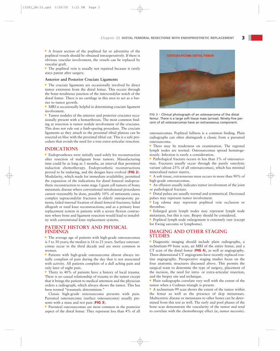
Classic high-grade osteosarcoma presents with pain.Parosteal osteosarcoma (surface osteosarcoma) usually pre-sents with a mass and not pain (FIG 3).■ Parosteal osteosarcomas are most common in the posterioraspect of the distal femur. They represent less than 4% of all
osteosarcomas. Popliteal fullness is a common finding. Plainradiographs can often distinguish a classic from a parostealosteosarcoma.■ There may be tenderness on examination. The regionallymph nodes are normal. Osteosarcomas spread hematoge-nously. Infection is rarely a consideration.■ Pathological fracture occurs in less than 1% of osteosarco-mas. Fractures usually occur through the purely osteolyticvariant (about 25% of all osteosarcomas), which has minimalmineralized tumor matrix.■ A soft tissue, extraosseous mass occurs in more than 90% ofhigh-grade osteosarcomas.■ An effusion usually indicates tumor involvement of the jointor pathological fracture.■ Distal pulses are usually normal and symmetrical. Decreasedpulses may represent tumor involvement.■ Leg edema may represent popliteal vein occlusion orthrombus.■ Enlarged groin lymph nodes may represent lymph nodemetastasis, but this is rare. Biopsy should be considered.■ Popliteal lymph node enlargement is extremely rare (exceptfor Ewing sarcoma or lymphoma).
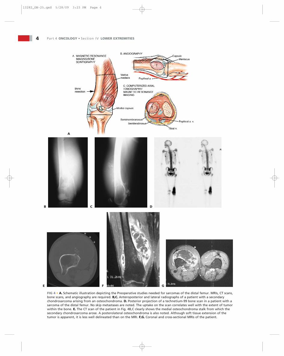
IMAGING AND OTHER STAGINGSTUDIES■ Diagnostic imaging should include plain radiographs, atechnetium-99 bone scan, an MRI of the entire femur, and aCT scan of the distal femur (FIG 4), as well as angiography.Three-dimensional CT angiograms have recently replaced rou-tine angiography. Preoperative staging studies focus on thefour anatomic structures discussed above. This permits thesurgical team to determine the type of surgery, placement ofthe incision, the need for intra- or extra-articular resection,and the biopsy site and technique.■ Plain radiographs correlate very well with the extent of thetumor when a Codman triangle is present.■ A technetium-99 scan shows the extent of the tumor withinthe femur as well as the presence of skip metastases.Multicentric disease or metastases to other bones can be deter-mined from this test as well. The early and pool phases of thebone scan demonstrate the vascularity of the tumor and tendto correlate with the chemotherapy effect (ie, tumor necrosis).
Chapter 25 DISTAL FEMORAL RESECTIONS WITH ENDOPROSTHETIC REPLACEMENT 3
FIG 3 • Clinical photograph of an osteosarcoma of the distalfemur. There is a large soft tissue mass (arrow). Ninety-five per-cent of all osteosarcomas have an extraosseous component.
13282_ON-25.qxd 5/28/09 3:23 PM Page 3
4 Part 4 ONCOLOGY • Section IV LOWER EXTREMITIES
A
CB D
F GE
FIG 4 • A. Schematic illustration depicting the Preoperative studies needed for sarcomas of the distal femur. MRIs, CT scans,bone scans, and angiography are required. B,C. Anteroposterior and lateral radiographs of a patient with a secondarychondrosarcoma arising from an osteochondroma. D. Posterior projection of a technetium-99 bone scan in a patient with asarcoma of the distal femur. No skip metastases are noted. The uptake on the scan correlates well with the extent of tumorwithin the bone. E. The CT scan of the patient in Fig. 4B,C clearly shows the medial osteochondroma stalk from which thesecondary chondrosarcoma arose. A posterolateral osteochondroma is also noted. Although soft tissue extension of thetumor is apparent, it is less well delineated than on the MRI. F,G. Coronal and cross-sectional MRIs of the patient.
13282_ON-25.qxd 5/28/09 3:23 PM Page 4

Chapter 25 DISTAL FEMORAL RESECTIONS WITH ENDOPROSTHETIC REPLACEMENT 5
■ A femoral MRI best shows the extraosseous extent of thetumor as well as its proximal and distal extent within themedullary canal. This study is the most accurate in detectingskip metastases.■ CT scans are complementary to MRI scans and can show the quality of the bone stock at the intended level ofresection.■ Angiography or three-dimensional CT angiography can beused to evaluate the superficial femoral and popliteal arteries.
This is especially important if there is a large posterior ormedial extraosseous component. The late arterial phase of theangiogram or venous phase will show residual tumor blush.The degree of remaining vascularity correlates well with thetumor necrosis (FIG 5A,B). An unresponsive tumor as shownby a tumor blush requires a wider margin than a good respon-der (no tumor blush). More recently, three-dimensional CTangiography has replaced traditional angiograms; it shows thevascular anatomy well (FIG 5C–F).
A B C
ED F
FIG 5 • A. Angiography after induction chemotherapy. A. Anteroposterior view. B. Lateral viewshowing the absence of a tumor blush. This is the most reliable finding of all preoperative stagingstudies that can predict tumor response. This patient had 100% chemonecrosis. C. Three-dimensionalangiography is being evaluated in the treatment of bony tumors. C,D. Lateral and posterior views ofthe distal sarcoma. The popliteal artery is displaced. The extraosseous component is not visualizedbecause there is no bony formation. E,F. Secondary chondrosarcoma of the proximal tibia. Lateraland posterior views showing excellent visualization of the popliteal artery and its trifurcation(arrows). A 64- or 246-slice CT scanner is required, similar to coronary angiography.
13282_ON-25.qxd 5/28/09 3:23 PM Page 5

6 Part 4 ONCOLOGY • Section IV LOWER EXTREMITIES
A B C
FIG 7 • A. CT scans of a sclerotic osteosarcoma of the distal femur. A needle biopsy is performed under CT guidance. Needle biopsiesare routinely performed to establish a diagnosis; less than 5% to 10% require an open biopsy. B,C. Radiographic response after induc-tion chemotherapy for a distal femoral osteosarcoma. B. Preoperative CT scan shows an extraosseous component. C. CT scan showsreossification of the entire lesion. CT scans are extremely valuable at determining tumor response (the percentage of tumor necrosis)and are routinely performed before and after induction chemotherapy.
FIG 6 • Primary distal femoral osteosarcoma: soft tissue resec-tion. The small black dots represent potential skin metastases.The planes of a marginal excision and a wide excision areshown.
■ Together these studies help to determine the resectability ofthe tumor as well as the desired level of the femoral osteotomy.■ A comprehensive knowledge of prosthetic stem lengthsand widths is necessary to be sure that adequate proximalbone stock is available to proceed with endoprosthetic re-construction.
SURGICAL MANAGEMENT■ Surgical guidelines for limb-sparing surgery are as follows:
■ The major neurovascular bundle (popliteal vessels) mustbe free of tumor.
■ The resection of the affected bone should leave a widemargin and a normal muscle cuff (ie, 1 to 2 cm) in all direc-tions (FIG 6).■ All previous biopsy sites and all potentially contaminatedtissues should be removed en bloc. All needle biopsy tractsmust be removed (FIG 7A).■ To avoid intraosseous tumor extension, bone should beresected 3 to 5 cm beyond abnormal uptake, as determinedby preoperative studies.■ The adjacent joint and joint capsule should be resected.■ Adequate motor reconstruction must be accomplished byregional muscle transfers.■ Adequate soft tissue coverage is needed to decrease therisk of skin flap necrosis and secondary infection. A medialgastrocnemius rotation flap provides excellent coverage ofthe prosthesis when required.
■ Careful attention to the patient’s general condition is criticalin the timing of limb-sparing procedures in cancer patients.Patients undergoing preoperative chemotherapy (FIG 7B,C) andradiation therapy (Ewing sarcoma) need an adequate hiatusbefore surgery. In general, surgery can proceed 2 to 3 weeksafter these treatments are completed. The white count andplatelet count need to be within a safe range and rising, and theskin must have recovered from the effects of radiation and mustbe nonerythematous.■ When the procedure is used for a salvage reconstructionafter failed internal fixation, failed total joint arthroplasty, orallograft procedures, a past history of infection can bodepoorly if it is not completely eradicated before surgery.
Preoperative Planning■ The intended level of resection should be determined beforethe patient comes to the operating room. A careful review ofthe diagnostic tests should confirm this location and should
13282_ON-25.qxd 5/28/09 3:23 PM Page 6

Chapter 25 DISTAL FEMORAL RESECTIONS WITH ENDOPROSTHETIC REPLACEMENT 7
A
B
FIG 8 • A. Traditional subcutaneous approach to the distalfemur and popliteal space versus the anterior transadductorpopliteal approach. B. Incision used for distal femoral resectionand distally if necessary for a medial gastrocnemius flap.
confirm that there is adequate bone stock left to accept thefemoral stem, in terms of both length and width. The distalfemur should be resected with a safe oncologic margin (3 to4 cm of normal marrow). The extremity lengths should beequal to within millimeters. To achieve this, intraoperativemarks and measurements are made to ensure that the lengthbefore resection equals the reconstruction length.■ When planning the primary resection and reconstruction,the surgeon should also be planning an amputation or revi-sion. Ideally the level of amputation should be at the samelevel it would have been had amputation been chosen as theoriginal procedure to achieve local control. The surgeonshould plan how he or she will revise this reconstruction in theevent of infection or mechanical failure. The real goal wouldbe to retain the patient’s own hip and not go to a total femurreplacement unless necessary, as this requires rehabilitatingtwo joints in series, which is always a greater challenge for thepatient.
Positioning■ In the preoperative area or as anesthesia is being induced,the patient is given intravenous antibiotics. One gram of van-
comycin is slowly infused over 1 hour, and this is repeatedevery 12 hours until the drains are removed. A single 80-mgdose of gentamicin or tobramycin is also given. An epiduralcatheter is routinely used for postoperative pain management.■ After anesthesia is induced, a urinary catheter is placed. Forthe medial approach the patient is placed in the supine posi-tion with the entire leg, including the inguinal area, prepared.This provides adequate access to the proximal femoral vessels.■ A tourniquet is not used. A folded sheet placed transverselyunder the sacrum can elevate the pelvis to permit better accessfor draping. If the lateral approach is used, then the patient isplaced in the lateral decubitus position on a beanbag with anaxillary roll. A standard 10-minute preparation is performed,generally iodine-based.
Approach■ The preferred approach is a medial longitudinal approachwith exploration of the superficial femoral and popliteal ves-sels. All vessels that feed the tumor and distal femur are tiedoff (FIG 8). A lateral approach is used only when access to theproximal femur is needed for cross-pin stem fixation or whenlittle residual proximal femur remains.
TECH
NIQ
UES
Position and Dissection■ The patient is in supine position with the leg and in-
guinal area prepared out (TECH FIG 1A).■ The incision is longitudinal, following the sartorius mus-
cle from proximally in the thigh distal to beyond the tib-ial tubercle (TECH FIG 1B).
■ Any biopsy tract should be kept in continuity with theunderlying tumor. Because the routine approach for pri-mary tumors is medial, lateral or anterior open biopsytracts need to be ellipsed and kept in continuity with theunderlying tumor.
■ The saphenous nerve is identified and protected (TECHFIG 1C).
■ The interval between the sartorius and the vastus medialisis opened, exposing the superficial femoral artery andvein along with the saphenous nerve (TECH FIG 1D,E).
■ The vessels and the saphenous nerve are dissected fromproximal to distal and are reflected posterior and medialalong with the sartorius muscle.
■ All vessels (geniculates) are tied off with 2-0 or 3-0 silk tiesas they course from the vessels toward the distal femur andtumor (TECH FIG 1F). The surgeon must not ligate the me-dial or lateral sural vessels, which are the main blood sup-ply to the respective gastrocnemius muscles. These vesselsare the basis of a gastrocnemius flap if required.
■ The surgeon should be careful at the canal of Hunter be-cause the vessels are just deep to the adductor tendon.
RESECTION AND RECONSTRUCTION OF THE DISTAL FEMUR THROUGH ALONGITUDINAL MEDIAL APPROACH AND PREPARATION FOR CEMENTINGTHE TIBIA, PATELLA, AND FEMORAL COMPONENTS
13282_ON-25.qxd 5/28/09 3:23 PM Page 7

F
8 Part 4 ONCOLOGY • Section IV LOWER EXTREMITIESTE
CH
NIQ
UES
A B
C D
E
TECH FIG 1 • A. Right leg with large secondary chondrosar-coma. B. A medial incision follows the sartorius proximally inthe thigh to below the tibial tubercle. This allows immediateand very adequate visualization of the femoral and poplitealvessels. C. After the incision through the skin and subcuta-neous tissue, a large posteromedial flap is developed deep tothe fascia. The first vital structure identified and protected isthe saphenous nerve. It accompanies the femoral vessels prox-imally in the thigh and follows the sartorius into the leg.Cutting the nerve results in numbness over the medial calf andoccasionally a painful neuroma. D. In the middle and distalthigh, retracting the sartorius posteromedially exposes the su-perficial femoral vessels. E. In the proximal thigh, retractingthe sartorius anterior and lateral allows exposure of thefemoral vessels, all the way to the inguinal ligament if neces-
sary. F. All vessels coursing toward the distal femur and tumor are tied with 2-0 or 3-0 silk sutures before they are cut. This min-imizes blood loss, improves exposure, and guarantees the integrity of these structures. G. At the canal of Hunter, the adductortendon is identified and cut. The main vessels are just beneath this structure, and care and patience at this point in the dissec-tion are necessary. Several collateral vessels come off the femoral vessels at this point, coursing toward the femur and tumor.They need to be tied off. The saphenous nerve is seen accompanying the sartorius muscle.
G
13282_ON-25.qxd 5/28/09 3:24 PM Page 8

Chapter 25 DISTAL FEMORAL RESECTIONS WITH ENDOPROSTHETIC REPLACEMENT 9TEC
HN
IQU
ES
■ Distal to the canal of Hunter the popliteal vessels are dis-sected free and reflected posterior and medially (TECHFIG 1G). The short head of the biceps muscle is now seencoursing proximal to distal to join the long head laterallyin the thigh.
■ The sciatic nerve is exposed and protected.■ Proximal and medial in the thigh above the tumor, the
junction between the adductors and vastus medialis canbe opened to the femur to reflect the quadriceps later-ally off the femur (TECH FIG 2A).
■ Deep to the medial intermuscular septum is the terminalprofunda artery and vein, which may be ligated.
■ The superficial femoral vessels, along with the saphenousnerve and popliteal vessels, are dissected free from thetumor throughout its length to below the joint line(TECH FIG 2B,C).
■ The medial gastrocnemius muscle can be incised. The sur-geon must not ligate the medial sural vessels (TECH FIG2D,E).
■ With the femoral vessels completely dissected and re-flected, the quadriceps or a portion of it, along with thepatella and patellar tendon, are now reflected over thetumor, leaving the vastus intermedialis as a very satisfac-tory oncologic margin.
■ Intra-articular resections are usually performed.■ The joint capsule is opened and the anterior and pos-
terior cruciate ligaments, the popliteus tendon, andthe collateral ligaments are cut with an electricalcautery.
■ The posterior capsule is incised, with the poplitealvessels kept in direct view or under the operator’s fin-ger to prevent injury.
A
C D
B
TECH FIG 2 • A. Proximally in the thigh, above the tumorthe adductor fascia meets the fascia of the vastus medi-alis. This interval is opened to permit exposure of thefemur. The profunda vessels course just below the ad-ductor fascia and follow the linea aspera. B. Saphenousnerve proximally in the thigh accompanies the superficialfemoral vessels as the sartorius has been retracted pos-teromedially. The adductor tendon has not yet been cut,but the popliteal vessels have been exposed and mobi-lized to below the knee joint to guarantee their in-tegrity. C. Completed medial dissection. The saphenousnerve follows the sartorius from proximal in the field.The femoral and popliteal vessels have been dissectedand accompany the sartorius muscle and the saphenous
nerve distally in the thigh. D. Medially at the knee, the medial gastrocnemius muscle is dissected and cut. E. Medialgeniculates are identified and cut. (continued)
E
13282_ON-25.qxd 5/28/09 3:24 PM Page 9
10 Part 4 ONCOLOGY • Section IV LOWER EXTREMITIES
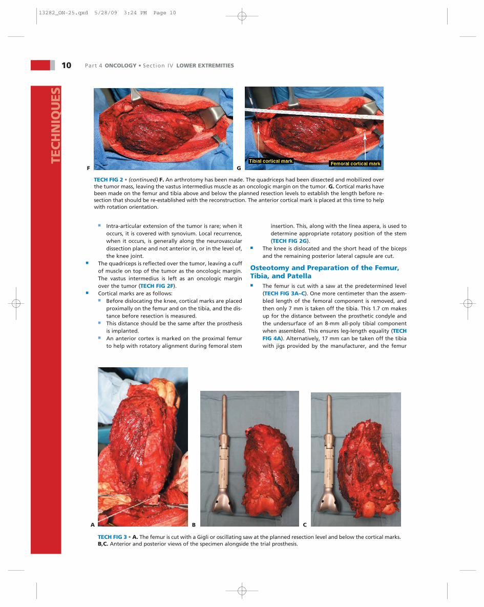
TECH FIG 2 • (continued) F. An arthrotomy has been made. The quadriceps had been dissected and mobilized overthe tumor mass, leaving the vastus intermedius muscle as an oncologic margin on the tumor. G. Cortical marks havebeen made on the femur and tibia above and below the planned resection levels to establish the length before re-section that should be re-established with the reconstruction. The anterior cortical mark is placed at this time to helpwith rotation orientation.
TEC
HN
IQU
ES
■ Intra-articular extension of the tumor is rare; when itoccurs, it is covered with synovium. Local recurrence,when it occurs, is generally along the neurovasculardissection plane and not anterior in, or in the level of,the knee joint.
■ The quadriceps is reflected over the tumor, leaving a cuffof muscle on top of the tumor as the oncologic margin.The vastus intermedius is left as an oncologic marginover the tumor (TECH FIG 2F).
■ Cortical marks are as follows:■ Before dislocating the knee, cortical marks are placed
proximally on the femur and on the tibia, and the dis-tance before resection is measured.
■ This distance should be the same after the prosthesisis implanted.
■ An anterior cortex is marked on the proximal femurto help with rotatory alignment during femoral stem
insertion. This, along with the linea aspera, is used todetermine appropriate rotatory position of the stem(TECH FIG 2G).
■ The knee is dislocated and the short head of the bicepsand the remaining posterior lateral capsule are cut.
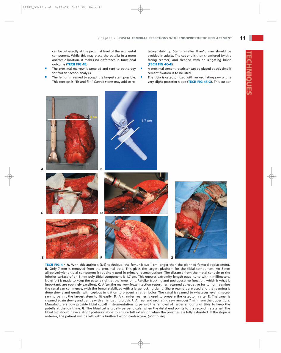
Osteotomy and Preparation of the Femur,Tibia, and Patella■ The femur is cut with a saw at the predetermined level
(TECH FIG 3A–C). One more centimeter than the assem-bled length of the femoral component is removed, andthen only 7 mm is taken off the tibia. This 1.7 cm makesup for the distance between the prosthetic condyle andthe undersurface of an 8-mm all-poly tibial componentwhen assembled. This ensures leg-length equality (TECHFIG 4A). Alternatively, 17 mm can be taken off the tibiawith jigs provided by the manufacturer, and the femur
TECH FIG 3 • A. The femur is cut with a Gigli or oscillating saw at the planned resection level and below the cortical marks.B,C. Anterior and posterior views of the specimen alongside the trial prosthesis.
A B C
F G
13282_ON-25.qxd 5/28/09 3:24 PM Page 10
Chapter 25 DISTAL FEMORAL RESECTIONS WITH ENDOPROSTHETIC REPLACEMENT 11TEC
HN
IQU
ES
can be cut exactly at the proximal level of the segmentalcomponent. While this may place the patella in a moreanatomic location, it makes no difference in functionaloutcome (TECH FIG 4B).
■ The proximal marrow is sampled and sent to pathologyfor frozen section analysis.
■ The femur is reamed to accept the largest stem possible.This concept is “fit and fill.” Curved stems may add to ro-
tatory stability. Stems smaller than13 mm should beavoided in adults. The cut end is then chamfered (with afacing reamer) and cleaned with an irrigating brush(TECH FIG 4C–E).
■ A proximal cement restrictor can be placed at this time ifcement fixation is to be used.
■ The tibia is osteotomized with an oscillating saw with avery slight posterior slope (TECH FIG 4F,G). This cut can
TECH FIG 4 • A. With this author’s (JJE) technique, the femur is cut 1 cm longer than the planned femoral replacement. B. Only 7 mm is removed from the proximal tibia. This gives the largest platform for the tibial component. An 8-mm all-polyethylene tibial component is routinely used in primary reconstructions. The distance from the metal condyle to theinferior surface of an 8-mm poly tibial component is 1.7 cm. This ensures extremity-length equality to within millimeters.No effort is made to keep the patella right at the knee joint. Patellar tracking and postoperative function, which is what isimportant, are routinely excellent. C. After the marrow frozen section report has returned as negative for tumor, reamingthe canal can commence, with the femur stabilized with a large locking clamp. Sharp reamers are used and the reaming isdone slowly and gently, with copious irrigation to prevent a fat embolus. The canal is reamed to whatever level is neces-sary to permit the largest stem to fit easily. D. A chamfer reamer is used to prepare the osteotomy site. E. The canal iscleaned again slowly and gently with an irrigating brush. F. A freehand oscillating saw removes 7 mm from the upper tibia.Manufacturers now provide tibial cutoff instrumentation to permit the removal of larger amounts of tibia to keep thepatella at the joint line. G. The tibial cut is usually perpendicular when the distal end points to the second metatarsal. Thetibial cut should have a slight posterior slope to ensure full extension when the prosthesis is fully extended. If the slope isanterior, the patient will be left with a built-in flexion contracture. (continued)
C
A B
D
E F G
13282_ON-25.qxd 5/28/09 3:24 PM Page 11
12 Part 4 ONCOLOGY • Section IV LOWER EXTREMITIESTE
CH
NIQ
UES
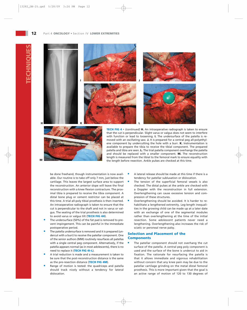
TECH FIG 4 • (continued) H. An intraoperative radiograph is taken to ensurethat the cut is perpendicular. Slight varus or valgus does not seem to interferewith function or lead to loosening. I. The undersurface of the patella is re-moved with an oscillating saw. J. It is prepared for a central peg all-polyethyl-ene component by undercutting the hole with a burr. K. Instrumentation isavailable to prepare the tibia to receive the tibial component. The preparedpatella and tibia are seen. L. The trial patella component overhangs the patellaand should be replaced with a smaller component. M. The reconstructionlength is measured from the tibial to the femoral mark to ensure equality withthe length before resection. Ankle pulses are checked at this time.
H
I J
K L
M
be done freehand, though instrumentation is now avail-able. Our routine is to take off only 7 mm, just below thecartilage. This leaves the largest surface area to supportthe reconstruction. An anterior slope will leave the finalreconstruction with a knee flexion contracture. The prox-imal tibia is prepared to receive the tibia component. Adistal bone plug or cement restrictor can be placed atthis time. A trial all-poly tibial prosthesis is then inserted.An intraoperative radiograph is taken to ensure that thecut is perpendicular to the shaft and not in varus or val-gus. The seating of the trial prosthesis is also determinedto avoid varus or valgus tilt (TECH FIG 4H).
■ The undersurface (50%) of the fat pad is removed to pre-vent impingement. This can be painful in the immediatepostoperative period.
■ The patella undersurface is removed and it is prepared (un-dercut with a burr) to receive the patellar component. Oneof the senior authors (MM) routinely resurfaces all patellaswith a single central peg component. Alternatively, if thepatella appears normal (as in most adolescents), there is noneed to replace it (TECH FIG 4I–L).
■ A trial reduction is made and a measurement is taken tobe sure that the post-reconstruction distance is the sameas the pre-resection distance (TECH FIG 4M).
■ Range of motion is tested: the quadriceps and patellashould track nicely without a tendency for lateraldislocation.
■ A lateral release should be made at this time if there is atendency for patellar subluxation or dislocation.
■ The tension of the superficial femoral vessels is alsochecked. The distal pulses at the ankle are checked witha Doppler with the reconstruction in full extension.Overlengthening can cause excessive tension and com-pression of these structures.
■ Overlengthening should be avoided. It is harder to re-habilitate a lengthened extremity. Leg-length inequali-ties in the growing child can be made up at a later datewith an exchange of one of the segmental modulesrather than overlengthening at the time of the initialresection. Some adolescent patients never need alengthening. Overlengthening also increases the risk ofsciatic or peroneal nerve palsy.
Selection and Placement of theComponents■ The patellar component should not overhang the cut
surface of the patella. A central peg poly component isused and the surface of the bone is undercut to aid infixation. The rationale for resurfacing the patella isthat it allows immediate and vigorous rehabilitationwithout concern that any knee pain may be due to thepatellar cartilage grinding on the metal distal femoralprosthesis. This is more important given that the goal isan active range of motion of 120 to 130 degrees of
13282_ON-25.qxd 5/28/09 3:24 PM Page 12
Chapter 25 DISTAL FEMORAL RESECTIONS WITH ENDOPROSTHETIC REPLACEMENT 13TEC
HN
IQU
ES
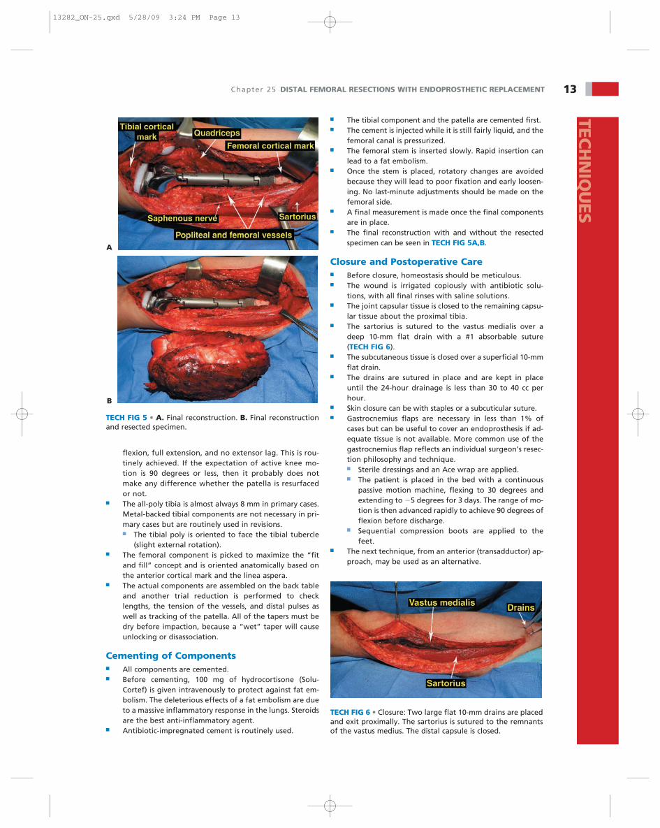
TECH FIG 5 • A. Final reconstruction. B. Final reconstructionand resected specimen.
A
B
TECH FIG 6 • Closure: Two large flat 10-mm drains are placedand exit proximally. The sartorius is sutured to the remnantsof the vastus medius. The distal capsule is closed.
flexion, full extension, and no extensor lag. This is rou-tinely achieved. If the expectation of active knee mo-tion is 90 degrees or less, then it probably does notmake any difference whether the patella is resurfacedor not.
■ The all-poly tibia is almost always 8 mm in primary cases.Metal-backed tibial components are not necessary in pri-mary cases but are routinely used in revisions.■ The tibial poly is oriented to face the tibial tubercle
(slight external rotation).■ The femoral component is picked to maximize the “fit
and fill” concept and is oriented anatomically based onthe anterior cortical mark and the linea aspera.
■ The actual components are assembled on the back tableand another trial reduction is performed to checklengths, the tension of the vessels, and distal pulses aswell as tracking of the patella. All of the tapers must bedry before impaction, because a “wet” taper will causeunlocking or disassociation.
Cementing of Components■ All components are cemented.■ Before cementing, 100 mg of hydrocortisone (Solu-
Cortef) is given intravenously to protect against fat em-bolism. The deleterious effects of a fat embolism are dueto a massive inflammatory response in the lungs. Steroidsare the best anti-inflammatory agent.
■ Antibiotic-impregnated cement is routinely used.
■ The tibial component and the patella are cemented first.■ The cement is injected while it is still fairly liquid, and the
femoral canal is pressurized.■ The femoral stem is inserted slowly. Rapid insertion can
lead to a fat embolism.■ Once the stem is placed, rotatory changes are avoided
because they will lead to poor fixation and early loosen-ing. No last-minute adjustments should be made on thefemoral side.
■ A final measurement is made once the final componentsare in place.
■ The final reconstruction with and without the resectedspecimen can be seen in TECH FIG 5A,B.
Closure and Postoperative Care■ Before closure, homeostasis should be meticulous.■ The wound is irrigated copiously with antibiotic solu-
tions, with all final rinses with saline solutions.■ The joint capsular tissue is closed to the remaining capsu-
lar tissue about the proximal tibia.■ The sartorius is sutured to the vastus medialis over a
deep 10-mm flat drain with a #1 absorbable suture(TECH FIG 6).
■ The subcutaneous tissue is closed over a superficial 10-mmflat drain.
■ The drains are sutured in place and are kept in placeuntil the 24-hour drainage is less than 30 to 40 cc perhour.
■ Skin closure can be with staples or a subcuticular suture.■ Gastrocnemius flaps are necessary in less than 1% of
cases but can be useful to cover an endoprosthesis if ad-equate tissue is not available. More common use of thegastrocnemius flap reflects an individual surgeon’s resec-tion philosophy and technique.■ Sterile dressings and an Ace wrap are applied.■ The patient is placed in the bed with a continuous
passive motion machine, flexing to 30 degrees andextending to �5 degrees for 3 days. The range of mo-tion is then advanced rapidly to achieve 90 degrees offlexion before discharge.
■ Sequential compression boots are applied to thefeet.
■ The next technique, from an anterior (transadductor) ap-proach, may be used as an alternative.
13282_ON-25.qxd 5/28/09 3:24 PM Page 13
14 Part 4 ONCOLOGY • Section IV LOWER EXTREMITIESTE
CH
NIQ
UES
A
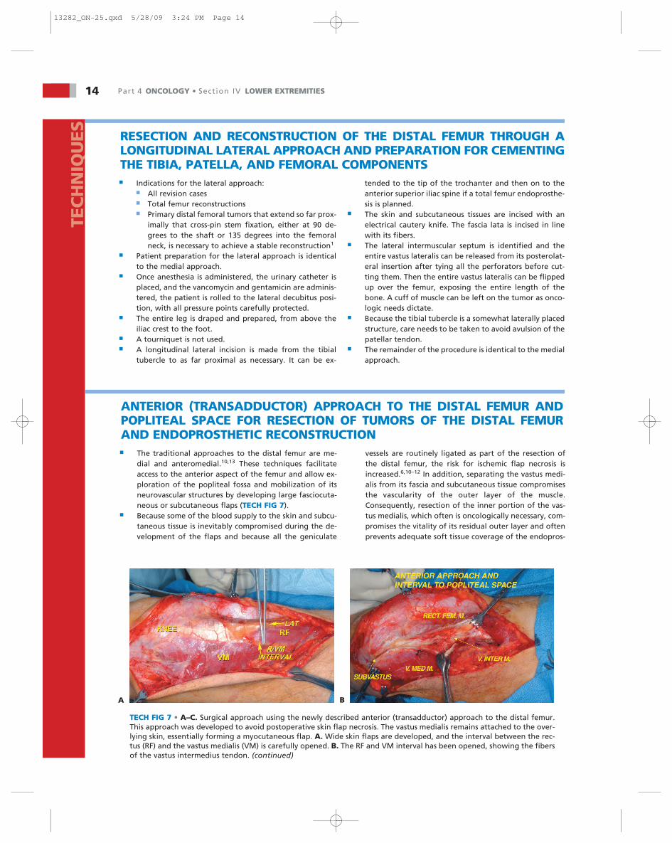
ANTERIOR (TRANSADDUCTOR) APPROACH TO THE DISTAL FEMUR ANDPOPLITEAL SPACE FOR RESECTION OF TUMORS OF THE DISTAL FEMURAND ENDOPROSTHETIC RECONSTRUCTION■ The traditional approaches to the distal femur are me-
dial and anteromedial.10,13 These techniques facilitateaccess to the anterior aspect of the femur and allow ex-ploration of the popliteal fossa and mobilization of itsneurovascular structures by developing large fasciocuta-neous or subcutaneous flaps (TECH FIG 7).
■ Because some of the blood supply to the skin and subcu-taneous tissue is inevitably compromised during the de-velopment of the flaps and because all the geniculate
vessels are routinely ligated as part of the resection ofthe distal femur, the risk for ischemic flap necrosis isincreased.6,10–12 In addition, separating the vastus medi-alis from its fascia and subcutaneous tissue compromisesthe vascularity of the outer layer of the muscle.Consequently, resection of the inner portion of the vas-tus medialis, which often is oncologically necessary, com-promises the vitality of its residual outer layer and oftenprevents adequate soft tissue coverage of the endopros-
B
RESECTION AND RECONSTRUCTION OF THE DISTAL FEMUR THROUGH ALONGITUDINAL LATERAL APPROACH AND PREPARATION FOR CEMENTINGTHE TIBIA, PATELLA, AND FEMORAL COMPONENTS■ Indications for the lateral approach:
■ All revision cases■ Total femur reconstructions■ Primary distal femoral tumors that extend so far prox-
imally that cross-pin stem fixation, either at 90 de-grees to the shaft or 135 degrees into the femoralneck, is necessary to achieve a stable reconstruction1
■ Patient preparation for the lateral approach is identicalto the medial approach.
■ Once anesthesia is administered, the urinary catheter isplaced, and the vancomycin and gentamicin are adminis-tered, the patient is rolled to the lateral decubitus posi-tion, with all pressure points carefully protected.
■ The entire leg is draped and prepared, from above theiliac crest to the foot.
■ A tourniquet is not used.■ A longitudinal lateral incision is made from the tibial
tubercle to as far proximal as necessary. It can be ex-
tended to the tip of the trochanter and then on to theanterior superior iliac spine if a total femur endoprosthe-sis is planned.
■ The skin and subcutaneous tissues are incised with anelectrical cautery knife. The fascia lata is incised in linewith its fibers.
■ The lateral intermuscular septum is identified and theentire vastus lateralis can be released from its posterolat-eral insertion after tying all the perforators before cut-ting them. Then the entire vastus lateralis can be flippedup over the femur, exposing the entire length of thebone. A cuff of muscle can be left on the tumor as onco-logic needs dictate.
■ Because the tibial tubercle is a somewhat laterally placedstructure, care needs to be taken to avoid avulsion of thepatellar tendon.
■ The remainder of the procedure is identical to the medialapproach.
TECH FIG 7 • A–C. Surgical approach using the newly described anterior (transadductor) approach to the distal femur.This approach was developed to avoid postoperative skin flap necrosis. The vastus medialis remains attached to the over-lying skin, essentially forming a myocutaneous flap. A. Wide skin flaps are developed, and the interval between the rec-tus (RF) and the vastus medialis (VM) is carefully opened. B. The RF and VM interval has been opened, showing the fibersof the vastus intermedius tendon. (continued)
13282_ON-25.qxd 5/28/09 3:24 PM Page 14

Chapter 25 DISTAL FEMORAL RESECTIONS WITH ENDOPROSTHETIC REPLACEMENT 15TEC
HN
IQU
ES
C D
TECH FIG 7 • (continued) C. The distal aspect of the VM is developed. D. The surgical approach shown in A–C. E. The vastusintermedius tendon is opened and the medialis is mobilized. F–H,K. Operative photographs showing the transadductor ap-proach. F. The interval between the RF and VM has been opened and the vastus intermedius has been mobilized. G. The termi-nation of the sartorial canal containing the superficial femoral artery and vein is dissected free. H. The sartorial canal has beenopened. I. The remaining attachment of the adductor magnus tendon to the distal femur is released. J. Relationship of the su-perficial femoral artery to the popliteal space and the adductor magnus tendon. (continued)
E F
G H
I J
13282_ON-25.qxd 5/28/09 3:24 PM Page 15

16 Part 4 ONCOLOGY • Section IV LOWER EXTREMITIESTE
CH
NIQ
UES
TECH FIG 7 • (continued) K. Exposure of the popliteal spaceand the neurovascular structures. The geniculate branches arecarefully ligated. The vastus medialis has remained attached tothe overlying skin, thus emphasizing the purpose of this ap-proach.
K
thesis. In a previous study by the senior author (MM) of110 distal femoral resections and endoprosthetic recon-structions using the anteromedial approach, 25 gastroc-nemius flaps were required due to insufficient soft tissuecoverage and flap necrosis.8
■ Kawai et al reported a flap necrosis incidence of 30% in aseries of 40 patients who underwent distal femoralresection and endoprosthetic reconstruction, and Safranet al concluded that perioperative chemotherapy and in-traoperative flap devascularization were the major causesof infection after limb-salvage procedures.14 In an attemptto decrease the occurrence of flap necrosis and to improvethe soft tissue coverage of the endoprosthesis, the seniorauthor (MM) has developed the following modifiedsurgical approach for distal femoral resection using a well-vascularized posteromedial myocutaneous flap:■ Indications
■ All high- and low-grade tumors of the distal femur■ All distal femoral revisions. If a lateral interlocking
hip nail is required, a separate proximal, lateral,standard hip-like incision is made. There is no needto connect these two incisions.
■ If soft tissue closure is considered to be a problem,this approach is recommended. This allows for amedial gastrocnemius flap by simply extending theincision distally. The medial gastrocnemius muscleis always preferred to a lateral gastrocnemius mus-cle because the medial gastrocnemius muscle islarger and longer than the lateral gastrocnemiusmuscle. It permits a larger area to be covered, bothlongitudinally and transversely across the prosthe-sis and knee joint, respectively. (See “MedialGastrocnemius Muscle Transfer” below.)
■ This incision creates a myocutaneous flap by keep-ing the vastus medialis muscle attached to its over-lying skin. Skin flap necrosis, wound dehiscence,hemarthrosis, effusions, and other wound prob-lems are rare (1% to 5%).
■ If vascular resection and reconstruction are preop-erative possibilities, the superficial femoral andpopliteal vessels are directly exposed. A lateral in-cision would make vascular reconstruction moredifficult.
■ If minimal muscle coverage remains a problem afterresection and attempted closure, the sartorius mus-cle can be rotated to cover small defects. In addi-tion, a formal sartorius muscle transfer can be per-formed through this incision to recreate or to re-place either partial or complete vastus medialis loss.
Position and Incision■ With the patient in the supine position and the surgeon
standing on the medial side of the knee (opposite side ofthe table), a long, medial paramedian skin incision ismade. The incision extends proximally along the junctionof the rectus femoris and vastus medialis muscles andcurves distally around the medial border of the patella tothe level of the pes anserinus.
Proximal Interval and Creation ofMusculocutaneous Flap■ The interval between the rectus femoris and vastus me-
dialis muscles is identified and opened to expose theunderlying vastus intermedius muscle. The fibers of thevastus intermedius are then carefully divided. It is impor-tant not to separate the overlying muscle from its fascio-cutaneous coverage, which would defeat the purpose ofthis approach. This can be ensured by suturing the vastusmedialis to the overlying skin.
Exposure of Intermuscular Septum andAdductor Hiatus■ The plane between the vastus medialis and the medial
femoral condyle is identified distally (similar to thesubvastus approach). The vastus medialis muscle is dis-sected off the medial femoral condyle in an extra-articu-lar fashion and retracted medially, away from the kneecapsule. By sweeping the fibers of the muscle from theintermuscular septum with a sponge, the intermuscularseptum, the adductor hiatus, and the adductor magnustendon are exposed.
Identification of the Superficial Femoraland Popliteal Vessels■ The sartorius muscle, which crosses over the proximal
portion of the vastus medialis, is mobilized posteriorly byopening the thin fascia between the vastus medialis andits superior border. The superficial femoral artery andvein are identified proximally at the level of the adduc-tor hiatus. With the surgeon’s finger placed into the ad-ductor hiatus to protect the underlying vessels, the distalportion of the adductor magnus tendon is dissected andreleased from the distal femur and adductor tubercle,partially exposing the popliteal space. The superficialfemoral vessels are carefully dissected and mobilizedalong their sheath, and vessel loops are placed aroundthem as they enter the popliteal fossa.
Completion of Popliteal Exposure■ The knee is placed in 90 degrees of flexion. With the vas-
tus medialis musculocutaneous flap retracted posteriorly,the entire popliteal space is visualized and the poplitealvessels are identified distally between the two heads ofthe gastrocnemius muscle. After the identification of the
13282_ON-25.qxd 5/28/09 3:24 PM Page 16

Chapter 25 DISTAL FEMORAL RESECTIONS WITH ENDOPROSTHETIC REPLACEMENT 17TEC
HN
IQU
ES
popliteal vessels, the medial head of the gastrocnemius isreleased from the femoral condyle; this should be per-formed with the surgeon’s finger placed underneath themuscle, protecting the popliteal artery and vein. In addi-tion, care should be taken to preserve the medial suralartery, which is the sole vascular supply to the medialhead of the gastrocnemius (TECH FIG 8).
Mobilization of the Popliteal Vessels andSciatic Nerve■ Mobilization of the popliteal vessels is facilitated by indi-
vidually ligating their geniculate branches from the levelof the adductor hiatus to the junction of the gastrocne-mius muscle. A downward traction maneuver of the ves-sels allows better identification of the geniculatebranches.
■ The sciatic nerve is then exposed over the proximal por-tion of the popliteal fat and followed distally to its bifur-cation into the tibial and common peroneal nerves. Thepopliteal vessels are then covered by a sponge soaked inpapaverine to prevent potential vasospasm.
Lateral Structures Release■ After complete exposure of the popliteal space, includ-
ing release of the medial head of the gastrocnemius andmobilization of the popliteal vessels, the lateral head of
the gastrocnemius muscle, the short head of the bicepsmuscle, and the entire posterior capsule are released.
Anterior (Intra-articular) Release and DistalFemoral Osteotomy■ To complete the soft tissue dissection of the distal femur,
the anterior capsule is opened transversely and both cru-ciate ligaments are divided. With the superficial femoralvessels mobilized, the femoral osteotomy, which is usu-ally made 15 to 20 cm proximal to joint line, above thelevel of the adductor hiatus, can now be safely per-formed. The following steps to complete the resectionand reconstruction are identical to those discussedabove:■ Intra-articular resections■ Cortical marks■ Osteotomy and preparation■ Trial reduction■ Selection and placement of the components■ Cementing■ Closure
Medial Gastrocnemius Muscle Transfer■ The medial gastrocnemius muscle is the mainstay of mus-
cle transfers of the distal femur. The technique of medialgastrocnemius transfer for difficult and complicated distal
TECH FIG 8 • A. The knee is flexed to permit exposure of the popliteal space and the vascular structures. The origin of the me-dial gastrocnemius muscle is released, permitting easy exposure of the distal end of the popliteal vessels. B. Surgical defect. Ingeneral, the defect ranges from 15 to 20 cm. C. Trial prosthesis. Care must be taken not to stretch the neurovascular structuresand to equalize leg length. D. The permanent prosthesis has been inserted. This design promotes fibrous and bony ingrowthand prevents stem loosening.
A B
C D
13282_ON-25.qxd 5/28/09 3:24 PM Page 17

18 Part 4 ONCOLOGY • Section IV LOWER EXTREMITIESTE
CH
NIQ
UES
femoral resections was initially described by Malawer andPrice in 1985 (TECH FIG 9).
■ This muscle transfer provides excellent coverage for smalland large medial and anterior defects after distalfemoral resection. It has been our experience that a freeflap has never been required after distal femoral resec-tion and endoprosthesis replacement.
■ The medial gastrocnemius muscle is dissected free of itstendinous and midline insertions in the calf after cement-ing of the prosthesis. It may then be rotated transverselyor proximally, depending on the area to be covered.Usually the skin can be closed directly over the trans-ferred muscle, but if there is any skin tension or swelling,the skin flaps are sutured directly to the muscle transferand the remaining defect is closed with a split-thicknessskin graft onto the muscle directly at the time of surgery.
■ There is a thick fascia covering both the anterior and pos-terior surfaces of the medial gastrocnemius muscle.These fascia coverings are routinely removed with asharp blade. This permits the muscle to expand about150% larger than normal. The muscle can then be ro-tated either proximally to cover large medial defects oranteriorly to cover the entire exposed knee joint. The arcof rotation may be increased by releasing the sartorius
and the other pes muscles. These muscles are then ten-odesed to the transferred gastrocnemius muscle after ro-tating the gastrocnemius into place.
■ The medial gastrocnemius muscle is fed by one majorbranch: the medial sural artery off the popliteal artery.The origin of this branch is at or below the knee jointline. At the time of popliteal exploration and dissection,it is essential to preserve this branch and not mistake itfor a geniculate vessel. Geniculate branches pass anteriorfrom the popliteal artery, whereas the medial suralartery passes posterior and medial. This vessel usuallytakes off at about the level of the inferior geniculatepedicle. The lateral gastrocnemius is rarely used becauseit is a much smaller muscle and its arc of rotation is de-creased by the peroneal nerve and the fibula.
Pain Control■ Silastic epineural catheters are routinely placed (MM) in
the femoral nerve sheath and a 10-cc bolus of 0.25% bupi-vacaine is infused before the patient is transferred to therecovery room. Four to 8 cc per hour is administered usingan infusion pump for up to 72 hours postoperatively. Thisprovides excellent pain control and decreases systemicnarcotic requirements by more than 50% (TECH FIG 10).
TECH FIG 9 • Development of the gastrocnemius flap. A.Incision. B. The medial gastrocnemius flap is detached distallyand through the midline between the medial and lateral gas-trocnemius muscles. This permits easy rotation. C. The gas-trocnemius muscle is now rotated to cover the prosthesis andthe joint. Muscle coverage is essential to permit wound heal-ing and to avoid infections. D,E. Medial gastrocnemius flap.D. The medial gastrocnemius flap has been mobilized. E. Nowsutured in place, this flap closes the defect. The muscle is ten-odesed to the vastus medialis muscle, the patella, and thesoleus muscle distally.
C
A
B
D
E
13282_ON-25.qxd 5/28/09 3:24 PM Page 18

Chapter 25 DISTAL FEMORAL RESECTIONS WITH ENDOPROSTHETIC REPLACEMENT 19TEC
HN
IQU
ES
POSTOPERATIVE CARE■ In the operating room the patient is placed in the bed with acontinuous passive motion machine, flexing to 35 degrees andextending to �5 degrees. That range of motion is maintaineduntil the third day, when it is advanced 10 to 15 degrees a dayto achieve 90 degrees before discharge.■ The inpatient stay is generally 7 to 10 days.■ A towel roll is placed under the heel three times a day for 60minutes to ensure that full extension is achieved and that aflexion contracture is avoided. This practice is continued forthe first 4 weeks after surgery.■ The patient is mobilized out of bed on the third postopera-tive day and ambulated initially with a walker and thencrutches and with a knee immobilizer, which is kept on for 4to 8 weeks when out of bed.■ Before discharge the patient should be able to flex to 90 de-grees and do 10 straight-leg raises with the knee immobilizeron, should be in and out of bed independently, and able to goup and down stairs.
TECH FIG 10 • Perineural technique of catheter place-ment for postoperative pain control. We use a continuousinfusion of 0.25% Marcaine at 4 to 8 cc per hour. A.Intraoperative photograph shows the nerve and catheterin relationship to the prosthesis. B. The sciatic nervesheath has been opened and the catheter is placed. C. Thecatheter is brought out of the wound via an angiocath,which is then removed.
A B
■ The drains are removed when the drainage in a 24-hour pe-riod is less than 30 to 40 cc in each drain; generally this is in 5to 6 days.■ Intravenous antibiotics are continued until the drains areremoved.■ Anticoagulation after surgery is based on the patient’s riskfactors.■ A circumferential compression Ace wrap is used for 2months, and sometime a Neoprene knee brace is used for sev-eral months.■ Outpatient physical therapy is begun 4 to 6 weeks aftersurgery and lasts for 12 weeks. The goals are to maximize kneeflexion, motor strength, and gait. Most patients achieve morethan 120 degrees of flexion, have full extension without a lag,and walk without a limp. By 4 months the patient should beable to walk down the hall and most observers would not beable to tell that he or she has had surgery.
C
13282_ON-25.qxd 5/28/09 3:25 PM Page 19
20 Part 4 ONCOLOGY • Section IV LOWER EXTREMITIES
PEARLS AND PITFALLSDifficulty closing the wound ■ Difficulty in wound closure usually occurs as a result of significant muscle resection due to the
oncologic need for adequate margins. A too-long leg can cause this problem. The level of thepatella should be checked.
■ A medial gastrocnemius muscle flap should be used if there is any question about the viability of the medial closure or if a significant amount of the vastus medialis has been resected.Occasionally, the sartorius muscle can be rotated to close a small defect instead of thegastrocnemius muscle.
Identifying surgical planes ■ The surgeon should carefully identify the vastus medialis and rectus femoris interval and themedially and the subvastus subsequent vastus intermedius below it. The vastus medialis is mobilized extra-articularly interval from the femoral condyle.
Mobilization of the ■ These vessels are identified within the sartorial canal and traced to the adductor hiatus. superficial femoral vessels The surgeon places a finger into the hiatus before releasing the adductor fibers and
intermuscular septum.
Difficulty identifying the ■ The surgeon should release the medial gastrocnemius muscle within 1 to 2 cm from its popliteal vessels, especially insertion onto the medial condyle. The popliteal vessels are found between the two heads distally of the gastrocnemius muscles.
Injury to or ligation of the ■ The medial sural artery is the main pedicle to the medial gastrocnemius muscle. This branchmedial sural artery comes off medial and posterior from the popliteal artery. The geniculates take off anteriorly.
The surgeon must not ligate any “geniculate” if it appears to be running medial or posterior.
Injury to the sciatic nerve, ■ The sciatic nerve is easily identified in the popliteal space covered by fat posterior to theespecially the peroneal branch popliteal vessel sheath. These two sheaths are separate in the proximal portion of the
popliteal space. Only after the sciatic nerve divides does the tibial nerve join the poplitealvessels within a common sheath. The peroneal nerve runs lateral to exit the popliteal spaceand runs lateral to the lateral gastrocnemius muscle. The nerve can easily be injured at thislevel, especially when the lateral gastrocnemius is released from the femoral condyle.
Injury to the popliteal artery ■ Although these vessels are initially identified and mobilized, they can be iatrogenically and vein injured later in the procedure. The surgeon must be careful when releasing the posterior
capsule. The popliteal vessels are tied down to the capsule at the joint line by the most infe-rior geniculate vessels. These vessels should be ligated early in the procedure so that thepopliteal vessels fall away from the entire femur and capsule.
■ Occasionally the popliteal vessels are punctured by the distal end of the osteotomized femur. The surgeon should pack off the distal femur with a laparotomy pad to avoid this.
Absent pulses after closing ■ This is most common in young patients with small-diameter vessels. It is often due to severethe wound vascular spasm, usually secondary to small vessels, long length of vessels exposed, and expo-
sure to the cold operating room air. It is best to avoid this situation by placing papaverine(vasodilator)-soaked sponges and warm laparotomy pads on the vessels throughout theprocedure.
■ If this does occur, the surgeon must ensure that the vessels are intact and not kinked off orthrombosed secondary to traction, intimal damage, or iatrogenic ligation. An intraoperativeangiogram and a vascular surgeon consultation should be obtained. In most cases, the woundshould be opened and the popliteal vessels quickly explored. The vascular surgeon maychoose to pass Fogarty catheters to make sure there is no thrombus, but this technique is alsoa good means of opening a severely spasmed artery.
Unequal leg lengths ■ Taking careful measurements before resection and after implantation ensures leg-lengthequality to within millimeters.
Fitting and filling the ■ Ream up the femoral canal to maximize fit.femoral canal
Tying vessels before cutting ■ Tying vessels before cutting minimizes blood loss and improves visibility.
Cementing all components, ■ We have had no patellar or proximal tibia poly failures in 25 years. Cementing permits including the patella and aggressive rehabilitation.all-poly tibia
Preventing fat embolism ■ Ream the canal slowly, insert the stem slowly, and pretreat with 100 mg of hydrocortisone(Solu-Cortef) before cementing.
Preventing patellar dislocation ■ Ensure soft tissue balance; perform a lateral release if necessary before closure.
Unwillingness to plan for ■ A surgeon unwilling to plan for and do revisions should probably not be doing the primary and do the revisions resections and reconstructions.
13282_ON-25.qxd 5/28/09 3:25 PM Page 20
Chapter 25 DISTAL FEMORAL RESECTIONS WITH ENDOPROSTHETIC REPLACEMENT 21
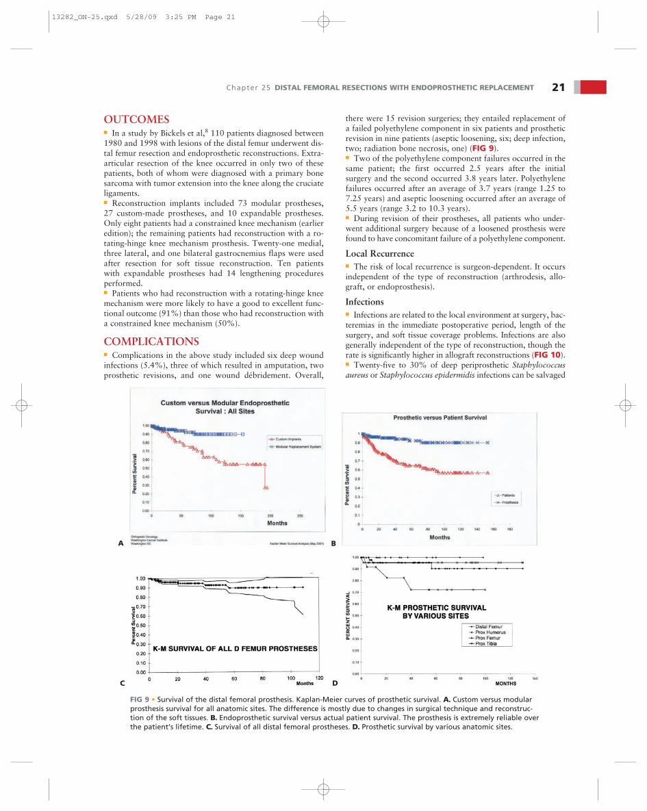
there were 15 revision surgeries; they entailed replacement ofa failed polyethylene component in six patients and prostheticrevision in nine patients (aseptic loosening, six; deep infection,two; radiation bone necrosis, one) (FIG 9).■ Two of the polyethylene component failures occurred in thesame patient; the first occurred 2.5 years after the initialsurgery and the second occurred 3.8 years later. Polyethylenefailures occurred after an average of 3.7 years (range 1.25 to7.25 years) and aseptic loosening occurred after an average of5.5 years (range 3.2 to 10.3 years).■ During revision of their prostheses, all patients who under-went additional surgery because of a loosened prosthesis werefound to have concomitant failure of a polyethylene component.
Local Recurrence■ The risk of local recurrence is surgeon-dependent. It occursindependent of the type of reconstruction (arthrodesis, allo-graft, or endoprosthesis).
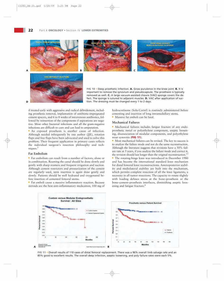
Infections■ Infections are related to the local environment at surgery, bac-teremias in the immediate postoperative period, length of thesurgery, and soft tissue coverage problems. Infections are alsogenerally independent of the type of reconstruction, though therate is significantly higher in allograft reconstructions (FIG 10).■ Twenty-five to 30% of deep periprosthetic Staphylococcusaureus or Staphylococcus epidermidis infections can be salvaged
OUTCOMES■ In a study by Bickels et al,8 110 patients diagnosed between1980 and 1998 with lesions of the distal femur underwent dis-tal femur resection and endoprosthetic reconstructions. Extra-articular resection of the knee occurred in only two of thesepatients, both of whom were diagnosed with a primary bonesarcoma with tumor extension into the knee along the cruciateligaments.■ Reconstruction implants included 73 modular prostheses,27 custom-made prostheses, and 10 expandable prostheses.Only eight patients had a constrained knee mechanism (earlieredition); the remaining patients had reconstruction with a ro-tating-hinge knee mechanism prosthesis. Twenty-one medial,three lateral, and one bilateral gastrocnemius flaps were usedafter resection for soft tissue reconstruction. Ten patientswith expandable prostheses had 14 lengthening proceduresperformed.■ Patients who had reconstruction with a rotating-hinge kneemechanism were more likely to have a good to excellent func-tional outcome (91%) than those who had reconstruction witha constrained knee mechanism (50%).
COMPLICATIONS■ Complications in the above study included six deep woundinfections (5.4%), three of which resulted in amputation, twoprosthetic revisions, and one wound débridement. Overall,
A B
C D
FIG 9 • Survival of the distal femoral prosthesis. Kaplan-Meier curves of prosthetic survival. A. Custom versus modularprosthesis survival for all anatomic sites. The difference is mostly due to changes in surgical technique and reconstruc-tion of the soft tissues. B. Endoprosthetic survival versus actual patient survival. The prosthesis is extremely reliable overthe patient’s lifetime. C. Survival of all distal femoral prostheses. D. Prosthetic survival by various anatomic sites.
13282_ON-25.qxd 5/28/09 3:25 PM Page 21
if treated early with aggressive and radical débridement, includ-ing prosthesis removal, implantation of antibiotic-impregnatedcement spacers, and 6 to 8 weeks of intravenous antibiotics, fol-lowed by reinsertion of the components if aspirations are nega-tive. Most other bacterial infections and all the gram-negativeinfections are difficult to cure and can lead to amputation.■ An exposed prosthesis is another cause of infection.Although needed infrequently by one author (JJE), rotationflaps and free flaps have been advocated and used to solve thisproblem. Their frequent application in primary cases reflectsthe individual surgeon’s resection philosophy and tech-niques.3
Fat Embolism■ Fat embolism can result from a number of factors, alone orin combination. Reaming the canal should be done slowly andgently with sharp reamers and frequent irrigation and suction.Although cement restrictors and pressurization of the cementare regularly used, stem insertion is again done gently andslowly. Patients should be well hydrated and oxygenated be-fore insertion of cemented femoral stems.■ Fat emboli cause a massive inflammatory reaction. Becausesteroids are the best anti-inflammatory medication, 100 mg of
hydrocortisone (Solu-Cortef) is routinely administered beforecementing and insertion of long intramedullary stems.■ Massive fat emboli can be fatal.
Mechanical Failures■ Mechanical failures includes fatigue fracture of any endo-prosthetic metal or polyethylene component, aseptic loosen-ing, disassociation of modular components, and polyethylenewear synovitis (FIG 11).■ Most mechanical failures can be revised. The key to success isto analyze the failure mode and not do the same reconstruction.Although the literature suggests that revisions have a 50% fail-ure rate at 5 years, if you analyze the failure mode and correct it,the revision should last longer than the original reconstruction.13
■ The rotating-hinge knee was introduced in December 1980and has become the international standard knee mechanismfor distal femoral knee reconstructions. Anteroposterior stabil-ity and mediolateral stability are built into the mechanism,which permits complete resection of all the knee ligaments, anecessity in all tumor resections. The capacity to rotate slightlywith loading defuses stress at the bone–prosthesis or thebone–cement–prosthesis interfaces, diminishing aseptic loos-ening and fatigue fracture.6
22 Part 4 ONCOLOGY • Section IV LOWER EXTREMITIES
FIG 10 • Deep prosthetic infection. A. Gross purulence in the knee joint. B. It isimportant to remove the synovium and pseudocapsule. The prosthesis is typicallyremoved as well. C. A large vacuum-assisted closure (VAC) sponge covers the de-fect. The sponge is sutured to adjacent muscles. D. VAC after application of suc-tion. The dressing must be changed every 1 to 2 days.
A B C
D
A B
FIG 11 • Overall results of 110 cases of distal femoral replacement. There was a 96% overall limb salvage rate and an85% good to excellent results. The overall deep infection, aseptic loosening, and poly failure rates were each 5%.
13282_ON-25.qxd 5/28/09 3:25 PM Page 22

■ The development of modular components with forged stemshas greatly reduced the incidence of fatigue fracture, especiallyof the femoral stems, compared with casted stems. This is unlessthere is a mismatch between the patient size and the implant size:an 11-mm stem in a 250-pound patient is a recipe for failure.
Bushing Failures and Pseudomeniscus Formation■ Bushing failures are heralded by the sudden onset of kneejoint pain and a sense of instability to the point that ambu-
latory aids are necessary. Only on rare occasions, whenthere is complete disintegration of the bushing and extensorstop, will the radiographs be positive with medial or lateralprotrusion of the axle. Surgical exploration, therefore, isdone because of a high index of suspicion. This tends to bea late complication: the median time to failure in a seriesof seven cases was 84 months (range 30 to 112 months;FIG 12).
Chapter 25 DISTAL FEMORAL RESECTIONS WITH ENDOPROSTHETIC REPLACEMENT 23
FIG 12 • A. Breakage of medial bushing. B. Close-up of residual bushing. C. Delamination of a bushing and bumper. D. Delaminationof a poly bumper removed 17 years after surgery. E. Clinical photograph showing gross instability to a varus stress test. This instabilityis characteristic of worn or broken bushings. F,G. Patients with pseudomenisci present with localized pain, lack of full extension, andno effusion. H. Gross specimen of a pseudomeniscus. This is formed by thick fibrous collagen without an inflammatory component.Pseudomeniscus rarely occurs before 5 to 7 years after surgery.
A
B
C
D
E
F
G
H
13282_ON-25.qxd 5/28/09 3:25 PM Page 23
Pseudomeniscus and Internal Derangement of the Knee■ The term “pseudomeniscus” refers to scar tissue formationbetween the moving components of the femoral condyles onthe tibial bearing component as well as under this componentand the cemented all-poly (within the tibia). Scar tissue overtime and with constant motion will form a true fibrocartilagetype of scar that takes the shape of a true meniscus.■ Pseudomeniscus formation occurs frequently but is sympto-matic in only a few patients. The symptoms are usually subtle,often presenting as an internal derangement of the knee. Themost suggestive signs are a feeling of instability, combined withslight valgus instability (more than 5% on a stress test), andwith or without a small effusion. There are no real diagnostictests. Suspicion is the key to diagnosis. These symptoms maymimic those of a bushing failure, but with less instability and asmaller effusion.■ The true incidence of symptomatic pseudomeniscus is 5% to7%. The treatment is resection of the pseudomeniscus as wellas the pseudocapsule in the hope of preventing a recurrence.
Stem Fracture■ The incidence of femoral stem fractures has been reducedsignificantly with the introduction of forged stems, but theycan occur, especially if the stem is undersized compared to theweight of the patient. Stem loosening usually precedes cata-strophic fatigue fractures and may present as an actual dis-placed bone fracture.■ If the stem cracks but does not displace, the patient will havepain at the site of fracture, but the radiographs will remain neg-ative until enough motion exists to cause displacement of themetal fracture pieces. Pain is significant and the patient will seekambulatory aids. The older casted stems tended to break about2 cm proximal to the forged junction with the body.
Disassociation of Morse Tapers■ Disassociation of the Morse taper locking mechanism is ex-ceedingly rare and most likely due to failure to impact thecomponents adequately. Surgical exploration and reassemblyof the components and full impaction are required.
Aseptic Loosening■ The incidence of aseptic loosening of the femoral stems hasbeen reduced by the incorporation of extramedullary porousingrowth beads at the junction of the segmental replacementand the stems. Soft tissue ingrowth into these beads in the di-aphysis isolates the joint debris from the bone–cement–pros-thesis composite, creating a “biologic purse-string” effect.11
■ Hydroxyapatite coatings can also enhance fixation.7■ Cross-pin stem fixation requires a custom component but
permits the use of a short stem or a metaphyseal position thatwould normally lead to early aseptic loosening.1■ On rare occasions patients develop a polyethylene debris syn-ovitis. Exploration of the reconstruction, resection of the “pseu-dosynovium” or periprosthetic capsule, and exchange of thebushings and extensor stop can manage this. JE never revisesthe all-poly tibia or poly patellar components if they are wellcemented unless there is an infection. If the cemented tibial polyis removed, then at reconstruction a metal-backed tibial compo-nent is used. Recementing an all-poly tibia component in revi-sion situations risks early aseptic loosening, as the cementationis never as good as the primary reconstruction.
REFERENCES1. Cannon CP, Eckardt JJ, Kabo JM, et al. Cross-pin fixation in 32
tumor endoprosthetic stems. Clin Orthop Relat Res 2003;417:285–292.
2. Eckardt JJ, Kabo JM, Kelly CK, et al. Endoprosthetic reconstruc-tion for bone metastasis. Clin Orthop Relat Res 2003;415(Supp):S254–S262.
3. Eckardt JJ, Lesavoy MA, Dubrow TJ, et al. Exposed endoprosthesis:management protocol using muscle and myocutaneous flaps. ClinOrthop Relat Res 1990;251:220–229.
4. Freedman EL, Hack DJ, Johnson EE, et al. Total knee replacement in-cluding a nodular distal femoral component in elderly patients withacute fractures and nonunion. J Orthop Trauma 1995;9:231–237.
5. Freidman EH, Eckardt JJ. A modular endoprosthetic system for tumorand non-tumor reconstructions: preliminary report. Orthopaedics1996;20:20–27.
6. Kabo JM, Yang RS, Dorey FJ, et al. In vivo rotational stability inthe kinematic rotating hinge knee. Clin Orthop Relat Res 1997;336:166–176.
7. Kay RM, Kabo JM, Seeger LL, et al. Hydroxyapatite-coated distalfemoral replacements: preliminary results. Clin Orthop Relat Res1994;302:92–100.
8. Bickels J, Wittig JC, Kollender Y, et al. Distal femur resection with en-doprosthetic reconstruction: a long-term followup study. Clin OrthopRelat Res 2002;400:225–235.
9. Ward WG, Eckardt JJ. Endoprosthetic reconstruction of the femurfollowing massive bone reconstruction. J South Orthop Assoc 1994;3:108–116.
10. Ward WG, Haight D, Ritchie P, et al. Dislocation of rotating totalknee arthroplasty: a biomechanical analysis. J Bone Joint Surg Am2003;85A:448–453.
11. Ward WG, Johnston KS, Dorey FJ, et al. Extramedullary porouscoating to prevent diaphyseal osteolysis and lines around proximaltibial replacements. J Bone Joint Surg Am 1993;75A:976–987.
12. Ward WG, Johnson KS, Dorey FJ, et al. Loosening of massivefemoral cemented endoprostheses. J Arthroplasty 1997;12:741–750.
13. Wirganowicz PZ, Eckardt JJ, Dorey FJ, et al. Etiology and results oftumor endoprosthesis revision surgery in 64 patients. Clin OrthopRelat Res 1999;358:64–74.
14. Wu CC, Pritsch T, Shehadeh A, et al. The anterior popiteal approachfor popiteal exploration, distal femoral resections, and endopros-thetic reconstruction. J Arthroplasty 2008;23:254–262.
24 Part 4 ONCOLOGY • Section IV LOWER EXTREMITIES
13282_ON-25.qxd 5/28/09 3:25 PM Page 24