2/24/2016 - university of northern iowa2/24/2016 5 (precentral gyrus) corticospinal tract begins in...
TRANSCRIPT

2/24/2016
1
Now it is time to focus on the pathways running down the cord( on both sides of the cord even though the diagram only shows the descending paths on the left)
•Descending Motor Pathways
• Pathways from brain carrying motor commands down to
• “lower motor neurons”
• (review the motor neurons which synapse on muscle fibers at the neuromuscular junction, using acetylcholine as the messenger to trigger contraction)
•“Lower Motor Neurons” (LMNs)
• Neurons whose axons synapse on skeletal muscle fibers (to stimulate contraction)
• aka “alpha motor neurons”
• aka “final common path” to all behavior
• majority are found in ventral horns of cord
• some are in the brainstem (cranial nerve motor neurons serving muscles of head)
•An Example of a LMN
Ventral
horn
LMN
•Motor Units• Motor unit = a single LMN + the extrafusal muscle
fibers it synapses on
• Some motor units are small (axon synapses on just a few fibers); some motor units are large (axon synapses on ~1000 fibers).
• All fibers of a motor unit would be stimulated by neurotransmitter (ACh) release at the same time.
• All movements depend on LMNs.
•Motor Unit

2/24/2016
2
Location
• Myotomes• Muscles innervated by each spinal nerve (not as easy to
diagram as “dermatomes”)
• Examples of myotomes of different levels• Arm extension: C-5
• Elbow extension: C-7
• Small finger abduction: T-1
• Knee extension: L-3
• Ankle flexion: S-1
•Spinal Anatomy and PhysiologySpinal Nerves
•Spinal Reflexes
• Spinal cord is not just a cable between brain and body. Segments of cord also have “local” functions relatively independent of the brain.
• Monosynaptic stretch reflex
• Polysynaptic withdrawal or flexor reflex
• Polysynaptic withdrawal plus crossed extensor reflex
Embedded between the muscle fibers that move us are sensory receptors that monitor or sense muscle tension. AKA “stretch receptor”
•Stretch ReflexMuscle spindle receptor is
stretched and begins to fire when muscle stretches or relaxes too much. This firing activates the LMN that will make that relaxed muscle fiber contract to re-establish muscle tone/tension. Notice reflex arc only involves 2 neurons (sensory neuron synpases on motor neuron) – a “monosynaptic reflex”
http://www.youtube.com/watch?v=TVawlvNU5gI&feature=relatedkneejerk
http://www.youtube.com/watch?v=vSOPtqnJQjk other stretch
Tapping tendon stretches muscle, activating stretch receptors. Reflex automatically maintains muscle tone.
Book 8.10
Knee jerk reflex – 1 example of stretch reflex or “deep tendon” reflex

2/24/2016
3
•The Flexor or Withdrawal Reflex is slightly more complicated because it involves an interneuron
Flexors and extensors in a limb are never active at the same time. It is wired into our nervous system that when one is stimulated the other is inhibited from contracting (so is relaxed).
Flexion
Extension
Withdrawal of a Supporting Limb
If we step on a tack and the withdrawal reflex is triggered in that leg, an even more complicated spinal reflex occurs because your opposite leg must support your weight while the other leg is withdrawing.
The Crossed Extensor Reflex
•Muscle Fibers Depend on Their Innervation by a LMN
• Muscle fibers that lose their LMN show:• no reflexive contraction (so no muscle tone)
• no voluntary contraction
• (This is called “flaccid paralysis”)
• atrophy of the muscle fibers over time
• (These are “symptoms of LMN damage”)
2/24/2016
4
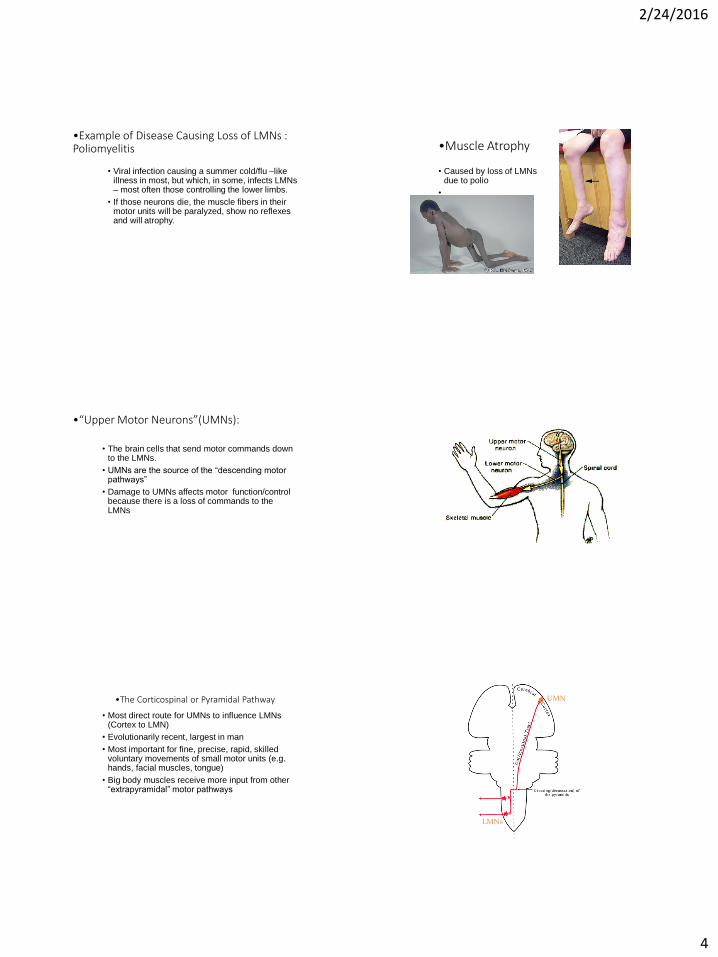
•Example of Disease Causing Loss of LMNs : Poliomyelitis
• Viral infection causing a summer cold/flu –like illness in most, but which, in some, infects LMNs – most often those controlling the lower limbs.
• If those neurons die, the muscle fibers in their motor units will be paralyzed, show no reflexes and will atrophy.
•Muscle Atrophy
• Caused by loss of LMNs due to polio
•
•“Upper Motor Neurons”(UMNs):
• The brain cells that send motor commands down to the LMNs.
• UMNs are the source of the “descending motor pathways”
• Damage to UMNs affects motor function/control because there is a loss of commands to the LMNs
•The Corticospinal or Pyramidal Pathway
• Most direct route for UMNs to influence LMNs (Cortex to LMN)
• Evolutionarily recent, largest in man
• Most important for fine, precise, rapid, skilled voluntary movements of small motor units (e.g. hands, facial muscles, tongue)
• Big body muscles receive more input from other “extrapyramidal” motor pathways
● UMN
LMNs
at spinal cord-medulla junction
To muscles
Notice- no synapses until cortex neurons reach spinal cord
2/24/2016
5
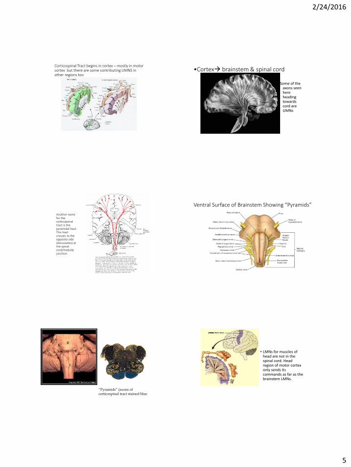
(precentral gyrus)
Corticospinal Tract begins in cortex – mostly in motor cortex but there are some contributing UMNS in other regions too
•Cortex brainstem & spinal cord
Some of the axons seen here heading towards cord are UMNs
Another name for the corticopsinal tract is the pyramidal tract.This tract crosses to the opposite side (decussates) at the spinal cord/medulla junction.
“pyramidal cells”
Ventral Surface of Brainstem Showing “Pyramids”
“Pyramids” (axons of corticospinal tract stained blue
• LMNs for muscles of head are not in the spinal cord. Head region of motor cortex only sends its commands as far as the brainstem LMNs.

2/24/2016
6
P.S. - Besides this direct cortexspinal route, brain motor areas also affect movement via several indirect “extrapyramidal” paths we’ll get to shortly.
Corticospinal or Pyramidal Pathway
Internal capsule
“Pyramids” of medulla
Most of axons in lateral white matter-“lateral corticospinal pathway”
The little bit of this path that goes to LMNS in the brainstem is “corticobulbar”
Head
Region“”
•Upper Motor Neurons Modulate Muscle Tone Via Gamma Motor Neurons
• “Gamma motor neurons” (the “other” motor neurons) do not go to the extrafusal fibers that move us.
• They synapse on “intrafusal muscle fibers” inside stretch receptors/muscle spindle receptors.
• Tensing these little fibers makes stretch receptors more sensitive.
• UMN also can inhibit stretch reflex to allow movement, so we are not stiff statues.
Gamma
Synapses inside stretch receptor
•UMN vs LMN
•Symptoms of UMN Damage
• Reflexes and muscle tone still present and, in fact, intensified “hyperreflexia”
• Voluntary control impaired
• “Spastic paralysis” (excess muscle tone & loss of voluntary control of movement)
• No denervation induced atrophy (LMNs are okay)
•Reflex Changes After UMN Damage
• hyperactive stretch reflex, particularly in anti-gravity muscles
• too much muscle tone (hypertonia or spasticity)
• clonus (rapid repetitive response to stretch)
• altered Babinski reflex after corticospinal damage

2/24/2016
7
•Normal Adult vs. Positive Babinski
https://www.youtube.com/watch?v=9nNb32VWA7Q
•Clonus
• Without descending modulation from brain, stretch reflex leads to repetitive contractions
http://www.youtube.com/watch?v=kZweyLV9SkA
•Other Descending Tracts: “Extrapyramidal Motor Pathways”• Rubrospinal pathway to regulate tone of flexors in
limbs for locomotion & to organize repetitive movements that involve the flexors (e.g. walking, running, crawling)
• Vestibulospinal pathway to stimulate extensors (antigravity) for standing, posture
• Tectospinal pathway for reflexive motor reactions to visual stimuli
• Reticulospinal pathway to regulate muscle tone by modulating the stretch reflex
• (don’t need to memorize exact route of these)
“Rubro” means from the red nucleus
Vestibulospinal and Tectospinal Pathways

2/24/2016
8
•Amyotrophic Lateral Sclerosis (ALS) or Lou Gehrig’s Disease
• Fatal progessive loss of LMNs as well as corticospinal pathway (UMNs). Several genes involved.
• Onset most often in late 50’s-early 60’s; more men affected
• 70% will die within 5 years (eventually cannot swallow, breathe)
•ALS – Symptoms• First symptoms usually muscle cramping &
twitching (fasciculations), with feelings of fatigue & weakness in a limb
• Loss of LMNs causes weakness, paralysis, loss of reflexes & atrophy in affected muscles. Loss of UMNs causes spasticity (muscle stiffness, cramping from too much tonus where LMNs have not been lost).
• Combination of UMN + LMN symptoms at multiple levels is fairly diagnostic
*Listen to ALS link in syllabus
•LMN Damage vs UMN Damage
either reflexively or voluntarily
Because of loss of stretch reflex