22. mutagenesis studies of rhodopsin phototransduction | isbn
TRANSCRIPT
Structures and Functions ofRetinal Proteins d. JL Rigaud. Colloque ENSERM /John Libbey Euroicxt Lid.© 1992, VoL 221, pp. 67-70
Mutagenesis studies of rhodopsinphototransduction
Thomas P. Sakmar, Karim Fahmy, Theresa Chan and Melissa Lee
Howard Hughes Medical Institute, Rockefeller University, WY 10021, USA
We employ the vertebrate visual proteins rhodopsin and transducin as a model system forstructure-function studies on the molecular mechanisms of transmembrane signaling. These visualproteins are members of a super-family of related guanine nucleotide-binding regulatory proteins (Gproteins) and 0 protein-coupled receptors. We are particularly interested in the structures of the retinalbinding pockets of visual pigments and in identifying the structural domains of both rhodopsin andtransducin involved in the G protein activation process.
AMINO ACID SUBSTITUIONS THAT CAUSE BATHOCHROMIC SPECTRAL SHIFTS INBOVINE RHODOPSIN
Nearly all vertebrate visual pigments share a common chromophore, 1 1-cis-retinal. In humans, thedifferences in absorption maxima of the cone pigments that underlie human red-green color vision mustresult from differences in the amino acid sequences of the respective opsin proteins. Fifteen amino acidsubstitutions distinguish the human green pigment (inax = 530 nm) from the human red pigment= 560 nm) (Nathans et a!., 1986). Three of these residues were suggested in a genetic analysisof eight primate visual pigments to produce this spectral difference of about 1000 cm-1 (Neitz et al.,1991). The amino acid at each of these three positions in the rod pigment rhodopsin = 500 nm),matches that of the green pigment (Table 1) (Nathans & Hogness, 1983; Nathans et a!., 1986).Therefore, it was postulated that the influence of these residues could be tested experimentally bysubstituting the amino acid residues of the red pigment into rhodopsin. A mutation resulting in a redshift in absorption maximum relative to rhodospin would indicate potential relevance in red-greenspectral tuning.
We recently reported seven bovine rhodopsin mutants involving the three amino acid positions: threesingle substitutions, Ala 164 replaced by Ser (A164S), Phe 261 replaced by Tyr (F261Y), and Ala 269replaced by Thr (A269T); three double substitutions; and one triple substitution (Chan et a!., 1992).Replacement ofAla 164 caused only a slight red shift effect ( Xmax = 502 nm). However, replacementof Phe 261 or Ala 269 caused red-shifted Xmax values of 510 nm and 514 rim, respectively. Thedouble replacement at both positions 261 and 269 simultaneously caused a red shift to 520 nm that wasgreater than either of the two substitutions alone but not strictly additive. Replacement at both positions164 and 261 caused a red shift (max = 512 nm) that was slightly greater than that of F261Y alone.Replacement at both positions 164 and 269 resulted in a Xmax value of 514 nm that was the same asthat of the single substitution at position 269. Two ofthe three positions (261 and 269) in combinationappear to account for the 775 cm' of the observed 1000 cm' difference between the human green andred pigments (Table 2). The triple mutant did not bind 1 1-cis-retinal to form a pigment. It is notknown whether the triple mutant, if itcould be induced to bind 11-cis -retinal, would display the full
67

1000 cm' red shift. However, the effects of all combinations of double replacements were
qualitatively additive but not synergistic. For the triple mutant to account for the entire 1000 cart shift,a synergistic effect would be required.
Table 1 Comparison of amino acids in various pigments at positions proposed to account for red-greenspectral tuning*
bovine human human humanrhodopsin rhodopsin green red
Ala 164 Ala 164 Ala 180k Ser 180Phe 261 Phe 261 Phe277 Tyr277Ala 269 Ala 269 Ala 285 Thr 285
*The numbering system shown is from previous reports of the deduced amino acid sequences ofbovine rhodopsin (Nathans & Hogness, 1983), human rhodopsin (Nathans & Hogness, 1984), andhuman cone pigments (Nathans et al., 1986).+a genetic polymorphism was reported at this position that could potentially result in Ser at this positionas well (Nathans et al., 1986).
The most likely explanation for the observed red-shifted Xmax values is that a newly introducedhydroxyl-bearing amino acid residue can interact directly with the chromophore. However, it ispossible that an individual amino acid replacement causes distant effects on the chromophore bindingpocket. The effect of a mutation on absorption maximum may result from an indirect effect as well as adirect interaction. However, whereas blue-shifted Xmax values indicate a relative loss ofchromophore-protein interactions, red-shifted Xmax values indicate an enhanced interaction. A mutantwith a red-shifted Xmax value has a larger opsin shift than that normally observed in rhodopsin.Attributing an effect on absorption maximum to a specific amino acid-chromophore interaction is likelyto be more valid in cases where a red shift rather than a blue shift is observed. A large number ofrhodopsin mutants have been previously reported that cause blue-shifted absorption maxima(Nakayama & Khorana, 1990; Nathans, 1990a). No significant red-shifted mutants have been reportedother than those involving the Schiff base counterion at position Glu 113 (Nathans, 1990b; Sakmar eta!., 1989; Sakmareta!., 1991, Zhukovsky&Oprian, 1989)..
Although residues in rhodopsin match those in the green pigment at the three positions tested, therhodopsin and the green pigment are only about 70% homologous (Nathans et al., 1986). Obviouslythe retinal binding pocket in rhodopsin is significantly different from that of the green pigment asdemonstrated by the 1,125 cm' difference between their spectral peaks. However, at the threepositions proposed from primary structure comparisions to account for red-green pigment spectraltuning, rhodopsin and the green pigment share the same residues. In addition, the design of thisexperiment involves testing a hypothesis by correlating mutagenesis with the appearance of ared-shifted absorption maximum (increase in opsin shift), and not with the loss of an existingretinal-protein interaction as indicated by a blue-shifted absorption maximum (decrease in opsin shift).
Neitz et at. (1991) hypothesized that additive effects of changes at amino acid position 180, 277, and285 should account for all shifts in spectra among a set of primate visual pigments (see Table 1 for acomparison of numbering systems in rhodopsin versus cone pigments) (Neitz et a14, 1991). Theyargued that the effects of changes at positions 180 and 285 were shifts of about 5 and 15.5 nmrespectively and that the remaining 9- to 10-nm difference was produced by the substitution at position277. We conclude that two of these residues (Tyr 277 and Thr 285) are primarily involved in spectraltuning that distinguishes red from green pigments, but that the effects of individual differences may notbe strictly additive. For example, single substitutions in rhodopsin at position 261 (F261Y) andpostion 269 (A269T) result in red shifts of 400 cm-1 and 550 cm-1, respectively. However, incombination these two replacements cause a red shift of 775 cm'. Also, replacement at position 164(A164S) results in a slight red shift (75 cm-1). This effect was additive in combination with F261 Y but
68
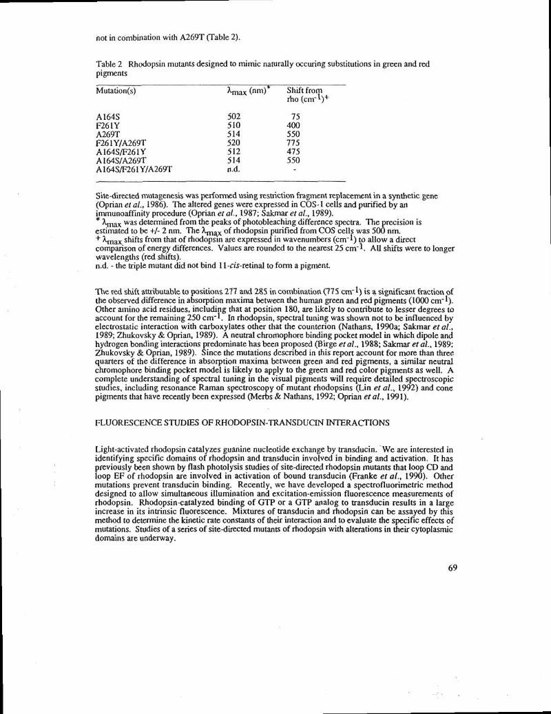
not in combination with A269T (Table 2).
Table 2 Rhodopsin mutants designed to mimic naturally occuring substitutions in green and redpigments
Mutation(s) max (nm)* Shift fromrho (cm- 1)
A 164S 502 75F261Y 510 400A269T 514 550F261Y/A269T 520 775A164S/F261Y 512 475A164S/A269T 514 550A 164SfF26 1Y/A269T n.d. -
Site-directed mutagenesis was performed using restriction fragment replacement in a synthetic gene(Oprian et at., 1986). The altered genes were expressed in COS-1 cells and purified by animmunoaffinity procedure (Oprian era!., 1987; Sakmar et al., 1989).*Xmax was determined from the peaks of photobleaching difference spectra. The precision is
estimated to be +1- 2 nm. The Xmax of rhodopsin purified from COS cells was 500 nm.+ Xlnax shifts from that of rhodopsin are expressed in wavenuinbers (cm-1) to allow a directcomparison ofenergy differences. Values are rounded to the nearest 25 cm-1. All shifts were to longerwavelengths (red shifts).n.d. - the triple mutant did not bind 1 1-cis-retinal to form a pigment
The red shift attributable to positions 277 and 285 in combination (775 cm') is a significant fraction ofthe observed difference in absorption maxima between the human green and red pigments (1000 cm-1).Other amino acid residues, including that at position 180, are likely to contribute to lesser degrees toaccount for the remaining 250 cm-t. In rhodopsin, spectral tuning was shown not to be influenced byelectrostatic interaction with carboxylates other that the counterion (Nathans, 1990a; Sakmar et al.,1989; Zhukovsky & Oprian, 1989). A neutral chromophore binding pocket model in which dipole andhydrogen bonding interactions predominate has been proposed (B irge et al., 1988; Sakmar et at., 1989;Zhukovsky & Oprian, 1989). Since the mutations described in this report account for more than threequarters of the difference in absorption maxima between green and red pigments, a similar neutralchromophore binding pocket model is likely to apply to the green and red color pigments as well. Acomplete understanding of spectral tuning in the visual pigments will require detailed spectroscopicstudies, including resonance Raman spectroscopy of mutant rhodopsins (Liii et al., 1992) and conepigments that have recently been expressed (Merbs & Nathans, 1992; Oprian et al., 1991).
FLUORESCENCE STUDIES OF RHODOPSIN-TRANSDUCfN INTERACTIONS
Light-activated rhodopsin catalyzes guanine nucleotide exchange by transducun. We are interested inidentifying specific domains of rhodopsin and transducin involved in binding and activation. It haspreviously been shown by flash photolysis studies of site-directed rhodopsun mutants that loop CD andloop EF of rhodopsin are involved in activation of bound transducun (Franke et a!., 1990). Othermutations prevent transducin binding. Recently, we have developed a spectrofluorimetric methoddesigned to allow simultaneous illumination and excitation-emission fluorescence measurements ofrhodopsin. Rhodopsin-catalyzed binding of GTP or a GTP analog to transducin results in a largeincrease in its intrinsic fluorescence. Mixtures of transducin and rhodopsin can be assayed by thismethod to determine the kinetic rate constants of their interaction and to evaluate the specific effects ofmutations. Studies ofa series of site-directed mutants of rhodopsin with alterations in their cytoplasmicdomains are underway.
69
REFERENCES
Birge, R.R., Einterz, C.M., Knapp, H.M., and Murray, LP. (1988): The nature of the primaryphotochemical events in rhodopsin and isorhodopsin. Biophys. J. 53, 367-385.
Chan, T., Lee, M., and Sakmar, T.P. (1992): Introduction of hydroxyl-bearing amino acids causesbathochromic spectral shifts in rhodopsin: Amino acid substitutions responsible for red-greencolor pigment spectral tuning. J. Biol. Chem. 267, 9478-9480.
Ferretti, L., Karnik, S.S., Khorana, H.G., Nassal, M. and Oprian, D.D. (1986): Total synthesis of a
gene for bovine rhodopsin. Proc. Nati. Acad. Sci. U.S.A. 83, 599-603.Franke, R.R., König, B., Sakmar, T.P., Khorana, [1G., and Hofmann, K.P. (1990): Rhodopsin
mutants that bind but fail to activate transducin. Science 250, 123-125.Karnik, S.S., Sakmar, T.P., Chen, H.-B., and Khorana, H.G. (1988): Cysteine residues 110 and 187
are essential for the formation ofcorrect structure in bovine rhodopsin. Proc. Nat!.Acad. Sci.U.S.A. 85, 8459-8463.
Lin, S.W., Sakmar, T.P., Franke, R.R., Khorana, H.G., and Mathies, R.A. (1992): ResonanceRanian microprobe spectroscopy of rhodopsin mutants: Effect of substitutions in the thirdtransmembrane helix. Biochemistry 31, 5105-5111.
Merbs, S.L., and Nathans, 1. (1992): Absorption spectra of human cone pigments. Nature 356,433-435.
Nakayama, T.A., and Khorana, H. G. (1990): Mapping of the amino acids in membrane-embeddedhelices that interact with the retinal chromophore in bovine rhodopsin. J. Bioi.Chem. 266,4269-4275.
Nathans, I., Thomas, D., and Hogness, D.S. (1986): Molecular genetics of human color vision: thegenes encoding blue, green, and red pigments. Science 232, 193-202.
Nathans, J., and [-logness, D. S. (1984): Isolation and nucleotide sequence of the gene encodinghuman rhodopsin. Proc. Nat!. Acad. Sci. U.S.A. 81, 4851-4855.
Nathans J., and Hogness, D. S. (1983): Isolation, sequence analysis, and intron-exon arrangement ofthe gene encoding bovine rhodopsin. Cell 34, 807-814.
Nathans, J. (1990): Determinants ofvisual pigment absorbance: role ofcharged amino acids in theputative transmembrane segments. Biochemistry 29, 937-942.
Nathans, J. (1990): Determinants of visual pigment absorbance: Identification of the retinylideneSchiffs base counterion in bovine rhodopsin. Biochemistry 29, 9746-9752.
Neitz, M., Neitz, J., and Jacobs, G.H. (1991): Spectral tuning ofpigments underlying red-green colorvision. Science 252, 971-973.
Oprian, D.D., Molday, R.S., Kaufman, R.J., and Khorana, H.G. (1987): Expression of a syntheticbovine rhodopsin gene in monkey kidney cells. Proc. Nat!. Acad. Sci. U.S.A. 84, 8874-8878.
Oprian, D.D., Asenjo, A.B., Lee, N., and Pelletier S.L. (1991): Design, chemical synthesis, and
expression of genes for the three human color vision pigments. Biochemistry 30, 11367-11372.Sakmar, T.P., Franke, R.R., and Khorana, H.G. (1989): Glutamic acid-113 serves as the retinylidene
Schiff base counterion in bovine rhodopsin. Proc. Nat!. Acad. Sci. U.S.A. 86, 8309-8313.Sakmar, T.P., Franke, R.R., and Khorana, H. 0. (1991): The role of the retinylidene Schiff base
counterion in rhodopsin in determining wavelength absorbance and Schiff base pKa. Proc. Natl.Acad. Sd. USA. 88, 3079-3083.
Zhukovsky, E.A., and Oprian, D.D. (1989): Effect of carboxylic acid side chains on the absorptionmaximum of visual pigments. Science 246, 928-930.
70