21c- schwartzmarboe vasculitis 3 - columbia university · 3 case presentation • findings were...
TRANSCRIPT

1
VASCULITIS
Case Presentation
• The patient is a 24 year old woman who presentedThe patient is a 24 year old woman who presented to the emergency room with left-sided weakness. She was confused and complained of a severe headache. She was noted to have asymmetric blood pressures. The left-sided weakness resolved over the next several hours.
• Blood studies of note included a positive ANA (1:160 with a speckled pattern) and a CRP of 23.4 (normal <3).

2
Case PresentationEvaluation included a transesophageal echo which showed va uat o c uded a t a sesop agea ec o w c s owed
diffuse thickening of the aorta and a CT and MRI which showed multiple abnormalities including:
• 1. thickened regions of the wall of the thoracic aorta which showed significant enhancement, suggestive of active inflammation
• 2. encasement and narrowing of the right pulmonary artery hi h l h dwhich also enhanced
• 3. marked narrowing of the proximal left carotid artery and the left subclavian artery and diffuse narrowing of the right subclavian and proximal portion of the right carotid.
Ultrasound B-mode and color-duplex flow imaging of the left common carotid artery (longitudinal section): homogeneous, midechoic, circumferential wall thickening
("macaroni sign") with luminal stenosis
Copyright ©2006 American Heart Association
Meini, S. et al. Circulation 2006;114:e544

3
Case Presentation
• Findings were felt to be consistent with• Findings were felt to be consistent with Takayasu’s arteritis, an inflammatory granulomatous disease of the medium and large vessels that is prevalent in young women.
Vasculitis
• Vacsulitis is an inflammation of the vessel• Vacsulitis is an inflammation of the vessel wall.
• Inflammation results from immune complex deposition or from cell-mediated immune reactions directed against the vessel wall.
• It can involved small, medium and large blood vessels.

4
Vasculitis: A classification by size and type of involved vessel.
Adapted from Jennette and Falk: Small-vessel vasculitis, NEJM 337: 1512, 1997.
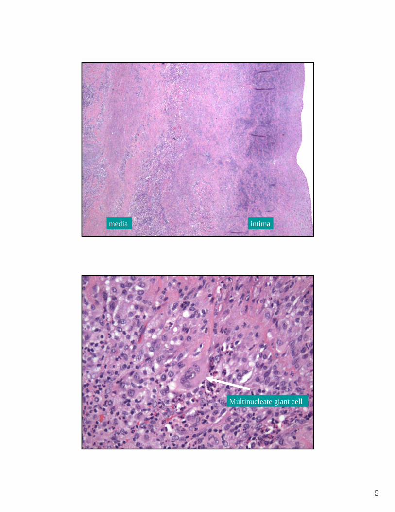
Human AortaIntima
Media
Adventitia
5
intimamedia
Multinucleate giant cell

6
Multinucleate giant cells
7
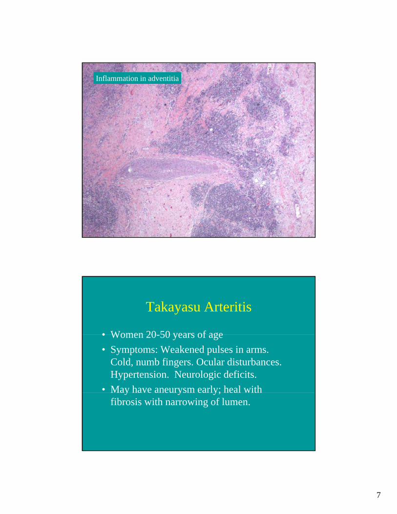
Inflammation in adventitia
Takayasu Arteritis
• Women 20 50 years of age• Women 20-50 years of age• Symptoms: Weakened pulses in arms.
Cold, numb fingers. Ocular disturbances. Hypertension. Neurologic deficits.
• May have aneurysm early; heal with y y y;fibrosis with narrowing of lumen.
8
Aortitis: Takayasu v. Giant Cell
• Both show female predilection• Takayasu patients 20 – 50 years old• Giant cell patients > age 70• Giant cell has mild intimal scarring• Takayasu has more adventitial
inflammation, scarring and endarteritis obliterans.
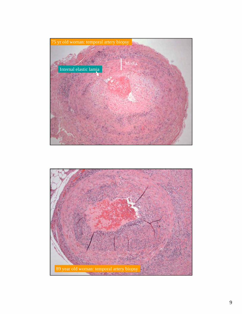
Giant Cell Arteritis
• Most common of the vasculitides• Large, medium and small arteries involved• Primarily temporal, vertebral, ophthalmic• Most in persons > 50 years of age• Fever, fatigue, weight loss, facial pain, headache.• Diagnose with temporal artery biopsy: Focal• Diagnose with temporal artery biopsy: Focal
thickening with granulomatous inflammation focused on the internal elastic lamina.
• Responds to anti-inflammatory therapy
9
75 yr old woman: temporal artery biopsy
MediaInternal elastic lamia
89 year old woman: temporal artery biopsy

10
75 year old woman with
giant cell arteritis in
temporal artery
Vasculitis: A classification by size and type of involved vessel.
Adapted from Jennette and Falk: Small-vessel vasculitis, NEJM 337: 1512, 1997.
11
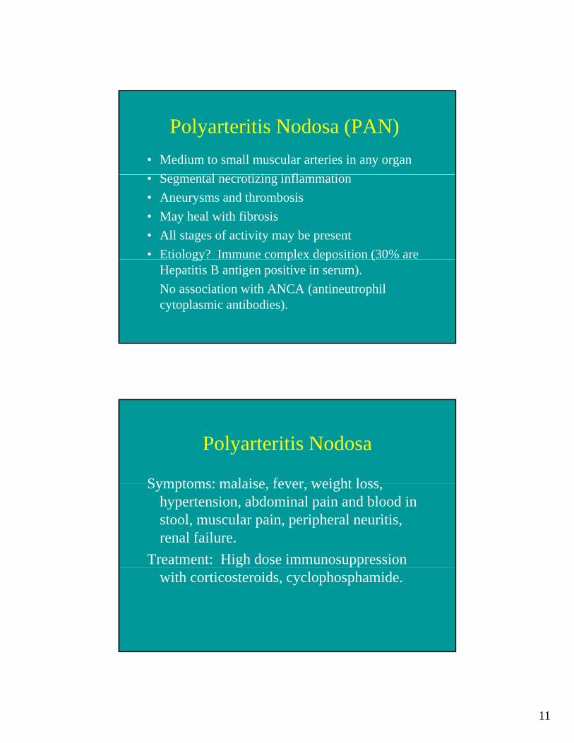
Polyarteritis Nodosa (PAN)• Medium to small muscular arteries in any organ
S l i i i fl i• Segmental necrotizing inflammation• Aneurysms and thrombosis• May heal with fibrosis• All stages of activity may be present• Etiology? Immune complex deposition (30% are gy p p (
Hepatitis B antigen positive in serum). No association with ANCA (antineutrophil cytoplasmic antibodies).
Polyarteritis Nodosa
Symptoms: malaise fever weight lossSymptoms: malaise, fever, weight loss, hypertension, abdominal pain and blood in stool, muscular pain, peripheral neuritis, renal failure.
Treatment: High dose immunosuppression with corticosteroids, cyclophosphamide.
12
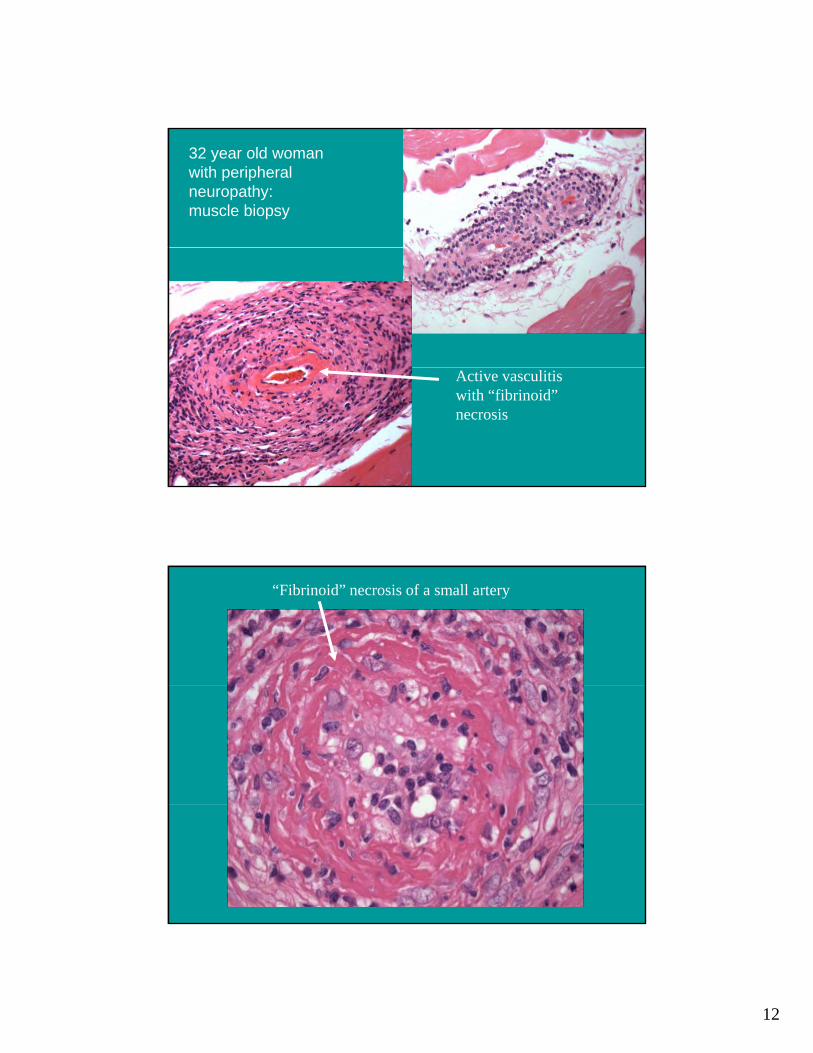
32 year old woman with peripheral neuropathy: muscle biopsy
Active vasculitis with “fibrinoid” necrosis
“Fibrinoid” necrosis of a small artery
13
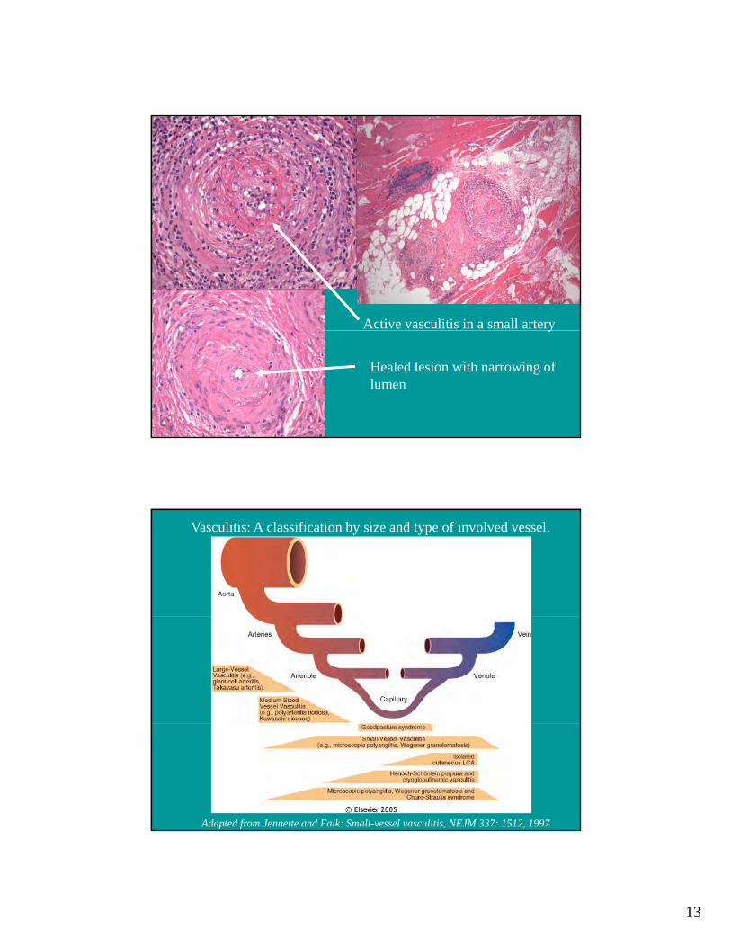
Active vasculitis in a small arteryy
Healed lesion with narrowing of lumen
Vasculitis: A classification by size and type of involved vessel.
Adapted from Jennette and Falk: Small-vessel vasculitis, NEJM 337: 1512, 1997.
14
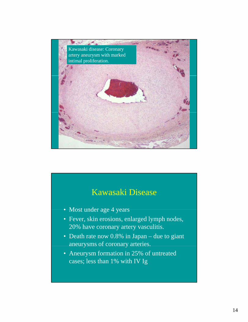
Kawasaki disease: Coronary artery aneurysm with marked intimal proliferation.
Kawasaki Disease
• Most under age 4 years• Most under age 4 years• Fever, skin erosions, enlarged lymph nodes,
20% have coronary artery vasculitis.• Death rate now 0.8% in Japan – due to giant
aneurysms of coronary arteries.y y• Aneurysm formation in 25% of untreated
cases; less than 1% with IV Ig

15
Vasculitis: A classification by size and type of involved vessel.
Adapted from Jennette and Falk: Small-vessel vasculitis, NEJM 337: 1512, 1997.
Microscopic Polyangiitis
• Necrotizing vasculitis of small vessels• Necrotizing vasculitis of small vessels (smaller than involved in PAN)
• Symptoms: skin nodules, hemoptysis, abdominal pain, hematuria, proteinuria.
• Glomerulonephritis in 90%.• Often an immunologic reaction to drug
(penicillin), microorganisms, administered proteins, or tumor antigens.

16
Wegener Granulomatosis
1 Acute necrotizing granulomas of ear1. Acute necrotizing granulomas of ear, nose, throat, or lung.
2. Necrotizing vasculitis of small to medium sized vessels
3. Renal disease - focal necrotizing gglomerulonephritis
Antineutrophil Cytoplasmic Antibodies (ANCA)
• Autoantibodies directed against enzymes in• Autoantibodies directed against enzymes in granules in neutrophils, lysosomes of monocytes, and in endothelial cells.
• Cytoplasmic (cANCA): proteinase-3• Perinuclear (pANCA): myeloperoxidase(p C ) y p• Wegener’s granulomatosis – cANCA• Microscopic polyangiitis - pANCA

17
Lung biopsy: granulomatous inflammation
Lung: small vessel vasculitis

18
Kidney: Glomerulonephritis
Kidney: granulomatous inflammation

19
Wegener’s Granulomatosis:• Etiology: ? Hypersensitivity to
undetermined antigens.g• Prognosis: Untreated – 90% mortality in 2
years• 85-90% of patients respond to
cyclophosphamide and prednisonebut 50% have relapses