hybrid aproach of takayasu’s aortic aneurysm · hybrid aproach of takayasu’s aortic aneurysm...
TRANSCRIPT
HYBRID APROACH OF TAKAYASU’S AORTIC
ANEURYSM Guilherme V Meirelles
Luiz Marcelo A Viarengo Aline M Martins
Meirelles Vascular Institute - Campinas –SP- Brazil
GUILHERME V MEIRELLES

Disclosure
Speaker name:
Guilherme V Meirelles
I have the following potential conflicts of interest to report:
Consulting
Employment in industry
Stockholder of a healthcare company
Owner of a healthcare company
Other(s)
I do not have any potential conflict of interest X

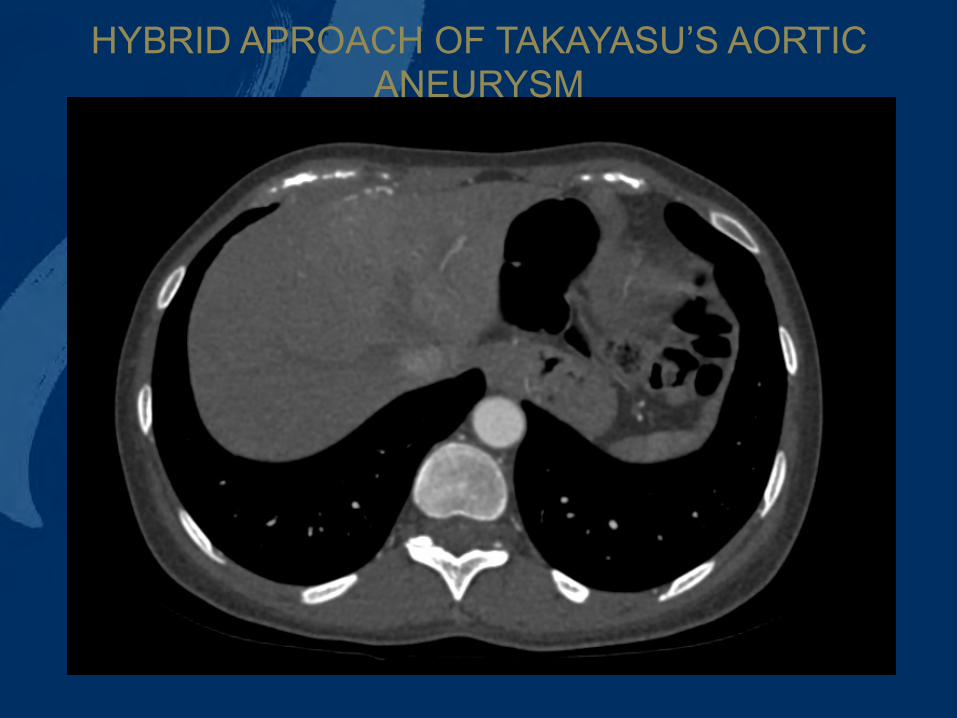
• 36-year-old female • Symptomatic arterial mesenteric angina • Weight lost of 10Kg in 2 month • Desnutrition • Angio-CT
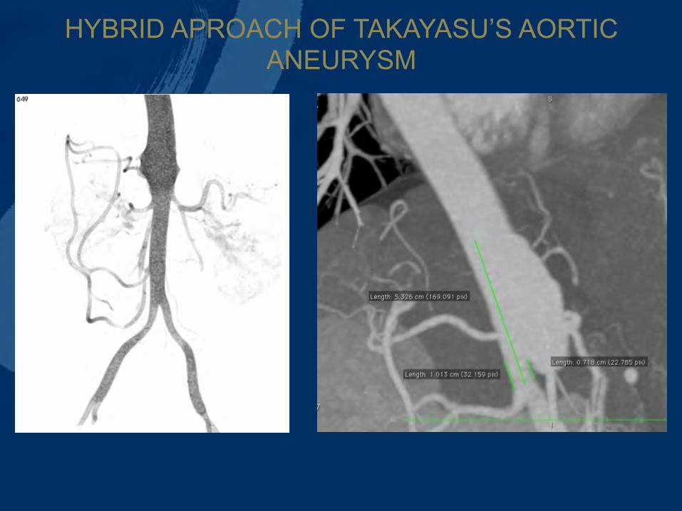
• Complete occlusion of superior mesenteric artery, • Subocclusion of celiac trunk • Saccular aneurysm 3,5X 60mm with 1 cm neck above
the renal arteries .
• Suprarenal aorta diameter was 2.0 cm in the proximal part and 1.3 cm in distal segment, infrarenal was 7,8 mm
HYBRID APROACH OF TAKAYASU’S AORTIC ANEURYSM
HYBRID APROACH OF TAKAYASU’S AORTIC ANEURYSM
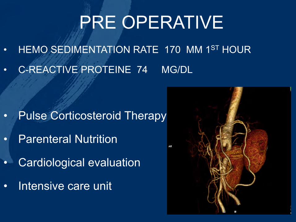
• HEMO SEDIMENTATION RATE 170 MM 1ST HOUR
• C-REACTIVE PROTEINE 74 MG/DL
• Pulse Corticosteroid Therapy
• Parenteral Nutrition
• Cardiological evaluation
• Intensive care unit
PRE OPERATIVE
HYBRID APROACH OF TAKAYASU’S AORTIC ANEURYSM

HYBRID APROACH OF TAKAYASU’S AORTIC ANEURYSM
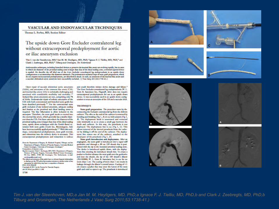
Tim J. van der Steenhoven, MD,a Jan M. M. Heyligers, MD, PhD,a Ignace F. J. Tielliu, MD, PhD,b and Clark J. Zeebregts, MD, PhD,b Tilburg and Groningen, The Netherlands J Vasc Surg 2011;53:1738-41.)
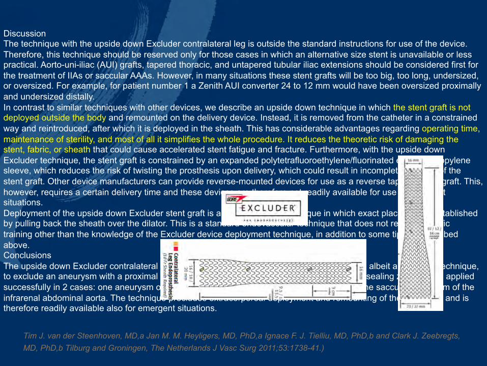
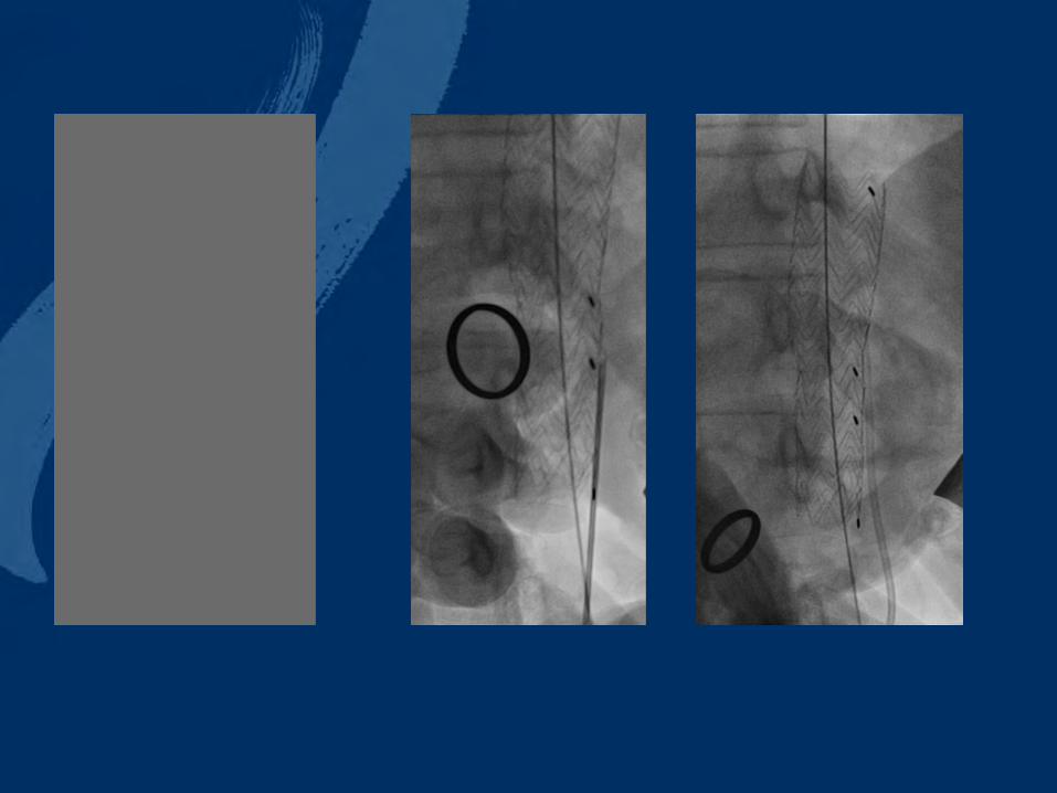
Discussion The technique with the upside down Excluder contralateral leg is outside the standard instructions for use of the device. Therefore, this technique should be reserved only for those cases in which an alternative size stent is unavailable or less practical. Aorto-uni-iliac (AUI) grafts, tapered thoracic, and untapered tubular iliac extensions should be considered first for the treatment of IIAs or saccular AAAs. However, in many situations these stent grafts will be too big, too long, undersized, or oversized. For example, for patient number 1 a Zenith AUI converter 24 to 12 mm would have been oversized proximally and undersized distally. In contrast to similar techniques with other devices, we describe an upside down technique in which the stent graft is not deployed outside the body and remounted on the delivery device. Instead, it is removed from the catheter in a constrained way and reintroduced, after which it is deployed in the sheath. This has considerable advantages regarding operating time, maintenance of sterility, and most of all it simplifies the whole procedure. It reduces the theoretic risk of damaging the stent, fabric, or sheath that could cause accelerated stent fatigue and fracture. Furthermore, with the upside down Excluder technique, the stent graft is constrained by an expanded polytetrafluoroethylene/fluorinated ethylene propylene sleeve, which reduces the risk of twisting the prosthesis upon delivery, which could result in incomplete opening of the stent graft. Other device manufacturers can provide reverse-mounted devices for use as a reverse tapered stent graft. This, however, requires a certain delivery time and these devices are therefore not readily available for use in emergent situations. Deployment of the upside down Excluder stent graft is a very controlled technique in which exact placement is established by pulling back the sheath over the dilator. This is a standard endovascular technique that does not require specific training other than the knowledge of the Excluder device deployment technique, in addition to some tips as described above. Conclusions The upside down Excluder contralateral leg technique offers an easy endovascular approach, albeit an off-label technique, to exclude an aneurysm with a proximal sealing zone that is larger in diameter than the distal sealing zone. It was applied successfully in 2 cases: one aneurysm of the internal iliac artery after coil embolization and one saccular aneurysm of the infrarenal abdominal aorta. The technique precludes extracorporeal deployment and remounting of the stent graft and is therefore readily available also for emergent situations.
Tim J. van der Steenhoven, MD,a Jan M. M. Heyligers, MD, PhD,a Ignace F. J. Tielliu, MD, PhD,b and Clark J. Zeebregts, MD, PhD,b Tilburg and Groningen, The Netherlands J Vasc Surg 2011;53:1738-41.)
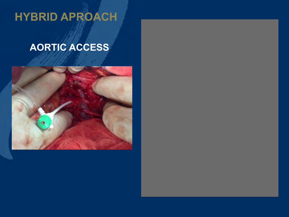
AORTIC ACCESS
HYBRID APROACH

DRYSEAL 18 FR
BELL BOTTON 23X100X16 MM

CUT THE STRING

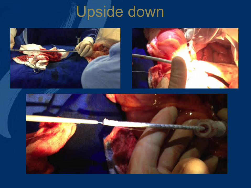
Upside down
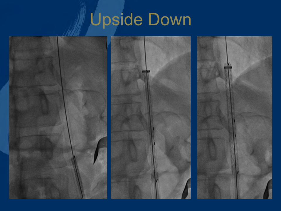
Upside Down
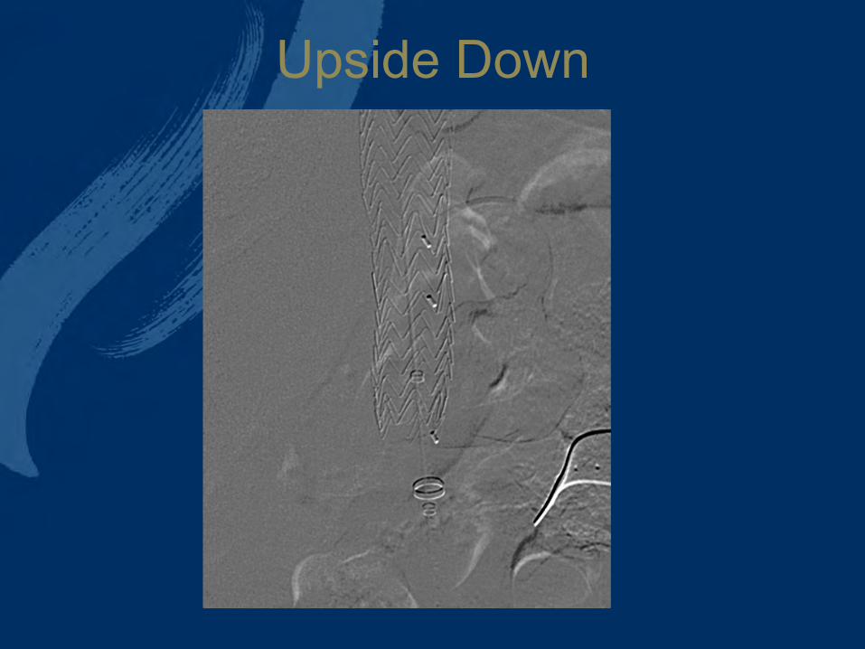
Upside Down

Aortic – Mesenteric Bypass Graft

Aortic – Mesenteric Bypass Graft


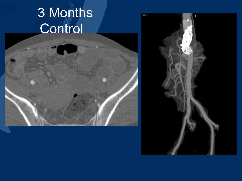
3 Months Control

EMBOLIZATION
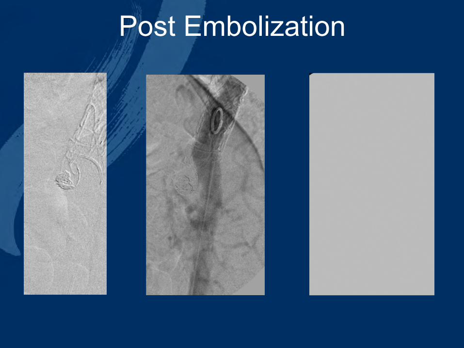
Post Embolization
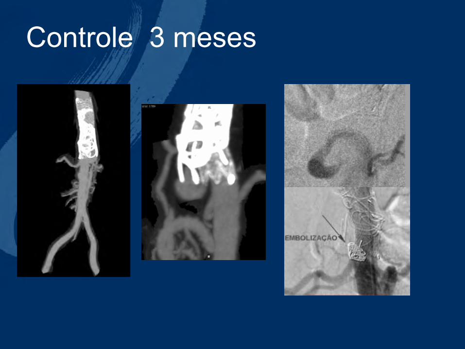
Controle 3 meses

Controle após embolização
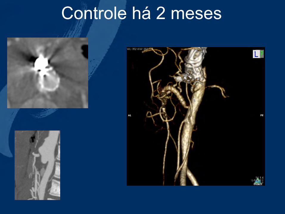
Controle há 2 meses

PREGNANT
BIS BALD

HYBRID APROACH OF TAKAYASU’S AORTIC
ANEURYSM Guilherme V Meirelles
Luiz Marcelo A Viarengo Aline M Martins
Meirelles Vascular Institute - Campinas –SP- Brazil
GUILHERME V MEIRELLES