2019 hepatology update r. warren sands md, phd
TRANSCRIPT

2019 Hepatology Update
R. Warren Sands MD, PhD
• 68 yo F with PMH of GERD and MS referred from OSH to
hematology at UPMC for severe iron deficiency anemia
• Denied overt bleeding
• Prior endoscopy notable for (GI?) AVMs
• Recent EGD and colonoscopy at OSH were normal
• Refused capsule endoscopy at OSH
Clinical Vignette
2

PMH:
Iron deficiency anemia
Breast carcinoma
GERD
Hypertension
Multiple sclerosis
PSH:
Tubal Ligation
CCY
Medical Histories
3
Medications:
None
FHx:
M: Bleeding disorder, DM
F: Bled to death
7 siblings without bleeding
diseases, brother with DM, no
other known illnesses
2 children healthy
No GI, liver, or pancreatic
diseases
SHx:
Waitress, married
No smoking,
EtOH, or illicits
• Vitals and PE unremarkable
• Labs: WBC 6.5, Hgb 9.1, MCV 75.2, RDW 31.8, plt 345;
CMP unremarkable, iron 26, % iron sat 6, ferritin 19
• Started on IV iron with monitoring of her labs
PE and Laboratory Studies
4

• ~1.5 years later she developed abdominal pain
• OSH CT chest/abd/pelvis with contrast: thyroid nodules and
a 6 mm left lower lobe nodular density
• ENT: cauterization for epistaxis
• 6 months later: bleeding in her nose, throat, and teeth
• Heme PE note: “Broken blood vessels over the tips of her
fingers” and “small red lesions” were noted on her tongue
Clinical Course
5
• Next two years: IV iron, aminocaproic acid not tolerated
• SOB
• OSH EGD: gastric and duodenal AVMs
• 2 months later: LE swelling
• Admitted later that month for LE and abdominal swelling
• CMP notable for AP 145, PT/PTT/INR 15.7/1.3/31.1, BNP
772
• EKG atrial flutter, TTE EF 55-60%, flattened septum, RVH,
severe tricuspid regurgitation, small rt to lt shunt by saline
(cardiac vs intrapulmonary)
• RHC: RA 19, RV: 61/3, PA 68/29/40, W: 28 Fick CO 8, Fick
CI: 4.7.
• Doppler u/s and CT C/A/P with contrast
Clinical Course
6
• Diagnosis?
• What is happening in the natural course of disease?
Diagnosis and Disease Course
7

8
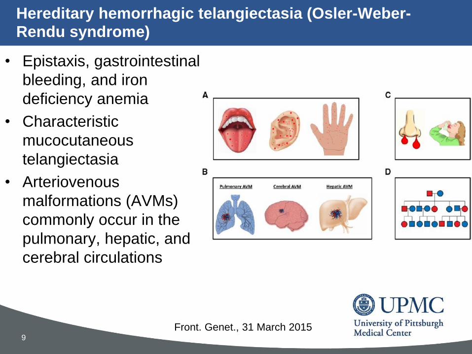
• Epistaxis, gastrointestinal
bleeding, and iron
deficiency anemia
• Characteristic
mucocutaneous
telangiectasia
• Arteriovenous
malformations (AVMs)
commonly occur in the
pulmonary, hepatic, and
cerebral circulations
Hereditary hemorrhagic telangiectasia (Osler-Weber-Rendu syndrome)
9
Front. Genet., 31 March 2015

• Spontaneous and recurrent epistaxis
• Multiple mucocutaneous telangiectasias at characteristic
sites
• Visceral involvement (e.g., gastrointestinal telangiectasia;
pulmonary, cerebral, or hepatic arteriovenous
malformations)
• A first-degree relative with HHT
Definite (3+ criteria), suspected (2 criteria), and unlikely (1
criterion)
Diagnostic Criteria: Curaçao diagnostic criteria
10Am J Med Genet. 2000;91(1):66

Pathophysiology
11
N Engl J Med 1995; 333:918-924
• AD disease with varying penetrance,
progressive
• 1:5000 to 1:8000 individuals
• 600 mutations described: ENG (endoglin),
ACVRL1 (activin receptor-like kinase 1,
ALK-1), SMAD4
• Result in diffuse defects in vascular
structure (telangiectasias and AVMs)
Tim
e

Management
12

Management
13
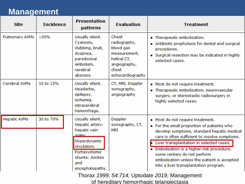
Management
14 Thorax 1999; 54:714; Uptodate 2019; Management
of hereditary hemorrhagic telangiectasia

• Diuresed with milrinone
• V/Q scan low probability
• Head MRI without cerebral AVM
• Hepatology: Discussed transplant, patient declined
• Heme: bevacizumab if no transplant
• Hepatic embolization thought too high of risk
• Palliative care consulted
• Discharged on diuretics and PPI with multidisciplinary follow
up
Clinical Course
15

• ~6 months later she presented to OSH with 2 days of
melena found to have to be hypotensive with a Hgb of 5.4,
received two units PRBC, Hgb to 7.7
• EGD OSH 11/11/17:
– A few 10 mm non bleeding angioectasias were found in the stomach
– Nd:YAG laser therapy was performed on the greater curvature of the
gastric antrum, in the duodenal bulb and in the first part of the
duodenum with 20 watts, 5 pulses, and 5 joules.
• She was lost to follow up
Clinical Course
16

• AD dominant vascular disease (AVMs and telangiectasias)
• Variable penetrance
• Clinical symptoms develop with increasing age
• Epistaxis earliest and most frequent symptom
• Iron deficiency anemia, GI bleeding, mucocutaneous
telangiectasias and AVMs in the pulmonary, hepatic, and
cerebral circulations are common
• Management with iron transfusions, local therapy, systemic
therapy, and screening for PAVMs, +/- cerebral AVMs
• Hepatic AVMs and cardiac failure, if medical management
fails consider transplantation
Summary: Hereditary hemorrhagic telangiectasia
17

Questions
Thank you
18
What was invented (or first) in greater Pittsburgh
A. The pound sign
B. The Big Mac
C. The hepatitis B vaccine
D. The first commercial television station
E. The first movie
Fun Fact Pittsburgh Trivia
19

What was invented (or first) in greater Pittsburgh
A. The pound sign -> emoticon at CMU
B. The Big Mac
C. The hepatitis B vaccine -> polio vaccine developed in
Pittsburgh
D. The first commercial television station -> radio station -
KDKA 1920 (CBS radio Pittsburgh)
E. The first movie -> first nickelodeon in Pittsburgh in 1905
Fun Fact Pittsburgh Trivia
20

• Basic clinical examination
• Evaluation for anemia and iron deficiency
• Pulmonary AVM (PAVM) screening
• Discussion of screening versus non-screening for other
systemic AVMs:
– Cerebral AVMs
– Hepatic AVMs – Screening is rarely performed
– Spinal AVMs – Screening is performed in pregnancy and for patients
undergoing surgery when epidural analgesia may be required
Screening
21