2016 european guidelines on cardiovascular disease ... · joint esc guidelines 2016 european...
TRANSCRIPT

JOINT ESC GUIDELINES
2016 European Guidelines on cardiovasculardisease prevention in clinical practiceThe Sixth Joint Task Force of the European Society of Cardiologyand Other Societies on Cardiovascular Disease Prevention inClinical Practice (constituted by representatives of 10 societiesand by invited experts)
Developed with the special contribution of the European Associationfor Cardiovascular Prevention & Rehabilitation (EACPR)
Authors/Task Force Members: Massimo F. Piepoli* (Chairperson) (Italy),Arno W. Hoes* (Co-Chairperson) (The Netherlands), Stefan Agewall (Norway)1,Christian Albus (Germany)9, Carlos Brotons (Spain)10, Alberico L. Catapano (Italy)3,Marie-Therese Cooney (Ireland)1, Ugo Corra (Italy)1, Bernard Cosyns (Belgium)1,Christi Deaton (UK)1, Ian Graham (Ireland)1, Michael Stephen Hall (UK)7,F. D. Richard Hobbs (UK)10, Maja-Lisa Løchen (Norway)1, Herbert Lollgen(Germany)8, Pedro Marques-Vidal (Switzerland)1, Joep Perk (Sweden)1, Eva Prescott(Denmark)1, Josep Redon (Spain)5, Dimitrios J. Richter (Greece)1, Naveed Sattar(UK)2, Yvo Smulders (The Netherlands)1, Monica Tiberi (Italy)1,H. Bart van der Worp (The Netherlands)6, Ineke van Dis (The Netherlands)4,W. M. Monique Verschuren (The Netherlands)1
Additional Contributor: Simone Binno (Italy)
* Corresponding authors: Massimo F. Piepoli, Heart Failure Unit, Cardiology Department, Polichirurgico Hospital G. Da Saliceto, Cantone Del Cristo, 29121 Piacenza, Emilia Romagna,Italy, Tel: +39 0523 30 32 17, Fax: +39 0523 30 32 20, E-mail: [email protected], [email protected].
Arno W. Hoes, Julius Center for Health Sciences and Primary Care, University Medical Center Utrecht, PO Box 85500 (HP Str. 6.131), 3508 GA Utrecht, The Netherlands,Tel: +31 88 756 8193, Fax: +31 88 756 8099, E-mail: [email protected].
ESC Committee for Practice Guidelines (CPG) and National Cardiac Societies document reviewers: listed in the Appendix.
ESC entities having participated in the development of this document:
Associations: European Association for Cardiovascular Prevention & Rehabilitation (EACPR), European Association of Cardiovascular Imaging (EACVI), European Association ofPercutaneous Cardiovascular Interventions (EAPCI), Heart Failure Association (HFA).
Councils: Council on Cardiovascular Nursing and Allied Professions, Council for Cardiology Practice, Council on Cardiovascular Primary Care.
Working Groups: Cardiovascular Pharmacotherapy
The content of these European Society of Cardiology (ESC) Guidelines has been published for personal and educational use only. No commercial use is authorized. No part of theESC Guidelines may be translated or reproduced in any form without written permission from the ESC. Permission can be obtained upon submission of a written request to OxfordUniversity Press, the publisher of the European Heart Journal and the party authorized to handle such permissions on behalf of the ESC.
Disclaimer. The ESC Guidelines represent the views of the ESC and were produced after careful consideration of the scientific and medical knowledge and the evidence available atthe time of their publication. The ESC is not responsible in the event of any contradiction, discrepancy and/or ambiguity between the ESC Guidelines and any other official recom-mendations or guidelines issued by the relevant public health authorities, in particular in relation to good use of healthcare or therapeutic strategies. Health professionals are encour-aged to take the ESC Guidelines fully into account when exercising their clinical judgment, as well as in the determination and the implementation of preventive, diagnostic ortherapeutic medical strategies; however, the ESC Guidelines do not override, in any way whatsoever, the individual responsibility of health professionals to make appropriate andaccurate decisions in consideration of each patient’s health condition and in consultation with that patient and, where appropriate and/or necessary, the patient’s caregiver. Nordo the ESC Guidelines exempt health professionals from taking into full and careful consideration the relevant official updated recommendations or guidelines issued by the competentpublic health authorities, in order to manage each patient’s case in light of the scientifically accepted data pursuant to their respective ethical and professional obligations. It is also thehealth professional’s responsibility to verify the applicable rules and regulations relating to drugs and medical devices at the time of prescription.
& The European Society of Cardiology 2016. All rights reserved. For permissions please email: [email protected].
European Heart Journal (2016) 37, 2315–2381doi:10.1093/eurheartj/ehw106
Downloaded from https://academic.oup.com/eurheartj/article-abstract/37/29/2315/1748952/2016-European-Guidelines-on-cardiovascular-diseaseby gueston 10 October 2017

Document Reviewers: Guy De Backer (CPG Review Coordinator) (Belgium), Marco Roffi (CPG Review Coordinator)(Switzerland), Victor Aboyans (France)1, Norbert Bachl (Austria)8, Hector Bueno (Spain)1, Scipione Carerj (Italy)1,Leslie Cho (USA)1, John Cox (Ireland)10, Johan De Sutter (Belgium)1, Gunther Egidi (Germany)1, Miles Fisher (UK)2,Donna Fitzsimons (UK)1, Oscar H. Franco (The Netherlands)1, Maxime Guenoun (France)1, Catriona Jennings (UK)1,Borut Jug (Slovenia)4, Paulus Kirchhof (UK/Germany)1, Kornelia Kotseva (UK)1, Gregory Y.H. Lip (UK)1,Francois Mach (Switzerland)1, Giuseppe Mancia (Italy)5, Franz Martin Bermudo (Spain)7, Alessandro Mezzani (Italy)1,Alexander Niessner (Austria)1, Piotr Ponikowski (Poland)1, Bernhard Rauch (Germany)1, Lars Ryden (Sweden)1,Adrienne Stauder (Hungary)9, Guillaume Turc (France)6, Olov Wiklund (Sweden)3, Stephan Windecker(Switzerland)1, Jose Luis Zamorano (Spain)1
Societies: 1European Society of Cardiology (ESC); 2European Association for the Study of Diabetes (EASD); 3EuropeanAtherosclerosis Society (EAS); 4European Heart Network (EHN); 5European Society of Hypertension (ESH); 6EuropeanStroke Organisation (ESO); 7International Diabetes Federation European Region (IDF Europe); 8International Federation ofSport Medicine (FIMS); 9International Society of Behavioural Medicine (ISBM); 10WONCA Europe.
The disclosure forms of all experts involved in the development of these guidelines are available on the ESC websitehttp://www.escardio.org/guidelines.
Online publish-ahead-of-print 23 May 2016
- - - - - - - - - - - - - - - - - - - - - - - - - - - - - - - - - - - - - - - - - - - - - - - - - - - - - - - - - - - - - - - - - - - - - - - - - - -- - - - - - - - - - - - - - - - - - - - - - - - - - - - - - - - - - - - - - - - - - - - - - - - - - - - - - - - - - - - - - - - - - - - - - - - - - -Keywords Guidelines † Blood pressure † Clinical settings † Diabetes † Healthy lifestyle † Lipid † Nutrition †
Physical activity † Population † Prevention † Primary care † Psychosocial factors † Rehabilitation †
Risk assessment † Risk management † Smoking † Stakeholder
Table of Contents
Abbreviations and acronyms . . . . . . . . . . . . . . . . . . . . . . . .2318
1. What is cardiovascular disease prevention? . . . . . . . . . . . . .2319
1.1 Definition and rationale . . . . . . . . . . . . . . . . . . . . . .2319
1.2 Development of the 6th Joint Task Force guidelines . . .2320
1.3 Cost-effectiveness of prevention . . . . . . . . . . . . . . . .2320
2. Who will benefit from prevention? When and how to assess
risk and prioritize . . . . . . . . . . . . . . . . . . . . . . . . . . . . . . .2321
2.1 Estimation of total cardiovascular risk . . . . . . . . . . . . .2321
2.2 When to assess total cardiovascular risk? . . . . . . . . . .2321
2.3 How to estimate total cardiovascular risk? . . . . . . . . . .2322
2.3.1 Ten-year cardiovascular risk . . . . . . . . . . . . . . . .2325
2.3.2 Cardiovascular risk age . . . . . . . . . . . . . . . . . . .2326
2.3.3 Lifetime vs. 10-year cardiovascular risk estimation . .2326
2.3.4 Low-risk, high-risk and very-high-risk countries . . . .2326
2.3.4.1 What are low-risk countries? . . . . . . . . . . . .2326
2.3.4.2 What are high-risk and very-high-risk
countries? . . . . . . . . . . . . . . . . . . . . . . . . . . . . .2326
2.3.5 How to use the risk estimation charts . . . . . . . . . .2326
2.3.6 Modifiers of calculated total cardiovascular risk . . . .2330
2.3.7 Risk categories: priorities . . . . . . . . . . . . . . . . . .2330
2.3.8 Risk factor targets . . . . . . . . . . . . . . . . . . . . . . .2330
2.3.9 Conclusions . . . . . . . . . . . . . . . . . . . . . . . . . . .2330
2.4 Other risk markers . . . . . . . . . . . . . . . . . . . . . . . . .2331
2.4.1 Family history/(epi)genetics . . . . . . . . . . . . . . . . .2331
2.4.1.1 Family history . . . . . . . . . . . . . . . . . . . . . .2331
2.4.1.2 Genetic markers . . . . . . . . . . . . . . . . . . . .2331
2.4.1.3 Epigenetics . . . . . . . . . . . . . . . . . . . . . . . .2332
2.4.2 Psychosocial risk factors . . . . . . . . . . . . . . . . . . .2332
2.4.3 Circulating and urinary biomarkers . . . . . . . . . . . .2333
2.4.4 Measurement of preclinical vascular damage . . . . . .2334
2.4.4.1 Coronary artery calcium . . . . . . . . . . . . . . .2334
2.4.4.2 Carotid ultrasound . . . . . . . . . . . . . . . . . .2335
2.4.4.3 Arterial stiffness . . . . . . . . . . . . . . . . . . . .2335
2.4.4.4 Ankle–brachial index . . . . . . . . . . . . . . . . .2335
2.4.4.5 Echocardiography . . . . . . . . . . . . . . . . . . .2335
2.4.5 Clinical conditions affecting cardiovascular disease risk .2335
2.4.5.1 Chronic kidney disease . . . . . . . . . . . . . . . .2335
2.4.5.2 Influenza . . . . . . . . . . . . . . . . . . . . . . . . .2336
2.4.5.3 Periodontitis . . . . . . . . . . . . . . . . . . . . . . .2336
2.4.5.4 Patients treated for cancer . . . . . . . . . . . . .2336
2.4.5.5 Autoimmune disease . . . . . . . . . . . . . . . . .2337
2.4.5.6 Obstructive sleep apnoea syndrome . . . . . . .2337
2.4.5.7 Erectile dysfunction . . . . . . . . . . . . . . . . . .2338
2.5 Relevant groups . . . . . . . . . . . . . . . . . . . . . . . . . . .2338
2.5.1 Individuals ,50 years of age . . . . . . . . . . . . . . . .2338
2.5.1.1 Assessing cardiovascular disease risk in people
,50 years of age . . . . . . . . . . . . . . . . . . . . . . . . .2338
2.5.1.2 Management of cardiovascular disease risk in
people ,50 years of age . . . . . . . . . . . . . . . . . . . .2338
2.5.2 Elderly . . . . . . . . . . . . . . . . . . . . . . . . . . . . . .2339
2.5.2.1 Hypertension . . . . . . . . . . . . . . . . . . . . . .2339
2.5.2.2 Diabetes mellitus . . . . . . . . . . . . . . . . . . . .2339
2.5.2.3 Hyperlipidaemia . . . . . . . . . . . . . . . . . . . .2339
2.5.3 Female-specific conditions . . . . . . . . . . . . . . . . .2339
2.5.3.1 Obstetric conditions . . . . . . . . . . . . . . . . .2339
2.5.3.2 Non-obstetric conditions . . . . . . . . . . . . . .2340
2.5.4 Ethnic minorities . . . . . . . . . . . . . . . . . . . . . . . .2340
3a. How to intervene at the individual level: risk factor
intervention . . . . . . . . . . . . . . . . . . . . . . . . . . . . . . . . . . .2341
3a.1 Behaviour change . . . . . . . . . . . . . . . . . . . . . . . . .2341
3a.2 Psychosocial factors . . . . . . . . . . . . . . . . . . . . . . . .2342
3a.3 Sedentary behaviour and physical activity . . . . . . . . . .2343
3a.3.1 Introduction . . . . . . . . . . . . . . . . . . . . . . . . . .2343
Joint ESC Guidelines2316
Downloaded from https://academic.oup.com/eurheartj/article-abstract/37/29/2315/1748952/2016-European-Guidelines-on-cardiovascular-diseaseby gueston 10 October 2017

3a.3.2 Physical activity prescription . . . . . . . . . . . . . . .2344
3a.3.2.1 Aerobic physical activity . . . . . . . . . . . . . .2344
3a.3.2.2 Muscle strength/resistance physical activity . .2344
3a.3.2.3 Neuromotor physical activity . . . . . . . . . . .2345
3a.3.2.4 Phases and progression of physical activity . .2345
3a.3.3 Risk assessment . . . . . . . . . . . . . . . . . . . . . . .2345
3a.4 Smoking intervention . . . . . . . . . . . . . . . . . . . . . . .2345
3a.4.1 Introduction . . . . . . . . . . . . . . . . . . . . . . . . . .2345
3a.4.2 Dosage and type . . . . . . . . . . . . . . . . . . . . . . .2346
3a.4.3 Passive smoking . . . . . . . . . . . . . . . . . . . . . . .2346
3a.4.4 Mechanisms by which tobacco smoking increases
risk . . . . . . . . . . . . . . . . . . . . . . . . . . . . . . . . . . . .2346
3a.4.5 Smoking cessation . . . . . . . . . . . . . . . . . . . . . .2346
3a.4.6 Evidence-based drug interventions . . . . . . . . . . .2346
3a.4.7 Electronic cigarettes . . . . . . . . . . . . . . . . . . . . .2347
3a.4.8 Other smoking cessation interventions . . . . . . . .2347
3a.5 Nutrition . . . . . . . . . . . . . . . . . . . . . . . . . . . . . . .2347
3a.5.1 Introduction . . . . . . . . . . . . . . . . . . . . . . . . . .2347
3a.5.2 Fatty acids . . . . . . . . . . . . . . . . . . . . . . . . . . .2347
3a.5.3 Minerals . . . . . . . . . . . . . . . . . . . . . . . . . . . .2348
3a.5.4 Vitamins . . . . . . . . . . . . . . . . . . . . . . . . . . . .2348
3a.5.5 Fibre . . . . . . . . . . . . . . . . . . . . . . . . . . . . . . .2348
3a.5.6 Foods and food groups . . . . . . . . . . . . . . . . . . .2349
3a.5.6.1 Fruits and vegetables . . . . . . . . . . . . . . . .2349
3a.5.6.2 Nuts . . . . . . . . . . . . . . . . . . . . . . . . . . .2349
3a.5.6.3 Fish . . . . . . . . . . . . . . . . . . . . . . . . . . . .2349
3a.5.6.4 Alcoholic beverages . . . . . . . . . . . . . . . . .2349
3a.5.6.5 Soft drinks and sugar . . . . . . . . . . . . . . . .2349
3a.5.7 Functional foods . . . . . . . . . . . . . . . . . . . . . . .2349
3a.5.8 Dietary patterns . . . . . . . . . . . . . . . . . . . . . . .2349
3a.6 Body weight . . . . . . . . . . . . . . . . . . . . . . . . . . . . .2349
3a.6.1 Introduction . . . . . . . . . . . . . . . . . . . . . . . . . .2350
3a.6.2 Which index of obesity is the best predictor of
cardiovascular risk? . . . . . . . . . . . . . . . . . . . . . . . . . .2350
3a.6.3 Does ‘metabolically healthy obesity’ exist? . . . . . .2350
3a.6.4 The obesity paradox in established heart disease . .2350
3a.6.5 Treatment goals and modalities . . . . . . . . . . . . .2350
3a.7 Lipid control . . . . . . . . . . . . . . . . . . . . . . . . . . . .2351
3a.7.1 Introduction . . . . . . . . . . . . . . . . . . . . . . . . . .2351
3a.7.2 Total and low-density lipoprotein cholesterol . . . .2351
3a.7.3 Apolipoprotein B . . . . . . . . . . . . . . . . . . . . . .2351
3a.7.4 Triglycerides . . . . . . . . . . . . . . . . . . . . . . . . .2351
3a.7.5 High-density lipoprotein cholesterol . . . . . . . . . .2351
3a.7.6 Lipoprotein(a) . . . . . . . . . . . . . . . . . . . . . . . .2352
3a.7.7 Apolipoprotein B/apolipoprotein A1 ratio . . . . . .2352
3a.7.8 Calculated lipoprotein variables . . . . . . . . . . . . .2352
3a.7.8.1 Low-density lipoprotein cholesterol . . . . . . .2352
3a.7.8.2 Non-high-density lipoprotein cholesterol
(accurate in non-fasting samples) . . . . . . . . . . . . . .2352
3a.7.8.3 Remnant cholesterol . . . . . . . . . . . . . . . . .2352
3a.7.9 Exclusion of secondary and familial dyslipidaemia . .2352
3a.7.10 Who should be treated and what are the goals? . .2352
3a.7.11 Patients with kidney disease . . . . . . . . . . . . . . .2353
3a.7.12 Drugs . . . . . . . . . . . . . . . . . . . . . . . . . . . . .2353
3a.7.13 Drug combinations . . . . . . . . . . . . . . . . . . . .2354
3a.8 Diabetes mellitus (type 2 and type 1) . . . . . . . . . . . .2355
3a.8.1 Lifestyle intervention . . . . . . . . . . . . . . . . . . . .2356
3a.8.2 Cardiovascular risk . . . . . . . . . . . . . . . . . . . . .2356
3a.8.3 Glucose control . . . . . . . . . . . . . . . . . . . . . . .2356
3a.8.4 Blood pressure . . . . . . . . . . . . . . . . . . . . . . . .2356
3a.8.5 Lipid-lowering therapy . . . . . . . . . . . . . . . . . . .2356
3a.8.6 Antithrombotic therapy . . . . . . . . . . . . . . . . . .2357
3a.8.7 Microalbuminuria . . . . . . . . . . . . . . . . . . . . . .2357
3a.8.8 Type 1 diabetes . . . . . . . . . . . . . . . . . . . . . . .2357
3a.9 Hypertension . . . . . . . . . . . . . . . . . . . . . . . . . . . .2358
3a.9.1 Introduction . . . . . . . . . . . . . . . . . . . . . . . . . .2359
3a.9.2 Definition and classifications of hypertension . . . .2359
3a.9.3 Blood pressure measurement . . . . . . . . . . . . . .2359
3a.9.4 Office or clinic blood pressure measurement . . . .2359
3a.9.5 Out-of-office blood pressure monitoring . . . . . . .2359
3a.9.6 Diagnostic evaluation in hypertensive patients . . . .2359
3a.9.7 Risk stratification in hypertension . . . . . . . . . . . .2360
3a.9.8 Who to treat, and when to initiate antihypertensive
treatment . . . . . . . . . . . . . . . . . . . . . . . . . . . . . . . .2360
3a.9.9 How to treat . . . . . . . . . . . . . . . . . . . . . . . . .2360
3a.9.9.1 Lifestyle changes . . . . . . . . . . . . . . . . . . .2360
3a.9.9.2 Blood pressure-lowering drugs . . . . . . . . . .2360
3a.9.9.3 Combination treatment . . . . . . . . . . . . . . .2361
3a.9.10 Blood pressure goals . . . . . . . . . . . . . . . . . . .2361
3a.9.11 Hypertension in special groups . . . . . . . . . . . . .2362
3a.9.11.1 Diabetes mellitus . . . . . . . . . . . . . . . . . .2362
3a.9.11.2 Elderly . . . . . . . . . . . . . . . . . . . . . . . . .2362
3a.9.12 Resistant hypertension . . . . . . . . . . . . . . . . . .2362
3a.9.13 Duration of treatment and follow-up . . . . . . . . .2362
3a.10 Antiplatelet therapy . . . . . . . . . . . . . . . . . . . . . . .2363
3a.10.1 Antiplatelet therapy in individuals without
cardiovascular disease . . . . . . . . . . . . . . . . . . . . . . . .2363
3a.10.2 Antiplatelet therapy in individuals with
cardiovascular or cerebrovascular disease . . . . . . . . . . .2363
3a.11 Adherence to medication . . . . . . . . . . . . . . . . . . .2364
3a.11.1 Polypill . . . . . . . . . . . . . . . . . . . . . . . . . . . .2365
3b. How to intervene at the individual level: disease-specific
intervention—atrial fibrillation, coronary artery disease, chronic
heart failure, cerebrovascular disease, peripheral artery disease
(web addenda) . . . . . . . . . . . . . . . . . . . . . . . . . . . . . . . . .2365
3c. How to intervene at the population level . . . . . . . . . . . . .2365
3c.1 Introduction (healthy lifestyle promotion) . . . . . . . . .2365
3c.2 Population-based approaches to diet . . . . . . . . . . . . .2366
3c.3 Population-based approaches to physical activity . . . . .2367
3c.4 Population-based approaches to smoking and other
tobacco use . . . . . . . . . . . . . . . . . . . . . . . . . . . . . . . .2369
3c.5 Alcohol abuse protection . . . . . . . . . . . . . . . . . . . .2370
3c.6 Healthy environment . . . . . . . . . . . . . . . . . . . . . . .2371
4a. Where to intervene at the individual level . . . . . . . . . . . . .2371
4a.1 Clinical settings and stakeholders . . . . . . . . . . . . . . .2371
4a.1.1 Cardiovascular disease prevention in primary care .2371
4a.1.2 Acute hospital admission setting . . . . . . . . . . . . .2372
4a.1.3 Specialized prevention programmes . . . . . . . . . .2372
4a.1.4 Alternative rehabilitation models . . . . . . . . . . . .2373
4a.1.4.1 Telerehabilitation . . . . . . . . . . . . . . . . . . .2373
4a.1.5 Maintaining lifestyle changes . . . . . . . . . . . . . . . .2373
4a.2 How to monitor preventive activities . . . . . . . . . . . .2373
4b. Where to intervene at the population level . . . . . . . . . . . .2374
4b.1 Government and public health . . . . . . . . . . . . . . . . . 60
4b.2 Non-governmental organizations . . . . . . . . . . . . . . .2374
5. To do and not to do messages from the Guidelines . . . . . . .2375
6. Appendix . . . . . . . . . . . . . . . . . . . . . . . . . . . . . . . . . . .2376
7. References . . . . . . . . . . . . . . . . . . . . . . . . . . . . . . . . . .2377
Joint ESC Guidelines 2317
Downloaded from https://academic.oup.com/eurheartj/article-abstract/37/29/2315/1748952/2016-European-Guidelines-on-cardiovascular-diseaseby gueston 10 October 2017
Abbreviations and acronymsABI ankle–brachial (blood pressure) indexABPM ambulatory blood pressure monitoringACCORD Action to Control Cardiovascular Risk in DiabetesACE-I angiotensin-converting enzyme inhibitorACS acute coronary syndromesADVANCE Action in Diabetes and Vascular disease: PreterAx
and Diamicron MR Controlled EvaluationAF atrial fibrillationAMI acute myocardial infarctionapoA1 apolipoprotein A1apoB apolipoprotein BARB angiotensin receptor blockerBEUC Bureau Europeen des Unions de ConsommateursBMI body mass index (weight (kg)/height (m2))BP blood pressureCAC coronary artery calciumCAD coronary artery diseaseCAPRIE Clopidogrel versus Aspirin in Patients at Risk for
Ischaemic EventsCARDS Collaborative Atorvastatin Diabetes StudyCHANCE Clopidogrel in High-risk patients with Acute
Non-disabling Cerebrovascular EventsCHARISMA Clopidogrel for High Atherothrombotic Risk
and Ischemic Stabilisation, Management, andAvoidance
CI confidence intervalCKD chronic kidney diseaseCR cardiac rehabilitationCT computed tomographyCTT Cholesterol Treatment Trialists’ CollaborationCURE Clopidogrel vs. Placebo in Patients with ACS
without ST-segment elevationCV cardiovascularCVD cardiovascular diseaseDALYs disability-adjusted life yearsDASH Dietary Approaches to Stop HypertensionDBP diastolic blood pressureDCCT Diabetes Control and Complications TrialDHA docosahexaenoic acidDM diabetes mellitusDPP-4 dipeptidyl peptidase-4eGFR estimated glomerular filtration rateECDA European Chronic Disease AllianceECG electrocardiogramED erectile dysfunctionEHN European Heart NetworkEMA European Medicines AgencyEPA eicosapentaenoic acidEPIC European Prospective Investigation into Cancer
and NutritionEPODE Ensemble Prevenons l’Obesite des EnfantsESC European Society of CardiologyEU European Union
FDA Food and Drug Administration (USA)FDC fixed dose combinationFH familial hypercholesterolaemiaGLP-1 glucagon-like peptide 1GP general practitionerGOSPEL Global Secondary Prevention Strategies to Limit
Event Recurrence After Myocardial InfarctionHbA1c glycated haemoglobinHBPM home blood pressure measurementsHDL-C high-density lipoprotein cholesterolHF heart failureHF-ACTION Heart Failure: A Controlled Trial Investigating
Outcomes of Exercise TrainingHOPE Heart Outcomes Prevention EvaluationHPS Heart Protection StudyHRQoL health-related quality of lifeHR heart ratehsCRP high-sensitivity C-reactive proteinHYVET Hypertension in the Very Elderly TrialICD International Classification of DiseasesIMT intima–media thicknessINVEST International Verapamil-Trandolapril StudyLDL-C low-density lipoprotein cholesterolLp(a) lipoprotein(a)LV left ventricle/left ventricularLVH left ventricular hypertrophyMET metabolic equivalentMHO metabolically healthy overweight/obesityMI myocardial infarctionMUFA monounsaturated fatty acidsNGO non-governmental organizationNHS National Health Service (UK)NICE National Institute for Health and Care ExcellenceNNT number needed to treatNRI net reclassification indexNRT nicotine replacement therapyOASIS Organization to Assess Strategies in Acute
Ischemic SyndromesONTARGET ONgoing Telmisartan Alone and in combination
with Ramipril Global Endpoint TrialOSAS obstructive sleep apnoea syndromeOR odds ratioPA physical activityPAD peripheral artery diseasePLATO Ticagrelor vs. Clopidogrel in Patients with ACS
with and without ST-segment elevationPCOS polycystic ovary syndromePCSK9 proprotein convertase subtilisin/kexin type 9PROactive Prospective Pioglitazone Clinical Trial in Macro-
vascular EventsPROGRESS Perindopril Protection Against Recurrent Stroke
StudyPROCAM Prospective Cardiovascular Munster StudyPWV pulse wave velocityRA rheumatoid arthritis
Joint ESC Guidelines2318
Downloaded from https://academic.oup.com/eurheartj/article-abstract/37/29/2315/1748952/2016-European-Guidelines-on-cardiovascular-diseaseby gueston 10 October 2017
RCT randomized controlled trialRESPONSE Randomised Evaluation of Secondary Prevention
by Outpatient Nurse SpecialistsRM repetition maximumROS reactive oxygen speciesRPE rating of perceived exertionRR relative riskSAVOR-TIMI53
Saxagliptin Assessment of Vascular OutcomesRecorded in Patients with Diabetes Mellitus –Trombolysis in Myocardial Infarction
SBP systolic blood pressureSGLT2 sodium-glucose co-transporter 2SNP single nucleotide polymorphismSCORE Systematic Coronary Risk EstimationSPARCL Stroke Prevention by Aggressive Reduction in
Cholesterol LevelsTIA transient ischaemic attackTRITON Prasugrel vs. Clopidogrel in Patients with ACSUKPDS United Kingdom Prospective Diabetes StudyVADT Veterans Affairs Diabetes TrialVALUE Valsartan Antihypertensive Long-Term Use
EvaluationVLDL very low-density lipoproteinVO2 oxygen uptakeWHO World Health Organization
1. What is cardiovascular diseaseprevention?
1.1 Definition and rationaleCardiovascular disease (CVD) prevention is defined as a coordinatedset of actions, at the population level or targeted at an individual, thatare aimed at eliminating or minimizing the impact of CVDs and theirrelated disabilities.1 CVD remains a leading cause of morbidity andmortality, despite improvements in outcomes. Age-adjusted coron-ary artery disease (CAD) mortality has declined since the 1980s, par-ticularly in high-income regions.2 CAD rates are now less than halfwhat they were in the early 1980s in many countries in Europe,due to preventive measures including the success of smoking legisla-tion. However, inequalities between countries persist and many riskfactors, particularly obesity3 and diabetes mellitus (DM),4 have beenincreasing substantially. If prevention was practised as instructed it
Classes of recommendations
Classes of recommendations
Definition Suggested wording to use
Class I Evidence and/or general agreement that a given treatment or procedure is beneficial, useful,
Conflicting evidence and/or a
usefulness/efficacy of the given
effective.
Is recommended/is indicated
Class II divergence of opinion about the
treatment or procedure.
Class IIa Weight of evidence/opinion is in favour of usefulness/efficacy.
Usefulness/efficacy is less well
Should be considered
Class IIbestablished by evidence/opinion.
May be considered
Class III Evidence or general agreement that the given treatment or procedure is not useful/effective, and in some cases may be harmful.
Is not recommended
Level of evidence
Level of evidence A
Data derived from multiple randomized clinical trials or meta-analyses.
Level of evidence B
Data derived from a single randomized clinical trial or large non-randomized studies.
Level of evidence C
Consensus of opinion of the experts and/or small studies, retrospective studies, registries.
Joint ESC Guidelines 2319
Downloaded from https://academic.oup.com/eurheartj/article-abstract/37/29/2315/1748952/2016-European-Guidelines-on-cardiovascular-diseaseby gueston 10 October 2017

would markedly reduce the prevalence of CVD. It is thus not onlyprevailing risk factors that are of concern, but poor implementationof preventive measures as well.5,6 Prevention should be delivered (i)at the general population level by promoting healthy lifestyle behav-iour7 and (ii) at the individual level, i.e. in those subjects at moderateto high risk of CVD or patients with established CVD, by tackling un-healthy lifestyles (e.g. poor-quality diet, physical inactivity, smoking)and by optimising risk factors. Prevention is effective: the eliminationof health risk behaviours would make it possible to prevent at least80% of CVDs and even 40% of cancers.8,9
1.2 Development of the 6th Joint TaskForce guidelinesThe present guidelines represent an evidence-based consensusof the 6th European Joint Task Force involving 10 professionalsocieties.
By appraising the current evidence and identifying remainingknowledge gaps in managing CVD prevention, the Task Force for-mulated recommendations to guide actions to prevent CVD in clin-ical practice. The Task Force followed the quality criteria fordevelopment of guidelines, which can be found at http://www.escardio.org/Guidelines-&-Education/Clinical-Practice-Guidelines/Guidelines-development/Writing-ESC-Guidelines. For simplifica-tion and in keeping with other European Society of Cardiology(ESC) guidelines, the ESC grading system based on classes of recom-mendation and levels of evidence has been maintained, recognisingthat this may be less suitable to measure the impact of preventionstrategies, particularly those related to behavioural issues andpopulation-based interventions.
This document has been developed to support healthcare profes-sionals communicating with individuals about their cardiovascular(CV) risk and the benefits of a healthy lifestyle and early modificationof their CV risk. In addition, the guidelines provide tools for health-care professionals to promote population-based strategies and inte-grate these into national or regional prevention frameworks and totranslate these in locally delivered healthcare services, in line withthe recommendations of the World Health Organization (WHO)global status report on non-communicable diseases 2010.10
As in the present guidelines, the model presented in the previousdocument from the Fifth European Joint Task Force11 has beenstructured around four core questions: (i) What is CVD prevention?(ii) Who will benefit from prevention? (iii) How to intervene?(iv) Where to intervene?
Compared with the previous guidelines, greater emphasis has beenplaced on a population-based approach, on disease-specific interven-tions and on female-specific conditions, younger individuals and eth-nic minorities. Due to space restrictions for the paper version, thechapter on disease-specific intervention is on the web, togetherwith a few tables and figures (for more detail see web addenda).
A lifetime approach to CV risk is important since both CV risk andprevention are dynamic and continuous as patients age and/or accumu-late co-morbidities. This implies that, apart from improving lifestyle andreducing risk factor levels in patients with established CVD and thoseat increased risk of developing CVD, healthy people of all ages shouldbe encouraged to adopt a healthy lifestyle. Healthcare professionalsplay an important role in achieving this in their clinical practice.
1.3 Cost-effectiveness of preventionKey messages† Prevention of CVD, either by implementation of lifestyle changes
or use of medication, is cost effective in many scenarios, includingpopulation-based approaches and actions directed at high-riskindividuals.
† Cost-effectiveness depends on several factors, including baselineCV risk, cost of drugs or other interventions, reimbursementprocedures and implementation of preventive strategies.
Recommendation for cost-effective prevention ofcardiovascular disease
Recommendation Classa Levelb Ref c
Measures aimed at promoting healthy lifestyles at the population level should be considered.
IIa B 12, 13
aClass of recommendation.bLevel of evidence.cReference(s) supporting recommendations.
In 2009, costs related to CVD amounted to E106 billion, represent-ing �9% of the total healthcare expenditure across the EuropeanUnion (EU).14 Thus, CVD represents a considerable economic bur-den to society and effective preventive measures are necessary.There is consensus in favour of an approach combining strategiesto improve CV health across the population at large from childhoodonward, with specific actions to improve CV health in individuals atincreased risk of CVD or with established CVD.
Most studies assessing the cost-effectiveness of CVD preventioncombine evidence from clinical research with simulation ap-proaches, while cost-effectiveness data from randomized controlledtrials (RCTs) are relatively scarce.15,16 Cost-effectiveness stronglydepends on parameters such as the target population’s age, theoverall population risk of CVD and the cost of interventions. Hence,results obtained in one country may not be valid in another. Further-more, changes such as the introduction of generic drugs can consid-erably change cost-effectiveness.17 According to the WHO, policyand environmental changes could reduce CVD in all countries forless than US$1/person/year.18 A report from the National Institutefor Health and Care Excellence (NICE) estimated that a UK nationalprogramme reducing population CV risk by 1% would prevent25 000 CVD cases and generate savings of E40 million/year. CADmortality rates could be halved by only modest risk factor reduc-tions and it has been suggested that eight dietary priorities alonecould halve CVD death.13
In the last three decades, more than half of the reduction in CVmortality has been attributed to changes in risk factor levels in thepopulation, primarily the reduction in cholesterol and blood pres-sure (BP) levels and smoking. This favourable trend is partly offsetby an increase in other risk factors, mainly obesity and type 2DM.19,20 Aging of the population also increases CVD events.21
Several population interventions have efficiently modified the life-style of individuals. For example, increased awareness of how healthylifestyles prevent CVD has helped to reduce smoking and cholesterol
Joint ESC Guidelines2320
Downloaded from https://academic.oup.com/eurheartj/article-abstract/37/29/2315/1748952/2016-European-Guidelines-on-cardiovascular-diseaseby gueston 10 October 2017
levels. Lifestyle interventions act on several CV risk factors and shouldbe applied prior to or in conjunction with drug therapies. Also, legis-lation aimed at decreasing salt and the trans fatty acid content of foodsand smoking habits is cost effective in preventing CVD.12,13,19
Cholesterol lowering using statins15,16 and improvement in BPcontrol are cost effective if targeted at persons with high CVrisk.22 Importantly, a sizable portion of patients on lipid-loweringor BP-lowering drug treatment fails to take their treatment ad-equately or to reach therapeutic goals,23,24 with clinical and eco-nomic consequences.
Gap in evidence† Most cost-effectiveness studies rely on simulation. More data,
mainly from RCTs, are needed.
2. Who will benefit fromprevention? When and how toassess risk and prioritize
2.1 Estimation of total cardiovascular riskAll current guidelines on the prevention of CVD in clinical practicerecommend the assessment of total CVD risk since atherosclerosisis usually the product of a number of risk factors. Prevention of CVDin an individual should be adapted to his or her total CV risk: thehigher the risk, the more intense the action should be.
The importance of total risk estimation in apparently healthypeople before management decisions are made is illustrated in sup-plementary Figure A (see web addenda) and in Table 1 derived fromthe high-risk Systemic Coronary Risk Estimation (SCORE) chart(http://www.escardio.org/Guidelines-&-Education/Practice-tools/CVD-prevention-toolbox/SCORE-Risk-Charts). This shows that aperson with a cholesterol level of 7 mmol/L can be at 10 times lowerrisk than someone with a cholesterol level of 5 mmol/L if the formeris a female and the latter is a male hypertensive smoker.
A recent meta-analysis on CV risk reduction by treatment withBP-lowering drugs does, however, support the concept that abso-lute risk reduction is larger in those individuals at higher baselinerisk.25 This was confirmed in a further meta-analysis that also
showed a greater residual risk during treatment in those at higherbaseline risk, supporting earlier intervention.26,27
Although clinicians often ask for decisional thresholds to triggerintervention, this is problematic since risk is a continuum and thereis no exact point above which, for example, a drug is automatically in-dicated nor below which lifestyle advice may not usefully be offered.
The risk categories presented later in this section are to assist thephysician in dealing with individual people. They acknowledge that al-though individuals at the highest levels of risk gain most from risk factorinterventions, most deaths in a community come from those at lowerlevels of risk, simply because they are more numerous compared withhigh-risk individuals. Thus a strategy for individuals at high risk must becomplemented by public health measures to encourage a healthy life-style and to reduce population levels of CV risk factors.
It is essential for clinicians to be able to assess CV risk rapidly andwith sufficient accuracy. This realization led to the development ofthe risk chart used in the 1994 and 1998 Guidelines. This chart,developed from a concept pioneered by Anderson,28 used age, sex,smoking status, blood cholesterol and systolic BP (SBP) to estimatethe 10- year risk of a first fatal or non-fatal CAD event. There wereseveral problems with this chart, which are outlined in the FourthJoint European Guidelines on prevention.11,29 This led to the present-ly recommended SCORE system, estimating an individual’s 10 yearrisk of fatal CVD.30 The SCORE charts have been developed toestimate risk in both high- and low-risk European populations; itsapplicability to non-Caucasian populations has not been examined.
2.2 When to assess total cardiovascularrisk?
Recommendations for cardiovascular risk assessment
Recommendations Classa Levelb
Systematic CV risk assessment is recommended in individuals at increased CV risk, i.e. with family history of premature CVD, familial hyperlipidaemia, major CV risk factors (such as smoking, high BP, DM or raised lipid levels) or comorbidities increasing CV risk.
I C
It is recommended to repeat CV risk assessment every 5 years, and more often for individuals with risks close to thresholds mandating treatment.
I C
Systematic CV risk assessment may be considered in men >40 years of age and in women >50 years of age or post-menopausal with no known CV risk factors.
IIb C
Systematic CV risk assessment in men <40 of age and women <50 years of age with no known CV risk factors is not recommended.
III C
BP ¼ blood pressure; CV ¼ cardiovascular; CVD ¼ cardiovascular disease;DM ¼ diabetes mellitus.aClass of recommendation.bLevel of evidence.
Screening is the identification of unrecognized disease or, in thiscase, of an unknown increased risk of CVD in individuals without
Table 1 Impact of combinations of risk factors on risk
Gender Age(years)
Cholesterol (mmol/L)
SBP(mmHg)
Smoker Risk (10 year risk of fatal CVD)
F 60 7 120 No 2%
F 60 7 140 Yes 5%
M 60 6 160 No 9%
M 60 5 180 Yes 21%
CVD ¼ cardiovascular disease; F ¼ female; M ¼ male; SBP ¼ systolic bloodpressure.
Joint ESC Guidelines 2321
Downloaded from https://academic.oup.com/eurheartj/article-abstract/37/29/2315/1748952/2016-European-Guidelines-on-cardiovascular-diseaseby gueston 10 October 2017
symptoms. CV risk assessment or screening can be done opportun-istically or systematically. Opportunistic screening means without apredefined strategy, but is done when the opportunity arises [e.g.when the individual is consulting his or her general practitioner(GP) for some other reason]. Systematic screening can be done inthe general population as part of a screening programme or in tar-geted subpopulations, such as subjects with a family history of pre-mature CVD or familial hyperlipidaemia.
While the ideal scenario would be for all adults to have their riskassessed, this is not practical in many societies. The decision aboutwho to screen must be made by individual countries and will be re-source dependent.
In a meta-analysis, GP-based health checks on cholesterol, BP, bodymass index (BMI) and smoking were effective in improving surrogateoutcomes, especially in high-risk patients.31 A large study of CV riskassessment in the general population found that although there wereoverall improvements in risk factors, there was no impact on CV out-comes at the population level.32 A Cochrane review of RCTs usingcounselling or education to modify CV risk factors in adults fromthe general population, occupational groups or those with specificrisk factors (i.e. DM, hypertension) concluded that risk factor im-provements were modest and interventions did not reduce total orCV mortality in general populations, but reduced mortality in high-risk hypertensive and DM populations.33 Although the benefits oftreating asymptomatic conditions such as hypertension, DM and dys-lipidaemia on morbidity and mortality outcomes have been docu-mented, a Cochrane review of the existing trials concluded thatgeneral health checks (including screening for these conditions) donot reduce all-cause or CV morbidity or mortality.34 However,most studies were performed three to four decades ago, and thusrisk factor interventions were not contemporary. Perhaps applicationof medical treatment in addition to the lifestyle interventions thatwere the core component of most trials would improve efficacy.
Most guidelines recommend a mixture of opportunistic and sys-tematic screening.11,35–38 Screening in people at relatively low riskof CVD is not particularly effective in reducing the risk of CV events.The costs of such screening interventions are high and these re-sources may be better used in people at higher CV risk or with estab-lished CVD. In many countries, GPs have a unique role in identifyingindividuals at risk of but without established CVD and assessing theireligibility for intervention (see section 4a.1.1). A modelling studybased on the European Prospective Investigation of Cancer–Norfolk(EPIC-Norfolk) cohort data concluded that, compared with the Na-tional Health Service (NHS) national strategy to screen all adults 40–74 years of age for CV risk, inviting the 60% of the population at thehighest risk according to an integrated risk score was equally effectivein preventing new cases of CVD and had potential cost savings.39
A general concern in screening, including CV risk assessment, is itspotential to do harm. False positive results can cause unnecessaryconcern and medical treatment. Conversely, false negative resultsmay lead to inappropriate reassurance and a lack of lifestyle changes.However, current data suggest that participating in CV screening ingeneral does not cause worry in those who are screened.40 – 43
More research is needed on how certain subgroups, such as olderpeople, the socially deprived and ethnic minorities, react to screening.
Despite limited evidence, these guidelines recommend a system-atic approach to CV risk assessment targeting populations likely tobe at higher CV risk, such as those with a family history of premature
CVD. Thus systematic CV risk assessment in men ,40 years of ageand women ,50 years of age with no known CV risk factors is notrecommended. Additionally, screening of specific groups with jobsthat place other people at risk, e.g. bus drivers and pilots, may be rea-sonable, as is screening for CV risk factors in women before prescrib-ing combined oral contraception, although there are no data tosupport the beneficial effects. Beyond this, systematic CV risk assess-ment in adults ,40 years of age with no known CV risk factors is notrecommended as a main strategy due to the low cost-effectiveness.Systematic CV assessment may be considered in adult men .40years of age and in women .50 years of age or post-menopausalwith no known CV risk factors. Risk assessment is not a one-timeevent; it should be repeated, for example, every 5 years.
2.3 How to estimate total cardiovascularrisk?Key messages† In apparently healthy persons, CV risk in general is the result of
multiple, interacting risk factors. This is the basis for the total CVrisk approach to prevention.
† SCORE, which estimates the 10 year risk of fatal CVD, is recom-mended for risk assessment and can assist in making logical man-agement decisions and may help to avoid both under- andovertreatment. Validated local risk estimation systems are usefulalternatives to SCORE.
† Individuals automatically at high to very high CV risk (Table 5) donot need the use of a risk score and require immediate attentionto risk factors.
† In younger persons, a low absolute risk may conceal a very highrelative risk and use of the relative risk chart or calculation oftheir “risk age” may help in advising them of the need for intensivepreventive efforts.
† While women are at lower CV risk than men, their risk is de-ferred by �10 years rather than avoided.
† The total risk approach allows flexibility; if perfection cannot beachieved with one risk factor, trying harder with others can stillreduce risk.
Recommendation for how to estimate cardiovascular risk
Recommendation Classa Levelb Ref c
Total CV risk estimation, using a risk estimation system such as SCORE, is recommended for adults >40 years of age, unless they are automatically categorised as being at high-risk or very high-risk based on documented CVD, DM (>40 years of age), kidney disease or highly elevated single risk factor (Table 5).
I C 11, 25
CV ¼ cardiovascular; DM ¼ diabetes mellitus; SCORE ¼ Systematic CoronaryRisk Estimation.aClass of recommendation.bLevel of evidence.cReference(s) supporting recommendations.
Joint ESC Guidelines2322
Downloaded from https://academic.oup.com/eurheartj/article-abstract/37/29/2315/1748952/2016-European-Guidelines-on-cardiovascular-diseaseby gueston 10 October 2017
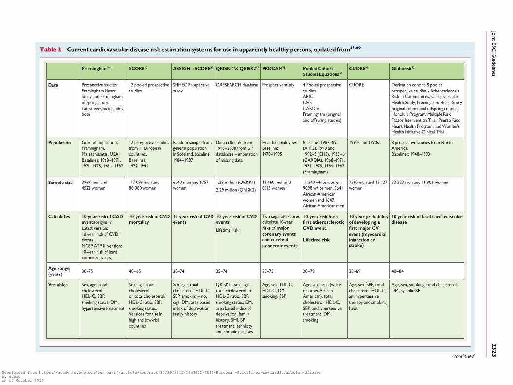
Table 2 Current cardiovascular disease risk estimation systems for use in apparently healthy persons, updated from59,60
Framingham44 SCORE30 ASSIGN – SCORE45 QRISK146 & QRISK247 PROCAM48 Pooled Cohort Studies Equations50
CUORE49 Globorisk52
Data Prospective studies:Framingham Heart Study and Framingham offspring study.Latest version includes both
12 pooled prospective studies
SHHEC Prospective study
QRESEARCH database Prospective study 4 Pooled prospective studiesARICCHSCARDIAFramingham (original and offspring studies)
CUORE Derivation cohort: 8 pooled prospective studies - Atherosclerosis Risk in Communities, Cardiovascular Health Study, Framingham Heart Study original cohort and offspring cohort, Honolulu Program, Multiple Risk Factor Intervention Trial, Puerto Rico Heart Health Program, and Women’s Health Initiative Clinical Trial
Population General population, Framingham,Massachusetts, USA.Baselines: 1968–1971, 1971–1975, 1984–1987
12 prospective studies from 11 European countries.Baselines: 1972–1991
Random sample fromgeneral populationin Scotland, baseline:1984–1987
Data collected from 1993–2008 from GP databases – imputation of missing data
Healthy employees.Baseline: 1978–1995
Baselines 1987–89 (ARIC), 1990 and 1992–3 (CHS), 1985–6 (CARDIA), 1968–1971, 1971–1975, 1984–1987 (Framingham)
1980s and 1990s 8 prospective studies from North America. Baselines: 1948–1993
Sample size 3969 men and 4522 women
117 098 men and88 080 women
6540 men and 6757 women
1.28 million (QRISK1)
2.29 million (QRISK2)
18 460 men and 8515 women
11 240 white women, 9098 white men, 2641 African-American women and 1647 African-American men
7520 men and 13 127 women
33 323 men and 16 806 women
Calculates 10-year risk of CADevents originally.Latest version:10-year risk of CVD eventsNCEP ATP III version: 10-year risk of hard coronary events
10-year risk of CVD mortality
10-year risk of CVD events
10-year risk of CVD events.
Lifetime risk
Two separate scorescalculate 10-year risks of major coronary events and cerebral ischaemic events
first atherosclerotic10-year risk for a
CVD event.
Lifetime risk
10-year probability of developing a
event (myocardial infarction orstroke)
first major CV
10 year risk of fatal cardiovascular disease
Age range (years)
30–75 40–65 30–74 35–74 20–75 20–79 35–69 40–84
Variables Sex, age, total cholesterol,HDL-C, SBP,smoking status, DM,hypertensive treatment
Sex, age, total cholesterolor total cholesterol/HDL-C ratio, SBP, smoking status.Versions for use in high and low-risk countries
Sex, age, total cholesterol, HDL-C, SBP, smoking – no. cigs, DM, area based index of deprivation, family history
QRISK1 - sex, age,total cholesterol to HDL-C ratio, SBP, smoking status, DM, area based index of deprivation, family history, BMI, BP treatment, ethnicity and chronic diseases
Age, sex, LDL-C,HDL-C, DM, smoking, SBP
Age, sex, race (white or other/African American), total cholesterol, HDL-C, SBP, antihypertensive treatment, DM, smoking
Age, sex, SBP, total cholesterol, HDL-C, antihypertensive therapy and smoking habit
Age, sex, smoking, total cholesterol,DM, systolic BP
continued
JointESC
Guidelines
2323
Downloaded from https://academic.oup.com/eurheartj/article-abstract/37/29/2315/1748952/2016-European-Guidelines-on-cardiovascular-diseaseby gueston 10 October 2017

Table 2 (continued)
Framingham44 SCORE30 ASSIGN – SCORE45 QRISK146 & QRISK247
PROCAM48 Pooled Cohort Studies Equations50
CUORE49 Globorisk52
Comments/ developments
Latest version includesversion based onnon-laboratory values only,substituting BMI from lipid measurements
National, updated recalibrations
QRISK2 includes interaction terms to adjust for the interactions between age and some of the variables
Recent change in the methods (Weibull) allows extension of risk estimation to women and broader age range
Race specific beta coefficients for
risk factors have been incorporated. Calculator shown to overestimate risk in external validations – this may indicate the need for recalibration in certain populations
Recalibrations have been undertaken for 11 countries
Recommendedby guidelines
NCEP guidelines,54
Canadian CV guidelines,55 other national guidelines recommend adapted versions including New Zealand56
European Guidelines on CVD Prevention29
SIGN37 NICE guidelines on lipid modification,57
QRISK Lifetime recommended by JBS3 guidelines58
International Task Force for Prevention of Coronary Disease Guidelines
2013 AHA ACC Guideline on the assessment of CVD risk50
ACC ¼ American College of Cardiology; AHA ¼ American Heart Association; ARIC ¼ Atherosclerosis Risk in Communities; ATP ¼ Adult Treatment Panel; BMI ¼ body mass index; BP ¼ blood pressure; CAD ¼ coronary artery disease;CARDIA ¼ Coronary Artery Risk Development in Young Adults; CHS ¼ Cardiovascular Health Study; CVD ¼ cardiovascular disease; DM ¼ diabetes mellitus; HDL-C ¼ high-density lipoprotein cholesterol; JBS ¼ Joint British Societies;LDL-C ¼ low-density lipoprotein cholesterol; NCEP ¼ National Cholesterol Education Program; NICE ¼ National Institute for Health and Care Excellence; no. cigs ¼ number of cigarettes; PROCAM ¼ Prospective Cardiovascular MunsterStudy; SBP ¼ systolic blood pressure; SIGN ¼ Scottish Intercollegiate Guidelines Network; SHHEC ¼ Scottish Heart Health Extended Cohort.
JointESC
Guidelines
2324
Downloaded from https://academic.oup.com/eurheartj/article-abstract/37/29/2315/1748952/2016-European-Guidelines-on-cardiovascular-diseaseby gueston 10 October 2017

2.3.1 Ten-year cardiovascular riskMany CV risk assessment systems are available for use in apparentlyhealthy individuals (Table 2), including Framingham,44 SCORE,30 AS-SIGN (CV risk estimation model from the Scottish IntercollegiateGuidelines Network),45 Q-Risk,46,47 PROCAM (Prospective Car-diovascular Munster Study),48 CUORE,49 the Pooled Cohort equa-tions,50 Arriba51 and Globorisk.52 In practice, most risk estimationsystems perform rather similarly when applied to populations rec-ognizably comparable to those from which the risk estimation sys-tem was derived. Since 2003, the European Guidelines on CVDprevention in clinical practice recommend use of the SCORE sys-tem, because it is based on large, representative European cohortdatasets. The SCORE risk function has been externally validated.53
Table 3 lists the advantages of the SCORE risk charts.The SCORE system estimates the 10 year risk of a first fatal ath-
erosclerotic event. All International Classification of Diseases (ICD)codes that could reasonably be assumed to be atherosclerotic areincluded, including CAD, stroke and aneurysm of the abdominalaorta. Traditionally most systems estimated CAD risk only; how-ever, more recently a number of risk estimation systems have chan-ged to estimate the risk of all CVDs.44,47,50,58
The choice of CV mortality rather than total (fatal plus non-fatal)events was deliberate, although not universally popular. Non-fatalevent rates are critically dependent upon definitions and the meth-ods used in their ascertainment. Critically, the use of mortality al-lows recalibration to allow for time trends in CV mortality. Anyrisk estimation system will overpredict in countries in which mortal-ity has fallen and underpredict in those in which it has risen. Recali-bration to allow for secular changes can be undertaken if goodquality, up-to-date mortality and risk factor prevalence data areavailable. Data quality does not permit this for non-fatal events.For these reasons, the CV mortality charts were produced andhave been recalibrated for a number of European countries.
Naturally, the risk of total fatal and non-fatal events is higher, andclinicians frequently ask for this to be quantified. The SCORE data in-dicate that the total CV event risk is about three times higher than therisk of fatal CVD for men, so that a SCORE risk of fatal CVD of 5%translates into a fatal plus non-fatal CV risk of �15%; the multiplier isabout four in women and somewhat lower than three in older per-sons, in whom a first event is more likely to be fatal.61
As noted in the introduction, thresholds to trigger certain inter-ventions are problematic since risk is a continuum and there is nothreshold at which, for example, a drug is automatically indicated.Obviously, decisions on whether treatment is initiated should alsobe based on patient preferences.
A particular problem relates to young people with high levels ofrisk factors, where a low absolute risk may conceal a very high rela-tive risk requiring intensive lifestyle advice. Several approaches tocommunicating about risk to younger people are presented below(refer also to section 2.5.1). These include use of the relative riskchart or ‘risk age’ or ‘lifetime risk’. The aim is to communicatethat lifestyle changes can reduce the relative risk substantially aswell as reduce the increase in risk that occurs with ageing.
Another problem relates to older people. In some age categories,the vast majority, especially of men, will have estimated CV deathrisks exceeding the 5–10% level, based on age (and gender) only,even when other CV risk factor levels are low. This could lead to
excessive use of drugs in the elderly. This issue is dealt with later(see section 2.3.5). It should be noted that RCT evidence to guidedrug treatments in older persons is limited (refer to section 2.5.2).
The role of high-density lipoprotein cholesterol (HDL-C) in risk es-timation has been systematically re-examined using the SCORE data-base.62–64 Overall HDL-C has a modest but useful effect in redefiningrisk estimation,63,64 but this may not be seen in some low-risk popu-lations.65 Assessing HDL-C is particularly important at levels of riskjust below the threshold for intensive risk modification of 5%, wheremany of these subjects will qualify for intensive advice if their HDL-Cis low.63 SCORE charts incorporating HDL-C are illustrated in sup-plementary Figures B–I (see web addenda). In these charts, HDL-Cis used categorically. The electronic version of SCORE, HeartScore(http://www.HeartScore.org), has been modified to take HDL-Cinto account on a continuous basis and is therefore more accurate.
The role of a plasma triglyceride as a predictor of CVD has beendebated for many years. Fasting triglycerides relate to risk in univari-able analyses, but the effect is attenuated by adjustment for otherfactors, especially HDL-C.66
Dealing with the impact of additional risk factors such as bodyweight, family history and newer risk markers is difficult within theconstraint of a paper chart. It should be stressed, however, that al-though many other risk factors have been identified, their contribu-tion is generally very modest to both absolute CV risk estimationsand in terms of reclassification of an individual to another riskcategory67 (Table 4).
The SCORE risk charts are shown in Figures 1–4, including a chartof relative risks (Figure 3). Instructions on their use follow.
Please note that Figure 3 shows relative not absolute risk. Thus a per-son in the top right-hand box, with multiple CV risk factors, has a riskthat is 12 times greater than a person in the bottom left with normal risk
Table 3 Advantages and limitations in using theSCORE risk charts
Advantages• Intuitive, easy to use tool.• Establishes a common language of risk for healthcare professionals.• Allows a more objective assessment of risk.• Takes account of the multifactorial nature of CVD.• Allows flexibility in management; if an ideal risk factor level cannot be
achieved, total risk can still be reduced by reducing other risk factors.• Deals with the problem of a low absolute risk in young people with
multiple risk factors: the relative risk chart helps to illustrate how a young person with a low absolute risk may be at a substantially high and reducible relative risk; calculation of an individual’s “risk age” may also be of use in this situation.
Limitations• Estimates risk of fatal but not total (fatal + non-fatal) CV risk for
reasons outlined in text. • Adapted to suit different European populations, but not different
ethnic groups within these populations.• Limited to the major determinants of risk.• Other systems have more functionality, although applicability to
multiple countries is uncertain.• Limited age range (40–65 years).
CVD = cardiovascular disease; SCORE = Systematic Coronary Risk Estimation.
Joint ESC Guidelines 2325
Downloaded from https://academic.oup.com/eurheartj/article-abstract/37/29/2315/1748952/2016-European-Guidelines-on-cardiovascular-diseaseby gueston 10 October 2017

factor levels. This may be helpful when advising a young person with alow absolute but high relative risk of the need for lifestyle change.
2.3.2 Cardiovascular risk ageThe risk age of a person with several CV risk factors is the age of aperson of the same gender with the same level of risk but with ideallevels of risk factors. Thus a 40-year-old with high levels of some riskfactors may have the risk age of a 60-year-old (Figure 4), because therisk equals that of a 60-year-old with ideal risk factor levels (i.e. non-smoking, total cholesterol of 4 mmol/L and BP of 120 mmHg).68 Riskage is an intuitive and easily understood way of illustrating the likelyreduction in life expectancy that a young person with a low absolutebut high relative risk of CVD will be exposed to if preventive mea-sures are not adopted.68 Table A showing different risk factor com-binations is included in the web addenda to provide a more accurateestimation of risk ages. Risk age is also automatically calculated aspart of the latest revision of HeartScore.
Risk age has been shown to be independent of the CV endpointused,68 which bypasses the dilemma of whether to use a risk estimationsystem based on CV mortality or on total CV events. Risk age can beused in any population regardless of baseline risk and secular changes inmortality, and therefore avoids the need for recalibration.69 At present,risk age is recommended to help communicate about risk, especially toyounger people with a low absolute risk but a high relative risk.
2.3.3 Lifetime vs. 10-year cardiovascular risk estimationConventional CV risk prediction schemes estimate the 10 year riskof CV events. Lifetime CV risk prediction models identify high-riskindividuals both in the short and long term. Such models account forpredicted risk in the context of competing risks from other diseasesover the remaining expected lifespan of an individual.
Notably, 10 year risk identifies individuals who are most likely tobenefit from drug therapy in the near term. Drug treatment starts towork quite rapidly, and drug treatment can be largely informed byshort-term risk, such as 10 year risk. One problem with short-termrisk is that it is mostly governed by age and consequently few youngerindividuals, in particular women, reach treatment thresholds. It hastherefore been argued that lifetime risk estimation may enhance riskcommunication, particularly among younger individuals and women.
Evidence for the role of lifetime risk in treatment decisions is lack-ing. Sufficient data for robust lifetime risk estimations, as well as
meaningful risk categorization thresholds, are also lacking. Providinglifetime CV risk estimates for some groups at high risk of mortalitydue to competing non-CVD causes can be difficult to interpret. Im-portantly, evidence of the benefits of lifelong preventive therapy(e.g. BP- or lipid-lowering drugs) in younger individuals with lowshort-term but higher lifetime risks is lacking. For these reasons,we do not recommend that risk stratification for treatment deci-sions be based on lifetime risk. However, like risk age and relativerisk, it may be a useful tool in communicating about risk to indivi-duals with high risk factor levels but who are at a low 10 year abso-lute risk of CV events, such as some younger people. Whateverapproach is used, if absolute risk is low, a high relative risk or riskage signals the need for active lifestyle advice and awareness thatdrug treatment may need consideration as the person ages. Bothrisk age and lifetime risk are closer to relative than absolute risk,and none provides an evidence base for drug treatment decisions.
2.3.4 Low-risk, high-risk and very-high-risk countriesThe countries considered here are those with national cardiologysocieties that belong to the ESC, both European and non-European.
2.3.4.1 What are low-risk countries?The fact that CVD mortality has declined in many European coun-tries means that more now fall into the low-risk category. While anycut-off point is arbitrary and open to debate, in these guidelines thecut-off points for calling a country ‘low risk’ are based onage-adjusted 2012 CVD mortality rates in those 45–74 years ofage (,225/100 000 in men and ,175/100 000 in women).70
Thus the following countries are defined as low risk: Andorra, Aus-tria, Belgium, Cyprus, Denmark, Finland, France, Germany, Greece,Iceland, Ireland, Israel, Italy, Luxembourg, Malta, Monaco, The Neth-erlands, Norway, Portugal, San Marino, Slovenia, Spain, Sweden,Switzerland and the United Kingdom.
2.3.4.2 What are high-risk and very-high-risk countries?High-risk countries are Bosnia and Herzegovina, Croatia, Czech Re-public, Estonia, Hungary, Lithuania, Montenegro, Morocco, Poland,Romania, Serbia, Slovakia, Tunisia and Turkey.
Very-high-risk countries present levels of risk that are more thandouble that of low-risk countries (i.e. CVD mortality .450/100 000for men and .350/100 000 for women). Additionally, the male:femaleratio is smaller than in low-risk countries, suggesting a major problemfor women. The very high-risk countries are Albania, Algeria, Armenia,Azerbaijan, Belarus, Bulgaria, Egypt, Georgia, Kazakhstan, Kyrgyzstan,Latvia, former Yugoslav Republic of Macedonia, Moldova, Russian Fed-eration, Syrian Arab Republic, Tajikistan, Turkmenistan, Ukraine andUzbekistan.
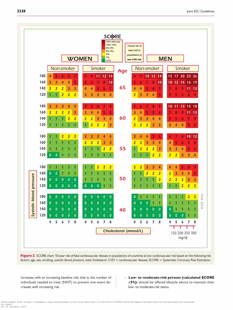
2.3.5 How to use the risk estimation charts† The SCORE charts are used in apparently healthy people, not for
those with established CVD or at very high risk or high risk forother reasons [e.g. DM (see section 3a.8) or chronic kidney disease(CKD; see section 2.4.5.1)], who need intensive risk advice anyway.
† Use of the low-risk chart is recommended for the countrieslisted above. Use of the high-risk chart is recommended for allother European and Mediterranean countries, taking into ac-count that the high-risk charts may underestimate the risk invery-high-risk countries (see above). Note that several countrieshave undertaken national recalibrations to allow for time trends
Table 4 Examples of risk modifiers that are likely tohave reclassification potential (see following sections fordetails)
Socio-economic status, social isolation, or lack of social support.
Family history of premature CVD.
BMI and central obesity.
CT coronary calcium score.
Atherosclerotic plaques determined by carotid artery scanning.
ABI.
ABI ¼ ankle–brachial blood pressure index; BMI ¼ body mass index; CVD ¼cardiovascular disease; CT ¼ computed tomography.
Joint ESC Guidelines2326
Downloaded from https://academic.oup.com/eurheartj/article-abstract/37/29/2315/1748952/2016-European-Guidelines-on-cardiovascular-diseaseby gueston 10 October 2017

in mortality and risk factor distributions. Such charts are likely tobetter represent risk levels.
† To estimate a person’s 10 year risk of CV death, find the table fortheir gender, smoking status and (nearest) age. Within the table,find the cell nearest to the person’s BP and total cholesterol. Risk
estimates will need to be adjusted upwards as the person ap-proaches the next age category.
While no threshold is universally applicable, the intensity of ad-vice should increase with increasing risk. The effect of interven-tions on the absolute probability of developing a CV event
Figure 1 SCORE chart: 10-year risk of fatal cardiovascular disease in populations of countries at high cardiovascular risk based on the following riskfactors: age, sex, smoking, systolic blood pressure, total cholesterol. CVD ¼ cardiovascular disease; SCORE ¼ Systematic Coronary Risk Estimation.
Joint ESC Guidelines 2327
Downloaded from https://academic.oup.com/eurheartj/article-abstract/37/29/2315/1748952/2016-European-Guidelines-on-cardiovascular-diseaseby gueston 10 October 2017
increases with an increasing baseline risk; that is, the number ofindividuals needed to treat (NNT) to prevent one event de-creases with increasing risk.
– Low- to moderate-risk persons (calculated SCORE<5%): should be offered lifestyle advice to maintain theirlow- to moderate-risk status.
Figure 2 SCORE chart: 10-year risk of fatal cardiovascular disease in populations of countries at low cardiovascular risk based on the following riskfactors: age, sex, smoking, systolic blood pressure, total cholesterol. CVD ¼ cardiovascular disease; SCORE¼ Systematic Coronary Risk Estimation.
Joint ESC Guidelines2328
Downloaded from https://academic.oup.com/eurheartj/article-abstract/37/29/2315/1748952/2016-European-Guidelines-on-cardiovascular-diseaseby gueston 10 October 2017

Figure 4 SCORE chart (for use in high-risk European countries) illustrating how the approximate risk age can be read off the chart. SCORE ¼Systematic Coronary Risk Estimation.
Figure 3 Relative risk chart, derived from SCORE Conversion of cholesterol mmol/L � mg/dL: 8 ¼ 310; 7 ¼ 270; 6 ¼ 230; 5 ¼ 190; 4 ¼ 155.
Joint ESC Guidelines 2329
Downloaded from https://academic.oup.com/eurheartj/article-abstract/37/29/2315/1748952/2016-European-Guidelines-on-cardiovascular-diseaseby gueston 10 October 2017

– High-risk persons (calculated SCORE ≥5% and <10%):qualify for intensive lifestyle advice and may be candidates fordrug treatment.
– Very-high-risk persons (calculated SCORE ≥10%):drug treatment is more frequently required. In persons .60years of age, these thresholds should be interpreted more le-niently, because their age-specific risk is normally aroundthese levels, even when other CV risk factor levels are ‘nor-mal’. In particular, uncritical initiation of drug treatments ofall elderly with risks greater than the 10% threshold shouldbe discouraged.
Use of the risk charts should be qualified by knowledge of the fol-lowing aspects:
† The charts assist in risk estimation but must be interpreted in lightof the clinician’s knowledge and experience and in view of the fac-tors that may modify the calculated risk (see below).
† Relative risks may be high in young persons, even if 10 year abso-lute risks are low, because events usually occur later in life. Therelative risk chart or estimating risk age may be helpful in identi-fying and counselling such persons.
† The lower risk in women is explained by the fact that risk is de-ferred by 10 years—the risk of a 60-year-old woman is similar tothat of a 50-year-old man. Ultimately, more women than men dieof CVD.
† The charts may be used to give some indication of the effects ofreducing risk factors, given that there will be a time lag before riskreduces and that the results of RCTs in general give better esti-mates of the benefits of interventions. Those who stop smokinggenerally halve their risk.
2.3.6 Modifiers of calculated total cardiovascular riskApart from the conventional major CV risk factors included in therisk charts, there are other risk factors that could be relevant for as-sessing total CVD risk. The Task Force recommends additional riskfactor assessment if such a risk factor improves risk classification[e.g. by calculation of a net reclassification index (NRI)] and if the as-sessment is feasible in daily practice. In general, reclassification is ofmost value when the individual’s risk lies close to a decisionalthreshold, such as a SCORE risk of 5%. In very-high-risk orvery-low-risk situations, the impact of additional risk factors is un-likely to alter management decisions. While the presence of riskmodifiers may move an individual’s estimated risk upward, absenceof these modifiers should lead to lowering an individual’s estimatedrisk.
Table 4 lists examples of factors that fulfil the aforementioned cri-teria. Several other factors that are frequently discussed in the litera-ture, but may not have the ability to reclassify subjects, are discussedin subsequent paragraphs. Also discussed further in this section arethe roles of ethnicity and of specific conditions or diseases that maybe associated with a higher than calculated risk, such as CKD, auto-immune diseases, etc. The way modifiers are related to CV risk maybe very different. Social deprivation and being overweight, for ex-ample, are important as ‘causes of the causes’ of CVD, in thatthey may be associated with higher levels of conventional risk fac-tors. Family history may reflect a shared environment, genetic fac-tors or both. Markers such as computed tomography (CT)calcium scoring are indicators of disease rather than risk factorsfor future disease.
2.3.7 Risk categories: prioritiesIndividuals at highest risk gain most from preventive efforts, and thisguides the priorities, which are detailed in Table 5.
2.3.8 Risk factor targetsRisk factor goals and target levels for important CV risk factors arepresented in Table 6.
2.3.9 ConclusionsEstimation of total CV risk remains a crucial part of the presentguidelines. The priorities (risk categories) defined in this sectionare for clinical use and reflect the fact that those at highest risk ofa CVD event gain most from preventive measures. This approachshould complement public actions to reduce community risk factorlevels and promote a healthy lifestyle. The principles of risk estima-tion and the definition of priorities reflect an attempt to make com-plex issues simple and accessible. Their very simplicity makes themvulnerable to criticism. Above all, they must be interpreted in light of
Table 5 Risk categories
Very high-risk Subjects with any of the following:• Documented CVD, clinical or unequivocal on imaging. Documented clinical CVD includes previous AMI, ACS, coronary revascularization and other arterial revascularization procedures, stroke and TIA, aortic aneurysm and PAD. Unequivocally documented CVD on imaging
includes plaque on coronary angiography or carotid ultrasound. It does NOT include some increase in continuous imaging parameters such as intima–media thickness of the carotid artery. • DM with target organ damage such as proteinuria or with a major risk factor such as smoking or marked hypercholesterolaemia or marked hypertension.• Severe CKD (GFR <30 mL/min/1.73 m2).• A calculated SCORE ≥10%.
High-risk Subjects with:• Markedly elevated single risk factors, in particular cholesterol >8 mmol/L (>310 mg/dL) (e.g. in familial hypercholesterolaemia) or BP ≥180/110 mmHg. • Most other people with DM (with the
exception of young people with type 1 DM and without major risk factors that may be at low or moderate risk).• Moderate CKD (GFR 30–59 mL/min/1.73 m2).• A calculated SCORE ≥5% and <10%.
Moderate-risk SCORE is ≥1% and <5% at 10 years. Many middle-aged subjects belong to this category.
Low-risk SCORE <1%.
ACS ¼ acute coronary syndrome; AMI ¼ acute myocardial infarction; BP ¼ bloodpressure; CKD ¼ chronic kidney disease; DM ¼ diabetes mellitus; GFR ¼glomerular filtration rate; PAD ¼ peripheral artery disease; SCORE ¼ systematiccoronary risk estimation; TIA ¼ transient ischaemic attack.
Joint ESC Guidelines2330
Downloaded from https://academic.oup.com/eurheartj/article-abstract/37/29/2315/1748952/2016-European-Guidelines-on-cardiovascular-diseaseby gueston 10 October 2017

the physician’s detailed knowledge of his/her patient and in light oflocal guidance and conditions.
Gaps in evidence† There are no recent RCTs of a total risk approach to risk assess-
ment or risk management.† The young, women, older people and ethnic minorities continue
to be underrepresented in clinical trials.† A systematic comparison of current international guidelines is
needed to define areas of agreement and the reasons fordiscrepancies.
2.4 Other risk markers2.4.1 Family history/(epi)genetics
Key messages† Family history of premature CVD in first-degree relatives, before
55 years of age in men and 65 years of age in women, increasesthe risk of CVD.
† Several genetic markers are associated with an increased risk ofCVD, but their use in clinical practice is not recommended.
Recommendations for assessment of family history/(epi)genetics
Recommendations Classa Levelb Ref c
Assessment of family history of premature CVD (defined as a fatalor non-fatal CVD event or/and established diagnosis of CVD in
first degree male relatives before 55years or female relatives before 65 years) is recommended as part of cardiovascular risk assessment.
I C 71
The generalized use of DNA-based tests for CVD risk assessment is not recommended.
III B 72, 73
CVD ¼ cardiovascular disease.aClass of recommendation.bLevel of evidence.cReference(s) supporting recommendations.
2.4.1.1 Family historyFamilial history of premature CVD is a crude but simple indicator ofthe risk of developing CVD, reflecting both the genetic trait and theenvironment shared among household members.71 A positive familyhistory of premature CV death is associated with an increased risk ofearly and lifetime CVD.74 In the few studies that simultaneously as-sessed and reported the effects of family history and genetic scores,family history remained significantly associated with the incidence ofCVD after adjusting for the genetic scores.75,76 Limited data exist re-garding the ability of family history to improve the prediction of CVDbeyond conventional CV risk factors.77–79 One possible explanationis the varying definitions of family history applied80 and that conven-tional CV risk factors can partly explain the impact of family history.
A family history of premature CVD is simple, inexpensive infor-mation that should be part of the CV risk assessment in all subjects.Family history can be a risk modifier to optimal management afterthe calculated risk using SCORE lies near a decisional threshold: apositive family history would favour more intensive interventions,while a negative family history would translate into less intensivetreatment.77
2.4.1.2 Genetic markersGenetic screening and counselling is effective in some conditions,such as familial hypercholesterolaemia (FH) (see section 3a.7.9).This paragraph will focus on genetic screening for high CV risk inthe general population.
Table 6 Risk factor goals and target levels forimportant cardiovascular risk factors
Smoking No exposure to tobacco in any form.
Diet Low in saturated fat with a focus on wholegrain products, vegetables, fruit and fish.
Physical activity
At least 150 minutes a week of moderate aerobic PA (30 minutes for 5 days/week) or 75 minutesa week of vigorous aerobic PA (15 minutes for 5 days/week) or a combination thereof.
Body weight BMI 20–25 kg/m2. Waist circumference <94 cm (men) or <80 cm (women).
Blood pressure
<140/90 mmHga
Lipidsb
LDLc is the primary target
HDL-C
Triglycerides
Very high-risk: <1.8 mmol/L (<70 mg/dL), or a reduction of at least 50% if the baseline is between1.8 and 3.5 mmol/L (70 and 135 mg/dL)d
High-risk: <2.6mmol/L (<100 mg/dL), or a reduction of at least 50% if the baseline is between 2.6 and 5.1 mmol/L (100 and 200 mg/dL)Low to moderate risk: <3.0 mmol/L (<115 mg/dL).
No target but >1.0 mmol/L (>40mg/dL) in men and >1.2 mmol/L (>45 mg/dL) in women indicate lower risk.
No target but <1.7 mmol/L (<150 mg/dL) indicates lower risk and higher levels indicate a need to look for other risk factors.
Diabetes HbA1c <7%. (<53 mmol/mol)
BMI ¼ body mass index; HbA1c ¼ glycated haemoglobin; HDL-C ¼ high-densitylipoprotein cholesterol; LDL-C ¼ low density lipoprotein cholesterol.aBlood pressure ,140/90 mmHg is the general target. The target can be higher infrail elderly, or lower in most patients with DM (see chapter 3.a.8) and in some(very) high-risk patients without DM who can tolerate multiple blood pressurelowering drugs (see chapter 3.a.9).bNon-HDL-C is a reasonable and practical alternative target because it does notrequire fasting. Non HDL-C secondary targets of ,2.6, ,3.3 and ,3.8 mmol/L(,100, ,130 and ,145 mg/dL) are recommended for very high, high and low tomoderate risk subjects, respectively. See section 3a.7.10 for more details.cA view was expressed that primary care physicians might prefer a single generalLDL-C goal of 2.6 mmol/L (100 mg/dL). While accepting the simplicity of thisapproach and that it could be useful in some settings, there is better scientificsupport for the three targets matched to level of risk.dThis is the general recommendation for those at very high-risk. It should be notedthat the evidence for patients with CKD is less strong.
Joint ESC Guidelines 2331
Downloaded from https://academic.oup.com/eurheartj/article-abstract/37/29/2315/1748952/2016-European-Guidelines-on-cardiovascular-diseaseby gueston 10 October 2017
Several recent genome-wide association studies have identifiedcandidate genes associated with CVD. Since the effect of eachgenetic polymorphism is small, most studies have used geneticscores to summarize the genetic component. There is a lack ofconsensus regarding which genes and their corresponding singlenucleotide polymorphisms (SNPs) should be included in a geneticrisk score and which method should be used to calculate the gen-etic score.
The association of genetic scores with incident CVD has beenprospectively studied, adjusting for the main CV risk factors, andmost studies have found a significant association, with the relativerisks varying between 1.02 and 1.49 per increase in one scoreunit.77 The ability of genetic scores to predict CV events beyondtraditional CV risk factors (i.e. defined by the NRI) was found inabout half of the studies. The NRI is a statistical measure quantify-ing the usefulness of adding new variables to a risk prediction equa-tion.77 The biggest improvements in the NRI were observed inparticipants at intermediate risk, while little or no improvementwas observed in participants at high risk.75,81 One study estimatedthat one additional CAD event for every 318 people screened atintermediate risk could be prevented by measuring the CAD-specific genetic score in addition to established risk factors.81 Im-portantly, since the frequency of polymorphisms might differ, theresults may vary between populations.76,82,83 Recently, a geneticrisk score based on 27 genetic variants enabled the identificationof subjects at increased risk of CAD, who would benefit themost from statin therapy, even after adjustment for family his-tory.84 Still, it is likely that some reported associations might bedue to chance,85 and replication studies are needed to confirmpositive findings.
Currently, many commercial tests are available, allowing analmost complete assessment of an individual’s genome, andstrong pressure is being applied to use this information topredict genetic risk and to make genetic testing a routine meas-ure.86 Given the lack of agreement regarding which genetic mar-kers should be included, how genetic risk scores should becalculated and uncertainties about improvement in CV risk predic-tion, the use of genetic markers for the prediction of CVD is notrecommended.
2.4.1.3 EpigeneticsEpigenetics studies the chemical changes in DNA that affect geneexpression. Methylation of genes related to CV risk factors is asso-ciated with variation in CV risk factor levels,87,88 and lower DNAmethylation levels are associated with an increased risk of CADor stroke.89 No information exists, however, regarding the effectof epigenetic markers in improving CVD risk prediction beyondconventional risk factors. Thus, epigenetic screening of CVD isnot recommended.
Gaps in evidence
† The impact of adding family history to the current SCORE riskequation should be assessed.
† Future studies should assess the power of different geneticrisk scores to improve CVD risk prediction in several differentpopulations, the number of events prevented and the cost-effectiveness of including genetic data in the risk assessment.
2.4.2 Psychosocial risk factors
Key messages† Low socio-economic status, lack of social support, stress at work
and in family life, hostility, depression, anxiety and other mental dis-orders contribute to the risk of developing CVD and a worse prog-nosis of CVD, with the absence of these items being associated witha lower risk of developing CVD and a better prognosis of CVD.
† Psychosocial risk factors act as barriers to treatment adherenceand efforts to improve lifestyle, as well as to promoting health inpatients and populations.
Recommendation for assessment of psychosocial riskfactors
Recommendation Classa Levelb Ref c
Psychosocial risk factor assessment, using clinical interview or standardized questionnaires, should be considered to identify possible barriers to lifestyle change or adherence to medication in individuals at high CVD risk or with established CVD.
IIa B 90–92
aClass of recommendation.bLevel of evidence.cReference(s) supporting recommendations.
Low socio-economic status, defined as low educational level, low in-come, holding a low-status job or living in a poor residential area,confer an increased risk of CAD; the relative risk (RR) of CAD mor-tality risk is 1.3–2.0.93,94 Compared with the Framingham risk score,adding social deprivation to CV risk assessment was able to reduceunattributed risk substantially.45
People who are isolated or disconnected from others are at in-creased risk of developing and dying prematurely from CAD. Simi-larly, a lack of social support increases CAD risk and worsens theprognosis of CAD.95
Acute mental stressors may act as triggers of acute coronary syn-drome (ACS). These stressors include exposure to natural cata-strophes, as well as personal stressors (e.g. defeat or other seriouslife events) resulting in acute strong negative emotions (e.g. outburstsof anger or grief).96 After the death of a significant person, the inci-dence rate of acute myocardial infarction (AMI) is elevated 21-fold dur-ing the first 24 hours, declining steadily during the subsequent days.97
Chronic stress at work (e.g. long working hours, extensive over-time work, high psychological demands, unfairness and job strain)predicts premature incident CAD in men [relative risk (RR)�1.2–1.5].98 In addition, long-term stressful conditions in familylife increase CAD risk (RR �2.7–4.0).99,100
Clinical depression and depressive symptoms predict incident CAD(RR 1.6 and 1.9, respectively)101 and worsen its prognosis (RR 1.6 and2.4, respectively).92,96,101,102 Vital exhaustion, most likely representingsomatic symptoms of depression, significantly contributed to incidentCAD (population attributable risk 21.1% in women and 27.7% in men).The NRI improved significantly.103 Panic attacks also increase the riskof incident CAD (RR 4.2).104 Anxiety is an independent risk factor for
Joint ESC Guidelines2332
Downloaded from https://academic.oup.com/eurheartj/article-abstract/37/29/2315/1748952/2016-European-Guidelines-on-cardiovascular-diseaseby gueston 10 October 2017
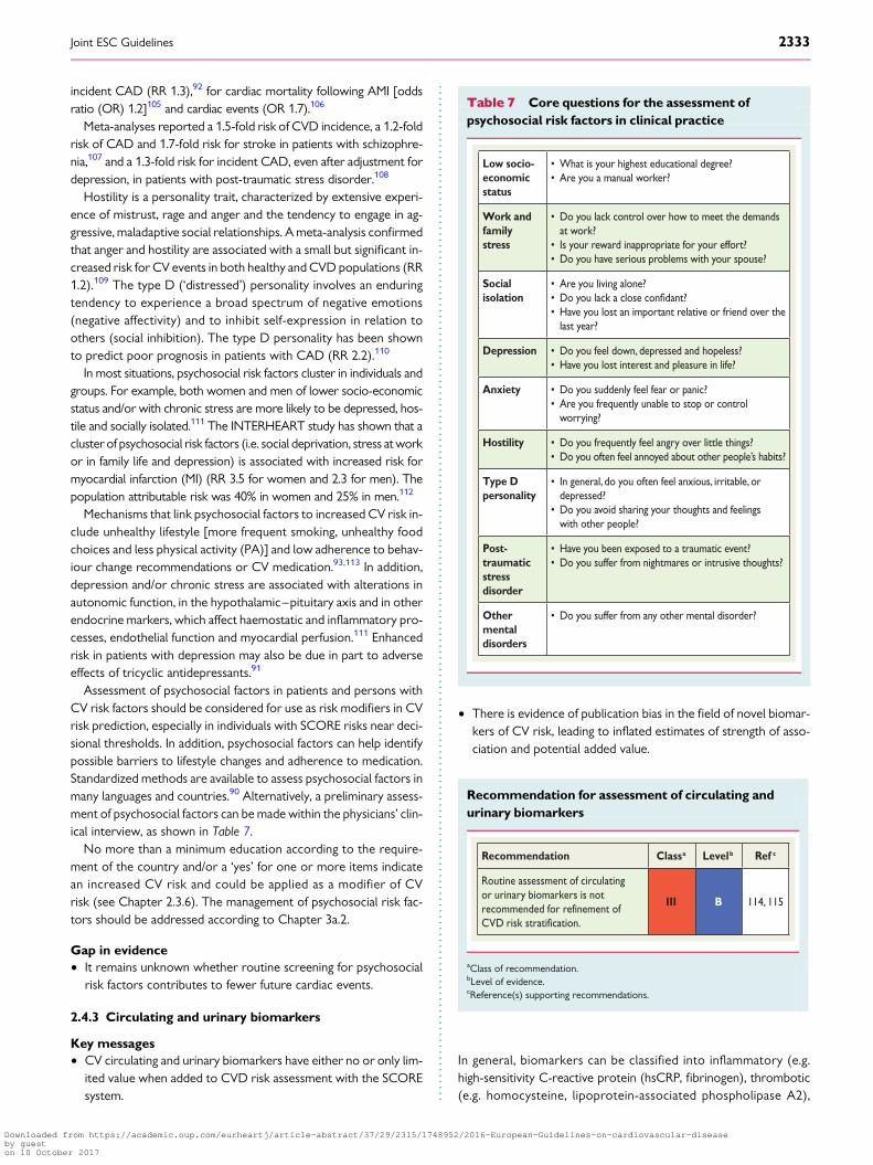
incident CAD (RR 1.3),92 for cardiac mortality following AMI [oddsratio (OR) 1.2]105 and cardiac events (OR 1.7).106
Meta-analyses reported a 1.5-fold risk of CVD incidence, a 1.2-foldrisk of CAD and 1.7-fold risk for stroke in patients with schizophre-nia,107 and a 1.3-fold risk for incident CAD, even after adjustment fordepression, in patients with post-traumatic stress disorder.108
Hostility is a personality trait, characterized by extensive experi-ence of mistrust, rage and anger and the tendency to engage in ag-gressive, maladaptive social relationships. A meta-analysis confirmedthat anger and hostility are associated with a small but significant in-creased risk for CV events in both healthy and CVD populations (RR1.2).109 The type D (‘distressed’) personality involves an enduringtendency to experience a broad spectrum of negative emotions(negative affectivity) and to inhibit self-expression in relation toothers (social inhibition). The type D personality has been shownto predict poor prognosis in patients with CAD (RR 2.2).110
In most situations, psychosocial risk factors cluster in individuals andgroups. For example, both women and men of lower socio-economicstatus and/or with chronic stress are more likely to be depressed, hos-tile and socially isolated.111 The INTERHEART study has shown that acluster of psychosocial risk factors (i.e. social deprivation, stress at workor in family life and depression) is associated with increased risk formyocardial infarction (MI) (RR 3.5 for women and 2.3 for men). Thepopulation attributable risk was 40% in women and 25% in men.112
Mechanisms that link psychosocial factors to increased CV risk in-clude unhealthy lifestyle [more frequent smoking, unhealthy foodchoices and less physical activity (PA)] and low adherence to behav-iour change recommendations or CV medication.93,113 In addition,depression and/or chronic stress are associated with alterations inautonomic function, in the hypothalamic–pituitary axis and in otherendocrine markers, which affect haemostatic and inflammatory pro-cesses, endothelial function and myocardial perfusion.111 Enhancedrisk in patients with depression may also be due in part to adverseeffects of tricyclic antidepressants.91
Assessment of psychosocial factors in patients and persons withCV risk factors should be considered for use as risk modifiers in CVrisk prediction, especially in individuals with SCORE risks near deci-sional thresholds. In addition, psychosocial factors can help identifypossible barriers to lifestyle changes and adherence to medication.Standardized methods are available to assess psychosocial factors inmany languages and countries.90 Alternatively, a preliminary assess-ment of psychosocial factors can be made within the physicians’ clin-ical interview, as shown in Table 7.
No more than a minimum education according to the require-ment of the country and/or a ‘yes’ for one or more items indicatean increased CV risk and could be applied as a modifier of CVrisk (see Chapter 2.3.6). The management of psychosocial risk fac-tors should be addressed according to Chapter 3a.2.
Gap in evidence† It remains unknown whether routine screening for psychosocial
risk factors contributes to fewer future cardiac events.
2.4.3 Circulating and urinary biomarkers
Key messages† CV circulating and urinary biomarkers have either no or only lim-
ited value when added to CVD risk assessment with the SCOREsystem.
† There is evidence of publication bias in the field of novel biomar-kers of CV risk, leading to inflated estimates of strength of asso-ciation and potential added value.
Recommendation for assessment of circulating andurinary biomarkers
Recommendation Classa Levelb Ref c
Routine assessment of circulating or urinary biomarkers is not recommended for refinement of CVD risk stratification.
III B 114, 115
aClass of recommendation.bLevel of evidence.cReference(s) supporting recommendations.
In general, biomarkers can be classified into inflammatory (e.g.high-sensitivity C-reactive protein (hsCRP, fibrinogen), thrombotic(e.g. homocysteine, lipoprotein-associated phospholipase A2),
Table 7 Core questions for the assessment ofpsychosocial risk factors in clinical practice
Low socio-economic status
• What is your highest educational degree?• Are you a manual worker?
Work and family stress
• Do you lack control over how to meet the demands at work?
• Is your reward inappropriate for your effort?• Do you have serious problems with your spouse?
Social isolation
• Are you living alone? • Do you lack a close • Have you lost an important relative or friend over the
last year?
Depression • Do you feel down, depressed and hopeless? • Have you lost interest and pleasure in life?
Anxiety • Do you suddenly feel fear or panic? • Are you frequently unable to stop or control
worrying?
Hostility • Do you frequently feel angry over little things? • Do you often feel annoyed about other people’s habits?
Type D personality
• In general, do you often feel anxious, irritable, ordepressed?
• Do you avoid sharing your thoughts and feelings with other people?
Post-traumatic stress disorder
• Have you been exposed to a traumatic event?• Do you suffer from nightmares or intrusive thoughts?
Other mental disorders
• Do you suffer from any other mental disorder?
Joint ESC Guidelines 2333
Downloaded from https://academic.oup.com/eurheartj/article-abstract/37/29/2315/1748952/2016-European-Guidelines-on-cardiovascular-diseaseby gueston 10 October 2017
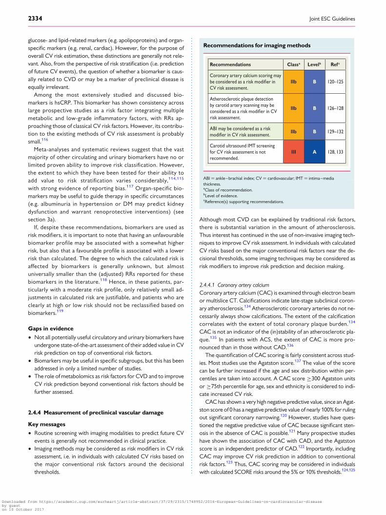
glucose- and lipid-related markers (e.g. apolipoproteins) and organ-specific markers (e.g. renal, cardiac). However, for the purpose ofoverall CV risk estimation, these distinctions are generally not rele-vant. Also, from the perspective of risk stratification (i.e. predictionof future CV events), the question of whether a biomarker is caus-ally related to CVD or may be a marker of preclinical disease isequally irrelevant.
Among the most extensively studied and discussed bio-markers is hsCRP. This biomarker has shown consistency acrosslarge prospective studies as a risk factor integrating multiplemetabolic and low-grade inflammatory factors, with RRs ap-proaching those of classical CV risk factors. However, its contribu-tion to the existing methods of CV risk assessment is probablysmall.116
Meta-analyses and systematic reviews suggest that the vastmajority of other circulating and urinary biomarkers have no orlimited proven ability to improve risk classification. However,the extent to which they have been tested for their ability toadd value to risk stratification varies considerably,114,115
with strong evidence of reporting bias.117 Organ-specific bio-markers may be useful to guide therapy in specific circumstances(e.g. albuminuria in hypertension or DM may predict kidneydysfunction and warrant renoprotective interventions) (seesection 3a).
If, despite these recommendations, biomarkers are used asrisk modifiers, it is important to note that having an unfavourablebiomarker profile may be associated with a somewhat higherrisk, but also that a favourable profile is associated with a lowerrisk than calculated. The degree to which the calculated risk isaffected by biomarkers is generally unknown, but almostuniversally smaller than the (adjusted) RRs reported for thesebiomarkers in the literature.118 Hence, in these patients, par-ticularly with a moderate risk profile, only relatively small ad-justments in calculated risk are justifiable, and patients who areclearly at high or low risk should not be reclassified based onbiomarkers.119
Gaps in evidence
† Not all potentially useful circulatory and urinary biomarkers haveundergone state-of-the-art assessment of their added value in CVrisk prediction on top of conventional risk factors.
† Biomarkers may be useful in specific subgroups, but this has beenaddressed in only a limited number of studies.
† The role of metabolomics as risk factors for CVD and to improveCV risk prediction beyond conventional risk factors should befurther assessed.
2.4.4 Measurement of preclinical vascular damage
Key messages
† Routine screening with imaging modalities to predict future CVevents is generally not recommended in clinical practice.
† Imaging methods may be considered as risk modifiers in CV riskassessment, i.e. in individuals with calculated CV risks based onthe major conventional risk factors around the decisionalthresholds.
Recommendations for imaging methods
Recommendations Classa Levelb Ref c
Coronary artery calcium scoring may be considered as a risk in CV risk assessment.
IIb B 120–125
Atherosclerotic plaque detection by carotid artery scanning may be considered as a risk in CV risk assessment.
IIb B 126–128
ABI may be considered as a risk in CV risk assessment.
IIb B 129–132
Carotid ultrasound IMT screening for CV risk assessment is not recommended.
III A 128, 133
ABI ¼ ankle–brachial index; CV ¼ cardiovascular; IMT ¼ intima–mediathickness.aClass of recommendation.bLevel of evidence.cReference(s) supporting recommendations.
Although most CVD can be explained by traditional risk factors,there is substantial variation in the amount of atherosclerosis.Thus interest has continued in the use of non-invasive imaging tech-niques to improve CV risk assessment. In individuals with calculatedCV risks based on the major conventional risk factors near the de-cisional thresholds, some imaging techniques may be considered asrisk modifiers to improve risk prediction and decision making.
2.4.4.1 Coronary artery calciumCoronary artery calcium (CAC) is examined through electron beamor multislice CT. Calcifications indicate late-stage subclinical coron-ary atherosclerosis.134 Atherosclerotic coronary arteries do not ne-cessarily always show calcifications. The extent of the calcificationcorrelates with the extent of total coronary plaque burden.134
CAC is not an indicator of the (in)stability of an atherosclerotic pla-que.135 In patients with ACS, the extent of CAC is more pro-nounced than in those without CAD.136
The quantification of CAC scoring is fairly consistent across stud-ies. Most studies use the Agatston score.137 The value of the scorecan be further increased if the age and sex distribution within per-centiles are taken into account. A CAC score ≥300 Agatston unitsor ≥75th percentile for age, sex and ethnicity is considered to indi-cate increased CV risk.
CAC has shown a very high negative predictive value, since an Agat-ston score of 0 has a negative predictive value of nearly 100% for rulingout significant coronary narrowing.120 However, studies have ques-tioned the negative predictive value of CAC because significant sten-osis in the absence of CAC is possible.121 Many prospective studieshave shown the association of CAC with CAD, and the Agatstonscore is an independent predictor of CAD.122 Importantly, includingCAC may improve CV risk prediction in addition to conventionalrisk factors.123 Thus, CAC scoring may be considered in individualswith calculated SCORE risks around the 5% or 10% thresholds.124,125
Joint ESC Guidelines2334
Downloaded from https://academic.oup.com/eurheartj/article-abstract/37/29/2315/1748952/2016-European-Guidelines-on-cardiovascular-diseaseby gueston 10 October 2017

Although recent studies also showed the presence of CAC inlow-risk populations, the added predictive value on CV events re-mains to be demonstrated.138– 140
There are concerns regarding costs and radiation exposure. ForCAC scoring, the radiation exposure with properly selected techni-ques is +1 mSv.
2.4.4.2 Carotid ultrasoundPopulation-based studies have shown correlations between the se-verity of atherosclerosis in one arterial territory and the involvementof other arteries.126 Therefore, early detection of arterial disease inapparently healthy individuals has focused on peripheral arteries,and in particular on the carotid arteries. Risk assessment using carotidultrasound focuses on the measurement of the intima–media thick-ness (IMT) and the presence and characteristics of plaques.
The IMT is not only a measure of early atherosclerosis, but also ofsmooth muscle hypertrophy/hyperplasia. There is a graded increasein CV risk with increasing IMT,126 and a value .0.9 mm is consid-ered abnormal. The risk of stroke associated with IMT is non-linear,with hazards increasing more rapidly at lower IMTs than at higherIMTs. The IMT-associated risk of cardiac events is also non-linear.127
The extent of carotid IMT is an independent predictor of CVD, butseems to be more predictive in women than in men.
The lack of standardization regarding the definition and measure-ment of IMT, its high variability and low intra-individual reproducibil-ity have raised concerns. A recent meta-analysis failed todemonstrate any added value of IMT compared to the FraminghamRisk Score in predicting future CVD, even in the intermediate riskgroup.128 Thus, the systematic use of carotid ultrasound IMT to im-prove risk assessment is not recommended.
Plaque is usually defined as the presence of a focal wall thickeningthat it is at least 50% greater than the surrounding vessel wall or as afocal region with an IMT measurement ≥1.5 mm that protrudesinto the lumen.141 Plaques may be characterized by their number,size, irregularity and echodensity (echolucent vs. calcified). Plaquesare related to both coronary and cerebrovascular events, and echo-lucent (as opposed to calcified) plaques increase ischaemic cerebro-vascular events.127 Many studies emphasize the greater value ofmeasures that include plaque area and thickness, rather than IMTalone, in predicting CVD. Therefore, even though formal reclassifi-cation analyses have not been undertaken, carotid artery plaque as-sessment using ultrasonography may be considered to be a riskmodifier in CV risk prediction in some cases.
2.4.4.3 Arterial stiffnessArterial stiffness is commonly measured using either aortic pulsewave velocity (PWV) or arterial augmentation index. An increasein arterial stiffness is usually related to damage in the arterial wall,as has been shown in hypertensive patients.142 Although the rela-tionship between aortic stiffness and CVD is continuous, a PWVthreshold of 12 m/s has been suggested as a conservative estimateof significant alterations of aortic function in middle-aged hyperten-sive patients. A meta-analysis showed that arterial stiffness predictsfuture CVD and improves risk classification.142 However, the valid-ity of this conclusion is offset by evidence of substantial publicationbias.117 The Task Force concludes that arterial stiffness may serve asa useful biomarker to improve CV risk prediction for patients close
to decisional thresholds, but its systematic use in the general popu-lation to improve risk assessment is not recommended.
2.4.4.4 Ankle–brachial indexThe ankle–brachial index (ABI) is an easy-to-perform and reprodu-cible test to detect asymptomatic atherosclerotic disease. An ABI,0.9 indicates ≥50% stenosis between the aorta and the distalleg arteries. Because of its acceptable sensitivity (79%) and specifi-city (90%),131 an ABI ,0.90 is considered to be a reliable markerof peripheral artery disease (PAD).129 An ABI value indicating signifi-cant PAD adds value to the medical history, because 50–89% of pa-tients with an ABI ,0.9 do not have typical claudication130 and it ispresent in 12–27% of asymptomatic individuals .55 years of age.
The ABI is inversely related to CV risk,132 but there is controversyregarding its potential to reclassify patients into different riskcategories.131,143
2.4.4.5. EchocardiographyEchocardiography is more sensitive than electrocardiography indiagnosing left ventricular hypertrophy (LVH) and it precisely quan-tifies left ventricular (LV) mass and geometric LVH patterns. Cardiacabnormalities detected by echocardiography have an additional pre-dictive power.144,145 In view of the lack of convincing evidence thatechocardiography improves CV risk reclassification, and because ofthe logistical challenges in performing it, this imaging tool is not re-commended to improve CV risk prediction.
Gaps in evidence† Currently, most imaging techniques have not been rigorously
tested as screening tools in CV risk assessment; more evidenceon calibration, reclassification and cost-effectiveness is still needed.
† The reduction of CVD risk in patients treated with lipid- orBP-lowering drugs because of reclassification with, for example,CAC or ABI remains to be demonstrated.
2.4.5 Clinical conditions affecting cardiovascular diseaserisk2.4.5.1 Chronic kidney disease
Key message† CKD is associated with an increased risk of CVD, independent of
conventional CVD risk factors.
Hypertension, dyslipidaemia and DM are common among patientswith CKD. In addition, inflammatory mediators and promoters ofcalcification cause vascular injury and may explain why CKD is asso-ciated with CVD even after adjustment for conventional risk fac-tors.146 A decreasing estimated glomerular filtration rate (eGFR)is an important sign of a gradually increasing risk for CVD-relatedmortality, starting at ,75 mL/min/1.73 m2 and gradually increasingto an approximate three-fold risk in patients with values of 15 mL/min/1.73 m2. End-stage renal disease is associated with a very highCV risk. Independent of eGFR, increased albumin excretion is alsoassociated with CV mortality risk; the RR is �2.5 in overt protein-uria.147 Studies assessing whether the accuracy of CV risk stratifica-tion improves with the addition of eGFR levels are emerging,148 butthere is no consensus on which measure of renal function (i.e. whichformula, and creatinine- or cystatine-C-based) best predicts
Joint ESC Guidelines 2335
Downloaded from https://academic.oup.com/eurheartj/article-abstract/37/29/2315/1748952/2016-European-Guidelines-on-cardiovascular-diseaseby gueston 10 October 2017

CVD.149,150 Based on the evidence, the Task Force decided to clas-sify patients with severe CKD (GFR ,30 mL/min/1.73 m2) as ‘veryhigh risk’ and those with moderate CKD (GFR 30–59 mL/min/1.73m2) as ‘high risk’ (see Table 5).
Gap in evidence† The contribution of various CKD markers to CVD risk stratifica-
tion remains unclear.
2.4.5.2 Influenza
Key message† There is an association between acute respiratory infections, es-
pecially those occurring at times of peak influenza virus circula-tion, and AMI.
Recommendation for influenza vaccination
Recommendation Classa Levelb Ref c
Annual vaccination may be considered in patients with established CVD.
IIb C 151–154
aClass of recommendation.bLevel of evidence.cReference(s) supporting recommendations.
Influenza can trigger a CV event. Studies show an increase in rates ofMI during the annual influenza season. The risk of MI or stroke wasmore than four times higher after a respiratory tract infection, withthe highest risk in the first 3 days.151 A recent meta-analysis suggeststhat preventing influenza, particularly by means of vaccination, canprevent influenza-triggered AMI,154 but there is concern thatsome studies are biased.151 –153,155
Gap in evidence† Large-scale RCTs are needed to assess the efficacy of influenza
vaccination in preventing influenza-triggered AMI.
2.4.5.3 PeriodontitisStudies have linked periodontal disease to both atherosclerosis andCVD,156,157 and serological studies have linked elevated periodontalbacteria antibody titres to atherosclerotic disease.158 A longitudinalstudy has suggested that an improvement in clinical and microbialperiodontal status is related to a decreased rate of carotid arteryIMT progression during a 3 year follow-up period,159 but IMT pro-gression does not seem to be associated with CV events.133 Thus, ifactive treatment or prevention of periodontitis improves, clinicalprognosis is still unclear.
2.4.5.4 Patients treated for cancer
Key messages† Patients surviving cancer after treatment with chemotherapy or
radiotherapy are at increased risk for CVD.† The increased incidence of CVD is correlated with the (combin-
ation of) treatments given and the administered dose.
† The presence of traditional CV risk factors in cancer patients fur-ther increases CV risk.
Recommendations for patients treated for cancer
Recommendations Classa Levelb Ref c
Cardio-protection in high-riskpatientsd receiving type I chemotherapy should be considered for LV dysfunction prevention
IIa B 160, 161
Optimization of the CV risk should be considered in cancer treated patients.
IIa C
CV ¼ cardiovascular; LV ¼ left ventricular.aClass of recommendation.bLevel of evidence.cReference(s) supporting recommendations.dHigh-risk patients are mainly those individuals receiving high cumulative doses oftype I chemotherapy and/or combined treatment with other chemotherapicagents and radiotherapy, and/or with CV uncontrolled risk factors.
Survivors of cancer represent an increasingly large population, mostof whom have received chemotherapy and/or radiotherapy. Cardi-otoxicity due to chemotherapy is related to a direct effect on thecell (anthracycline-like) through the generation of reactive oxygenspecies (ROS). It can be mediated by topoisomerase IIb in cardio-myocytes through the formation of ternary complexes (topoisom-erase IIb–anthracycline–DNA) inducing DNA double-strandbreaks and transcriptome changes responsible for defective mito-chondrial biogenesis and ROS formation. Some agents (fluorouracil,bevacizumab, sorafenib and sunitinib) can induce a direct ischaemiceffect not related to the premature development of atheroscleroticlesions. Moreover, they can increase risk factors such as hyperten-sion and accelerate atherosclerosis, especially in older patients.These effects can be irreversible (type I agents) or partially revers-ible (type II agents) and can develop many years after treatment ex-posure. Typically, anthracyclines are the prototype of type I agentsand trastuzumab of type II agents.162
Cardiotoxicity due to chest radiotherapy can induce micro- andmacrovascular injury. It can accelerate atherosclerosis, but this mayoccur many years after the initial exposure.163– 169 The latency andseverity of radiotherapy cardiotoxicity is related to multiple factors,including the dose (total per fraction), the volume of the heart irra-diated, concomitant administration of other cardiotoxic drugs andpatient factors (younger age, traditional risk factors,170 history ofheart disease).
The first step in the identification of higher risk for cardio-toxicity consists of a careful baseline assessment of CV risk factors.Primary care, cardiology and oncology should work togetherto deliver optimal survivorship care that addresses CVD risk fac-tors as well as prevalent disease. Positive health-promoting behav-iour, including lifestyle factors (healthy diet, smoking cessation,regular exercise, weight control) should be strongly advised. Inparticular, aerobic exercise is considered as a promising non-pharmacological strategy to prevent and/or treat chemotherapy-induced cardiotoxicity.171
Joint ESC Guidelines2336
Downloaded from https://academic.oup.com/eurheartj/article-abstract/37/29/2315/1748952/2016-European-Guidelines-on-cardiovascular-diseaseby gueston 10 October 2017

Signs or symptoms of cardiac dysfunction should be monitoredbefore and periodically during treatment for early detection ofeven asymptomatic abnormalities in patients receiving potentiallycardiotoxic chemotherapy, and heart failure (HF) guideline recom-mendations should be followed if indicated.172 Thus, pretreatmentevaluation of LV function is required.173 A targeted approach totreat patients with early LV dysfunction, in combination with globallongitudinal strain abnormalities and biomarker (notably troponin)elevation, has been proposed.173,174
In the case of a decrease in LV function during or after chemother-apy, the use of cardiotoxic agents should be avoided or delayed, ifpossible, until after discussion with the oncology team. This callsfor adequate communication between oncology and cardiology.
To reduce chemotherapy type I cardiotoxicity, a variety ofprophylactic treatments, including b-blockers, angiotensin-converting enzyme inhibitors (ACE-Is), dexrazozane and statins,has been tested and compiled in a recent meta-analysis.161 It hasbeen stressed that early preventive treatment is mandatory to exerta maximum effect.173 –176
Gaps in evidence† Evidence on the effect of early preventive measures to reduce
type I cardiotoxicity is inconclusive.† The most appropriate strategy to improve risk stratification and
prevent CVD in patients treated for cancer needs to be testedprospectively.
2.4.5.5 Autoimmune disease
Key messages† Rheumatoid arthritis (RA) enhances CV risk independently of
traditional risk factors, with an RR of 1.4 and 1.5 in men and wo-men, respectively.
† There is mounting evidence that other immune diseases, such asankylosing spondylitis or early severe psoriasis, also increase CVrisk, with RRs approaching those in RA.
† Post hoc analysis of two statin trials suggests that the relative re-duction in CVD incidence in autoimmune diseases is comparableto that seen in the other conditions.
Recommendations for autoimmune disease
Recommendations Classa Levelb Ref c
The use of a 1.5 factor risk multiplier for CV risk in rheumatoid arthritis should be considered, particularly if disease activity is high.
IIa B 177
The use of a 1.5 risk multiplier for CV risk in immune diseases other than rheumatoid arthritis may be considered on a patient-by-patient basis, depending on disease activity/severity.
IIb C 177
aClass of recommendation.bLevel of evidence.cReference(s) supporting recommendations.
There is now clear evidence implicating high-grade inflammation as apathway for accelerated vascular disease.178 Systemic inflammationappears to enhance CV risk directly and indirectly via accentuationof existing risk pathways.178 While early small studies suggested RAincreases CV risk beyond other risk markers, the recent analysis ofthe national QRESEARCH database in 2.3 million people providesthe best available evidence for this.47 Such evidence has nowbeen implemented in some national risk scores58 and Europeanguidelines.177
Evidence in psoriasis is less rigorous, but a recent paper demon-strates broadly comparable CV risks in RA and in early severe psor-iasis.179 Robust data for independently elevated CV risks in otherautoimmune conditions are generally lacking. Hence, clinical judg-ment should be applied on a case-by-case basis. There is evidencefrom post hoc analysis of randomized trials to support astatin-associated reduction in CV risk in autoimmune conditions.180
Finally, in all autoimmune diseases, drug interactions of anti-inflammatory and immunosuppressive drugs with, for example, sta-tins, antiplatelet agents and antihypertensive agents deserveattention.
Gaps in evidence† The association between non-RA immune inflammatory disease
and CVD is less clear than for RA.† The relationship between anti-rheumatic drugs and CV risk is
unknown.
2.4.5.6 Obstructive sleep apnoea syndrome
Key message† There is evidence of a positive relationship between obstructive
sleep apnoea syndrome (OSAS) and hypertension, CAD, atrialfibrillation (AF), stroke, and HF.
OSAS is characterized by recurrent partial or complete collapseof the upper airway during sleep. It affects an estimated 9% ofadult women and 24% of adult men and has been associatedwith an RR of 1.7 for CV morbidity and mortality.181 Repetitivebursts of sympathetic activity, surges of BP and oxidative stressbrought on by pain and episodic hypoxaemia associated withincreased levels of mediators of inflammation are thought topromote endothelial dysfunction and atherosclerosis.181 Screen-ing for OSAS can be performed using the Berlin Questionnaireand daytime sleepiness can be assessed by the Epworth SleepinessScale and overnight oximetry.182 Definitive diagnosis oftenrequires polysomnography, usually during a night in a sleeplaboratory during which multiple physiological variables arecontinuously recorded. Treatment options include behaviouralchanges, such as avoiding alcohol, caffeine or other stimulantsof wakefulness before sleep, increased PA, discontinuation ofsedating drugs and obesity control. Continuous positive airwaypressure is the gold-standard therapy and reduces CV mortalityand events.183
Gap in evidence† More studies are needed to determine whether routine screen-
ing reduces (non)fatal CVD.
Joint ESC Guidelines 2337
Downloaded from https://academic.oup.com/eurheartj/article-abstract/37/29/2315/1748952/2016-European-Guidelines-on-cardiovascular-diseaseby gueston 10 October 2017
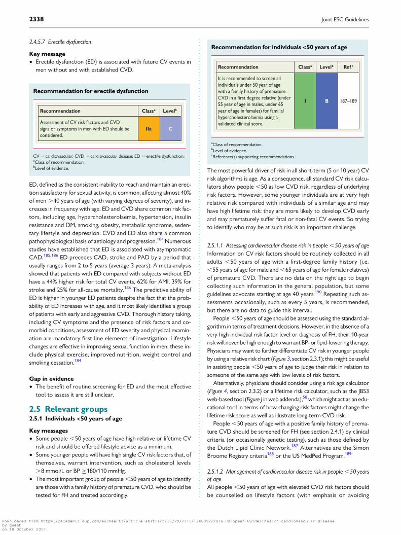
2.4.5.7 Erectile dysfunction
Key message† Erectile dysfunction (ED) is associated with future CV events in
men without and with established CVD.
Recommendation for erectile dysfunction
Recommendation Classa Levelb
Assessment of CV risk factors and CVD signs or symptoms in men with ED should be considered.
IIa C
CV ¼ cardiovascular; CVD ¼ cardiovascular disease; ED ¼ erectile dysfunction.aClass of recommendation.bLevel of evidence.
ED, defined as the consistent inability to reach and maintain an erec-tion satisfactory for sexual activity, is common, affecting almost 40%of men .40 years of age (with varying degrees of severity), and in-creases in frequency with age. ED and CVD share common risk fac-tors, including age, hypercholesterolaemia, hypertension, insulinresistance and DM, smoking, obesity, metabolic syndrome, seden-tary lifestyle and depression. CVD and ED also share a commonpathophysiological basis of aetiology and progression.184 Numerousstudies have established that ED is associated with asymptomaticCAD.185,186 ED precedes CAD, stroke and PAD by a period thatusually ranges from 2 to 5 years (average 3 years). A meta-analysisshowed that patients with ED compared with subjects without EDhave a 44% higher risk for total CV events, 62% for AMI, 39% forstroke and 25% for all-cause mortality.186 The predictive ability ofED is higher in younger ED patients despite the fact that the prob-ability of ED increases with age, and it most likely identifies a groupof patients with early and aggressive CVD. Thorough history taking,including CV symptoms and the presence of risk factors and co-morbid conditions, assessment of ED severity and physical examin-ation are mandatory first-line elements of investigation. Lifestylechanges are effective in improving sexual function in men: these in-clude physical exercise, improved nutrition, weight control andsmoking cessation.184
Gap in evidence† The benefit of routine screening for ED and the most effective
tool to assess it are still unclear.
2.5 Relevant groups2.5.1 Individuals <50 years of age
Key messages† Some people ,50 years of age have high relative or lifetime CV
risk and should be offered lifestyle advice as a minimum.† Some younger people will have high single CV risk factors that, of
themselves, warrant intervention, such as cholesterol levels.8 mmol/L or BP ≥180/110 mmHg.
† The most important group of people ,50 years of age to identifyare those with a family history of premature CVD, who should betested for FH and treated accordingly.
Recommendation for individuals <50 years of age
Recommendation Classa Levelb Ref c
It is recommended to screen all individuals under 50 year of age with a family history of premature CVD in a degree relative (under 55 year of age in males, under 65 year of age in females) for familial hypercholesterolaemia using a validated clinical score.
I B 187–189
aClass of recommendation.bLevel of evidence.cReference(s) supporting recommendations.
The most powerful driver of risk in all short-term (5 or 10 year) CVrisk algorithms is age. As a consequence, all standard CV risk calcu-lators show people ,50 as low CVD risk, regardless of underlyingrisk factors. However, some younger individuals are at very highrelative risk compared with individuals of a similar age and mayhave high lifetime risk: they are more likely to develop CVD earlyand may prematurely suffer fatal or non-fatal CV events. So tryingto identify who may be at such risk is an important challenge.
2.5.1.1 Assessing cardiovascular disease risk in people ,50 years of ageInformation on CV risk factors should be routinely collected in alladults ,50 years of age with a first-degree family history (i.e.,55 years of age for male and ,65 years of age for female relatives)of premature CVD. There are no data on the right age to begincollecting such information in the general population, but someguidelines advocate starting at age 40 years.190 Repeating such as-sessments occasionally, such as every 5 years, is recommended,but there are no data to guide this interval.
People ,50 years of age should be assessed using the standard al-gorithm in terms of treatment decisions. However, in the absence of avery high individual risk factor level or diagnosis of FH, their 10-yearrisk will never be high enough to warrant BP- or lipid-lowering therapy.Physicians may want to further differentiate CV risk in younger peopleby using a relative risk chart (Figure 3, section 2.3.1); this might be usefulin assisting people ,50 years of age to judge their risk in relation tosomeone of the same age with low levels of risk factors.
Alternatively, physicians should consider using a risk age calculator(Figure 4, section 2.3.2) or a lifetime risk calculator, such as the JBS3web-based tool (Figure J in web addenda),58 which might act as an edu-cational tool in terms of how changing risk factors might change thelifetime risk score as well as illustrate long-term CVD risk.
People ,50 years of age with a positive family history of prema-ture CVD should be screened for FH (see section 2.4.1) by clinicalcriteria (or occasionally genetic testing), such as those defined bythe Dutch Lipid Clinic Network.187 Alternatives are the SimonBroome Registry criteria188 or the US MedPed Program.189
2.5.1.2 Management of cardiovascular disease risk in people ,50 yearsof ageAll people ,50 years of age with elevated CVD risk factors shouldbe counselled on lifestyle factors (with emphasis on avoiding
Joint ESC Guidelines2338
Downloaded from https://academic.oup.com/eurheartj/article-abstract/37/29/2315/1748952/2016-European-Guidelines-on-cardiovascular-diseaseby gueston 10 October 2017

smoking, overweight and sedentary behaviour) and the relationshipbetween risk factors and subsequent disease. There are no data onwhat are the most effective methods of changing health behavioursin younger people. However, smoking cessation, healthy weightmaintenance and regular aerobic activity are all important beha-viours on which to provide advice and support.
Younger people with very high BP levels warranting treatmentshould be managed in the same way as older people with hyperten-sion. In younger people who are judged eligible for a statin on thegrounds of either FH or very high lipid levels, the management of-fered is the same as for older people. Very importantly, for all pa-tients deemed to suffer with FH, the physician making themanagement decisions should arrange for FH screening for familymembers (see section 3a.7.9).
Gaps in evidence† Age to commence formal CV risk estimation.† Whether and how to screen populations for FH.
2.5.2 ElderlyAge is the dominant driver of cardiovascular risk, and most individualsare already at (very) high risk at the age of 65 years (see section 2.3.1).Especially in the oldest old, cardiovascular risk management is contro-versial. Opponents argue that risk should not be treated when it is es-sentially age-driven. Proponents, on the other hand, point out thatmany preventive treatments are still effective at advanced age in termsof postponing morbidity and mortality.
The Task Force has taken the position that epidemiological evi-dence of absolute risk reduction in clinical trials is the main driverfor recommendations in this guideline. Still, we encourage a discus-sion with patients regarding quality of life and life potentially gained,as well as regarding the ethical dilemmas of treating risk inherent toageing, the total burden of drug treatment and the inevitable uncer-tainties of benefit.
In this guideline, sections on treatment of the main risk factorscontain recommendations or considerations specific to the elderlywhen evidence is available.
2.5.2.1 HypertensionMost of the elderly-specific evidence is available for BP (section3a.9). In general, more lenient treatment targets are advocated inthe elderly. The hypertension literature also contains increasing evi-dence that biological rather than calendar age is important.191
2.5.2.2 Diabetes mellitusEvidence supporting more lenient glycaemic control targets in theelderly is also available for DM (section 3a.8). The role of biologicalage/frailty is less well established than for BP, but nonetheless, aClass IIa recommendation is given to relax glycaemic targets in eld-erly or frail patients.
2.5.2.3 HyperlipidaemiaFew areas in CVD prevention are more controversial than the massuse of statins in the elderly. As the section on lipid control points out,there is no evidence of decreasing effectiveness of statins in patients.75 years of age (section 3a.7). On the other hand, the cost-effectiveness of statins in these patients is offset by even smallgeriatric-specific adverse effects.192 Also, evidence supporting
effectiveness in the oldest old (i.e. .80 years of age) is very limited.A recent trial suggested no harm of stopping statins in the elderly witha limited life expectancy.193 Taken together, the recommendations ofcholesterol-lowering treatment in the elderly should be followed withcaution and common sense, adverse effects should be monitoredclosely and treatment should be reconsidered periodically.
2.5.3 Female-specific conditions
Key messages† Several obstetric complications, in particular pre-eclampsia and
pregnancy-related hypertension, are associated with a higherrisk of CVD later in life. This higher risk is explained, at least part-ly, by hypertension and DM.
† Polycystic ovary syndrome (PCOS) confers a significant risk forfuture development of DM.
Recommendations for female-specific conditions
Recommendations Classa Levelb Ref c
In women with a history of pre-eclampsia and/or pregnancy-induced hypertension, periodic screening for hypertension and DM should be considered.
IIa B 194–197
In women with a history of polycystic ovary syndrome or gestational DM, periodic screening for DM should be considered.
IIa B 198–201
In women with a history of giving premature birth, periodic screening for hypertension and DM may be considered.
IIb B 202, 203
DM ¼ diabetes mellitus; PCOS ¼ polycystic ovary syndrome.aClass of recommendation.bLevel of evidence.cReference(s) supporting recommendations.
Specific conditions that may occur in females only and may have animpact on CVD risk can be separated into obstetric and non-obstetric conditions.
2.5.3.1 Obstetric conditionsPre-eclampsia (defined as pregnancy-related hypertension accom-panied by proteinuria) occurs in 1–2% of all pregnancies. Studiessuggest that pre-eclampsia is associated with an increase in CVrisk by a factor 1.5–2.5,194,195 while the RR of developing hyperten-sion is �3196 and DM �2.194,197 Because most studies did not adjustthe elevated risk of future CVD for the development of convention-al risk factors, it cannot be established whether the increased CVrisk after pre-eclampsia occurs independent of CV risk factors.The rationale for screening these women for the occurrence ofhypertension and DM is, however, quite strong.
Pregnancy-related hypertension affects 10–15% of all pregnan-cies. The associated risk of later CVD is lower than for pre-eclampsia, but is still elevated (RR 1.9–2.5).202 Also, the risk for
Joint ESC Guidelines 2339
Downloaded from https://academic.oup.com/eurheartj/article-abstract/37/29/2315/1748952/2016-European-Guidelines-on-cardiovascular-diseaseby gueston 10 October 2017

sustained or future hypertension is elevated (RRs vary widely, from2.0 to 7.2 or even higher).196,204 Again, however, there was incom-plete adjustment for conventional risk factors. The risk of develop-ing DM is probably also elevated in these women, but exactestimates are not available.
There are no data to suggest that recurrent pregnancy loss is as-sociated with an increased CV risk. A history of premature birth ispossibly associated with an increased risk of CVD in offspring (RR1.5–2.0),202,203 which may be partially explained by an increased in-cidence of hypertension and DM.
Finally, gestational diabetes confers a sharply elevated risk of fu-ture DM, with up to 50% developing DM within 5 years after preg-nancy.200 Previously, oral glucose tolerance testing was advocatedto screen for DM in such patients, but screening by fasting glucoseor glycated haemoglobin may be preferable.201
2.5.3.2 Non-obstetric conditionsPCOS affects �5% of all women in their fertile years. PCOShas been associated with an increased risk for future development ofCVD, but larger studies have produced conflicting results.198,205 Therisk of developing hypertension is probably somewhat increased,but again the data are conflicting.205 PCOS does seem to be asso-ciated with a higher risk of developing DM (RR 2–4),198,199 suggest-ing that periodic screening for DM is appropriate.
Premature menopause, better defined as primary ovarian insuffi-ciency, occurs in roughly 1% in women ≤40 years of age. It hasbeen reported to be associated with an increased risk of CVD (RR�1.5),206 but studies are sparse. There are insufficient data to drawconclusions on a possible increased risk of hypertension or DM.
Gaps in evidence† The degree to which increased CVD risk associated with several
of the female-specific conditions occurs independent of conven-tional CVD risk factors is unknown.
† Information on whether female-specific conditions improve riskclassification in women is unknown.
2.5.4 Ethnic minorities
Key messages† CVD risk varies considerably between immigrant groups. South
Asians and sub-Saharan Africans have a higher risk, while Chineseand South Americans have a lower risk.
† South Asians are characterized by a high prevalence and inad-equate management of DM.
† Current risk estimation equations do not provide adequate esti-mations of CVD risk in ethnic minorities.
Recommendation for ethnic minorities
Recommendation Classa Levelb Ref c
Ethnicity should be considered in CVD risk assessment.
IIa A 207, 208
CVD ¼ cardiovascular disease.aClass of recommendation.bLevel of evidence.cReference(s) supporting recommendations.
Europe welcomes a large number of non-EU immigrants per year,mainly from India, China, North Africa and Pakistan. One of 25 Eur-opeans comes from outside Europe, but data regarding CVD risk orCVD risk factors among immigrants are scarce and of differingquality.209
First-generation migrants usually display lower CVD mortalityrates than natives of the host country,210 but with time, migrantstend to approach the CVD risk in their host country.210,211 Rela-tive to natives of the host country, CVD mortality risk, as well asthe prevalence and management of CVD risk factors among mi-grants, varies according to country of origin and host coun-try.211 – 213 Given the considerable variability in CVD risk factorsbetween immigrant groups, no single CVD risk score performs ad-equately in all groups and the use of ethnic-specific scores might benecessary.207
Immigrants from South Asia (notably India and Pakistan) presenthigh CVD rates214 – 216 and have a much higher prevalence ofDM,217,218 while the prevalence of other CV risk factors is slightlylower than or comparable to natives of the host country.217,219
Interestingly, the increased prevalence of DM increases the CVDrisk in South Asians in some studies214 but not in others. Manage-ment of DM is also significantly worse, while management ofhigh BP and hypercholesterolaemia is better among South Asiansthan host country natives.220 The higher CVD risk among SouthAsians makes screening more cost effective than in other immi-grant groups, but risk prediction using SCORE might not beoptimal.221
Immigrants from China and Vietnam present lower CVDrisk than natives of the host country,214 although thisfinding has been challenged.215 This lower risk seems attributableto lower levels of CV risk factors217 and higher HDL-Clevels.222
Immigrants from Turkey have higher estimated CVD riskand higher CVD mortality rates212 than host country natives.This seems mainly due to the higher prevalence of smoking,DM, dyslipidaemia, hypertension and obesity rates.222 – 224 Man-agement of CVD risk factors also varies according to the hostcountry; there are no differences in hypertension control com-pared with natives in The Netherlands,224 but there is worse con-trol in Denmark.225
Immigrants from Morocco present lower CVD rates than nativesfrom the host country.212 Possible explanations include lower BPand cholesterol levels and smoking rates,223,224 although a higherprevalence of DM and obesity has also been found.224 No differ-ences between Moroccan immigrants and Dutch natives were foundregarding hypertension control.223
Immigrants from sub-Saharan Africa and the Caribbean presenthigher CVD rates than natives from the host country in some stud-ies,213,214,226 but not all.214 African immigrants have higher DMrates218 but smoke less219 than natives from the host country. Man-agement of CVD risk factors was worse than among natives in onestudy,220 but not in another.227
Immigrants from South America have lower CVD mortalityrates than natives in Spain,228 while no difference was foundin Denmark.229 South American immigrants in Spain have alower prevalence of CV risk factors and CVD rates than nativesin Spain, but these differences decrease with increasing lengthof stay.230
Joint ESC Guidelines2340
Downloaded from https://academic.oup.com/eurheartj/article-abstract/37/29/2315/1748952/2016-European-Guidelines-on-cardiovascular-diseaseby gueston 10 October 2017
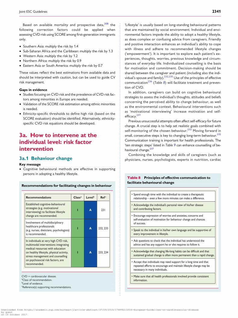
Based on available mortality and prospective data,208 thefollowing correction factors could be applied whenassessing CVD risk using SCORE among first-generation immigrantsonly.
† Southern Asia: multiply the risk by 1.4† Sub-Saharan Africa and the Caribbean: multiply the risk by 1.3† Western Asia: multiply the risk by 1.2† Northern Africa: multiply the risk by 0.9† Eastern Asia or South America: multiply the risk by 0.7
These values reflect the best estimations from available data andshould be interpreted with caution, but can be used to guide CVrisk management.
Gaps in evidence† Studies focusing on CVD risk and the prevalence of CVD risk fac-
tors among minorities in Europe are needed.† Validation of the SCORE risk estimation among ethnic minorities
is needed.† Ethnicity-specific thresholds to define high risk (based on the
SCORE evaluation) should be identified. Alternatively, ethnicity-specific CVD risk equations should be developed.
3a. How to intervene at theindividual level: risk factorintervention
3a.1 Behaviour changeKey message† Cognitive behavioural methods are effective in supporting
persons in adopting a healthy lifestyle.
Recommendations for facilitating changes in behaviour
Recommendations Classa Levelb Ref c
Established cognitive-behavioural strategies (e.g. motivational interviewing) to facilitate lifestyle change are recommended.
I A 231
Involvement of multidisciplinary healthcare professionals (e.g. nurses, dieticians, psychologists) is recommended.
I A 232, 233
In individuals at very high CVD risk, multimodal interventions integrating medical resources with education on healthy lifestyle, physical activity, stress management and counselling on psychosocial risk factors, are recommended.
I A 233, 234
CVD ¼ cardiovascular disease.aClass of recommendation.bLevel of evidence.cReference(s) supporting recommendations.
‘Lifestyle’ is usually based on long-standing behavioural patternsthat are maintained by social environment. Individual and envi-ronmental factors impede the ability to adopt a healthy lifestyle,as does complex or confusing advice from caregivers. Friendlyand positive interaction enhances an individual’s ability to copewith illness and adhere to recommended lifestyle changes(‘empowerment’). It is important to explore each patient’s ex-periences, thoughts, worries, previous knowledge and circum-stances of everyday life. Individualized counselling is the basisfor motivation and commitment. Decision-making should beshared between the caregiver and patient (including also the indi-vidual’s spouse and family).232,235 Use of the principles of effectivecommunication236 (Table 8) will facilitate treatment and preven-tion of CVD.
In addition, caregivers can build on cognitive behaviouralstrategies to assess the individual’s thoughts, attitudes and beliefsconcerning the perceived ability to change behaviour, as wellas the environmental context. Behavioural interventions suchas ‘motivational interviewing’ increase motivation and self-efficacy.231
Previous unsuccessful attempts often affect self-efficacy for futurechange. A crucial step is to help set realistic goals combined withself-monitoring of the chosen behaviour.232 Moving forward insmall, consecutive steps is key to changing long-term behaviour.232
Communication training is important for health professionals. The‘ten strategic steps’ listed in Table 9 can enhance counselling of be-havioural change.237
Combining the knowledge and skills of caregivers (such asphysicians, nurses, psychologists, experts in nutrition, cardiac
Table 8 Principles of effective communication tofacilitate behavioural change
• Spend enough time with the individual to create a therapeutic relationship – even a few more minutes can make a difference.
• Acknowledge the individual’s personal view of his/her disease and contributing factors.
• Encourage expression of worries and anxieties, concerns andself-evaluation of motivation for behaviour change and chances of success.
• Speak to the individual in his/her own language and be supportive of every improvement in lifestyle.
• Ask questions to check that the individual has understood the advice and has any support he or she requires to follow it.
• Acknowledge that changing life-long habits can be and thatsustained gradual change is often more permanent than a rapid change.
• Accept that individuals may need support for a long time and that repeated efforts to encourage and maintain lifestyle change may be necessary in many individuals.
• Make sure that all health professionals involved provide consistent information.
Joint ESC Guidelines 2341
Downloaded from https://academic.oup.com/eurheartj/article-abstract/37/29/2315/1748952/2016-European-Guidelines-on-cardiovascular-diseaseby gueston 10 October 2017

rehabilitation and sports medicine) into multimodal behaviouralinterventions can optimize preventive efforts.232 – 234 Multimodalbehavioural interventions are especially recommended for indivi-duals at very high risk.232 – 234 These interventions include promot-ing a healthy lifestyle through behaviour changes, includingnutrition, PA, relaxation training, weight management and smokingcessation programmes for resistant smokers.233,234 They enhancecoping with illness and improve adherence and CV outcome.238,239
Psychosocial risk factors (stress, social isolation, and negativeemotions) that may act as barriers against behaviour change shouldbe addressed in tailored individual or group counsellingsessions.233,234
There is evidence that more extensive/longer interventionslead to better long-term results with respect to behaviour changeand prognosis.232 Individuals of low socio-economic status, olderage or female sex may need tailored programmes in order tomeet their specific needs regarding information and emotionalsupport.232,240,241
Gap in evidence† There is limited evidence to determine which interventions are
most effective in specific groups (e.g. young–old, male–female,high vs. low socio-economic status).
3a.2 Psychosocial factorsKey messages† Treatment of psychosocial risk factors can counteract psycho-
social stress, depression and anxiety, thus facilitating behaviourchange and improving quality of life and prognosis.
† The caregiver–patient interaction should follow the principles ofpatient-centred communication. Age- and sex-specific psycho-social aspects should be considered.
Recommendations for psychosocial factors
Recommendations Classa Levelb Ref c
Multimodal behavioural interventions, integrating health education, physical exercise and psychological therapy, for psychosocial risk factors and coping with illness are recommended in patients with established CVD and psychosocial symptoms in order to improve psychosocial health.
I A 242
Referral for psychotherapy, medication or collaborative care should be considered in the case of clinically symptoms of depression, anxiety or hostility.
IIa A 243, 244
Treatment of psychosocial risk factors with the aim of preventing CAD should be considered when the risk factor itself is a diagnosable disorder (e.g. depression) or when the factor worsens classical risk factors.
IIa B 245, 246
CAD ¼ coronary artery disease; CVD ¼ cardiovascular disease.aClass of recommendation.bLevel of evidence.cReference(s) supporting recommendations.
Caregivers in clinical practice are in a unique position to directly sup-port their patients regarding psychosocial risk factors in individualswith high CV risk or with established disease. Empathic, patient-centred communication helps to establish and maintain a trustful re-lationship and is a powerful source of emotional support and profes-sional guidance in coping with psychosocial stressors, depression,anxiety, CV risk factors and CVD.247,248 The principles of a support-ive caregiver–patient interaction are247,248
† Spend enough time with the patient, listen carefully and repeatessential keywords.
† Consider age- and sex-specific psychosocial aspects.† Encourage expression of emotions, do not trivialize psychosocial
burdens and worries.† Explain essential medical facts in the patient’s own language, con-
vey hope and relief from feelings of guilt and reinforce adaptivethoughts and actions.
† In the case of severe mental symptoms, obtain treatment prefer-ences and perform shared decision-making regarding furtherdiagnostic and therapeutic steps.
† Summarize important aspects of the consultation to confirm thatthe patient has been understood.
† Offer regular follow-up contacts.
Table 9 Ten strategic steps to facilitate behaviourchange
1. Develop a therapeutic alliance.
2. Counsel all individuals at risk of or with manifest cardiovascular disease.
3. Assist individuals to understand the relationship between their behaviour and health.
4. Help individuals assess the barriers to behaviour change.
5. Gain commitments from individuals to own their behaviour change.
6. Involve individuals in identifying and selecting the risk factors to change.
7. Use a combination of strategies including reinforcement of the individual’s capacity for change.
8. Design a plan.
9. Involve other healthcare staff whenever possible.
10. Monitor progress through follow-up contact.
Joint ESC Guidelines2342
Downloaded from https://academic.oup.com/eurheartj/article-abstract/37/29/2315/1748952/2016-European-Guidelines-on-cardiovascular-diseaseby gueston 10 October 2017

Specialized psychological interventions have additional beneficialeffects on distress, depressiveness and anxiousness, even whenadded to standard rehabilitation.242 These interventions includeindividual or group counselling on psychosocial risk factors andcoping with illness, stress management programmes, meditation,autogenic training, biofeedback, breathing, yoga and/or muscularrelaxation.
Large and consistent effects on depression have been shown in‘collaborative care’, which may involve a systematic assessment ofdepression, a (non-physician) care manager to perform longitudin-al symptom monitoring, treatment interventions and care coord-ination and specialist-provided stepped care recommendationsand treatment.244 Collaborative care for depression resulted in a48% lower risk for developing first CAD events 8 years after treat-ment compared with usual care [RR 0.52 (95% CI 0.31, 0.86)].245
Internet-delivered cognitive behavioural therapy in depressed pa-tients with high CVD risk produced small, but robust, improve-ment of depressive symptoms, adherence and some healthbehaviours.246
In patients with established CAD, mental health treatments fordepression (psychotherapy and/or medication) have moderate effi-cacy for reducing cardiac events (NNT 34), but do not reduce totalmortality.243 Collaborative care is especially effective on depressivesymptoms and partially effective on cardiac prognosis.249,250 Fur-thermore, there is evidence that PA can effectively improve depres-sion in patients with CAD.251
In addition to the treatment of mood symptoms, there are severalother approaches to psychosocial intervention that have proveduseful. Two RCTs252,253 have shown the favourable impact of stressmanagement and social support groups on the prognosis of clinicalCAD. Nurse-led interventions reveal beneficial effects on anxiety,depression and general well-being in CAD patients.254,255
In hostile CAD patients, a group-based hostility-control interven-tion may lead not only to decreases in behaviourally assessed hos-tility levels, but also to decreased levels of depression, resting heartrate (HR) and CV reactivity to mental stress, as well as to increasedsocial support and satisfaction with life.256 Work reorganizationsaimed at improving autonomy and increasing control at work mayresult in improved social support and a reduction in physiologicalstress responses. Hence, a reduction of work stress in managersand supervisors may have beneficial health effects on the target in-dividuals and may also improve perceived social support in theirsubordinates.257
Gap in evidence† Evidence that treatment of clinically significant depression and
anxiety alone will prevent CVD and improve outcomes isinconclusive.
3a.3 Sedentary behaviour and physicalactivityKey messages† Regular PA is a mainstay of CV prevention; participation de-
creases all-cause and CV mortality.† PA increases fitness and improves mental health.
† Sedentary subjects should be encouraged to start light-intensityaerobic PA.
Recommendations for physical activity
Recommendations Classa Levelb Ref c
It is recommended for healthy adults of all ages to perform at least 150 minutes a week of moderate intensity or 75 minutes a week of vigorous intensity aerobic PA or an equivalent combination thereof.
I A 258–261
For additional in healthy adults, a gradual increase in aerobic PA to 300 minutes a week of moderate intensity, or 150 minutes a week of vigorous intensity aerobic PA, or an equivalent combination thereof is recommended.
I A 259, 260
Regular assessment and counselling on PA is recommended to promote the engagement and, if necessary, to support an increase in PA volume over time.d
I B 262–264
PA is recommended in low-riskindividuals without further assessment.
I C 265, 266
Multiple sessions of PA should be considered, each lasting ≥10 minutes and evenly spread throughout the week, i.e. on 4–5 days a week and preferably every day of the week.
IIa B 267, 268
Clinical evaluation, including exercise testing, should be considered for sedentary people with CV risk factors who intend to engage in vigorous PAs or sports.
IIa C 265
CV ¼ cardiovascular; PA ¼ physical activity.aClass of recommendation.bLevel of evidence.cReference(s) supporting recommendations.dVolume is the total weekly dose of PA.
3a.3.1 IntroductionRegular PA reduces the risk of many adverse health outcomes over awide age range: all-cause and CVD mortality are reduced in healthyindividuals by 20–30% in a dose–response fashion,258 – 260,267,269 insubjects with coronary risk factors269 and in cardiac patients.270 PAhas a positive effect on many risk factors, including hypertension,low-density lipoprotein cholesterol (LDL-C) and non-HDL-C,body weight and type 2 DM.267 This applies to both men and wo-men and across a broad range of ages from childhood to the veryelderly. A sedentary lifestyle is one of the major risk factors forCVD independent of participation in PA.271
Joint ESC Guidelines 2343
Downloaded from https://academic.oup.com/eurheartj/article-abstract/37/29/2315/1748952/2016-European-Guidelines-on-cardiovascular-diseaseby gueston 10 October 2017
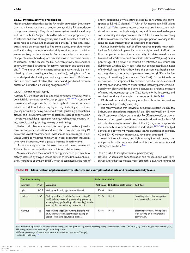
3a.3.2 Physical activity prescriptionHealth providers should assess the PA level in any subject (how manydays and minutes per day are spent on average doing PA at moderateor vigorous intensity). They should warn against inactivity and helpadd PA to daily life. Subjects should be advised on appropriate typesof activities and ways of progressing and should be helped to set per-sonal goals to achieve and maintain the benefits. To this end, indivi-duals should be encouraged to find some activity they either enjoyand/or that they can include in their daily routines, as such activitiesare more likely to be sustainable. For a more effective behaviourchange, clinicians should explore practical ways to overcome barriersto exercise. For this reason, the link between primary care and localcommunity-based structures for activity, recreation and sport is cru-cial.262 The amount of time spent being sedentary should be mini-mized by active travelling (cycling or walking), taking breaks fromextended periods of sitting and reducing screen time.272 Brief exer-cises are more cost effective than supervised gym-based exerciseclasses or instructor-led walking programmes.264
3a.3.2.1 Aerobic physical activityAerobic PA, the most studied and recommended modality, with abeneficial dose–response effect on prognosis,259,260,268 consists ofmovements of large muscle mass in a rhythmic manner for a sus-tained period. It includes everyday activity, including active travel(cycling or walking), heavy household work, gardening, occupationalactivity and leisure time activity or exercise such as brisk walking,Nordic walking, hiking, jogging or running, cycling, cross-country ski-ing, aerobic dancing, skating, rowing or swimming.
Similar to all other interventions, its prescription can be adjusted interms of frequency, duration and intensity. However, practising PAbelow the lowest recommended levels should be encouraged in indi-viduals unable to meet the minimum or in those sedentary individualswho have just started, with a gradual increase in activity level.
Moderate or vigorous aerobic exercise should be recommended.This can be expressed either in absolute or relative terms.
Absolute intensity is the amount of energy expended per minute ofactivity, assessed by oxygen uptake per unit of time (mL/min or L/min)or by metabolic equivalent (MET), which is estimated as the rate of
energy expenditure while sitting at rest. By convention this corre-sponds to 3.5 mL O2/kg/min).273 A list of PA intensities in MET valuesis available.274 An absolute measure does not take into account indi-vidual factors such as body weight, sex, and fitness level: older per-sons exercising at a vigorous intensity of 6 METs may be exercisingat their maximum intensity, while a younger person working at thesame absolute intensity may be exercising moderately.
Relative intensity is the level of effort required to perform an activ-ity. Less fit individuals generally require a higher level of effort thanfitter people to perform the same activity. It is determined relativeto an individual’s level of cardiorespiratory fitness (V?O2max) or as apercentage of a person’s measured or estimated maximum HR(%HRmax), which is 220 2 age. It also can be expressed as an indexof individual rate of effort (how hard the person feels he/she is ex-ercising), that is, the rating of perceived exertion (RPE) or by fre-quency of breathing (the so-called Talk Test). For individuals onmedication, it is important to consider possible modification ofHR response and to refer to other relative intensity parameters. Es-pecially for older and deconditioned individuals, a relative measureof intensity is more appropriate. Classification for both absolute andrelative intensity and examples are presented in Table 10.
PA should occur at a frequency of at least three to five sessionsper week, but preferably every day.
It is recommended that individuals accumulate at least 30 min/day,5 days/week of moderate intensity PA (i.e. 150 min/week) or 15 min/day, 5 days/week of vigorous intensity PA (75 min/week), or a com-bination of both, performed in sessions with a duration of at least 10min. Shorter exercise sessions (i.e. ,10 min) may also be appropri-ate, especially in very deconditioned individuals.267,276,277 For lipidcontrol or body weight management, longer durations of exercise,40 and 60–90 min/day, respectively, have been proposed.278
Aerobic interval training and high-intensity interval training can-not yet be broadly recommended until further data on safety andefficacy are available.266
3a.3.2.2 Muscle strength/resistance physical activityIsotonic PA stimulates bone formation and reduces bone loss; it pre-serves and enhances muscle mass, strength, power and functional
Table 10 Classification of physical activity intensity and examples of absolute and relative intensity levels
Absolute intensity Relative intensity
Intensity MET Examples %HRmax RPE (Borg scale score) Talk Test
Light 1.1–2.9 Walking <4.7 km/h, light household work. 50–63 10–11
Moderate 3–5.9 Walking briskly (4.8–6.5 km/h), slow cycling (15 km/h), painting/decorating, vacuuming, gardening (mowing lawn), golf (pulling clubs in trolley), tennis (doubles), ballroom dancing, water aerobics.
64–76 12–13 Breathing is faster but compatible with speaking full sentences.
Vigorous ≥6 Race-walking, jogging or running, bicycling >15 km/h, heavy gardening (continuous digging or hoeing), swimming laps, tennis (single).
77–93 14–16 Breathing very hard, incompatible with carrying on a conversation comfortably.
MET (metabolic equivalent) is estimated as the energy cost of a given activity divided by resting energy expenditure: 1 MET = 3.5 mL O2 kg-1 min-1 oxygen consumption (VO2).RPE, rating of perceived exertion (20 value Borg score).%HRmax, percentage of measured or estimated maximum heart rate (220-age).Modified from Howley.275
Joint ESC Guidelines2344
Downloaded from https://academic.oup.com/eurheartj/article-abstract/37/29/2315/1748952/2016-European-Guidelines-on-cardiovascular-diseaseby gueston 10 October 2017
ability, with some evidence of benefit in lipid and BP control and in-sulin sensitivity, especially in combination with aerobic exer-cise.267,279 It should target the major muscle groups (agonist andantagonist) and include multijoint or compound movementsthrough the full range of motion of the joints, such as workingwith resistance bands, calisthenics using body weight for resistance,carrying heavy loads and heavy gardening. For each exercise session,the suggested prescription is two to three sets of 8–12 repetitionsat the intensity of 60–80% of the individual’s 1 repetition maximum(1 RM, the maximum load that can be lifted one time) at a frequencyof least 2 days a week. For older adults or very deconditioned indi-viduals, it is suggested to start with one set of 10–15 repetitions at60–70% of 1 RM.280
3a.3.2.3 Neuromotor physical activityFor older adults at risk of falls, neuromotor exercise helps to maintainand improve balance and motor skills (balance, agility, coordinationand gait). This includes multifaceted activities such as tai chi andyoga, and recreational activities using paddles or sport balls to chal-lenge hand–eye coordination. The optimal volume is not known.276
3a.3.2.4 Phases and progression of physical activityPA sessions should include the following phases: warm-up, condi-tioning (aerobic, muscle strength/resistance and neuromotor exer-cise), cool-down and stretching/flexibility. Progressive warm-upbefore and cool-down after exercise may prevent injuries and ad-verse cardiac events. Inactive adults should start gradually, at lightor moderate intensity for short periods of time (even ,10 min),with sessions spread throughout the week. With the improvementin exercise tolerance, each subject progresses in the level of PA, butincreases in any components (i.e. frequency, duration and intensity)should be gradual, to minimize risks of muscle soreness, injury, fa-tigue and the long-term risk of overtraining.276 Following any adjust-ments, the individual should check for adverse effects (e.g. excessiveshortness of breath) and if there are any such effects, downward ad-justments should be made.276
3a.3.3 Risk assessmentThe risk of an adverse CV response during PA is extremely lowfor apparently healthy adults (5–17 sudden deaths/millionpopulation/year).281 The risk of participation is outweighed by thesubstantial health benefits conferred by PA.267 Risk during light-or moderate-intensity exercise is lower than during vigorous activ-ity,267 thus in healthy individuals who wish to undertake moderatePA, such as a walking programme, a preliminary medical evaluationis not needed.266
Before starting more intensive leisure time activities (i.e. struc-tured or competitive activity, amateur sports, exercise and fitnesstraining), a risk assessment should be tailored to the individual’s clin-ical (i.e. metabolic, musculoskeletal condition/disease) and cardiacrisk profile, the current level of habitual PA and the intended levelof PA.265 Individuals who exercise only occasionally seem to have anincreased risk of acute coronary events and sudden cardiac deathduring or after exercise.282 Sedentary subjects and those with CVrisk factors should start aerobic PA at low-intensity activity and pro-gress gradually. Clinical evaluation, including exercise testing, may beconsidered for sedentary people with CV risk factors who intend toengage in vigorous PA and sports. The information gathered from
exercise tests may be useful in establishing a safe and effective exer-cise prescription. Validated self-assessment questionnaires havebeen proposed for sedentary individuals entering low-intensity leis-ure time sports activity or starting moderate-intensity activities265
(see Table B in web addenda).
Gaps in evidence† The lower and upper limit of aerobic PA intensity, duration and
frequency to exert a beneficial effect is unknown.† The effectiveness of PA monitoring vs. simple counselling to op-
timize the motivation of patients to adhere to active lifestyle isunknown.
† The role and sustainability of modern technology (such as wear-able technology, ‘exergaming’ and smartphone apps) for motivat-ing people to undertake more PA has not been established.
3a.4 Smoking interventionKey messages† Stopping smoking is the most cost-effective strategy for CVD
prevention.† There is a strong evidence base for brief interventions with advice
to stop smoking, all types of nicotine replacement therapy (NRT),bupropion, varenicline and greater effectiveness of drugs in com-bination, except for NRT plus varenicline. The most effective arebrief interventions plus assistance with stopping using drug ther-apy and follow-up support.
† Electronic cigarettes (e-cigarettes) may help in smoking cessationbut should be covered by the same marketing restrictions ascigarettes.
† Passive secondary smoking carries significant risk, with the needto protect non-smokers.
Recommendations for smoking intervention strategies
Recommendations Class a Level b Ref c
It is recommended to identify smokers and provide repeated advice on stopping with offers to help, by the use of follow up support, nicotine replacement therapies, varenicline, and bupropion individually or in combination.
I A 283–286
It is recommended to stop all smoking of tobacco or herbal products, as this is strongly and independently causal of CVD.
I B 287–291
It is recommended to avoid passive smoking.
I B 292, 293
CVD ¼ cardiovascular disease.aClass of recommendation.bLevel of evidence.cReference(s) supporting recommendations.
3a.4.1 IntroductionSmoking is a lethal addictive disorder. A lifetime smoker has a 50%probability of dying due to smoking, and on average will lose 10
Joint ESC Guidelines 2345
Downloaded from https://academic.oup.com/eurheartj/article-abstract/37/29/2315/1748952/2016-European-Guidelines-on-cardiovascular-diseaseby gueston 10 October 2017

years of life,287 contrasting with ,3 years with severe hypertensionand ,1 year with mild hypertension.288 Smoking is an establishedcause of a plethora of diseases and is responsible for 50% of allavoidable deaths in smokers, half of these due to CVD. The10-year fatal CVD risk is approximately doubled in smokers. TheRR in smokers ,50 years of age is five-fold higher than innon-smokers.289
Slightly less than half of lifetime smokers will continue smoking untildeath. Approximately 70% of UK smokers want to stop smoking atsome time in the future,290 with �43% trying to stop in the pastyear; however, only 2–3% of the population succeed in stopping.291
Even modest and low levels of smoking confer vascular risk.294
Although the rate of smoking is declining in Europe, it remainsvery common and is increasing in women, adolescents and the so-cially disadvantaged.295 Widening education-related inequalities insmoking cessation rates have been observed in many Europeancountries. In the EUROASPIRE IV survey among CAD patients,16% smoked after a mean follow-up time of 16 months, and nearlyhalf of the participants who smoked at the time of their coronaryevent were persistent smokers. The survey also found thatevidence-based treatment for smoking cessation was underused.6
3a.4.2 Dosage and typeThe risks associated with smoking show a dose–response relation-ship with no lower limit for deleterious effects.296 Duration alsoplays a role, and while cigarette smoking is the most common, alltypes of smoked tobacco, including low-tar (‘mild’ or ‘light’) cigar-ettes, filtered cigarettes, cigars and pipes, are harmful.292 Smokingis deleterious regardless of how it is done, including by waterpipe. Tobacco smoke is more harmful when inhaled, but smokerswho claim not to inhale the smoke (e.g. pipe smokers) are also atincreased risk of CVD. Smokeless tobacco is also associated witha small but statistically significant increased risk of MI and stroke.
3a.4.3 Passive smokingPassive smoking increases the risk of CAD.293,297 A smoking spouseor workplace exposure increases CVD risk by an estimated 30%. Ma-jor health benefits result from reduced environmental tobaccosmoke, with public smoking bans in various different geographical lo-cations leading to significant decreases in MI rates (see section 3c.4).
3a.4.4 Mechanisms by which tobacco smokingincreases riskSmoking enhances the development of both atherosclerosis andsuperimposed thrombotic phenomena. Smoking affects endothelialfunction, oxidative processes, platelet function, fibrinolysis, inflamma-tion, lipid oxidation and vasomotor function. In experimental studies,several of these effects are fully or partly reversible within a very shorttime. Plaque formation is not thought to be fully reversible and thussmokers would never be expected to reach the risk level of neversmokers concerning CVD. Nicotine replacement shows no adverseeffect on outcomes in patients with cardiac disease.298,299
3a.4.5 Smoking cessationThe benefits of smoking cessation have a large evidence base. Someadvantages are almost immediate; others take more time. CVD riskin former smokers is in between that of current and never smokers.
Stopping smoking after an MI is potentially the most effective of allpreventive measures: a systematic review and meta-analysis showedreductions in MIs and in the composite endpoints of death/MI (RR0.57 and 0.74, respectively) compared with continued smoking.300
The benefit is consistent over gender, duration of follow-up, studysite and time period. Significant morbidity reductions occur withinthe first 6 months.301 Randomized trials also support smoking ces-sation, with the risk of CVD approaching (but never equalling) therisk of never smokers within 10–15 years.
Smoking reduction has not been shown to increase the probabil-ity of future smoking cessation, but some advocate nicotine-assistedsmoking reduction in smokers unable or unwilling to quit. Quittingmust be encouraged in all smokers (Table 11). There is no age limitto the benefits of smoking cessation. Passive smoking should also beavoided.
Professional support can increase the odds of stopping [RR 1.66(95% CI 1.42, 1.94)].302 An impetus for smoking cessation occurs atthe time of diagnosing or (invasive) treatment of CVD. Prompting aperson to try to quit, brief reiteration of CV and other health ha-zards and agreeing on a specific plan with a follow-up arrangementare evidence-based interventions (see Figure K in web addenda).
Smoking cessation programmes initiated during hospital admis-sion should continue for a prolonged period after discharge. Asmoking history including daily tobacco consumption and degreeof addiction (most commonly assessed by the Fagerstrom test302)may guide the degree of support and pharmacological aids. Smokersshould be advised about expected weight gain of, on average, 5 kgand that the health benefits of tobacco cessation far outweigh therisks from weight gain.
3a.4.6 Evidence-based drug interventionsFollowing the failure of advice, encouragement and motivational in-terventions, or in addition to them, NRT, varenicline or bupropionshould be offered to assist cessation.285 All forms of NRT (chewinggum, transdermal nicotine patches, nasal spray, inhaler, sublingual ta-blets) are effective: in a systematic review, the RR for abstinencewith NRT vs. control was 1.60; NRTs increase the rate of quittingby 50–70%, regardless of setting.303
The antidepressant bupropion aids long-term smoking cessationwith a similar efficacy to NRT.286 A meta-analysis of 44 trials
Table 11 The “Five As” for a smoking cessationstrategy for routine practice
A–ASK:Systematically inquire about smoking status at every opportunity.
A–ADVISE: Unequivocally urge all smokers to quit.
A–ASSESS:Determine the person’s degree of addiction and readiness to quit
A–ASSIST:Agree on a smoking cessation strategy, including setting a quit date, behavioural counselling, and pharmacological support.
A–ARRANGE: Arrange a schedule of follow-up.
Joint ESC Guidelines2346
Downloaded from https://academic.oup.com/eurheartj/article-abstract/37/29/2315/1748952/2016-European-Guidelines-on-cardiovascular-diseaseby gueston 10 October 2017

comparing long-term cessation rates using bupropion vs. controlyielded a relative success rate of 1.62.283 Bupropion carries a knownrisk of seizures (reported as �1/1000 users),286 without increasedrisks of neuropsychiatric or heart and circulatory problems. Overall,NRT and bupropion help �80% more people to quit than placebo;this means that for every 10 people who quit with placebo, �18could be expected to quit with NRT or with bupropion.285
The partial nicotine receptor agonist varenicline at the standarddose increases the chances of quitting more than two-fold com-pared with placebo (14 trials, 6166 people).283 The number of peo-ple stopping smoking with varenicline is higher than with bupropion(three trials, 1622 people). Varenicline more than doubles thechances of quitting compared with placebo, so that for every 10who quit with placebo, �28 could be expected to quit with vareni-cline. Varenicline helps �50% more people to quit than nicotinepatch and ‘other’ NRTs (tablets, sprays, lozenges and inhalers)and �70% more people than nicotine gum. So for every 10 peoplewho quit with an NRT patch or with ‘other’ NRTs, �15 would beexpected to quit with varenicline, and for every 10 who quit withNRT gum, �17 would be expected to quit with varenicline.285
Low-dose varenicline (four trials, 1272 people) roughly doublesthe chances of quitting and reduces the number and severity ofside effects. The main side effect of varenicline is nausea, but this ismostly mild or moderate and usually subsides over time.285 Althoughconcerns have been raised, retrospective cohort studies and anRCT304 indicate no severe adverse events with varenicline in the set-ting of ACS patients, with the large EVITA trial in ACS ongoing.
Clonidine has helped people to quit, but causes side effects and istherefore a second-line agent. It is not clear whether mecamylamineused with NRT helps people to quit. Other treatments did not seemto help. So far, nicotine vaccines are not licensed for use anywherein the world.285
Combining two types of NRT is as effective as using varenicline,and helps more people to quit than a single type of NRT.285
3a.4.7 Electronic cigarettesElectronic cigarettes (e-cigarettes) are battery-operated devicesthat simulate combustible cigarettes by heating nicotine and otherchemicals into a vapour that is inhaled. Electronic cigarettes deliverthe addictive nicotine without the vast majority of tobacco chemi-cals, and are probably less harmful than tobacco.305
Evidence on the effectiveness of e-cigarettes is limited due to thesmall number of trials, low event rates and wide confidence inter-vals.306 Data from some observational studies and a randomized trialsuggest that the efficacy of first-generation e-cigarettes is similar tothat of transdermal NRT patches307 or NRT inhalers.308 The benefitmay come from low nicotine delivery or just the non-nicotine behav-ioural components of e-cigarette use. About 6% of former smokerswho used e-cigarettes daily relapsed to smoking after 1 month and 6%after 1 year, and nearly half of dual users of both tobacco ande-cigarettes stopped smoking after 1 year, indicating that e-cigaretteuse might be effective in relapse prevention and smoking cessation.309
These studies and real-world data indicate that e-cigarettes are mod-erately effective as smoking cessation and harm reduction aids, butthat a significant component of that effect is due to changes in behav-iour rather than in nicotine delivery. Recent evidence indicates thate-cigarettes, as currently being used, are associated with significantly
less quitting among smokers.310 Although no safety issues have beenobserved in the short term (2 years), determining the long-termhealth effects of e-cigarettes (and in particular dual use with cigar-ettes) will require more research.305
3a.4.8 Other smoking cessation interventionsBoth individual and group behavioural interventions are effective inhelping smokers quit. Support from the individual’s partner and fam-ily is important. There are no reliable data that acupuncture, acu-pressure, laser therapy, hypnotherapy or electrostimulation areeffective for smoking cessation.
Gap in evidence† More efficient, safe and cost-effective smoking cessation aids are
required.
3a.5 NutritionKey messages† Dietary habits influence the risk of CVD and other chronic dis-
eases such as cancer.† Energy intake should be limited to the amount of energy needed
to maintain (or obtain) a healthy weight, that is, a BMI .20.0 but,25.0 kg/m2.
† In general, when following the rules for a healthy diet, no dietarysupplements are needed.
Recommendation on nutrition
Recommendation Class a Level b Ref c
A healthy diet is recommended as a cornerstone of CVD prevention in all individuals.
I B 311
CVD ¼ cardiovascular disease.aClass of recommendation.bLevel of evidence.cReference(s) supporting recommendations.
3a.5.1 IntroductionDietary habits influence CV risk, either through an effect on risk fac-tors such as cholesterol, BP, body weight and DM, or through othereffects.311 Table 12 summarizes the characteristics of a healthy diet.
Most evidence on the relation between nutrition and CVD isbased on observational studies; randomized clinical trials estimatingthe impact of diet on endpoints are scarce. The impact of diet isstudied on three levels: specific nutrients, specific foods/foodgroups and specific dietary patterns, of which the Mediterraneandiet is the most studied.
The nutrients of interest with respect to CVD are fatty acids(which mainly affect lipoprotein levels), minerals (which mainly af-fect BP), vitamins and fibre.
3a.5.2 Fatty acidsFor prevention of CVD, the types of fatty acids consumed are moreimportant than the total fat content.
Joint ESC Guidelines 2347
Downloaded from https://academic.oup.com/eurheartj/article-abstract/37/29/2315/1748952/2016-European-Guidelines-on-cardiovascular-diseaseby gueston 10 October 2017

The risk of CAD is reduced by 2–3% when 1% of energy intakefrom saturated fatty acids is replaced by polyunsaturated fatty acids.The same has not been clearly shown for replacement with carbo-hydrates and monounsaturated fatty acids (MUFAs). Saturated fattyacid intake should be reduced to a maximum of 10% of energy intakeby replacing it with polyunsaturated fatty acids.312
MUFAs have a favourable effect on HDL-C levels when they re-place saturated fatty acids or carbohydrates,313 but there is little evi-dence that MUFAs lower CAD risk.
Polyunsaturated fatty acids lower LDL-C levels, and to a lesserextent HDL-C levels, when they replace saturated fatty acids. Thepolyunsaturated fatty acids can be divided into two subgroups:omega-6 fatty acids, mainly from plant foods, and omega-3 fattyacids, mainly from fish oils and fats. Within the subclass of omega-3fatty acids, eicosapentaenoic acid and docosahexaenoic acid (EPA/DHA) are especially important. They do not change serum choles-terol levels and, with currently available cardioprotective therapies,it is debatable whether they exert a favourable effect on all-cause,CAD, and stroke mortality.314,315
The trans fatty acids, a subclass of unsaturated fatty acids, havebeen shown to be especially harmful due to their unfavourable im-pact on both total cholesterol (increase) and HDL-C (decrease).These fatty acids are formed during industrial processing (harden-ing) of fats and are present in, for example, margarine and bakeryproducts. A meta-analysis of prospective cohort studies has shownthat, on average, a 2% increase in energy intake from trans fatty acidsincreases CAD risk by 23%.316 It is recommended to derive ,1% oftotal energy intake from trans fatty acids—the less the better.
The impact of dietary cholesterol on serum cholesterol levels isweak compared with that of the fatty acid composition of the diet.When guidelines are followed to lower saturated fat intake, this usu-ally also leads to a reduction in dietary cholesterol intake. Therefore,
some guidelines (including this one) on healthy diet do not give spe-cific guidelines on the intake of dietary cholesterol; others recom-mend a limited intake of ,300 mg/day.
3a.5.3 MineralsA meta-analysis estimated that even a modest reduction in sodiumintake of 1 g/day reduces SBP by 3.1 mmHg in hypertensive patientsand 1.6 mmHg in normotensive patients.317 The Dietary Ap-proaches to Stop Hypertension (DASH) trial showed a dose–re-sponse relation between sodium reduction and BP reduction.318
In most western countries, salt intake is high (�9–10 g/day), where-as the recommended maximum intake is 5 g/day. Optimal intake le-vels might be as low as �3 g/day. Although the relation between saltintake and BP remains controversial, the totality of evidence war-rants salt reduction as an important way to prevent CAD andstroke. On average, 80% of salt intake comes from processed foods,while only 20% is added later on. Salt reduction can be achieved bymaking different dietary choices (fewer processed foods, more basicfoods) and the reformulation of foods (lowering salt content)(see Chapter 3c.2).
Potassium has favourable effects on BP. The main sources ofpotassium are fruits and vegetables. An inverse statistically signifi-cant association exists between potassium intake and the risk of in-cident stroke [RR 0.76 (95% CI 0.66, 0.89)].319 Apart from reducingsodium intake, increasing potassium intake contributes to the low-ering of BP.
3a.5.4 VitaminsMany case–control and prospective observational studies have ob-served inverse associations between levels of vitamin A and E andthe risk of CVD. However, intervention trials have failed to confirmthese observational studies. Also, for the B vitamins (B6, folic acidand B12) and vitamin C, trials have shown no beneficial effects.
In the bottom tertile of serum levels of vitamin D, CV and totalmortality is 35% higher [RR 1.35 (95% CI 1.13, 1.61)] than in thehighest tertile.320 A 41% higher risk of CV mortality [RR 1.41(95% CI 1.18, 1.68)] and 57% higher risk of all-cause mortality [RR1.57 (95% CI 1.36, 1.81)] has been reported in the lowest vs. highestquintile.321 A much smaller effect was observed in RCTs: an 11% riskreduction in all-cause mortality was observed for vitamin D3 supple-mentation [RR 0.89 (95% CI 0.80, 0.99)], but not for vitamin D2 sup-plementation.320 Due to a lack of power, it was not possible to lookat CV mortality specifically. Therefore, conclusions about vitamin Dsupplementation [type of supplement (D2 or D3), dosage and dur-ation] for CV prevention cannot yet be drawn.
3a.5.5. FibreRecent meta-analyses of prospective cohort studies show that a 7 g/day higher intake of total fibre is associated with a 9% lower risk ofCAD [RR 0.91 (95% CI 0.87, 0.94)]322 and a 10 g/day higher fibreintake is associated with a 16% lower risk of stroke [RR 0.84 (95%CI 0.75, 0.94)]323 and a 6% lower risk of type 2 DM [RR 0.94 (95% CI0.91, 0.97)].324 There is no evidence yet for a similar association withfibre from fruits and vegetables. Although the mechanism has notbeen elucidated completely, it is known that a high fibre intake re-duces postprandial glucose responses after carbohydrate-rich mealsand lowers total cholesterol and LDL-C levels.
Table 12 Healthy diet characteristics
• Saturated fatty acids to account for <10% of total energy intake, through replacement by polyunsaturated fatty acids.
• Trans unsaturated fatty acids: as little as possible, preferably no intake from processed food, and <1% of total energy intake from natural origin.
• <5 g of salt per day.
• 30–45 g of per day, preferably from wholegrain products.
• ≥200 g of fruit per day (2–3 servings).
• ≥200 g of vegetables per day (2–3 servings).
• Fish 1–2 times per week, one of which to be oily
• 30 grams unsalted nuts per day.
• Consumption of alcoholic beverages should be limited to 2 glasses per day (20 g/d of alcohol) for men and 1 glass per day (10 g/d of alcohol) for women.
• Sugar-sweetened soft drinks and alcoholic beverages consumption must be discouraged.
Joint ESC Guidelines2348
Downloaded from https://academic.oup.com/eurheartj/article-abstract/37/29/2315/1748952/2016-European-Guidelines-on-cardiovascular-diseaseby gueston 10 October 2017

3a.5.6 Foods and food groups3a.5.6.1 Fruits and vegetablesProspective cohort studies have shown a protective effect of theconsumption of fruits and vegetables on CVD, but RCTs are scarce.A meta-analysis reported a decrease of 4% [RR 0.96 (95% CI 0.92,0.99)] in CV mortality for each additional serving of fruits (equivalentto 77 g) and vegetables (equivalent to 80 g) per day, while all-cause mor-tality did not reduce further with intakes of more than five servings.325 Ameta-analysis reported a risk reduction for stroke of 11% [RR 0.89 (95%CI 0.83, 0.97)] for three to five daily servings of fruits and vegetables andof 26% [RR 0.74 (95% CI 0.69, 0.79)] for more than five servingscompared with less than three servings.326 A meta-analysis onCAD reported a 4% decrease in CAD risk [RR 0.96 (95% CI 0.93,0.99)] for each additional serving of fruits and vegetables per day.327
3a.5.6.2 NutsA meta-analysis of prospective cohort studies has shown that dailyconsumption of 30 g of nuts reduces the risk of CVD by �30% [RR0.71 (95% CI 0.59, 0.85)].328 It must be noted that the energy densityof nuts is high.
3a.5.6.3 FishThe protective effect of fish on CVD is attributed to the n-3 fattyacid content. Pooled risk estimates from prospective cohort studiesshow that eating fish at least once a week results in a 16% reductionin the risk of CAD [RR 0.85 (95% CI 0.75, 0.95)] compared with eat-ing less fish.329 A recent meta-analysis showed that eating fish two tofour times a week reduces the risk of stroke by 6% [RR 0.94 (95% CI0.90, 0.98)] compared with eating fish less than once a week.330 Therelation between fish intake and CV risk is not linear. Especially inthe range of no or very low intake, risk is increased. The publichealth impact of a small increase in fish consumption in the generalpopulation is therefore potentially large.
For fish oil, three randomized controlled prevention trials havebeen published. All three trials, in post-AMI or CAD patients whoreceived an extra amount of 400–1000 g EPA/DHA daily, did notobserve a reduction in CV events in the intervention group. A re-cent meta-analysis of 20 trials, mostly prevention of recurrent CVevents and mostly using fish oil supplements, showed no benefitof fish oil supplementation on CV outcomes.315
3a.5.6.4 Alcoholic beveragesDrinking three or more alcoholic beverages per day is associatedwith elevated CVD risk. Results from epidemiological studies sug-gest a lower risk of CVD occurring with moderate (one to two unitsper day) alcohol consumption compared with non-drinkers. This as-sociation appears not to be explained by special characteristics ofabstainers,331 although the potential for residual confounding andreverse causality cannot be fully excluded. Moreover, a recent Men-delian randomization study including analyses from 59 epidemio-logical studies has shed doubt on any beneficial effect ofmoderate alcohol consumption,332 suggesting that the lowest risksfor CV outcomes were in abstainers and that any amount of alcoholis associated with elevated BP and BMI.
3a.5.6.5 Soft drinks and sugarSugar-sweetened soft drinks are the largest single food source ofcalories in the US diet and are important in Europe. In children
and adolescents, beverages may now even account for 10–15% ofthe calories consumed. Regular consumption of soft drinks has beenassociated with overweight, metabolic syndrome and type 2 DM.Substitution of sugar-sweetened soft drinks with artificially swee-tened drinks resulted in less weight gain in children over an18-month period.333 Sugar-sweetened beverages also cause weightgain in adults. Regular consumption of sugar-sweetened beverages(i.e. two servings per day compared with one serving per month)was associated with a 35% higher risk of CAD in women, even afterother unhealthy lifestyle and dietary factors were accounted for,whereas artificially sweetened beverages were not associated withCAD. The WHO guideline recommends a maximum intake of 10%of energy from sugar (mono- and disaccharides), which includesadded sugars as well as sugars present in fruits and fruit juices.334
3a.5.7 Functional foodsFunctional foods containing phytosterols (plant sterols and stanols)are effective in lowering LDL-C levels by an average of 10% whenconsumed in amounts of 2 g/day. The cholesterol-lowering effectis in addition to that obtained with a low-fat diet or use of statins.Further cholesterol reduction can be obtained with higher dosesof phytosterols.335 No studies with clinical endpoints have been per-formed yet.
3a.5.8 Dietary patternsStudying the impact of a total dietary pattern theoretically showsthe full preventive potential of diet since it yields a combined esti-mate of the impact of several favourable dietary habits. The Medi-terranean diet comprises many of the nutrients and foods thathave been discussed previously: high intake of fruits, vegetables, le-gumes, wholegrain products, fish and unsaturated fatty acids (espe-cially olive oil); moderate consumption of alcohol (mostly wine,preferably consumed with meals) and low consumption of (red)meat, dairy products and saturated fatty acids. A meta-analysis ofprospective cohort studies has demonstrated that greater adher-ence to a Mediterranean diet is associated with a 10% reductionin CV incidence or mortality [pooled RR 0.90 (95% CI 0.87,0.93)] and an 8% reduction in all-cause mortality [pooled RR0.92 (95% CI 0.90, 0.94)].336 An RCT in high-risk individuals sug-gested that following a Mediterranean diet over a 5 year period,compared with a control diet, was related to a 29% lower risk ofCVD [RR 0.71 (95% CI 0.56, 0.90)].337
Gaps in evidence† The biggest challenge in dietary prevention of CVD is to develop
more effective strategies to make people change their diet (bothquantitatively and qualitatively) and to maintain that healthy dietand a normal weight.
† Research into the substances in foods that underlie the protect-ive effects is ongoing.
3a.6 Body weightKey messages† Both overweight and obesity are associated with an increased risk of
CVD death and all-cause mortality. All-cause mortality is lowestwith a BMI of 20–25 kg/m2 (in those ,60 years of age); furtherweight reduction cannot be considered protective against CVD.
Joint ESC Guidelines 2349
Downloaded from https://academic.oup.com/eurheartj/article-abstract/37/29/2315/1748952/2016-European-Guidelines-on-cardiovascular-diseaseby gueston 10 October 2017

† Healthy weight in the elderly is higher than in the young andmiddle-aged.
† Achieving and maintaining a healthy weight has a favourable effecton metabolic risk factors (BP, blood lipids, glucose tolerance) andlower CV risk.
Recommendation for body weight
Recommendation Classa Levelb Ref c
It is recommended that subjects with healthy weightd maintain their weight. It is recommended that overweight and obese people achieve a healthy weight (or aim for a reduction in weight) in order to reduce BP, dyslipidaemia and risk of developing type 2 DM, and thus improve the CV risk
I A 338, 339
BP ¼ blood pressure; CVD ¼ cardiovascular disease; DM ¼ diabetes mellitus.aClass of recommendation.bLevel of evidence.cReference(s) supporting recommendations.dBMI 20–25 kg/m2. There is evidence that optimal weight in elderly is higher thanin the young and middle-aged.339
3a.6.1 IntroductionIn many countries, favourable trends in major risk factors such asblood cholesterol, BP and smoking prevalence have been ob-served, translating into reduced CV mortality. However, BMI hasgreatly increased in all countries over recent decades, resultingin a concomitant increase in the prevalence of type 2 DM. In theUSA, it has been projected that if obesity trends from 2005 to2020 continue, obesity will increasingly offset the positive effectsof declining smoking rates.340 The main clinical complications of in-creasing body weight are increases in BP, dyslipidaemia, insulin re-sistance, systemic inflammation and prothrombotic state andalbuminuria and the development of DM and CV events (HF,CAD, AF, stroke).
3a.6.2 Which index of obesity is the best predictor ofcardiovascular risk?BMI [weight (kg)/height (m2)] can be measured easily and is used ex-tensively to define categories of body weight (see Table C in the webaddenda).341 In addition to the amount of body fat, its distribution isimportant. Body fat stored in the abdomen (intra-abdominal fat)carries a higher risk than subcutaneous fat.
Several measures of body fatness are available (see Table D in theweb addenda). Most data are available for BMI, waist:hip circumfer-ence ratio and simple waist circumference. The optimal level formeasurement of waist circumference is midway from the lowerrib margin to the anterior superior iliac crest, in the standing pos-ition. The WHO thresholds for waist circumference are the mostwidely accepted in Europe. Based on these thresholds, two actionlevels are recommended:
(i) waist circumference ≥94 cm in men and ≥80 cm in womenrepresents the threshold at which no further weight should begained and
(ii) waist circumference ≥102 cm in men and ≥88 cm in womenrepresents the threshold at which weight reduction should beadvised.
These thresholds have been calculated based on Caucasians, andit is apparent that different cut-offs for anthropometric measure-ments are required in different races and ethnicities. A meta-analysisconcluded that both BMI and waist circumference are similarlystrong and continuously associated with CVD and type 2 DM.342
Therefore, BMI generally suffices in routine practice.
3a.6.3 Does ‘metabolically healthy obesity’ exist?The phenotype of ‘metabolically healthy obesity’ (MHO), defined bythe presence of obesity in the absence of metabolic risk factors, hasgained a lot of interest. Some studies argue that a specific subgroupof obese individuals is resistant to metabolic complications such asarterial hypertension and insulin resistance. However, MHO indivi-duals present a higher all-cause mortality compared with normalweight metabolically healthy individuals.343,344 Long-term resultsfrom the Whitehall study support the notion that MHO is a transi-ent phase345 moving towards glucometabolic abnormalities ratherthan a specific ‘state’.
3a.6.4 The obesity paradox in established heart diseaseAt the population level, obesity is associated with CVD risk. How-ever, among those with established CAD, the evidence is contradic-tory. Systematic reviews of patients with CAD or undergoingpercutaneous coronary intervention have suggested an ‘obesityparadox’ whereby obesity appears protective.338,346 This is alsothe case for HF patients. However, this evidence should not bemisinterpreted to recommend higher target BMIs for those with es-tablished CVD since reverse causality may be operating. Cardio-respiratory fitness might influence relationships between adiposityand clinical prognosis in the obesity paradox. Normal weight unfitindividuals have a higher risk of mortality than fit individuals, regard-less of their BMI. Overweight and obese fit individuals have mortalityrisks similar to normal weight fit individuals.347 Furthermore, the re-sults of the EPIC study suggest that the influence of physical inactiv-ity on mortality appears to be greater than that of high BMI.348
3a.6.5 Treatment goals and modalitiesCVD risk has a continuous positive relationship with BMI and othermeasures of body fat. Because all-cause mortality appears to in-crease at BMI levels ,20,339 we do not recommend such lowBMI levels as treatment goals.
Although diet, exercise and behaviour modifications are the main-stay therapies for overweight and obesity, they are often unsuccessfulfor long-term treatment. Medical therapy with orlistat and/or bariatricsurgery are additional options. A recent meta-analysis indicates thatpatients undergoing bariatric surgery have a reduced risk of MI, stroke,CV events and mortality compared with non-surgical controls.349
Gaps in evidence† Knowledge and implementation of effective strategies to achieve
weight loss and maintain a long-term healthy weight.† Identification of the relative roles of diet, exercise and behaviour
modification in the management of overweight and obese people.† The optimal level of BMI over the life course (at older ages and
after a CV event).
Joint ESC Guidelines2350
Downloaded from https://academic.oup.com/eurheartj/article-abstract/37/29/2315/1748952/2016-European-Guidelines-on-cardiovascular-diseaseby gueston 10 October 2017

3a.7 Lipid controlKey messages† Elevated levels of plasma LDL-C are causal to atherosclerosis.† Reduction of LDL-C decreases CV events.† Low HDL-C is associated with increased CV risk, but man-
oeuvres to increase HDL-C have not been associated with a de-creased CV risk.
† Lifestyle and dietary changes are recommended for all.† Total CV risk should guide the intensity of the intervention.† Total cholesterol and HDL-C are adequately measured on non-
fasting samples, thus allowing non-HDL-C to be derived.
Recommendations for lipid control
Recommendations d e Classa Levelb Ref c
In patients at VERY HIGH CV risk, an LDL-C goal <1.8 mmol/L (<70 mg/dL), or a reduction of at least 50% if the baseline is between 1.8 and 3.5 mmol/L (70 and 135 mg/dL) is recommended.f
I B 350–353
In patients at HIGH CV risk, an LDL-C goal <2.6 mmol/L (<100 mg/dL), or a reduction of at least 50% if the baseline is between 2.6 and 5.1 mmol/L (100 and200 mg/dL) is recommended.
I B 350–353
In the remaining patients on LDL-C lowering treatment, an LDL-C goal <3.0 mmol/L (<115 mg/dL) should be considered.
IIa C 350–353
CV ¼ cardiovascular; HDL-C ¼ high-density lipoprotein cholesterol;LDL-C ¼ low-density lipoprotein cholesterol.aClass of recommendation.bLevel of evidence.cReference(s) supporting recommendations.dNon-HDL-C is a reasonable and practical alternative target because it does notrequire fasting. Non HDL-C secondary targets of ,2.6, ,3.3 and ,3.8 mmol/L(,100, ,130 and ,145 mg/dL) are recommended for very high, high and low tomoderate risk subjects, respectively See section 3a.7.10 for more details.eA view was expressed that primary care physicians might prefer a single LDL-Cgoal of 2.6 mmol/L (100 mg/dL). While accepting the simplicity of this approachand that it could be useful in some settings, there is better scientific support for thethree targets matched to level of risk.fThis is the general recommendation for those at very high-risk. It should be notedthat the evidence for patients with CKD is less strong.
3a.7.1 IntroductionThe crucial role of dyslipidaemia, especially hypercholesterolaemia,in the development of CVD is documented beyond any doubt bygenetic, pathology, observational and intervention studies.
In blood plasma, lipids such as cholesterol and triglycerides circu-late as lipoproteins in association with various proteins (apolipopro-teins). The main carrier of cholesterol in plasma (LDL-C) isatherogenic. The role of triglyceride-rich lipoproteins is currentlyunder active investigation: chylomicrons and large very-low-densitylipoproteins (VLDLs) appear not to be atherogenic, but very highconcentrations of these triglyceride-rich lipoproteins can cause pan-creatitis. Remnant lipoproteins [total cholesterol 2 (LDL-C +
HDL-C)] have recently been identified in Mendelian randomizationstudies as pro-atherogenic lipoproteins.
3a.7.2 Total and low-density lipoprotein cholesterolMost cholesterol is normally carried in LDL-C. Over a wide range ofplasma cholesterol concentrations, there is a strong and gradedpositive association between total as well as LDL-C and risk ofCVD.354 This association applies to men and women, and to thosewithout CVD as well as with established CVD.
The evidence that reducing plasma LDL-C reduces CVD risk isunequivocal; the results of epidemiological studies and trials withand without statins using angiographic or clinical endpoints confirmthat the reduction of LDL-C is of prime concern in the prevention ofCVD.38
Meta-analyses of many statin trials show a dose-dependent rela-tive reduction in CVD with LDL-C lowering. Every 1.0 mmol/L re-duction in LDL-C is associated with a corresponding 20–25%reduction in CVD mortality and non-fatal MI.350
3a.7.3 Apolipoprotein BApolipoprotein B (apoB; the main apoprotein of atherogeniclipoproteins) levels have also been measured in outcome studiesin parallel with LDL-C.355 Based on the available evidence, it appearsthat apoB is a similar risk marker to LDL-C.356 Also, there appearsto be less laboratory error in the determination of apoB thanLDL-C, particularly in patients with marked hypertriglyceridaemia[.3.4 mmol/L (.300 mg/dL)], but there is no evidence that apoBis a better predictor of CVD than LDL-C.357
3a.7.4 TriglyceridesHypertriglyceridaemia is a significant independent CVD risk factor,but the association is far weaker than for hypercholesterolaemia.358
The risk is associated more strongly with moderate than with verysevere hypertriglyceridaemia [.10 mmol/L (.�900 mg/dL)],which is a risk factor for pancreatitis. There are, however, no rando-mized trials to provide sufficient evidence to derive target levels fortriglycerides. Meta-analyses suggest that targeting triglycerides mayreduce CVD in specific subgroups with high triglycerides and lowHDL-C. At present, fasting triglycerides .1.7 mmol/L (.
�150 mg/dL) continue to be considered a marker of increasedrisk, but concentrations ≤1.7 mmol/L are not evidence-based tar-get levels for therapy.
3a.7.5 High-density lipoprotein cholesterolLow HDL-C is independently associated with higher CVD risk.359
Low HDL-C may even rival hypercholesterolaemia (due to highconcentrations of LDL-C) as a risk factor for CAD.360 The combin-ation of moderately elevated triglycerides and low concentrationsof HDL-C is very common in patients with type 2 DM, abdominalobesity and insulin resistance and in those who are physicallyinactive. This lipid pattern is also characterized by the presenceof small, dense, atherogenic LDL particles. An HDL-C level,1.0 mmol/L (,40 mg/dL) in men and ,1.2 mmol/L (,45 mg/dL) in women may be regarded as a marker of increased risk. RecentMendelian randomization studies, however, cast doubt on the causalrole of HDL-C in CVD.361 Physical activity and other lifestyle fac-tors, rather than drug treatment, remain important means of in-creasing HDL-C levels.
Joint ESC Guidelines 2351
Downloaded from https://academic.oup.com/eurheartj/article-abstract/37/29/2315/1748952/2016-European-Guidelines-on-cardiovascular-diseaseby gueston 10 October 2017

3a.7.6 Lipoprotein(a)Lipoprotein(a) [Lp(a)] is a low-density lipoprotein to which an add-itional protein called apolipoprotein(a) is attached. High concentra-tions of Lp(a) are associated with increased risk of CAD andischaemic stroke and Mendelian randomization studies support a cau-sal role in CVD for Lp(a). There is no randomized intervention studyshowing that reducing Lp(a) decreases CVD risk.362 At present thereis no justification for screening the general population for Lp(a), but itmay be considered in patients at moderate risk to refine risk evalu-ation or in subjects with a family history of early CVD.
3a.7.7 Apolipoprotein B/apolipoprotein A1 ratioApolipoprotein A1 (apoA1) is the major apoprotein of high-densitylipoprotein. It is beyond doubt that the apoB:apoA1 ratio is one ofthe strongest risk markers.112,355 However, there is insufficient evi-dence to support this variable as a treatment goal. Since the meas-urement of apolipoproteins is not available to all physicians inEurope, is more costly than currently used lipid variables and onlyadds moderately to the information derived from currently appliedlipid parameters, its use is not recommended.
3a.7.8 Calculated lipoprotein variables3a.7.8.1 Low-density lipoprotein cholesterolLDL-C can be measured directly, but in most studies and in manylaboratories LDL-C is calculated using the Friedewald formula:363
† In mmol/L: LDL-C ¼ total cholesterol 2 HDL-C 2 (0.45 ×triglycerides)
† In mg/dL: LDL-C ¼ total cholesterol 2 HDL-C 2 (0.2 ×triglycerides)
The calculation is valid only when the concentration of triglycerides is,4.5 mmol/L (, �400 mg/dL). Similar problems may be faced whenLDL-C is low [, �1.3 mmol/L (,50 mg/dL)]. Direct methods maybe less sensitive to plasma triglyceride levels. However, recent datashowthat thedirectmethodsmayalsobebiasedwhentriglyceride levelsare high. Also, the values obtained with the different direct methods arenot necessarily identical, especially for low and high LDL-C values.
3a.7.8.2 Non-high-density lipoprotein cholesterol(accurate in non-fasting samples)Non-HDL-C comprises the cholesterol in low-density lipoprotein,intermediate-density lipoprotein, remnant and VLDL, thus capturingall the information regarding pro-atherogenic lipoproteins.Non-HDL-C predicts CVD risk even better than LDL-C.351
LDL-C limits may be transferred to non-HDL-C limits by adding0.8 mmol/L (30 mg/dL). Calculated by simply subtracting HDL-Cfrom total cholesterol, non-HDL-C, unlike LDL-C, does not requirethe triglyceride concentration to be ,4.5 mmol/L (,400 mg/dL).Therefore, it is certainly a better measure than calculated LDL-Cfor patients with increased plasma triglyceride concentrations, butalso has an additional advantage of not requiring patients to fast be-fore blood sampling. There is evidence for a role of non-HDL-C as atreatment target.364 Since non-HDL-C is capturing the informationregarding all the atherogenic apoB-containing lipoproteins, we sug-gest that it is a reasonable alternative treatment goal while acknow-ledging that it has not been an endpoint in therapeutic trials.
3a.7.8.3 Remnant cholesterolRecently the remnant cholesterol [total cholesterol 2 (HDL-C +LDL-C)] has been shown to be causally related to atherosclerosis
in Mendelian randomization studies. This parameter, however, isnot suggested as a predictor or main target for therapy and furtherpopulation data and clinical studies are awaited.
3a.7.9 Exclusion of secondary and familial dyslipidaemiaThe presence of dyslipidaemias secondary to other conditions mustbe excluded before beginning treatment, as treatment of underlyingdisease improves hyperlipidaemia without requiring antilipidaemictherapy. This is particularly true for hypothyroidism. Secondarydyslipidaemias can also be caused by alcohol abuse, DM, Cushing’ssyndrome, diseases of the liver and kidneys and several drugs (e.g.corticosteroids). Patients who could have genetic dyslipidaemias,such as FH, can be identified by extreme lipid abnormalities and/or family history. If possible, these patients should be referred forspecialist evaluation. The treatment recommendations in this guide-line may not apply to these specific patients, who are dealt with indetail in the ESC/European Atherosclerosis Society guidelines ondyslipidaemias.38,352 An LDL-C .5.1 mmol/L (.200 mg/dL) intherapy-naive patients requires careful evaluation for possible FH.However, in the presence of premature CVD or family history, pos-sible FH should be considered at lower LDL-C levels.
3a.7.10 Who should be treated and what are the goals?In general, RCTs are the ideal evidence base for decisional thresh-olds and treatment goals. For treatment goals, this requires RCTsrandomly allocating subjects to different lipid goal levels. However,most evidence in terms of treatment goals is derived from ob-servational studies and from post hoc analyses of RCTs (andmeta-regression analyses thereof) randomly allocating differenttreatment strategies (and not treatment goals). Hence, recommen-dations reflect consensus based on large-scale epidemiological dataand RCTs comparing treatment regimens, not on RCTs comparingdifferent lipid goal levels.
In the past, an LDL-C of 2.6 mmol/L (100 mg/dL) has been con-sidered a treatment threshold and goal. This goal remains reason-able for most patients who have an indication for LDL-C-loweringtherapy based on calculation of the CV risk (see section 2).
Evidence from trials has suggested that lowering LDL-C to≤1.8 mmol/L (,70 mg/dL) is associated with a lower risk of recur-rent CVD events.365 Therefore, an LDL-C level of 1.8 mmol/L(70 mg/dL) appears to be a reasonable goal for prevention of recur-rent CV events and in other very-high-risk subjects. A treatmentgoal of an LDL-C reduction of at least 50% is also recommendedif the baseline LDL-C level is 1.8–3.5 mmol/L (70–135 mg/dL).
Non-HDL-C target values may be an alternate target if non-fasting samples are obtained, and goals should be ,2.6, ,3.3 and,3.8 mmol/L (,100, ,130 and ,145 mg/dL) with very high,high and low to moderate CV risk, respectively. In addition, this isa secondary goal in people with elevated triglycerides. In the samesubjects, although not generally recommended, apoB levels at ,80and ,100 mg/dL can be reasonable goals for subjects with very highand high CV risk, respectively.
The benefit of cholesterol-lowering therapy depends on initial le-vels of risk: the higher the risk, the greater the benefit in absoluterisk reduction (Table 13). There are no differences in the relative re-duction between men and women and between younger and olderage or between those with and without DM.366
Joint ESC Guidelines2352
Downloaded from https://academic.oup.com/eurheartj/article-abstract/37/29/2315/1748952/2016-European-Guidelines-on-cardiovascular-diseaseby gueston 10 October 2017
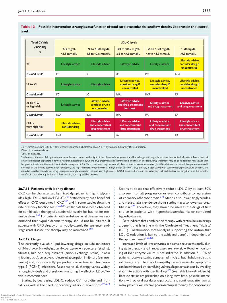
3a.7.11 Patients with kidney diseaseCKD can be characterized by mixed dyslipidaemia (high triglycer-ides, high LDL-C and low HDL-C).367 Statin therapy has a beneficialeffect on CVD outcomes in CKD368 and in some studies slows therate of kidney function loss.369,370 Similar data have been observedfor combination therapy of a statin with ezetimibe, but not for eze-timibe alone.368 For patients with end-stage renal disease, we rec-ommend that hypolipidaemic therapy should not be initiated. Ifpatients with CKD already on a hypolipidaemic therapy enter end-stage renal disease, the therapy may be maintained.368
3a.7.12 DrugsThe currently available lipid-lowering drugs include inhibitorsof 3-hydroxy-3-methylglutaryl-coenzyme A reductase (statins),fibrates, bile acid sequestrants (anion exchange resins), niacin(nicotinic acid), selective cholesterol absorption inhibitors (e.g. eze-timibe) and, more recently, proprotein convertase subtilisin/kexintype 9 (PCSK9) inhibitors. Response to all therapy varies widelyamong individuals and therefore monitoring the effect on LDL-C le-vels is recommended.
Statins, by decreasing LDL-C, reduce CV morbidity and mor-tality as well as the need for coronary artery interventions.371,372
Statins at doses that effectively reduce LDL-C by at least 50%also seem to halt progression or even contribute to regressionof coronary atherosclerosis.373 Statins also lower triglycerides,and meta-analysis evidence shows statins may also lower pancrea-titis risk.374 Therefore, they should be used as the drugs of firstchoice in patients with hypercholesterolaemia or combinedhyperlipidaemia.
Data indicate that combination therapy with ezetimibe also bringsa benefit that is in line with the Cholesterol Treatment Trialists’(CTT) Collaboration meta-analysis supporting the notion thatLDL-C reduction is key to the achieved benefit independent ofthe approach used.353,375
Increased levels of liver enzymes in plasma occur occasionally dur-ing statin therapy, and in most cases are reversible. Routine monitor-ing of liver enzyme values is not indicated. In addition, 5–10% ofpatients receiving statins complain of myalgia, but rhabdomyolysis isextremely rare. The risk of myopathy (severe muscular symptoms)can be minimized by identifying vulnerable patients and/or by avoidingstatin interactions with specific drugs376 (see Table E in web addenda).Because statins are prescribed on a long-term basis, possible interac-tions with other drugs deserve particular and continuous attention, asmany patients will receive pharmacological therapy for concomitant
Table 13 Possible intervention strategies as a function of total cardiovascular risk and low-density lipoprotein cholesterollevel
Total CV risk
(SCORE)
%
LDL-C levels
<70 mg/dL
<1.8 mmol/L
70 to <100 mg/dL
1.8 to <2.6 mmol/L
100 to <155 mg/dL
2.6 to <4.0 mmol/L
155 to <190 mg/dL
4.0 to <4.9 mmol/L
≥190 mg/dL
≥4.9 mmol/L
<1 Lifestyle advice Lifestyle advice Lifestyle advice Lifestyle adviceLifestyle advice, consider drug if
uncontrolled
Classa /Levelb I/C I/C I/C I/C IIa/A
≥1 to <5 Lifestyle advice Lifestyle adviceLifestyle advice, consider drug if
uncontrolled
Lifestyle advice, consider drug if
uncontrolled
Lifestyle advice, consider drug if
uncontrolled
Classa /Levelb I/C I/C IIa/A IIa/A I/A
≥5 to <10, or high-risk Lifestyle advice
Lifestyle advice, consider drug if
uncontrolled
Lifestyle adviceand drug treatment
for most
Lifestyle adviceand drug treatment
Lifestyle adviceand drug treatment
Classa /Levelb IIa/A IIa/A IIa/A I/A I/A
≥10 or very high-risk
Lifestyle advice, consider drug
Lifestyle adviceand concomitantdrug treatment
Lifestyle adviceand concomitantdrug treatment
Lifestyle adviceand concomitantdrug treatment
Lifestyle adviceand concomitantdrug treatment
Classa /Levelb IIa/A IIa/A I/A I/A I/A
CV ¼ cardiovascular;; LDL-C ¼ low-density lipoprotein cholesterol; SCORE ¼ Systematic Coronary Risk Estimation.aClass of recommendation.bLevel of evidence.Guidance on the use of drug treatment must be interpreted in the light of the physician’s judgement and knowledge with regards to his or her individual patient. Note that riskstratification is not applicable in familial hypercholesterolaemia, where drug treatment is recommended, and that, in this table, drug treatment may be considered at risks lower thanthe generic treatment thresholds indicated in paragraph 2.3.5. Thus treatment may occasionally be considered in moderate risk (1–5%) individuals, provided that patients are well-informed of the limited absolute risk reduction, and high numbers needed to treat. In higher risk (5–10%), drug therapy is associated with somewhat larger absolute benefits, andshould at least be considered. Drug therapy is strongly advised in those at very high risk (≥10%). If baseline LDL-C in this category is already below the target level of 1.8 mmol/L,benefit of statin therapy initiation is less certain, but may still be present.
Joint ESC Guidelines 2353
Downloaded from https://academic.oup.com/eurheartj/article-abstract/37/29/2315/1748952/2016-European-Guidelines-on-cardiovascular-diseaseby gueston 10 October 2017
conditions.377 In practice, management of a patient with myalgia butwithout a major creatinine kinase increase is based on trial and errorand usually involves a trial of a different statin or the use of a very lowdosage several days a week with a gradual increase.376
In general, the safety profile of statins is acceptable, and earlierobservations that lipid-lowering treatment may contribute to an in-crease in non-CV mortality (e.g. cancers, suicides, depression) ormental disorders were not confirmed in a large meta-analysis.378 In-creased blood sugar and glycated haemoglobin (HbA1c) levels (i.e.increased risk of type 2 DM) occur after statin treatment and aredose dependent, in part linked to very slight weight gain, but thebenefits of statins outweigh the risks for the vast majority of pa-tients.377 –379 Patients should be reminded that adhering to lifestylechanges when prescribed a statin should lessen any modest DMrisk.379– 382
For non-statin treatments, selective cholesterol absorption inhi-bitors (e.g. ezetimibe) are not usually used as monotherapy to de-crease LDL-C concentrations, unless patients are intolerant tostatins. They are recommended as combination therapy with statinsin selected patients when a specific goal is not reached with themaximal tolerated dose of a statin.
Bile acid sequestrants also decrease total cholesterol and LDL-Cbut are poorly tolerated and tend to increase plasma triglycerideconcentrations. They are therefore not recommended for routineuse in CVD prevention.
Fibrates and niacin are used primarily for triglyceride lowering andincreasing HDL-C, while fish oils (n-3 fatty acids) in doses of 2–4 g/day are used for triglyceride lowering.360 Evidence supporting theuse of these drugs for CVD event reduction is limited and, giventhe strong evidence favouring statins, routine use of these drugs inCVD prevention is not recommended. In order to prevent pancrea-titis, when triglycerides are .10 mmol/L (.900 mg/dL) they mustbe reduced not only by drugs but also by restriction of alcohol,treatment of DM, withdrawal of oestrogen therapy, etc. In thoserare patients with severe primary hypertriglyceridaemia, specialistreferral must be considered.
Regarding new therapies, recent data from phase I– III trials showthat PCSK9 inhibitors sharply decrease LDL-C by up to 60%, eitheras monotherapy or in addition to the maximal statin dose. Whetherthis approach results in the predicted reduction in CV events isbeing addressed in large outcome trials; preliminary evidence sug-gests that this is the case.383– 385
3a.7.13 Drug combinationsPatients with dyslipidaemia, particularly those with establishedCVD, DM or asymptomatic high-risk individuals, may not alwaysreach treatment goals, even with the highest tolerated statindose. Therefore, combination treatment may be needed. It mustbe stressed, however, that the only combination that has evidenceof clinical benefit (one large RCT) is that of a statin combined withezetimibe.353 Based on the relatively limited body of evidence, clin-icians may restrict the use of this combination to patients at high orvery-high risk of CVD.
Combinations of niacin and a statin increase HDL-C and decreasetriglycerides better than either of these drugs alone, but flushingis the main adverse effect of niacin, which may affect compliance.Furthermore, there is no evidence of clinical benefit for thiscombination.386
Fibrates, particularly fenofibrate, may be useful, not only fordecreasing high triglyceride concentrations and increasing lowHDL-C, but for lowering LDL-C further when used with a statin.There is limited evidence for this combination in terms of a reduc-tion in CVD events. In selected cases, however, this approach maybe considered, such as when, during statin treatment, triglyceridesremain high and/or HDL-C is very low. Other drugs metabolizedthrough cytochrome P450 should be avoided when this com-bination is prescribed. Fibrates should preferably be taken in themorning and statins in the evening to minimize peak dose con-centrations and decrease the risk of myopathy. Patients have tobe instructed about warning symptoms (myalgia), even thoughsuch adverse effects are very rare. Gemfibrozil should not be addedto a statin treatment, because of the high potential for interactions.
If target levels cannot be reached even on maximal doses oflipid-lowering therapy or drug combinations, patients will still bene-fit from treatment to the extent that the dyslipidaemia has been im-proved. In these patients, increased attention to other risk factorsmay help to reduce total risk.
Gaps in evidence† Triglyceride or HDL-C values as a target for therapy.† Whether Lp(a) lowering against background statin therapy can
reduce the risk of CVD.† How to increase adoption of non-HDL-C and non-fasting sam-
ples in clinical practice.† Whether functional foods and food supplements with a
lipid-lowering effect can safely reduce the risk of CVD.
3a.8 Diabetes mellitus (type 2 and type 1)Key messages† The multifactorial approach is very important in patients with
type 2 DM.† Lifestyle management to aid weight control by sustainable dietary
changes and increased PA levels should be central in the manage-ment of patients with type 2 DM.
† Intensive management of hyperglycaemia reduces the risk ofmicrovascular complications and, to a lesser extent, the risk ofCVD. However, targets should be relaxed in the elderly, frail,those with long-duration DM and those with existing CVD.
† Intensive treatment of BP in DM, with a target of 140 mmHg sys-tolic for the majority, reduces the risk of macrovascular andmicrovascular outcomes. A lower SBP target of 130 mmHg fur-ther lessens the risks for stroke, retinopathy and albuminuria andshould be applied to selected patients.
† Lipid lowering is a key mechanism to lower CVD risk in both type 2and type 1 DM. All patients .40 years of age and selected youngerpatients at elevated risk are recommended for statin therapy.
† In DM patients with existing CVD, the use of a sodium-glucoseco-transporter-2 (SGLT2) inhibitor substantially lessened CVDand total mortality and HF hospitalisation without major adverseeffects. SGLT2 inhibitors should be considered early in thecourse of DM management in such patients.
† Recent evidence points to sizeable reductions in CVD mortalityin DM patients via improvements in risk factor management, al-though the increasing worldwide DM prevalence will create ma-jor challenges. More should be done to prevent DM.
Joint ESC Guidelines2354
Downloaded from https://academic.oup.com/eurheartj/article-abstract/37/29/2315/1748952/2016-European-Guidelines-on-cardiovascular-diseaseby gueston 10 October 2017

People with DM are on average at double the risk of CVD.399 Asimple DM risk questionnaire can guide which patients without
CVD should be tested for DM.400
Keeping close to the recommended targets for BP, lipid control,glycaemia and HbA1c is important for the prevention of CVD. Clear
reductions have occurred in CVD death rates in DM consistent with
better management of risk factors, although the increasing prevalence
of DM continues to create pressures on all health care systems.The targets, especially the glycaemic and in some cases lipids,
should be less stringently implemented in older people with DM,
those with a longer duration of DM, those with evidence of CVD
and the frail.401
There is mounting evidence for a very high relative risk in youngerindividuals with type 2 DM (age ,40 years),402 and additional guid-ance on care is needed.
Except for glucose management, prevention of CVD follows thesame general principles as for people without DM. Achieving lowBP levels and low LDL-C and total cholesterol concentrations is par-ticularly important. Many treatment targets are more stringent for pa-tients with DM. Typically, patients with type 2 DM have multiple CVDrisk factors, each requiring treatment according to existing guidelines.
3a.8.1 Lifestyle interventionThe ESC and European Association for the Study of Diabetesscientific statements advocate lifestyle management as a first
Recommendations for management of diabetes
Recommendations Classa Levelb Ref c
Lifestyle changes including smoking cessation, low fat diet, high diet, aerobic physical activity, and strength training are recommended.
I A 387
Reduction in energy intake is recommended to patients to help achieve lower weight or prevent weight gain. I B 387
A target HbA1c for the reduction in risk of CVD and microvascular complications in DM of <7.0% (<53 mmol/mol) is recommended for the majority of non-pregnant adults with either type 1 or type 2 DM.
I A 388, 389
For patients with a long duration of DM, the elderly, frail, or those with existing CVD, a relaxing of the HbA1c targets (i.e. less stringent) should be considered.
IIa B 389
A target HbA1c of ≤6.5% (≤48 mmol/mol) should be considered at diagnosis or early in the course of type 2 DM in patients, who are not frail and do not have CVD.
IIa B 389
When screening for DM in individuals with or without CVD, assessment of HbA1c (which can be done non-fasting) or fasting blood glucose should be considered. An oral glucose tolerance test can be offered when there is still doubt.
IIa A 390
Metformin is recommended as therapy, if tolerated and not contra-indicated, following evaluation of renal function. I B 391
Avoidance of hypoglycaemia and excessive weight gain should be considered and individual approaches (with respect to both treatment targets and drug choices) should be considered in patients with advanced disease.
IIa B389, 392,
393
In patients with type 2 DM and CVD, the use of an SGLT2 inhibitor should be considered early in the course of the disease to reduce CV and total mortality.
IIa B 394
Lipid lowering agents (principally statins) are recommended to reduce CV risk in all patients with type 2 or type 1 DM above the age of 40 years.
I A 371, 372
Lipid lowering agents (principally statins) may be considered also in individuals below 40 years of age if at elevated risk, based on the presence of micro-vascular complications or of multiple CV risk factors.
IIb A 371, 372
In DM patients at very high-risk (see table 5), a LDL-C target <1.8 mmol/L (<70 mg/dL), or a reduction of at least 50% if the baseline LDL-C is between 1.8 and 3.5 mmol/L (70 and 135 mg/dL), is recommended.d
In DM patients with high-risk (see table 5), LDL-C target <2.6 mmol/L (<100mg/dL) or a reduction of at least 50% if the baseline LDL-C is between 2.6 and 5.1 mmol/L (100 and 200 mg/dL) is recommended.d
I B 395
BP targets in type 2 DM are generally recommended to be <140/85 mmHg, but a lower target of <130/80 mmHg is recommended in selected patients (e.g. younger patients at elevated risk for complications) for additional gains on stroke, retinopathy and albuminuria risk. Renin-angiotensin-aldosterone system blocker is recommended in the treatment of hypertension in DM, particularly in the presence of proteinuria or micro- albuminuria. Recommended BP target in patients with type 1 DM is <130/80 mmHg.
I B 396, 397
The use of drugs that increase HDL-C to prevent CVD in type 2 DM is not recommended. III A 386
Antiplatelet therapy (e.g. with aspirin) is not recommended for people with DM who do not have CVD. III A 398
BP ¼ blood pressure; CV ¼ cardiovascular; DM ¼ diabetes mellitus; HbA1c ¼ glycated haemoglobin; HDL-C ¼ high-density lipoprotein cholesterol; LDL-C ¼ low-densitylipoprotein cholesterol; SGLT2 ¼ Sodium-glucose co-transporter-2.aClass of recommendation.bLevel of evidence.cReference(s) supporting recommendations.dNon-HDL-C is a reasonable and practical alternative target because it does not require fasting. Non HDL-C secondary targets of ,2.6 and ,3.3 mmol/L (,100 and ,130 mg/dL) arerecommended for very high, and high-risk subjects, respectively See section 3a.7.10 for more details.
Joint ESC Guidelines 2355
Downloaded from https://academic.oup.com/eurheartj/article-abstract/37/29/2315/1748952/2016-European-Guidelines-on-cardiovascular-diseaseby gueston 10 October 2017

measure for the prevention and management of DM.387 Most pa-tients with DM are obese, so weight control is a central compo-nent. Several dietary patterns can be adopted where thepredominance of fruits, vegetables, wholegrain cereals and low-fatprotein sources is more important than the precise proportions oftotal energy provided by the major macronutrients. Salt intakeshould be restricted. Specific dietary recommendations includelimiting saturated and trans fats and alcohol intake, monitoringcarbohydrate consumption and increasing dietary fibre. AMediterranean-type diet is acceptable, where fat sources are de-rived primarily from monounsaturated oils.
A combination of aerobic and resistance exercise training iseffective in the prevention of the progression of DM and forthe control of glycaemia. Little is known about how to promoteand sustain PA; however, reinforcement by health care providers topatients to find sustainable ways to increase PA is crucial. Smoking in-creases the risk of DM, CVD and premature death and should bestrongly discouraged (see section 3a.4.5).387,403 Lifestyle interventioncan also prevent DM development in those at elevated risk and, inturn, lowers future microvascular and macrovascular risks.404
3a.8.2 Cardiovascular riskAt diagnosis or in those with a short duration of disease, DM is not aCAD risk equivalent state.405,406 In general, risk levels approach CADrisk equivalence after about a decade or in those with proteinuria orlow eGFR.406–408 Emerging data suggest that patients who developDM at a younger age have a high complication burden.402 Peoplewith DM with existing CAD have a vascular risk well in excess of thosewithCADbutwithout DManda substantially lower lifeexpectancy.409
Statins are recommended for all those newly diagnosed with type2 DM beyond a certain age (.40 years is currently recommended).This recommendation reflects greater lifetime vascular risk trajec-tories in these individuals. However, a proportion of DM patientsat 40–50 years of age may have a low 10 year risk of CVD due tonormal BP and lipid levels and being non-smokers, and in such casesthere remains a role for physician judgement. Equally, in some pa-tients ,40 years of age with type 2 DM with evidence of end-organdamage or significant risk factors, statins may be indicated.
3a.8.3 Glucose controlThe UK Prospective Diabetes Study (UKPDS) established the im-portance of intensive glucose lowering with respect to CVD risk re-duction in newly diagnosed patients with DM but not treated withmodern BP- or lipid-lowering therapies, with the best evidence tosupport metformin, leading to its position as first-line therapy.Three trials were conducted to see if CV events could be reducedfurther with more intensive glycaemia treatment and lower targetHbA1c levels.389,393,410 However, the results were surprising, withunexpected increases in total and CVD deaths in the Action toControl Cardiovascular Risk in Diabetes (ACCORD) trial and atrend towards an increase in CVD death in the Veterans AffairsDiabetes Trial (VADT). The results prompted concerns aboutthe safety of intensive glucose lowering and the appropriatenessof pursuing tight glucose control, particularly in older peoplewith DM and in those with existing CVD. Subsequentmeta-analyses of intensive glucose control, including data fromUKPDS, Prospective Pioglitazone Clinical Trial in MacrovascularEvents (PROactive), ACCORD, Action in Diabetes and Vascular
disease: PreterAx and Diamicron MR Controlled Evaluation (AD-VANCE) and VADT,411 showed significant reductions in non-fatalAMI and CAD events, but no effect on stroke or total mortal-ity.412,413 The additional analyses of these trials suggested thatCVD benefits for an average HbA1c reduction of �0.9% over 5years were far less than via usual reductions in cholesterol andBP seen with statins and available BP-lowering agents. Four recenttrials of newer DM therapies (DPP-4 and GLP-1)414 – 417 in patientswith DM and existing CVD or at high risk demonstrated non-inferiority (i.e. safety) but not superiority with respect to CVDrisk. There was, however, an increase in the rate of hospitalizationfor HF with saxagliptin in the Saxagliptin Assessment of VascularOutcomes Recorded in Patients with Diabetes Mellitus – Trombo-lysis in Myocardial Infarction (SAVOR-TIMI 53) trial.416
Very recently, the SGLT2 inhibitor empagliflozin demonstratedsubstantial reductions in CVD death (by 38%) and all-cause mortality(by 32%), as well as in hospitalisation for HF (by 35%), as comparedwith standard care, suggesting use of an SLGT2 inhibitor should comevery early in the course of management of patients with DM andCVD.394 The pattern of trial results whereby non-fatal MI and strokewere not reduced by active treatment, as well as the rapid separationof mortality curves, suggest that the mechanism of benefit was likelyto relate more to cardio-renal haemodynamic effects than to athero-thrombotic actions or effects of glucose lowering per se. More re-search on understanding the trial results is needed.
3a.8.4 Blood pressureIn people with type 2 DM, apart from lifestyle interventions, the re-duction of BP (along with cholesterol) should be targeted as strictlyas targeting glucose/HbA1c levels. BP targets should be consideredregardless of overall CV risk score in patients with type 2 DM.
Hypertension is more common in patients with type 2 DM com-pared with the general population. A recent systematic review andmeta-analysis of randomized trials of BP-lowering agents in .100000 patients with type 2 DM confirmed that lowering BP reducesthe risk of all-cause mortality, CV events, CAD events, stroke, HF,retinopathy, new or worsening albuminuria and renal failure.418
The results were similar when trials with low risk of bias were se-lected. Furthermore, a systolic target ,140 mmHg lessens therisk of total mortality and most separate outcomes. Further reduc-tions in the risk for albuminuria, retinopathy and stroke, but not inoverall survival or aggregate clinical endpoints, were achieved with asystolic target ,130 mmHg. In people .80 years of age, targetsshould be set higher, aiming for ,150/90 mmHg, unless renal im-pairment is present.
Combination treatment is commonly needed to lower BP effect-ively in DM. An ACE-I or an angiotensin receptor blocker (ARB),where tolerated, should always be included as first-line therapy be-cause of the evidence of superior protective effects against initiationor progression of nephropathy.
3a.8.5 Lipid-lowering therapyThe Heart Protection Study (HPS) demonstrated that treatmentwith simvastatin 40 mg reduced the risk of CAD and stroke in peo-ple with DM and individuals without DM who had no prior AMI orangina pectoris.372 Further robust support for statin benefit camefrom the Collaborative Atorvastatin Diabetes Study (CARDS),which compared 10 mg atorvastatin with placebo,371 and from the
Joint ESC Guidelines2356
Downloaded from https://academic.oup.com/eurheartj/article-abstract/37/29/2315/1748952/2016-European-Guidelines-on-cardiovascular-diseaseby gueston 10 October 2017
CTT meta-analysis in DM patients.419 There is also trial evidence toshow greater CVD risk reduction with more intense statin therapyin DM patients.395 More recent trial evidence shows a clear CVDbenefit of lowering LDL-C with ezetimibe on top of a statin in pa-tients with type 2 DM.353 Emerging evidence also shows thatPCSK9 inhibitors are equally efficacious in lowering LDL-C in type2 DM patients, although results of CV outcome trials are awaited.Lower treatment targets should be pursued in patients with type2 DM who have overt CVD or CKD.
While the most common lipid abnormality in type 2 DM is ele-vated triglyceride and low HDL-C, trials examining possible CVDbenefits of lipid (mainly triglyceride) lowering with fibrates in DMhave not been positive. The US Food and Drug Administration(FDA) states that the current evidence base is insufficient to supportfibrates for CVD protection and that more trial evidence isneeded.420
Prescribing of lipid-lowering agents in older people with DM(.85 years) requires special consideration because exposure tohigher doses (or higher potency) may not increase life expectancy,but may increase the risk of adverse effects.
3a.8.6 Antithrombotic therapyPatients with type 1 or type 2 DM have an increased tendency todevelop thrombotic phenomena. The Antiplatelet Trialists’ Collab-oration meta-analysis demonstrated the benefits of antithrombotictherapy (mainly aspirin) in patients with DM with clinically estab-lished CAD, cerebrovascular disease or other forms of thromboticdisease, with a 25% reduction in risk of CV events.421
The role of aspirin in patients without CVD remains unproven. Ameta-analysis of six RCTs found no statistically significant reductionin the risk of major CV events or all-cause mortality when aspirinwas compared with placebo or no aspirin in people with DM andno pre-existing CVD.398 Further trials are ongoing.
3a.8.7 MicroalbuminuriaMicroalbuminuria (urinary albumin excretion from 30 to 300 mg/24 h) predicts the development of overt nephropathy in patientswith type 1 or type 2 DM, while the presence of overt proteinuria(300 mg/24 h) generally indicates established renal parenchymaldamage. In patients with DM and hypertension, microalbuminur-ia—even below the current threshold values—predicts CV events,and a continuous relationship between CV as well as non-CV mor-tality and urinary protein:creatinine ratios has been reported. Micro-albuminuria can be measured from spot urine samples (due toinaccuracy in sampling, 24 h or night-time urine collection is discour-aged) by indexing the urinary albumin concentration to the urinarycreatinine concentration (2.5/3.5–25/35 mg/mmol). Patients withDM and microalbuminuria or proteinuria should be treated withan ACE-I or ARB regardless of baseline BP.
Gaps in evidence† There is a need to examine whether a type 2 DM CV risk score
based on either 10 year or lifetime risk helps to improve targetingof preventative therapies and leads to a reduction in CV risk or again in lifetime years free from disease.
† Further trial data are needed to establish if the empagliflozin out-come findings hold for other classes of SGLT2 inhibitors and tobetter understand the mechanisms of benefit. It would also be
useful to know if SGLT2 inhibitors lessen CV mortality and HFrisks in patients with DM but without CVD.
† More research on the benefits of glucagon-like peptide 1(GLP-1) receptor agonists on CVD risk is needed and trialsare due to be reported in subsequent years. Early evidence sug-gests no CVD benefit with short-term use of dipeptidyl peptid-ase 4 (DPP-4) inhibitors in people at high risk for CVD, asreviewed.422
3a.8.8 Type 1 diabetes
Key messages† CVD and mortality risks have decreased in type 1 DM patients
but remain unacceptably elevated in those with very poor gly-caemic control or any evidence of kidney disease.
† Intensive management of hyperglycaemia in DM reducesthe risk of macrovascular complications and prematuremortality; a target of 6.5–7.5% (48–58 mmol/mol) HbA1c isrecommended.
† The recommended BP target in the majority of patients with type1 DM is 130/80 mmHg.
† Lipid-lowering agents targeting LDL-C reduction should be re-commended to the majority of patients .40 years of age andto those younger than this with evidence of nephropathy orwith multiple risk factors.
Type 1 DM is the result of a lack of insulin production in the pan-creas, confirmed by absent or virtually absent C-peptide levels.The average age of onset is �14 years, although persons of anyage can develop type 1 DM. Type 1 DM should be suspected inany patient who progresses to insulin within the first year of diagno-sis. A large contemporary study in Scotland observed a relative riskfor CVD events of 2.3 in men and 3 in women with type 1 DM com-pared with the general population,423 suggesting CVD risks mayhave declined over time, commensurate with improvements in lifeexpectancy.424 Another report from Sweden demonstrated CVDmortality rates in type 1 DM to be twice the rates of the generalpopulation in those with HbA1c levels ,6.9% (52 mmol/mol),whereas risk was especially high (�10-fold) in those with verypoor control [≥9.7% (≥83 mmol/mol)].425 In the majority of stud-ies, the risk of CVD events or mortality was highest among thosewith diabetic nephropathy, macroalbuminuria or CKD. The pres-ence of proliferative retinopathy and autonomic neuropathy alsosignalled an elevated CVD risk.
The Diabetes Control and Complications Trial (DCCT) estab-lished the importance of tight glucose control to lessen the risks ofboth microvascular and macrovascular disease. A 27 year follow-up of this trial showed that 6.5 years of initial intensive DM therapyin type 1 DM was associated with a modestly lower all-causemortality rate when compared with conventional therapy.426
A glycaemic target for HbA1c of 6.5–7.5% (48–58 mmol/mol) ap-pears to be a balanced approach for long-term care of patientswith type 1 DM. The use of insulin analogues, insulin pumps andcontinuous glucose monitoring to improve glycaemic controlwhile minimizing hypoglycaemia is the subject of intense research,as is the use of agents (e.g. metformin, GLP-1 agonists) commonlyused in type 2 DM.
The CTT suggested lipid lowering with statins is as equally effect-ive in type 1 patients as in type 2.427 All patients .40 years of age
Joint ESC Guidelines 2357
Downloaded from https://academic.oup.com/eurheartj/article-abstract/37/29/2315/1748952/2016-European-Guidelines-on-cardiovascular-diseaseby gueston 10 October 2017

with type 1 DM should be recommended for statins unless they havea short duration of DM and no other risk factors. Younger patientswith multiple risk factors or evidence of end organ damage (albu-minuria, low eGFR, proliferative retinopathy or neuropathy) shouldbe considered for statin therapy.
A target BP of 130/80 mmHg is accepted practice in type 1 DM,with evidence of specific benefits of ACE-Is or ARBs on the earlydevelopment and later progression of microvascular disease inyounger type 1 DM patients. A lower target BP of 120/75–80mmHg may be helpful in younger type 1 DM patients (,40 yearsof age) with persistent microalbuminuria. Studies supporting im-proved CVD outcome in type 1 DM through BP reduction are lack-ing. As more patients with type 1 DM are living to older age, SBPtargets may need to be relaxed (140 mmHg) in some to avoidside effects.
Current evidence suggests many patients with type 1 DM .40years of age continue to smoke, are still not receiving statins and,perhaps most importantly, have very poor glucose control.423 Fur-ther efforts to target these established risk factors are needed.
Gaps in evidence† Further studies are needed on metformin and GLP-1 receptor
agonists in (subgroups of) patients with type 1 DM to determinewhether they improve glycaemic control, aid in weight reductionand improve clinical outcomes.
† There is a need for a CVD risk score in type 1 DM to better guideinitiation of preventative therapies in younger patients.
3a.9 HypertensionKey messages† Elevated BP is a major risk factor for CAD, HF, cerebrovascular
disease, PAD, CKD and AF.† The decision to start BP-lowering treatment depends on the BP
level and total CV risk.† Benefits of treatment are mainly driven by BP reduction per se,
not by drug type.† Combination treatment is needed to control BP in most
patients.
Recommendations for management of hypertension
Recommendations Classa Levelb Ref c
Lifestyle measures (weight control, increased physical activity, alcohol moderation, sodium restriction, and increased consumption of fruits, vegetables, and low-fat dairy products) are recommended in all patients with hypertension and in individuals with high normal BP.
I A337,
428-430
All major BP lowering drug classes (i.e. diuretics, ACE-I, calcium antagonists, ARBs, and ß-blockers) do not differ in their BP-lowering and thus are recommended as BP lowering treatment.
I A 431, 432
In asymptomatic subjects with hypertension but free of CVD, CKD, and DM, total CV risk using the SCORE model is recommended.
I B 30
Drug treatment is recommended in patients with grade 3 hypertension irrespective of CV risk, as well as in patients with grade 1 or 2 hypertension who are at very high CV risk.
I B 433
Drug treatment should be considered in patients with grade 1 or 2 hypertension who are at high CV risk. IIa B 433
In patients at low to moderate total CV risk and with grade 1 or 2 hypertension, lifestyle measures are recommended. I B 433
In patients at low to moderate total CV risk and with grade 1 or 2 hypertension, if lifestyle measures fail to reduce BP, drug treatment may be considered.
IIb B 433
SBP <140 mmHg and DBP <90 mmHg are recommended in all treated hypertensive patients <60 years old. I B 433
In patients >60 years old with SBP ≥160 mmHg, it is recommended to reduce SBP to between 150 and 140 mmHg. I B 434
In patients <80 years old, a target SBP <140 mmHg may be considered if treatment is well tolerated. In some of these patients a target SBP <120 mmHg may be considered if at (very) high-risk and tolerate multiple BP lowering drugs.
IIb B 434, 435
In individuals >80 years and with initial SBP ≥160 mmHg, it is recommended to reduce SBP to between 150 and 140 mmHg, provided they are in good physical and mental conditions.
I B 434
In frail elderly patients, a careful treatment intensity (e.g. number of BP lowering drugs) and BP targets should be considered, and clinical effects of treatment should be carefully monitored.
IIa B 436
Initiation of BP lowering therapy with a two-drug combination may be considered in patients with markedly elevated baseline BP or at high CV risk. Combination of two drugs at doses in a single pill may be considered because of improved adherence.
IIb C 437
ß-blockers and thiazide diuretics are not recommended in hypertensive patients with multiple metabolic risk factors,d due to the increased risk of DM.
III B 438
ACE-I ¼ angiotensin-converting enzyme inhibitor; ARBs ¼ angiotensin receptor blockers; BP ¼ blood pressure; CKD ¼ chronic kidney disease; CV ¼ cardiovascular; CVD ¼cardiovascular disease; DBP ¼ diastolic blood pressure; SBP ¼ systolic blood pressure; SCORE ¼ Systematic Coronary Risk Estimation.aClass of recommendation.bLevel of evidence.cReference(s) supporting recommendations.dOverweight, obesity, dyslipidaemia, impaired glucose tolerance.
Joint ESC Guidelines2358
Downloaded from https://academic.oup.com/eurheartj/article-abstract/37/29/2315/1748952/2016-European-Guidelines-on-cardiovascular-diseaseby gueston 10 October 2017

3a.9.1. IntroductionHigh BP is a leading risk factor for disease burden globally, account-ing for 9.4 million deaths and 7.0% of global disability-adjusted life-years (DALYs) in 2010.439 Compared with 1990, the impact of highBP has increased by �2.1 million deaths.439 Overall, the prevalenceof hypertension is �30–45% in adult persons ≥18 years of age,with a steep increase with ageing.
Elevated BP is a risk factor for CAD, HF, cerebrovascular disease,PAD, CKD and AF. The risk of death from either CAD or stroke in-creases progressively and linearly from BP levels as low as 115mmHg systolic and 75 mmHg diastolic upwards,440 although for ab-solute risk the curves flatten in the lower BP ranges.
3a.9.2 Definition and classifications of hypertensionThe definition and classifications of hypertension are shown inTable 14.11
3a.9.3 Blood pressure measurementOffice BP is recommended for screening and diagnosis of hyperten-sion, which should be based on at least two BP measurements pervisit and on at least two visits. If the BP is only slightly elevated, re-peated measurements should be made over a period of severalmonths to achieve an acceptable definition of the individual’s ‘usual’BP and to decide about initiating drug treatment. If BP is more mark-edly elevated or accompanied by target organ damage, other CVfactors or established CV or renal disease, repeated BP measure-ments are required within a shorter period in order to make treat-ment decisions.
3a.9.4 Office or clinic blood pressure measurementAuscultatory or oscillometric semi-automatic sphygmoman-ometers should be validated and checked periodically.441 Meas-urement of BP at the upper arm is preferred, and cuff andbladder dimensions should be adapted to the arm circumference.If feasible, automated recording of multiple BP readings in the of-fice, with the patient seated in an isolated room, might be
considered as a means of improving reproducibility and matchingoffice BP values closer to those provided by daytime ambulatoryBP monitoring (ABPM) or home BP measurements (HBPMs).442
Note that automated devices are not validated for BP measure-ment in patients with AF.
3a.9.5 Out-of-office blood pressure monitoringOut-of-office BP is commonly assessed by ABPM or HBPM, usuallyby self-measurement; it is usually lower than the office BP and thedifference increases as office BP increases (Table 15).443
The following general principles and remarks should be takeninto account: (i) the procedure should be adequately explainedto the patient, with verbal and written instructions; (ii) interpret-ation of the results should take into account that the reproducibil-ity of out-of-office BP measurements is reasonably good for 24 h,day and night BP averages, but less so for shorter periods;(iii) ABPM and HBPM provide somewhat different informationon the subject’s BP status and risk, and the two methods shouldthus be regarded as complementary rather than competitive;(iv) devices should be validated and regularly calibrated, at leastevery 6 months.
Both ABPM and HBPM values are closely related to prognosis.444
Night-time BP seems to be a stronger predictor than daytime BP.Out-of-office measurement may be useful not only in untreatedsubjects, but also in treated patients, with the aim of monitoringthe effects of treatment and increasing compliance with drug ther-apy (Table 16).
3a.9.6 Diagnostic evaluation in hypertensive patientsLaboratory tests should include haemoglobin, fasting plasma glucose(HbA1c if not fasting) and serum tests for total cholesterol, HDL-C,triglycerides, potassium, uric acid, creatinine (and calculated renalfunction) and thyrotropin (in postmenopausal women). Urinalysisshould include albumin:creatinine ratio, dipstick test, sediment andquantitative proteinuria if the dipstick test is positive. Echocardiog-raphy and fundoscopy can be considered. The routine measurementof additional biomarkers and/or the use of vascular imaging methodsis not recommended.
Table 14 Definition and classification of bloodpressure levelsa
Category Systolic BP (mmHg)
Diastolic BP (mmHg)
Optimal <120 and <80
Normal 120–129 and/or 80–84
High-normal 130–139 and/or 85–89
Grade 1 hypertension 140–159 and/or 90–99
Grade 2 hypertension 160–179 and/or 100–109
Grade 3 hypertension ≥180 and/or ≥110
Isolated systolic hypertension
≥140 and <90
BP ¼ blood pressure.aBP levels in untreated individuals.
Table 15 Blood pressure thresholds for definition ofhypertension with different types of BP measurement
SBP (mmHg) DBP (mmHg)
or clinic 140 90
24-hour 125 –130 80
Day 130–135 85
Night 120 70
Home 130–135 85
DPB ¼ diastolic blood pressure; SBP ¼ systolic blood pressure.
Joint ESC Guidelines 2359
Downloaded from https://academic.oup.com/eurheartj/article-abstract/37/29/2315/1748952/2016-European-Guidelines-on-cardiovascular-diseaseby gueston 10 October 2017
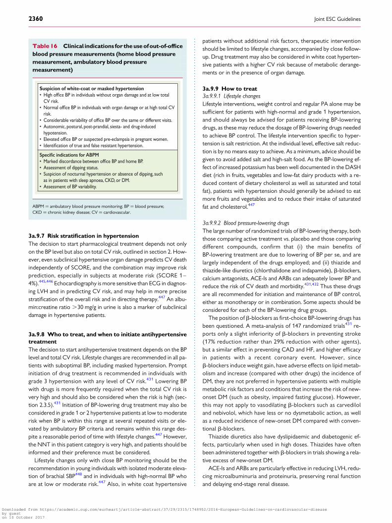
3a.9.7 Risk stratification in hypertensionThe decision to start pharmacological treatment depends not onlyon the BP level but also on total CV risk, outlined in section 2. How-ever, even subclinical hypertensive organ damage predicts CV deathindependently of SCORE, and the combination may improve riskprediction, especially in subjects at moderate risk (SCORE 1–4%).445,446 Echocardiography is more sensitive than ECG in diagnos-ing LVH and in predicting CV risk, and may help in more precisestratification of the overall risk and in directing therapy.447 An albu-min:creatine ratio .30 mg/g in urine is also a marker of subclinicaldamage in hypertensive patients.
3a.9.8 Who to treat, and when to initiate antihypertensivetreatmentThe decision to start antihypertensive treatment depends on the BPlevel and total CV risk. Lifestyle changes are recommended in all pa-tients with suboptimal BP, including masked hypertension. Promptinitiation of drug treatment is recommended in individuals withgrade 3 hypertension with any level of CV risk.431 Lowering BPwith drugs is more frequently required when the total CV risk isvery high and should also be considered when the risk is high (sec-tion 2.3.5).431 Initiation of BP-lowering drug treatment may also beconsidered in grade 1 or 2 hypertensive patients at low to moderaterisk when BP is within this range at several repeated visits or ele-vated by ambulatory BP criteria and remains within this range des-pite a reasonable period of time with lifestyle changes.447 However,the NNT in this patient category is very high, and patients should beinformed and their preference must be considered.
Lifestyle changes only with close BP monitoring should be therecommendation in young individuals with isolated moderate eleva-tion of brachial SBP448 and in individuals with high-normal BP whoare at low or moderate risk.447 Also, in white coat hypertensive
patients without additional risk factors, therapeutic interventionshould be limited to lifestyle changes, accompanied by close follow-up. Drug treatment may also be considered in white coat hyperten-sive patients with a higher CV risk because of metabolic derange-ments or in the presence of organ damage.
3a.9.9 How to treat3a.9.9.1 Lifestyle changesLifestyle interventions, weight control and regular PA alone may besufficient for patients with high-normal and grade 1 hypertension,and should always be advised for patients receiving BP-loweringdrugs, as these may reduce the dosage of BP-lowering drugs neededto achieve BP control. The lifestyle intervention specific to hyper-tension is salt restriction. At the individual level, effective salt reduc-tion is by no means easy to achieve. As a minimum, advice should begiven to avoid added salt and high-salt food. As the BP-lowering ef-fect of increased potassium has been well documented in the DASHdiet (rich in fruits, vegetables and low-fat dairy products with a re-duced content of dietary cholesterol as well as saturated and totalfat), patients with hypertension should generally be advised to eatmore fruits and vegetables and to reduce their intake of saturatedfat and cholesterol.447
3a.9.9.2 Blood pressure-lowering drugsThe large number of randomized trials of BP-lowering therapy, boththose comparing active treatment vs. placebo and those comparingdifferent compounds, confirm that (i) the main benefits ofBP-lowering treatment are due to lowering of BP per se, and arelargely independent of the drugs employed; and (ii) thiazide andthiazide-like diuretics (chlorthalidone and indapamide), b-blockers,calcium antagonists, ACE-Is and ARBs can adequately lower BP andreduce the risk of CV death and morbidity.431,432 Thus these drugsare all recommended for initiation and maintenance of BP control,either as monotherapy or in combination. Some aspects should beconsidered for each of the BP-lowering drug groups.
The position of b-blockers as first-choice BP-lowering drugs hasbeen questioned. A meta-analysis of 147 randomized trials431 re-ports only a slight inferiority of b-blockers in preventing stroke(17% reduction rather than 29% reduction with other agents),but a similar effect in preventing CAD and HF, and higher efficacyin patients with a recent coronary event. However, sinceb-blockers induce weight gain, have adverse effects on lipid metab-olism and increase (compared with other drugs) the incidence ofDM, they are not preferred in hypertensive patients with multiplemetabolic risk factors and conditions that increase the risk of new-onset DM (such as obesity, impaired fasting glucose). However,this may not apply to vasodilating b-blockers such as carvediloland nebivolol, which have less or no dysmetabolic action, as wellas a reduced incidence of new-onset DM compared with conven-tional b-blockers.
Thiazide diuretics also have dyslipidaemic and diabetogenic ef-fects, particularly when used in high doses. Thiazides have oftenbeen administered together with b-blockers in trials showing a rela-tive excess of new-onset DM.
ACE-Is and ARBs are particularly effective in reducing LVH, redu-cing microalbuminuria and proteinuria, preserving renal functionand delaying end-stage renal disease.
Table 16 Clinical indications for the use of out-of-officeblood pressure measurements (home blood pressuremeasurement, ambulatory blood pressuremeasurement)
Suspicion of white-coat or masked hypertension• High BP in individuals without organ damage and at low total CV risk.• Normal BP in individuals with organ damage or at high total CV risk.• Considerable variability of BP over the same or different visits.• Autonomic, postural, post-prandial, siesta- and drug-induced hypotension.• Elevated BP or suspected pre-eclampsia in pregnant women.
of true and false resistant hypertension.
• Marked discordance between BP and home BP.• Assessment of dipping status.• Suspicion of nocturnal hypertension or absence of dipping, such as in patients with sleep apnoea, CKD, or DM.• Assessment of BP variability.
ABPM ¼ ambulatory blood pressure monitoring; BP ¼ blood pressure;CKD ¼ chronic kidney disease; CV ¼ cardiovascular.
Joint ESC Guidelines2360
Downloaded from https://academic.oup.com/eurheartj/article-abstract/37/29/2315/1748952/2016-European-Guidelines-on-cardiovascular-diseaseby gueston 10 October 2017
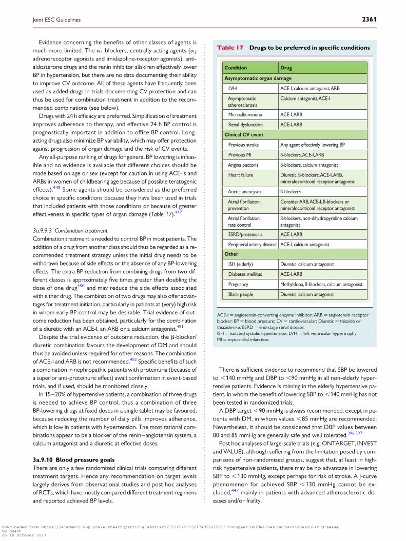
Evidence concerning the benefits of other classes of agents ismuch more limited. The a1 blockers, centrally acting agents (a2
adrenoreceptor agonists and imidazoline-receptor agonists), anti-aldosterone drugs and the renin inhibitor aliskiren effectively lowerBP in hypertension, but there are no data documenting their abilityto improve CV outcome. All of these agents have frequently beenused as added drugs in trials documenting CV protection and canthus be used for combination treatment in addition to the recom-mended combinations (see below).
Drugs with 24 h efficacy are preferred. Simplification of treatmentimproves adherence to therapy, and effective 24 h BP control isprognostically important in addition to office BP control. Long-acting drugs also minimize BP variability, which may offer protectionagainst progression of organ damage and the risk of CV events.
Any all-purpose ranking of drugs for general BP lowering is infeas-ible and no evidence is available that different choices should bemade based on age or sex (except for caution in using ACE-Is andARBs in women of childbearing age because of possible teratogeniceffects).449 Some agents should be considered as the preferredchoice in specific conditions because they have been used in trialsthat included patients with those conditions or because of greatereffectiveness in specific types of organ damage (Table 17).447
3a.9.9.3 Combination treatmentCombination treatment is needed to control BP in most patients. Theaddition of a drug from another class should thus be regarded as a re-commended treatment strategy unless the initial drug needs to bewithdrawn because of side effects or the absence of any BP-loweringeffects. The extra BP reduction from combining drugs from two dif-ferent classes is approximately five times greater than doubling thedose of one drug450 and may reduce the side effects associatedwith either drug. The combination of two drugs may also offer advan-tages for treatment initiation, particularly in patients at (very) high riskin whom early BP control may be desirable. Trial evidence of out-come reduction has been obtained, particularly for the combinationof a diuretic with an ACE-I, an ARB or a calcium antagonist.451
Despite the trial evidence of outcome reduction, the b-blocker/diuretic combination favours the development of DM and shouldthus be avoided unless required for other reasons. The combinationof ACE-I and ARB is not recommended.452 Specific benefits of sucha combination in nephropathic patients with proteinuria (because ofa superior anti-proteinuric effect) await confirmation in event-basedtrials, and if used, should be monitored closely.
In 15–20% of hypertensive patients, a combination of three drugsis needed to achieve BP control, thus a combination of threeBP-lowering drugs at fixed doses in a single tablet may be favoured,because reducing the number of daily pills improves adherence,which is low in patients with hypertension. The most rational com-binations appear to be a blocker of the renin–angiotensin system, acalcium antagonist and a diuretic at effective doses.
3a.9.10 Blood pressure goalsThere are only a few randomized clinical trials comparing differenttreatment targets. Hence any recommendation on target levelslargely derives from observational studies and post hoc analysesof RCTs, which have mostly compared different treatment regimensand reported achieved BP levels.
There is sufficient evidence to recommend that SBP be loweredto ,140 mmHg and DBP to ,90 mmHg in all non-elderly hyper-tensive patients. Evidence is missing in the elderly hypertensive pa-tient, in whom the benefit of lowering SBP to ,140 mmHg has notbeen tested in randomized trials.
A DBP target ,90 mmHg is always recommended, except in pa-tients with DM, in whom values ,85 mmHg are recommended.Nevertheless, it should be considered that DBP values between80 and 85 mmHg are generally safe and well tolerated.396,397
Post hoc analyses of large-scale trials (e.g. ONTARGET, INVESTand VALUE), although suffering from the limitation posed by com-parisons of non-randomized groups, suggest that, at least in high-risk hypertensive patients, there may be no advantage in loweringSBP to ,130 mmHg, except perhaps for risk of stroke. A J-curvephenomenon for achieved SBP ,130 mmHg cannot be ex-cluded,447 mainly in patients with advanced atherosclerotic dis-eases and/or frailty.
Table 17 Drugs to be preferred in specific conditions
Condition Drug
Asymptomatic organ damage
LVH ACE-I, calcium antagonist, ARB
Asymptomatic atherosclerosis
Calcium antagonist, ACE-I
Microalbuminuria ACE-I, ARB
Renal dysfunction ACE-I, ARB
Clinical CV event
Previous stroke Any agent effectively lowering BP
Previous MI ß-blockers, ACE-I, ARB
Angina pectoris ß-blockers, calcium antagonist
Heart failure Diuretic, ß-blockers, ACE-I, ARB, mineralocorticoid receptor antagonist
Aortic aneurysm ß-blockers
Atrial prevention
Consider ARB, ACE-I, ß-blockers or mineralocorticoid receptor antagonist
Atrial rate control
ß-blockers, non-dihydropyridine calcium antagonist
ESRD/proteinuria ACE-I, ARB
Peripheral artery disease ACE-I, calcium antagonist
Other
ISH (elderly) Diuretic, calcium antagonist
Diabetes mellitus ACE-I, ARB
Pregnancy Methyldopa, ß-blockers, calcium antagonist
Black people Diuretic, calcium antagonist
ACE-I ¼ angiotensin-converting enzyme inhibitor; ARB ¼ angiotensin receptorblocker; BP ¼ blood pressure; CV ¼ cardiovascular; Diuretic ¼ thiazide orthiazide-like; ESRD ¼ end-stage renal disease;ISH ¼ isolated systolic hypertension; LVH ¼ left ventricular hypertrophy;MI ¼ myocardial infarction.
Joint ESC Guidelines 2361
Downloaded from https://academic.oup.com/eurheartj/article-abstract/37/29/2315/1748952/2016-European-Guidelines-on-cardiovascular-diseaseby gueston 10 October 2017
The publication of the primary results of the Systolic BloodPressure Intervention Trial (SPRINT), which compared the benefitof treatment of SBP to a target of ,120 mmHg with treatment to atarget of ,140 mmHg, challenged the above goal recommenda-tions in high-risk patients without DM.435 Frail elderly were under-represented in this trial. Targeting an SBP ,120 mmHg comparedwith ,140 mmHg (average values 121 mmHg and 136 mmHg, re-spectively, at the first year) resulted in lower rates of a combinedoutcome of fatal and non-fatal major CV events and death from anycause. However, significantly higher rates of serious adverseevents, hypotension, syncope, electrolyte abnormalities and acutekidney injury or failure, but not injurious falls, were observed in theintensive-treatment group. The fact that the study was open labelin a strategy close to usual care with frequent visits may havehelped to adjust the antihypertensive treatment if serious side ef-fects occurred and thus minimized the risk of events. Generaliz-ability of the findings of SPRINT to patients with DM and to frailelderly is problematic.
Based on current data, it may still be prudent to recommend low-ering SBP/DBP to values within the range 130–139/80–85 mmHg,and possibly close to the lower values in this range, in all hyperten-sive patients.
3a.9.11 Hypertension in special groups3a.9.11.1 Diabetes mellitusSee section 3a.8.4.
3a.9.11.2 ElderlyLarge meta-analyses confirm that treatment is highly beneficial inthe elderly hypertensive patient. The proportional benefit in pa-tients .60 years of age is no less than that for younger patients.
In patients .60 years of age with SBP ≥160 mmHg, there issolid evidence to recommend reducing SBP to 140–150 mmHg.However, in fit patients ,80 years of age, BP-lowering treatmentmay be considered at SBP values ≥140 mmHg, with a target SBP,140 mmHg if treatment is well tolerated.
Evidence is now available from an outcome trial that BP-loweringtreatment also has benefits in patients ≥80 years of age. Because pa-tients in the Hypertension in the VeryElderly Trial (HYVET) were gen-erally in good condition, the extent to which HYVET data can beextrapolated to more frail octogenarians is uncertain. In individuals.80 years of age with an initial SBP ≥160 mmHg, it is recommendedto reduce SBP to 140–150 mmHg, provided the individual is in goodphysical and mental condition.436 The decision to treat should bemade on an individual basis, and patients should always be carefullymonitored during treatment, with BP also measured in the standingposition. In frail elderly patients, it is recommended to be careful andreachadecisionbasedonmonitoringoftheclinicaleffectsoftreatment.
3a.9.12 Resistant hypertensionThe definition of hypertension resistant to treatment is when atherapeutic strategy that includes appropriate lifestyle measuresplus a diuretic and two other BP-lowering drugs belonging to differ-ent classes at adequate doses (but not necessarily including a min-eralocorticoid receptor antagonist) fails to lower SBP and DBP
values to ,140 mmHg and ,90 mmHg, respectively. Dependingon the population examined and the level of medical screening,the prevalence of resistant hypertension has been reported to rangefrom 5 to 30% of the overall hypertensive population, with figures,10% probably representing the true prevalence. Resistant hyper-tension is associated with a high risk of CV and renal events.453 Be-fore a patient is considered treatment resistant, considerationshould be given to a lack of treatment adherence, white coat syn-drome or high salt or alcohol intake, as well as drug intake with apotential pressor effect, the use of recreational drugs or secondaryhypertension. In these patients, physicians should check whetherthe drugs included in the existing multiple drug regimen have anyBP-lowering effect and withdraw them if their effect is absent orminimal. Anti-aldosterone drugs, amiloride or the a1 blocker doxa-zosin should be considered as the fourth or fifth drug, if no contra-indication exists (eGFR ,45 mL/min/m2 and/or serum potassium.4.5 mmol/L for mineralocorticoid receptor antagonists).
In the case of drug treatment ineffectiveness (i.e. resistant hyper-tension), specialist referral should be considered. Any invasive ap-proach in these patients should be considered only for trulyresistant hypertensive patients, with clinic values ≥160 mmHg SBPor ≥110 mmHg DBP and with BP elevation confirmed by ABPM.
3a.9.13 Duration of treatment and follow-upGenerally, BP-lowering therapy should be maintained indefinitely.Cessation of therapy in hypertensive patients is mostly followedby the return of BP to pretreatment levels. In some patients, inwhom treatment is accompanied by effective BP control for an ex-tended period, it may be possible to reduce the number and/ordosage of drugs. This may be particularly the case if BP controlis accompanied by healthy lifestyle changes. A reduction of med-ications should be made gradually and the patient should bechecked frequently because of the risk of reappearance ofhypertension.
Patient follow-up should be carried out by the health care team,which should include physicians, nurses and pharmacists in a con-certed activity, although wide variations exist in the organizationof health care systems across Europe. In some countries, the task re-lies more on physicians, while in others, specially educated andtrained nurses play a more prominent role. Once the target isreached, a visit interval of a few months is reasonable; there is nodifference in BP control between 3 and 6 month intervals. The re-gression of asymptomatic organ damage occurring during treatmentreflects the treatment-induced reduction of morbid and fatal CVevents;454 however, a cost-effectiveness analysis in which signs of or-gan damage should best be assessed in the follow-up has never beendone.447
Gaps in evidence† Drug treatment in white coat hypertension.† If and when drug treatment should be started in the high-normal
BP range.† The optimal office BP values (i.e. the most protective and safe) for
patients to achieve by treatment in different demographic andclinical conditions.
Joint ESC Guidelines2362
Downloaded from https://academic.oup.com/eurheartj/article-abstract/37/29/2315/1748952/2016-European-Guidelines-on-cardiovascular-diseaseby gueston 10 October 2017
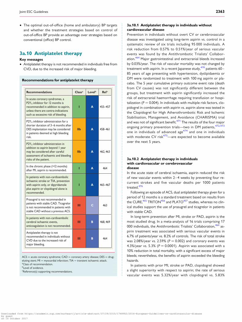
† The optimal out-of-office (home and ambulatory) BP targetsand whether the treatment strategies based on control ofout-of-office BP provide an advantage over strategies based onconventional (office) BP control.
3a.10 Antiplatelet therapyKey messages† Antiplatelet therapy is not recommended in individuals free from
CVD, due to the increased risk of major bleeding.
Recommendations for antiplatelet therapy
Recommendations Classa Levelb Ref c
In acute coronary syndromes, a P2Y12 inhibitor for 12 months is recommended in addition to aspirin, unless there are contra-indications such as excessive risk of bleeding.
I A 455–457
P2Y12 inhibitor administration for a shorter duration of 3–6 months after DES implantation may be considered in patients deemed at high bleeding risk.
IIb A 458–461
P2Y12 inhibitor administration in addition to aspirin beyond 1 year may be considered after careful assessment of ischaemic and bleeding risks of the patient.
IIb A 462, 463
In the chronic phase (>12 months) after MI, aspirin is recommended.
I A 464
In patients with non-cardioembolic ischaemic stroke or TIA, prevention with aspirin only, or dipyridamole plus aspirin or clopidogrel alone is recommended.
I A 465–467
Prasugrel is not recommended in patients with stable CAD. Ticagrelor is not recommended in patients with stable CAD without a previous ACS.
III C 463
In patients with non-cardioembolic cerebral ischaemic events, anticoagulation is not recommended.
III B 468, 469
Antiplatelet therapy is not recommended in individuals without CVD due to the increased risk of major bleeding.
III B 464
ACS ¼ acute coronary syndrome; CAD ¼ coronary artery disease; DES ¼ drug-eluting stent; MI ¼ myocardial infarction; TIA ¼ transient ischaemic attack.aClass of recommendation.bLevel of evidence.cReference(s) supporting recommendations.
3a.10.1 Antiplatelet therapy in individuals withoutcardiovascular diseasePrevention in individuals without overt CV or cerebrovasculardisease was investigated using long-term aspirin vs. control in asystematic review of six trials including 95 000 individuals. Arisk reduction from 0.57% to 0.51%/year of serious vascularevents was found by the Antithrombotic Trialists’ Collabor-ation.464 Major gastrointestinal and extracranial bleeds increasedby 0.03%/year. The risk of vascular mortality was not changed bytreatment with aspirin. In a recent Japanese study,470 patients 60–85 years of age presenting with hypertension, dyslipidaemia orDM were randomized to treatment with 100 mg aspirin or pla-cebo. The 5 year cumulative primary outcome event rate (deathfrom CV causes) was not significantly different between thegroups, but treatment with aspirin significantly increased therisk of extracranial haemorrhage requiring transfusion or hospi-talization (P ¼ 0.004). In individuals with multiple risk factors, clo-pidogrel in combination with aspirin vs. aspirin alone was tested inthe Clopidogrel for High Atherothrombotic Risk and IschemicStabilisation, Management, and Avoidance (CHARISMA) trialand was not of significant benefit.471 The results of the four majorongoing primary prevention trials—two in DM patients,472,473
one in individuals of advanced age474 and one in individualswith moderate CV risk475—are expected to become availableover the next 5 years.
3a.10.2 Antiplatelet therapy in individualswith cardiovascular or cerebrovasculardiseaseIn the acute state of cerebral ischaemia, aspirin reduced the riskof new vascular events within 2–4 weeks by preventing four re-current strokes and five vascular deaths per 1000 patientstreated.476
Following an episode of ACS, dual antiplatelet therapy given for aperiod of 12 months is a standard treatment based on results fromthe CURE,455 TRITON456 and PLATO457 studies, whereas no clin-ical studies support the use of prasugrel and ticagrelor in patientswith stable CAD.
In long-term prevention after MI, stroke or PAD, aspirin is themost studied drug. In a meta-analysis of 16 trials comprising 17000 individuals, the Antithrombotic Trialists’ Collaboration,464 as-pirin treatment was associated with serious vascular events in6.7% of patients/year vs. 8.2% of controls. The risk of total strokewas 2.08%/year vs. 2.59% (P ¼ 0.002) and coronary events was4.3%/year vs. 5.3% (P ¼ 0.0001). Aspirin was associated with a10% reduction in total mortality, with a significant excess of majorbleeds; nevertheless, the benefits of aspirin exceeded the bleedinghazards.
In patients with prior MI, stroke or PAD, clopidogrel showeda slight superiority with respect to aspirin; the rate of seriousvascular events was 5.32%/year with clopidogrel vs. 5.83%
Joint ESC Guidelines 2363
Downloaded from https://academic.oup.com/eurheartj/article-abstract/37/29/2315/1748952/2016-European-Guidelines-on-cardiovascular-diseaseby gueston 10 October 2017

with aspirin (P ¼ 0.043). There were slightly more bleedswith aspirin.477
Adding aspirin to clopidogrel in high-risk patients with recent is-chaemic stroke or transient ischaemic attack (TIA) was associatedwith a non-significant difference in reducing major vascular events.However, the risk of life-threatening or major bleeding was signifi-cantly increased by the addition of aspirin.478
On the other hand, the Clopidogrel in High-risk patientswith Acute Non-disabling Cerebrovascular Events (CHANCE)trial showed that the combined treatment of clopidogrel andaspirin decreased the 90 day risk of stroke without increasinghaemorrhage compared with aspirin alone in 5170 Chinesepatients randomized within 24 h after symptom onset ofminor stroke or TIA to clopidogrel – aspirin or to aspirinalone.479
In patients with prior non-cardioembolic ischaemic stroke, dualantiplatelet therapy with dipyridamole plus aspirin showed super-iority over aspirin.465 In such patients, oral vitamin K antagonistsare not superior to aspirin and are associated with a higher bleedingrisk.468,469
In patients with ischaemic stroke, a direct comparison of dipyrid-amole plus aspirin vs. clopidogrel alone466 showed similar rates ofrecurrent stroke, including haemorrhagic stroke. There was a higherfrequency of major haemorrhagic events with dipyridamole plus as-pirin (4.1% vs. 3.6%).
Vorapaxar is a novel antiplatelet agent that selectively inhibitsthe cellular actions of thrombin through antagonism of PAR-1.In 26 449 patients who had a history of MI, ischaemic stroke orPAD, the primary composite endpoint—CV death, MI orstroke—was significantly reduced with vorapaxar in additionto standard antiplatelet therapy, but with increased risk ofmoderate or severe bleeding.480 Vorapaxar cannot be re-commended systematically in patients with stable atheroscleroticdisease.
Gap in evidence† Experience with the new antiplatelet drugs in patients with stable
CAD is still limited and so is their use in combination with anti-coagulant treatment.
3a.11 Adherence to medicationKey messages† Adherence to medication in individuals at high risk and in patients
with CVD is low.† Several types of interventions are effective in improving medica-
tion adherence.† The polypill may increase adherence to treatment and improve
CV risk factor control.
Recommendations for achieving medication adherence
Recommendations Classa Levelb Ref c
Simplifying the treatment regimen to the lowest acceptable level is recommended, with repetitive monitoring and feedback. In case of persistent non-adherence, multi-session or combined behavioural interventions are recommended.
I A 481
It is recommended that physicians assess medication adherence, and identify reasons for non-adherence in order to tailor further interventions.
I C 482–484
The use of the polypill and combination therapy to increase adherence to drug therapy may be considered.
IIb B 485, 486
aClass of recommendation.bLevel of evidence.cReference(s) supporting recommendations.
Adherence to medication in individuals at high risk and in patientswith CVD is low, resulting in worse outcomes and higher healthcare costs.487 One month after AMI, 25–30% of patients stop atleast one drug, with a progressive decline in adherence over time.After 1 year, only 50% of patients report persistent use of statins,b-blockers or BP-lowering therapy.483,484 The reasons for poor ad-herence are multifactorial (Table F in web addenda).483
Cost-related non-adherence is a relevant problem in many healthcare systems. For example, in American veterans, adherence tolipid-lowering medication decreased as co-payments increased.488
Depression also independently doubles the risk for non-adherence.489 Reasons for non-adherence tend to cluster; for ex-ample, complex medication regimens may be important in indivi-duals with chronic disease or multiple risk factors. This places highdemands on caregivers to provide clear advice and continuouscare.484 Physicians often fail to communicate critical elements ofmedication use (e.g. possible adverse effects, how long to take themedication and the frequency or timing of dosing).490 Thus there is aneed to train physicians to identify risk factors for non-adherenceand promote adherence to medication.
Several interventions are effective in improving adherence inchronic conditions.481 Solely reducing dosage demands resulted instrong effects, but other interventions such as repetitive monitoringand feedback, multisession information and combined behaviouralinterventions have shown effects ranging from minor to strong.481
Collaboration with pharmacists or pharmacist-directed care was
Joint ESC Guidelines2364
Downloaded from https://academic.oup.com/eurheartj/article-abstract/37/29/2315/1748952/2016-European-Guidelines-on-cardiovascular-diseaseby gueston 10 October 2017

superior to standard care with respect to BP, total cholesterol andLDL-C levels.491 Knowledge of one’s CAC score may increase riskperception and adherence to medication.492
In clinical practice, physicians should assess adherence to medica-tion, identify reasons for possible non-adherence and promote ad-herence according to the following established principles:
† Provide clear advice regarding the benefits and possible adverseeffects of the medication and the duration and timing of dosing.
† Consider patients’ habits and preferences (shared decision-making).
† Simplify the treatment regimen to the lowest feasible level.† Ask patients in a non-judgmental way how the medication
works for them and discuss possible reasons for non-adherence(e.g. side effects, worries).
† Implement repetitive monitoring and feedback.† Introduce physician assistants and/or trained nurses or pharma-
cists whenever it is necessary and feasible.† In case of persistent non-adherence, offer multisession or
combined behavioural interventions [e.g. for patients aftermyocardial revascularization in a cardiac rehabilitation (CR)setting].
3a.11.1 PolypillMore than a decade ago, Wald and Law quantified the efficacy andadverse effects of a fixed dose combination (FDC) from publishedtrials and proposed that an FDC consisting of a statin, BP-loweringagents, aspirin and folate could potentially reduce CVD by 80% inindividuals .55 years of age.493
A recent systematic review and meta-analysis485 summarizesnine randomized trials (n ¼ 7047) on FDCs, largely conducted inhigher-risk populations and primarily designed to evaluate changesin CV risk factors and adherence. However, FDCs included inthe analysis were single pills of diverse composition and doses(although all contained a statin and at least one BP-lowering agent)and had a range of comparators (placebo, single-drug active com-ponent or ‘usual care’). No convincing evidence of either benefitor risk for FDCs in terms of all-cause mortality or CV eventswas found. FDC therapy improved adherence (only one trial) com-pared with a multidrug strategy by 33% (95% CI 26%, 41%) com-pared with usual care.
In another international study, not included in the previousmeta-analysis, 695 CAD patients were randomized to test the effectof an FDC polypill containing aspirin, simvastatin and ramipril, or thethree drugs separately. The study showed that FDC improved ad-herence compared with separate medications after 9 months offollow-up (adherence 63% vs. 52%; P ¼ 0.006).486
The polypill should not be considered in isolation, but as an inte-gral part of a comprehensive CVD prevention strategy that includesefforts to reduce tobacco use, increase PA and increase consump-tion of a heart-healthy diet.494 However, potential adverse effects ofa single drug component of the FDC cannot be specifically cor-rected and therefore may also affect treatment adherence to theother components. Until we have the results of ongoing trialswith major CVD as the endpoint, the polypill cannot be recom-mended for prevention of CVD and cannot be prescribed to allindividuals.
Gaps in evidence† There is limited evidence about which interventions for improv-
ing adherence to medication are the most effective and in whom(e.g. young–old, male– female, high vs. low socio-economicstatus).
† The effect of the polypill as a global strategy to reduce CVD re-mains uncertain.
3b. How to intervene at theindividual level: disease-specificintervention—atrial fibrillation,coronary artery disease, chronicheart failure, cerebrovasculardisease, peripheral artery disease(web addenda)
3c. How to intervene at thepopulation level
3c.1 Introduction (healthy lifestylepromotion)The population level approach follows the Geoffrey Rose para-digm: small shifts in the risk of disease (or risk factor) across awhole population consistently lead to greater reductions in dis-ease burden than a large shift in high-risk individuals only. Thispopulation-wide approach has further advantages: it addressesCV health over the entire life course and reduces healthinequalities.
Individual behaviour is enacted in an environment with hierarch-ical levels, which encompass individual choice, family influence, cul-tural and ethnic grouping, workplace, health care and policy at thestate and global levels (e.g. EU policies and international trade agree-ments).The aim of this section is to provide stakeholders withevidence-based suggestions for the most effective interventions toimprove CVD risk that can be implemented at a group, community,regional, national or global level. Health care professionals play animportant role in advocating evidence-based population-levelinterventions.
Strategies such as ‘nudging’ (to push mildly) and ‘default’ havebeen proposed as tools. By changing the context to make healthydecisions the default, the individual is nudged in the healthy direc-tion. The task for both national and local authorities is to createsocial environments that provide healthier defaults.
The evidence presented here builds on recent comprehen-sive reviews311,495 – 497 and individual studies and summarizesthe ‘totality of evidence’. It is rarely feasible to use an RCTto evaluate population-level interventions (in contrast toindividual-level interventions). The guidelines committee has cho-sen to follow the definition of ‘level of evidence’ for population-level approaches. Thus consistent findings from several high-quality studies were considered sufficient to merit strongrecommendations.
Joint ESC Guidelines 2365
Downloaded from https://academic.oup.com/eurheartj/article-abstract/37/29/2315/1748952/2016-European-Guidelines-on-cardiovascular-diseaseby gueston 10 October 2017
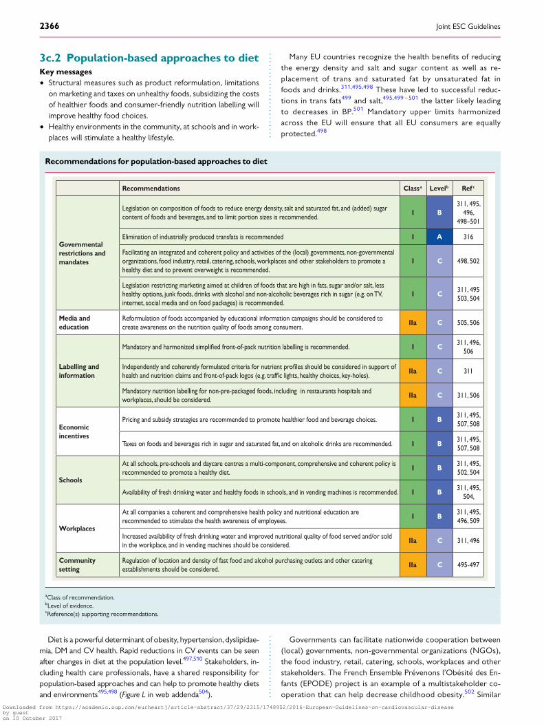
3c.2 Population-based approaches to dietKey messages† Structural measures such as product reformulation, limitations
on marketing and taxes on unhealthy foods, subsidizing the costsof healthier foods and consumer-friendly nutrition labelling willimprove healthy food choices.
† Healthy environments in the community, at schools and in work-places will stimulate a healthy lifestyle.
Diet is a powerful determinant of obesity, hypertension, dyslipidae-mia, DM and CV health. Rapid reductions in CV events can be seenafter changes in diet at the population level.497,510 Stakeholders, in-cluding health care professionals, have a shared responsibility forpopulation-based approaches and can help to promote healthy dietsand environments495,498 (Figure L in web addenda504).
Many EU countries recognize the health benefits of reducingthe energy density and salt and sugar content as well as re-placement of trans and saturated fat by unsaturated fat infoods and drinks.311,495,498 These have led to successful reduc-tions in trans fats499 and salt,495,499 – 501 the latter likely leadingto decreases in BP.501 Mandatory upper limits harmonizedacross the EU will ensure that all EU consumers are equallyprotected.498
Governments can facilitate nationwide cooperation between(local) governments, non-governmental organizations (NGOs),the food industry, retail, catering, schools, workplaces and otherstakeholders. The French Ensemble Prevenons l’Obesite des En-fants (EPODE) project is an example of a multistakeholder co-operation that can help decrease childhood obesity.502 Similar
Recommendations for population-based approaches to diet
Recommendations Classa Levelb Ref c
Governmental restrictions and mandates
Legislation on composition of foods to reduce energy density, salt and saturated fat, and (added) sugar content of foods and beverages, and to limit portion sizes is recommended.
I B311, 495,
496, 498–501
Elimination of industrially produced transfats is recommended I A 316
Facilitating an integrated and coherent policy and activities of the (local) governments, non-governmental organizations, food industry, retail, catering, schools, workplaces and other stakeholders to promote a healthy diet and to prevent overweight is recommended.
I C 498, 502
Legislation restricting marketing aimed at children of foods that are high in fats, sugar and/or salt, less healthy options, junk foods, drinks with alcohol and non-alcoholic beverages rich in sugar (e.g. on TV, internet, social media and on food packages) is recommended.
I C311, 495 503, 504
Media and education
Reformulation of foods accompanied by educational information campaigns should be considered to create awareness on the nutrition quality of foods among consumers.
IIa C 505, 506
Labelling and information
Mandatory and harmonized front-of-pack nutrition labelling is recommended. I C311, 496,
506
Independently and coherently formulated criteria for nutrient should be considered in support of health and nutrition claims and front-of-pack logos (e.g. lights, healthy choices, key-holes).
IIa C 311
Mandatory nutrition labelling for non-pre-packaged foods, including in restaurants hospitals and workplaces, should be considered.
IIa C 311, 506
Economic incentives
Pricing and subsidy strategies are recommended to promote healthier food and beverage choices. I B311, 495, 507, 508
Taxes on foods and beverages rich in sugar and saturated fat, and on alcoholic drinks are recommended. I B311, 495, 507, 508
Schools
At all schools, pre-schools and daycare centres a multi-component, comprehensive and coherent policy is recommended to promote a healthy diet.
I B311, 495, 502, 504
Availability of fresh drinking water and healthy foods in schools, and in vending machines is recommended. I B311, 495,
504,
Workplaces
At all companies a coherent and comprehensive health policy and nutritional education are recommended to stimulate the health awareness of employees.
I B311, 495, 496, 509
Increased availability of fresh drinking water and improved nutritional quality of food served and/or sold in the workplace, and in vending machines should be considered.
IIa C 311, 496
Community setting
Regulation of location and density of fast food and alcohol purchasing outlets and other catering establishments should be considered.
IIa C 495-497
aClass of recommendation.bLevel of evidence.cReference(s) supporting recommendations.
Joint ESC Guidelines2366
Downloaded from https://academic.oup.com/eurheartj/article-abstract/37/29/2315/1748952/2016-European-Guidelines-on-cardiovascular-diseaseby gueston 10 October 2017

projects are in place in Belgium, Spain, The Netherlands, Greeceand Australia.
Educational tools and intervention in the media may lead to a re-duction in childhood obesity (e.g. limiting children’s exposure to ad-vertising of unhealthy foods).311,495,497,502,503 In 2013, the EuropeanHeart Network (EHN) published a report summarizing recent devel-opments in relation to the marketing of unhealthy foods to chil-dren.504 Accompanying consumer awareness campaigns on healthyfoods505 and nutrition labelling can be effective. Consumers under-stand different systems of labelling and their use has a positive impacton sales.506 The EHN is calling for a simplified, colour-coded,front-of-pack scheme indicating high, medium and low levels of nutri-ents.311,495,497 This scheme can be applied to all foods and could beexpanded to certain restaurants.311 Labelling also stimulates the re-formulation of foods,504 and thus it has the potential to improve diet-ary intake and reduce diet-related chronic diseases.
Pricing strategies can lead to a decline in the sales of unhealthyfoods and an increase in the sales of fruits and vegetables. Modellingstudies have demonstrated that food taxes could improve energyand nutrient intake, BMI and health.495,507,508 An increasing numberof countries have introduced taxes on unhealthy foods and drinks[e.g. the fat tax in Denmark (caused a 10–15% decrease in con-sumption; now repealed) and the junk food tax in Hungary (sales de-clined by 27%)].504
Consideration should be given to balanced economic incentives:subsidies and taxes to counteract any unbalanced effect on the so-cially disadvantaged.
To tackle obesity, every school and workplace should have a pol-icy to promote a healthy environment and provide healthy foodsand meals.495,504 Ideally, health education should be part of theschool curriculum. Workplace dietary modification interventions
alone and in combination with nutrition education or environmentalchanges have shown improvements in the consumption of fruits andvegetables and/or fats.509
In the community, planning the location and density of fast foodoutlets and good access to supermarkets is needed, especially in de-prived areas.495 – 497
Gaps in evidence† Scientific evidence of the impact of food and nutrition policy in-
struments on outcome measures such as food intake and CVhealth is largely lacking.
† Cost-effectiveness studies of the impact of different policy op-tions are also limited.
3c.3 Population-based approachesto physical activityKey messages† A sedentary lifestyle and physical inactivity affects more than half
of the population worldwide.† Regular PA is recommended for all men and women as a lifelong
part of lifestyle, with at least 150 min/week of moderate activityor at least 75 min/week of vigorous activity or an equivalent com-bination thereof. Any activity is better than none and more activ-ity is better than some.
† Population-based interventions are effective in promoting PA.† Early childhood education in PA and movement should start at
preschool/kindergarten.† Daily PA at school should be at least 30 minutes, and preferably
60 minutes.† Good neighbourhoods and a safe environment enhances and en-
courages PA in everyday life.
Recommendations for population-based approaches to physical activity
Recommendations Classa Levelb Ref c
Governmental restrictions and mandates
Consideration of PA when planning new landscaping/buildings or towns is recommended. I C311,
511–513
Media and educationSee also section 3c.2 for multi-component interventions
Sustained, focused, media and educational campaigns, using multiple media modes (e.g. apps, posters, and signage) may be considered to promote PA.
IIb C 496
Short term community-based educational programmes and wearable devices promoting healthy behaviours, such as walking should be considered.
IIa C 514–516
Labelling and information
Point-of-decision prompts should be considered to encourage use of stairs. IIa B 516, 517
Exercise prescription for health promotion by physicians, especially GPs, similar to drug prescription should be considered.
IIa C 517, 518
Economic incentives
Increased fuel (gasoline) taxes should be considered to increase active transport/commuting. IIa C 512, 518
Tax reduction incentives for individuals to purchase exercise equipment or health memberships
may
be
considered.
IIb C 512, 518
Sustained individual incentives may be considered for increased or weight loss. IIb C512, 513,
518
Tax reduction incentives to employers to offer comprehensive worksite wellness programmes with
nutrition,
PA,
and tobacco cessation/prevention components may be considered.
IIb C 512, 518
continued
Joint ESC Guidelines 2367
Downloaded from https://academic.oup.com/eurheartj/article-abstract/37/29/2315/1748952/2016-European-Guidelines-on-cardiovascular-diseaseby gueston 10 October 2017

In most countries, the majority of adults and children donot achieve the minimum activity levels recommended by
health organizations: every person should engage in moderate exer-
cise for at least 150 min/week and/or vigorous activity for at least
75 min/week or an equivalent thereof.258,520 For population-based
prevention, the statement of ‘seven best investments’512 gives the
universal and comprehensive advice to promote PA.512
Specific national guidelines developed for PA include frequency,intensity, time (duration) and type of activity (FITT), which can influ-
ence legislative initiatives, such as ‘active cities’ with bicycle lanes and
walking paths and reallocation of road space.Focused media and educational campaigns can initiate physical ac-
tivities.519 Recent campaigns from sports medicine societies have
endorsed PA prescriptions from GPs (http://www.efsma.net). The
PA should be assessed at every medical encounter.A simple strategy for increasing daily exercise is to encourage the
use of stairs rather than the elevator or escalator, along with signage
directing people to the stairs and health promotion materials en-
dorsing the positive effects of stair climbing.516
Interestingly, an increase in fuel prices may reduce car driving andincrease active commuting for those who live within reasonable
walking or biking distances, with the exception of diseased or dis-
abled persons.496
PA education should be started in preschool/kindergarten andcontinue for all levels of primary and secondary education. Forschool education, a multicomponent intervention should focus onimproving lifelong PA by trained teachers. At least 3 h/week, andpreferably 60 min/day, of sports or PA should be performed duringschool time.511 Regular activity also improves cognitive competencefor learning.513,521 This activity can be supplemented with activecommuting to school and supervised walking routes to and fromschool, with less reliance on buses.514
Workplaces can offer different opportunities for PA promo-tion. Some larger companies offer a fitness centre on companygrounds without fees for employees. Workplace-based interven-tions may increase regular physical exercise for employees, butresults demonstrate that a high proportion of workers do notparticipate.522 Therefore, supervisors and managers should en-dorse workplace interventions by encouraging employees toundertake PA.
Improved accessibility to recreation and exercise facilities with in-creased operating hours and utilizing community resources such asschool playgrounds may increase regular PA in all age groups and re-duce socio-economic inequality of access.517
Gap in evidence
† Sustainability and long-term outcomes of population-based ac-tions to promote PA.
Recommendations for population-based approaches to physical activity (continued)
Recommendations Classa Levelb Ref c
Schools See also section 3c.2 for multi-component interventions
Increased availability and types of school playground spaces and equipment for exercise activity and sports are recommended.
I C 512, 519
Regular classroom PA breaks during academic lessons should be considered. IIa B 511
Increasing active commuting to school should be considered e.g. a walking school bus programme with supervised walking routes to and from school for safety.
IIa C 512, 514
Increased number and duration of PA classes, with revised PA curricula to implement at least moderate activity and trained teachers in exercise and sports may be considered.
IIb B 511, 513
WorkplacesSee also section 3c.2 for multi-component interventions
Comprehensive worksite wellness programmes should be considered with nutrition and PA components. IIa B
512, 520–522
Structured worksite programmes that encourage PA and provide a set time for PA during work hours should be considered. Improving stairway access and appeal, potentially in combination with “skip-stop” elevators that skip some should be considered.
IIa C
Promoting worksite centres should be considered. IIa C 517
Community setting
Health care providers should consider inquiring about PA in every medical encounter and adding it to the record. In addition, they should consider to motivate the individual and promote PA.
IIa C 512, 520
Improved accessibility of recreation and PA spaces and facilities (e.g. building of parks and playgrounds, increasing operating hours, use of school facilities during non-school hours), improved walkability should be considered.
IIa C 512, 520
Improved neighbourhood aesthetics (to increase activity in adults) should be considered. IIa C 512, 520
GPs ¼ general practitioners; PA ¼ physical activity.aClass of recommendation.bLevel of evidence.cReference(s) supporting recommendations.
Joint ESC Guidelines2368
Downloaded from https://academic.oup.com/eurheartj/article-abstract/37/29/2315/1748952/2016-European-Guidelines-on-cardiovascular-diseaseby gueston 10 October 2017
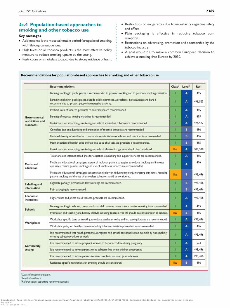
3c.4 Population-based approaches tosmoking and other tobacco useKey messages† Adolescence is the most vulnerable period for uptake of smoking,
with lifelong consequences.† High taxes on all tobacco products is the most effective policy
measure to reduce smoking uptake by the young.† Restrictions on smokeless tobacco due to strong evidence of harm.
† Restrictions on e-cigarettes due to uncertainty regarding safetyand effect.
† Plain packaging is effective in reducing tobacco con-sumption.
† Restrictions on advertising, promotion and sponsorship by thetobacco industry.
† A goal would be to make a common European decision toachieve a smoking-free Europe by 2030.
Recommendations for population-based approaches to smoking and other tobacco use
Recommendations Classa Levelb Ref c
Governmental restrictions and mandates
Banning smoking in public places is recommended to prevent smoking and to promote smoking cessation. I A 495
Banning smoking in public places, outside public entrances, workplaces, in restaurants and bars is recommended to protect people from passive smoking.
I A 496, 523
Prohibit sales of tobacco products to adolescents are recommended. I A 495
Banning of tobacco vending machines is recommended. I A 495
Restrictions on advertising, marketing and sale of smokeless tobacco are recommended. I A 524-527
Complete ban on advertising and promotion of tobacco products are recommended. I B 496
Reduced density of retail tobacco outlets in residential areas, schools and hospitals is recommended. I B 496
Harmonization of border sales and tax free sales of all tobacco products is recommended. I B 495
Restrictions on advertising, marketing and sale of electronic cigarettes should be considered. IIa A 305, 528
Media and education
Telephone and internet based lines for cessation counselling and support services are recommended. I A 496
Media and educational campaigns as part of multicomponent strategies to reduce smoking and increase quit rates, reduce passive smoking and use of smokeless tobacco are recommended.
I A496
Media and educational campaigns concentrating solely on reducing smoking, increasing quit rates, reducing passive smoking and the use of smokeless tobacco should be considered.
IIa B 495, 496
Labelling and information
Cigarette package pictorial and text warnings are recommended. I B 495, 496
Plain packaging is recommended. I B 495, 496
Economic incentives
Higher taxes and prices on all tobacco products are recommended. I A 495, 496
SchoolsBanning smoking in schools, pre-schools and child care to protect from passive smoking is recommended. I A 495
Promotion and teaching of a healthy lifestyle including tobacco-free life should be considered in all schools. IIa B 496
WorkplacesWorkplace bans on smoking to reduce passive smoking and increase quit rates are recommended. I A 495, 496
Workplace policy on healthy choices including tobacco cessation/prevention is recommended. I A 496
Community setting
It is recommended that health personnel, caregivers and school personnel set an example by not smoking or using tobacco products at work.
I A 495, 496
It is recommended to advise pregnant women to be tobacco-free during pregnancy. I A 524
It is recommended to advise parents to be tobacco-free when children are present. I A 495, 496
It is recommended to advise parents to never smoke in cars and private homes. I A 495, 496
restrictions on smoking should be considered. IIa B 496
aClass of recommendation.bLevel of evidence.cReference(s) supporting recommendations.
Joint ESC Guidelines 2369
Downloaded from https://academic.oup.com/eurheartj/article-abstract/37/29/2315/1748952/2016-European-Guidelines-on-cardiovascular-diseaseby gueston 10 October 2017

The WHO Framework Convention on Tobacco Control recom-mends smoke-free laws: protecting people from tobacco smoke andbanning smoke in public places, warning about the dangers of tobacco,raising taxes on tobacco and enforcing advertising bans.523 Childrenand low socio-economic groups are sensitive to population-based to-bacco intervention. Passive smoking increases CVD risk,495,496 moreso in women than in men.529 All smoking, including smoking a waterpipe, is deleterious. Smokeless tobacco (in Europe usually snus, a moistpowder tobacco placed under the upper lip) increases the risk of fatalCVD events,525–527 and the use of snus during pregnancy increasesthe risk of stillbirth.530 There is no evidence that snus increases smok-ing cessation more than nicotine replacement products or medication.Many smokers use e-cigarettes to quit. There are many unansweredquestions about their safety, efficacy for harm reduction and cessationand impact on public health. International legislation should be harmo-nized to prevent a new tobacco epidemic.495
Multicomponent strategies are best. Advertising bans reduce to-bacco consumption, and mass media campaigns reduce smoking up-take by teenagers and increase adult quitting.495 Media andeducational campaigns in schools reduce smoking and promotesmoking cessation. Editors should increase the coverage of tobaccoand health in the media.531 Telephone or Internet-based cessationsupport reduces tobacco use.496
Packs with pictorial and text warnings raise awareness of tobaccodangers.495 Plain and standardized packaging without brand labelsenhances the effectiveness.
Higher taxes reduce tobacco consumption and encourage quitting,particularly among young and lower socio-economic groups.495,496
School-based smoking bans should be implemented.496 Smokingbans at workplaces reduce exposure to passive smoking, decreasesmoking and increase quitting rates.495 Tobacco outlet densitynear homes, hospitals and schools should be reduced. Pregnant wo-men should avoid tobacco, and parents should be tobacco freewhen children are present. Health personnel, caregivers and tea-chers must set an example by not using tobacco products at work.
Gaps in evidence
† Effect of school-based smoking restrictions.† Health harm of e-cigarettes.† More evidence on environmental smoking is needed, as smoke
particles may remain in rooms for many years.
3c.5 Alcohol abuse protectionKey messages
† Excessive alcohol intake is associated with increased CV mortal-ity, and alcohol ranks as the second-leading cause of DALYs lostin high-income countries.
† The interventions for addressing the harmful use of alcohol arecost effective, with good return (i.e. increasing alcoholic beverageexcise taxes, restricting access to alcoholic beverages and imple-menting comprehensive restrictions and bans on advertising andpromotion of alcoholic beverages).
Recommendations for protecting against alcohol abuse
Recommendations Classa Levelb Ref c
Governmental restrictions and mandates
Regulating physical availability of alcoholic beverages is recommended, including minimum legal purchase age, restrictions on outlet density and time and place of sales, public health oriented licensing systems, and governmental monopolies of retail sales.
I B 532–536
Drink-driving countermeasures are recommended such as lowered blood alcohol concentration limits and “zero tolerance”, random breath testing and sobriety check points.
I B 534, 537
Implementing comprehensive restrictions and bans on advertising and promotion of alcoholic beverages is recommended.
I C 532
Media and education
Educational information campaigns may be considered to create awareness on the hazardous effects of alcohol.
IIb B 532, 538
Labelling and information
Labelling alcohol with information on caloric content and health warning messages of the harmful effects of alcohol may be considered.
IIb B 532, 538
Economic incentives
Taxes on alcoholic beverages are recommended. I B 533
SchoolsAt every school, pre-school and day care a multi-component, comprehensive and coherent education may be considered to prevent alcohol abuse.
IIb B 532, 538
WorkplacesAt every company a coherent and comprehensive health policy and nutritional education on stimulating the health of employees, including limiting excessive alcohol intake, are recommended.
I B 495
Community setting
Measures to support and empower primary care to adopt effective approaches to prevent and reduce harmful use of alcohol are recommended.
I B 539
Enacting management policies relating to responsible serving of alcoholic beverages should be considered to reduce the negative consequences of drinking.
IIa B 534, 538
Planning of location and density of alcohol purchasing outlets and other catering establishments should be considered.
IIa C
aClass of recommendation.bLevel of evidence.cReference(s) supporting recommendations.
Joint ESC Guidelines2370
Downloaded from https://academic.oup.com/eurheartj/article-abstract/37/29/2315/1748952/2016-European-Guidelines-on-cardiovascular-diseaseby gueston 10 October 2017

At the population level, alcohol consumption is associated withmultiple health risks that clearly outweigh any potential benefits. In2012, �3.3 million deaths (5.9% of all global deaths) and 139 millionDALYs (5.1% of the global burden of disease and injury) were attrib-utable to alcohol consumption. The highest numbers of deaths arefrom CVDs, with 33.3% of the alcohol-attributable deaths due toCVDs.534 Ischaemic heart disease mortality is 65% higher in male hea-vy drinkers and more than double in female heavy drinkers.540
The relationship between alcohol consumption and CAD andcerebrovascular diseases is complex. It depends on both the leveland pattern of alcohol consumption. Low alcohol consumption, ran-ging from one to three alcohol units per day (a unit equates to about80 mL of wine, 250 mL of normal strength beer or 30–50 mL of spir-its) in some segments of the population is associated with the lowestall-cause mortality, largely due to lower coronary mortality.541
SBP and DBP levels increase as alcohol consumption increases to.3 units/day, as does the risk of cardiac arrhythmias, cardiomyop-athy, sudden death and haemorrhagic stroke.542 The pattern of alco-hol use has an effect on CVD risk; binge drinking is associated with ahigher risk of sudden death and stroke.543
The following strategies and interventions have the highest levelof effectiveness to prevent the harmful use of alcohol: age limits forsale and serving;535 drink-driving strategies;537 government retailmonopolies on the sale of alcohol and reducing the hours ofsale;536 banning alcohol advertising, promotion and sponsorship ofevents532 and an increase in retail prices.533,538
In the absence of other population-level measures, such as tax-ation and advertising restrictions, labelling alcohol with informationon caloric content and health warning messages of the harmful ef-fects of alcohol has been shown to have a limited effect.538
Alcohol regulations in the policies of workplaces, educationalcentres and schools are effective.532
Brief intervention in primary care to prevent alcohol abuse hasbeen shown to be effective.539
In the community, excessive alcohol intake can be limited by re-strictions in the number and opening hours of outlets and by in-creasing the minimum age for sales and servings.495
Gap in evidence† Better quality evidence is needed with regard to potential con-
founding in studies on the effects of alcohol consumption.
3c.6 Healthy environmentAir pollution contributes to the risk of respiratory and CV diseases.544
Important sources of fine particles in the EU are motorized road traf-fic, power plants and industrial and residential heating using oil, coal orwood. Up to a third of Europeans living in urban areas are exposed tolevels exceeding EU air quality standards. In particular, young and oldindividuals and subjects with a high risk of CVD are more prone to thedetrimental effects of air pollution on the circulation and the heart.
The EU Commission released a policy package to be implemen-ted by the year 2030 with measures to reduce harmful emissionsfrom traffic, energy plants and agriculture. Further efforts to reduceair pollution should be encouraged and taken up by national govern-ments (e.g. through appropriate and effective legislation). Patient or-ganizations and health professionals have an important role to playin supporting education and policy initiatives and provide a strongvoice in the call for action at the governmental level.544
The media can inform the population on air quality (e.g. by apps)and by providing smog alerts. Information on patients’ behaviour
during smog is needed. Economic incentives such as reduced taxeson electric and hybrid cars can contribute to the improvement of airquality. New houses and schools can be built in areas remote fromhighways and polluting industries.
4a. Where to intervene at theindividual levelThe question of ‘where’ prevention should take place requires onlya simple answer: everywhere! Prevention of CVD should be valuedand implemented at all levels of society and in all health care settings.This should include increased spending on prevention in health careand on actions that make communities healthier. All cliniciansshould consider prevention and promotion of healthy lifestyles aprofessional responsibility with individual patients and should sup-port policies that promote healthier lifestyles. Patients should alsobe empowered and have the knowledge and support to make in-formed decisions and to demand robust prevention efforts fromhealth care groups and society.
4a.1 Clinical settings and stakeholders4a.1.1 Cardiovascular disease prevention in primary careKey messages† The prevention of CVD should be delivered in all health care set-
tings, including primary care.† Where appropriate, all health professionals should assess CV risk
factors to determine the individual’s total CV risk score.† GPs and nurses should work together as a team to provide the
most effective multidisciplinary care.
Recommendation for cardiovascular disease preventionin primary care
Recommendation Classa Levelb
It is recommended that GPs, nurses and allied health professionals within primary care deliver CVD prevention for high-risk patients.
I C
aClass of recommendation.bLevel of evidence.
The physician in general practice is the key person to initiate, coord-inate and provide long-term follow-up for CVD prevention. In mostcountries, GPs deliver .90% of consultations and provide mostpublic health medicine, including preventive care and chronic dis-ease monitoring. In the case of CVD prevention, they have a uniquerole in identifying individuals at risk of CVD and assessing their eli-gibility for intervention based on their risk profile. How to maximizeattendance rates and adherence, particularly in those who are athighest risk, remains an issue.
As mentioned in section 2.2, a systematic approach is recom-mended to risk assessment, giving priority to persons with a priorihigher risk (such as family history of premature CVD, presence ofhypertension, etc.); opportunistic screening of persons ,40 yearsof age without CV risk factors is not recommended.
Intensive and structured intervention in general practice contri-butes to the prevention of recurrent CV events and reduces hos-pital admissions in CAD patients.545
Joint ESC Guidelines 2371
Downloaded from https://academic.oup.com/eurheartj/article-abstract/37/29/2315/1748952/2016-European-Guidelines-on-cardiovascular-diseaseby gueston 10 October 2017

The successful implementation of CVD prevention guidelines re-lies heavily on GPs providing risk factor evaluation, intervention andpatient education. However, CV targets in general practice are oftennot achieved. The EUROASPIRE III survey (primary prevention arm)showed that the lifestyle of people being treated as high CV risk—defined as patients treated with BP- and lipid-lowering drugs as wellas anti-diabetes drugs—showed much persistent smoking and a highprevalence of both obesity and central obesity. BP, lipid and glucosecontrol is poor, with most patients not achieving the targets definedin the prevention guidelines.5
Surveys done among GPs and physicians in several European re-gions found that most were aware of the European guidelines onCVD prevention, but that only 36–57% were using the guidelinesin practice, and less than half performed comprehensive risk assess-ments. The main barrier was time, but GPs also cited that therewere too many guidelines, unrealistic targets for risk factor control,a preference for using their own experience and a lack of knowledgeregarding comprehensive risk assessment.546 –549 Online resources,mobile apps, pocket guidelines and summary cards may contributeas a means to overcome the implementation challenge.
Evidence for an effective role for nurses in primary care exists.A study of nurse-coordinated preventive cardiology programmesfor primary prevention of CVD compared with routine practiceconducted in a matched, paired-cluster RCT in six pairs of generalpractices in six European countries showed more high-risk pa-tients achieved the lifestyle and risk factor targets in the nurse-coordinated arm compared with usual care.550
In 2009, a randomized trial in The Netherlands on CVD risk manage-ment and preventive care found that practice nurses achieved resultsequal to GPs after 1 year of follow-up.551 A clinical trial (n ¼ 525) inthe USA also showed that advanced practice nurses working with com-munity health workers can achieve significant improvements in CV riskfactors (BP, cholesterol, DM control) in underserved inner-city popula-tions compared with enhanced usual care, and it was cost-effective.552
Gap in evidence† Further research is needed in order to explore what is the best
strategy to improve implementation of CVD prevention guide-lines in general practice, taking into account the heterogeneityamong countries in terms of health systems and local resources.
4a.1.2 Acute hospital admission setting
Recommendation for CVD prevention strategies in theacute hospital admission setting
Recommendation Classa Levelb Ref c
It is recommended to implement strategies for prevention in CVD patients, including lifestyle changes, risk factor management and pharmacological optimization, after an acute event before hospital discharge to lower risk of mortality and morbidity.
I A 300, 553
aClass of recommendation.bLevel of evidence.cReference(s) supporting recommendations.
The importance of starting appropriate prevention before hospitaldischarge cannot be overemphasized, as prevention treatmenttends to decrease rather than increase post-hospitalization, withproportions of patients on appropriate therapy declining overtime and patients not reaching risk factor targets.295,554
The acute care team should emphasize the importance of thepreventive measures directly to the patient, because failure to doso may suggest that these measure are valueless; and they interactwith other health professionals (e.g. physicians, nurses) to ensurethat prevention strategies initiated during hospitalization are sus-tained and supported in other settings.
Thus patients, while in acute care, should receive appropriate in-terventions to optimize prevention strategies. These include fullclinical assessment to guide optimization of medical therapy, indivi-dualized behavioural education for risk factor modification and re-ferral to exercise-based CR.
Education should be person-centred with full participation of pa-tients and caregivers, providing explanations for each intervention,while early mobilization and physical conditioning programmesshould vary according to the individual’s clinical status.
4a.1.3 Specialized prevention programmes
Recommendations for specialized preventionprogrammes
Recommendations Classa Levelb Ref c
Participation in a CR programme for patients hospitalized for an acute coronary event or revascularization, and for patients with HF, is recommended to improve patient outcomes.
I A 555, 556
Preventive programmes for therapy optimisation, adherence and risk factor management are recommended for stable patients with CVD to reduce disease recurrence.
I B 557–560
Methods to increase referral to and uptake of CR should be considered such as electronic prompts or automatic referrals, referral and liaison visits, structured follow-up by physicians, nurses or therapists, and early starts to programmes after discharge.
IIa B 557, 558
Nurses and allied health professional led programmes should be considered to deliver CVD prevention across healthcare settings.
IIa B550–552,
561
CR ¼ cardiac rehabilitation; CVD ¼ cardiovascular disease; HF ¼ heart failure.aClass of recommendation.bLevel of evidence.cReference(s) supporting recommendations.
Specialized prevention programmes are delivered as CR or otherprevention programmes for all patients with CVD or at high riskfor CVD. The core components and goals of CR have been standar-dized,562 but the structure, length and type of programme offered
Joint ESC Guidelines2372
Downloaded from https://academic.oup.com/eurheartj/article-abstract/37/29/2315/1748952/2016-European-Guidelines-on-cardiovascular-diseaseby gueston 10 October 2017

differs widely by country, affected by national guidelines and stan-dards, legislation and payment factors.563
CR is a comprehensive programme involving exercise training,risk factor modification, education and psychological support.An overview of six Cochrane systematic reviews of CR (148RCTs with 98 093 subjects) concluded that for low- to moderate-risk patients with HF, or who are post-MI or revascularization,exercise-based CR decreased hospital admissions and improvedhealth-related quality of life (HRQoL) compared with usual care,and may reduce mortality longer term.555 A limitation of current re-views is the inclusion of trials prior to modern treatment, differingpatients groups and heterogeneous programmes of CR. Thusmore research is needed to determine the optimal intervention.A number of recent controlled cohort studies have found a survivalbenefit for patients receiving CR compared with no CR. An ongoingmeta-analysis of CR in the modern era may provide more definitiveresults regarding patient programmes and outcomes. At present,the benefit of CR appears to be through direct physiological effectsof exercise training and through CR’s effects on risk factors, behav-iour and mood.555 CR also provides an opportunity for social sup-port and to screen patients for psychosocial risk factors.
Referral and participation in CR varies widely across countries: manyCR programmes do not include unstable patients or patients with HF,devices or PAD, and referral and retention of women and older,higher-risk patients remains suboptimal.563,564 Referrals to CR can beincreased through electronic prompts or automatic referrals, while pa-tient uptake may be improved with structured follow-up by nurses ortherapists and early starts to programmes after discharge.557,558,565
Nurse-led programmes can also deliver effective preventive pro-grammes in patients with CVD. The EUROACTION trial used a 16week family-centred approach that led to healthier lifestyle changesin activity and diet and more effective control of risk factors in pa-tients and their partners compared with usual care.550 The Rando-mised Evaluation of Secondary Prevention by Outpatient NurseSpecialists (RESPONSE) trial randomized patients after ACS tousual care or to nurse-coordinated prevention intervention of out-patient visits over 6 months: at 1 year, patients in the interventiongroup had better control of risk factors, fewer readmissions andemergency department visits and a predicted RR of mortality (usingSCORE) 17% lower than the control group.561
4a.1.4 Alternative rehabilitation modelsKey message† Home-based rehabilitation with and without telemonitoring
holds promise for increasing participation and supporting behav-ioural change.
CR has predominantly been implemented in hospitals or in commu-nity centres with trained staff. Home-based rehabilitation pro-grammes have the potential to increase patient participation byoffering greater flexibility and options for activities. A systematic re-view of 12 trials (with 1978 patients) of home- vs. centre-based re-habilitation found no difference in outcomes, adherence or costbetween the two in the short term and up to 24 months.566 The ma-jority of studies recruited low-risk, predominantly male patients, andactivities were self-regulated with intermittent support, usually bytelephone. Home-based rehabilitation thus offers an alternative
for some patients, although relatively few programmes in Europe of-fer it.563
4a.1.4.1 TelerehabilitationTelerehabilitation, i.e. the use of electronic communication and in-formation technologies to provide and support remote clinical careafter an acute event, has been found to be more effective than usualcare in achieving behavioural change, and equally effective as a CRprogramme.557,567 Simple telemonitoring, including ECG transmis-sion by telephone in patients with CVD, has been found to besafe and acceptable to patients and results in improvements in phys-ical capacity.568 Recent studies are also using smartphone applica-tions for monitoring and delivery of content and support, withimprovements in uptake, adherence and completion of rehabilita-tion in younger patients.569
Thus telerehabilitation could further widen participation to morepatients and provide monitoring and greater individualized behav-ioural support, but large-scale randomized trials are needed.
4a.1.5 Maintaining lifestyle changesMaintaining healthy behaviours after a specialized prevention pro-gramme is problematic for many patients.
Specialized prevention programmes and patient consultationsshould use a patient-centred approach that focuses on the patient’spriorities and goals and incorporates lifestyle changes within thecontext of the patient’s life. Behavioural change of personal valueto the individual is more likely to be maintained (see section 3a.1).
Longer-term support for behaviour change may be needed andcommunity maintenance programmes may be useful. In the GlobalSecondary Prevention Strategies to Limit Event Recurrence After MI(GOSPEL) trial, 3241 patients were randomized post-CR pro-gramme to an intensive multifactorial intervention over 3 years orusual care. Patients in the intervention group received monthly ex-ercise and counselling sessions for 6 months, then every 6 monthsfor 3 years. Compared with usual care, the intervention group hadimproved PA, diet and total cholesterol maintained throughout thestudy. The intervention significantly decreased several combinedendpoints, such as CV mortality plus non-fatal MI and stroke by33%, cardiac death plus non-fatal MI by 36% and non-fatal MI by48% compared with usual care.570
Gaps in evidence
† The optimal CR programme in the era of modern cardiology andthe incremental benefits of various components of CR pro-grammes, especially for underserved patient groups.
† Alternative and cost-effective models of CR are needed to en-sure participation globally, including low- and middle-incomecountries.
4a.2 How to monitor preventive activitiesKey message
† Standards of performance in CVD prevention may serve as vehi-cles to accelerate appropriate translation of scientific evidenceinto clinical practice.
Joint ESC Guidelines 2373
Downloaded from https://academic.oup.com/eurheartj/article-abstract/37/29/2315/1748952/2016-European-Guidelines-on-cardiovascular-diseaseby gueston 10 October 2017
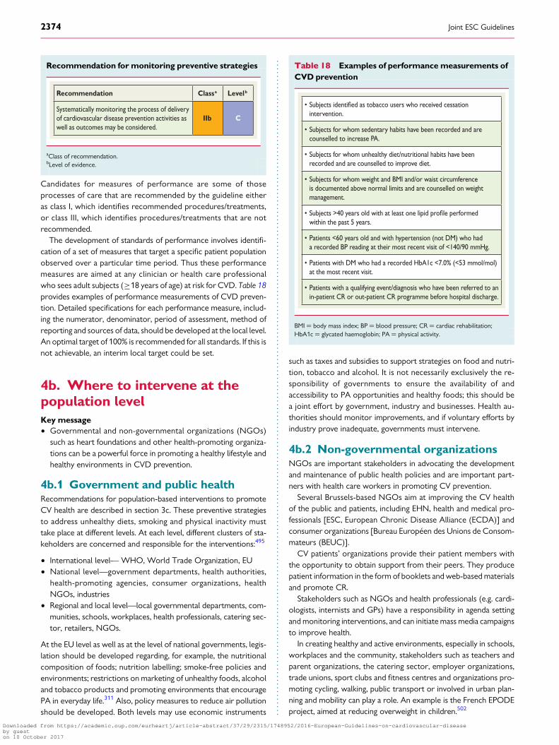
Recommendation for monitoring preventive strategies
Recommendation Classa Levelb
Systematically monitoring the process of delivery of cardiovascular disease prevention activities as well as outcomes may be considered.
IIb C
aClass of recommendation.bLevel of evidence.
Candidates for measures of performance are some of thoseprocesses of care that are recommended by the guideline eitheras class I, which identifies recommended procedures/treatments,or class III, which identifies procedures/treatments that are notrecommended.
The development of standards of performance involves identifi-cation of a set of measures that target a specific patient populationobserved over a particular time period. Thus these performancemeasures are aimed at any clinician or health care professionalwho sees adult subjects (≥18 years of age) at risk for CVD. Table 18provides examples of performance measurements of CVD preven-tion. Detailed specifications for each performance measure, includ-ing the numerator, denominator, period of assessment, method ofreporting and sources of data, should be developed at the local level.An optimal target of 100% is recommended for all standards. If this isnot achievable, an interim local target could be set.
4b. Where to intervene at thepopulation levelKey message† Governmental and non-governmental organizations (NGOs)
such as heart foundations and other health-promoting organiza-tions can be a powerful force in promoting a healthy lifestyle andhealthy environments in CVD prevention.
4b.1 Government and public healthRecommendations for population-based interventions to promoteCV health are described in section 3c. These preventive strategiesto address unhealthy diets, smoking and physical inactivity musttake place at different levels. At each level, different clusters of sta-keholders are concerned and responsible for the interventions:495
† International level— WHO, World Trade Organization, EU† National level—government departments, health authorities,
health-promoting agencies, consumer organizations, healthNGOs, industries
† Regional and local level—local governmental departments, com-munities, schools, workplaces, health professionals, catering sec-tor, retailers, NGOs.
At the EU level as well as at the level of national governments, legis-lation should be developed regarding, for example, the nutritionalcomposition of foods; nutrition labelling; smoke-free policies andenvironments; restrictions on marketing of unhealthy foods, alcoholand tobacco products and promoting environments that encouragePA in everyday life.311 Also, policy measures to reduce air pollutionshould be developed. Both levels may use economic instruments
such as taxes and subsidies to support strategies on food and nutri-tion, tobacco and alcohol. It is not necessarily exclusively the re-sponsibility of governments to ensure the availability of andaccessibility to PA opportunities and healthy foods; this should bea joint effort by government, industry and businesses. Health au-thorities should monitor improvements, and if voluntary efforts byindustry prove inadequate, governments must intervene.
4b.2 Non-governmental organizationsNGOs are important stakeholders in advocating the developmentand maintenance of public health policies and are important part-ners with health care workers in promoting CV prevention.
Several Brussels-based NGOs aim at improving the CV healthof the public and patients, including EHN, health and medical pro-fessionals [ESC, European Chronic Disease Alliance (ECDA)] andconsumer organizations [Bureau Europeen des Unions de Consom-mateurs (BEUC)].
CV patients’ organizations provide their patient members withthe opportunity to obtain support from their peers. They producepatient information in the form of booklets and web-based materialsand promote CR.
Stakeholders such as NGOs and health professionals (e.g. cardi-ologists, internists and GPs) have a responsibility in agenda settingand monitoring interventions, and can initiate mass media campaignsto improve health.
In creating healthy and active environments, especially in schools,workplaces and the community, stakeholders such as teachers andparent organizations, the catering sector, employer organizations,trade unions, sport clubs and fitness centres and organizations pro-moting cycling, walking, public transport or involved in urban plan-ning and mobility can play a role. An example is the French EPODEproject, aimed at reducing overweight in children.502
Table 18 Examples of performance measurements ofCVD prevention
• Subjects as tobacco users who received cessation intervention.
• Subjects for whom sedentary habits have been recorded and are counselled to increase PA.
• Subjects for whom unhealthy diet/nutritional habits have been recorded and are counselled to improve diet.
• Subjects for whom weight and BMI and/or waist circumference is documented above normal limits and are counselled on weight management.
• Subjects >40 years old with at least one lipid performed within the past 5 years.
• Patients <60 years old and with hypertension (not DM) who had a recorded BP reading at their most recent visit of <140/90 mmHg.
• Patients with DM who had a recorded HbA1c <7.0% (<53 mmol/mol) at the most recent visit.
• Patients with a qualifying event/diagnosis who have been referred to an in-patient CR or out-patient CR programme before hospital discharge.
BMI ¼ body mass index; BP ¼ blood pressure; CR ¼ cardiac rehabilitation;HbA1c ¼ glycated haemoglobin; PA ¼ physical activity.
Joint ESC Guidelines2374
Downloaded from https://academic.oup.com/eurheartj/article-abstract/37/29/2315/1748952/2016-European-Guidelines-on-cardiovascular-diseaseby gueston 10 October 2017
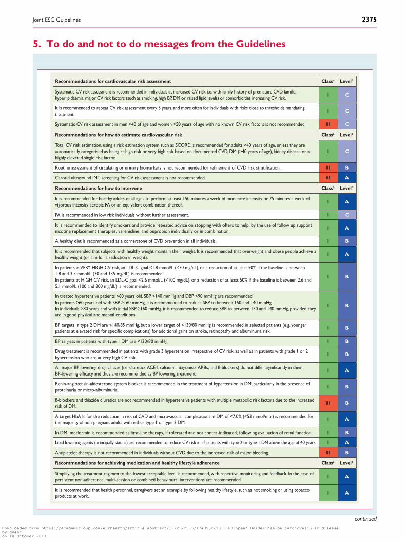
5. To do and not to do messages from the Guidelines
Recommendations for cardiovascular risk assessment Classa Levelb
Systematic CV risk assessment is recommended in individuals at increased CV risk, i.e. with family history of premature CVD, familial hyperlipidaemia, major CV risk factors (such as smoking, high BP, DM or raised lipid levels) or comorbidities increasing CV risk.
I C
It is recommended to repeat CV risk assessment every 5 years, and more often for individuals with risks close to thresholds mandating treatment.
I C
Systematic CV risk assessment in men <40 of age and women <50 years of age with no known CV risk factors is not recommended. III C
Recommendations for how to estimate cardiovascular risk Classa Levelb
Total CV risk estimation, using a risk estimation system such as SCORE, is recommended for adults >40 years of age, unless they are automatically categorised as being at high risk or very high risk based on documented CVD, DM (>40 years of age), kidney disease or a highly elevated single risk factor.
I C
Routine assessment of circulating or urinary biomarkers is not recommended for of CVD risk III B
Carotid ultrasound IMT screening for CV risk assessment is not recommended. III A
Recommendations for how to intervene Classa Levelb
It is recommended for healthy adults of all ages to perform at least 150 minutes a week of moderate intensity or 75 minutes a week of vigorous intensity aerobic PA or an equivalent combination thereof.
I A
PA is recommended in low risk individuals without further assessment. I C
It is recommended to identify smokers and provide repeated advice on stopping with offers to help, by the use of follow up support, nicotine replacement therapies, varenicline, and bupropion individually or in combination.
I A
A healthy diet is recommended as a cornerstone of CVD prevention in all individuals. I B
It is recommended that subjects with healthy weight maintain their weight. It is recommended that overweight and obese people achieve a healthy weight (or aim for a reduction in weight).
I A
In patients at VERY HIGH CV risk, an LDL-C goal <1.8 mmol/L (<70 mg/dL), or a reduction of at least 50% if the baseline is between 1.8 and 3.5 mmol/L (70 and 135 mg/dL) is recommended.In patients at HIGH CV risk, an LDL-C goal <2.6 mmol/L (<100 mg/dL), or a reduction of at least 50% if the baseline is between 2.6 and 5.1 mmol/L (100 and 200 mg/dL) is recommended.
I B
In treated hypertensive patients <60 years old, SBP <140 mmHg and DBP <90 mmHg are recommendedIn patients >60 years old with SBP ≥160 mmHg, it is recommended to reduce SBP to between 150 and 140 mmHg.In individuals >80 years and with initial SBP ≥160 mmHg, it is recommended to reduce SBP to between 150 and 140 mmHg, provided they are in good physical and mental conditions.
I B
BP targets in type 2 DM are <140/85 mmHg, but a lower target of <130/80 mmHg is recommended in selected patients (e.g. younger patients at elevated risk for complications) for additional gains on stroke, retinopathy and albuminuria risk.
I B
BP targets in patients with type 1 DM are <130/80 mmHg. I B
Drug treatment is recommended in patients with grade 3 hypertension irrespective of CV risk, as well as in patients with grade 1 or 2 hypertension who are at very high CV risk.
I B
All major BP lowering drug classes (i.e. diuretics, ACE-I, calcium antagonists, ARBs, and ß-blockers) do not differ in theirBP-lowering and thus are recommended as BP lowering treatment.
I A
Renin-angiotensin-aldosterone system blocker is recommended in the treatment of hypertension in DM, particularly in the presence of proteinuria or micro-albuminuria.
I B
ß-blockers and thiazide diuretics are not recommended in hypertensive patients with multiple metabolic risk factors due to the increased risk of DM.
III B
A target HbA1c for the reduction in risk of CVD and microvascular complications in DM of <7.0% (<53 mmol/mol) is recommended for the majority of non-pregnant adults with either type 1 or type 2 DM.
I A
In DM, metformin is recommended as therapy, if tolerated and not contra-indicated, following evaluation of renal function. I B
Lipid lowering agents (principally statins) are recommended to reduce CV risk in all patients with type 2 or type 1 DM above the age of 40 years. I A
Antiplatelet therapy is not recommended in individuals without CVD due to the increased risk of major bleeding. III B
Recommendations for achieving medication and healthy lifestyle adherence Classa Levelb
Simplifying the treatment regimen to the lowest acceptable level is recommended, with repetitive monitoring and feedback. In the case of persistent non-adherence, multi-session or combined behavioural interventions are recommended.
I A
It is recommended that health personnel, caregivers set an example by following healthy lifestyle, such as not smoking or using tobacco products at work.
I A
continued
Joint ESC Guidelines 2375
Downloaded from https://academic.oup.com/eurheartj/article-abstract/37/29/2315/1748952/2016-European-Guidelines-on-cardiovascular-diseaseby gueston 10 October 2017

6. AppendixESC Committee for Practice Guidelines (CPG): Jose Luis Za-morano (Chairperson) (Spain), Victor Aboyans (France), StephanAchenbach (Germany), Stefan Agewall (Norway), Lina Badimon(Spain), Gonzalo Baron-Esquivias (Spain), Helmut Baumgartner(Germany), Jeroen J. Bax (The Netherlands), Hector Bueno (Spain),Scipione Carerj (Italy), Veronica Dean (France), Çetin Erol (Turkey),Donna Fitzsimons (UK), Oliver Gaemperli (Switzerland), PaulusKirchhof (UK/Germany), Philippe Kolh (Belgium), Patrizio Lancellot-ti (Belgium), Gregory Y.H. Lip (UK), Petros Nihoyannopoulos (UK),Massimo F. Piepoli (Italy), Piotr Ponikowski (Poland), Marco Roffi(Switzerland), Adam Torbicki (Poland), Antonio Vaz Carneiro(Portugal), Stephan Windecker (Switzerland).
ESC National Cardiac Societies actively involved in the re-view process of the 2016 European Guidelines on cardiovasculardisease prevention in clinical practice:
Austria: Austrian Society of Cardiology, Bernhard Metzler;Azerbaijan: Azerbaijan Society of Cardiology, Ruslan Najafov;Belarus: Belorussian Scientific Society of Cardiologists, ValeriyStelmashok; Belgium: Belgian Society of Cardiology, CatherineDe Maeyer; Bosnia and Herzegovina: Association of Cardiolo-gists of Bosnia and Herzegovina, Mirza Dilic; Bulgaria: BulgarianSociety of Cardiology, Ivan Gruev; Croatia: Croatian Cardiac Soci-ety, Davor Milicic; Czech Republic: Czech Society of Cardiology,Helena Vaverkova; Denmark: Danish Society of Cardiology, IdaGustafsson; Egypt: Egyptian Society of Cardiology, Ihab Attia; Es-tonia: Estonian Society of Cardiology, Davit Duishvili; FormerYugoslav Republic of Macedonia: Macedonian FYR Society ofCardiology, Nela Kostova; France: French Society of Cardiology,Jean Ferrieres; Georgia: Georgian Society of Cardiology, Zurab
Klimiashvili; Germany: German Cardiac Society, Rainer Ham-brecht; Greece: Hellenic Cardiological Society, KonstantinosTsioufis; Hungary: Hungarian Society of Cardiology, Eszter Szaba-dos; Iceland: Icelandic Society of Cardiology, Karl Andersen; Ire-land: Irish Cardiac Society, Carl Vaughan; Israel: Israel HeartSociety, Barak Zafrir; Italy: Italian Federation of Cardiology, Salva-tore Novo; Kazakhstan: Association of Cardiologists of Kazakh-stan, Kairat Davletov; Kosovo: Kosovo Society of Cardiology,Fisnik Jashari; Kyrgyzstan: Kyrgyz Society of Cardiology, Alina Ker-imkulova, Latvia: Latvian Society of Cardiology, Iveta Mintale; Leba-non: Lebanese Society of Cardiology, Georges Saade; Lithuania:Lithuanian Society of Cardiology, Zaneta Petrulioniene; Luxem-bourg: Luxembourg Society of Cardiology, Charles Delagardelle;Malta: Maltese Cardiac Society, Caroline J. Magri; Moldova:Moldavian Society of Cardiology, Victor Rudi; Morocco: MoroccanSociety of Cardiology, Latifa Oukerraj; The Netherlands: Nether-lands Society of Cardiology, B. Ersen Çolkesen; Norway: NorwegianSociety of Cardiology, Henrik Schirmer; Poland: Polish CardiacSociety, Piotr Jankowski; Portugal: Portuguese Society of Cardi-ology, Roberto Palma dos Reis; Romania: Romanian Society ofCardiology, Daniel Gherasim; Russian Federation: Russian Societyof Cardiology, Sergey Nedogoda; San Marino: San Marino Societyof Cardiology, Marco Zavatta; Serbia: Cardiology Society of Serbia,Vojislav Giga; Slovakia: Slovak Society of Cardiology, Slavomira Fili-pova; Spain: Spanish Society of Cardiology, Luis Rodrıguez Padial;Sweden: Swedish Society of Cardiology, Anna Kiessling; Switzer-land: Swiss Society of Cardiology, Francois Mach; Tunisia: TunisianSociety of Cardiology and Cardio-Vascular Surgery, Abdallah Mahd-haoui; Turkey: Turkish Society of Cardiology, Dilek Ural; Ukraine:Ukrainian Association of Cardiology, Elena Nesukay; United King-dom: British Cardiovascular Society, Chris Gale.
To do and not to do messages from the Guidelines (continued)
Recommendation for CVD prevention implementation Classa Levelb
In primary care, it is recommended that GPs, nurses and allied health professionals within primary care deliver CVD prevention for high-risk patients.
I C
In acute hospital setting, it is recommended to implement strategies for prevention in CVD patients, including lifestyle changes, risk factor management and pharmacological optimization, after an acute event before hospital discharge to lower risk of mortality and morbidity.
I A
Participation in a cardiac rehabilitation programme for patients hospitalized for an acute coronary event or revascularization, and for patients with HF, is recommended.
I A
ACE-I¼ angiotensin-converting enzyme inhibitor; ACS ¼ acute coronary syndrome; ARBs¼ angiotensin receptor blockers; BP¼ blood pressure; CAD ¼ coronary artery disease;CKD ¼ chronic kidney disease; CV¼ cardiovascular; CVD¼ cardiovascular disease; DBP ¼ diastolic blood pressure; DM¼ diabetes mellitus; GPs ¼ general practitioners; HbA1c ¼glycated haemoglobin; HDL-C¼ high-density lipoprotein cholesterol; HF ¼ heart failure; IMT¼ intima–media thickness; LDL-C¼ low-density lipoprotein cholesterol; PA¼ physicalactivity; SBP ¼ systolic blood pressure; SCORE¼ Systematic Coronary Risk Estimation; TIA¼ transient ischaemic attack.aClass of recommendation.bLevel of evidence.
The CME text ‘2016 European Guidelines on cardiovascular disease prevention in clinical practice’ is accredited by the European Board for Accreditation in Cardiology (EBAC). EBACworks according to the quality standards of the European Accreditation Council for Continuing Medical Education (EACCME), which is an institution of the European Union of MedicalSpecialists (UEMS). In compliance with EBAC/EACCME Guidelines, all authors participating in this programme have disclosed any potential conflicts of interest that might cause a bias inthe article. The Organizing Committee is responsible for ensuring that all potential conflicts of interest relevant to the programme are declared to the participants prior to the CMEactivities.
CME questions for this article are available at: European Heart Journal http://www.oxforde-learning.com/eurheartj and European Society of Cardiology http://www.escardio.org/guidelines
Joint ESC Guidelines2376
Downloaded from https://academic.oup.com/eurheartj/article-abstract/37/29/2315/1748952/2016-European-Guidelines-on-cardiovascular-diseaseby gueston 10 October 2017
7. References1. A Dictionary of Epidemiology. 4th ed. New York: Oxford University Press.2. Moran AE, Forouzanfar MH, Roth GA, Mensah GA, Ezzati M, Murray CJ,
Naghavi M. Temporal trends in ischemic heart disease mortality in 21 world re-gions, 1980 to 2010: the Global Burden of Disease 2010 study. Circulation 2014;129:1483–1492.
3. Finucane MM, Stevens GA, Cowan MJ, Danaei G, Lin JK, Paciorek CJ, Singh GM,Gutierrez HR, Lu Y, Bahalim AN, Farzadfar F, Riley LM, Ezzati M. National, region-al, and global trends in body-mass index since 1980: systematic analysis of healthexamination surveys and epidemiological studies with 960 country-years and 9.1million participants. Lancet 2011;377:557–567.
4. Danaei G, Finucane MM, Lu Y, Singh GM, Cowan MJ, Paciorek CJ, Lin JK,Farzadfar F, Khang YH, Stevens GA, Rao M, Ali MK, Riley LM, Robinson CA,Ezzati M. National, regional, and global trends in fasting plasma glucose and dia-betes prevalence since 1980: systematic analysis of health examination surveysand epidemiological studies with 370 country-years and 2.7 million participants.Lancet 2011;378:31–40.
5. Kotseva K, Wood D, De Backer G, De Bacquer D, Pyorala K, Reiner Z, Keil U,EUROASPIRE III. Management of cardiovascular risk factors in asymptomatichigh-risk patients in general practice: cross-sectional survey in 12 European coun-tries. Eur J Cardiovasc Prev Rehabil 2010;17:530–540.
6. Kotseva K, Wood D, De Bacquer D, De Backer G, Ryden L, Jennings C, Gyberg V,Amouyel P, Bruthans J, Castro Conde A, Cifkova R, Deckers JW, De Sutter J,Dilic M, Dolzhenko M, Erglis A, Fras Z, Gaita D, Gotcheva N, Goudevenos J,Heuschmann P, Laucevicius A, Lehto S, Lovic D, Milicic D, Moore D,Nicolaides E, Oganov R, Pajak A, Pogosova N, Reiner Z, Stagmo M, Stork S,Tokgozoglu L, Vulic D. EUROASPIRE IV: a European Society of Cardiology surveyon the lifestyle, risk factor and therapeutic management of coronary patients from24 European countries. Eur J Prev Cardiol 2016;23:636–648.
7. Cooney MT, Dudina A, Whincup P, Capewell S, Menotti A, Jousilahti P, Njolstad I,Oganov R, Thomsen T, Tverdal A, Wedel H, Wilhelmsen L, Graham I. Re-evaluating the Rose approach: comparative benefits of the population and high-risk preventive strategies. Eur J Cardiovasc Prev Rehabil 2009;16:541–549.
8. Liu K, Daviglus ML, Loria CM, Colangelo LA, Spring B, Moller AC,Lloyd-Jones DM. Healthy lifestyle through young adulthood and the presenceof low cardiovascular disease risk profile in middle age: the Coronary ArteryRisk Development in (Young) Adults (CARDIA) study. Circulation 2012;125:996–1004.
9. NICE Public Health Guidance 25. Prevention of Cardiovascular Disease. http://www.nice.org.uk/guidance/PH25.
10. World Health Organization. Global status report on non-communicable diseases2010. http://apps.who.int/iris/bitstream/10665/44579/1/9789240686458_eng.pdf.
11. Perk J, De Backer G, Gohlke H, Graham I, Reiner Z, Verschuren M, Albus C,Benlian P, Boysen G, Cifkova R, Deaton C, Ebrahim S, Fisher M, Germano G,Hobbs R, Hoes A, Karadeniz S, Mezzani A, Prescott E, Ryden L, Scherer M,Syvanne M, Scholte op Reimer WJ, Vrints C, Wood D, Zamorano JL, Zannad F.European Guidelines on cardiovascular disease prevention in clinical practice(version 2012): the Fifth Joint Task Force of the European Society of Cardiologyand Other Societies on Cardiovascular Disease Prevention in Clinical Practice(constituted by representatives of nine societies and by invited experts). Eur HeartJ 2012;33:1635–701.
12. Cobiac LJ, Magnus A, Lim S, Barendregt JJ, Carter R, Vos T. Which interventionsoffer best value for money in primary prevention of cardiovascular disease? PLoSOne 2012;7:e41842.
13. Collins M, Mason H, O’Flaherty M, Guzman-Castillo M, Critchley J, Capewell S. Aneconomic evaluation of salt reduction policies to reduce coronary heart disease inEngland: a policy modeling study. Value Health 2014;17:517–524.
14. Nichols M TN, Scarborough P, Rayner P. European Cardiovascular Disease Statistics,2012 edition. http://www.escardio.org/static_file/Escardio/Press-media/press-releases/2013/EU-cardiovascular-disease-statistics-2012.pdf.
15. McConnachie A, Walker A, Robertson M, Marchbank L, Peacock J, Packard CJ,Cobbe SM, Ford I. Long-term impact on healthcare resource utilization of statintreatment, and its cost effectiveness in the primary prevention of cardiovasculardisease: a record linkage study. Eur Heart J 2014;35:290–298.
16. Mistry H, Morris S, Dyer M, Kotseva K, Wood D, Buxton M. Cost-effectiveness ofa European preventive cardiology programme in primary care: a Markov model-ling approach. BMJ Open 2012;2:e001029.
17. Plans-Rubio P. The cost effectiveness of statin therapies in Spain in 2010, after theintroduction of generics and reference prices. Am J Cardiovasc Drugs 2010;10:369–382.
18. World Health Organization. Scaling up action agains noncommunicable diseases: howmuch will it cost? Geneva: World Health Organization, 2011.
19. Mason H, Shoaibi A, Ghandour R, O’Flaherty M, Capewell S, Khatib R, Jabr S,Unal B, Sozmen K, Arfa C, Aissi W, Ben Romdhane H, Fouad F, Al-Ali R,Husseini A, MedCHAMPS project team. A cost effectiveness analysis of salt re-duction policies to reduce coronary heart disease in four Eastern Mediterraneancountries. PLoS One 2014;9:e84445.
20. O’Keeffe C, Kabir Z, O’Flaherty M, Walton J, Capewell S, Perry IJ. Modelling theimpact of specific food policy options on coronary heart disease and strokedeaths in Ireland. BMJ Open 2013;3:e002837.
21. Roth GA, Forouzanfar MH, Moran AE, Barber R, Nguyen G, Feigin VL, Naghavi M,Mensah GA, Murray CJ. Demographic and epidemiologic drivers of global cardio-vascular mortality. N Engl J Med 2015;372:1333–1341.
22. Pereira M, Azevedo A, Lunet N, Carreira H, O’Flaherty M, Capewell S, Bennett K.Explaining the decline in coronary heart disease mortality in Portugal between1995 and 2008. Circ Cardiovasc Qual Outcomes 2013;6:634–642.
23. Banegas JR, Lopez-Garcia E, Dallongeville J, Guallar E, Halcox JP, Borghi C,Masso-Gonzalez EL, Jimenez FJ, Perk J, Steg PG, De Backer G,Rodriguez-Artalejo F. Achievement of treatment goals for primary preventionof cardiovascular disease in clinical practice across Europe: the EURIKA study.Eur Heart J 2011;32:2143–2152.
24. De Smedt D, Kotseva K, De Bacquer D, Wood D, De Backer G, Dallongeville J,Seppo L, Pajak A, Reiner Z, Vanuzzo D, Georgiev B, Gotcheva N, Annemans L.Cost-effectiveness of optimizing prevention in patients with coronary heart dis-ease: the EUROASPIRE III health economics project. Eur Heart J 2012;33:2865–2872.
25. Blood Pressure Lowering Treatment Trialists’ Collaboration, Sundstrom J,Arima H, Woodward M, Jackson R, Karmali K, Lloyd-Jones D, Baigent C,Emberson J, Rahimi K, MacMahon S, Patel A, Perkovic V, Turnbull F, Neal B. Bloodpressure-lowering treatment based on cardiovascular risk: a meta-analysis of in-dividual patient data. Lancet 2014;384:591–598.
26. Thomopoulos C, Parati G, Zanchetti A. Effects of blood pressure lowering onoutcome incidence in hypertension: 3. Effects in patients at different levels of car-diovascular risk—overview and meta-analyses of randomized trials. J Hypertens2014;32:2305–2314.
27. Thomopoulos C, Parati G, Zanchetti A. Effects of blood pressure lowering onoutcome incidence in hypertension: 2. Effects at different baseline and achievedblood pressure levels—overview and meta-analyses of randomized trials.J Hypertens 2014;32:2296–2304.
28. Anderson KM, Odell PM, Wilson PW, Kannel WB. Cardiovascular disease riskprofiles. Am Heart J 1991;121(1 Pt 2):293–298.
29. Graham I, Atar D, Borch-Johnsen K, Boysen G, Burell G, Cifkova R, Dallongeville J,De Backer G, Ebrahim S, Gjelsvik B, Herrmann-Lingen C, Hoes A, Humphries S,Knapton M, Perk J, Priori SG, Pyorala K, Reiner Z, Ruilope L, Sans-Menendez S, OpReimer WS, Weissberg P, Wood D, Yarnell J, Zamorano JL, Walma E, Fitzgerald T,Cooney MT, Dudina A, Vahanian A, Camm J, De Caterina R, Dean V, Dickstein K,Funck-Brentano C, Filippatos G, Hellemans I, Kristensen SD, McGregor K,Sechtem U, Silber S, Tendera M, Widimsky P, Zamorano JL, Altiner A,Bonora E, Durrington PN, Fagard R, Giampaoli S, Hemingway H, Hakansson J,Kjeldsen SE, Larsen ML, Mancia G, Manolis AJ, Orth-Gomer K, Pedersen T,Rayner M, Ryden L, Sammut M, Schneiderman N, Stalenhoef AF, Tokgozoglu L,Wiklund O, Zampelas A. European guidelines on cardiovascular disease preven-tion in clinical practice: full text. Fourth Joint Task Force of the European Societyof Cardiology and other societies on cardiovascular disease prevention in clinicalpractice (constituted by representatives of nine societies and by invited experts).Eur J Cardiovasc Prev Rehabil 2007;14(Suppl 2):S1–S113.
30. Conroy RM, Pyorala K, Fitzgerald AP, Sans S, Menotti A, De Backer G, DeBacquer D, Ducimetiere P, Jousilahti P, Keil U, Njolstad I, Oganov RG,Thomsen T, Tunstall-Pedoe H, Tverdal A, Wedel H, Whincup P, Wilhelmsen L,Graham IM. Estimation of ten-year risk of fatal cardiovascular disease in Europe:the SCORE project. Eur H J 2003;24:987–1003.
31. Si S, Moss JR, Sullivan TR, Newton SS, Stocks NP. Effectiveness of general practice-based health checks: a systematic review and meta-analysis. Br J Gen Pract 2014;64:e47–e53.
32. Jorgensen T, Jacobsen RK, Toft U, Aadahl M, Glumer C, Pisinger C. Effect ofscreening and lifestyle counselling on incidence of ischaemic heart disease in gen-eral population: Inter99 randomised trial. BMJ 2014;348:g3617.
33. Ebrahim S, Taylor F, Ward K, Beswick A, Burke M, Davey Smith G. Multiple riskfactor interventions for primary prevention of coronary heart disease. CochraneDatabase Syst Rev 2011;1:CD001561.
34. Krogsboll LT, Jorgensen KJ, Gronhoj Larsen C, Gotzsche PC. General healthchecks in adults for reducing morbidity and mortality from disease. Cochrane Data-base Syst Rev 2012;10:CD009009.
35. National Institute for Health and Care Excellence. Lipid Modification: CardiovascularRisk Assessment and the Modification of Blood Lipids for the Primary and Secondary Pre-vention of Cardiovascular Disease. London: National Institute for Health and CareExcellence, 2014.
Joint ESC Guidelines 2377
Downloaded from https://academic.oup.com/eurheartj/article-abstract/37/29/2315/1748952/2016-European-Guidelines-on-cardiovascular-diseaseby gueston 10 October 2017

36. American Heart Association. Heart-Health Screenings. http://www.heart.org/HEARTORG/Conditions/Heart-Health-Screenings_UCM_428687_Article.jsp#.VtxbjseZaPU.
37. Scottish Intercollegiate Guidelines Network. Risk estimation and the prevention ofcardiovascular disease. A national clinical guideline. Edinburgh: Scottish IntercollegiateGuidelines Network, 2007.
38. European Association for Cardiovascular Prevention & Rehabilitation, Reiner Z,Catapano AL, De Backer G, Graham I, Taskinen MR, Wiklund O, Agewall S,Alegria E, Chapman MJ, Durrington P, Erdine S, Halcox J, Hobbs R, Kjekshus J,Filardi PP, Riccardi G, Storey RF, Wood D. ESC/EAS Guidelines for the manage-ment of dyslipidaemias: the Task Force for the management of dyslipidaemias ofthe European Society of Cardiology (ESC) the European Atherosclerosis Society(EAS). Eur Heart J 2011;32:1769–1818.
39. Chamnan P, Simmons RK, Khaw KT, Wareham NJ, Griffin SJ. Estimating the popu-lation impact of screening strategies for identifying and treating people at high riskof cardiovascular disease: modelling study. BMJ 2010;340:c1693.
40. Nielsen AD, Videbech P, Gerke O, Petersen H, Jensen JM, Sand NP, Egstrup K,Larsen ML, Mickley H, Diederichsen AC. Population screening for coronary arterycalcification does not increase mental distress and the use of psychoactive medi-cation. J Thorac Imaging 2012;27:202–206.
41. Christensen B, Engberg M, Lauritzen T. No long-term psychological reaction toinformation about increased risk of coronary heart disease in general practice.Eur J Cardiovasc Prev Rehabil 2004;11:239–243.
42. Lokkegaard T, Andersen JS, Jacobsen RK, Badsberg JH, Jorgensen T, Pisinger C.Psychological consequences of screening for cardiovascular risk factors in an un-selected general population: results from the Inter99 randomised interventionstudy. Scand J Public Health 2015;43:102–110.
43. Jorgensen T, Ladelund S, Borch-Johnsen K, Pisinger C, Schrader AM, Thomsen T,Glumer C, Ibsen H, Mortensen EL. Screening for risk of cardiovascular disease isnot associated with mental distress: the Inter99 study. Prev Med 2009;48:242–246.
44. D’Agostino RB Sr, Vasan RS, Pencina MJ, Wolf PA, Cobain M, Massaro JM,Kannel WB. General cardiovascular risk profile for use in primary care: the Fra-mingham Heart Study. Circulation 2008;117:743–753.
45. Woodward M, Brindle P, Tunstall-Pedoe H. Adding social deprivation and familyhistory to cardiovascular risk assessment: the ASSIGN score from the ScottishHeart Health Extended Cohort (SHHEC). Heart 2007;93:172–176.
46. Hippisley-Cox J, Coupland C, Vinogradova Y, Robson J, May M, Brindle P. Deriv-ation and validation of QRISK, a new cardiovascular disease risk score for the Uni-ted Kingdom: prospective open cohort study. BMJ 2007;335:136.
47. Hippisley-Cox J, Coupland C, Vinogradova Y, Robson J, Minhas R, Sheikh A,Brindle P. Predicting cardiovascular risk in England and Wales: prospective deriv-ation and validation of QRISK2. BMJ 2008;336:1475–1482.
48. Assmann G, Cullen P, Schulte H. Simple scoring scheme for calculating the risk ofacute coronary events based on the 10-year follow-up of the prospective cardio-vascular Munster (PROCAM) study. Circulation 2002;105:310–315.
49. Giampaoli S. CUORE: a sustainable cardiovascular disease prevention strategy.Eur J Cardiovasc Prev Rehabil 2007;14:161–162.
50. Goff DC Jr, Lloyd-Jones DM, Bennett G, Coady S, D’Agostino RB Sr, Gibbons R,Greenland P, Lackland DT, Levy D, O’Donnell CJ, Robinson JG, Schwartz JS,Shero ST, Smith SC Jr, Sorlie P, Stone NJ, Wilson PW. 2013 ACC/AHA guidelineon the assessment of cardiovascular risk: a report of the American College of Car-diology/American Heart Association Task Force on Practice Guidelines. J Am CollCardiol 2014;63(25 Pt B):2935–2959.
51. Krones T, Keller H, Sonnichsen A, Sadowski EM, Baum E, Wegscheider K,Rochon J, Donner-Banzhoff N. Absolute cardiovascular disease risk and shareddecision making in primary care: a randomized controlled trial. Ann Fam Med2008;6:218–227.
52. Hajifathalian K, Ueda P, Lu Y, Woodward M, Ahmadvand A, Aguilar-Salinas CA,Azizi F, Cifkova R, Di Cesare M, Eriksen L, Farzadfar F, Ikeda N, Khalili D,Khang YH, Lanska V, Leon-Munoz L, Magliano D, Msyamboza KP, Oh K,Rodriguez-Artalejo F, Rojas-Martinez R, Shaw JE, Stevens GA, Tolstrup J,Zhou B, Salomon JA, Ezzati M, Danaei G. A novel risk score to predict cardiovas-cular disease risk in national populations (Globorisk): a pooled analysis of pro-spective cohorts and health examination surveys. Lancet Diabetes Endocrinol2015;3:339–355.
53. Aktas MK, Ozduran V, Pothier CE, Lang R, Lauer MS. Global risk scores and ex-ercise testing for predicting all-cause mortality in a preventive medicine program.JAMA 2004;292:1462–1468.
54. Grundy SM, Cleeman JI, Merz CN, Brewer HB Jr, Clark LT, Hunninghake DB,Pasternak RC, Smith SC Jr, Stone NJ. Implications of recent clinical trials for theNational Cholesterol Education Program Adult Treatment Panel III guidelines. Ar-terioscler Thromb Vasc Biol 2004;24:e149–e161.
55. Genest J, McPherson R, Frohlich J, Anderson T, Campbell N, Carpentier A,Couture P, Dufour R, Fodor G, Francis GA, Grover S, Gupta M, Hegele RA,
Lau DC, Leiter L, Lewis GF, Lonn E, Mancini GB, Ng D, Pearson GJ,Sniderman A, Stone JA, Ur E. 2009 Canadian Cardiovascular Society/Canadianguidelines for the diagnosis and treatment of dyslipidemia and prevention of car-diovascular disease in the adult – 2009 recommendations. Can J Cardiol 2009;25:567–579.
56. Williams M. Risk assessment and management of cardiovascular disease in NewZealand. N Z Med J 2003;116:U661.
57. Rabar S, Harker M, O’Flynn N, Wierzbicki AS, Guideline Development G. Lipidmodification and cardiovascular risk assessment for the primary and secondaryprevention of cardiovascular disease: summary of updated NICE guidance. BMJ2014;349:g4356.
58. Board JBS. Joint British Societies’ consensus recommendations for the preventionof cardiovascular disease (JBS3). Heart 2014;100(Suppl 2):ii1– ii67.
59. Cooney MT, Dudina A, D’Agostino R, Graham IM. Cardiovascular risk-estimationsystems in primary prevention: do they differ? Do they make a difference? Can wesee the future? Circulation 2010;122:300–310.
60. Cooney MT, Dudina AL, Graham IM. Value and limitations of existing scores forthe assessment of cardiovascular risk: a review for clinicians. J Am Coll Cardiol 2009;54:1209–1227.
61. van Dis I, Geleijnse JM, Boer JM, Kromhout D, Boshuizen H, Grobbee DE, van derSchouw YT, Verschuren WM. Effect of including nonfatal events in cardiovascularrisk estimation, illustrated with data from The Netherlands. Eur J Prev Cardiol 2014;21:377–383.
62. Pyorala K, De Backer G, Graham I, Poole-Wilson P, Wood D. Prevention of cor-onary heart disease in clinical practice. Recommendations of the Task Force of theEuropean Society of Cardiology, European Atherosclerosis Society and EuropeanSociety of Hypertension. Eur Heart J 1994;15:1300–1331.
63. Cooney MT, Dudina A, De Bacquer D, Fitzgerald A, Conroy R, Sans S, Menotti A,De Backer G, Jousilahti P, Keil U, Thomsen T, Whincup P, Graham I. How muchdoes HDL cholesterol add to risk estimation? A report from the SCORE Investi-gators. Eur J Cardiovasc Prev Rehabil 2009;16:304–314.
64. Cooney MT, Dudina A, De Bacquer D, Wilhelmsen L, Sans S, Menotti A, DeBacker G, Jousilahti P, Keil U, Thomsen T, Whincup P, Graham IM. HDL choles-terol protects against cardiovascular disease in both genders, at all ages and at alllevels of risk. Atherosclerosis 2009;206:611–616.
65. Mortensen MB, Afzal S, Nordestgaard BG, Falk E. The high-densitylipoprotein-adjusted SCORE model worsens SCORE-based risk classification ina contemporary population of 30,824 Europeans: the Copenhagen General Popu-lation Study. Eur Heart J 2015;36:2446–2453.
66. Emerging Risk Factors Collaboration, Di Angelantonio E, Sarwar N, Perry P,Kaptoge S, Ray KK, Thompson A, Wood AM, Lewington S, Sattar N,Packard CJ, Collins R, Thompson SG, Danesh J. Major ipids, apolipoproteins,andrisk of vascular disease. JAMA 2009;302:1993–2000.
67. Wilson PW, Pencina M, Jacques P, Selhub J, D’Agostino R Sr, O’Donnell CJ.C-reactive protein and reclassification of cardiovascular risk in the FraminghamHeart Study. Circ Cardiovasc Qual Outcomes 2008;1:92–97.
68. Cooney MT, Vartiainen E, Laatikainen T, De Bacquer D, McGorrian C, Dudina A,Graham I. Cardiovascular risk age: concepts and practicalities. Heart 2012;98:941–946.
69. Cuende JI, Cuende N, Calaveras-Lagartos J. How to calculate vascular age with theSCORE project scales: a new method of cardiovascular risk evaluation. Eur Heart J2010;31:2351–2358.
70. World Health Organisation. WHO Global Health Repository. Cardiovascular diseases,deaths per 100 000. Data by country. http://apps.who.int/gho/data/node.main.A865CARDIOVASCULAR?lang=en.
71. Banerjee A. A review of family history of cardiovascular disease: risk factor andresearch tool. Int J Clin Pract 2012;66:536–543.
72. Di Angelantonio E, Butterworth AS. Clinical utility of genetic variants for cardio-vascular risk prediction: a futile exercise or insufficient data? Circ Cardiovasc Genet2012;5:387–390.
73. Ioannidis JP. Prediction of cardiovascular disease outcomes and established car-diovascular risk factors by genome-wide association markers. Circ Cardiovasc Genet2009;2:7–15.
74. Bachmann JM, Willis BL, Ayers CR, Khera A, Berry JD. Association between familyhistory and coronary heart disease death across long-term follow-up in men: theCooper Center Longitudinal Study. Circulation 2012;125:3092–3098.
75. Tikkanen E, Havulinna AS, Palotie A, Salomaa V, Ripatti S. Genetic risk predictionand a 2-stage risk screening strategy for coronary heart disease. Arterioscler ThrombVasc Biol 2013;33:2261–2266.
76. Ripatti S, Tikkanen E, Orho-Melander M, Havulinna AS, Silander K, Sharma A,Guiducci C, Perola M, Jula A, Sinisalo J, Lokki ML, Nieminen MS, Melander O,Salomaa V, Peltonen L, Kathiresan S. A multilocus genetic risk score for coronaryheart disease: case-control and prospective cohort analyses. Lancet 2010;376:1393–1400.
Joint ESC Guidelines2378
Downloaded from https://academic.oup.com/eurheartj/article-abstract/37/29/2315/1748952/2016-European-Guidelines-on-cardiovascular-diseaseby gueston 10 October 2017
77. Sivapalaratnam S, Boekholdt SM, Trip MD, Sandhu MS, Luben R, Kastelein JJ,Wareham NJ, Khaw KT. Family history of premature coronary heart diseaseand risk prediction in the EPIC-Norfolk prospective population study. Heart2010;96:1985–1989.
78. Veronesi G, Gianfagna F, Giampaoli S, Chambless LE, Mancia G, Cesana G,Ferrario MM. Improving long-term prediction of first cardiovascular event: thecontribution of family history of coronary heart disease and social status. PrevMed 2014;64:75–80.
79. Yeboah J, McClelland RL, Polonsky TS, Burke GL, Sibley CT, O’Leary D, Carr JJ,Goff DC, Greenland P, Herrington DM. Comparison of novel risk markers for im-provement in cardiovascular risk assessment in intermediate-risk individuals.JAMA 2012;308:788–795.
80. Hughes MF, Saarela O, Stritzke J, Kee F, Silander K, Klopp N, Kontto J, Karvanen J,Willenborg C, Salomaa V, Virtamo J, Amouyel P, Arveiler D, Ferrieres J,Wiklund PG, Baumert J, Thorand B, Diemert P, Tregouet DA, Hengstenberg C,Peters A, Evans A, Koenig W, Erdmann J, Samani NJ, Kuulasmaa K,Schunkert H. Genetic markers enhance coronary risk prediction in men: theMORGAM prospective cohorts. PLoS One 2012;7:e40922.
81. Ganna A, Magnusson PK, Pedersen NL, de Faire U, Reilly M, Arnlov J, Sundstrom J,Hamsten A, Ingelsson E. Multilocus genetic risk scores for coronary heart diseaseprediction. Arterioscl Thromb Vasc Biol 2013;33:2267–2272.
82. Brautbar A, Pompeii LA, Dehghan A, Ngwa JS, Nambi V, Virani SS, Rivadeneira F,Uitterlinden AG, Hofman A, Witteman JC, Pencina MJ, Folsom AR, Cupples LA,Ballantyne CM, Boerwinkle E. A genetic risk score based on direct associationswith coronary heart disease improves coronary heart disease risk prediction inthe Atherosclerosis Risk in Communities (ARIC), but not in the Rotterdam andFramingham Offspring, Studies. Atherosclerosis 2012;223:421–426.
83. Bressler J, Folsom AR, Couper DJ, Volcik KA, Boerwinkle E. Genetic variants iden-tified in a European genome-wide association study that were found to predictincident coronary heart disease in the atherosclerosis risk in communities study.Am J Epidemiol 2010;171:14–23.
84. Mega JL, Stitziel NO, Smith JG, Chasman DI, Caulfield MJ, Devlin JJ, Nordio F,Hyde CL, Cannon CP, Sacks FM, Poulter NR, Sever PS, Ridker PM,Braunwald E, Melander O, Kathiresan S, Sabatine MS. Genetic risk, coronary heartdisease events, and the clinical benefit of statin therapy: an analysis of primary andsecondary prevention trials. Lancet 2015;385:2264–2271.
85. Floyd CN, Mustafa A, Ferro A. The PlA1/A2 polymorphism of glycoprotein IIIa asa risk factor for myocardial infarction: a meta-analysis. PLoS One 2014;9:e101518.
86. Singleton A, Erby LH, Foisie KV, Kaphingst KA. Informed choice indirect-to-consumer genetic testing (DTCGT) websites: a content analysis of ben-efits, risks, and limitations. J Genet Couns 2012;21:433–439.
87. Guay SP, Brisson D, Lamarche B, Marceau P, Vohl MC, Gaudet D, Bouchard L.DNA methylation variations at CETP and LPL gene promoter loci: new molecularbiomarkers associated with blood lipid profile variability. Atherosclerosis 2013;228:413–420.
88. Wang X, Falkner B, Zhu H, Shi H, Su S, Xu X, Sharma AK, Dong Y, Treiber F,Gutin B, Harshfield G, Snieder H. A genome-wide methylation study on essentialhypertension in young African American males. PLoS One 2013;8:e53938.
89. Baccarelli A, Wright R, Bollati V, Litonjua A, Zanobetti A, Tarantini L, Sparrow D,Vokonas P, Schwartz J. Ischemic heart disease and stroke in relation to bloodDNA methylation. Epidemiology 2010;21:819–828.
90. Albus C, Jordan J, Herrmann-Lingen C. Screening for psychosocial risk factors inpatients with coronary heart disease-recommendations for clinical practice. Eur JCardiovasc Prev Rehabil 2004;11:75–79.
91. Lichtman JH, Froelicher ES, Blumenthal JA, Carney RM, Doering LV,Frasure-Smith N, Freedland KE, Jaffe AS, Leifheit-Limson EC, Sheps DS,Vaccarino V, Wulsin L. Depression as a risk factor for poor prognosis among pa-tients with acute coronary syndrome: systematic review and recommendations: ascientific statement from the American Heart Association. Circulation 2014;129:1350–1369.
92. Pogosova N, Saner H, Pedersen SS, Cupples ME, McGee H, Hofer S, Doyle F,Schmid JP, von Kanel R. Psychosocial aspects in cardiac rehabilitation: from theoryto practice. A position paper from the Cardiac Rehabilitation Section of the Euro-pean Association of Cardiovascular Prevention and Rehabilitation of the Euro-pean Society of Cardiology. Eur J Prev Cardiol 2015;22:1290–1306.
93. Albert MA, Glynn RJ, Buring J, Ridker PM. Impact of traditional and novel risk fac-tors on the relationship between socioeconomic status and incident cardiovascu-lar events. Circulation 2006;114:2619–2626.
94. Alter DA, Franklin B, Ko DT, Austin PC, Lee DS, Oh PI, Stukel TA, Tu JV. Socio-economic status, functional recovery, and long-term mortality among patientssurviving acute myocardial infarction. PLoS One 2014;8:e65130.
95. Barth J, Schneider S, von Kanel R. Lack of social support in the etiology and theprognosis of coronary heart disease: a systematic review and meta-analysis. Psy-chosom Med 2010;72:229–238.
96. Nawrot TS, Perez L, Kunzli N, Munters E, Nemery B. Public health importance oftriggers of myocardial infarction: a comparative risk assessment. Lancet 2011;377:732–740.
97. Mostofsky E, Penner EA, Mittleman MA. Outbursts of anger as a trigger of acutecardiovascular events: a systematic review and meta-analysis. Eur Heart J 2014;35:1404–1410.
98. Kivimaki M, Nyberg ST, Batty GD, Fransson EI, Heikkila K, Alfredsson L,Bjorner JB, Borritz M, Burr H, Casini A, Clays E, De Bacquer D, Dragano N,Ferrie JE, Geuskens GA, Goldberg M, Hamer M, Hooftman WE, Houtman IL,Joensuu M, Jokela M, Kittel F, Knutsson A, Koskenvuo M, Koskinen A,Kouvonen A, Kumari M, Madsen IE, Marmot MG, Nielsen ML, Nordin M,Oksanen T, Pentti J, Rugulies R, Salo P, Siegrist J, Singh-Manoux A,Suominen SB, Vaananen A, Vahtera J, Virtanen M, Westerholm PJ,Westerlund H, Zins M, Steptoe A, Theorell T. Job strain as a risk factor for cor-onary heart disease: a collaborative meta-analysis of individual participant data.Lancet 2012;380:1491–1497.
99. Eaker ED, Sullivan LM, Kelly-Hayes M, D’Agostino RB Sr, Benjamin EJ. Marital sta-tus, marital strain, and risk of coronary heart disease or total mortality: the Fra-mingham Offspring Study. Psychosom Med 2007;69:509–513.
100. Kivimaki M, Jokela M, Nyberg ST, Singh-Manoux A, Fransson EI, Alfredsson L,Bjorner JB, Borritz M, Burr H, Casini A, Clays E, De Bacquer D, Dragano N,Erbel R, Geuskens GA, Hamer M, Hooftman WE, Houtman IL, Jockel KH,Kittel F, Knutsson A, Koskenvuo M, Lunau T, Madsen IE, Nielsen ML,Nordin M, Oksanen T, Pejtersen JH, Pentti J, Rugulies R, Salo P, Shipley MJ,Siegrist J, Steptoe A, Suominen SB, Theorell T, Vahtera J, Westerholm PJ,Westerlund H, O’Reilly D, Kumari M, Batty GD, Ferrie JE, Virtanen M. Long work-ing hours and risk of coronary heart disease and stroke: a systematic review andmeta-analysis of published and unpublished data for 603 838 individuals. Lancet2015;386:1739–1746.
101. Spindler H, Pedersen SS. Posttraumatic stress disorder in the wake of heart dis-ease: prevalence, risk factors, and future research directions. Psychosom Med 2005;67:715–723.
102. Orth-Gomer K, Wamala SP, Horsten M, Schenck-Gustafsson K, Schneiderman N,Mittleman MA. Marital stress worsens prognosis in women with coronary heartdisease: the Stockholm Female Coronary Risk Study. JAMA 2000;284:3008–3014.
103. Schnohr P, Marott JL, Kristensen TS, Gyntelberg F, Gronbaek M, Lange P,Jensen MT, Jensen GB, Prescott E. Ranking of psychosocial and traditional risk fac-tors by importance for coronary heart disease: the Copenhagen City Heart Study.Eur Heart J 2015;36:1385–1393.
104. Smoller JW, Pollack MH, Wassertheil-Smoller S, Jackson RD, Oberman A,Wong ND, Sheps D. Panic attacks and risk of incident cardiovascular eventsamong postmenopausal women in the Women’s Health Initiative ObservationalStudy. Arch Gen Psychiatry 2007;64:1153–1160.
105. Roest AM, Martens EJ, de Jonge P, Denollet J. Anxiety and risk of incident coronaryheart disease: a meta-analysis. J Am Coll Cardiol 2010;56:38–46.
106. Roest AM, Martens EJ, Denollet J, de Jonge P. Prognostic association of anxietypost myocardial infarction with mortality and new cardiac events: a meta-analysis.Psychosom Med 2010;72:563–569.
107. Fan Z, Wu Y, Shen J, Ji T, Zhan R. Schizophrenia and the risk of cardiovascular dis-eases: a meta-analysis of thirteen cohort studies. J Psychiatr Res 2013;47:1549–1556.
108. Edmondson D, Kronish IM, Shaffer JA, Falzon L, Burg MM. Posttraumatic stressdisorder and risk for coronary heart disease: a meta-analytic review. Am Heart J2013;166:806–814.
109. Chida Y, Steptoe A. The association of anger and hostility with future coronaryheart disease: a meta-analytic review of prospective evidence. J Am Coll Cardiol2009;53:936–946.
110. Grande G, Romppel M, Barth J. Association between type D personality and prog-nosis in patients with cardiovascular diseases: a systematic review andmeta-analysis. Ann Behav Med 2012;43:299–310.
111. Chandola T, Britton A, Brunner E, Hemingway H, Malik M, Kumari M, Badrick E,Kivimaki M, Marmot M. Work stress and coronary heart disease: what are the me-chanisms? Eur Heart J 2008;29:640–648.
112. Yusuf S, Hawken S, Ounpuu S, Dans T, Avezum A, Lanas F, McQueen M, Budaj A,Pais P, Varigos J, Lisheng L. Effect of potentially modifiable risk factors associatedwith myocardial infarction in 52 countries (the INTERHEART study): case-controlstudy. Lancet 2004;364:937–952.
113. Stringhini S, Sabia S, Shipley M, Brunner E, Nabi H, Kivimaki M, Singh-Manoux A.Association of socioeconomic position with health behaviors and mortality. JAMA2010;303:1159–1166.
114. Gilstrap LG, Wang TJ. Biomarkers and cardiovascular risk assessment for primaryprevention: an update. Clin Chem 2012;58:72–82.
115. Ioannidis JP, Tzoulaki I. Minimal and null predictive effects for the most popularblood biomarkers of cardiovascular disease. Circ Res 2012;110:658–662.
Joint ESC Guidelines 2379
Downloaded from https://academic.oup.com/eurheartj/article-abstract/37/29/2315/1748952/2016-European-Guidelines-on-cardiovascular-diseaseby gueston 10 October 2017
116. Emerging Risk Factors Collaboration, Kaptoge S, Di Angelantonio E, Pennells L,Wood AM, White IR, Gao P, Walker M, Thompson A, Sarwar N, Caslake M,Butterworth AS, Amouyel P, Assmann G, Bakker SJ, Barr EL, Barrett-Connor E,Benjamin EJ, Bjorkelund C, Brenner H, Brunner E, Clarke R, Cooper JA,Cremer P, Cushman M, Dagenais GR, D’Agostino RB Sr, Dankner R,Davey-Smith G, Deeg D, Dekker JM, Engstrom G, Folsom AR, Fowkes FG,Gallacher J, Gaziano JM, Giampaoli S, Gillum RF, Hofman A, Howard BV,Ingelsson E, Iso H, Jorgensen T, Kiechl S, Kitamura A, Kiyohara Y, Koenig W,Kromhout D, Kuller LH, Lawlor DA, Meade TW, Nissinen A,Nordestgaard BG, Onat A, Panagiotakos DB, Psaty BM, Rodriguez B,Rosengren A, Salomaa V, Kauhanen J, Salonen JT, Shaffer JA, Shea S, Ford I,Stehouwer CD, Strandberg TE, Tipping RW, Tosetto A, Wassertheil-Smoller S,Wennberg P, Westendorp RG, Whincup PH, Wilhelmsen L, Woodward M,Lowe GD, Wareham NJ, Khaw KT, Sattar N, Packard CJ, Gudnason V,Ridker PM, Pepys MB, Thompson SG, Danesh J. C-reactive protein, fibrinogen,and cardiovascular disease prediction. N Engl J Med 2012;367:1310–1320.
117. Tzoulaki I, Siontis KC, Evangelou E, Ioannidis JP. Bias in associations of emergingbiomarkers with cardiovascular disease. JAMA Intern Med 2013;173:664–671.
118. Kooter AJ, Kostense PJ, Groenewold J, Thijs A, Sattar N, Smulders YM. Integratinginformation from novel risk factors with calculated risks: the critical impact of riskfactor prevalence. Circulation 2011;124:741–745.
119. Wurtz P, Havulinna AS, Soininen P, Tynkkynen T, Prieto-Merino D, Tillin T,Ghorbani A, Artati A, Wang Q, Tiainen M, Kangas AJ, Kettunen J, Kaikkonen J,Mikkila V, Jula A, Kahonen M, Lehtimaki T, Lawlor DA, Gaunt TR, Hughes AD,Sattar N, Illig T, Adamski J, Wang TJ, Perola M, Ripatti S, Vasan RS,Raitakari OT, Gerszten RE, Casas JP, Chaturvedi N, Ala-Korpela M, Salomaa V.Metabolite profiling and cardiovascular event risk: a prospective study of 3population-based cohorts. Circulation 2015;131:774–85.
120. Haberl R, Becker A, Leber A, Knez A, Becker C, Lang C, Bruning R, Reiser M,Steinbeck G. Correlation of coronary calcification and angiographically documen-ted stenoses in patients with suspected coronary artery disease: results of 1,764patients. J Am Coll Cardiol 2001;37:451–457.
121. Marwan M, Ropers D, Pflederer T, Daniel WG, Achenbach S. Clinical character-istics of patients with obstructive coronary lesions in the absence of coronary cal-cification: an evaluation by coronary CT angiography. Heart 2009;95:1056–1060.
122. Hecht HS, Superko HR. Electron beam tomography and National CholesterolEducation Program guidelines in asymptomatic women. J Am Coll Cardiol 2001;37:1506–1511.
123. Hadamitzky M, Freissmuth B, Meyer T, Hein F, Kastrati A, Martinoff S, Schomig A,Hausleiter J. Prognostic value of coronary computed tomographic angiography forprediction of cardiac events in patients with suspected coronary artery disease.JACC Cardiovasc Imaging 2009;2:404–411.
124. van Werkhoven JM, Gaemperli O, Schuijf JD, Jukema JW, Kroft LJ, Leschka S,Alkadhi H, Valenta I, Pundziute G, de Roos A, van der Wall EE, Kaufmann PA,Bax JJ. Multislice computed tomography coronary angiography for risk stratifica-tion in patients with an intermediate pretest likelihood. Heart 2009;95:1607–1611.
125. Peters SA, den Ruijter HM, Bots ML, Moons KG. Improvements in risk stratifica-tion for the occurrence of cardiovascular disease by imaging subclinical athero-sclerosis: a systematic review. Heart 2012;98:177–184.
126. O’Leary DH, Polak JF, Kronmal RA, Manolio TA, Burke GL, Wolfson SK Jr.Carotid-artery intima and media thickness as a risk factor for myocardial infarc-tion and stroke in older adults. Cardiovascular Health Study Collaborative Re-search Group. N Engl J Med 1999;340:14–22.
127. Chambless LE, Heiss G, Folsom AR, Rosamond W, Szklo M, Sharrett AR,Clegg LX. Association of coronary heart disease incidence with carotid arterialwall thickness and major risk factors: the Atherosclerosis Risk in Communities(ARIC) Study, 1987–1993. Am J Epidemiol 1997;146:483–494.
128. Den Ruijter HM, Peters SA, Anderson TJ, Britton AR, Dekker JM, Eijkemans MJ,Engstrom G, Evans GW, de Graaf J, Grobbee DE, Hedblad B, Hofman A,Holewijn S, Ikeda A, Kavousi M, Kitagawa K, Kitamura A, Koffijberg H,Lonn EM, Lorenz MW, Mathiesen EB, Nijpels G, Okazaki S, O’Leary DH,Polak JF, Price JF, Robertson C, Rembold CM, Rosvall M, Rundek T, Salonen JT,Sitzer M, Stehouwer CD, Witteman JC, Moons KG, Bots ML. Common carotidintima-media thickness measurements in cardiovascular risk prediction: ameta-analysis. JAMA 2012;308:796–803.
129. Hiatt WR. Medical treatment of peripheral arterial disease and claudication. NEngl J Med 2001;344:1608–21.
130. McDermott MM, Greenland P, Liu K, Guralnik JM, Celic L, Criqui MH, Chan C,Martin GJ, Schneider J, Pearce WH, Taylor LM, Clark E. The ankle brachial indexis associated with leg function and physical activity: the Walking and Leg Circula-tion Study. Ann Intern Med 2002;136:873–883.
131. Fowkes FG, Murray GD, Newman AB, Lee RJ. Ankle brachial index combined withFramingham Risk Score to predict cardiovascular events and mortality: ameta-analysis. JAMA 2008;300:197–208.
132. Fowkes FG, Price JF, Stewart MC, Butcher I, Leng GC, Pell AC, Sandercock PA,Fox KA, Lowe GD, Murray GD. Aspirin for prevention of cardiovascular eventsin a general population screened for a low ankle brachial index: a randomized con-trolled trial. JAMA 2010;303:841–848.
133. Lorenz MW, Polak JF, Kavousi M, Mathiesen EB, Volzke H, Tuomainen TP,Sander D, Plichart M, Catapano AL, Robertson CM, Kiechl S, Rundek T,Desvarieux M, Lind L, Schmid C, DasMahapatra P, Gao L, Ziegelbauer K,Bots ML, Thompson SG. Carotid intima-media thickness progression to predictcardiovascular events in the general population (the PROG-IMT collaborativeproject): a meta-analysis of individual participant data. Lancet 2012;379:2053–2062.
134. Tinana A, Mintz GS, Weissman NJ. Volumetric intravascular ultrasound quantifi-cation of the amount of atherosclerosis and calcium in nonstenotic arterial seg-ments. Am J Cardiol 2002;89:757–760.
135. Burke AP, Kolodgie FD, Farb A, Weber D, Virmani R. Morphological predictors ofarterial remodeling in coronary atherosclerosis. Circulation 2002;105:297–303.
136. Schmermund A, Schwartz RS, Adamzik M, Sangiorgi G, Pfeifer EA, Rumberger JA,Burke AP, Farb A, Virmani R. Coronary atherosclerosis in unheralded sudden cor-onary death under age 50: histo-pathologic comparison with ‘healthy’ subjects dy-ing out of hospital. Atherosclerosis 2001;155:499–508.
137. Silber S. Comparison of spiral and electron beam tomography in the evaluation ofcoronary calcification in asymptomatic persons. Int J Cardiol 2002;82:297–298; au-thor reply 299.
138. Gibson AO, Blaha MJ, Arnan MK, Sacco RL, Szklo M, Herrington DM, Yeboah J.Coronary artery calcium and incident cerebrovascular events in an asymptomaticcohort. The MESA Study. JACC Cardiovasc Imaging 2014;7:1108–1115.
139. Fernandez-Friera L, Penalvo JL, Fernandez-Ortiz A, Ibanez B, Lopez-Melgar B,Laclaustra M, Oliva B, Mocoroa A, Mendiguren J, Martinez de Vega V, Garcia L,Molina J, Sanchez-Gonzalez J, Guzman G, Alonso-Farto JC, Guallar E, Civeira F,Sillesen H, Pocock S, Ordovas JM, Sanz G, Jimenez-Borreguero LJ, Fuster V. Preva-lence, vascular distribution, and multiterritorial extent of subclinical atheroscler-osis in a middle-aged cohort: the PESA (Progression of Early SubclinicalAtherosclerosis) study. Circulation 2015;131:2104–2113.
140. Nasir K, Bittencourt MS, Blaha MJ, Blankstein R, Agatson AS, Rivera JJ,Miemdema MD, Sibley CT, Shaw LJ, Blumenthal RS, Budoff MJ, Krumholz HM. Im-plications of coronary artery calcium testing among statin candidates according toAmerican College of Cardiology/American Heart Association cholesterol man-agement guidelines: MESA (Multi-Ethnic Study of Atherosclerosis). J Am Coll Car-diol 2015;66:1657–1668.
141. Stein JH, Korcarz CE, Hurst RT, Lonn E, Kendall CB, Mohler ER, Najjar SS,Rembold CM, Post WS. Use of carotid ultrasound to identify subclinical vasculardisease and evaluate cardiovascular disease risk: a consensus statement from theAmerican Society of Echocardiography Carotid Intima-Media Thickness TaskForce. Endorsed by the Society for Vascular Medicine. J Am Soc Echocardiogr2008;21:93–111; quiz 189–190.
142. Vlachopoulos C, Aznaouridis K, Stefanadis C. Prediction of cardiovascular eventsand all-cause mortality with arterial stiffness: a systematic review andmeta-analysis. J Am Coll Cardiol 2010;55:1318–1327.
143. U.S. Preventive Services Task Force. Using nontraditional risk factors in coronaryheart disease risk assessment: U.S. Preventive Services Task Force recommenda-tion statement. Ann Intern Med 2009;151:474–482.
144. Taylor HA, Penman AD, Han H, Dele-Michael A, Skelton TN, Fox ER, Benjamin EJ,Arnett DK, Mosley TH Jr. Left ventricular architecture and survival inAfrican-Americans free of coronary heart disease (from the AtherosclerosisRisk in Communities [ARIC] study). Am J Cardiol 2007;99:1413–1420.
145. Muiesan ML, Salvetti M, Monteduro C, Bonzi B, Paini A, Viola S, Poisa P, Rizzoni D,Castellano M, Agabiti-Rosei E. Left ventricular concentric geometry during treat-ment adversely affects cardiovascular prognosis in hypertensive patients. Hyper-tension 2004;43:731–738.
146. Schiffrin EL, Lipman ML, Mann JF. Chronic kidney disease: effects on the cardiovas-cular system. Circulation 2007;116:85–97.
147. Chronic Kidney Disease Prognosis Consortium, Matsushita K, van der Velde M,Astor BC, Woodward M, Levey AS, de Jong PE, Coresh J, Gansevoort RT. Asso-ciation of estimated glomerular filtration rate albuminuria with all-cause cardio-vascular mortality in general population cohorts: a collaborative meta-analysis.Lancet 2010;375:2073–2081.
148. Matsushita K, Coresh J, Sang Y, Chalmers J, Fox C, Guallar E, Jafar T, Jassal SK,Landman GW, Muntner P, Roderick P, Sairenchi T, Schottker B, Shankar A,Shlipak M, Tonelli M, Townend J, van Zuilen A, Yamagishi K, Yamashita K,Gansevoort R, Sarnak M, Warnock DG, Woodward M, Arnlov J,Consortium CKDP. Estimated glomerular filtration rate and albuminuria for pre-diction of cardiovascular outcomes: a collaborative meta-analysis of individualparticipant data. Lancet Diabetes Endocrinol 2015;3:514–525.
149. Shlipak MG, Matsushita K, Arnlov J, Inker LA, Katz R, Polkinghorne KR,Rothenbacher D, Sarnak MJ, Astor BC, Coresh J, Levey AS, Gansevoort RT.
Joint ESC Guidelines2380
Downloaded from https://academic.oup.com/eurheartj/article-abstract/37/29/2315/1748952/2016-European-Guidelines-on-cardiovascular-diseaseby gueston 10 October 2017
Cystatin C versus creatinine in determining risk based on kidney function. N Engl JMed 2013;369:932–943.
150. Matsushita K, Mahmoodi BK, Woodward M, Emberson JR, Jafar TH, Jee SH,Polkinghorne KR, Shankar A, Smith DH, Tonelli M, Warnock DG, Wen CP,Coresh J, Gansevoort RT, Hemmelgarn BR, Levey AS. Comparison of risk predic-tion using the CKD-EPI equation and the MDRD study equation for estimatedglomerular filtration rate. JAMA 2012;307:1941–51.
151. Smeeth L, Thomas SL, Hall AJ, Hubbard R, Farrington P, Vallance P. Risk of myo-cardial infarction and stroke after acute infection or vaccination. N Engl J Med2004;351:2611–2618.
152. Siriwardena AN, Gwini SM, Coupland CA. Influenza vaccination, pneumococcalvaccination and risk of acute myocardial infarction: matched case-control study.CMAJ 2010;182:1617–1623.
153. Gwini SM, Coupland CA, Siriwardena AN. The effect of influenza vaccination onrisk of acute myocardial infarction: self-controlled case-series study. Vaccine 2011;29:1145–1149.
154. Udell JA, Zawi R, Bhatt DL, Keshtkar-Jahromi M, Gaughran F, Phrommintikul A,Ciszewski A, Vakili H, Hoffman EB, Farkouh ME, Cannon CP. Association betweeninfluenza vaccination and cardiovascular outcomes in high-risk patients: ameta-analysis. JAMA 2013;310:1711–1720.
155. Warren-Gash C, Hayward AC, Hemingway H, Denaxas S, Thomas SL,Timmis AD, Whitaker H, Smeeth L. Influenza infection and risk of acute myocar-dial infarction in England and Wales: a CALIBER self-controlled case series study.J Infect Dis 2012;206:1652–1659.
156. Jimenez M, Krall EA, Garcia RI, Vokonas PS, Dietrich T. Periodontitis and incidenceof cerebrovascular disease in men. Ann Neurol 2009;66:505–512.
157. Dietrich T, Jimenez M, Krall Kaye EA, Vokonas PS, Garcia RI. Age-dependent as-sociations between chronic periodontitis/edentulism and risk of coronary heartdisease. Circulation 2008;117:1668–1674.
158. Beck JD, Eke P, Lin D, Madianos P, Couper D, Moss K, Elter J, Heiss G,Offenbacher S. Associations between IgG antibody to oral organisms and carotidintima-medial thickness in community-dwelling adults. Atherosclerosis 2005;183:342–348.
159. Desvarieux M, Demmer RT, Jacobs DR, Papapanou PN, Sacco RL, Rundek T.Changes in clinical and microbiological periodontal profiles relate to progressionof carotid intima-media thickness: the Oral Infections and Vascular Disease Epi-demiology study. J Am Heart Assoc 2013;2:e000254.
160. Bosch X, Rovira M, Sitges M, Domenech A, Ortiz-Perez JT, de Caralt TM,Morales-Ruiz M, Perea RJ, Monzo M, Esteve J. Enalapril and carvedilol for prevent-ing chemotherapy-induced left ventricular systolic dysfunction in patients withmalignant hemopathies: the OVERCOME trial (preventiOn of left Ventriculardysfunction with Enalapril and caRvedilol in patients submitted to intensiveChemOtherapy for the treatment of Malignant hEmopathies). J Am Coll Cardiol2013;61:2355–2362.
161. Kalam K, Marwick TH. Role of cardioprotective therapy for prevention of cardi-otoxicity with chemotherapy: a systematic review and meta-analysis. Eur J Cancer2013;49:2900–2909.
162. Chen J, Long JB, Hurria A, Owusu C, Steingart RM, Gross CP. Incidence of heartfailure or cardiomyopathy after adjuvant trastuzumab therapy for breast cancer.J Am Coll Cardiol 2012;60:2504–2512.
163. Darby S, McGale P, Peto R, Granath F, Hall P, Ekbom A. Mortality from cardiovas-cular disease more than 10 years after radiotherapy for breast cancer: nationwidecohort study of 90 000 Swedish women. BMJ 2003;326:256–257.
164. Smith GL, Smith BD, Buchholz TA, Giordano SH, Garden AS, Woodward WA,Krumholz HM, Weber RS, Ang KK, Rosenthal DI. Cerebrovascular disease riskin older head and neck cancer patients after radiotherapy. J Clin Oncol 2008;26:5119–5125.
165. Fajardo LF. Is the pathology of radiation injury different in small vs large blood ves-sels? Cardiovasc Radiat Med 1999;1:108–110.
166. van der Pal HJ, van Dalen EC, van Delden E, van Dijk IW, Kok WE, Geskus RB,Sieswerda E, Oldenburger F, Koning CC, van Leeuwen FE, Caron HN,Kremer LC. High risk of symptomatic cardiac events in childhood cancer survi-vors. J Clin Oncol 2012;30:1429–1437.
167. Hooning MJ, Botma A, Aleman BM, Baaijens MH, Bartelink H, Klijn JG, Taylor CW,van Leeuwen FE. Long-term risk of cardiovascular disease in 10-year survivors ofbreast cancer. J Natl Cancer Instit 2007;99:365–375.
168. Yeh JM, Nohria A, Diller L. Routine echocardiography screening for asymptomaticleft ventricular dysfunction in childhood cancer survivors: a model-based estima-tion of the clinical and economic effects. Ann Intern Med 2014;160:661–671.
169. Jensen BV, Skovsgaard T, Nielsen SL. Functional monitoring of anthracycline car-diotoxicity: a prospective, blinded, long-term observational study of outcome in120 patients. Ann Oncology 2002;13:699–709.
170. Darby SC, Ewertz M, McGale P, Bennet AM, Blom-Goldman U, Bronnum D,Correa C, Cutter D, Gagliardi G, Gigante B, Jensen MB, Nisbet A, Peto R,
Rahimi K, Taylor C, Hall P. Risk of ischemic heart disease in women after radio-therapy for breast cancer. N Engl J Med 2013;368:987–998.
171. Mishra SI, Scherer RW, Snyder C, Geigle PM, Berlanstein DR, Topaloglu O. Exer-cise interventions on health-related quality of life for people with cancer duringactive treatment. Cochrane Database Syst Rev 2012;8:CD008465.
172. McMurray JJ, Adamopoulos S, Anker SD, Auricchio A, Bohm M, Dickstein K,Falk V, Filippatos G, Fonseca C, Gomez-Sanchez MA, Jaarsma T, Kober L,Lip GY, Maggioni AP, Parkhomenko A, Pieske BM, Popescu BA, Ronnevik PK,Rutten FH, Schwitter J, Seferovic P, Stepinska J, Trindade PT, Voors AA,Zannad F, Zeiher A. ESC Guidelines for the diagnosis and treatment of acuteand chronic heart failure 2012: the Task Force for the Diagnosis and Treatmentof Acute and Chronic Heart Failure 2012 of the European Society of Cardiology.Developed in collaboration with the Heart Failure Association (HFA) of the ESC.Eur Heart J 2012;33:1787–1847.
173. Plana JC, Galderisi M, Barac A, Ewer MS, Ky B, Scherrer-Crosbie M, Ganame J,Sebag IA, Agler DA, Badano LP, Banchs J, Cardinale D, Carver J, Cerqueira M,DeCara JM, Edvardsen T, Flamm SD, Force T, Griffin BP, Jerusalem G, Liu JE,Magalhaes A, Marwick T, Sanchez LY, Sicari R, Villarraga HR, Lancellotti P. Expertconsensus for multimodality imaging evaluation of adult patients during and aftercancer therapy: a report from the American Society of Echocardiography and theEuropean Association of Cardiovascular Imaging. Eur Heart J Cardiovasc Imaging2014;15:1063–1093.
174. Curigliano G, Cardinale D, Suter T, Plataniotis G, de Azambuja E, Sandri MT,Criscitiello C, Goldhirsch A, Cipolla C, Roila F. Cardiovascular toxicity inducedby chemotherapy, targeted agents and radiotherapy: ESMO Clinical PracticeGuidelines. Ann Oncology 2012;23(Suppl 7):vii155–vii166.
175. Cardinale D, Colombo A, Lamantia G, Colombo N, Civelli M, De Giacomi G,Rubino M, Veglia F, Fiorentini C, Cipolla CM. Anthracycline-induced cardiomyop-athy: clinical relevance and response to pharmacologic therapy. J Am Coll Cardiol2010;55:213–220.
176. Felker GM, Thompson RE, Hare JM, Hruban RH, Clemetson DE, Howard DL,Baughman KL, Kasper EK. Underlying causes and long-term survival in patientswith initially unexplained cardiomyopathy. N Engl J Med 2000;342:1077–1084.
177. Peters MJ, Symmons DP, McCarey D, Dijkmans BA, Nicola P, Kvien TK,McInnes IB, Haentzschel H, Gonzalez-Gay MA, Provan S, Semb A,Sidiropoulos P, Kitas G, Smulders YM, Soubrier M, Szekanecz Z, Sattar N,Nurmohamed MT. EULAR evidence-based recommendations for cardiovascularrisk management in patients with rheumatoid arthritis and other forms of inflam-matory arthritis. Ann Rheum Dis 2010;69:325–331.
178. Sattar N, McCarey DW, Capell H, McInnes IB. Explaining how “high-grade” sys-temic inflammation accelerates vascular risk in rheumatoid arthritis. Circulation2003;108:2957–63.
179. Ogdie A, Yu Y, Haynes K, Love TJ, Maliha S, Jiang Y, Troxel AB, Hennessy S,Kimmel SE, Margolis DJ, Choi H, Mehta NN, Gelfand JM. Risk of major cardiovas-cular events in patients with psoriatic arthritis, psoriasis and rheumatoid arthritis:a population-based cohort study. Ann Rheum Dis 2015;74:326–332.
180. Semb AG, Kvien TK, DeMicco DA, Fayyad R, Wun CC, LaRosa JC, Betteridge J,Pedersen TR, Holme I. Effect of intensive lipid-lowering therapy on cardiovascularoutcome in patients with and those without inflammatory joint disease. ArthritisRheum 2012;64:2836–2846.
181. Gonzaga C, Bertolami A, Bertolami M, Amodeo C, Calhoun D. Obstructive sleepapnea, hypertension and cardiovascular diseases. J Hum Hypertens 2015;29:705–712.
182. Johns MW. A new method for measuring daytime sleepiness: the Epworth sleepi-ness scale. Sleep 1991;14:540–545.
183. Somers VK, White DP, Amin R, Abraham WT, Costa F, Culebras A, Daniels S,Floras JS, Hunt CE, Olson LJ, Pickering TG, Russell R, Woo M, Young T. Sleep ap-nea and cardiovascular disease: an American Heart Association/American Collegeof Cardiology Foundation Scientific Statement from the American Heart Associ-ation Council for High Blood Pressure Research Professional Education Commit-tee, Council on Clinical Cardiology, Stroke Council, and Council onCardiovascular Nursing. In collaboration with the National Heart, Lung, andBlood Institute National Center on Sleep Disorders Research (National Institutesof Health). Circulation 2008;118:1080–1111.
184. Vlachopoulos C, Jackson G, Stefanadis C, Montorsi P. Erectile dysfunction in thecardiovascular patient. Eur Heart J 2013;34:2034–2046.
185. Montorsi P, Ravagnani PM, Galli S, Salonia A, Briganti A, Werba JP, Montorsi F. As-sociation between erectile dysfunction and coronary artery disease: matching theright target with the right test in the right patient. Eur Urol 2006;50:721–731.
186. Vlachopoulos CV, Terentes-Printzios DG, Ioakeimidis NK, Aznaouridis KA,Stefanadis CI. Prediction of cardiovascular events and all-cause mortality witherectile dysfunction: a systematic review and meta-analysis of cohort studies.Circ Cardiovasc Qual Outcomes 2013;6:99–109.
187. World Health Organization. Familial hypercholesterolemia—report of a second WHOconsultation. Geneva: World Health Organization, 1999.
Joint ESC Guidelines 2381
Downloaded from https://academic.oup.com/eurheartj/article-abstract/37/29/2315/1748952/2016-European-Guidelines-on-cardiovascular-diseaseby gueston 10 October 2017
188. Scientific Steering Committee on behalf of the Simon Broome Register Group.Risk of fatal coronary heart disease in familial hypercholesterolaemia. BMJ 1991;303:893–896.
189. Williams RR, Hunt SC, Schumacher MC, Hegele RA, Leppert MF, Ludwig EH,Hopkins PN. Diagnosing heterozygous familial hypercholesterolemia using newpractical criteria validated by molecular genetics. Am J Cardiol 1993;72:171–176.
190. Stone NJ, Robinson JG, Lichtenstein AH, Bairey Merz CN, Blum CB, Eckel RH,Goldberg AC, Gordon D, Levy D, Lloyd-Jones DM, McBride P, Schwartz JS,Shero ST, Smith SC Jr, Watson K, Wilson PW. 2013 ACC/AHA guideline onthe treatment of blood cholesterol to reduce atherosclerotic cardiovascularrisk in adults: a report of the American College of Cardiology/American Heart As-sociation Task Force on Practice Guidelines. J Am Coll Cardiol 2014;63(25 Pt B):2889–2934.
191. Muller M, Smulders YM, de Leeuw PW, Stehouwer CD. Treatment of hyperten-sion in the oldest old: a critical role for frailty? Hypertension 2014;63:433–441.
192. Odden MC, Pletcher MJ, Coxson PG, Thekkethala D, Guzman D, Heller D,Goldman L, Bibbins-Domingo K. Cost-effectiveness and population impact of sta-tins for primary prevention in adults aged 75 years or older in the United States.Ann Intern Med 2015;162:533–541.
193. Kutner JS, Blatchford PJ, Taylor DH Jr, Ritchie CS, Bull JH, Fairclough DL,Hanson LC, LeBlanc TW, Samsa GP, Wolf S, Aziz NM, Currow DC, Ferrell B,Wagner-Johnston N, Zafar SY, Cleary JF, Dev S, Goode PS, Kamal AH,Kassner C, Kvale EA, McCallum JG, Ogunseitan AB, Pantilat SZ, Portenoy RK,Prince-Paul M, Sloan JA, Swetz KM, Von Gunten CF, Abernethy AP. Safety andbenefit of discontinuing statin therapy in the setting of advanced, life-limiting ill-ness: a randomized clinical trial. JAMA Intern Med 2015;175:691–700.
194. Lykke JA, Langhoff-Roos J, Sibai BM, Funai EF, Triche EW, Paidas MJ. Hypertensivepregnancy disorders and subsequent cardiovascular morbidity and type 2 dia-betes mellitus in the mother. Hypertension 2009;53:944–51.
195. Skjaerven R, Wilcox AJ, Klungsoyr K, Irgens LM, Vikse BE, Vatten LJ, Lie RT. Car-diovascular mortality after pre-eclampsia in one child mothers: prospective, popu-lation based cohort study. BMJ 2012;345:e7677.
196. Wilson BJ, Watson MS, Prescott GJ, Sunderland S, Campbell DM, Hannaford P,Smith WC. Hypertensive diseases of pregnancy and risk of hypertension andstroke in later life: results from cohort study. BMJ 2003;326:845.
197. Engeland A, Bjorge T, Daltveit AK, Skurtveit S, Vangen S, Vollset SE, Furu K. Risk ofdiabetes after gestational diabetes and preeclampsia. A registry-based study of230,000 women in Norway. Eur J Epidemiol 2011;26:157–163.
198. Morgan CL, Jenkins-Jones S, Currie CJ, Rees DA. Evaluation of adverse outcomein young women with polycystic ovary syndrome versus matched, reference con-trols: a retrospective, observational study. J Clin Endocrinol Metab 2012;97:3251–3260.
199. Moran LJ, Misso ML, Wild RA, Norman RJ. Impaired glucose tolerance, type 2 dia-betes and metabolic syndrome in polycystic ovary syndrome: a systematic reviewand meta-analysis. Hum Reprod Update 2010;16:347–363.
200. Bellamy L, Casas JP, Hingorani AD, Williams D. Type 2 diabetes mellitus after ges-tational diabetes: a systematic review and meta-analysis. Lancet 2009;373:1773–1779.
201. Venkataraman H, Sattar N, Saravanan P. Postnatal testing following gestational dia-betes: time to replace the oral glucose tolerance test? Lancet Diabetes Endocrinol2015;3:754–756.
202. Lykke JA, Paidas MJ, Damm P, Triche EW, Kuczynski E, Langhoff-Roos J. Pretermdelivery and risk of subsequent cardiovascular morbidity and type-II diabetes inthe mother. BJOG 2010;117:274–281.
203. Bonamy AK, Parikh NI, Cnattingius S, Ludvigsson JF, Ingelsson E. Birth character-istics and subsequent risks of maternal cardiovascular disease: effects of gestation-al age and fetal growth. Circulation 2011;124:2839–2846.
204. Marin R, Gorostidi M, Portal CG, Sanchez M, Sanchez E, Alvarez J. Long-termprognosis of hypertension in pregnancy. Hypertens Pregnancy 2000;19:199–209.
205. Shaw LJ, Bairey Merz CN, Azziz R, Stanczyk FZ, Sopko G, Braunstein GD,Kelsey SF, Kip KE, Cooper-Dehoff RM, Johnson BD, Vaccarino V, Reis SE,Bittner V, Hodgson TK, Rogers W, Pepine CJ. Postmenopausal women with a his-tory of irregular menses and elevated androgen measurements at high risk forworsening cardiovascular event-free survival: results from the National Institutesof Health–National Heart, Lung, and Blood Institute sponsored Women’s Ische-mia Syndrome Evaluation. J Clin Endocrinol Metab 2008;93:1276–1284.
206. Hong JS, Yi SW, Kang HC, Jee SH, Kang HG, Bayasgalan G, Ohrr H. Age at meno-pause and cause-specific mortality in South Korean women: Kangwha CohortStudy. Maturitas 2007;56:411–419.
207. Tillin T, Hughes AD, Whincup P, Mayet J, Sattar N, McKeigue PM, Chaturvedi N.Ethnicity and prediction of cardiovascular disease: performance of QRISK2 andFramingham scores in a U.K. tri-ethnic prospective cohort study (SABRE–South-all And Brent REvisited). Heart 2014;100:60–67.
208. Cappuccio FP, Oakeshott P, Strazzullo P, Kerry SM. Application of Framinghamrisk estimates to ethnic minorities in United Kingdom and implications for primary
prevention of heart disease in general practice: cross sectional population basedstudy. BMJ 2002;325:1271.
209. Gadd M, Johansson SE, Sundquist J, Wandell P. Are there differences in all-causeand coronary heart disease mortality between immigrants in Sweden and in theircountry of birth? A follow-up study of total populations. BMC Public Health 2006;6:102.
210. Patel JV, Vyas A, Cruickshank JK, Prabhakaran D, Hughes E, Reddy KS,Mackness MI, Bhatnagar D, Durrington PN. Impact of migration on coronaryheart disease risk factors: comparison of Gujaratis in Britain and their contempor-aries in villages of origin in India. Atherosclerosis 2006;185:297–306.
211. Rafnsson SB, Bhopal RS, Agyemang C, Fagot-Campagna A, Harding S, Hammar N,Hedlund E, Juel K, Primatesta P, Rosato M, Rey G, Wild SH, Mackenbach JP,Stirbu I, Kunst AE. Sizable variations in circulatory disease mortality by regionand country of birth in six European countries. Eur J Public Health 2013;23:594–605.
212. van Oeffelen AA, Vaartjes I, Stronks K, Bots ML, Agyemang C. Incidence of acutemyocardial infarction in first and second generation minority groups: does the se-cond generation converge towards the majority population? Int J Cardiol 2013;168:5422–5429.
213. Harding S, Rosato M, Teyhan A. Trends for coronary heart disease and strokemortality among migrants in England and Wales, 1979–2003: slow declines not-able for some groups. Heart 2008;94:463–470.
214. Bhopal RS, Bansal N, Fischbacher CM, Brown H, Capewell S, Scottish H. Ethnicvariations in the incidence and mortality of stroke in the Scottish Health and Eth-nicity Linkage Study of 4.65 million people. Eur J Prev Cardiol 2012;19:1503–1508.
215. Tran AT, Straand J, Diep LM, Meyer HE, Birkeland KI, Jenum AK. Cardiovasculardisease by diabetes status in five ethnic minority groups compared to ethnic Nor-wegians. BMC Public Health 2011;11:554.
216. Bansal N, Fischbacher CM, Bhopal RS, Brown H, Steiner MF, Capewell S. Myocar-dial infarction incidence and survival by ethnic group: Scottish Health and EthnicityLinkage retrospective cohort study. BMJ Open 2013;3:e003415.
217. Bhopal RS, Humphry RW, Fischbacher CM. Changes in cardiovascular risk factorsin relation to increasing ethnic inequalities in cardiovascular mortality: comparisonof cross-sectional data in the Health Surveys for England 1999 and 2004. BMJ Open2013;3:e003485.
218. Agyemang C, Kunst AE, Bhopal R, Anujuo K, Zaninotto P, Nazroo J, Nicolaou M,Unwin N, van Valkengoed I, Redekop WK, Stronks K. Diabetes prevalence in po-pulations of South Asian Indian and African origins: a comparison of England andthe Netherlands. Epidemiology 2011;22:563–567.
219. Agyemang C, Stronks K, Tromp N, Bhopal R, Zaninotto P, Unwin N, Nazroo J,Kunst AE. A cross-national comparative study of smoking prevalence and cessa-tion between English and Dutch South Asian and African origin populations: therole of national context. Nicotine Tob Res 2010;12:557–566.
220. Mathur R, Hull SA, Badrick E, Robson J. Cardiovascular multimorbidity: the effectof ethnicity on prevalence and risk factor management. Br J Gen Pract 2011;61:e262–e270.
221. Bhopal R, Fischbacher C, Vartiainen E, Unwin N, White M, Alberti G. Predictedand observed cardiovascular disease in South Asians: application of FINRISK, Fra-mingham and SCORE models to Newcastle Heart Project data. J Public Health2005;27:93–100.
222. Glenday K, Kumar BN, Tverdal A, Meyer HE. Cardiovascular disease risk factorsamong five major ethnic groups in Oslo, Norway: the Oslo Immigrant HealthStudy. Eur J Cardiovasc Prev Rehabil 2006;13:348–355.
223. Agyemang C, Ujcic-Voortman J, Uitenbroek D, Foets M, Droomers M. Prevalenceand management of hypertension among Turkish, Moroccan and native Dutchethnic groups in Amsterdam, the Netherlands: the Amsterdam Health MonitorSurvey. J Hypertens 2006;24:2169–2176.
224. El Fakiri F, Bruijnzeels MA, Foets MM, Hoes AW. Different distribution of cardio-vascular risk factors according to ethnicity: a study in a high risk population.J Immigrant Minority Health 2008;10:559–65.
225. Hempler NF, Diderichsen F, Larsen FB, Ladelund S, Jorgensen T. Do immigrantsfrom Turkey, Pakistan and Yugoslavia receive adequate medical treatment withbeta-blockers and statins after acute myocardial infarction compared with Danish-born residents? A register-based follow-up study. Eur J Clin Pharmacol 2010;66:735–742.
226. Regidor E, de La Fuente L, Martinez D, Calle ME, Dominguez V. Heterogeneity incause-specific mortality according to birthplace in immigrant men residing in Ma-drid, Spain. Ann Epidemiol 2008;18:605–13.
227. Schofield P, Saka O, Ashworth M. Ethnic differences in blood pressure monitoringand control in south east London. Br J Gen Pract 2011;61:190–196.
228. Regidor E, Ronda E, Pascual C, Martinez D, Elisa Calle M, Dominguez V. [Mortalityfrom cardiovascular diseases in immigrants residing in Madrid]. Med Clin 2009;132:621–624.
229. Bo A, Zinckernagel L, Krasnik A, Petersen JH, Norredam M. Coronary heart dis-ease incidence among non-Western immigrants compared to Danish-born
Joint ESC Guidelines2381a
Downloaded from https://academic.oup.com/eurheartj/article-abstract/37/29/2315/1748952/2016-European-Guidelines-on-cardiovascular-diseaseby gueston 10 October 2017
people: effect of country of birth, migrant status, and income. Eur J Prev Cardiol2015;22:1281–1289.
230. Lozano Sanchez ML, Leal Hernandez M, Abellan Huerta J, Gomez Jara P, OrtinOrtin EJ, Abellan Aleman J. [Cardiovascular risk of immigrants living in Spain ac-cording to origin years of stay]. Aten Primaria 2013;45:92–100.
231. Rubak S, Sandbaek A, Lauritzen T, Christensen B. Motivational interviewing: a sys-tematic review and meta-analysis. Br J Gen Pract 2005;55:305–312.
232. Artinian NT, Fletcher GF, Mozaffarian D, Kris-Etherton P, Van Horn L,Lichtenstein AH, Kumanyika S, Kraus WE, Fleg JL, Redeker NS, Meininger JC,Banks J, Stuart-Shor EM, Fletcher BJ, Miller TD, Hughes S, Braun LT, Kopin LA,Berra K, Hayman LL, Ewing LJ, Ades PA, Durstine JL, Houston-Miller N,Burke LE. Interventions to promote physical activity and dietary lifestyle changesfor cardiovascular risk factor reduction in adults: a scientific statement from theAmerican Heart Association. Circulation 2010;122:406–441.
233. Balady GJ, Williams MA, Ades PA, Bittner V, Comoss P, Foody JM, Franklin B,Sanderson B, Southard D, American Heart Association Exercise CR. Core com-ponents of cardiac rehabilitation/secondary prevention programs: 2007 update: ascientific statement from the American Heart Association Exercise, Cardiac Re-habilitation, and Prevention Committee, the Council on Clinical Cardiology; theCouncils on Cardiovascular Nursing, Epidemiology and Prevention, and Nutri-tion, Physical Activity, and Metabolism; and the American Association of Cardio-vascular and Pulmonary Rehabilitation. Circulation 2007;115:2675–2682.
234. Piepoli MF, Corra U, Benzer W, Bjarnason-Wehrens B, Dendale P, Gaita D,McGee H, Mendes M, Niebauer J, Zwisler AD, Schmid JP. Secondary preventionthrough cardiac rehabilitation: from knowledge to implementation. A position pa-per from the Cardiac Rehabilitation Section of the European Association of Car-diovascular Prevention and Rehabilitation. Eur J Cardiovasc Prev Rehabil 2010;17:1–17.
235. General Medical Council. Consent: patients and doctors making decisions to-gether. Manchester, UK: General Medical Council, 2008.
236. Martin LR, DiMatteo MR, eds. The Oxford Handbook of Health Communication, Be-haviour Change, and Treatment Adherence. New York: Oxford University Press,2014.
237. U.S. Preventive Services Task Force. Guide to Clinical Preventive Services. 2nd ed. Bal-timore: Williams & Wilkins, 1996.
238. Auer R, Gaume J, Rodondi N, Cornuz J, Ghali WA. Efficacy of in-hospital multidi-mensional interventions of secondary prevention after acute coronary syndrome:a systematic review and meta-analysis. Circulation 2008;117:3109–3117.
239. Janssen V, De Gucht V, Dusseldorp E, Maes S. Lifestyle modification programmesfor patients with coronary heart disease: a systematic review and meta-analysis ofrandomized controlled trials. Eur J Prev Cardiol 2013;20:620–640.
240. Hazelton G, Williams JW, Wakefield J, Perlman A, Kraus WE, Wolever RQ. Psy-chosocial benefits of cardiac rehabilitation among women compared with men.J Cardiopulm Rehab Prev 2014;34:21–28.
241. Burell G, Granlund B. Women’s hearts need special treatment. Int J Bbehav Med2002;9:228–242.
242. Whalley B, Thompson DR, Taylor RS. Psychological interventions for coronaryheart disease: cochrane systematic review and meta-analysis. Int J Behav Med2014;21:109–121.
243. Rutledge T, Redwine LS, Linke SE, Mills PJ. A meta-analysis of mental health treat-ments and cardiac rehabilitation for improving clinical outcomes and depressionamong patients with coronary heart disease. Psychosom Med 2013;75:335–349.
244. Huffman JC, Mastromauro CA, Beach SR, Celano CM, DuBois CM, Healy BC,Suarez L, Rollman BL, Januzzi JL. Collaborative care for depression and anxiety dis-orders in patients with recent cardiac events: the Management of Sadness andAnxiety in Cardiology (MOSAIC) randomized clinical trial. JAMA Intern Med2014;174:927–935.
245. Stewart JC, Perkins AJ, Callahan CM. Effect of collaborative care for depression onrisk of cardiovascular events: data from the IMPACT randomized controlled trial.Psychosom Med 2014;76:29–37.
246. Glozier N, Christensen H, Naismith S, Cockayne N, Donkin L, Neal B,Mackinnon A, Hickie I. Internet-delivered cognitive behavioural therapy for adultswith mild to moderate depression and high cardiovascular disease risks: a rando-mised attention-controlled trial. PLoS One 2013;8:e59139.
247. Albus C, Ladwig KH, Herrmann-Lingen C. [Psychocardiology: clinically relevantrecommendations regarding selected cardiovascular diseases]. Dtsch Med Wo-chenschr 2014;139:596–601.
248. Ladwig KH, Lederbogen F, Albus C, Angermann C, Borggrefe M, Fischer D,Fritzsche K, Haass M, Jordan J, Junger J, Kindermann I, Kollner V, Kuhn B,Scherer M, Seyfarth M, Voller H, Waller C, Herrmann-Lingen C. Position paperon the importance of psychosocial factors in cardiology: update 2013. Ger Med Sci2014;12:Doc09.
249. Huffman JC, Niazi SK, Rundell JR, Sharpe M, Katon WJ. Essential articles on col-laborative care models for the treatment of psychiatric disorders in medical
settings: a publication by the academy of psychosomatic medicine research andevidence-based practice committee. Psychosomatics 2014;55:109–122.
250. Katon WJ, Lin EH, Von Korff M, Ciechanowski P, Ludman EJ, Young B, Peterson D,Rutter CM, McGregor M, McCulloch D. Collaborative care for patients with de-pression and chronic illnesses. N Engl J Med 2010;363:2611–2620.
251. Blumenthal JA, Sherwood A, Babyak MA, Watkins LL, Smith PJ, Hoffman BM,O’Hayer CV, Mabe S, Johnson J, Doraiswamy PM, Jiang W, Schocken DD,Hinderliter AL. Exercise and pharmacological treatment of depressive symptomsin patients with coronary heart disease: results from the UPBEAT (Understandingthe Prognostic Benefits of Exercise and Antidepressant Therapy) study. J Am CollCardiol 2012;60:1053–1063.
252. Orth-Gomer K, Schneiderman N, Wang HX, Walldin C, Blom M, Jernberg T.Stress reduction prolongs life in women with coronary disease: the StockholmWomen’s Intervention Trial for Coronary Heart Disease (SWITCHD). Circ Car-diovasc Qual Outcomes 2009;2:25–32.
253. Gulliksson M, Burell G, Vessby B, Lundin L, Toss H, Svardsudd K. Randomizedcontrolled trial of cognitive behavioral therapy vs standard treatment to preventrecurrent cardiovascular events in patients with coronary heart disease: Second-ary Prevention in Uppsala Primary Health Care project (SUPRIM). Arch Intern Med2011;171:134–140.
254. Lie I, Arnesen H, Sandvik L, Hamilton G, Bunch EH. Effects of a home-based inter-vention program on anxiety and depression 6 months after coronary artery by-pass grafting: a randomized controlled trial. J Ppsychosom Res 2007;62:411–418.
255. Page T, Lockwood C, Conroy-Hiller T. Effectiveness of nurse-led cardiac clinics inadult patients with a diagnosis of coronary heart disease. Int J Evid Based Healthcare2005;3:2–26.
256. Bishop GD, Kaur D, Tan VL, Chua YL, Liew SM, Mak KH. Effects of a psychosocialskills training workshop on psychophysiological and psychosocial risk in patientsundergoing coronary artery bypass grafting. Am Heart J 2005;150:602–609.
257. Theorell T, Emdad R, Arnetz B, Weingarten AM. Employee effects of an educa-tional program for managers at an insurance company. Psychosom Med 2001;63:724–733.
258. Lollgen H, Bockenhoff A, Knapp G. Physical activity and all-cause mortality: an up-dated meta-analysis with different intensity categories. Int J Sports Med 2009;30:213–224.
259. Sattelmair J, Pertman J, Ding EL, Kohl HW 3rd, Haskell W, Lee IM. Dose responsebetween physical activity risk of coronary heart disease: a meta-analysis. Circulation2011;124:789–795.
260. Moore SC, Patel AV, Matthews CE, Berrington de Gonzalez A, Park Y, Katki HA,Linet MS, Weiderpass E, Visvanathan K, Helzlsouer KJ, Thun M, Gapstur SM,Hartge P, Lee IM. Leisure time physical activity of moderate to vigorous intensityand mortality: a large pooled cohort analysis. PLoS Med 2012;9:e1001335.
261. Samitz G, Egger M, Zwahlen M. Domains of physical activity and all-cause mortal-ity: systematic review and dose-response meta-analysis of cohort studies. Int J Epi-demiol 2011;40:1382–1400.
262. Campbell F BL, Messina J, Day M, Buckley Wood H, Payne N, Goyder E,Armitage C. National Institute for Health and Clinical Excellence (NICE) public healthintervention guidance physical activity: BA for adults in primary care. Review of effective-ness evidence. London: National Institute for Health and Clinical Excellence, 2012.
263. Elley CR, Kerse N, Arroll B, Robinson E. Effectiveness of counselling patients onphysical activity in general practice: cluster randomised controlled trial. BMJ 2003;326:793.
264. Garrett S, Elley CR, Rose SB, O’Dea D, Lawton BA, Dowell AC. Are physical ac-tivity interventions in primary care and the community cost-effective? A system-atic review of the evidence. Br J Gen Pract 2011;61:e125–e133.
265. Borjesson M, Urhausen A, Kouidi E, Dugmore D, Sharma S, Halle M,Heidbuchel H, Bjornstad HH, Gielen S, Mezzani A, Corrado D, Pelliccia A,Vanhees L. Cardiovascular evaluation of middle-aged/senior individuals engagedin leisure-time sport activities: position stand from the sections of exercise physi-ology and sports cardiology of the European Association of Cardiovascular Pre-vention and Rehabilitation. Eur J Cardiovasc Prev Rehabil 2011;18:446–458.
266. Fletcher GF, Ades PA, Kligfield P, Arena R, Balady GJ, Bittner VA, Coke LA, Fleg JL,Forman DE, Gerber TC, Gulati M, Madan K, Rhodes J, Thompson PD,Williams MA. Exercise standards for testing and training: a scientific statementfrom the American Heart Association. Circulation 2013;128:873–934.
267. Physical Activity Guidelines Advisory Committee. Physical Activity Guidelines Advis-ory Committee Report, 2008. Washington, DC: U.S. Department of Health and Hu-man Services, 2008.
268. Lee DC, Pate RR, Lavie CJ, Sui X, Church TS, Blair SN. Leisure-time running re-duces all-cause and cardiovascular mortality risk. J Am Coll Cardiol 2014;64:472–481.
269. Talbot LA, Morrell CH, Fleg JL, Metter EJ. Changes in leisure time physical activityand risk of all-cause mortality in men and women: the Baltimore LongitudinalStudy of Aging. Prev Med 2007;45:169–176.
Joint ESC Guidelines 2381b
Downloaded from https://academic.oup.com/eurheartj/article-abstract/37/29/2315/1748952/2016-European-Guidelines-on-cardiovascular-diseaseby gueston 10 October 2017

270. Piepoli MF, Davos C, Francis DP, Coats AJ, ExTra MC. Exercise trainingmeta-analysis of trials in patients with chronic heart failure (ExTraMATCH).BMJ 2004;328:189.
271. Lee IM, Shiroma EJ, Lobelo F, Puska P, Blair SN, Katzmarzyk PT. Effect of physicalinactivity on major non-communicable diseases worldwide: an analysis of burdenof disease and life expectancy. Lancet 2012;380:219–229.
272. Department of Health, Physical Activity, Health Improvement and Protection.Stay Active: A report on physical activity for health from the four home countries’ ChiefMedical Officers. https://www.sportengland.org/media/388152/dh_128210.pdf.
273. Vanhees L, Geladas N, Hansen D, Kouidi E, Niebauer J, Reiner Z, Cornelissen V,Adamopoulos S, Prescott E, Borjesson M, Bjarnason-Wehrens B, Bjornstad HH,Cohen-Solal A, Conraads V, Corrado D, De Sutter J, Doherty P, Doyle F,Dugmore D, Ellingsen O, Fagard R, Giada F, Gielen S, Hager A, Halle M,Heidbuchel H, Jegier A, Mazic S, McGee H, Mellwig KP, Mendes M, Mezzani A,Pattyn N, Pelliccia A, Piepoli M, Rauch B, Schmidt-Trucksass A, Takken T, vanBuuren F, Vanuzzo D. Importance of characteristics and modalities of physical ac-tivity and exercise in the management of cardiovascular health in individuals withcardiovascular risk factors: recommendations from the EACPR. Part II. Eur J PrevCardiol 2012;19:1005–1033.
274. Ainsworth BE, Haskell WL, Herrmann SD, Meckes N, Bassett DR Jr,Tudor-Locke C, Greer JL, Vezina J, Whitt-Glover MC, Leon AS. 2011 Compen-dium of Physical Activities: a second update of codes and MET values. Med SciSports Exerc 2011;43:1575–1581.
275. Howley ET. Type of activity: resistance, aerobic and leisure versus occupationalphysical activity. Med Sci Sports Exerc 2001;33(6 Suppl):S364–S369; discussionS419–S420.
276. Garber CE, Blissmer B, Deschenes MR, Franklin BA, Lamonte MJ, Lee IM,Nieman DC, Swain DP. American College of Sports Medicine position stand.Quantity and quality of exercise for developing and maintaining cardiorespiratory,musculoskeletal, and neuromotor fitness in apparently healthy adults: guidancefor prescribing exercise. Med Sci Sports Exerc 2011;43:1334–1359.
277. Glazer NL, Lyass A, Esliger DW, Blease SJ, Freedson PS, Massaro JM, Murabito JM,Vasan RS. Sustained and shorter bouts of physical activity are related to cardiovas-cular health. Med Sci Sports Exerc 2013;45:109–15.
278. Donnelly JE, Blair SN, Jakicic JM, Manore MM, Rankin JW, Smith BK. American Col-lege of Sports Medicine position stand. Appropriate physical activity interventionstrategies for weight loss and prevention of weight regain for adults. Med Sci SportsExerc 2009;41:459–471.
279. Schwingshackl L, Missbach B, Dias S, Konig J, Hoffmann G. Impact of differenttraining modalities on glycaemic control and blood lipids in patients with type 2diabetes: a systematic review and network meta-analysis. Diabetologia 2014;57:1789–1797.
280. American College of Sports Medicine. ACSM’s Guidelines for Exercise Testing andPrescription. Philadelphia: Wolters Kluwer/Lippincott Williams & Wilkins Health,2014.
281. Marijon E, Tafflet M, Celermajer DS, Dumas F, Perier MC, Mustafic H, Toussaint JF,Desnos M, Rieu M, Benameur N, Le Heuzey JY, Empana JP, Jouven X. Sports-related sudden death in the general population. Circulation 2011;124:672–681.
282. Thompson PD, Franklin BA, Balady GJ, Blair SN, Corrado D, Estes NA 3rd,Fulton JE, Gordon NF, Haskell WL, Link MS, Maron BJ, Mittleman MA,Pelliccia A, Wenger NK, Willich SN, Costa F. Exercise and acute cardiovascularevents placing the risks into perspective: a scientific statement from the AmericanHeart Association Council on Nutrition, Physical Activity, and Metabolism andthe Council on Clinical Cardiology. Circulation 2007;115:2358–2368.
283. Cahill K, Stevens S, Perera R, Lancaster T. Pharmacological interventions forsmoking cessation: an overview and network meta-analysis. Cochrane DatabaseSyst Rev 2013;5:CD009329.
284. Hughes JR, Stead LF, Lancaster T. Antidepressants for smoking cessation. CochraneDatabase Syst Rev 2007;1:CD000031.
285. Cahill K, Stead LF, Lancaster T. Nicotine receptor partial agonists for smoking ces-sation. Cochrane Database Syst Rev 2012;4:CD006103.
286. Hughes JR, Stead LF, Hartmann-Boyce J, Cahill K, Lancaster T. Antidepressants forsmoking cessation. Cochrane Database Syst Rev 2014;1:CD000031.
287. Doll R, Peto R, Boreham J, Sutherland I. Mortality in relation to smoking: 50 years’observations on male British doctors. BMJ 2004;328:1519.
288. Kiiskinen U, Vartiainen E, Puska P, Aromaa A. Long-term cost and life-expectancyconsequences of hypertension. J Hypertens 1998;16:1103–1112.
289. Prescott E, Hippe M, Schnohr P, Hein HO, Vestbo J. Smoking and risk of myocar-dial infarction in women and men: longitudinal population study. BMJ 1998;316:1043–1047.
290. Taylor T LD, Bryant A, Keyse L, Joloza MT Smoking-related behaviour and attitudes,2005. London: Office for National Statistics, 2006.
291. West R. Key performance indicators: findings from the Smoking Toolkit Study. http://www.smokinginengland.info.
292. He J, Vupputuri S, Allen K, Prerost MR, Hughes J, Whelton PK. Passive smokingand the risk of coronary heart disease—a meta-analysis of epidemiologic studies.N Engl J Med 1999;340:920–926.
293. Law MR, Morris JK, Wald NJ. Environmental tobacco smoke exposure and ischae-mic heart disease: an evaluation of the evidence. BMJ 1997;315:973–980.
294. Prescott E, Scharling H, Osler M, Schnohr P. Importance of light smoking and in-halation habits on risk of myocardial infarction and all cause mortality. A 22 yearfollow up of 12 149 men and women in the Copenhagen City Heart Study.J Epidemiol Community Health 2002;56:702–706.
295. Kotseva K, Wood D, De Backer G, De Bacquer D, Pyorala K, Keil U, Group ES.EUROASPIRE III: a survey on the lifestyle, risk factors and use of cardioprotectivedrug therapies in coronary patients from 22 European countries. Eur J CardiovascPrev Rehabil 2009;16:121–137.
296. Centers for Disease Control and Prevention. How Tobacco Smoke Causes Disease:The Biology and Behavioural Basis for Smoking-attributable Disease 2010. A Report ofthe Surgeon General. http://www.surgeongeneral.gov/library/tobaccosmoke/index.html.
297. Joseph AM, Norman SM, Ferry LH, Prochazka AV, Westman EC, Steele BG,Sherman SE, Cleveland M, Antonuccio DO, Hartman N, McGovern PG. Thesafety of transdermal nicotine as an aid to smoking cessation in patients with car-diac disease. N Engl J Med 1996;335:1792–1798.
298. Critchley J, Capewell S. Smoking cessation for the secondary prevention of cor-onary heart disease. Cochrane Database Syst Rev 2004;1:CD003041.
299. Woolf KJ, Zabad MN, Post JM, McNitt S, Williams GC, Bisognano JD. Effect ofnicotine replacement therapy on cardiovascular outcomes after acute coronarysyndromes. Am J Cardiol 2012;110:968–970.
300. Chow CK, Jolly S, Rao-Melacini P, Fox KA, Anand SS, Yusuf S. Association of diet,exercise, and smoking modification with risk of early cardiovascular events afteracute coronary syndromes. Circulation 2010;121:750–758.
301. Stead LF, Bergson G, Lancaster T. Physician advice for smoking cessation. CochraneDatabase Syst Rev 2008;2:CD000165.
302. Heatherton TF, Kozlowski LT, Frecker RC, Fagerstrom KO. The Fagerstrom Testfor Nicotine Dependence: a revision of the Fagerstrom Tolerance Questionnaire.Br J Addict 1991;86:1119–1127.
303. Stead LF, Perera R, Bullen C, Mant D, Hartmann-Boyce J, Cahill K, Lancaster T.Nicotine replacement therapy for smoking cessation. Cochrane Database SystRev 2012;11:CD000146.
304. Eisenberg MJ, Windle SB, Roy N, Old W, Grondin F, Bata I, Iskander A, Lauzon C,Srivastava N, Clarke A, Cassavar D, Dion D, Haught H, Mehta SR, Baril JF,Lambert CR, Madan M, Abramson B, Dehghani P. Varenicline for smoking cessa-tion in hospitalized patients with acute coronary syndrome. Circulation 2016;133:21–30.
305. Pisinger C, Dossing M. A systematic review of health effects of electronic cigar-ettes. Prev Med 2014;69:248–260.
306. McRobbie H, Bullen C, Hartmann-Boyce J, Hajek P. Electronic cigarettes forsmoking cessation and reduction. Cochrane Database Syst Rev 2014;12:CD010216.
307. Bullen C, Howe C, Laugesen M, McRobbie H, Parag V, Williman J, Walker N. Elec-tronic cigarettes for smoking cessation: a randomised controlled trial. Lancet 2013;382:1629–1637.
308. Henningfield JE. Nicotine medications for smoking cessation. N Engl J Med 1995;333:1196–1203.
309. Etter JF, Bullen C. A longitudinal study of electronic cigarette users. Addict Behav2014;39:491–494.
310. Kalkhoran S, Glantz SA. E-cigarettes and smoking cessation in real-world and clin-ical settings: a systematic review and meta-analysis. Lancet Respir Med 2016;4:116–128.
311. European Heart Network. Diet, Physical Activity and Cardiovascular Disease Preven-tion in Europe. Brussels, Belguim: European Heart Network, 2011.
312. Astrup A, Dyerberg J, Elwood P, Hermansen K, Hu FB, Jakobsen MU, Kok FJ,Krauss RM, Lecerf JM, LeGrand P, Nestel P, Riserus U, Sanders T, Sinclair A,Stender S, Tholstrup T, Willett WC. The role of reducing intakes of saturatedfat in the prevention of cardiovascular disease: where does the evidence standin 2010? Am J Clin Nutr 2011;93:684–688.
313. Mensink RP, Katan MB. Effect of dietary fatty acids on serum lipids and lipopro-teins. A meta-analysis of 27 trials. Arterioscler Thromb 1992;12:911–919.
314. Wen YT, Dai JH, Gao Q. Effects of omega-3 fatty acid on major cardiovascularevents and mortality in patients with coronary heart disease: a meta-analysis ofrandomized controlled trials. Nutrition, metabolism, and cardiovascular diseases.Nutr Metab Cardiovasc Dis 2014;24:470–475.
315. Rizos EC, Ntzani EE, Bika E, Kostapanos MS, Elisaf MS. Association betweenomega-3 fatty acid supplementation and risk of major cardiovascular diseaseevents: a systematic review and meta-analysis. JAMA 2012;308:1024–1033.
316. Mozaffarian D, Katan MB, Ascherio A, Stampfer MJ, Willett WC. Trans fatty acidsand cardiovascular disease. N Engl J Med 2006;354:1601–1613.
Joint ESC Guidelines2381c
Downloaded from https://academic.oup.com/eurheartj/article-abstract/37/29/2315/1748952/2016-European-Guidelines-on-cardiovascular-diseaseby gueston 10 October 2017

317. He FJ, MacGregor GA. Effect of modest salt reduction on blood pressure: ameta-analysis of randomized trials. Implications for public health. J Hum Hypertens2002;16:761–770.
318. Sacks FM, Svetkey LP, Vollmer WM, Appel LJ, Bray GA, Harsha D, Obarzanek E,Conlin PR, Miller ER 3rd, Simons-Morton DG, Karanja N, Lin PH. Effects on bloodpressure of reduced dietary sodium the Dietary Approaches to Stop Hyperten-sion (DASH) diet. DASH-Sodium Collaborative Research Group. N Engl J Med2001;344:3–10.
319. Aburto NJ, Hanson S, Gutierrez H, Hooper L, Elliott P, Cappuccio FP. Effect ofincreased potassium intake on cardiovascular risk factors and disease: systematicreview and meta-analyses. BMJ 2013;346:f1378.
320. Chowdhury R, Stevens S, Gorman D, Pan A, Warnakula S, Chowdhury S, Ward H,Johnson L, Crowe F, Hu FB, Franco OH. Association between fish consumption,long chain omega 3 fatty acids, and risk of cerebrovascular disease: systematic re-view and meta-analysis. BMJ 2012;345:e6698.
321. Schottker B, Jorde R, Peasey A, Thorand B, Jansen EH, Groot L, Streppel M,Gardiner J, Ordonez-Mena JM, Perna L, Wilsgaard T, Rathmann W, Feskens E,Kampman E, Siganos G, Njolstad I, Mathiesen EB, Kubinova R, Pajak A,Topor-Madry R, Tamosiunas A, Hughes M, Kee F, Bobak M, Trichopoulou A,Boffetta P, Brenner H. Vitamin D and mortality: meta-analysis of individual partici-pant data from a large consortium of cohort studies from Europe and the UnitedStates. BMJ 2014;348:g3656.
322. Threapleton DE, Greenwood DC, Evans CE, Cleghorn CL, Nykjaer C,Woodhead C, Cade JE, Gale CP, Burley VJ. Dietary fibre intake and risk of cardio-vascular disease: systematic review and meta-analysis. BMJ 2013;347:f6879.
323. Zhang Z, Xu G, Liu D, Zhu W, Fan X, Liu X. Dietary fiber consumption and risk ofstroke. Eur J Epidemiol 2013;28:119–130.
324. Yao B, Fang H, Xu W, Yan Y, Xu H, Liu Y, Mo M, Zhang H, Zhao Y. Dietary fiberintake and risk of type 2 diabetes: a dose-response analysis of prospective studies.Eur J Epidemiol 2014;29:79–88.
325. Wang X, Ouyang Y, Liu J, Zhu M, Zhao G, Bao W, Hu FB. Fruit and vegetable con-sumption and mortality from all causes, cardiovascular disease, and cancer: sys-tematic review and dose-response meta-analysis of prospective cohort studies.BMJ 2014;349:g4490.
326. He FJ, Nowson CA, MacGregor GA. Fruit and vegetable consumption and stroke:meta-analysis of cohort studies. Lancet 2006;367:320–326.
327. Dauchet L, Amouyel P, Hercberg S, Dallongeville J. Fruit and vegetable consump-tion and risk of coronary heart disease: a meta-analysis of cohort studies. J Nutr2006;136:2588–2593.
328. Luo C, Zhang Y, Ding Y, Shan Z, Chen S, Yu M, Hu FB, Liu L. Nut consumption andrisk of type 2 diabetes, cardiovascular disease, and all-cause mortality: a systematicreview and meta-analysis. Am J Clin Nutr 2014;100:256–269.
329. Zheng J, Huang T, Yu Y, Hu X, Yang B, Li D. Fish consumption and CHD mortality:an updated meta-analysis of seventeen cohort studies. Public Health Nutr 2012;15:725–737.
330. Chowdhury R, Kunutsor S, Vitezova A, Oliver-Williams C, Chowdhury S,Kiefte-de-Jong JC, Khan H, Baena CP, Prabhakaran D, Hoshen MB, Feldman BS,Pan A, Johnson L, Crowe F, Hu FB, Franco OH. Vitamin D and risk of cause spe-cific death: systematic review and meta-analysis of observational cohort and ran-domised intervention studies. BMJ 2014;348:g1903.
331. Ronksley PE, Brien SE, Turner BJ, Mukamal KJ, Ghali WA. Association of alcoholconsumption with selected cardiovascular disease outcomes: a systematic reviewand meta-analysis. BMJ 2011;342:d671.
332. Holmes MV, Dale CE, Zuccolo L, Silverwood RJ, Guo Y, Ye Z, Prieto-Merino D,Dehghan A, Trompet S, Wong A, Cavadino A, Drogan D, Padmanabhan S, Li S,Yesupriya A, Leusink M, Sundstrom J, Hubacek JA, Pikhart H, Swerdlow DI,Panayiotou AG, Borinskaya SA, Finan C, Shah S, Kuchenbaecker KB, Shah T,Engmann J, Folkersen L, Eriksson P, Ricceri F, Melander O, Sacerdote C,Gamble DM, Rayaprolu S, Ross OA, McLachlan S, Vikhireva O, Sluijs I,Scott RA, Adamkova V, Flicker L, Bockxmeer FM, Power C, Marques-Vidal P,Meade T, Marmot MG, Ferro JM, Paulos-Pinheiro S, Humphries SE, Talmud PJ, Ma-teo Leach I, Verweij N, Linneberg A, Skaaby T, Doevendans PA, Cramer MJ, vander Harst P, Klungel OH, Dowling NF, Dominiczak AF, Kumari M, Nicolaides AN,Weikert C, Boeing H, Ebrahim S, Gaunt TR, Price JF, Lannfelt L, Peasey A,Kubinova R, Pajak A, Malyutina S, Voevoda MI, Tamosiunas A, Maitland-van derZee AH, Norman PE, Hankey GJ, Bergmann MM, Hofman A, Franco OH,Cooper J, Palmen J, Spiering W, de Jong PA, Kuh D, Hardy R, Uitterlinden AG,Ikram MA, Ford I, Hypponen E, Almeida OP, Wareham NJ, Khaw KT,Hamsten A, Husemoen LL, Tjonneland A, Tolstrup JS, Rimm E, Beulens JW,Verschuren WM, Onland-Moret NC, Hofker MH, Wannamethee SG,Whincup PH, Morris R, Vicente AM, Watkins H, Farrall M, Jukema JW,Meschia J, Cupples LA, Sharp SJ, Fornage M, Kooperberg C, LaCroix AZ,Dai JY, Lanktree MB, Siscovick DS, Jorgenson E, Spring B, Coresh J, Li YR,Buxbaum SG, Schreiner PJ, Ellison RC, Tsai MY, Patel SR, Redline S,Johnson AD, Hoogeveen RC, Hakonarson H, Rotter JI, Boerwinkle E, de
Bakker PI, Kivimaki M, Asselbergs FW, Sattar N, Lawlor DA, Whittaker J, DaveySmith G, Mukamal K, Psaty BM, Wilson JG, Lange LA, Hamidovic A, Hingorani AD,Nordestgaard BG, Bobak M, Leon DA, Langenberg C, Palmer TM, Reiner AP,Keating BJ, Dudbridge F, Casas JP. Association between alcohol and cardiovasculardisease: Mendelian randomisation analysis based on individual participant data.BMJ 2014;349:g4164.
333. de Ruyter JC, Olthof MR, Seidell JC, Katan MB. A trial of sugar-free orsugar-sweetened beverages and body weight in children. N Engl J Med 2012;367:1397–1406.
334. World Health Organization. Guideline: Sugars Intake for Adults and Children. Gen-eva: World Health Organization, 2015.
335. Ras RT, Geleijnse JM, Trautwein EA. LDL-cholesterol-lowering effect of plantsterols and stanols across different dose ranges: a meta-analysis of randomisedcontrolled studies. Br J Nutr 2014;112:214–219.
336. Sofi F, Abbate R, Gensini GF, Casini A. Accruing evidence on benefits of adher-ence to the Mediterranean diet on health: an updated systematic review andmeta-analysis. Am J Clin Nutr 2010;92:1189–1196.
337. Estruch R, Ros E, Salas-Salvado J, Covas MI, Corella D, Aros F, Gomez-Gracia E,Ruiz-Gutierrez V, Fiol M, Lapetra J, Lamuela-Raventos RM, Serra-Majem L,Pinto X, Basora J, Munoz MA, Sorli JV, Martinez JA, Martinez-Gonzalez MA. Pri-mary prevention of cardiovascular disease with a Mediterranean diet. N Engl J Med2013;368:1279–1290.
338. Oreopoulos A, Padwal R, Norris CM, Mullen JC, Pretorius V, Kalantar-Zadeh K.Effect of obesity on short- and long-term mortality postcoronary revasculariza-tion: a meta-analysis. Obesity 2008;16:442–450.
339. Berrington de Gonzalez A, Hartge P, Cerhan JR, Flint AJ, Hannan L, MacInnis RJ,Moore SC, Tobias GS, Anton-Culver H, Freeman LB, Beeson WL, Clipp SL,English DR, Folsom AR, Freedman DM, Giles G, Hakansson N, Henderson KD,Hoffman-Bolton J, Hoppin JA, Koenig KL, Lee IM, Linet MS, Park Y, Pocobelli G,Schatzkin A, Sesso HD, Weiderpass E, Willcox BJ, Wolk A,Zeleniuch-Jacquotte A, Willett WC, Thun MJ. Body-mass index and mortalityamong 1.46 million white adults. N Engl J Med 2010;363:2211–2219.
340. Stewart ST, Cutler DM, Rosen AB. Forecasting the effects of obesity and smokingon U.S. life expectancy. N Engl J Med 2009;361:2252–2260.
341. Poirier P, Alpert MA, Fleisher LA, Thompson PD, Sugerman HJ, Burke LE,Marceau P, Franklin BA. Cardiovascular evaluation and management of severelyobese patients undergoing surgery: a science advisory from the American HeartAssociation. Circulation 2009;120:86–95.
342. Emerging Risk Factors Collaboration, Wormser D, Kaptoge S, Di Angelantonio E,Wood AM, Pennells L, Thompson A, Sarwar N, Kizer JR, Lawlor DA,Nordestgaard BG, Ridker P, Salomaa V, Stevens J, Woodward M, Sattar N,Collins R, Thompson SG, Whitlock G, Danesh J. Separate and combined associa-tions of body-mass index and abdominal adiposity with cardiovascular disease:collaborative analysis of 58 prospective studies. Lancet 2011;377:1085–1095.
343. Kramer CK, Zinman B, Retnakaran R. Are metabolically healthy overweight andobesity benign conditions?: a systematic review and meta-analysis. Ann InternMed 2013;159:758–769.
344. van der AD, Nooyens AC, van Duijnhoven FJ, Verschuren MM, Boer JM. All-causemortality risk of metabolically healthy abdominal obese individuals: the EPIC-MORGEN study. Obesity 2014;22:557–564.
345. Bell JA, Hamer M, Sabia S, Singh-Manoux A, Batty GD, Kivimaki M. The naturalcourse of healthy obesity over 20 years. J Am Coll Cardiol 2015;65:101–102.
346. Romero-Corral A, Montori VM, Somers VK, Korinek J, Thomas RJ, Allison TG,Mookadam F, Lopez-Jimenez F. Association of bodyweight with total mortalityand with cardiovascular events in coronary artery disease: a systematic reviewof cohort studies. Lancet 2006;368:666–678.
347. Barry VW, Baruth M, Beets MW, Durstine JL, Liu J, Blair SN. Fitness vs. fatness onall-cause mortality: a meta-analysis. Prog Cardiovasc Dis 2014;56:382–390.
348. Ekelund U, Ward HA, Norat T, Luan J, May AM, Weiderpass E, Sharp SJ,Overvad K, Ostergaard JN, Tjonneland A, Johnsen NF, Mesrine S, Fournier A,Fagherazzi G, Trichopoulou A, Lagiou P, Trichopoulos D, Li K, Kaaks R,Ferrari P, Licaj I, Jenab M, Bergmann M, Boeing H, Palli D, Sieri S, Panico S,Tumino R, Vineis P, Peeters PH, Monnikhof E, Bueno-de-Mesquita HB,Quiros JR, Agudo A, Sanchez MJ, Huerta JM, Ardanaz E, Arriola L, Hedblad B,Wirfalt E, Sund M, Johansson M, Key TJ, Travis RC, Khaw KT, Brage S,Wareham NJ, Riboli E. Physical activity and all-cause mortality across levels ofoverall and abdominal adiposity in European men and women: the European Pro-spective Investigation into Cancer and Nutrition Study (EPIC). Am J Clin Nutr 2015;101:613–621.
349. Kwok CS, Pradhan A, Khan MA, Anderson SG, Keavney BD, Myint PK, Mamas MA,Loke YK. Bariatric surgery and its impact on cardiovascular disease and mortality:a systematic review and meta-analysis. Int J Cardiol 2014;173:20–28.
350. Cholesterol Treatment Trialists’ Collaboration, Mihaylova B, Emberson J,Blackwell L, Keech A, Simes J, Barnes EH, Voysey M, Gray A, Collins R,Baigent C. The effects of lowering LDL cholesterol with statin therapy in people
Joint ESC Guidelines 2381d
Downloaded from https://academic.oup.com/eurheartj/article-abstract/37/29/2315/1748952/2016-European-Guidelines-on-cardiovascular-diseaseby gueston 10 October 2017

at low risk of vascular disease: meta-analysis of individual data from 27 rando-mised trials. Lancet 2012;380:581–590.
351. Robinson JG, Wang S, Smith BJ, Jacobson TA. Meta-analysis of the relationship be-tween non-high-density lipoprotein cholesterol reduction and coronary heart dis-ease risk. J Am Coll Cardiol 2009;53:316–322.
352. Nordestgaard BG, Chapman MJ, Humphries SE, Ginsberg HN, Masana L,Descamps OS, Wiklund O, Hegele RA, Raal FJ, Defesche JC, Wiegman A,Santos RD, Watts GF, Parhofer KG, Hovingh GK, Kovanen PT, Boileau C,Averna M, Boren J, Bruckert E, Catapano AL, Kuivenhoven JA, Pajukanta P,Ray K, Stalenhoef AF, Stroes E, Taskinen MR, Tybjaerg-Hansen A. Familial hyper-cholesterolaemia is underdiagnosed and undertreated in the general population:guidance for clinicians to prevent coronary heart disease: consensus statement ofthe European Atherosclerosis Society. Eur Heart J 2013;34:3478–3490a.
353. Cannon CP, Blazing MA, Giugliano RP, McCagg A, White JA, Theroux P, Darius H,Lewis BS, Ophuis TO, Jukema JW, De Ferrari GM, Ruzyllo W, De Lucca P, Im K,Bohula EA, Reist C, Wiviott SD, Tershakovec AM, Musliner TA, Braunwald E,Califf RM. Ezetimibe added to statin therapy after acute coronary syndromes. NEngl J Med 2015;372:2387–2397.
354. Neaton JD, Blackburn H, Jacobs D, Kuller L, Lee DJ, Sherwin R, Shih J, Stamler J,Wentworth D. Serum cholesterol level and mortality findings for men screened inthe Multiple Risk Factor Intervention Trial. Multiple Risk Factor Intervention TrialResearch Group. Arch Intern Med 1992;152:1490–1500.
355. Thompson A, Danesh J. Associations between apolipoprotein B, apolipoproteinAI, the apolipoprotein B/AI ratio and coronary heart disease: a literature-basedmeta-analysis of prospective studies. J Intern Med 2006;259:481–492.
356. McQueen MJ, Hawken S, Wang X, Ounpuu S, Sniderman A, Probstfield J, Steyn K,Sanderson JE, Hasani M, Volkova E, Kazmi K, Yusuf S. Lipids, lipoproteins, and apo-lipoproteins as risk markers of myocardial infarction in 52 countries (the INTER-HEART study): a case-control study. Lancet 2008;372:224–233.
357. Emerging Risk Factors Collaboration, Di Angelantonio E, Sarwar N, Perry P, Kap-toge S, Ray KK, Thompson A, Wood AM, Lewington S, Sattar N, Packard CJ, Col-lins R, Thompson SG, Danesh J. Major lipids, apolipoproteins, and risk of vasculardisease. JAMA 2009;302:1993–2000.
358. Sarwar N, Danesh J, Eiriksdottir G, Sigurdsson G, Wareham N, Bingham S,Boekholdt SM, Khaw KT, Gudnason V. Triglycerides and the risk of coronaryheart disease: 10,158 incident cases among 262,525 participants in 29 Westernprospective studies. Circulation 2007;115:450–458.
359. Chapman MJ, Ginsberg HN, Amarenco P, Andreotti F, Boren J, Catapano AL,Descamps OS, Fisher E, Kovanen PT, Kuivenhoven JA, Lesnik P, Masana L,Nordestgaard BG, Ray KK, Reiner Z, Taskinen MR, Tokgozoglu L,Tybjaerg-Hansen A, Watts GF. Triglyceride-rich lipoproteins and high-densitylipoprotein cholesterol in patients at high risk of cardiovascular disease: evidenceand guidance for management. Eur Heart J 2011;32:1345–1361.
360. Fruchart JC, Sacks F, Hermans MP, Assmann G, Brown WV, Ceska R,Chapman MJ, Dodson PM, Fioretto P, Ginsberg HN, Kadowaki T,Lablanche JM, Marx N, Plutzky J, Reiner Z, Rosenson RS, Staels B, Stock JK,Sy R, Wanner C, Zambon A, Zimmet P. The Residual Risk Reduction Initiative:a call to action to reduce residual vascular risk in patients with dyslipidemia. AmJ Cardiol 2008;102(10 Suppl):1K–34K.
361. Voight BF, Peloso GM, Orho-Melander M, Frikke-Schmidt R, Barbalic M,Jensen MK, Hindy G, Holm H, Ding EL, Johnson T, Schunkert H, Samani NJ,Clarke R, Hopewell JC, Thompson JF, Li M, Thorleifsson G, Newton-Cheh C,Musunuru K, Pirruccello JP, Saleheen D, Chen L, Stewart A, Schillert A,Thorsteinsdottir U, Thorgeirsson G, Anand S, Engert JC, Morgan T, Spertus J,Stoll M, Berger K, Martinelli N, Girelli D, McKeown PP, Patterson CC,Epstein SE, Devaney J, Burnett MS, Mooser V, Ripatti S, Surakka I,Nieminen MS, Sinisalo J, Lokki ML, Perola M, Havulinna A, de Faire U,Gigante B, Ingelsson E, Zeller T, Wild P, de Bakker PI, Klungel OH, Maitland-vander Zee AH, Peters BJ, de Boer A, Grobbee DE, Kamphuisen PW, Deneer VH,Elbers CC, Onland-Moret NC, Hofker MH, Wijmenga C, Verschuren WM,Boer JM, van der Schouw YT, Rasheed A, Frossard P, Demissie S, Willer C,Do R, Ordovas JM, Abecasis GR, Boehnke M, Mohlke KL, Daly MJ, Guiducci C,Burtt NP, Surti A, Gonzalez E, Purcell S, Gabriel S, Marrugat J, Peden J,Erdmann J, Diemert P, Willenborg C, Konig IR, Fischer M, Hengstenberg C,Ziegler A, Buysschaert I, Lambrechts D, Van de Werf F, Fox KA, ElMokhtari NE, Rubin D, Schrezenmeir J, Schreiber S, Schafer A, Danesh J,Blankenberg S, Roberts R, McPherson R, Watkins H, Hall AS, Overvad K,Rimm E, Boerwinkle E, Tybjaerg-Hansen A, Cupples LA, Reilly MP,Melander O, Mannucci PM, Ardissino D, Siscovick D, Elosua R, Stefansson K,O’Donnell CJ, Salomaa V, Rader DJ, Peltonen L, Schwartz SM, Altshuler D,Kathiresan S. Plasma HDL cholesterol and risk of myocardial infarction: a mendel-ian randomisation study. Lancet 2012;380:572–580.
362. Nordestgaard BG, Chapman MJ, Ray K, Boren J, Andreotti F, Watts GF,Ginsberg H, Amarenco P, Catapano A, Descamps OS, Fisher E, Kovanen PT,Kuivenhoven JA, Lesnik P, Masana L, Reiner Z, Taskinen MR, Tokgozoglu L,
Tybjaerg-Hansen A. Lipoprotein(a) as a cardiovascular risk factor: current status.Eur Heart J 2010;31:2844–2853.
363. Friedewald WT, Levy RI, Fredrickson DS. Estimation of the concentration of low-density lipoprotein cholesterol in plasma, without use of the preparative ultracen-trifuge. Clin Chem 1972;18:499–502.
364. Boekholdt SM, Arsenault BJ, Mora S, Pedersen TR, LaRosa JC, Nestel PJ, Simes RJ,Durrington P, Hitman GA, Welch KMA, DeMicco DA, Zwinderman AH,Clearfield MB, Downs JR, Tonkin AM, Colhoun HM, Gotto AM, Ridker PM,Kastelein JJP. Association of LDL cholesterol, non-HDL cholesterol, and apolipo-protein B levels with risk of cardiovascular events among patients treated with sta-tins. JAMA 2012;307:1302–1309.
365. Cholesterol Treatment Trialists’ Collaboration, Baigent C, Blackwell L, EmbersonJ, Holland LE, Reith C, Bhala N, Peto R, Barnes EH, Keech A, Simes J, CollinsR. Efficacy and safety of more intensive lowering of LDL cholesterol: ameta-analysis of data from 170,000 participants in 26 randomised trials. Lancet2010;376:1670–1681.
366. Preis SR, Hwang SJ, Coady S, Pencina MJ, D’Agostino RB Sr, Savage PJ, Levy D,Fox CS. Trends in all-cause and cardiovascular disease mortality among womenand men with and without diabetes mellitus in the Framingham Heart Study,1950 to 2005. Circulation 2009;119:1728–1735.
367. Kwan BC, Kronenberg F, Beddhu S, Cheung AK. Lipoprotein metabolism and lipidmanagement in chronic kidney disease. J Am Soc Nephrol 2007;18:1246–1261.
368. Sharp Collaborative Group. Study of Heart and Renal Protection (SHARP): ran-domized trial to assess the effects of lowering low-density lipoprotein cholesterolamong 9,438 patients with chronic kidney disease. Am Heart J 2010;160:p785–794.e10.
369. Fellstrom BC, Jardine AG, Schmieder RE, Holdaas H, Bannister K, Beutler J,Chae DW, Chevaile A, Cobbe SM, Gronhagen-Riska C, De Lima JJ, Lins R,Mayer G, McMahon AW, Parving HH, Remuzzi G, Samuelsson O, Sonkodi S,Sci D, Suleymanlar G, Tsakiris D, Tesar V, Todorov V, Wiecek A, Wuthrich RP,Gottlow M, Johnsson E, Zannad F. Rosuvastatin and cardiovascular events in pa-tients undergoing hemodialysis. N Engl J Med 2009;360:1395–1407.
370. Palmer SC, Navaneethan SD, Craig JC, Johnson DW, Perkovic V, Hegbrant J,Strippoli GF. HMG CoA reductase inhibitors (statins) for people with chronic kid-ney disease not requiring dialysis. Cochrane Database Syst Rev 2014;5:CD007784.
371. Colhoun HM, Betteridge DJ, Durrington PN, Hitman GA, Neil HA, Livingstone SJ,Thomason MJ, Mackness MI, Charlton-Menys V, Fuller JH. Primary prevention ofcardiovascular disease with atorvastatin in type 2 diabetes in the CollaborativeAtorvastatin Diabetes Study (CARDS): multicentre randomised placebo-controlled trial. Lancet 2004;364:685–696.
372. Collins R, Armitage J, Parish S, Sleigh P, Peto R. MRC/BHF Heart Protection Studyof cholesterol-lowering with simvastatin in 5963 people with diabetes: a rando-mised placebo-controlled trial. Lancet 2003;361:2005–2016.
373. Nissen SE, Nicholls SJ, Sipahi I, Libby P, Raichlen JS, Ballantyne CM, Davignon J,Erbel R, Fruchart JC, Tardif JC, Schoenhagen P, Crowe T, Cain V, Wolski K,Goormastic M, Tuzcu EM. Effect of very high-intensity statin therapy on regres-sion of coronary atherosclerosis: the ASTEROID trial. JAMA 2006;295:1556–1565.
374. Preiss D, Tikkanen MJ, Welsh P, Ford I, Lovato LC, Elam MB, LaRosa JC,DeMicco DA, Colhoun HM, Goldenberg I, Murphy MJ, MacDonald TM,Pedersen TR, Keech AC, Ridker PM, Kjekshus J, Sattar N, McMurray JJ. Lipid-modifying therapies and risk of pancreatitis: a meta-analysis. JAMA 2012;308:804–811.
375. Robinson JG, Smith B, Maheshwari N, Schrott H. Pleiotropic effects of statins:benefit beyond cholesterol reduction? A meta-regression analysis. J Am Coll Car-diol 2005;46:1855–1862.
376. Stroes ES, Thompson PD, Corsini A, Vladutiu GD, Raal FJ, Ray KK, Roden M,Stein E, Tokgozoglu L, Nordestgaard BG, Bruckert E, De Backer G, Krauss RM,Laufs U, Santos RD, Hegele RA, Hovingh GK, Leiter LA, Mach F, Marz W,Newman CB, Wiklund O, Jacobson TA, Catapano AL, Chapman MJ,Ginsberg HN. Statin-associated muscle symptoms: impact on statin therapy—European Atherosclerosis Society Consensus Panel Statement on Assessment,Aetiology and Management. Eur Heart J 2015;36:1012–1022.
377. Reiner Z, Galic M, Hanzevacki M, Tedeschi-Reiner E. [Concomitant use of statinsand cytochrome P 450 inhibitors in Croatia]. Lijecnicki vjesnik 2005;127:65–68.
378. Cholesterol Treatment Trialists’ Collaboration, Emberson JR, Kearney PM, Black-well L, Newman C, Reith C, Bhala N, Holland L, Peto R, Keech A, Collins R, SimesJ, Baigent C. Lack of effect of lowering LDL cholesterol on cancer: meta-analysis ofindividual data from 175,000 people in 27 randomised trials of statin therapy. PLoSOne 2012;7:e29849.
379. Waters DD, Ho JE, DeMicco DA, Breazna A, Arsenault BJ, Wun CC, Kastelein JJ,Colhoun H, Barter P. Predictors of new-onset diabetes in patients treated withatorvastatin: results from 3 large randomized clinical trials. J Am Coll Cardiol2011;57:1535–1545.
Joint ESC Guidelines2381e
Downloaded from https://academic.oup.com/eurheartj/article-abstract/37/29/2315/1748952/2016-European-Guidelines-on-cardiovascular-diseaseby gueston 10 October 2017

380. Culver AL, Ockene IS, Balasubramanian R, Olendzki BC, Sepavich DM,Wactawski-Wende J, Manson JE, Qiao Y, Liu S, Merriam PA, Rahilly-Tierny C,Thomas F, Berger JS, Ockene JK, Curb JD, Ma Y. Statin use and risk of diabetesmellitus in postmenopausal women in the Women’s Health Initiative. Arch InternMed 2012;172:144–152.
381. Sattar N, Preiss D, Murray HM, Welsh P, Buckley BM, de Craen AJ, Seshasai SR,McMurray JJ, Freeman DJ, Jukema JW, Macfarlane PW, Packard CJ, Stott DJ,Westendorp RG, Shepherd J, Davis BR, Pressel SL, Marchioli R, Marfisi RM,Maggioni AP, Tavazzi L, Tognoni G, Kjekshus J, Pedersen TR, Cook TJ,Gotto AM, Clearfield MB, Downs JR, Nakamura H, Ohashi Y, Mizuno K,Ray KK, Ford I. Statins and risk of incident diabetes: a collaborative meta-analysisof randomised statin trials. Lancet 2010;375:735–742.
382. Swerdlow DI, Preiss D, Kuchenbaecker KB, Holmes MV, Engmann JE, Shah T,Sofat R, Stender S, Johnson PC, Scott RA, Leusink M, Verweij N, Sharp SJ,Guo Y, Giambartolomei C, Chung C, Peasey A, Amuzu A, Li K, Palmen J,Howard P, Cooper JA, Drenos F, Li YR, Lowe G, Gallacher J, Stewart MC,Tzoulaki I, Buxbaum SG, van der AD, Forouhi NG, Onland-Moret NC, van derSchouw YT, Schnabel RB, Hubacek JA, Kubinova R, Baceviciene M,Tamosiunas A, Pajak A, Topor-Madry R, Stepaniak U, Malyutina S,Baldassarre D, Sennblad B, Tremoli E, de Faire U, Veglia F, Ford I, Jukema JW,Westendorp RG, de Borst GJ, de Jong PA, Algra A, Spiering W, Maitland-vander Zee AH, Klungel OH, de Boer A, Doevendans PA, Eaton CB, Robinson JG,Duggan D, DIAGRAM Consortium, MAGIC Consortium, InterAct Consortium,Kjekshus J, Downs JR, Gotto AM, Keech AC, Marchioli R, Tognoni G, Sever PS,Poulter NR, Waters DD, Pedersen TR, Amarenco P, Nakamura H,McMurray JJ, Lewsey JD, Chasman DI, Ridker PM, Maggioni AP, Tavazzi L,Ray KK, Seshasai SR, Manson JE, Price JF, Whincup PH, Morris RW, Lawlor DA,Smith GD, Ben-Shlomo Y, Schreiner PJ, Fornage M, Siscovick DS, Cushman M,Kumari M, Wareham NJ, Verschuren WM, Redline S, Patel SR, Whittaker JC,Hamsten A, Delaney JA, Dale C, Gaunt TR, Wong A, Kuh D, Hardy R,Kathiresan S, Castillo BA, van der Harst P, Brunner EJ, Tybjaerg-Hansen A,Marmot MG, Krauss RM, Tsai M, Coresh J, Hoogeveen RC, Psaty BM,Lange LA, Hakonarson H, Dudbridge F, Humphries SE, Talmud PJ, Kivimaki M,Timpson NJ, Langenberg C, Asselbergs FW, Voevoda M, Bobak M, Pikhart H,Wilson JG, Reiner AP, Keating BJ, Hingorani AD, Sattar N. HMG-coenzyme A re-ductase inhibition, type 2 diabetes, and bodyweight: evidence from genetic ana-lysis and randomised trials. Lancet 2015;385:351–361.
383. Robinson JG, Farnier M, Krempf M, Bergeron J, Luc G, Averna M, Stroes ES,Langslet G, Raal FJ, El Shahawy M, Koren MJ, Lepor NE, Lorenzato C, Pordy R,Chaudhari U, Kastelein JJ. Efficacy and safety of alirocumab in reducing lipidsand cardiovascular events. N Engl J Med 2015;372:1489–1499.
384. Sabatine MS, Giugliano RP, Wiviott SD, Raal FJ, Blom DJ, Robinson J,Ballantyne CM, Somaratne R, Legg J, Wasserman SM, Scott R, Koren MJ,Stein EA. Efficacy and safety of evolocumab in reducing lipids and cardiovascularevents. N Engl J Med 2015;372:1500–1509.
385. Ballantyne CM, Neutel J, Cropp A, Duggan W, Wang EQ, Plowchalk D,Sweeney K, Kaila N, Vincent J, Bays H. Results of bococizumab, a monoclonal anti-body against proprotein convertase subtilisin/kexin type 9, from a randomized,placebo-controlled, dose-ranging study in statin-treated subjects with hyperchol-esterolemia. Am J Cardiol 2015;115:1212–1221.
386. AIM-HIGH Investigators, Boden WE, Probstfield JL, Anderson T, Chaitman BR,Desvignes-Nickens P, Koprowicz K, McBride R, Teo K, Weintraub W. Niacin inpatients with low HDL cholesterol levels receiving intensive statin therapy. NEngl J Med 2011;365:2255–2267.
387. Ryden L, Grant PJ, Anker SD, Berne C, Cosentino F, Danchin N, Deaton C,Escaned J, Hammes HP, Huikuri H, Marre M, Marx N, Mellbin L, Ostergren J,Patrono C, Seferovic P, Uva MS, Taskinen MR, Tendera M, Tuomilehto J,Valensi P, Zamorano JL, Achenbach S, Baumgartner H, Bax JJ, Bueno H,Dean V, Deaton C, Erol C, Fagard R, Ferrari R, Hasdai D, Hoes AW,Kirchhof P, Knuuti J, Kolh P, Lancellotti P, Linhart A, Nihoyannopoulos P,Piepoli MF, Ponikowski P, Sirnes PA, Tamargo JL, Tendera M, Torbicki A,Wijns W, Windecker S, De Backer G, Sirnes PA, Ezquerra EA, Avogaro A,Badimon L, Baranova E, Baumgartner H, Betteridge J, Ceriello A, Fagard R,Funck-Brentano C, Gulba DC, Hasdai D, Hoes AW, Kjekshus JK, Knuuti J,Kolh P, Lev E, Mueller C, Neyses L, Nilsson PM, Perk J, Ponikowski P, Reiner Z,Sattar N, Schachinger V, Scheen A, Schirmer H, Stromberg A, Sudzhaeva S,Tamargo JL, Viigimaa M, Vlachopoulos C, Xuereb RG. ESC guidelines on diabetes,pre-diabetes, and cardiovascular diseases developed in collaboration with theEASD: the Task Force on diabetes, pre-diabetes, and cardiovascular diseases ofthe European Society of Cardiology (ESC) and developed in collaboration withthe European Association for the Study of Diabetes (EASD). Eur Heart J 2013;34:3035–3087.
388. UK Prospective Diabetes Study (UKPDS) Group. Intensive blood-glucose controlwith sulphonylureas or insulin compared with conventional treatment and risk of
complications in patients with type 2 diabetes (UKPDS 33). Lancet 1998;352:837–853.
389. The ADVANCE Collaborative Group, Patel A, MacMahon S, Chalmers J, Neal B,Billot L, Woodward M, Marre M, Cooper M, Glasziou P, Grobbee D, Hamet P,Harrap S, Heller S, Liu L, Mancia G, Mogensen CE, Pan C, Poulter N,Rodgers A, Williams B, Bompoint S, de Galan BE, Joshi R, Travert F. Intensiveblood glucose control and vascular outcomes in patients with type 2 diabetes.N Engl J Med 2008;358:2560–2572.
390. Sattar N, Preiss D. HbA1c in type 2 diabetes diagnostic criteria: addressing theright questions to move the field forwards. Diabetologia 2012;55:1564–1567.
391. UK Prospective Diabetes Study (UKPDS) Group. Effect of intensive blood-glucose control with metformin on complications in overweight patients withtype 2 diabetes (UKPDS 34). Lancet 1998;352:854–865.
392. Heidenreich PA, Trogdon JG, Khavjou OA, Butler J, Dracup K, Ezekowitz MD,Finkelstein EA, Hong Y, Johnston SC, Khera A, Lloyd-Jones DM, Nelson SA,Nichol G, Orenstein D, Wilson PW, Woo YJ. Forecasting the future of cardiovas-cular disease in the United States: a policy statement from the American HeartAssociation. Circulation 2011;123:933–944.
393. Duckworth W, Abraira C, Moritz T, Reda D, Emanuele N, Reaven PD, Zieve FJ,Marks J, Davis SN, Hayward R, Warren SR, Goldman S, McCarren M, Vitek ME,Henderson WG, Huang GD, Investigators V. Glucose control and vascular com-plications in veterans with type 2 diabetes. N Engl J Med 2009;360:129–139.
394. Zinman B, Wanner C, Lachin JM, Fitchett D, Bluhmki E, Hantel S, Mattheus M,Devins T, Johansen OE, Woerle HJ, Broedl UC, Inzucchi SE. Empagliflozin, cardio-vascular outcomes, and mortality in type 2 diabetes. N Engl J Med 2015;373:2117–2128.
395. Shepherd J, Barter P, Carmena R, Deedwania P, Fruchart JC, Haffner S, Hsia J,Breazna A, LaRosa J, Grundy S, Waters D. Effect of lowering LDL cholesterol sub-stantially below currently recommended levels in patients with coronary heartdisease and diabetes: the Treating to New Targets (TNT) study. Diabetes Care2006;29:1220–1226.
396. Hansson L, Zanchetti A, Carruthers SG, Dahlof B, Elmfeldt D, Julius S, Menard J,Rahn KH, Wedel H, Westerling S. Effects of intensive blood-pressure loweringand low-dose aspirin in patients with hypertension: principal results of the Hyper-tension Optimal Treatment (HOT) randomised trial. HOT Study Group. Lancet1998;351:1755–1762.
397. UK Prospective Diabetes Study Group. Tight blood pressure control and risk ofmacrovascular and microvascular complications in type 2 diabetes: UKPDS 38.BMJ 1998;317:703–713.
398. De Berardis G, Sacco M, Strippoli GF, Pellegrini F, Graziano G, Tognoni G,Nicolucci A. Aspirin for primary prevention of cardiovascular events in peoplewith diabetes: meta-analysis of randomised controlled trials. BMJ 2009;339:b4531.
399. Emerging Risk Factors Collaboration, Sarwar N, Gao P, Seshasai SR, Gobin R,Kaptoge S, Di Angelantonio E, Ingelsson E, Lawlor DA, Selvin E, Stampfer M,Stehouwer CD, Lewington S, Pennells L, Thompson A, Sattar N, White IR,Ray KK, Danesh J. Diabetes mellitus, fasting blood glucose concentration, andrisk of vascular disease: a collaborative meta-analysis of 102 prospective studies.Lancet 2010;375:2215–2222.
400. Lindstrom J, Tuomilehto J. The diabetes risk score: a practical tool to predict type2 diabetes risk. Diabetes Care 2003;26:725–731.
401. Inzucchi SE, Bergenstal RM, Buse JB, Diamant M, Ferrannini E, Nauck M, Peters AL,Tsapas A, Wender R, Matthews DR. Management of hyperglycemia in type 2 dia-betes, 2015: a patient-centered approach: update to a position statement of theAmerican Diabetes Association and the European Association for the Study ofDiabetes. Diabetes Care 2015;38:140–149.
402. Constantino MI, Molyneaux L, Limacher-Gisler F, Al-Saeed A, Luo C, Wu T,Twigg SM, Yue DK, Wong J. Long-term complications and mortality in young-onset diabetes: type 2 diabetes is more hazardous and lethal than type 1 diabetes.Diabetes Care 2013;36:3863–3869.
403. Pan A, Wang Y, Talaei M, Hu FB, Wu T. Relation of active, passive, and quittingsmoking with incident type 2 diabetes: a systematic review and meta-analysis. Lan-cet Diabetes Endocrinol 2015;3:958–967.
404. Li G, Zhang P, Wang J, An Y, Gong Q, Gregg EW, Yang W, Zhang B, Shuai Y,Hong J, Engelgau MM, Li H, Roglic G, Hu Y, Bennett PH. Cardiovascular mortality,all-cause mortality, and diabetes incidence after lifestyle intervention for peoplewith impaired glucose tolerance in the Da Qing Diabetes Prevention Study: a23-year follow-up study. Lancet Diabetes Endocrinol 2014;2:474–480.
405. Evans JM, Wang J, Morris AD. Comparison of cardiovascular risk between patientswith type 2 diabetes and those who had had a myocardial infarction: cross section-al and cohort studies. BMJ 2002;324:939–942.
406. Wannamethee SG, Shaper AG, Whincup PH, Lennon L, Sattar N. Impact of dia-betes on cardiovascular disease risk and all-cause mortality in older men: influ-ence of age at onset, diabetes duration, and established and novel risk factors.Arch Intern Med 2011;171:404–410.
Joint ESC Guidelines 2381f
Downloaded from https://academic.oup.com/eurheartj/article-abstract/37/29/2315/1748952/2016-European-Guidelines-on-cardiovascular-diseaseby gueston 10 October 2017

407. Preiss D, Sattar N, McMurray JJ. A systematic review of event rates in clinical trialsin diabetes mellitus: the importance of quantifying baseline cardiovascular diseasehistory and proteinuria and implications for clinical trial design. Am Heart J 2011;161:p210–219.e1.
408. Tonelli M, Muntner P, Lloyd A, Manns BJ, Klarenbach S, Pannu N, James MT,Hemmelgarn BR. Risk of coronary events in people with chronic kidney diseasecompared with those with diabetes: a population-level cohort study. Lancet 2012;380:807–814.
409. Emerging Risk Factors Collaboration, Di Angelantonio E, Kaptoge S, Wormser D,Willeit P, Butterworth AS, Bansal N, O’Keeffe LM, Gao P, Wood AM, Burgess S,Freitag DF, Pennells L, Peters SA, Hart CL, Haheim LL, Gillum RF,Nordestgaard BG, Psaty BM, Yeap BB, Knuiman MW, Nietert PJ, Kauhanen J,Salonen JT, Kuller LH, Simons LA, van der Schouw YT, Barrett-Connor E,Selmer R, Crespo CJ, Rodriguez B, Verschuren WM, Salomaa V, Svardsudd K,van der Harst P, Bjorkelund C, Wilhelmsen L, Wallace RB, Brenner H,Amouyel P, Barr EL, Iso H, Onat A, Trevisan M, D’Agostino RB Sr, Cooper C,Kavousi M, Welin L, Roussel R, Hu FB, Sato S, Davidson KW, Howard BV,Leening MJ, Rosengren A, Dorr M, Deeg DJ, Kiechl S, Stehouwer CD,Nissinen A, Giampaoli S, Donfrancesco C, Kromhout D, Price JF, Peters A,Meade TW, Casiglia E, Lawlor DA, Gallacher J, Nagel D, Franco OH,Assmann G, Dagenais GR, Jukema JW, Sundstrom J, Woodward M, Brunner EJ,Khaw KT, Wareham NJ, Whitsel EA, Njolstad I, Hedblad B,Wassertheil-Smoller S, Engstrom G, Rosamond WD, Selvin E, Sattar N,Thompson SG, Danesh J. Association of cardiometabolic multimorbidity withmortality. JAMA 2015;314:52–60.
410. Action to Control Cardiovascular Risk in Diabetes Study Group, Gerstein HC,Miller ME, Byington RP, Goff DC Jr, Bigger JT, Buse JB, Cushman WC,Genuth S, Ismail-Beigi F, Grimm RH Jr, Probstfield JL, Simons-Morton DG,Friedewald WT. Effects of intensive glucose lowering in type 2 diabetes. N EnglJ Med 2008;358:2545–2559.
411. Ray KK, Seshasai SR, Wijesuriya S, Sivakumaran R, Nethercott S, Preiss D, Erqou S,Sattar N. Effect of intensive control of glucose on cardiovascular outcomes anddeath in patients with diabetes mellitus: a meta-analysis of randomised controlledtrials. Lancet 2009;373:1765–1772.
412. Dormandy JA, Charbonnel B, Eckland DJ, Erdmann E, Massi-Benedetti M,Moules IK, Skene AM, Tan MH, Lefebvre PJ, Murray GD, Standl E, Wilcox RG,Wilhelmsen L, Betteridge J, Birkeland K, Golay A, Heine RJ, Koranyi L,Laakso M, Mokan M, Norkus A, Pirags V, Podar T, Scheen A, Scherbaum W,Schernthaner G, Schmitz O, Skrha J, Smith U, Taton J. Secondary prevention ofmacrovascular events in patients with type 2 diabetes in the PROactive Study(PROspective pioglitAzone Clinical Trial In macroVascular Events): a randomisedcontrolled trial. Lancet 2005;366:1279–1289.
413. Kelly TN, Bazzano LA, Fonseca VA, Thethi TK, Reynolds K, He J. Systematic re-view: glucose control and cardiovascular disease in type 2 diabetes. Ann InternMed 2009;151:394–403.
414. Green JB, Bethel MA, Armstrong PW, Buse JB, Engel SS, Garg J, Josse R,Kaufman KD, Koglin J, Korn S, Lachin JM, McGuire DK, Pencina MJ, Standl E,Stein PP, Suryawanshi S, Van de Werf F, Peterson ED, Holman RR. Effect of sita-gliptin on cardiovascular outcomes in type 2 diabetes. N Engl J Med 2015;373:232–242.
415. Pfeffer MA, Claggett B, Diaz R, Dickstein K, Gerstein HC, Kober LV, Lawson FC,Ping L, Wei X, Lewis EF, Maggioni AP, McMurray JJ, Probstfield JL, Riddle MC,Solomon SD, Tardif JC. Lixisenatide in patients with type 2 diabetes and acute cor-onary syndrome. N Engl J Med 2015;373:2247–2257.
416. Scirica BM, Bhatt DL, Braunwald E, Steg PG, Davidson J, Hirshberg B, Ohman P,Frederich R, Wiviott SD, Hoffman EB, Cavender MA, Udell JA, Desai NR,Mosenzon O, McGuire DK, Ray KK, Leiter LA, Raz I. Saxagliptin and cardiovascu-lar outcomes in patients with type 2 diabetes mellitus. N Engl J Med 2013;369:1317–1326.
417. White WB, Cannon CP, Heller SR, Nissen SE, Bergenstal RM, Bakris GL,Perez AT, Fleck PR, Mehta CR, Kupfer S, Wilson C, Cushman WC, Zannad F. Alo-gliptin after acute coronary syndrome in patients with type 2 diabetes. N Engl JMed 2013;369:1327–1335.
418. Emdin CA, Rahimi K, Neal B, Callender T, Perkovic V, Patel A. Blood pressure low-ering in type 2 diabetes: a systematic review and meta-analysis. JAMA 2015;313:603–615.
419. Baigent C, Keech A, Kearney PM, Blackwell L, Buck G, Pollicino C, Kirby A,Sourjina T, Peto R, Collins R, Simes R. Efficacy and safety of cholesterol-loweringtreatment: prospective meta-analysis of data from 90,056 participants in 14 ran-domised trials of statins. Lancet 2005;366:1267–1278.
420. Goldfine AB, Kaul S, Hiatt WR. Fibrates in the treatment of dyslipidemias—timefor a reassessment. N Engl J Med 2011;365:481–484.
421. Antiplatelet Trialists’ Collaboration. Collaborative overview of randomised trialsof antiplatelet therapy—I: Prevention of death, myocardial infarction, and stroke
by prolonged antiplatelet therapy in various categories of patients. BMJ 1994;308:81–106.
422. Schernthaner G, Sattar N. Lessons from SAVOR and EXAMINE: some importantanswers, but many open questions. J Diabetes Complic 2014;28:430–433.
423. Livingstone SJ, Looker HC, Hothersall EJ, Wild SH, Lindsay RS, Chalmers J,Cleland S, Leese GP, McKnight J, Morris AD, Pearson DW, Peden NR,Petrie JR, Philip S, Sattar N, Sullivan F, Colhoun HM. Risk of cardiovascular diseaseand total mortality in adults with type 1 diabetes: Scottish registry linkage study.PLoS Med 2012;9:e1001321.
424. Livingstone SJ, Levin D, Looker HC, Lindsay RS, Wild SH, Joss N, Leese G, Leslie P,McCrimmon RJ, Metcalfe W, McKnight JA, Morris AD, Pearson DW, Petrie JR,Philip S, Sattar NA, Traynor JP, Colhoun HM. Estimated life expectancy in a Scot-tish cohort with type 1 diabetes, 2008–2010. JAMA 2015;313:37–44.
425. Lind M, Svensson AM, Kosiborod M, Gudbjornsdottir S, Pivodic A, Wedel H,Dahlqvist S, Clements M, Rosengren A. Glycemic control and excess mortalityin type 1 diabetes. N Engl J Med 2014;371:1972–1982.
426. Writing Group for the DCCT/EDIC Research Group, Orchard TJ, Nathan DM,Zinman B, Cleary P, Brillon D, Backlund JY, Lachin JM. Association between 7years of intensive treatment of type 1 diabetes and long-term mortality. JAMA2015;313:45–53.
427. Cholesterol Treatment Trialists’ Collaboration, Kearney PM, Blackwell L,Collins R, Keech A, Simes J, Peto R, Armitage J, Baigent C. Efficacy of cholesterol-lowering therapy in 18,686 people with diabetes in 14 randomised trials of statins:a meta-analysis. Lancet 2008;371:117–125.
428. Cook NR, Cutler JA, Obarzanek E, Buring JE, Rexrode KM, Kumanyika SK,Appel LJ, Whelton PK. Long term effects of dietary sodium reduction on cardio-vascular disease outcomes: observational follow-up of the trials of hypertensionprevention (TOHP). BMJ 2007;334:885–888.
429. Fagard RH. Exercise therapy in hypertensive cardiovascular disease. Prog Cardio-vasc Dis 2011;53:404–411.
430. Doll R, Peto R, Wheatley K, Gray R, Sutherland I. Mortality in relation to smoking:40 years’ observations on male British doctors. BMJ 1994;309:901–911.
431. Law MR, Morris JK, Wald NJ. Use of blood pressure lowering drugs in the preven-tion of cardiovascular disease: meta-analysis of 147 randomised trials in the con-text of expectations from prospective epidemiological studies. BMJ 2009;338:b1665.
432. Zanchetti A, Mancia G. Longing for clinical excellence: a critical outlook into theNICE recommendations on hypertension management—is nice always good?J Hypertens 2012;30:660–668.
433. Mancia G, Fagard R, Narkiewicz K, Redon J, Zanchetti A, Bohm M, Christiaens T,Cifkova R, De Backer G, Dominiczak A, Galderisi M, Grobbee DE, Jaarsma T,Kirchhof P, Kjeldsen SE, Laurent S, Manolis AJ, Nilsson PM, Ruilope LM,Schmieder RE, Sirnes PA, Sleight P, Viigimaa M, Waeber B, Zannad F. 2013ESH/ESC practice guidelines for the management of arterial hypertension. BloodPressure 2014;23:3–16.
434. Zanchetti A, Grassi G, Mancia G. When should antihypertensive drug treatmentbe initiated and to what levels should systolic blood pressure be lowered? A crit-ical reappraisal. J Hypertens 2009;27:923–934.
435. SPRINT Research Group. A randomized trial of intensive versus standard blood-pressure control. N Engl J Med 2015;373:2103–2116.
436. Beckett NS, Peters R, Fletcher AE, Staessen JA, Liu L, Dumitrascu D,Stoyanovsky V, Antikainen RL, Nikitin Y, Anderson C, Belhani A, Forette F,Rajkumar C, Thijs L, Banya W, Bulpitt CJ. Treatment of hypertension in patients80 years of age or older. N Engl J Med 2008;358:1887–1898.
437. Gupta AK, Arshad S, Poulter NR. Compliance, safety, and effectiveness of fixed-dose combinations of antihypertensive agents: a meta-analysis. Hypertension 2010;55:399–407.
438. Elliott WJ, Meyer PM. Incident diabetes in clinical trials of antihypertensive drugs: anetwork meta-analysis. Lancet 2007;369:201–207.
439. Lim SS, Vos T, Flaxman AD, Danaei G, Shibuya K, Adair-Rohani H, Amann M,Anderson HR, Andrews KG, Aryee M, Atkinson C, Bacchus LJ, Bahalim AN,Balakrishnan K, Balmes J, Barker-Collo S, Baxter A, Bell ML, Blore JD, Blyth F,Bonner C, Borges G, Bourne R, Boussinesq M, Brauer M, Brooks P, Bruce NG,Brunekreef B, Bryan-Hancock C, Bucello C, Buchbinder R, Bull F, Burnett RT,Byers TE, Calabria B, Carapetis J, Carnahan E, Chafe Z, Charlson F, Chen H,Chen JS, Cheng AT, Child JC, Cohen A, Colson KE, Cowie BC, Darby S,Darling S, Davis A, Degenhardt L, Dentener F, Des Jarlais DC, Devries K,Dherani M, Ding EL, Dorsey ER, Driscoll T, Edmond K, Ali SE, Engell RE,Erwin PJ, Fahimi S, Falder G, Farzadfar F, Ferrari A, Finucane MM, Flaxman S,Fowkes FG, Freedman G, Freeman MK, Gakidou E, Ghosh S, Giovannucci E,Gmel G, Graham K, Grainger R, Grant B, Gunnell D, Gutierrez HR, Hall W,Hoek HW, Hogan A, Hosgood HD 3rd, Hoy D, Hu H, Hubbell BJ,Hutchings SJ, Ibeanusi SE, Jacklyn GL, Jasrasaria R, Jonas JB, Kan H, Kanis JA,Kassebaum N, Kawakami N, Khang YH, Khatibzadeh S, Khoo JP, Kok C,Laden F, Lalloo R, Lan Q, Lathlean T, Leasher JL, Leigh J, Li Y, Lin JK,
Joint ESC Guidelines2381g
Downloaded from https://academic.oup.com/eurheartj/article-abstract/37/29/2315/1748952/2016-European-Guidelines-on-cardiovascular-diseaseby gueston 10 October 2017

Lipshultz SE, London S, Lozano R, Lu Y, Mak J, Malekzadeh R, Mallinger L,Marcenes W, March L, Marks R, Martin R, McGale P, McGrath J, Mehta S,Mensah GA, Merriman TR, Micha R, Michaud C, Mishra V, Mohd Hanafiah K,Mokdad AA, Morawska L, Mozaffarian D, Murphy T, Naghavi M, Neal B,Nelson PK, Nolla JM, Norman R, Olives C, Omer SB, Orchard J, Osborne R,Ostro B, Page A, Pandey KD, Parry CD, Passmore E, Patra J, Pearce N,Pelizzari PM, Petzold M, Phillips MR, Pope D, Pope CA 3rd, Powles J, Rao M,Razavi H, Rehfuess EA, Rehm JT, Ritz B, Rivara FP, Roberts T, Robinson C,Rodriguez-Portales JA, Romieu I, Room R, Rosenfeld LC, Roy A, Rushton L,Salomon JA, Sampson U, Sanchez-Riera L, Sanman E, Sapkota A, Seedat S,Shi P, Shield K, Shivakoti R, Singh GM, Sleet DA, Smith E, Smith KR,Stapelberg NJ, Steenland K, Stockl H, Stovner LJ, Straif K, Straney L,Thurston GD, Tran JH, Van Dingenen R, van Donkelaar A, Veerman JL,Vijayakumar L, Weintraub R, Weissman MM, White RA, Whiteford H,Wiersma ST, Wilkinson JD, Williams HC, Williams W, Wilson N, Woolf AD,Yip P, Zielinski JM, Lopez AD, Murray CJ, Ezzati M, AlMazroa MA, Memish ZA.A comparative risk assessment of burden of disease and injury attributable to67 risk factors and risk factor clusters in 21 regions, 1990–2010: a systematic ana-lysis for the Global Burden of Disease Study 2010. Lancet 2012;380:2224–2260.
440. Lewington S, Clarke R, Qizilbash N, Peto R, Collins R. Age-specific relevance ofusual blood pressure to vascular mortality: a meta-analysis of individual data forone million adults in 61 prospective studies. Lancet 2002;360:1903–1913.
441. O’Brien E, Waeber B, Parati G, Staessen J, Myers MG. Blood pressure measuringdevices: recommendations of the European Society of Hypertension. BMJ 2001;322:531–536.
442. Myers MG, Godwin M, Dawes M, Kiss A, Tobe SW, Kaczorowski J. Measurementof blood pressure in the office: recognizing the problem and proposing the solu-tion. Hypertension 2010;55:195–200.
443. Parati G, Stergiou GS, Asmar R, Bilo G, de Leeuw P, Imai Y, Kario K, Lurbe E,Manolis A, Mengden T, O’Brien E, Ohkubo T, Padfield P, Palatini P, Pickering T,Redon J, Revera M, Ruilope LM, Shennan A, Staessen JA, Tisler A, Waeber B,Zanchetti A, Mancia G. European Society of Hypertension guidelines for bloodpressure monitoring at home: a summary report of the Second International Con-sensus Conference on Home Blood Pressure Monitoring. J Hypertens 2008;26:1505–1526.
444. Mancia G, Facchetti R, Bombelli M, Grassi G, Sega R. Long-term risk of mortalityassociated with selective and combined elevation in office, home, and ambulatoryblood pressure. Hypertension 2006;47:846–853.
445. Sehestedt T, Jeppesen J, Hansen TW, Wachtell K, Ibsen H, Torp-Pedersen C,Hildebrandt P, Olsen MH. Risk prediction is improved by adding markers of sub-clinical organ damage to SCORE. Eur Heart J 2010;31:883–891.
446. Pascual JM, Rodilla E, Costa JA, Garcia-Escrich M, Gonzalez C, Redon J. Prognosticvalue of microalbuminuria during antihypertensive treatment in essential hyper-tension. Hypertension 2014;64:1228–1234.
447. ESH/ESC Task Force for the Management of Arterial Hypertension. 2013 Practiceguidelines for the management of arterial hypertension of the European Society ofHypertension (ESH) and the European Society of Cardiology (ESC): ESH/ESCTask Force for the Management of Arterial Hypertension. J Hypertens 2013;31:1925–1938.
448. O’Rourke MF, Adji A. Guidelines on guidelines: focus on isolated systolic hyper-tension in youth. J Hypertens 2013;31:649–654.
449. Blood Pressure Lowering Treatment Trialists’ Collaboration, Turnbull F, Neal B,Ninomiya T, Algert C, Arima H, Barzi F, Bulpitt C, Chalmers J, Fagard R,Gleason A, Heritier S, Li N, Perkovic V, Woodward M, MacMahon S. Effects ofdifferent regimens to lower blood pressure on major cardiovascular events in old-er and younger adults: meta-analysis of randomised trials. BMJ 2008;336:1121–1123.
450. Wald DS, Law M, Morris JK, Bestwick JP, Wald NJ. Combination therapy versusmonotherapy in reducing blood pressure: meta-analysis on 11,000 participantsfrom 42 trials. Am J Med 2009;122:290–300.
451. Jamerson K, Weber MA, Bakris GL, Dahlof B, Pitt B, Shi V, Hester A, Gupte J,Gatlin M, Velazquez EJ. Benazepril plus amlodipine or hydrochlorothiazide forhypertension in high-risk patients. N Engl J Med 2008;359:2417–2428.
452. ONTARGET Investigators, Yusuf S, Teo KK, Pogue J, Dyal L, Copland I,Schumacher H, Dagenais G, Sleight P, Anderson C. Telmisartan, ramipril, orboth in patients at high risk for vascular events. N Engl J Med 2008;358:1547–1559.
453. Daugherty SL, Powers JD, Magid DJ, Tavel HM, Masoudi FA, Margolis KL,O’Connor PJ, Selby JV, Ho PM. Incidence and prognosis of resistant hypertensionin hypertensive patients. Circulation 2012;125:1635–1642.
454. Wachtell K, Okin PM, Olsen MH, Dahlof B, Devereux RB, Ibsen H, Kjeldsen SE,Lindholm LH, Nieminen MS, Thygesen K. Regression of electrocardiographic leftventricular hypertrophy during antihypertensive therapy and reduction in suddencardiac death: the LIFE Study. Circulation 2007;116:700–705.
455. Yusuf S, Zhao F, Mehta SR, Chrolavicius S, Tognoni G, Fox KK. Effects of clopido-grel in addition to aspirin in patients with acute coronary syndromes withoutST-segment elevation. N Engl J Med 2001;345:494–502.
456. Wiviott SD, Braunwald E, McCabe CH, Montalescot G, Ruzyllo W, Gottlieb S,Neumann FJ, Ardissino D, De Servi S, Murphy SA, Riesmeyer J, Weerakkody G,Gibson CM, Antman EM. Prasugrel versus clopidogrel in patients with acute cor-onary syndromes. N Engl J Med 2007;357:2001–2015.
457. Wallentin L, Becker RC, Budaj A, Cannon CP, Emanuelsson H, Held C, Horrow J,Husted S, James S, Katus H, Mahaffey KW, Scirica BM, Skene A, Steg PG, Storey RF,Harrington RA, Freij A, Thorsen M. Ticagrelor versus clopidogrel in patients withacute coronary syndromes. N Engl J Med 2009;361:1045–1057.
458. Kim BK, Hong MK, Shin DH, Nam CM, Kim JS, Ko YG, Choi D, Kang TS, Park BE,Kang WC, Lee SH, Yoon JH, Hong BK, Kwon HM, Jang Y. A new strategy for dis-continuation of dual antiplatelet therapy: the RESET Trial (REal Safety and Efficacyof 3-month dual antiplatelet Therapy following Endeavor zotarolimus-elutingstent implantation). J Am Coll Cardiol 2012;60:1340–1348.
459. Feres F, Costa RA, Abizaid A, Leon MB, Marin-Neto JA, Botelho RV, King SB 3rd,Negoita M, Liu M, de Paula JE, Mangione JA, Meireles GX, Castello HJ Jr,Nicolela EL Jr, Perin MA, Devito FS, Labrunie A, Salvadori D Jr, Gusmao M,Staico R, Costa JR Jr, de Castro JP, Abizaid AS, Bhatt DL. Three vs twelve monthsof dual antiplatelet therapy after zotarolimus-eluting stents: the OPTIMIZE rando-mized trial. JAMA 2013;310:2510–2522.
460. Gwon HC, Hahn JY, Park KW, Song YB, Chae IH, Lim DS, Han KR, Choi JH,Choi SH, Kang HJ, Koo BK, Ahn T, Yoon JH, Jeong MH, Hong TJ, Chung WY,Choi YJ, Hur SH, Kwon HM, Jeon DW, Kim BO, Park SH, Lee NH, Jeon HK,Jang Y, Kim HS. Six-month versus 12-month dual antiplatelet therapy after im-plantation of drug-eluting stents: the Efficacy of Xience/Promus Versus Cypherto Reduce Late Loss After Stenting (EXCELLENT) randomized, multicenterstudy. Circulation 2012;125:505–513.
461. Schulz-Schupke S, Byrne RA, Ten Berg JM, Neumann FJ, Han Y, Adriaenssens T,Tolg R, Seyfarth M, Maeng M, Zrenner B, Jacobshagen C, Mudra H, vonHodenberg E, Wohrle J, Angiolillo DJ, von Merzljak B, Rifatov N, Kufner S,Morath T, Feuchtenberger A, Ibrahim T, Janssen PW, Valina C, Li Y, Desmet W,Abdel-Wahab M, Tiroch K, Hengstenberg C, Bernlochner I, Fischer M,Schunkert H, Laugwitz KL, Schomig A, Mehilli J, Kastrati A. ISAR-SAFE: a rando-mized, double-blind, placebo-controlled trial of 6 vs. 12 months of clopidogreltherapy after drug-eluting stenting. Eur Heart J 2015;36:1252–1263.
462. Mauri L, Kereiakes DJ, Yeh RW, Driscoll-Shempp P, Cutlip DE, Steg PG,Normand SL, Braunwald E, Wiviott SD, Cohen DJ, Holmes DR Jr,Krucoff MW, Hermiller J, Dauerman HL, Simon DI, Kandzari DE, Garratt KN,Lee DP, Pow TK, Ver Lee P, Rinaldi MJ, Massaro JM. Twelve or 30 months ofdual antiplatelet therapy after drug-eluting stents. N Engl J Med 2014;371:2155–2166.
463. Bonaca MP, Bhatt DL, Cohen M, Steg PG, Storey RF, Jensen EC, Magnani G,Bansilal S, Fish MP, Im K, Bengtsson O, Oude Ophuis T, Budaj A, Theroux P,Ruda M, Hamm C, Goto S, Spinar J, Nicolau JC, Kiss RG, Murphy SA,Wiviott SD, Held P, Braunwald E, Sabatine MS. Long-term use of ticagrelor in pa-tients with prior myocardial infarction. N Engl J Med 2015;372:1791–1800.
464. Antithrombotic Trialists’ Collaboration, Baigent C, Blackwell L, Collins R,Emberson J, Godwin J, Peto R, Buring J, Hennekens C, Kearney P, Meade T,Patrono C, Roncaglioni MC, Zanchetti A. Aspirin in the primary and secondaryprevention of vascular disease: collaborative meta-analysis of individual partici-pant data from randomised trials. Lancet 2009;373:1849–1860.
465. De Schryver EL, Algra A, van Gijn J. Dipyridamole for preventing stroke and othervascular events in patients with vascular disease. Cochrane Database Syst Rev 2007;3:CD001820.
466. Sacco RL, Diener HC, Yusuf S, Cotton D, Ounpuu S, Lawton WA, Palesch Y,Martin RH, Albers GW, Bath P, Bornstein N, Chan BP, Chen ST, Cunha L,Dahlof B, De Keyser J, Donnan GA, Estol C, Gorelick P, Gu V, Hermansson K,Hilbrich L, Kaste M, Lu C, Machnig T, Pais P, Roberts R, Skvortsova V, Teal P,Toni D, Vandermaelen C, Voigt T, Weber M, Yoon BW. Aspirin andextended-release dipyridamole versus clopidogrel for recurrent stroke. N Engl JMed 2008;359:1238–1251.
467. Farrell B, Godwin J, Richards S, Warlow C. The United Kingdom transient ischae-mic attack (UK-TIA) aspirin trial: final results. J Neurol Neurosurg Psychiatry 1991;54:1044–1054.
468. Mohr JP, Thompson JL, Lazar RM, Levin B, Sacco RL, Furie KL, Kistler JP,Albers GW, Pettigrew LC, Adams HP Jr, Jackson CM, Pullicino P. A comparisonof warfarin and aspirin for the prevention of recurrent ischemic stroke. N Engl JMed 2001;345:1444–1451.
469. Liu M, Counsell C, Sandercock P. Anticoagulants for preventing recurrence fol-lowing ischaemic stroke or transient ischaemic attack. Cochrane Database SystRev 2000;2:CD000248.
470. Ikeda Y, Shimada K, Teramoto T, Uchiyama S, Yamazaki T, Oikawa S, Sugawara M,Ando K, Murata M, Yokoyama K, Ishizuka N. Low-dose aspirin for primary
Joint ESC Guidelines 2381h
Downloaded from https://academic.oup.com/eurheartj/article-abstract/37/29/2315/1748952/2016-European-Guidelines-on-cardiovascular-diseaseby gueston 10 October 2017

prevention of cardiovascular events in Japanese patients 60 years or older withatherosclerotic risk factors: a randomized clinical trial. JAMA 2014;312:2510–2520.
471. Bhatt DL, Fox KA, Hacke W, Berger PB, Black HR, Boden WE, Cacoub P,Cohen EA, Creager MA, Easton JD, Flather MD, Haffner SM, Hamm CW,Hankey GJ, Johnston SC, Mak KH, Mas JL, Montalescot G, Pearson TA,Steg PG, Steinhubl SR, Weber MA, Brennan DM, Fabry-Ribaudo L, Booth J,Topol EJ. Clopidogrel and aspirin versus aspirin alone for the prevention of ather-othrombotic events. N Engl J Med 2006;354:1706–1717.
472. ASCEND. A Study of Cardiovascular Events in Diabetes. http://www.ctsu.ox.ac.uk/ascend.
473. De Berardis G, Sacco M, Evangelista V, Filippi A, Giorda CB, Tognoni G,Valentini U, Nicolucci A. Aspirin and Simvastatin Combination for CardiovascularEvents Prevention Trial in Diabetes (ACCEPT-D): design of a randomized studyof the efficacy of low-dose aspirin in the prevention of cardiovascular events insubjects with diabetes mellitus treated with statins. Trials 2007;8:21.
474. Nelson MR, Reid CM, Ames DA, Beilin LJ, Donnan GA, Gibbs P, Johnston CI,Krum H, Storey E, Tonkin A, Wolfe R, Woods R, McNeil JJ. Feasibility of conduct-ing a primary prevention trial of low-dose aspirin for major adverse cardiovascularevents in older people in Australia: results from the ASPirin in Reducing Events inthe Elderly (ASPREE) pilot study. Med J Aust 2008;189:105–109.
475. Aspirin to Reduce Risk of Initial Vascular Events (ARRIVE) Trial. http://www.arrive-study.com.
476. Chen ZM, Sandercock P, Pan HC, Counsell C, Collins R, Liu LS, Xie JX, Warlow C,Peto R. Indications for early aspirin use in acute ischemic stroke: a combined ana-lysis of 40 000 randomized patients from the Chinese Acute Stroke Trial and theInternational Stroke Trial. On behalf of the CAST and IST collaborative groups.Stroke 2000;31:1240–1249.
477. CAPRIE Steering Committee. A randomised, blinded, trial of clopidogrel versusaspirin in patients at risk of ischaemic events (CAPRIE). Lancet 1996;348:1329–1339.
478. Diener HC, Bogousslavsky J, Brass LM, Cimminiello C, Csiba L, Kaste M, Leys D,Matias-Guiu J, Rupprecht HJ. Aspirin and clopidogrel compared with clopidogrelalone after recent ischaemic stroke or transient ischaemic attack in high-risk pa-tients (MATCH): randomised, double-blind, placebo-controlled trial. Lancet 2004;364:331–337.
479. Wang Y, Pan Y, Zhao X, Li H, Wang D, Johnston SC, Liu L, Meng X, Wang A,Wang C, Wang Y. Clopidogrel with aspirin in acute minor stroke or transient is-chemic attack (CHANCE) trial: one-year outcomes. Circulation 2015;132:40–46.
480. Morrow DA, Braunwald E, Bonaca MP, Ameriso SF, Dalby AJ, Fish MP, Fox KA,Lipka LJ, Liu X, Nicolau JC, Ophuis AJ, Paolasso E, Scirica BM, Spinar J,Theroux P, Wiviott SD, Strony J, Murphy SA. Vorapaxar in the secondary preven-tion of atherothrombotic events. N Engl J Med 2012;366:1404–1413.
481. Kripalani S, Yao X, Haynes RB. Interventions to enhance medication adherence inchronic medical conditions: a systematic review. Arch Intern Med 2007;167:540–550.
482. Chowdhury R, Khan H, Heydon E, Shroufi A, Fahimi S, Moore C, Stricker B,Mendis S, Hofman A, Mant J, Franco OH. Adherence to cardiovascular therapy:a meta-analysis of prevalence and clinical consequences. Eur Heart J 2013;34:2940–2948.
483. Ho PM, Bryson CL, Rumsfeld JS. Medication adherence: its importance in cardio-vascular outcomes. Circulation 2009;119:3028–3035.
484. Osterberg L, Blaschke T. Adherence to medication. N Engl J Med 2005;353:487–497.
485. de Cates AN, Farr MR, Wright N, Jarvis MC, Rees K, Ebrahim S, Huffman MD.Fixed-dose combination therapy for the prevention of cardiovascular disease. Co-chrane Database Syst Rev 2014;4:CD009868.
486. Castellano JM, Sanz G, Penalvo JL, Bansilal S, Fernandez-Ortiz A, Alvarez L,Guzman L, Linares JC, Garcia F, D’Aniello F, Arnaiz JA, Varea S, Martinez F,Lorenzatti A, Imaz I, Sanchez-Gomez LM, Roncaglioni MC, Baviera M, Smith SCJr, Taubert K, Pocock S, Brotons C, Farkouh ME, Fuster V. A polypill strategy toimprove adherence: results from the FOCUS project. J Am Coll Cardiol 2014;64:2071–2082.
487. Simpson SH, Eurich DT, Majumdar SR, Padwal RS, Tsuyuki RT, Varney J,Johnson JA. A meta-analysis of the association between adherence to drug ther-apy and mortality. BMJ 2006;333:15.
488. Doshi JA, Zhu J, Lee BY, Kimmel SE, Volpp KG. Impact of a prescription copay-ment increase on lipid-lowering medication adherence in veterans. Circulation2009;119:390–397.
489. Gehi A, Haas D, Pipkin S, Whooley MA. Depression and medication adherence inoutpatients with coronary heart disease: findings from the Heart and Soul Study.Arch Intern Med 2005;165:2508–2513.
490. Tarn DM, Heritage J, Paterniti DA, Hays RD, Kravitz RL, Wenger NS. Physiciancommunication when prescribing new medications. Arch Intern Med 2006;166:1855–1862.
491. Santschi V, Chiolero A, Burnand B, Colosimo AL, Paradis G. Impact of pharmacistcare in the management of cardiovascular disease risk factors: a systematic reviewand meta-analysis of randomized trials. Arch Intern Med 2011;171:1441–1453.
492. Mamudu HM, Paul TK, Veeranki SP, Budoff M. The effects of coronary artery cal-cium screening on behavioral modification, risk perception, and medication ad-herence among asymptomatic adults: a systematic review. Atherosclerosis 2014;236:338–350.
493. Wald NJ, Law MR. A strategy to reduce cardiovascular disease by more than 80%.BMJ 2003;326:1419.
494. Lonn E, Bosch J, Teo KK, Pais P, Xavier D, Yusuf S. The polypill in the prevention ofcardiovascular diseases: key concepts, current status, challenges, and future direc-tions. Circulation 2010;122:2078–2088.
495. Jorgensen T, Capewell S, Prescott E, Allender S, Sans S, Zdrojewski T, DeBacquer D, de Sutter J, Franco OH, Logstrup S, Volpe M, Malyutina S,Marques-Vidal P, Reiner Z, Tell GS, Verschuren WM, Vanuzzo D. Population-level changes to promote cardiovascular health. Eur J Prev Cardiol 2013;20:409–421.
496. Mozaffarian D, Afshin A, Benowitz NL, Bittner V, Daniels SR, Franch HA,Jacobs DR Jr, Kraus WE, Kris-Etherton PM, Krummel DA, Popkin BM,Whitsel LP, Zakai NA. Population approaches to improve diet, physical activity,and smoking habits: a scientific statement from the American Heart Association.Circulation 2012;126:1514–1563.
497. National Institute for Health and Care Excellence. Prevention of cardiovascular dis-ease at the population level. London: National Institute for Health and Care Excel-lence, 2010.
498. EU platform on diet, physical activity and health. European Union, 2005. http://ec.europa.eu/health/archive/ph_determinants/life_style/nutrition/platform/docs/platform_charter.pdf
499. Bureau Europe’en des Unions de Consommateurs. The consumer case for EUlegal restrictions on the use of artificial trans-fats in food. Position paper. Brussels:European Union, 2014. http://www.beuc.eu/publications/beuc-x-2014-010_the_consumer_case_for_eu_legal_restrictions_on_the_use_of_artificial_trans.pdf.
500. Webster J, Trieu K, Dunford E, Hawkes C. Target salt 2025: a global overview ofnational programs to encourage the food industry to reduce salt in foods. Nutri-ents 2014;6:3274–87.
501. He FJ, Pombo-Rodrigues S, Macgregor GA. Salt reduction in England from 2003 to2011: its relationship to blood pressure, stroke and ischaemic heart disease mor-tality. BMJ Open 2014;4:e004549.
502. Romon M, Lommez A, Tafflet M, Basdevant A, Oppert JM, Bresson JL,Ducimetiere P, Charles MA, Borys JM. Downward trends in the prevalence ofchildhood overweight in the setting of 12-year school- and community-basedprogrammes. Public Health Nutr 2009;12:1735–1742.
503. Veerman JL, Van Beeck EF, Barendregt JJ, Mackenbach JP. By how much would lim-iting TV food advertising reduce childhood obesity? Eur J Public Health 2009;19:365–39.
504. Hawkes C, Smith TG, Jewell J, Wardle J, Hammond RA, Friel S, Thow AM, Kain J.Smart food policies for obesity prevention. Lancet 2015;385:2410–2421.
505. Wakefield MA, Loken B, Hornik RC. Use of mass media campaigns to changehealth behaviour. Lancet 2010;376:1261–1271.
506. Grunert KG, Wills JM. A review of European research on consumer response tonutrition information on food labels. J Public Health 2007;15:385–399.
507. Eyles H, Ni Mhurchu C, Nghiem N, Blakely T. Food pricing strategies, populationdiets, and non-communicable disease: a systematic review of simulation studies.PLoS Med 2012;9:e1001353.
508. Powell LM, Chriqui JF, Khan T, Wada R, Chaloupka FJ. Assessing the potential ef-fectiveness of food and beverage taxes and subsidies for improving public health: asystematic review of prices, demand and body weight outcomes. Obesity Rev 2013;14:110–128.
509. Geaney F, Kelly C, Greiner BA, Harrington JM, Perry IJ, Beirne P. The effectivenessof workplace dietary modification interventions: a systematic review. Prev Med2013;57:438–447.
510. Capewell S, O’Flaherty M. Rapid mortality falls after risk-factor changes in popula-tions. Lancet 2011;378:752–753.
511. Kriemler S, Zahner L, Schindler C, Meyer U, Hartmann T, Hebestreit H, Brunner-La Rocca HP, van Mechelen W, Puder JJ. Effect of school based physical activityprogramme (KISS) on fitness and adiposity in primary schoolchildren: cluster ran-domised controlled trial. BMJ 2010;340:c785.
512. Global Advocacy Council for Physical Activity, International Society for PhysicalActivity and Health. The Toronto Charter for Physical Activity: a global call foraction. J Physical Activity Health 2010;7(Suppl 3):S370–S385.
513. Hillman CH, Pontifex MB, Castelli DM, Khan NA, Raine LB, Scudder MR,Drollette ES, Moore RD, Wu CT, Kamijo K. Effects of the FITKids randomizedcontrolled trial on executive control and brain function. Pediatrics 2014;134:e1063–e1071.
Joint ESC Guidelines2381i
Downloaded from https://academic.oup.com/eurheartj/article-abstract/37/29/2315/1748952/2016-European-Guidelines-on-cardiovascular-diseaseby gueston 10 October 2017

514. Mendoza JA, Levinger DD, Johnston BD. Pilot evaluation of a walking school busprogram in a low-income, urban community. BMC Public Health 2009;9:122.
515. Patel MS, Asch DA, Volpp KG. Wearable devices as facilitators, not drivers, ofhealth behavior change. JAMA 2015;313:459–460.
516. Kerr J, Eves F, Carroll D. Six-month observational study of prompted stair climb-ing. Prev Med 2001;33:422–427.
517. Exercise prescription for health—the green prescription. Dublin: RCSI House, 2013.518. Mitchell MS, Goodman JM, Alter DA, John LK, Oh PI, Pakosh MT, Faulkner GE.
Financial incentives for exercise adherence in adults: systematic review andmeta-analysis. Am J Prev Med 2013;45:658–667.
519. Huhman ME, Potter LD, Duke JC, Judkins DR, Heitzler CD, Wong FL. Evaluationof a national physical activity intervention for children: VERB campaign, 2002–2004. Am J Prev Med 2007;32:38–43.
520. American College of Sports Medicine. ACSM’s guidelines for exercise testing and pre-scription. 9th ed. Baltimore: Wolters Kluwer/Lippincott, 2014.
521. Colcombe SJ, Kramer AF, Erickson KI, Scalf P, McAuley E, Cohen NJ, Webb A,Jerome GJ, Marquez DX, Elavsky S. Cardiovascular fitness, cortical plasticity,and aging. Proc Natl Acad Sci USA 2004;101:3316–3321.
522. Leyk D, Rohde U, Hartmann ND, Preuss PA, Sievert A, Witzki A. Results of aworkplace health campaign: what can be achieved? Dtsch Arztebl Int 2014;111:320–327.
523. World Health Organization. Guidelines for implementation. WHO FrameworkConvention on Tobacco Control. Articles 5.3, 8–14. Geneva: World Health Or-ganization, 2011.
524. Lochen ML, Henrichsen SH, Grimsrud TK, Holmen TL, Gallefoss F. Use of snusduring pregnancy is not without risk. Tidsskrift for den Norske laegeforening : tidsskriftfor praktisk medicin, ny raekke 2012;132:932–933.
525. Arefalk G, Hambraeus K, Lind L, Michaelsson K, Lindahl B, Sundstrom J. Discon-tinuation of smokeless tobacco and mortality risk after myocardial infarction. Cir-culation 2014;130:325–332.
526. Hansson J, Galanti MR, Hergens MP, Fredlund P, Ahlbom A, Alfredsson L,Bellocco R, Eriksson M, Hallqvist J, Hedblad B, Jansson JH, Nilsson P,Pedersen N, Trolle Lagerros Y, Ostergren PO, Magnusson C. Use of snus andacute myocardial infarction: pooled analysis of eight prospective observationalstudies. Eur J Epidemiol 2012;27:771–779.
527. International Agency for Research on Cancer. Monographs on the evaluation of car-cinogenic risks to humans. Volume 100E. Personal Habits and Indoor Combustions.Smokeless Tabacco. 2012 [http://monographs.iarc.fr/ENG/Monographs/vol100E/mono100E-8.pdf].
528. Grana R, Benowitz N, Glantz SA. E-cigarettes: a scientific review. Circulation 2014;129:1972–1986.
529. Iversen B, Jacobsen BK, Lochen ML. Active and passive smoking and the risk ofmyocardial infarction in 24,968 men and women during 11 year of follow-up:the Tromso Study. Eur J Epidemiol 2013;28:659–667.
530. Gallefoss F, Holmen T, Grimsrud T, Henrichsen S, Løchen M-L. Snus under svan-gerskap er ikke ufarlig. Tidsskrift for Den norske legeforening 2012;132:932–933.
531. Lochen ML, Gram IT, Skattebo S, Kolstrup N. Tobacco images and texts in Nor-wegian magazines and newspapers. Scand J Public Health 2007;35:31–38.
532. Anderson P. Global alcohol policy and the alcohol industry. Curr Opin Psychiatry2009;22:253–257.
533. Wagenaar AC, Salois MJ, Komro KA. Effects of beverage alcohol price and tax le-vels on drinking: a meta-analysis of 1003 estimates from 112 studies. Addiction2009;104:179–190.
534. World Health Organization. Global status report on alcohol and health. http://apps.who.int/iris/bitstream/10665/112736/1/9789240692763_eng.pdf.
535. National Institute for Health and Care Excellence. Alcohol-use disorders: preventingthe development of hazardous and harmful drinking. https://www.nice.org.uk/guidance/ph24.
536. Her M, Giesbrecht N, Room R, Rehm J. Privatizing alcohol sales and alcohol con-sumption: evidence and implications. Addiction 1999;94:1125–1139.
537. Mann RE, Macdonald S, Stoduto LG, Bondy S, Jonah B, Shaikh A. The effects ofintroducing or lowering legal per se blood alcohol limits for driving: an inter-national review. Accid Anal Prev 2001;33:569–583.
538. Stockwell T. A review of research into the impacts of alcohol warning labels on attitudesand behaviour. British Colombia, Canada: Center for Addiction Research of BC,University of Victoria, 2006.
539. Kaner EF, Beyer F, Dickinson HO, Pienaar E, Campbell F, Schlesinger C,Heather N, Saunders J, Burnand B. Effectiveness of brief alcohol interventionsin primary care populations. Cochrane Database Syst Rev 2007;2:CD004148.
540. Brien SE, Ronksley PE, Turner BJ, Mukamal KJ, Ghali WA. Effect of alcohol con-sumption on biological markers associated with risk of coronary heart disease:systematic review and meta-analysis of interventional studies. BMJ 2011;342:d636.
541. Mukamal KJ, Chen CM, Rao SR, Breslow RA. Alcohol consumption and cardiovas-cular mortality among U.S. adults, 1987 to 2002. J Am Coll Cardiol 2010;55):1328–1335.
542. Klatsky AL. Alcohol and cardiovascular mortality: common sense and scientifictruth. J Am Coll Cardiol 2010;55:1336–1338.
543. Britton A, McKee M. The relation between alcohol and cardiovascular disease inEastern Europe: explaining the paradox. J Epidemiol Community Health 2000;54:328–332.
544. Newby DE, Mannucci PM, Tell GS, Baccarelli AA, Brook RD, Donaldson K,Forastiere F, Franchini M, Franco OH, Graham I, Hoek G, Hoffmann B,Hoylaerts MF, Kunzli N, Mills N, Pekkanen J, Peters A, Piepoli MF,Rajagopalan S, Storey RF. Expert position paper on air pollution and cardiovascu-lar disease. Eur Heart J 2015;36:83–93b.
545. Murphy AW, Cupples ME, Smith SM, Byrne M, Byrne MC, Newell J. Effect of tai-lored practice and patient care plans on secondary prevention of heart disease ingeneral practice: cluster randomised controlled trial. BMJ 2009;339:b4220.
546. Brotons C, Lobos JM, Royo-Bordonada MA, Maiques A, de Santiago A,Castellanos A, Diaz S, Obaya JC, Pedro-Botet J, Moral I, Lizarbe V, Moreno R,Perez A, Cordero A, Fornes-Ubeda F, Serrano-Saiz B, Camafort-Babkowski M,Elosua R, Sans S, de Pablo C, Gil-Nunez A, de Alvaro-Moreno F, Armario P,Rico OC, Villar F, Lizcano A. Implementation of Spanish adaptation of the Euro-pean guidelines on cardiovascular disease prevention in primary care. BMC FamPract 2013;14:36.
547. Reiner Z, Sonicki Z, Tedeschi-Reiner E. Physicians’ perception, knowledge andawareness of cardiovascular risk factors and adherence to prevention guidelines:the PERCRO-DOC survey. Atherosclerosis 2010;213:598–603.
548. Dallongeville J, Banegas JR, Tubach F, Guallar E, Borghi C, De Backer G, Halcox JP,Masso-Gonzalez EL, Perk J, Sazova O, Steg PG, Artalejo FR. Survey of physicians’practices in the control of cardiovascular risk factors: the EURIKA study. Eur J PrevCardiol 2012;19:541–550.
549. Byrne D, O’Connor L, Jennings S, Bennett K, Murphy AW. A survey of GPs aware-ness and use of risk assessment tools and cardiovascular disease prevention guide-lines. Irish Med J 2015;108:204–207.
550. Wood DA, Kotseva K, Connolly S, Jennings C, Mead A, Jones J, Holden A, DeBacquer D, Collier T, De Backer G, Faergeman O. Nurse-coordinated multidis-ciplinary, family-based cardiovascular disease prevention programme (EUROAC-TION) for patients with coronary heart disease and asymptomatic individuals athigh risk of cardiovascular disease: a paired, cluster-randomised controlled trial.Lancet 2008;371:1999–2012.
551. Voogdt-Pruis HR, Beusmans GH, Gorgels AP, Kester AD, Van Ree JW. Effective-ness of nurse-delivered cardiovascular risk management in primary care: a rando-mised trial. Br J Gen Pract 2010;60:40–46.
552. Allen JK, Dennison-Himmelfarb CR, Szanton SL, Bone L, Hill MN, Levine DM,West M, Barlow A, Lewis-Boyer L, Donnelly-Strozzo M, Curtis C, Anderson K.Community Outreach and Cardiovascular Health (COACH) Trial: a randomized,controlled trial of nurse practitioner/community health worker cardiovasculardisease risk reduction in urban community health centers. Circ Cardiovasc QualOutcomes 2011;4:595–602.
553. Bramlage P, Messer C, Bitterlich N, Pohlmann C, Cuneo A, Stammwitz E,Tebbenjohanns J, Gohlke H, Senges J, Tebbe U. The effect of optimal medical ther-apy on 1-year mortality after acute myocardial infarction. Heart 2010;96:604–609.
554. Andrikopoulos G, Tzeis S, Nikas N, Richter D, Pipilis A, Gotsis A, Tsaknakis T,Kartalis A, Kitsiou A, Toli K, Mantas I, Olympios C, Pras A, Lampropoulos S,Oikonomou K, Pappas C, Kranidis A, Anastasiou-Nana M, Triposkiadis F,Goudevenos I, Theodorakis G, Vardas P. Short-term outcome and attainmentof secondary prevention goals in patients with acute coronary syndrome–re-sults from the countrywide TARGET study. Int J Cardiol 2013;168:922–927.
555. Anderson L, Taylor RS. Cardiac rehabilitation for people with heart disease: anoverview of Cochrane systematic reviews. Cochrane Database Syst Rev 2014;12:CD011273.
556. Anderson L, Oldridge N, Thompson DR, Zwisler A-D, Rees K, Martin N,Taylor RS. Exercise-based cardiac rehabilitation for coronary heart disease.J Am Coll Cardiol 2016;67:1–12.
557. Clark RA, Conway A, Poulsen V, Keech W, Tirimacco R, Tideman P. Alternativemodels of cardiac rehabilitation: a systematic review. Eur J Prev Cardiol 2015;22:35–74.
558. Karmali KN, Davies P, Taylor F, Beswick A, Martin N, Ebrahim S. Promoting pa-tient uptake and adherence in cardiac rehabilitation. Cochrane Database Syst Rev2014;6:CD007131.
559. McAlister FA, Lawson FM, Teo KK, Armstrong PW. Randomised trials of second-ary prevention programmes in coronary heart disease: systematic review. BMJ2001;323:957–962.
560. Murchie P, Campbell NC, Ritchie LD, Simpson JA, Thain J. Secondary preventionclinics for coronary heart disease: four year follow up of a randomised controlledtrial in primary care. BMJ 2003;326:84.
561. Jorstad HT, von Birgelen C, Alings AM, Liem A, van Dantzig JM, Jaarsma W, Lok DJ,Kragten HJ, de Vries K, de Milliano PA, Withagen AJ, Scholte Op Reimer WJ,Tijssen JG, Peters RJ. Effect of a nurse-coordinated prevention programme on
Joint ESC Guidelines 2381j
Downloaded from https://academic.oup.com/eurheartj/article-abstract/37/29/2315/1748952/2016-European-Guidelines-on-cardiovascular-diseaseby gueston 10 October 2017

cardiovascular risk after an acute coronary syndrome: main results of the RE-SPONSE randomised trial. Heart 2013;99:1421–1430.
562. Piepoli MF, Corra U, Adamopoulos S, Benzer W, Bjarnason-Wehrens B,Cupples M, Dendale P, Doherty P, Gaita D, Hofer S, McGee H, Mendes M,Niebauer J, Pogosova N, Garcia-Porrero E, Rauch B, Schmid JP, Giannuzzi P.Secondary prevention in the clinical management of patients with cardiovasculardiseases. Core components, standards and outcome measures for referral anddelivery: a policy statement from the cardiac rehabilitation section of the Euro-pean Association for Cardiovascular Prevention & Rehabilitation. Endorsed bythe Committee for Practice Guidelines of the European Society of Cardiology.Eur J Prev Cardiol 2014;21:664–681.
563. Bjarnason-Wehrens B, McGee H, Zwisler AD, Piepoli MF, Benzer W, Schmid JP,Dendale P, Pogosova NG, Zdrenghea D, Niebauer J, Mendes M. Cardiac rehabili-tation in Europe: results from the European Cardiac Rehabilitation Inventory Sur-vey. Eur J Cardiovasc Prev Rehabil 2010;17:410–418.
564. Kotseva K, Wood D, De Backer G, De Bacquer D. Use and effects of cardiac re-habilitation in patients with coronary heart disease: results from the EUROAS-PIRE III survey. Eur J Prev Cardiol 2013;20:817–826.
565. Gravely-Witte S, Leung YW, Nariani R, Tamim H, Oh P, Chan VM, Grace SL. Ef-fects of cardiac rehabilitation referral strategies on referral and enrollment rates.Nat Rev Cardiol 2010;7:87–96.
566. Taylor RS, Dalal H, Jolly K, Moxham T, Zawada A. Home-based versuscentre-based cardiac rehabilitation. Cochrane Database Syst Rev 2010;1:CD007130.
567. Neubeck L, Redfern J, Fernandez R, Briffa T, Bauman A, Freedman SB. Telehealthinterventions for the secondary prevention of coronary heart disease: a system-atic review. Eur J Cardiovasc Prev Rehabil 2009;16:281–289.
568. Piotrowicz E, Korzeniowska-Kubacka I, Chrapowicka A, Wolszakiewicz J,Dobraszkiewicz-Wasilewska B, Batogowski M, Piotrowski W, Piotrowicz R. Feasi-bility of home-based cardiac telerehabilitation: results of TeleInterMed study. Car-diol J 2014;21:539–546.
569. Varnfield M, Karunanithi M, Lee CK, Honeyman E, Arnold D, Ding H, Smith C,Walters DL. Smartphone-based home care model improved use of cardiac re-habilitation in postmyocardial infarction patients: results from a randomised con-trolled trial. Heart 2014;100:1770–1779.
570. Giannuzzi P, Temporelli PL, Marchioli R, Maggioni AP, Balestroni G, Ceci V,Chieffo C, Gattone M, Griffo R, Schweiger C, Tavazzi L, Urbinati S, Valagussa F,Vanuzzo D. Global secondary prevention strategies to limit event recurrenceafter myocardial infarction: results of the GOSPEL study, a multicenter, rando-mized controlled trial from the Italian Cardiac Rehabilitation Network. Arch InternMed 2008;168:2194–2204.
Joint ESC Guidelines2381k
Downloaded from https://academic.oup.com/eurheartj/article-abstract/37/29/2315/1748952/2016-European-Guidelines-on-cardiovascular-diseaseby gueston 10 October 2017