2) summary and test explanation 3) test description … · 1 2) summary and test explanation the...
TRANSCRIPT

0
Items Contents
1) Intended use
2) Summary and Test Explanation
3) Test Description
4) Description of Materials Provided & Product Code
System
4.1) Storage Conditions and Stability of Kit and
Components
5) Instructions for Use
5.1) Warnings
5.2) Specimen Collection and Preparation for Analysis
5.3) Reagents Storage
5.4) Plate Washing Procedure
5.5) Test Procedure
5.6) Calculation of Test Results
5.7) Validity of Test Runs
5.8) Interpretation of Results
5.9) Troubleshooting
5.10) Limitations and Interferences
5.11) Performance Characteristics
5.11.1) Diagnostic Specificity
5.11.2) Analytical Sensitivity & Linearity
5.11.3) Diagnostic Sensitivity
5.11.4) Precision
5.11.5) Traceability
5.11.6) Antibody Excess/High-Dose Hook Effect
5.12) Flow Chart of Test Procedure
6) References
1) Intended Use
Anti-HBc IgG ELISA Test Kit is an enzyme immunoassay for in vitro qualitative detection
of total antibody to hepatitis B virus core antigen (Anti-HBc Total) in human serum or
plasma (heparin, EDTA or citrate)

1
2) Summary and Test Explanation
The hepatitis B virus (HBV) consists of an external envelope (HBsAg) and an inner core
(HBcAg). In acute HBV infection, antibody to HBcAg (Anti-HBc) is detectable in serum or
plasma shortly before clinical symptoms and slightly after the appearance of HBsAg. In
cases in which HBV infection resolves, anti-HBc appears in blood during the window
period following loss of HBsAg and prior to the development of antibody to HBsAg (anti-
HBs). Anti-HBc is found in acute and chronic hepatitis B patients and also indicates past
resolved infection. Therefore, detection of anti-HBc is indicative of exposure to HBV and
thus of infection by this virus. Further testing of HBV serological markers is required to
define the specific disease state.*1-6
Anti-HBc IgG ELISA Test is a fast test for the qualitative detection of the presence of
antibodies to HBcAg in serum or plasma (heparin, citrate or EDTA) specimens. The test
utilizes HBcAg on microtiter wells and human peroxidase-conjugated Anti-HBc in a
competition principle to detect Anti-HBc levels in serum or plasma.
Specimens with absorbance values greater than 1.1 x Cutoff Value are considered
NEGATIVE for Anti-HBc.
Specimens with absorbance values less than 0.9 x Cutoff Value are considered POSITIVE
for Anti-HBc.
The test has to be repeated in duplicate for specimens with absorbance value within the
retest range (Cutoff Value ± 10 %) and interpreted as above.
If the absorbance of any of the specimens retested in duplicate is still within the retest
range, it is suggested to test follow-up samples of the patient.
3) Test Description
Anti-HBc IgG ELISA Test is a solid-phase enzyme immunoassay (ELISA= enzyme-
linked immune assay) - based on a competitive principle. The solid phase of the
microtiter plate is made of polystyrene wells coated with HBcAg and the liquid
phase of human peroxidase conjugated Anti-HBc.
When a serum or plasma specimen containing Anti-HBc is added to the HBcAg-
coated wells together with the human peroxidase conjugated Anti-HBc and
incubated, a competition will take place for the binding to the HBcAg on the wells.
(HBcAg)-(Anti-HBc • peroxidase) complex and/or (HBcAg)-(Anti-HBc) complex
will form on the wells. After washing the microtiter plate to remove unbound
material, a solution of TMB substrate is added to the wells and incubated. Due to the
competitive principle a color develops inversely proportional to the amount of Anti-
HBc bound to HBcAg deriving from the specimen. The peroxidase-TMB reaction is
stopped by addition of sulfuric acid. The optical density of developed color is read
with a suitable photometer at 450 nm with a selected reference wavelength within
620 to 690 nm*8
.
The above test principle is shown also in the following figure.
A Specimen containing Anti-HBc:
1. Plate well (HBcAg) + specimen (Anti-HBc) + Anti-HBc · peroxidase → Plate-
HBcAg-Anti-HBc
complex and Plate-HBcAg-Anti-HBc · peroxidase complex
2. + TMB substrate solution → blue to light to pale blue color/even colorless

3. Add sulfuric acid to stop the color development → Read OD at 450nm with a
selected reference
wavelength within 620 to 690nm*8
.
B Specimen without Anti-HBc:
1. Plate well (HBcAg) + specimen (without Anti-HBc) + Anti-HBc · peroxidase
→ Plate-HBcAg-Anti-HBc-peroxidase complex
2. + TMB substrate solution → blue to light blue color
3. Add sulfuric acid to stop the color development, read OD at 450nm with a
selected reference
wavelength within 620 to 690nm*8
.
2
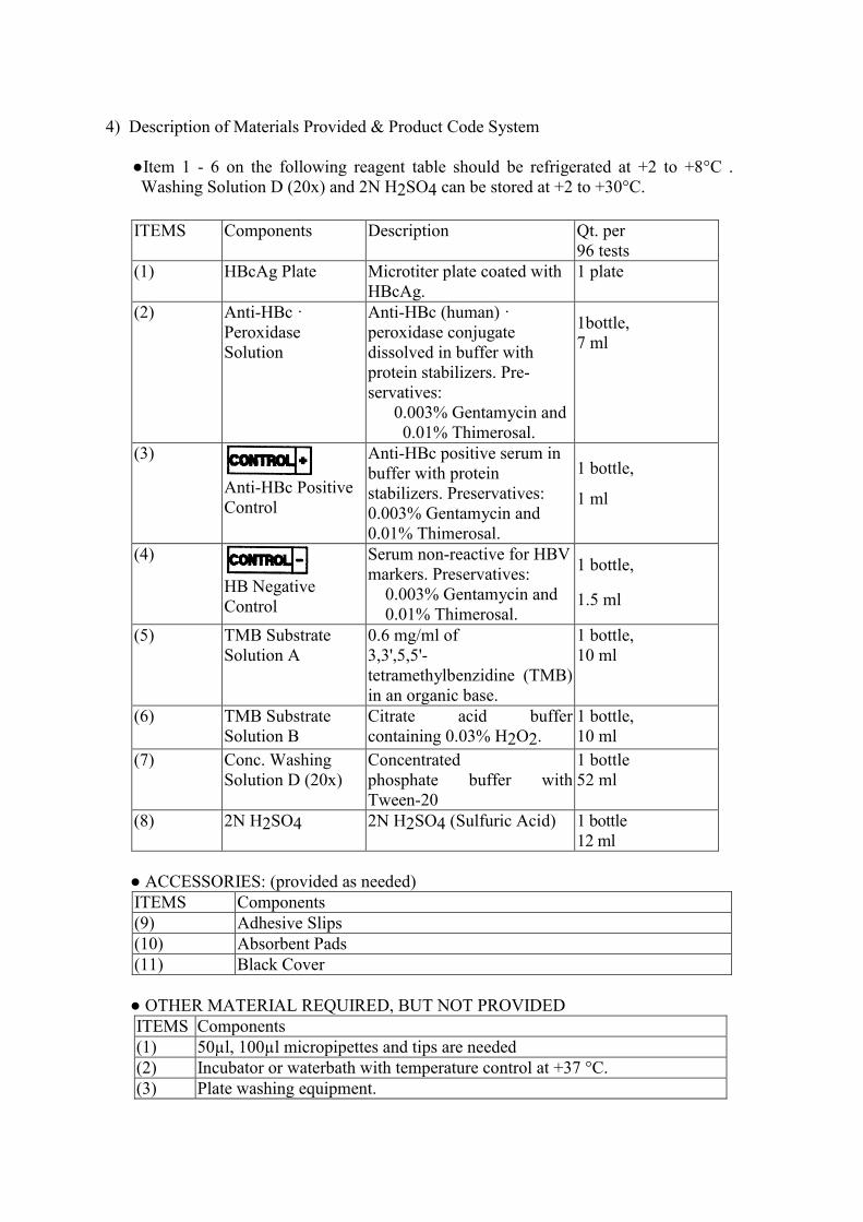
4) Description of Materials Provided & Product Code System
●Item 1 - 6 on the following reagent table should be refrigerated at +2 to +8°C .
Washing Solution D (20x) and 2N H2SO4 can be stored at +2 to +30°C.
ITEMS Components Description Qt. per
96 tests
(1) HBcAg Plate Microtiter plate coated with
HBcAg.
1 plate
(2) Anti-HBc ·
Peroxidase
Solution
Anti-HBc (human) ·
peroxidase conjugate
dissolved in buffer with
protein stabilizers. Pre-
servatives:
0.003% Gentamycin and
0.01% Thimerosal.
1bottle,
7 ml
(3)
Anti-HBc Positive
Control
Anti-HBc positive serum in
buffer with protein
stabilizers. Preservatives:
0.003% Gentamycin and
0.01% Thimerosal.
1 bottle,
1 ml
(4)
HB Negative
Control
Serum non-reactive for HBV
markers. Preservatives:
0.003% Gentamycin and
0.01% Thimerosal.
1 bottle,
1.5 ml
(5) TMB Substrate
Solution A
0.6 mg/ml of
3,3',5,5'-
tetramethylbenzidine (TMB)
in an organic base.
1 bottle,
10 ml
(6) TMB Substrate
Solution B
Citrate acid buffer
containing 0.03% H2O2.
1 bottle,
10 ml
(7) Conc. Washing
Solution D (20x)
Concentrated
phosphate buffer with
Tween-20
1 bottle
52 ml
(8) 2N H2SO4 2N H2SO4 (Sulfuric Acid) 1 bottle
12 ml
● ACCESSORIES: (provided as needed)
ITEMS Components
(9) Adhesive Slips
(10) Absorbent Pads
(11) Black Cover
● OTHER MATERIAL REQUIRED, BUT NOT PROVIDED
ITEMS Components
(1) 50µl, 100µl micropipettes and tips are needed
(2) Incubator or waterbath with temperature control at +37 °C.
(3) Plate washing equipment.

(4) ELISA microwell reader:
Dual wavelength 450nm with 620-690nm as reference wavelength*8
,
bandwidth 10nm.
(5) Fully automatic EIA micro-plate analyzer is optional. User should validate
the automatic EIA micro-plate analyzer in combination with the kit.
3

4.1) Storage Conditions and Stability of Kit and Components*
Kit/Components Storage
condition
State Stability
Anti-HBc IgG ELISA Test
Plate +2~+8 °C Original 18 months
Once open 1 month
Anti-HBc Positive Control +2~+8 °C Original 18 months
Once open 1 month
HB Negative Control +2~+8 °C Original 18 months
Once open 1 month
HBcAg Plate +2~+8 °C Original 24 months
Once open 2 month
Anti-HBc · Peroxidase
Conjugate Solution +2~+8 °C Original 18 months
Once open 1 month
Concentrated Washing Solution
D (20x)
Room temp. Original 24 months
Once open 1 month
20x Diluted Washing Solution Room temp. Diluted 2 days
+2~+8 °C Diluted 1 week
TMB Substrate Solution A +2~+8 °C Original 18 months
Once open 1 month
TMB Substrate Solution B +2~+8 °C Original 18 months
Once open 1 month
2N Sulfuric Acid Room temp. Original 24 months
Once open 1 month
5) Instructions for Use
5.1) Warnings:
5.1.1) This reagent kit is for professional use only.
5.1.2) This reagent kit is for in vitro diagnostic use only.
5.1.3) Bring all kit reagents and samples to room temperature (+20 to +30°C) and
mix carefully before use.
5.1.4) Do not use reagent beyond its expiration date.
5.1.5) Do not interchange reagents between different lots.
5.1.6) Do not pipette in the mouth.
5.1.7) Do not smoke or eat in areas where specimens or reagents are handled.
5.1.8) The positive control, negative control, conjugate solution and specimens
should be regarded as potential hazards to health. They should be used and
discarded according to the user’s laboratory safety procedures. Such safety
procedures probably will include the wearing of protective gloves and avoiding
the generation of aerosols.
5.1.9) Potential infectious specimens and nonacid containing spills or leakages should
be wiped up thoroughly with 5% sodium hypochlorite or treated in accordance
with the laboratory’s practice for potential bio-hazard control.
5.1.10)Prior to dispose the waste of used specimens and kit reagents as general waste,
it should be treated in accordance with the local procedures for potential bio-
hazardous waste or treated as follows:
Both liquid and solid waste should be autoclaved maintaining +121°C for at
least 30 minutes.
Solid waste can also be incinerated.
Non-acidic liquid waste can be treated with sodium hypochlorite diluted to a

final concentration of 1%.
Acidic liquid wastes must be neutralized before treatment with sodium
hypochlorite as mentioned above and should stand for 30 minutes to obtain
effective disinfection.
5.1.11) 2N sulfuric acid is an irritant to skin, eyes, respiratory tract and mucous
membranes. Avoid contact of the 2N sulfuric acid with skin and mucous
membranes. In case of contact, clean with large lots of water immediately.
In case of inhalation, supply fresh air
and seek medical advice in case of complaints.
5.1.12) TMB substrate solution A contains organic solvent, which is flammable.
TMB substrate solution A contains dimethyl sulfoxide, an irritant to skin and
mucous membranes.
5.1.13) Although all human sourced material are tested non-reactive for Anti-HCV
and Anti-HIV, and inactivated at +56 °C for one hour, the reagent shall be
handled as potential infectious material *7
.
5.2) Specimen Collection and Preparation for Analysis
5.2.1) No special preparation of the patient is required prior to blood collection.
Blood should be collected by approved medical techniques.
5.2.2) Either serum or plasma can be used with this diagnostic kit. Whole blood
specimen should be separated as soon as possible in order to avoid hemolysis.
Any particulates (e.g. fibrin clots, erythrocytes) contained in the specimen
should be removed prior to use.
5.2.3) Specimens must be stored at +2 to +8°C and avoided heat-inactivation to
minimize deterioration. For long-term storage, specimens should be frozen
below -20°C. Storage in self-defrosting freezers is not recommended.
5.2.4) Frozen specimens must be thoroughly thawed and mixed homogenously before
test.
5.2.5) Avoid multiple freeze-thaw procedures
5.2.6) WARNINGS
1. The specimen must not contain any AZIDE compounds which can inhibit the
peroxidase activity of the conjugate.
2. Incompletely coagulated serum samples and microbial-contaminated
specimens should not be used.
5.3) Reagents Storage
5.3.1) The kit must be stored at +2 to +8°C. Do not freeze.
5.3.2) Strips of the plate should be used within 2 month after opening the original
aluminum foil bag. The unused strips should be kept in the aluminum foil bag
and taped the opening tightly.
5.3.3) Return reagents to +2 to +8°C immediately after use.
5.3.4) Washing Solution D (20x) Concentrate is stored and shipped at +2 to +8°C,
which can cause crystallization. If the crystal has been precipitated before use,
warm up the solution in +37°C water bath till the crystal is dissolved.
5.4) Plate Washing Procedure
5.4.1) Preparation of washing solution:
Dilute Washing Solution D (20x) Concentrate with distilled or de-ionized water
to 1:20 dilution. Do not use tap water.
4

5.4.2) Plate washing:
(a) For plate washer with overflow aspirating function: 6 cycles with at least
0.5ml washing buffer per well per cycle.
or
(b) For plate washer without overflow aspirating function: 8 cycles with at least
0.35ml washing buffer per well per cycle.
5.4.3) Blot dry by inverting the plate and tapping firmly onto absorbent paper. Too
much residual wash buffer will cause false results.
WARNING
Improper washing will cause false results.
5.5) Test Procedure
5.5.1) Bring all reagents and specimens to room temperature (+20 to +30°C) before
assay. Adjust water bath or incubator to +37±1°C.
5.5.2) Reserve 2 wells for blanks. Add 50µl of each control or specimen to appro-
priate wells of reaction plate (3 Negative Controls and 2 Positive Controls).
NOTE:
a) Use a new pipette tip for each sampling to avoid cross-
contamination
b) Each plate needs its own negative controls, positive controls and
blank wells.
c) Do not use cut-off value established for other plates of Anti-HBc
IgG ELISA Test Kit.
5.5.3) Add 50 µl of Anti-HBc· Peroxidase solution to each well except the 2 blanks.
NOTE: Do not touch the well wall for preventing contamination.
5.5.4) Gently tap the plate.
5.5.5) Remove the protective backing from the adhesive slip and press it onto the
reaction plate, so that it is tightly sealed.
5.5.6) Incubate the reaction plate in a +37±1°C water bath or incubator for 1 hour.
5.5.7) At the end of the incubation period, remove and discard the adhesive slip and
wash the plate in accordance with 5.4) Plate washing procedure.
5.5.8) Select one of the following two methods for color development:
A. Mix equal volumes of TMB Substrate Solution A and B in a clean container
immediately prior to use. Add 100 µl of the mixture solution to each well
including 2 blank wells.
B. Add 50 µl of TMB Substrate Solution A first, then add 50 µl of TMB
Substrate Solution B into each well including the 2 blanks. Mix well gently.
NOTE: TMB Substrate Solution A should be colorless to light blue, otherwise, it
should be discarded. The mixture of TMB Substrate Solution A and B should be
used within 30 minutes after mix. The mixture should be protected from
exposition to intense light.
5.5.9) Cover the plate with black cover and incubate at room temperature for 15
minutes.
5.5.10) Stop the reaction by adding 100µl of 2N H2SO4 to each well including the
two blanks.
5.5.11)Determine the absorbance of controls and test specimens within 30 minutes
with a precision photometer at 450 / 620-690 nm (450 nm reading wavelength
with 620-690 nm reference wavelength)*1
. Use the first blank well to blank the
photometer.
5

NOTE:
The blanks should be colorless to light yellowish in color; otherwise, the test
results are invalid.
Substrate blank : absorbance value must be less than 0.100.
5.6) Calculation of Test Results
5.6.1) Calculation of the NCx (Mean Absorbance of Negative Control).
Example:
Sample No. Absorbance
1 0.939
2 0.944
3 0.925
NCx = (0.939 + 0.944 + 0.925) / 3 = 0.936
NCx must be≥ 0.4 , otherwise, the test run is invalid.
5.6.2) Calculation of the PCx (Mean Absorbance of Positive Control)
Example:
Sample No. Absorbance
1 0.068
2 0.052
PCx = (0.068 + 0.052) /2 = 0.060
PC x must be ≤ 0.1 , otherwise, the test run is invalid.
5.6.3) Calculation of the N - P Value
N - P = NC x – PC x
Example:
N – P = 0.936 – 0.060 = 0.876
N - P Value must be ≥ 0.3, otherwise, the test run is invalid.
5.6.4) Calculation of the Cutoff Value
Cutoff Value = 0.4 NCx + 0.6 PCx
Example:
Cutoff Value = (0.4 x 0.936) + (0.6 x 0.060) = 0.410
5.6.5) Calculation of the Retest Range
Retest Range = Cutoff Value ± 10%
Example: Cutoff Value = 0.410
Retest Range = (0.410 - 0.041) to (0.410 + 0.041) = 0.369 to 0.451
5.7) Validity of Test Runs
5.7.1) NC x must be ≥ 0.4 , otherwise, the test run is invalid.
5.7.2) PC x must be ≤ 0.1, otherwise, the test run is invalid.
5.7.3) N-P Value must be ≥ 0.3, otherwise, the test is invalid.
5.8) Interpretation of Results
If the signal/cut-off ratio is within Retest Range (0.9-1.1 x cutoff), the test must be
repeated in duplicate and interpreted as above. If both results are non-reactive the
final result is non-reactive, if both results are reactive the final result is reactive. Any
6

other combination is an indeterminate result. Testing of follow up samples and other
hepatitis B serological markers should be taken into account in case of an
indeterminate result.
5.9) Troubleshooting
If the result cannot be reproduced, a preliminary troubleshooting should be
performed by checking the possibilities listed below:
5.9.1) Improper washing procedure.
5.9.2) Contaminated with positive specimen.
5.9.3) Wrong volume of sample, conjugate or substrate.
5.9.4) Contamination of well rim with conjugate.
5.9.5) Improper specimen such as hemolyzed serum or plasma, specimen containing
precipitate and
specimen not thoroughly mixed before use.
5.9.6) Wrong incubation time or temperature.
5.9.7) Obstructed or partial obstructed washer aspirate/dispense head and needles.
5.9.8) Insufficient aspiration.
5.10) Limitations and Interferences
5.10.1) This reagent kit is to be used for un-pooled human serum or plasma samples
only.
5.10.2) The reagent kit has not been validated for use with cadaveric samples.
5.10.3) Non-repeatable false positive results may be obtained with any enzyme
immunoassay kit, largely due to technical error either on the part of the
operator or malfunction of apparatus used.
5.10.4) Potential interfering substances:
Potential interfering samples, i.e. samples with hyperlipemia, hemolysis,
hyper-bilirubinemia, and samples with monoclonal immunoglobulin
components, samples containing elevated levels of autoimmune antibodies
(rheumatoid factor-RF, antinuclear antibodies-ANA, or anti-mitochondrial
antibodies-ANA) did not affect the test result with Anti-HBc IgG ELISA Test.
5.10.5) The anticoagulants heparin, EDTA and sodium citrate have no influence on
the specificity of Anti-HBc IgG ELISA and can be used to obtain plasma
samples for analysis with the Anti-HBc Total kit.
5.11) Performance Characteristics
5.11.1) Diagnostic Specificity
Negative specimens/Specimens used to evaluate the specificity
True Negative Samples Anti-HBc
IgG ELISA
Type of sample Number
of samples
No. negative
samples
Blood donor samples 5020 5010
Samples from
hospitalized persons 200 200
Samples contain
potential interfering factors 97 97
Samples with added 12 11
7

5.11.1.1) Potential
interfering substances
Potential interferences
with Anti-HBc IgG
ELISA Test were investigated.
For each potential interfering substance, at least two serum samples containing
different amounts of the potentially interfering substance were mixed in fixed
ratios of 10 + 0; 7 + 3; 5 + 5; 3 + 7; 0 + 10 with other serum samples containing
increased Anti-HBc Total levels but no interfering factors. The neat samples as
well as the mixtures were analyzed.
In particular the specificity study included:
lipemic (turbid) samples (hyperlipidemia) before and after high speed
centrifugation
hemolytic samples or hemolysate
icteric samples (=hyperbilirubinemia)
samples with monoclonal immunoglobulin components
(hyperimmunoglobulinemia)
samples containing elevated levels of autoimmune antibodies (rheumatoid factor
- RF, antinuclear antibodies –ANA, or antimitochondrial antibodies-AMA).
No interferences were detected with both used lots. Neither the type of
anticoagulant had an influence on both tested lots of Anti-HBc IgG ELISA Test.
5.11.2) Analytical Sensitivity and Linearity:
To evaluate the sensitivity of Anti-HBc IgG ELISA serial dilutions of the Standard
Material for Anti-HBc Total of Paul Ehrlich Institute (PEI) (Langen, Germany)
(100 PEI U/ml) were used.
Semilog PEI Standard Dilution chart
0.0
1.0
2.0
3.0
4.0
-5.0 -4.0 -3.0 -2.0 -1.0 0.0 1.0 2.0
Log(Anti-HBc Total Conc. PEI U/ml)
S/CO
Lot# B34330PT Lot# B34331PT
possible interfering factors
Samples with different
anticoagulants 48 48
Total 5377 5366
Diagnostic Specificity ------------- 5366/5377 =
99.8%
8
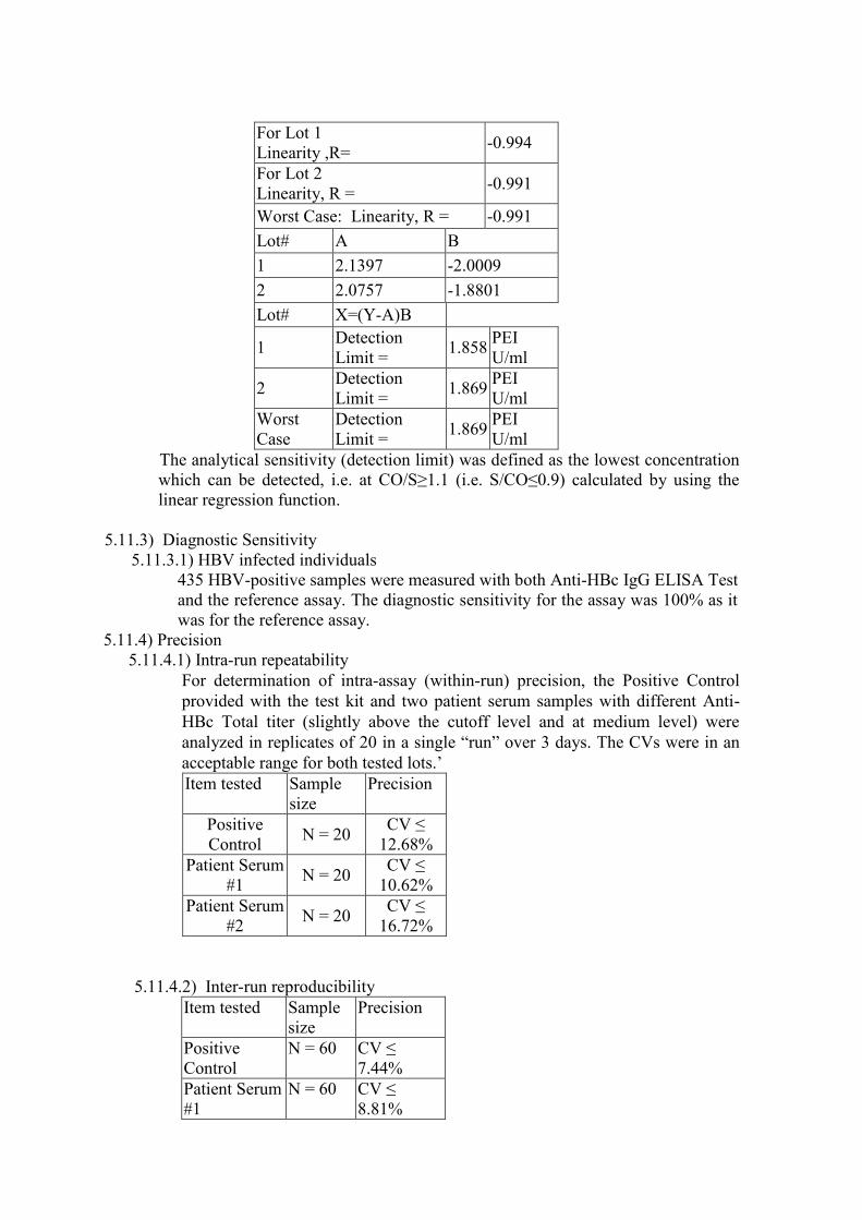
For Lot 1
Linearity ,R= -0.994
For Lot 2
Linearity, R = -0.991
Worst Case: Linearity, R = -0.991
Lot# A B
1 2.1397 -2.0009
2 2.0757 -1.8801
Lot# X=(Y-A)B
1 Detection
Limit = 1.858
PEI
U/ml
2 Detection
Limit = 1.869
PEI
U/ml
Worst
Case
Detection
Limit = 1.869
PEI
U/ml
The analytical sensitivity (detection limit) was defined as the lowest concentration
which can be detected, i.e. at CO/S≥1.1 (i.e. S/CO≤0.9) calculated by using the
linear regression function.
5.11.3) Diagnostic Sensitivity
5.11.3.1) HBV infected individuals
435 HBV-positive samples were measured with both Anti-HBc IgG ELISA Test
and the reference assay. The diagnostic sensitivity for the assay was 100% as it
was for the reference assay.
5.11.4) Precision
5.11.4.1) Intra-run repeatability
For determination of intra-assay (within-run) precision, the Positive Control
provided with the test kit and two patient serum samples with different Anti-
HBc Total titer (slightly above the cutoff level and at medium level) were
analyzed in replicates of 20 in a single “run” over 3 days. The CVs were in an
acceptable range for both tested lots.’
Item tested Sample
size
Precision
Positive
Control N = 20
CV ≤
12.68%
Patient Serum
#1 N = 20
CV ≤
10.62%
Patient Serum
#2 N = 20
CV ≤
16.72%
5.11.4.2) Inter-run reproducibility
Item tested Sample
size
Precision
Positive
Control
N = 60 CV ≤
7.44%
Patient Serum
#1
N = 60 CV ≤
8.81%

Patient Serum
#2
N = 60 CV
≤14.67%
5.11.5) Traceability
Concentration of Positive Control of Anti-HBc IgG ELISA Test referred to the
PEI Anti-HBc Total Reference Material = 70 PEI U/ml ± 30%
5.11.6) Antibody Excess/High-Dose Hook Effect
The effect of antibody excess was tested by consecutive dilution of a standard
material having very high Anti-HBc levels (PEI Anti-HBc Total Reference
Material).
Semilog Dilution chart
0.0
1.0
2.0
3.0
4.0
-5.0 -4.0 -3.0 -2.0 -1.0 0.0 1.0 2.0
Log(Anti-HBc Total Conc. PEI U/ml)
S/CO
Lot# B34330PT Lot# B34331PT
The Semilog PEI Standard Dilution chart
illustrates that an antigen/antibody excess is
not occurring also because of the reverse
reaction used in this assay format.
An antigen/antibody excess will not influence
the reactive/non-reactive interpretation.
5.12) Flow Chart of Test Procedure
Add 50 µl controls (3 x NC, 2 x PC)
and
add 50 µl of each specimen into wells.
Reserve 2 wells for blanks.
↓
Add 50 µl of Anti-HBc·Peroxidase
Solution into each reaction well, except
2 blanks.
↓
Incubate the plate at +37±1°C for 1
hour.
↓
Wash the plate.
(Choice one of the following two methods for color development)
/ \
Mix equal volumes
of TMB Substrate
Solution A and B.
Add 100 µl of the
mixed solution to
wells.
Add 50 µl of
TMB Substrate
Solution A to
wells and then add
50 µl of TMB
Substrate Solution
B. Mix well,
gently.
9
Semilog Dilution Chart

\ /
Incubate at R.T. for 30 minutes.
↓
Add 100 µl of 2N H2SO4 into each
well.
↓
Determine absorbance using 450 nm as
reading wavelength with 620-690nm
reference wavelength*8
6) Bibliography
1. Aach RD, Grisham JW, Paker CW. Detection of Australia antigen by
radioimmunoassay. Proc Natl Acad
Sci. USA 1971; 68:1056-1060.
2. Kim CY, Tikes JG. Purification and biophysical characterization of hepatitis antigen. J
Clin Invest. 1973;
52:1176-1186.
3. Hoofnagle JH, Gerety RJ, Barker LF, Antibody to hepatitis B virus core in man.
Lancet. 1973: 2(7834):
869-873.
4. Barker LF, Almeida JD, Hoofnagle JH, et al. Hepatitis B core antigen: immunology
and electron
microscopy. J Virol. 1974 Dec;14:1552-1558.
5. Hoofnagle. JH. Gerety, RJ.. Ni, LY.. Barker, LF. Antibody 10 Hepatitis B core antigen:
A sensitive
lndicator of hepatitis B virus replication. New Engl J Med. 1974; 290:1336-1340.
6. Niermeijer, P., Gips, C. H., Huizenga, J. R. et al. IgM Anti-HBc as a marker of
persistent and IgG anti-
HBc as a marker of past hepatitis B infection. A longitudinal study over 5 years. Acta
Hepato-Gastroenterol 1978; 25: 360–364.
7. Shikata T, Karasawa T, Abe K, et al. Incomplete inactivation of hepatitis B virus after
heat treatment at
+60°C for 10 hours, J. Infect. Dis. 1978; 138:242-244.
8. The reference wavelength of spectrometer can be 620nm to 690nm. However, user
should validate the
photometer in combination with this kit before use.
10

Symbols Key / Symbolschlussel / Explication des Symboles / Interpretazione
simboli /Clave dos Simbolos
In Vitro Diagnostic Medical Device / In-Vitro-Diagnostikum / Producto
sanitario para diagnóstico in vitro / Dispositivo medico-diagnostico in vitro /
Dispositif médical de diagnostic in vitro / Dispositivo médico para diagnóstico
in vitro
Batch code / Chargenbezeichnung / Código de lote / Codice del lotto / Code du
lot / Código do lote
Use By / Verwendbar bis / Fecha de caducidad / Utilizzare entro / Utiliser
jusque / Prazo de validade
Temperature limitation / Temperaturbegrenzung / Límite de temperatura /
Limiti di temperatura /Limites de température / Limites de temperatura
CE Mark / CE-Zeichen / Marquage CE / Marchio CE / CE Marca / Marca CE
Catalogue number / Bestellnummer / Número de catálogo / Numero di catalogo
/ Référence du catalogue / Referência de catálogo
Consult Instructions for Use- / Gebrauchsanweisung beachten / Consulte las
instrucciones de uso / Consultare le istruzioni per l'uso / Consulter les
instructions d'utilisation / Consulte as instruções de utilização
Caution,consult accompanying documents / Achtung, Begleitdokumente
beachten / Atención,ver instrucciones de uso / Attenzione, vedere le istruzioni
per l'uso / Attention voir notice d'instructions / Atenção, consulte a
documentação incluída