2. introduction to mechanics prokaryotic cells
TRANSCRIPT
1me239 mechanics of the cell
2. introduction to mechanics
the inner life of a cell, viel & lue, harvard [2006]
21.2 introduction to the cell
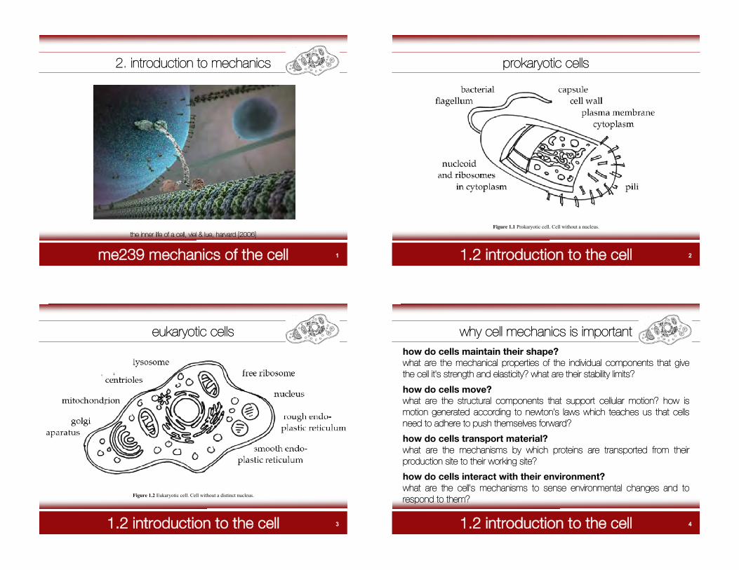
prokaryotic cells
Figure 1.1 Prokaryotic cell. Cell without a nucleus.
31.2 introduction to the cell
eukaryotic cells
Figure 1.2 Eukaryotic cell. Cell without a distinct nucleus.
41.2 introduction to the cell
why cell mechanics is importanthow do cells maintain their shape?what are the mechanical properties of the individual components that givethe cell it's strength and elasticity? what are their stability limits?
how do cells move?what are the structural components that support cellular motion? how ismotion generated according to newton's laws which teaches us that cellsneed to adhere to push themselves forward?
how do cells transport material?what are the mechanisms by which proteins are transported from theirproduction site to their working site?
how do cells interact with their environment?what are the cell's mechanisms to sense environmental changes and torespond to them?

51.3 introduction to biopolymers
biopolymers
Figure 3.1. Biopolymers. Characteristic length scales on the cellular and sucellular level..
61.3 introduction to biopolymers
biopolymers
typical examples of biopolymers• genes: RNA and DNA• gene products: peptides and proteins• biopolymers not coded by genes: lipids, polysaccharides, and carbohydrates
biopolymers are made up of monomers and polymers.monomers are smaller micromolecules such as nucleic acids, aminoacids, fatty acid, and sugar. assembled together as repeatingsubunits, monomers form long macromolecules which are referred toas polymers.
biopolymers are extremely flexible. upon thermal fluctua-tions, they may bend from side to side and jiggle around. this is thenature of soft matter related to the notion of entropy.
71.4 introduction to the cytoskeleton
the cytoskeleton
Figure 1.3. Eukaryotic cytoskeleton. The cytoskeleton provides structural stability and is responsible for force transmissionduring cell locomotion. Microtubules are thick hollow cylinders reaching out from the nucleus to the membrane, intermediatefilaments can be found anywhere in the cytosol, and actin filaments are usually concentrated close to the cell membrane.
81.4 introduction to the cytoskeleton
the cytoskeleton
actin filaments are 7nm in diameter and consist of two intertwined actin chains.they are tension bearing members of the cell. being located close to the cellmembrane, they are responsible for inter- and intracellular transduction. togetherwith myosin, they from the contraction apparatus to generate muscular contractionof skeletal and cardiac muscle.
intermediate filaments are 8-12nm in diameter and thus more stable than actinfilaments. they are also tension bearing within a cell. anchoring at organelles, theyorganize and maintain the three dimensional structure of the cell.
microtubules are hollow cylinders, 25nm in diameter with a 15nm lumen. they arecomprised of 13 protofilaments consisting of ! and " tubulin. microtubules areorganized by the centrosome, but reassemble dynamically. unlike actin andintermediate filaments, microtubules can also bear compression. in addition, theyform a highway for intracellular transport.
91.5 introduction to biomembranes
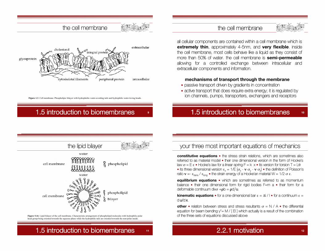
the cell membrane
Figure 1.3. Cell membrane. Phospholipic bilayer with hydrophobic water-avoiding tails and hydrophilic water-loving heads.
101.5 introduction to biomembranes
the cell membrane
mechanisms of transport through the membrane• passive transport driven by gradients in concentration• active transport that does require extra energy; it is regulated by ion channels, pumps, transporters, exchangers and receptors
all cellular components are contained within a cell membrane which isextremely thin, approximately 4-5nm, and very flexible. insidethe cell membrane, most cells behave like a liquid as they consist ofmore than 50% of water. the cell membrane is semi-permeableallowing for a controlled exchange between intracellular andextracellular components and information.
111.5 introduction to biomembranes
the lipid bilayer
Figure 5.16. Lipid bilayer of the cell membrane. Characteristic arrangement of phospholipid molecules with hydrophilic polarhead group being oriented towards the aqueous phase while the hydrophilic tails are oriented towards the non-polar inside.
122.2.1 motivation
your three most important equations of mechanics
constitutive equations • the stress strain relations, which are sometimes alsoreferred to as material model • their one dimensional version in the form of Hooke'slaw # = E $ • Hooke's law for a linear spring F = k x • its version for torsion T = L%• its three dimensional version $x = 1/E [#x - & #y - & #z] • the definition of Poisson'sratio & =- $trans / $long • the strain energy of a Hooke'an material W = 1/2 # $.
equilibrium equations • which are sometimes as referred to as momentumbalance • their one dimensional form for rigid bodies F=m a • their form for adeformable continuum div# +'b = 'dt
2u.
kinematic equations • for a one-dimensional bar $ = (l / l • for a continuum $ =d u/d x.
other • relation between stress and stress resultants # = N / A • the differentialequation for beam bending y''= M / [ EI ] which actually is a result of the combinationof the three sets of equations discussed above

132.2.1 motivation
one-dimensional bar
142.2.1 motivation
one-dimensional bar
152.2.1 motivation
one-dimensional bar
162.2.1 motivation
one-dimensional bar

172.2.1 motivation
one-dimensional bar
182.2.1 motivation
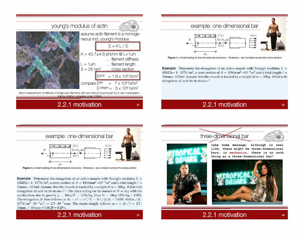
young’s modulus of actin
direct measurement of stiffness of single actin filaments with and without tropomyosin by in vitro manipulation.kojima, ishijima, yanagida, pnas [1994]
192.2.1 motivation
young’s modulus of actin
direct measurement of stiffness of single actin filaments with and without tropomyosin by in vitro manipulation.kojima, ishijima, yanagida, pnas [1994]
Fc = x KcFN = [ x0- x ] KN
x
x0
F
and
Kc = [ x0- x -1 ] KN
from FN = Fc = Fneedle force composite force
composite stiffness
202.2.1 motivation
young’s modulus of actin
figure. relationship between reciprocal values of composite stiffness (1/Kc) and actin filament length. • actinfilaments only. º actin filaments with tropomyosin. kojima, ishijima, yanagida, pnas [1994]
212.2.1 motivation
young’s modulus of actinassume actin filament is a homoge-neous rod, young’s modulus
E = K L / S
K = 43.7±4.6 pN/nm @ L=1um… filament stiffness
L = 1um … filament lengthS = 25 nm2 … cross section
Eactin = 1.8 x 10⁹ N/m2
compare Esilk = 7 x 10⁹ N/m2
Ecollagen = 5 x 10⁹ N/m2
direct measurement of stiffness of single actin filaments with and without tropomyosin by in vitro manipulation.kojima, ishijima, yanagida, pnas [1994]
222.2.1 motivation
example: one-dimensional bar
232.2.1 motivation
example: one-dimensional bar
242.2.1 motivation
three-dimensional bartake home message: although in reallife, there might be three-dimensionalbars, in mechanics, there is no suchthing as a three-dimensional bar!

252.2.2 kinematics
three-dimensional strain displacement relationnormal strains
shear strains
262.2.3 constitutive equations
three-dimensional stress strain relation
generalized (3d) hooke’s law
inverse hooke’s law
272.2.3 constitutive equations
who was robert hooke?robert hooke. 18 july 1635 - 3 march 1703.english natural philosopher and architect whoplayed an important role in the scientificrevolution, through both his experimental andtheoretical contributions. in 1660, hookediscovered the law of elasticity which bearshis name and which describes the linear
wikipedia.
variation of tension with extension in anelastic spring. in 1665, hooke publishedmicrographia, a book describing hismicroscopic and telescopic observations, andsome original work in biology. hook coinedthe term “cell” for describing biologicalorganisms, the term being suggested by theresemblance of plant cells to monk’s cells.
282.2.4 equilibrium
three-dimensional stress force relation
292.2.5 structural elements
trusses, beams, walls, plates, membranes, shells
/ bar
302.2.5 structural elements
typical example - the cell membrane
31 32