(19) tzz t - patentimages.storage.googleapis.com · be used to tesfto rconge nitalhypot hyroidism(...
TRANSCRIPT

Note: Within nine months of the publication of the mention of the grant of the European patent in the European PatentBulletin, any person may give notice to the European Patent Office of opposition to that patent, in accordance with theImplementing Regulations. Notice of opposition shall not be deemed to have been filed until the opposition fee has beenpaid. (Art. 99(1) European Patent Convention).
Printed by Jouve, 75001 PARIS (FR)
(19)E
P2
516
669
B1
(Cont. next page)
TEPZZ 5_6669B_T(11) EP 2 516 669 B1
(12) EUROPEAN PATENT SPECIFICATION
(45) Date of publication and mention of the grant of the patent: 12.10.2016 Bulletin 2016/41
(21) Application number: 10842658.6
(22) Date of filing: 17.12.2010
(51) Int Cl.:B01L 3/00 (2006.01) C07D 311/18 (2006.01)
C07D 311/20 (2006.01) C07H 15/203 (2006.01)
C12Q 1/25 (2006.01) C12Q 1/34 (2006.01)
G01N 33/543 (2006.01) C07D 311/16 (2006.01)
C07D 405/12 (2006.01) C07H 17/075 (2006.01)
G01N 21/64 (2006.01) C07D 413/12 (2006.01)
(86) International application number: PCT/US2010/061118
(87) International publication number: WO 2011/084703 (14.07.2011 Gazette 2011/28)
(54) ENZYME ASSAYS ON A DROPLET ACTUATOR
ENZYMASSAYS AUF EINEM TROPFENAKTUATOR
ANALYSES D’ENZYMES SUR UN DIFFUSEUR À GOUTTELETTES
(84) Designated Contracting States: AL AT BE BG CH CY CZ DE DK EE ES FI FR GB GR HR HU IE IS IT LI LT LU LV MC MK MT NL NO PL PT RO RS SE SI SK SM TR
(30) Priority: 25.10.2010 US 406380 P13.10.2010 US 392633 P14.09.2010 US 382564 P31.08.2010 US 378705 P30.06.2010 US 359943 P13.05.2010 US 334376 P19.04.2010 US 325580 P28.12.2009 US 290296 P21.12.2009 US 288633 P
(43) Date of publication of application: 31.10.2012 Bulletin 2012/44
(73) Proprietor: Advanced Liquid Logic, Inc.San Diego, CA 92122 (US)
(72) Inventors: • ECKHARDT, Allen, E.
DurhamNorth Carolina 27705 (US)
• GRAHAM, CarrieRaleighNorth Carolina 27614 (US)
• SISTA, RamakrishnaMorrisvilleNorth Carolina 27560 (US)
• PAMULA, Vamsee, K.DurhamNorth Carolina 27703 (US)
• WINGER, TheodoreMorrisvilleNorth Carolina 27560 (US)
• WANG, TongCaryNorth Carolina 27519 (US)
• XIONG, YalinMorrisvilleNorth Carolina 27560 (US)
• WU, NingRaleighNorth Carolina 27606 (US)
• SAHA, SanjayHyderabad-500076Andhra Pradesh (IN)
• RAOLJI, Gajendrasinh, BalvantsinhHyderabad-500076Andhra Pradesh (IN)
• DAYAM, RaveendraHyderabad-502032Andhra Pradesh (IN)
(74) Representative: Cooper, JohnMurgitroyd & Company Scotland House 165-169 Scotland StreetGlasgow G5 8PL (GB)

2
EP 2 516 669 B1
(56) References cited: WO-A1-91/11172 WO-A1-2008/116209WO-A1-2008/116209 WO-A1-2009/021173WO-A2-2007/120241 WO-A2-2008/098236JP-A- 5 025 159 US-A- 5 559 134US-A1- 2003 148 538 US-A1- 2005 158 845US-A1- 2006 054 503 US-A1- 2008 039 636US-B2- 7 109 222
• BRIAN D. WAGNER: "The Use of Coumarins as Environmentally-Sensitive Fluorescent Probes of Heterogeneous Inclusion Systems", MOLECULES, vol. 14, no. 1, 6 January 2009 (2009-01-06), pages 210-237, XP055061199, DOI: 10.3390/molecules14010210
• GYÖRGYI J KOLOSSVARY ET AL: "MOLECULAR DYNAMICS SIMULATION OF CYCLODEXTRIN INCLUSION COMPLEXES IN ENZYMATIC LIPID HYDROLYSIS GySrgyi", BIOTECHNOLOGY LETTERS, vol. 18, no. 4, 4 April 1996 (1996-04-04), pages 440-444, XP055061201,
• YA. V. VOZNYI ET AL.: ’A fluorimetric enzyme assay for the diagnosis of MPS II (Hunter disease)’ J. INHERIT. METAB. DIS. vol. 24, no. 6, November 2001, pages 675 - 680, XP002575046
EP 2 516 669 B1
3
5
10
15
20
25
30
35
40
45
50
55
Description
Field of the Invention
[0001] The invention generally relates to droplet actuator assay methods. In particular, the invention relates to dropletactuator devices and assay methods for multiplexed newborn testing for metabolic disorders.
Background
[0002] A droplet actuator may include one or more substrates configured to form a surface or gap for conductingdroplet operations. The one or more substrates establish a droplet operations surface or gap for conducting dropletoperations and may also include electrodes arrange to conduct the droplet operations. The droplet operations substrateor the gap between the substrates may be coated or filled with a filler fluid that is immiscible with the liquid that formsthe droplets.[0003] WO2008/116209 discloses a method of conducting a droplet-based enzymatic assay making use of a dropletactuator wherein the droplets are surrounded by an oil and a surfactant.[0004] Droplet actuators are used in a variety of applications, including molecular diagnostic assays, such as enzymaticassays and immunoassays. In one application, enzymatic assays and immunoassays are used as part of a routinetesting process to test newborn infants for various genetic disorders. For example, enzymatic assays may be used totest for various lysosomal storage diseases (LSD), galactosemia and biotinidase deficiency (BIOT). Immunoassays maybe used to test for congenital hypothyroidism (CH), congenital adrenal hyperplasia (CAH) and cystic fibrosis (CF). Currentenzymatic assay and immunoassay technologies used in newborn testing are based on 96-well microtiter plate compatiblesystems. Specimens are punched automatically from a neonatal dried blood spot (DBS) sample into several plates (i.e.,one punch for each test to be performed) and each plate is manipulated according to a specific assay protocol. Eachassay may require a separate laboratory section with a manager, one or more technologists, and equipment dedicatedto the assay. Overall, the system is labor intensive (although one or more steps are at least partially automated) andreagent and equipment costs can be high. Because the current system is labor intensive and costly, testing is generallyrestricted to centralized laboratories and often unavailable in developing countries. Therefore, there is a need for newapproaches to newborn testing.
Summary of the Invention
[0005] The present invention is in its broadest sense defined by the appended claims. Provided are droplet actuatorassay methods for multiplexed newborn testing for metabolic disorders. The methods include, among other things,droplet-based enzymatic assays for testing for metabolic disorders. The methods include conducting multiple assaysfor different metabolic disorders on a single droplet actuator, as well as multiple assays for the same metabolic disorderusing samples from different subjects and/or multiple samples from the same subject on a single droplet actuator. Invarious embodiments, the methods are included for conducting enzymatic activity assays in a single fresh blood and/orplasma samples and dried blood and/or plasma samples.[0006] Provided are assay methods for detection of one or more (i.e., multiplex detection) lysosomal storage diseases(LSDs) on a droplet actuator. Provided are assay methods for detection of MSP II (Hunter’s syndrome) on a dropletactuator. The methods include, among other things, droplet-based enzymatic assays for iduronate-2-sulfate sulphatase(IDS) enzyme activity. In certain embodiments, the Hunter’s assay may be performed at room temperature or at analternate temperature, such as 37 °C. In other embodiments the Hunter’s assay may be performed for 8 hours or less.The Hunter’s assay is a single-step homogenous assay that is performed at a single pH (i.e., pH 5.0) with a time to resultof 8 hours or less.[0007] Provided are methods for performing multiplexed enzymatic assays on a single droplet actuator using a singledried blood spot (DBS) sample. In one example, the integrated droplet actuator device and methods may be used formultiplexed detection of congenital adrenal hyperplasia (CAH), congenital hypothyroidism (CH), cystic fibrosis (CF),galactosemia and biotinidase deficiency (BIOT).[0008] Using digital microfluidics technology, sub-microliter-sized droplets may be manipulated using high-speed trans-port of droplets, reliable dispensing, rapid mixing, dilution, and disposal to provide rapid sample-to-result testing. Becausesubstantially all of the steps in a sample testing protocol are performed on-chip (automated), the risk of operator erroris substantially reduced. The flexibility, programmability and modular format of the microfluidic platform, additional assayprotocols (i.e., for other disorders) may be readily added to an on-chip testing panel.[0009] The droplet actuator methods of the invention complement tandem mass spectrometry (MS/MS) testing bymultiplexing testing of metabolic disorders that are not suited for MS/MS.[0010] Provided are bench-based methods for enzymatic detection of Hunter’s syndrome and Fabry disease.

EP 2 516 669 B1
4
5
10
15
20
25
30
35
40
45
50
55
[0011] Provided are a digital microfluidic platform and methods for multiplexed testing for hyperbilirubinemia, glucose-6-phosphate dehydrogenase (G6PD) deficiency, and congenital hypothyroidism (CH).
Definitions
[0012] As used herein, the following terms have the meanings indicated.[0013] "Activate," with reference to one or more electrodes, means affecting a change in the electrical state of the oneor more electrodes which, in the presence of a droplet, results in a droplet operation. Activation of an electrode can beaccomplished using alternating or direct current. Any suitable voltage may be used. For example, an electrode may beactivated using a voltage which is greater than about 150 V, or greater than about 200 V, or greater than about 250 V,or from about 275 V to about 375 V, or about 300 V. Where alternating current is used, any suitable frequency may beemployed. For example, an electrode may be activated using alternating current having a frequency from about 1 Hz toabout 100 Hz, or from about 10 Hz to about 60 Hz, or from about 20 Hz to about 40 Hz, or about 30 Hz.[0014] "Bead," with respect to beads on a droplet actuator, means any bead or particle that is capable of interactingwith a droplet on or in proximity with a droplet actuator. Beads may be any of a wide variety of shapes, such as spherical,generally spherical, egg shaped, disc shaped, cubical, amorphous and other three dimensional shapes. The bead may,for example, be capable of being subjected to a droplet operation in a droplet on a droplet actuator or otherwise configuredwith respect to a droplet actuator in a manner which permits a droplet on the droplet actuator to be brought into contactwith the bead on the droplet actuator and/or off the droplet actuator. Beads may be provided in a droplet, in a dropletoperations gap, or on a droplet operations surface. Beads may be provided in a reservoir that is external to a dropletoperations gap or situated apart from a droplet operations surface, and the reservoir may be associated with a fluid paththat permits a droplet including the beads to be brought into a droplet operations gap or into contact with a dropletoperations surface. Beads may be manufactured using a wide variety of materials, including for example, resins, andpolymers. The beads may be any suitable size, including for example, microbeads, microparticles, nanobeads andnanoparticles. In some cases, beads are magnetically responsive; in other cases beads are not significantly magneticallyresponsive. For magnetically responsive beads, the magnetically responsive material may constitute substantially all ofa bead, a portion of a bead, or only one component of a bead. The remainder of the bead may include, among otherthings, polymeric material, coatings, and moieties which permit attachment of an assay reagent. Examples of suitablebeads include flow cytometry microbeads, polystyrene microparticles and nanoparticles, functionalized polystyrene mi-croparticles and nanoparticles, coated polystyrene microparticles and nanoparticles, silica microbeads, fluorescent mi-crospheres and nanospheres, functionalized fluorescent microspheres and nanospheres, coated fluorescent micro-spheres and nanospheres, color dyed microparticles and nanoparticles, magnetic microparticles and nanoparticles,superparamagnetic microparticles and nanoparticles (e.g., DYNABEADS® particles, available from Invitrogen Group,Carlsbad, CA), fluorescent microparticles and nanoparticles, coated magnetic microparticles and nanoparticles, ferro-magnetic microparticles and nanoparticles, coated ferromagnetic microparticles and nanoparticles, and those describedin U.S. Patent Publication Nos. 20050260686, entitled "Multiplex flow assays preferably with magnetic particles as solidphase," published on November 24, 2005; 20030132538, entitled "Encapsulation of discrete quanta of fluorescentparticles," published on July 17, 2003; 20050118574, entitled "Multiplexed Analysis of Clinical Specimens Apparatusand Method," published on June 2, 2005; 20050277197. Entitled "Microparticles with Multiple Fluorescent Signals andMethods of Using Same," published on December 15, 2005; 20060159962, entitled "Magnetic Microspheres for use inFluorescence-based Applications," published on July 20, 2006; their teaching concerning beads and magnetically re-sponsive materials and beads. Beads may be pre-coupled with a biomolecule or other substance that is able to bind toand form a complex with a biomolecule. Beads may be pre-coupled with an antibody, protein or antigen, DNA/RNAprobe or any other molecule with an affinity for a desired target. Examples of droplet actuator techniques for immobilizingmagnetically responsive beads and/or non-magnetically responsive beads and/or conducting droplet operations proto-cols using beads are described in U.S. Patent Application No. 11/639,566, entitled "Droplet-Based Particle Sorting,"filed on December 15, 2006; U.S. Patent Application No. 61/039,183, entitled "Multiplexing Bead Detection in a SingleDroplet," filed on March 25, 2008; U.S. Patent Application No. 61/047,789, entitled "Droplet Actuator Devices and DropletOperations Using Beads," filed on April 25, 2008; U.S. Patent Application No. 61/086,183, entitled "Droplet ActuatorDevices and Methods for Manipulating Beads," filed on August 5, 2008; International Patent Application No.PCT/US2008/053545, entitled "Droplet Actuator Devices and Methods Employing Magnetic Beads," filed on February11, 2008; International Patent Application No. PCT/US2008/058018, entitled "Bead-based Multiplexed Analytical Meth-ods and Instrumentation," filed on March 24, 2008; International Patent Application No. PCT/US2008/058047, "BeadSorting on a Droplet Actuator," filed on March 23, 2008; and International Patent Application No. PCT/US2006/047486,entitled "Droplet-based Biochemistry," filed on December 11, 2006. Bead characteristics may be employed in the mul-tiplexing aspects of the invention. Examples of beads having characteristics suitable for multiplexing, as well as methodsof detecting and analyzing signals emitted from such beads, may be found in U.S. Patent Publication No. 20080305481,entitled "Systems and Methods for Multiplex Analysis of PCR in Real Time," published on December 11, 2008; U.S.

EP 2 516 669 B1
5
5
10
15
20
25
30
35
40
45
50
55
Patent Publication No. 20080151240, "Methods and Systems for Dynamic Range Expansion," published on June 26,2008; U.S. Patent Publication No. 20070207513, entitled "Methods, Products, and Kits for Identifying an Analyte in aSample," published on September 6, 2007; U.S. Patent Publication No. 20070064990, entitled "Methods and Systemsfor Image Data Processing," published on March 22, 2007; U.S. Patent Publication No. 20060159962, entitled "MagneticMicrospheres for use in Fluorescence-based Applications," published on July 20, 2006; U.S. Patent Publication No.20050277197, entitled "Microparticles with Multiple Fluorescent Signals and Methods of Using Same," published onDecember 15, 2005; and U.S. Patent Publication No. 20050118574, entitled "Multiplexed Analysis of Clinical SpecimensApparatus and Method," published on June 2, 2005.[0015] "Blood Sample" includes whole blood, whole blood constituents, such as serum or plasma, and dried bloodspot extracts. The dried blood can be on tissue paper or standard blood collection card or any other suitable substratethat does not eliminate the usefulness of the blood as a sample for the target of interest. The blood may be from thesubject, and the subject may be a human subject of any age, such as an adult, infant or a fetus. In the case of a fetus,the blood may be from the mother.[0016] "Droplet" means a volume of liquid on a droplet actuator. A droplet may be at least partially bounded by a fillerfluid. For example, a droplet may be completely surrounded by a filler fluid or may be bounded by filler fluid and one ormore surfaces of the droplet actuator. As another example, a droplet may be bounded by filler fluid, one or more surfacesof the droplet actuator, and/or the atmosphere. As yet another example, a droplet may be bounded by filler fluid and theatmosphere. Droplets may, for example, be aqueous or non-aqueous or may be mixtures or emulsions including aqueousand non-aqueous components. A 1X droplet may be about 300 nl. Droplets may take a wide variety of shapes; non-limiting examples include generally disc shaped, slug shaped, truncated sphere, ellipsoid, spherical, partially compressedsphere, hemispherical, ovoid, cylindrical, combinations of such shapes, and various shapes formed during droplet op-erations, such as merging or splitting or formed as a result of contact of such shapes with one or more surfaces of adroplet actuator. For examples of droplet fluids that may be subjected to droplet operations using the approach of theinvention, see International Patent Application No. PCT/US 06/47486, entitled, "Droplet-Based Biochemistry," filed onDecember 11, 2006. In various embodiments, a droplet may include a biological sample, such as whole blood, lymphaticfluid, serum, plasma, sweat, tear, saliva, sputum, cerebrospinal fluid, amniotic fluid, seminal fluid, vaginal excretion,serous fluid, synovial fluid, pericardial fluid, peritoneal fluid, pleural fluid, transudates, exudates, cystic fluid, bile, urine,gastric fluid, intestinal fluid, fecal samples, liquids containing single or multiple cells, liquids containing organelles, fluidizedtissues, fluidized organisms, liquids containing multi-celled organisms, biological swabs and biological washes. Moreover,a droplet may include a reagent, such as water, deionized water, saline solutions, acidic solutions, basic solutions,detergent solutions and/or buffers. Other examples of droplet contents include reagents, such as a reagent for a bio-chemical protocol, such as a nucleic acid amplification protocol, an affinity-based assay protocol, an enzymatic assayprotocol, a sequencing protocol, and/or a protocol for analyses of biological fluids. In various embodiments the dropletsmay include surfactants to improve droplet operations. It will be appreciated that where specific surfactants are mentioned,these may readily be supplemented or replaced with other similar surfactants, such as surfactants having similar HLBprofile.[0017] "Droplet Actuator" means a device for manipulating droplets. For examples of droplet actuators, see Pamulaet al., U.S. Patent 6,911,132, entitled "Apparatus for Manipulating Droplets by Electrowetting-Based Techniques," issuedon June 28, 2005; Pamula et al., U.S. Patent Application No. 11/343,284, entitled "Apparatuses and Methods for Ma-nipulating Droplets on a Printed Circuit Board," filed on filed on January 30, 2006; Pollack et al., International PatentApplication No. PCT/US2006/047486, entitled "Droplet-Based Biochemistry," filed on December 11, 2006; Shenderov,U.S. Patents 6,773,566, entitled "Electrostatic Actuators for Microfluidics and Methods for Using Same," issued on August10, 2004 and 6,565,727, entitled "Actuators for Microfluidics Without Moving Parts," issued on January 24, 2000; Kimand/or Shah et al., U.S. Patent Application Nos. 10/343,261, entitled "Electrowetting-driven Micropumping," filed onJanuary 27, 2003, 11/275,668, entitled "Method and Apparatus for Promoting the Complete Transfer of Liquid Dropsfrom a Nozzle," filed on January 23, 2006, 11/460,188, entitled "Small Object Moving on Printed Circuit Board," filed onJanuary 23, 2006, 12/465,935, entitled "Method for Using Magnetic Particles in Droplet Microfluidics," filed on May 14,2009, and 12/513,157, entitled "Method and Apparatus for Real-time Feedback Control of Electrical Manipulation ofDroplets on Chip," filed on April 30, 2009; Velev, U.S. Patent 7,547,380, entitled "Droplet Transportation Devices andMethods Having a Fluid Surface," issued on June 16, 2009; Sterling et al., U.S. Patent 7,163,612, entitled "Method,Apparatus and Article for Microfluidic Control via Electrowetting, for Chemical, Biochemical and Biological Assays andthe Like," issued on January 16, 2007; Becker and Gascoyne et al., U.S. Patent Nos. 7,641,779, entitled "Method andApparatus for Programmable fluidic Processing," issued on January 5, 2010, and 6,977,033, entitled "Method andApparatus for Programmable fluidic Processing," issued on December 20, 2005; Decre et al., U.S. Patent 7,328,979,entitled "System for Manipulation of a Body of Fluid," issued on February 12, 2008; Yamakawa et al., U.S. Patent Pub.No. 20060039823, entitled "Chemical Analysis Apparatus," published on February 23, 2006; Wu, International PatentPub. No. WO/2009/003184, entitled "Digital Microfluidics Based Apparatus for Heat-exchanging Chemical Processes,"published on December 31, 2008; Fouillet et al., U.S. Patent Pub. No. 20090192044, entitled "Electrode Addressing

EP 2 516 669 B1
6
5
10
15
20
25
30
35
40
45
50
55
Method," published on July 30, 2009; Fouillet et al., U.S. Patent 7,052,244, entitled "Device for Displacement of SmallLiquid Volumes Along a Micro-catenary Line by Electrostatic Forces," issued on May 30, 2006; Marchand et al., U.S.Patent Pub. No. 20080124252, entitled "Droplet Microreactor," published on May 29, 2008; Adachi et al., U.S. PatentPub. No. 20090321262, entitled "Liquid Transfer Device," published on December 31, 2009; Roux et al., U.S. PatentPub. No. 20050179746, entitled "Device for Controlling the Displacement of a Drop Between two or Several SolidSubstrates," published on August 18, 2005; Dhindsa et al., "Virtual Electrowetting Channels: Electronic Liquid Transportwith Continuous Channel Functionality," Lab Chip, 10:832-836 (2010). Certain droplet actuators will include one or moresubstrates arranged with a gap therebetween and electrodes associated with (e.g., layered on, attached to, and/orembedded in) the one or more substrates and arranged to conduct one or more droplet operations. For example, certaindroplet actuators will include a base (or bottom) substrate, droplet operations electrodes associated with the substrate,one or more dielectric layers atop the substrate and/or electrodes, and optionally one or more hydrophobic layers atopthe substrate, dielectric layers and/or the electrodes forming a droplet operations surface. A top substrate may also beprovided, which is separated from the droplet operations surface by a gap, commonly referred to as a droplet operationsgap. Various electrode arrangements on the top and/or bottom substrates are discussed in the above-referenced patentsand applications and certain novel electrode arrangements are discussed in the description of the invention. Duringdroplet operations it is preferred that droplets remain in continuous contact or frequent contact with a ground or referenceelectrode. A ground or reference electrode may be associated with the top substrate facing the gap, the bottom substratefacing the gap, in the gap. Where electrodes are provided on both substrates, electrical contacts for coupling the electrodesto a droplet actuator instrument for controlling or monitoring the electrodes may be associated with one or both plates.In some cases, electrodes on one substrate are electrically coupled to the other substrate so that only one substrate isin contact with the droplet actuator. In one embodiment, a conductive material (e.g., an epoxy, such as MASTER BOND™Polymer System EP79, available from Master Bond, Inc., Hackensack, NJ) provides the electrical connection betweenelectrodes on one substrate and electrical paths on the other substrates, e.g., a ground electrode on a top substratemay be coupled to an electrical path on a bottom substrate by such a conductive material. Where multiple substratesare used, a spacer may be provided between the substrates to determine the height of the gap therebetween and definedispensing reservoirs. The spacer height may, for example, be from about 5 mm to about 600 mm, or about 100 mm toabout 400 mm, or about 200 mm to about 350 mm, or about 250 mm to about 300 mm, or about 275 mm. The spacermay, for example, be formed of a layer of projections form the top or bottom substrates, and/or a material insertedbetween the top and bottom substrates. One or more openings may be provided in the one or more substrates for forminga fluid path through which liquid may be delivered into the droplet operations gap. The one or more openings may insome cases be aligned for interaction with one or more electrodes, e.g., aligned such that liquid flowed through theopening will come into sufficient proximity with one or more droplet operations electrodes to permit a droplet operationto be effected by the droplet operations electrodes using the liquid. The base (or bottom) and top substrates may insome cases be formed as one integral component. One or more reference electrodes may be provided on the base (orbottom) and/or top substrates and/or in the gap. Examples of reference electrode arrangements are provided in theabove referenced patents and patent applications. In various embodiments, the manipulation of droplets by a dropletactuator may be electrode mediated, e.g., electrowetting mediated or dielectrophoresis mediated or Coulombic forcemediated. Examples of other techniques for controlling droplet operations that may be used in the droplet actuators ofthe invention include using devices that induce hydrodynamic fluidic pressure, such as those that operate on the basisof mechanical principles (e.g. external syringe pumps, pneumatic membrane pumps, vibrating membrane pumps, vacuumdevices, centrifugal forces, piezoelectric/ultrasonic pumps and acoustic forces); electrical or magnetic principles (e.g.electroosmotic flow, electrokinetic pumps, ferrofluidic plugs, electrohydrodynamic pumps, attraction or repulsion usingmagnetic forces and magnetohydrodynamic pumps); thermodynamic principles (e.g. gas bubble generation/phase-change-induced volume expansion); other kinds of surface-wetting principles (e.g. electrowetting, and optoelectrowetting,as well as chemically, thermally, structurally and radioactively induced surface-tension gradients); gravity; surface tension(e.g., capillary action); electrostatic forces (e.g., electroosmotic flow); centrifugal flow (substrate disposed on a compactdisc and rotated); magnetic forces (e.g., oscillating ions causes flow); magnetohydrodynamic forces; and vacuum orpressure differential. In certain embodiments, combinations of two or more of the foregoing techniques may be employedto conduct a droplet operation in a droplet actuator of the invention. Similarly, one or more of the foregoing may be usedto deliver liquid into a droplet operations gap, e.g., from a reservoir in another device or from an external reservoir ofthe droplet actuator (e.g., a reservoir associated with a droplet actuator substrate and a fluid path from the reservoir intothe droplet operations gap). Droplet operations surfaces of certain droplet actuators of the invention may be made fromhydrophobic materials or may be coated or treated to make them hydrophobic. For example, in some cases some portionor all of the droplet operations surfaces may be derivatized with low surface-energy materials or chemistries, e.g., bydeposition or using in situ synthesis using compounds such as poly- or per-fluorinated compounds in solution or polym-erizable monomers. Examples include TEFLON® AF (available from DuPont, Wilmington, DE), members of the cytopfamily of materials, coatings in the FLUOROPEL® family of hydrophobic and superhydrophobic coatings (available fromCytonix Corporation, Beltsville, MD), silane coatings, fluorosilane coatings, hydrophobic phosphonate derivatives (e.g..,

EP 2 516 669 B1
7
5
10
15
20
25
30
35
40
45
50
55
those sold by Aculon, Inc), and NOVEC™ electronic coatings (available from 3M Company, St. Paul, MN), and otherfluorinated monomers for plasma-enhanced chemical vapor deposition (PECVD). In some cases, the droplet operationssurface may include a hydrophobic coating having a thickness ranging from about 10 nm to about 1,000 nm. Moreover,in some embodiments, the top substrate of the droplet actuator includes an electrically conducting organic polymer,which is then coated with a hydrophobic coating or otherwise treated to make the droplet operations surface hydrophobic.For example, the electrically conducting organic polymer that is deposited onto a plastic substrate may be poly (3,4-ethylenedioxythiophene) poly (styrenesulfonate) (PEDOT:PSS). Other examples of electrically conducting organic pol-ymers and alternative conductive layers are described in Pollack et al., International Patent Application No.PCT/US2010/040705, entitled "Droplet Actuator Devices and Methods,". One or both substrates may be fabricated usinga printed circuit board (PCB), glass, indium tin oxide (ITO)-coated glass or polymer, and/or semiconductor materials asthe substrate. When the substrate is ITO-coated glass, the ITO coating is preferably a thickness in the range of about20 to about 200 nm, preferably about 50 to about 150 nm, or about 75 to about 125 nm, or about 100 nm. In some cases,the top and/or bottom substrate includes a PCB substrate that is coated with a dielectric, such as a polyimide dielectric,which may in some cases also be coated or otherwise treated to make the droplet operations surface hydrophobic.When the substrate includes a PCB, the following materials are examples of suitable materials: MITSUI™ BN-300(available from MITSUI Chemicals America, Inc., San Jose CA); ARLON™ 11N (available from Arlon, Inc, Santa Ana,CA).; NELCO® N4000-6 and N5000-30/32 (available from Park Electrochemical Corp., Melville, NY); ISOLA™ FR406(available from Isola Group, Chandler, AZ), especially IS620; fluoropolymer family (suitable for fluorescence detectionsince it has low background fluorescence); polyimide family; polyester; polyethylene naphthalate; polycarbonate; poly-etheretherketone; liquid crystal polymer; cyclo-olefin copolymer (COC); cyclo-olefin polymer (COP); aramid; THER-MOUNT® nonwoven aramid reinforcement (available from DuPont, Wilmington, DE); NOMEX® brand fiber (availablefrom DuPont, Wilmington, DE); and paper. Various materials are also suitable for use as the dielectric component of thesubstrate. Examples include: vapor deposited dielectric, such as PARYLENE™ C (especially on glass) arid PARYLENE™N (available from Parylene Coating Services, Inc., Katy, TX); TEFLON® AF coatings; cytop; soldermasks, such as liquidphotoimageable soldermasks (e.g., on PCB) like TAIYO™ PSR4000 series, TAIYO™ PSR and AUS series (availablefrom Taiyo America, Inc. Carson City, NV) (good thermal characteristics for applications involving thermal control), andPROBIMER™ 8165 (good thermal characteristics for applications involving thermal control (available from HuntsmanAdvanced Materials Americas Inc., Los Angeles, CA); dry film soldermask, such as those in the VACREL® dry filmsoldermask line (available from DuPont, Wilmington, DE); film dielectrics, such as polyimide film (e.g., KAPTON® poly-imide film, available from DuPont, Wilmington, DE), polyethylene, and fluoropolymers (e.g., FEP), polytetrafluoroethylene;polyester; polyethylene naphthalate; cyclo-olefin copolymer (COC); cyclo-olefin polymer (COP); any other PCB substratematerial listed above; black matrix resin; and polypropylene. Droplet transport voltage and frequency may be selectedfor performance with reagents used in specific assay protocols. Design parameters may be varied, e.g., number andplacement of on-chip reservoirs, number of independent electrode connections, size (volume) of different reservoirs,placement of magnets/bead washing zones, electrode size, inter-electrode pitch, and gap height (between top andbottom substrates) may be varied for use with specific reagents, protocols, droplet volumes, etc. In some cases, asubstrate of the invention may derivatized with low surface-energy materials or chemistries, e.g., using deposition or insitu synthesis using poly- or per-fluorinated compounds in solution or polymerizable monomers. Examples includeTEFLON® AF coatings and FLUOROPEL® coatings for dip or spray coating, and other fluorinated monomers for plasma-enhanced chemical vapor deposition (PECVD). Additionally, in some cases, some portion or all of the droplet operationssurface may be coated with a substance for reducing background noise, such as background fluorescence from a PCBsubstrate. For example, the noise-reducing coating may include a black matrix resin, such as the black matrix resinsavailable from Toray industries, Inc., Japan. Electrodes of a droplet actuator may be controlled by a controller or aprocessor, which is itself provided as part of a system, which may include processing functions as well as data andsoftware storage and input and output capabilities.[0018] "Droplet operation" means any manipulation of a droplet on a droplet actuator. A droplet operation may, forexample, include: loading a droplet into the droplet actuator; dispensing one or more droplets from a source droplet;splitting, separating or dividing a droplet into two or more droplets; transporting a droplet from one location to anotherin any direction; merging or combining two or more droplets into a single droplet; diluting a droplet; mixing a droplet;agitating a droplet; deforming a droplet; retaining a droplet in position; incubating a droplet; heating a droplet; vaporizinga droplet; cooling a droplet; disposing of a droplet; transporting a droplet out of a droplet actuator; other droplet operationsdescribed herein; and/or any combination of the foregoing. The terms "merge," "merging," "combine," "combining" andthe like are used to describe the creation of one droplet from two or more droplets. It should be understood that whensuch a term is used in reference to two or more droplets, any combination of droplet operations that are sufficient toresult in the combination of the two or more droplets into one droplet may be used. For example, "merging droplet Awith droplet B," can be achieved by transporting droplet A into contact with a stationary droplet B, transporting dropletB into contact with a stationary droplet A, or transporting droplets A and B into contact with each other. The terms"splitting," "separating" and "dividing" are riot intended to imply any particular outcome with respect to volume of the

EP 2 516 669 B1
8
5
10
15
20
25
30
35
40
45
50
55
resulting droplets (i.e., the volume of the resulting droplets can be the same or different) or number of resulting droplets(the number of resulting droplets may be 2, 3, 4, 5 or more). The term "mixing" refers to droplet operations which resultin more homogenous distribution of one or more components within a droplet. Examples of "loading" droplet operationsinclude microdialysis loading, pressure assisted loading, robotic loading, passive loading, and pipette loading. Dropletoperations may be electrode-mediated. In some cases, droplet operations are further facilitated by the use of hydrophilicand/or hydrophobic regions on surfaces and/or by physical obstacles. For examples of droplet operations, see the patentsand patent applications cited above under the definition of "droplet actuator." Impedance or capacitance sensing orimaging techniques may sometimes be used to determine or confirm the outcome of a droplet operation. Examples ofsuch techniques are described in Sturmer et al., International Patent Pub. No. WO/2008/101194, entitled "CapacitanceDetection in a Droplet Actuator," published on August 21, 2008. Generally speaking, the sensing or imaging techniquesmay be used to confirm the presence or absence of a droplet at a specific electrode. For example, the presence of adispensed droplet at the destination electrode following a droplet dispensing operation confirms that the droplet dispensingoperation was effective. Similarly, the presence of a droplet at a detection spot at an appropriate step in an assay protocolmay confirm that a previous set of droplet operations has successfully produced a droplet for detection. Droplet transporttime can be quite fast. For example, in various embodiments, transport of a droplet from one electrode to the next mayexceed about 1 sec, or about 0.1 sec, or about 0.01 sec, or about 0.001 sec. In one embodiment, the electrode isoperated in AC mode but is switched to DC mode for imaging. It is helpful for conducting droplet operations for thefootprint area of droplet to be similar to electrowetting area; in other words, 1x-, 2x- 3x-droplets are usefully controlledoperated using 1, 2, and 3 electrodes, respectively. If the droplet footprint is greater than the number of electrodesavailable for conducting a droplet operation at a given time, the difference between the droplet size and the number ofelectrodes should typically not be greater than 1; in other words, a 2x droplet is usefully controlled using 1 electrode anda 3x droplet is usefully controlled using 2 electrodes. When droplets include beads, it is useful for droplet size to beequal to the number of electrodes controlling the droplet, e.g., transporting the droplet.[0019] "Filler fluid" means a fluid associated with a droplet operations substrate of a droplet actuator, which fluid issufficiently immiscible with a droplet phase to render the droplet phase subject to electrode-mediated droplet operations.For example, the gap of a droplet actuator may be filled with a filler fluid. The filler fluid may, for example, be a low-viscosity oil, such as silicone oil or an alkane filler fluid, such as hexadecane filler fluid. The filler fluid may fill the entiregap of the droplet actuator or may coat one or more surfaces of the droplet actuator. Filler fluids may be conductive ornon-conductive. Filler fluids may, for example, be doped with surfactants or other additives. For example, additives maybe selected to improve droplet operations and/or reduce loss of reagent or target substances from droplets, formationof microdroplets, cross contamination between droplets, contamination of droplet actuator surfaces, degradation ofdroplet actuator materials, etc. Composition of the filler fluid, including surfactant doping, may be selected for performancewith reagents used in the specific assay protocols and effective interaction or non-interaction with droplet actuatormaterials. Examples of filler fluids and filler fluid formulations suitable for use with the invention are provided in Srinivasanet al, International Patent Pub. Nos. WO/2010/027894, entitled "Droplet Actuators, Modified Fluids and Methods," pub-lished on March 11, 2010, and WO/2009/021173, entitled "Use of Additives for Enhancing Droplet Operations," publishedon February 12, 2009; Sista et al., International Patent Pub. No. WO/2008/098236, entitled "Droplet Actuator Devicesand Methods Employing Magnetic Beads," published on August 14, 2008; and Monroe et al., U.S. Patent PublicationNo. 20080283414, entitled "Electrowetting Devices," filed on May 17, 2007.[0020] "Immobilize" with respect to magnetically responsive beads, means that the beads are substantially restrainedin position in a droplet or in filler fluid on a droplet actuator. For example, in one embodiment, immobilized beads aresufficiently restrained in position in a droplet to permit execution of a droplet splitting operation, yielding one droplet withsubstantially all of the beads and one droplet substantially lacking in the beads.[0021] "Magnetically responsive" means responsive to a magnetic field. "Magnetically responsive beads" include orare composed of magnetically responsive materials. Examples of magnetically responsive materials include paramag-netic materials, ferromagnetic materials, ferrimagnetic materials, and metamagnetic materials. Examples of suitableparamagnetic materials include iron, nickel, and cobalt, as well as metal oxides, such as Fe3O4, BaFe12O19, CoO, NiO,Mn2O3, Cr2O3, and CoMnP.[0022] "Washing" with respect to washing a bead means reducing the amount and/or concentration of one or moresubstances in contact with the bead or exposed to the bead from a droplet in contact with the bead. The reduction inthe amount and/or concentration of the substance may be partial, substantially complete, or even complete. The sub-stance may be any of a wide variety of substances; examples include target substances for further analysis, and unwantedsubstances, such as components of a sample, contaminants, and/or excess reagent. In some embodiments, a washingoperation begins with a starting droplet in contact with a magnetically responsive bead, where the droplet includes aninitial amount and initial concentration of a substance. The washing operation may proceed using a variety of dropletoperations. The washing operation may yield a droplet including the magnetically responsive bead, where the droplethas a total amount and/or concentration of the substance which is less than the initial amount and/or concentration ofthe substance. Examples of suitable washing techniques are described in Pamula et al., U.S. Patent 7,439,014, entitled

EP 2 516 669 B1
9
5
10
15
20
25
30
35
40
45
50
55
"Droplet-Based Surface Modification and Washing," granted on October 21, 2008.[0023] The terms "top," "bottom," "over," "under," and "on" are used throughout the description with reference to therelative positions of components of the droplet actuator, such as relative positions of top and bottom substrates of thedroplet actuator. It will be appreciated that the droplet actuator is functional regardless of its orientation in space.[0024] When a liquid in any form (e.g., a droplet or a continuous body, whether moving or stationary) is described asbeing "on", "at", or "over" an electrode, array, matrix or surface, such liquid could be either in direct contact with theelectrode/array/matrix/surface, or could be in contact with one or more layers or films that are interposed between theliquid and the electrode/array/matrix/surface.[0025] When a droplet is described as being "on" or "loaded on" a droplet actuator, it should be understood that thedroplet is arranged on the droplet actuator in a manner which facilitates using the droplet actuator to conduct one ormore droplet operations on the droplet, the droplet is arranged on the droplet actuator in a manner which facilitatessensing of a property of or a signal from the droplet, and/or the droplet has been subjected to a droplet operation on thedroplet actuator.
Brief Description of the Drawings
[0026]
Figure 1 shows a flow diagram of an enzymatic assay protocol for Hunter’s syndrome;
Figure 2 shows a plot of the effect of plasma dilution on a modified Hunter’s assay;
Figure 3 shows a plot of the concentration dependence of recombinant iduronidase on the modified Hunter’s assay;
Figures 4A and 4B show plots of affected and normal plasma samples screened for MSPII using the modifiedHunter’s assay of Figure 2;
Figure 5 shows a plot of a standard curve for recombinant iduronidase activity in the presence of BSA;
Figures 6A and 6B show plots of a Hunter’s assay using recombinant iduronidase formulated with BSA;
Figure 7 shows a plot of the effect of extraction volume on Hunter’s assay using DBS extracts;
Figure 8 illustrates a top view of an example of a droplet actuator that is suitable for use in conducting multiplexedenzymatic assays in a newborn testing protocol;
Figure 9 shows an example of a plot of multiplexed testing for Pompe and Fabry diseases using extracts from singleDBS punch;
Figure 10 shows an example of a plot of a standard curve for thyroid stimulating hormone (TSH);
Figure 11 illustrates a top view of an example of a droplet actuator that is suitable for use in conducting an integratedenzymatic assay and immunoassay newborn testing protocol;
Figure 12 illustrates a side view of an example of a detection system for simultaneous fluorescence and absorbancedetection of multiple droplets on a single droplet actuator;
Figure 13 shows a diagram of an assay protocol for evaluating the effect of β-cyclodextrins on 4-MU partitioning;
Figures 14A and 14B show plots of the concentration free 4-MU as a function of β-cyclodextrin:4-MU molar ratio;
Figure 15 shows a plot of an expected BE signal (back extraction) as a function of free 4-MU concentration atequilibrium in FE (forward extraction);
Figures 16A through 16G show screenshots of an example of an experiment designed to evaluate the effect ofcyclodextrins, pH and ionic strength on aqueous containment of 4-MU;
Figures 17A and 17B show data tables of fluorescence readings for forward transfer and backward transfer, respec-

EP 2 516 669 B1
10
5
10
15
20
25
30
35
40
45
50
55
tively, of the 4-MU partitioning assay of Tables 5 and 6;
Figures 18A and 18B show data tables of another example of a 4-MU partitioning assay;
Figure 19 shows a data table of relative fluorescence readings for backward transfer partitioning (backward extraction;BE) of a 4-MU partitioning assay used to evaluate the effect of aqueous phase surfactants on 4-MU containment;
Figures 20A and 20B show bar graphs of the effect of hydroxypropyl-β-cyclodextrin and methyl-β-cyclodextrin,respectively, on substrate hydrolysis in a Fabry assay;
Figures 21A and 21B show bar graphs of the effect of hydroxypropyl-β-cyclodextrin and methyl-β-cyclodextrin,respectively, on substrate hydrolysis in a Gaucher assay;
Figures 22A and 22B show bar graphs of the effect of lower concentrations of hydroxypropyl-β-cyclodextrin andmethyl-β-cyclodextrin, respectively, on substrate hydrolysis in a Fabry assay;
Figures 23A and 23B show bar graphs of the effect of lower concentrations of hydroxypropyl-β-cyclodextrin andmethyl-β-cyclodextrin, respectively, on substrate hydrolysis in a Gaucher assay;
Figure 24 illustrates a top view of an example of an electrode arrangement of a droplet actuator configured forperforming multiplexed Pompe, Fabry and Hunter’s assays;
Figure 25 shows a plot of IDS activity in a Hunter’s assay performed on the digital microfluidic platform using extractsfrom DBS samples;
Figure 26 shows a bar graph of fluorescence data of a single-step on bench assay for Hunter’s syndrome;
Figure 27 shows a bar graph of another example of fluorescence data of a single-step assay for Hunter’s syndrome;
Figures 28A and 28B show plots of bilirubin calibration curves obtained on-chip and a scatter plot of values, respec-tively;
Figure 29 shows a plot of the G6PD assay performed on-chip;
Figure 30 shows a plot of a TSH calibration curve generated on-chip;
Figure 31 illustrates a flow diagram of an example of a protocol for multiplexed newborn testing for total bilirubin,G6PD and TSH on a droplet actuator;
Figure 32 shows a top view of an example of an electrode arrangement of a droplet actuator configured for performingmultiplexed total bilirubin, G6PD and TSH assays on a droplet actuator;
Figure 33 illustrates a top view of a portion of a droplet actuator that includes optically transparent detection electrodessuitable for detection of colorimetric reaction products; and
Figure 34 illustrates a perspective view of an example of a detection system for detection of colorimetric reactionproducts.
Figure 35 A-H illustrates examples of individual 4-HMU analog structures.
Figure 36 A-I illustrates examples of individual 6-HMU analog structures.
Description
[0027] The invention provides droplet actuator methods of conducting testing. The methods of testing include methodsof testing samples for activity of certain enzymes. In one example, the enzymes tested are enzymes associated withmetabolic disorders. The testing may be associated with screening programs (e.g., screening newborn infants for met-abolic disorders), diagnostics, monitoring, screening of modified enzymes, or for any other useful purpose.

EP 2 516 669 B1
11
5
10
15
20
25
30
35
40
45
50
55
[0028] Described are methods and devices for conducting multiple assays for different metabolic disorders on a singledroplet actuator, as well as multiple assays for the same metabolic disorder using samples from different subjects and/ormultiple samples from the same subject on a single droplet actuator.[0029] Described are modified assays for detecting altered enzymatic activity. Among the enzyme assays which maybe conducted according to the methods are those methods useful in the diagnosis of defects in glycosidases, such aslysosomal storage diseases. Enzymatic indicators of lysosomal storage diseases can be identified using droplet basedassays on a droplet actuator. Assays of the appropriate glycosidase activity can be used to detect altered activity of aparticular glycosidase, which may be an indicator of a particular lysosomal storage disease. A deficiencyin .alpha.-glucosidase activity, for example, is a diagnostic indicator of Pompe disease. Similarly, a deficiencyin .alpha.-galactosidase activity is a diagnostic indicator of Fabry disease. Multiple diseases and/or multiple samplescan be tested simultaneously on a single droplet actuator.[0030] Described are diagnostic techniques for metabolic disorders that result from defects in lysosomal function.Examples include, without limitation: activator deficiency/GM2 gangliosidosis; alpha-mannosidosis; aspartylglu-cosaminuria; cholesteryl ester storage disease; chronic hexosaminidase a deficiency; cystinosis; Danon disease; Fabrydisease; Farber disease; fucosidosis; galactosialidosis; Gaucher disease (Type I, Type II, Type III); GM1 gangliosidosis(infantile, late infantile/juvenile, adult/chronic); I-cell disease/mucolipidosis II; infantile free sialic acid storage dis-ease/ISSD; juvenile hexosaminidase A deficiency; Krabbe disease (infantile onset, late onset); metachromatic leukod-ystrophy; mucopolysaccharidoses disorders (pseudo-hurler polydystrophy/mucolipidosis IIIA, PSI Hurler syndrome, MP-SI Scheie syndrome, MPS I Hurler-Scheie syndrome, MPS II Hunter syndrome, Sanfilippo syndrome Type A/MPS III A,Sanfilippo syndrome Type B/MPS III B, Sanfilippo syndrome Type C/MPS III C, Sanfilippo syndrome Type D/MPS III D,Morquio type A/MPS IVA, morquio Type B/MPS IVB, MPS IX hyaluronidase deficiency, MPS VI Maroteaux-Lamy, MPSVII Sly syndrome, mucolipidosis I/Sialidosis, mucolipidosis IIIC, mucolipidosis type IV); Maroteaux-Lamy; multiple sul-fatase deficiency; Niemann-Pick disease (Type A, Type B, Type C); Neuronal ceroid lipofuscinoses (CLN6 disease--Atypical late infantile, late onset variant, early juvenile, Batten-Spielmeyer-Vogt/juvenile NCL/CLN3 disease, Finnishvariant late infantile CLN5, Jansky-Bielschowsky disease/late infantile CLN2/TPP1 disease, Kufs/adult-onset NCL/CLN4disease, northern epilepsy/variant late infantile CLN8, Santavuori-Haltia/infantile CLN1/PPT disease, beta-mannosido-sis); Pompe disease/glycogen storage disease type II; Pycnodysostosis; Sandhoff disease/GM2 gangliosidosis (AdultOnset, Infantile, Juvenile); Schindler disease; Salla disease/sialic acid storage disease; Tay-Sachs/GM2 gangliosidosis;and Wolman disease. Various enzyme-related conditions, including without limitation lysosomal storage diseases, aredescribed in the Merck Manual, 18 ed., Apr. 7, 2006.[0031] Described are assay methods for detection of MSP II (Hunter’s syndrome) on a droplet actuator. The methodsinclude, among other things, droplet-based enzymatic assays for iduronate-2-sulfate sulphatase (IDS) enzyme activity.In certain examples, the Hunter’s assay may be performed at room temperature or at an alternate temperature, suchas 37 °C. In other examples the Hunter’s assay may be performed for 8 hours or less. The Hunter’s assay is a single-step homogenous assay that is performed at a single pH (i.e., pH 5.0) with a time to result of 8 hours or less.[0032] Described are assay methods for detection of one or more diseases related to insufficient quantity or activityof a specific enzyme or enzymes, such as Hunter’s, Gaucher and Niemann-Pick diseases, Pompe and Fabry diseases,and Morquio B syndrome on a droplet actuator.[0033] Described are methods for performing multiplexed enzymatic assays on a single droplet actuator using a singledried blood spot (DBS) sample. In one example, methods may be used for one or more of congenital adrenal hyperplasia(CAH), congenital hypothyroidism (CH), cystic fibrosis (CF), galactosemia and biotinidase deficiency (BIOT).[0034] It will be appreciated that while the methods of the invention are primarily directed at droplet-based testingusing microfluidic devices or droplet actuators, the invention also provides novel chemistries that may be conductedusing manual techniques, pipetting, robotics, or other devices or techniques.[0035] The invention provides techniques for conducting enzymatic screening in droplets associated with, surroundedby, or otherwise in contact with, an immiscible oil phase, such as a silicone oil phase. The invention provides for the useof relatively lipophilic signal molecules, such as 4-MU and its analogs, in solution with molecules that associate with andincrease retention of the signal molecules in the aqueous phase, i.e. cyclodextrins. Suitable signal retaining moleculescan be selected by screening for molecules that result in greater retention of signal in their presence than in their absence.[0036] Described are substrates for conducting enzymatic assays that exhibit improved aqueous solubility. Suchmolecules are particularly useful in conducting enzymatic screening in droplets surrounded by or in contact with animmiscible oil phase, such as a silicone oil phase, because less signal is lost to the oil phase. Modified substratesdescribed herein are 4-MU and HMU molecules modified to add moieties that improve their aqueous solubility withoutsignificantly diminishing their fluorescence or eliminating their suitability as an enzyme substrate. Preferred modifiedsubstrates are those which retain sufficient fluorescence and exhibit sufficient water solubility to provide greater signalunder the same assay conditions relative to the corresponding 4-MU or HMU substrates, in particular where the assayis conducted in a droplet surrounded by or in contact with, an immiscible oil phase, such as a silicone oil phase.
EP 2 516 669 B1
12
5
10
15
20
25
30
35
40
45
50
55
7.1 Enzymatic Assay for Hunter’s Syndrome
[0037] Described is a two-step enzymatic assay. The assay may be performed in a standard laboratory setting it mayhave about a 24 hour turn-around time, it may have a turn-around time of less than or about 12 hours, it may have aturn-around time of less than or about 6 hours. Blood samples are obtained from a subject and spotted onto a solidmedium such as filter paper, dried and sent to a central laboratory. Because the blood samples are spotted onto a solidmedium and dried, they must be reconstituted before analysis, a step that requires dilution of the sample into a suitableliquid medium. There is a need for an improved Hunter’s assay that provides for a rapid, single-step protocol that maybe used on fresh and/or dried blood samples (i.e., whole blood samples, plasma samples). Hunter’s syndrome is causedby a reduction (or absence) of the enzyme iduronate-2-sulfate sulphatase (IDS). Figure 1 shows a flow diagram of anenzymatic assay protocol 100 for Hunter’s syndrome. The assay may be performed using a microtiter-plate based assayand microtiter plate reader (e.g., Biotek KC4 plate reader). The assay for Hunter’s syndrome uses 4-methylumbelliferyl-α-L-iduronide-2-sulfate (MU-αldoA-2S) as a substrate. The assay is a two enzyme, two step assay where IDS first actson the MU-αIdoA-2S substrate fluid to hydrolyze the sulfates yielding a 4-MU-IdoA intermediate (Reaction 1). A secondaryenzyme α-L-iduronidase acts on the sulfate free intermediate (4-MU-IdoA) to release the 4-methylumbelliferone (4-MU),generating a fluorescent signal (Reaction 2). In the absence of active IDS, the 4-MU-IdoA intermediate is not formedand no fluorescent signal is produced. Reactions 1 and 2 are performed at different pHs, pH 5.0 and pH 3.5, respectively.[0038] The α-L-iduronidase (or any active iduronidase) may, for example, be partially purified from bovine testis orrabbit liver and may introduce significant concentrations of contaminating sulphatases into the reaction. To inactivatecontaminating sulphatases in the L-iduronidase extracts, a bolus of phosphate/citric acid buffer (McIlvain’s buffer) isadded to the reaction prior to the second enzymatic reaction. Purified or synthetically produced α-L-iduronidase oranalogues or derivatives with similar activity may be used as alternatives to purified α-L-iduronidase.[0039] In one example, the assay protocol includes the following steps:
1. A 5x diluted plasma sample (10 mL) is incubated with 20 mL of 1.25 mM MU-αIdoA-2S (0.1M Na acetate, 0.1Macetic acid buffer, pH 5.0 containing 10 mM Pb-Acetate) at 37 °C for 4 h (Reaction 1);
2. Add 20 mL McIlvains’ phosphate/citric acid buffer (Pi/Ci buffer) to all samples (including substrate blank) and mix(Pi/Ci buffer is added to all samples to quench the activity of all sulphatases in the sample and in the added LEBTsolution (step 3));
3. Add 10 mL LEBT solution, pH 3.5 (partially purified lysosomal extract bovine testis) to plasma samples and mix;
4. Incubate 24 h at 37 °C (Reaction 2); and
[0040] Add 200 mL stop buffer (sodium carbonate pH 10.1, 0.01% Tween® 20 solution available from Promega Cor-poration, Madison, WI) to all samples and read fluorescence of 4-MU.[0041] Also described is that 4MU fluid substrates may be replaced or supplemented with 4-trifluoromethylumbelliferylsubstrates. For example, 4-trifluoromethylumbelliferyl glycosides may be used as substrates for the assay of LSD hy-drolyases (sulfatases, etc.). The 4-trifluoromethylumbelliferone leaving group exhibits a greater signal and the signal isshifted more to the red when compared to 4-methylumbelliferone.
7.2 Enzyme Assay for Hunter’s Syndrome on a Droplet Actuator
[0042] Described is a droplet actuator-based assay for Hunter’s syndrome. The assay may be a homogeneous assaythat uses purified recombinant iduronidase. Because purified recombinant iduronidase is used, contaminating sul-phatases from partially purified lysosmal iduronidase from bovine testis (i.e., LEBT solution) are not present and theaddition of Mcllvains’ phosphate/citric acid buffer (Pi/Ci buffer) is not required. The Hunter’s assay is adapted for pH,sample concentration and iduronidase enzyme activity. The substrate MU-αIdoA-2S and the recombinant iduronidaseare added to the diluted plasma sample at the same time and incubated together for the entire reaction at, for example,25 °C (room temperature). The droplet actuator-based Hunter’s assay is a homogeneous, single step assay that isperformed without requiring a substrate change in pH.[0043] Sample droplets and reagent droplets for use in conducting the enzymatic assays may be dispensed and/orcombined according to appropriate assay protocols using droplet operations on a droplet actuator. Incubation of assaydroplets, including temperature adjustments as needed, may also be performed on a droplet actuator. Further, detectionof signals from assay droplets, such as detection of fluorescence may be conducted while the droplet is present on thedroplet actuator. Further, each of these processes may be conducted while the droplet is partially or completely sur-rounded by a filler fluid on the droplet actuator.

EP 2 516 669 B1
13
5
10
15
20
25
30
35
40
45
50
55
[0044] Certain assay steps may be conducted outside of a droplet actuator and certain assay steps may be conductedon a droplet actuator. For example, sample and reagents may be prepared outside the droplet actuator and combined,incubated and detected on the droplet actuator.
7.2.1 Effect of Plasma Dilution
[0045] Anions, such as chloride, sulfate, and phosphate, inherently present in a plasma sample are inhibitors ofsulphatase activity (e.g., iduronate-2-sulfate sulphatase (IDS)). For example, at 30 mM chloride, IDS activity is inhibitedby 50% and at 250 mM chloride IDS activity is completely inhibited. A plasma sample may have a chloride concentrationof 150 mM, a concentration that may reduce the signal output in a Hunter’s assay. The amount of chloride present in aplasma sample may, for example, be reduced by dilution of the sample with a suitable liquid, such as water. Anions(e.g., chloride, sulfate, and phosphate) present in a plasma sample may also be reduced by precipitation with lead-acetate (Pb-acetate).[0046] Figure 2 shows a plot 200 of the effect of plasma dilution on a modified Hunter’s assay. Reagent solutionswere prepared on-bench, and the experiment was performed on a droplet actuator using a digital microfluidic protocol.Different dilutions (1/10X, 1/4X, 1/3X, 1/2X and 1X) of plasma samples from normal (n=1) and affected (n=1) subjectswere prepared using molecular grade water. A blank sample was prepared using 1 mg/mL of recombinant iduronidasein 0.05 M Na-Acetate/0.05 M Acetic acid buffer pH 5.0. A working stock of 10 mg/mL recombinant iduronidase in 0.05M Na-Acetate/0.05 M Acetic acid buffer pH 5.0 was prepared from a stock of 0.5 mg/mL iduronidase (supplied in 0.05M Na-Acetate with 150 mM NaCl, 0.02% Brij-35 (w/v) pH 3.5). A working stock of 1.25 mM MU-αIdoA-S substrate fluidwas prepared in 0.1 M Na-Acetate, 0.1M Acetic Acid buffer pH 5.0 containing 10 mM Pb-Acetate. A substrate fluid wasprepared by mixing 1 mL of 10 mg/mL recombinant iduronidase and 9 mL of 1.25 mM MU-αIdoA-S.[0047] In one example, the digital microfluidic protocol for testing plasma sample dilutions included the following steps:
1. Dispense eleven 1x droplets of the substrate fluid from a reagent reservoir onto 11 reaction lanes of a dropletactuator;
2. Dispense 1x droplets of plasma samples from the respective sample reservoirs;
3. Merge the plasma sample droplets with the substrate fluid droplets to yield 2x reaction droplets;
4. Split each 2x reaction droplet into two 1x reaction droplets;
5. Dispense 11 droplets of stop buffer (sodium carbonate pH 10.1, 0.01% Tween® 20) and merge them using dropletoperations with the first set of 1x reaction droplets;
6. Detect fluorescence at 364 nm (t = 0 h);
7. Incubate the second set of 1x reaction droplets for 8 h at room temperature;
8. After 8 h, dispense 11 droplets of stop buffer and combine them with the second set of 1x reaction droplets; and
9. Detect fluorescence at 364 nm (t = 8 h).
[0048] Referring to Figure 2, the maximum separation of fluorescence signal between the normal and affected plasmasamples was observed at a dilution of 1:4. Because one droplet of plasma sample was mixed with one droplet of thesubstrate fluid, the final effective dilution of the plasma sample is 1:8. The chloride concentration at this dilution has aminimal impact on the iduraonate-2-sulfate sulphatase activity.[0049] The plasma blood sample is diluted from about 1:2 to about 1:15 plasma:buffer, or from any of about 1:2, 1:3,1:4, 1:5, 1:6 or 1:7 to any of about 1:8, 1:9, 1:10, 1:11, 1:12, 1:13, 1:14 or 1:15 plasma:buffer. The plasma blood sampleis diluted to at least 1:2 plasma:buffer, to at least 1:3 plasma:buffer, to at least 1:4 plasma:buffer, to at least 1:5 plas-ma:buffer, to at least 1:6 plasma:buffer, to at least 1:7 plasma:buffer, to at least 1:8 plasma:buffer.
7.2.2 Concentration of Recombinant Iduronidase
[0050] Figure 3 shows a plot 300 of the concentration dependence of recombinant iduronidase on the modified Hunter’sassay. Eight different normal plasma samples were analyzed using two concentrations of recombinant iduronidase (i.e.,1 mg/mL and 10 mg/mL) in the modified Hunter’s assay. The activity of the Hunter’s assay shows a significant concentration

EP 2 516 669 B1
14
5
10
15
20
25
30
35
40
45
50
55
dependence on the amount of recombinant iduronidase used (i.e., higher activity at 10 mg/mL). In plot 300, the thicksolid line represents the mean of all the readings. In plot 300, the error bars represent the standard deviation amongthe readings. Because of the concentration dependence on the amount of recombinant iduronidase used in the modifiedHunter’s assay, an even higher concentration (>10 mg/mL) of recombinant iduronidase may be used.
7.2.3 Effect of Carrier Protein on Recombinant Iduronidase Activity
[0051] Reagent solutions were prepared outside a droplet actuator (i.e., on-bench) and the experiments were performedon a droplet actuator using a digital microfluidic protocol, such as the protocol of Figure 2.[0052] Figures 4A and 4B show plots 400 and 450 of affected and normal plasma samples screened for MSPII usingthe modified Hunter’s assay of Figure 2. Affected (n=10) and normal (n=10) plasma samples were diluted 1:4 andanalyzed on two separate days on the same droplet actuator. Samples 1 through 5 of both affected and normal sampleswere analyzed on day 1. Samples 6 through 10 of both affected and normal samples were analyzed on day 2. Plot 400of Figure 4A shows the distribution of all 10 affected and 10 normal samples (i.e., days 1 and 2). Plot 450 of Figure 4Bshows the distribution of samples at day 1 (1-5 affected, 1-5 normal) and day 2 (6-10 affected, 6-10 normal). Referringto plot 450 of Figure 4B, the signal for normal samples was decreased between days 1 and 2. The decrease in signalfor normal plasma samples at day 2 may be due to loss of iduronidase activity with time. The thick solid line representsthe mean of all the readings. The error bars represent the standard deviation among the readings.[0053] The stability of recombinant iduronidase activity may be sufficiently increased by incorporation of a carrierprotein, such as bovine serum albumin (BSA), into the substrate fluid.[0054] Figure 5 shows a plot 500 of a standard curve for recombinant iduronidase activity in the presence of BSA.Serial dilutions of recombinant iduronidase were prepared in 0.05 M Na-Acetate/0.05 M Acetic acid buffer, pH 5.0 froma stock of 0.5 mg/mL recombinant iduronidase containing 1 mg/mL molecular biology grade bovine serum albumin(BSA). A substrate fluid was prepared in 0.05 M Na Acetate, pH 5.0 containing 1mM 4-methylumbelliferyl-α-L-idopyra-nosiduronic acid (4-MUI) and 100 mM D-saccharic acid 1,4-lactone.[0055] The digital microfluidic protocol for the iduronidase standard curve included the following steps:
1. Mix one 1x droplet of iduronidase and one 1x droplet of substrate fluid to yield a 2x reaction droplet;
2. Split the 2x reaction droplet into two 1x reaction droplets;
3. Dispense 11 droplets of stop buffer (sodium carbonate pH 10.1, 0.01% Tween® 20) and combine them with thefirst set of 1x reaction droplets;
4. Detect end point fluorescence at 365 nm (t = 0);
5. Incubate the second set of 1x reaction droplets for 1 h at room temperature;
6. After 1 h of incubation, dispense 11 droplets of stop buffer and combine with the reaction droplets; and
7. Detect end point fluorescence at 365 nm (t = 1 h).
[0056] Figures 6A and 6B show plots 600 and 650, respectively, of a Hunter’s assay using recombinant iduronidase
Table 1 shows the relative fluorescence of the iduronidase standard curve at t = 1 h incubation after the addition of stop buffer.
Table 1 Relative fluorescence at t = 1 hrs incubation
Idu (mg/mL) Rfu
0 17.99915
0.001 34.6948
0.01 179.1927
0.1 999.3997
1 7427.665

EP 2 516 669 B1
15
5
10
15
20
25
30
35
40
45
50
55
formulated with BSA. Plasma samples from normal (n=9; samples 2-10) and affected (n=9; samples 2-10) subjects werediluted 1:4 using molecular grade water. A blank sample was prepared using 1 mg/mL of recombinant iduronidase in0.05 M Na-Acetate/0.05 M Acetic acid buffer pH 5.0. A working stock of 10 mg/mL recombinant iduronidase in 0.05 MNa-Acetate/0.05 M Acetic acid buffer pH 5.0 containing 1 mg/mL of molecular grade BSA was prepared from a stock of0.5 mg/mL iduronidase (supplied in 0.05 M Na-Acetate with 150 mM NaCl, 0.02% Brij-33 (w/v) pH 3.5). A stock of 1.25mM MU-IdoA-S substrate fluid was supplied in 0.1 M Na-Acetate, 0.1 M Acetic Acid buffer pH 5.0 containing 10 mM Pb-Acetate. A substrate fluid was prepared by mixing 1 mL of 10 mg/mL recombinant iduronidase and 9 mL of 1.25 mM MU-αIdoA-S. Final concentrations of recombinant iduronidase and MU-αIdoA-S in the reaction mixture in the droplet are0.5 mg/mL and 0.5625 mM respectively.[0057] Plasma samples (#2-10 normal and #2-10 Hunter) were analyzed on two different days. Referring to plot 600of Figure 6A, on day 1 a first aliquot of normal and Hunter patient samples were analyzed on a first droplet actuator andinstrument. Referring to plot 650 of Figure 6B, on day 2, a second aliquot of normal and Hunter patient samples wereanalyzed on a second droplet actuator and instrument. The assays on day 1 and day 2 were performed by differentoperators.[0058] The digital microfluidic protocol for testing plasma samples included the following steps:
1. Dispense eleven 1x droplets of the substrate fluid from a reagent reservoir onto 11 reaction lanes of a dropletactuator;
2. Dispense 1x droplets of plasma samples from the respective sample reservoirs;
3. Merge the plasma sample droplets with the substrate fluid droplet to yield a 2x reaction droplet;
4. Split the 2x reaction droplet into two 1x reaction droplets;
5. Dispense 11 droplets of stop buffer (0.1 M sodium bicarbonate pH 10.1, 0.01% Tween® 20) and merge themusing droplet operations with the first set of 1x reaction droplets;
6. Detect fluorescence at 365 nm (t = 0 h);
7. Incubate the second set of 1x reaction droplets for 8 h at room temperature;
8. After 8 h, dispense 11 droplets of stop buffer and combine them with the 1x reaction droplets; and
9. Detect fluorescence at 365 nm (t = 8 h).
[0059] Referring again to plot 600 of Figure 6A and plot 650 of Figure 6B, at an incubation time of t = 8 h there is asignificant separation in the fluorescence signal between a Hunter patient’s plasma sample and a normal plasma sample.The fluorescence signals for normal samples analyzed on day 1 and day 2 are consistent indicating stability in the assayprotocol. In plots 600 and 650 the thick solid line represents the mean of all the readings. In plots 600 and 650 the errorbars represent the standard deviation among the readings. All data points for the Hunter affected samples overlappedone another.[0060] Because there is about a 20-100 fold separation in the fluorescence signal between a Hunter patient’s plasmasample and a normal plasma sample, the time to result and throughput of the modified Hunter’s assay may be significantlyincreased further. The overall incubation time for the assay may be significantly reduced to less than about 12, 8, 6, 4,or 2 hours. The time to result of the Hunter assay may be reduced by performing the reaction at 37 °C.
7.3 Hunter’s Assay on DBS and DPS
[0061] Hunter’s assays may be performed in a central laboratory on a dried blood spot (DBS) sample. To evaluate toefficacy of the droplet actuator-based Hunter assay on dried samples, plasma samples, fresh blood and plasma fromfresh blood were spotted on 1/8" diameter filter paper and air dried.[0062] Frozen plasma samples (n=10 affected and n=10 normal) were thawed and 1.4 mL of the sample was spottedonto filter paper and air dried for about 3 hours. Each DBS was placed in a separate centrifuge tube and stored at -20 °C.[0063] Fresh whole blood was collected in a lithium heparin tube. Whole blood (3.1 mL) was spotted onto filter paperand air dried for about 3 hours. Plasma was prepared from the blood sample by centrifugation and spotted onto filterpaper and air dried for about 3 hours. Each DBS and DPS was placed in a separate centrifuge tube.[0064] Samples were extracted from the filter paper using water containing extraction buffer, which may include a

EP 2 516 669 B1
16
5
10
15
20
25
30
35
40
45
50
55
surfactant, such as 0.1% Tween® 20. Dried plasma spots from frozen plasma samples (n=10 affected and n=10 normal)and plasma obtained from fresh blood samples were extracted in 40 mL and 100 mL water containing 0.1% Tween® 20.DBS prepared from fresh blood were extracted in 100 mL and 150 mL water containing 0.1% Tween® 20. All sampleswere incubated for 30 minutes with agitation. The tubes were centrifuged to deposit the filter paper at the bottom of thetube and the sample extracts were transferred into sample reservoirs on a droplet actuator. All the extracts were assayedfor MPS II (Hunter’s syndrome) along with fresh plasma obtained from whole blood. The digital microfluidic protocol fortesting DBS and DPS samples was as described in reference to Figures 6A and 6B. The data is shown in Table 2.
[0065] The data from Table 2 clearly shows that there is separation between a Hunter patient DPS and a normal DPS.The extracts from DPS and DBS prepared from fresh whole blood gave similar fluorescence values using 100 mLextraction volumes. Assay on fresh plasma prepared from whole blood and diluted 1:4 using water was used as a positivecontrol. The fluorescence signal obtained from fresh plasma was within the range as the fluorescence signals obtainedfrom plasma samples in Figures 6A and 6B.
7.3.1 Effect of Temperature on Hunter Assay
[0066] To examine the effect of temperature on the Hunter assay, the assay (referring to the protocol of Figures 6Aand 6B) was performed at 37 °C with an incubation time of 8 hours. Plasma samples were diluted 1:4 as described inreference to Figure 6A and 6B. Archived DBS samples were extracted in 150 mL of water containing 0.1% Tween® 20as described in reference to Table 2. The data is shown in Table 3. A clear separation in fluorescence signal is observedbetween Hunter affected plasma samples and normal plasma samples. The fluorescence signal from archived DBS arealso increased compared to Hunter affected samples and are comparable to the signal from fresh DBS.
Table 2 Hunter Assay on DPS and DBS
# Sample t = 0 t = 8hrs Difference
1 Blank 58 89 31
2 Hunter Affected#10 DPS extracted in 40 mL 58 178 119
3 Hunter Affected#10 DPS extracted in 100 mL 59 103 44
4 Hunter Normal#10 DPS extracted in 40 mL 58 303 244
5 Hunter Normal#10 DPS extracted in 100 mL 56 341 284
6 Fresh DPS extracted in 40 mL 52 1938 1886
7 Fresh DPS extracted in 100 mL 53 704 650
8 Fresh DBS extracted in 100 mL 49 744 694
9 Fresh DBS extracted in 150 mL 51 739 688
10 Fresh Plasma diluted 1/4x 56 6023 5967
11 Fresh Plasma diluted 1/4x 56 8471 8415
Table 3 Effect of Temperature On Hunter Assay
Blank 31
Hunter Affected Plasma sample#2 diluted 1/4x 45
Hunter Affected Plasma sample#3 diluted 1/4x 34
Hunter Affected Plasma sample#4 diluted 1/4x 108
Hunter Normal Plasma sample#2 diluted 1/4x 2136
Hunter Normal Plasma sample#3diluted 1/4x 3761
Hunter Normal Plasma sample#4 diluted 1/4x 6878
Archived DBS (1230N) extracted in 150 mL 1084
Archived DBS (1240N) extracted in 150 mL 570

EP 2 516 669 B1
17
5
10
15
20
25
30
35
40
45
50
55
7.3.2 Effect of Extraction Volume on Hunter Assay Using Dried blood spot extracts
[0067] To examine the effect of extraction volume on the Hunter assay, archived normal DBS (1264N, 1265N) andfresh normal DBS were extracted in 50, 100, or 150 mL of water containing 0.1% Tween® 20. The samples were analyzedon a droplet actuator using a digitial microfluidic protocol, such as the protocol of Figures 6A and 6B.[0068] Figure 7 shows a plot 700 of the effect of extraction volume on Hunter assay using DBS extracts. The data isshown in Table 4. The data show that as the extraction volume is decreased (i.e., from 150 to 50 mL) the relativefluorescence signal of the sample increases. The increase in signal intensity may be due to an increase in the concen-tration of iduronate-2-sulfate sulphatase (IDS) in the samples at lower extraction volumes. In a preferred embodiment,about 100 mL extraction volume may be used to prepare DBS extracts.
7.4 Enzyme Assays for Gaucher and Niemann-Pick Diseases
[0069] Described are methods for detection of Gaucher and Niemann-Pick diseases on a droplet actuator. Gaucher’sdisease and Niemann-Pick’s disease are caused by deficient activity of the lysosomal enzymes acid β-D-glucosidaseand acid sphingomyelinase, respectively. In one embodiment, the invention provides methods for a droplet-based en-zymatic assay for acid β-D-glucosidase activity. For example, a multifluoroethylumbelliferyl-β-D-glucoside, such as a 4-pentafluoroethylumbelliferyl-β-D-glucoside, may be used as a substrate fluid in a droplet-based enzymatic assay foracid β-D-glucosidase enzyme activity. The increased number of fluorines makes the substrate fluid more labile to hy-drolysis. The excitation/emission wavelengths for 4-pentafluoroethylumbelliferyl-β-D-glucoside are 385/495 nm. Whentesting for Gaucher’s disease in a droplet actuator, the enzymatic activity may, for example, be as much as eight-foldgreater when using the 4-pentafluoroethylumbelliferyl-B-D-glucoside substrate fluid compared with using the 4-methy-lumbelliferyl-β-D-glucoside substrate fluid.[0070] 4MU fluid substrates may be replaced or supplemented with 4-trifluoromethylumbelliferyl substrates. For ex-ample, 4-trifluoromethylumbelliferyl glycosides may be used as substrates for the assay of LSD hydrolyases (glycosi-dases, etc.). The 4-trifluoromethylumbelliferone leaving group exhibits a greater signal and the signal is shifted more tothe red when compared to 4-methylumbelliferone. In addition, the 4-trifluoromethylumbelliferyl glycosides maybe cleavedmore readily than the 4-methylumbelliferyl glycosides leading to a larger signal.[0071] Described are methods for droplet-based enzymatic assays for chitotriosidase activity. Chitotriosidase is sig-nificantly increased in the plasma of patients with certain conditions, such as Gaucher’s disease. In one example,chitotriosidase activity may be used as a surrogate plasma marker for a condition in which chitotriosidase activity isaltered, such as Gaucher’s disease. In another example, chitotriosidase activity may be used as a surrogate plasmamarker for Niemann-Pick’s disease. The substrate fluid for chitotriosidase in an enzymatic assay may, for example, bethe fluorogenic substrate 4-trifluoromethylumbelliferylchitroside. The sample for the enzymatic assay may, for example,be a plasma droplet or a dried blood extract droplet. Because chitotriosidase is increased in the plasma of patients withGaucher and Niemann-Pick diseases, there may be a significant separation in the fluorescence signal between a patient’splasma sample and a normal plasma sample. The use of chitotriosidase in may also reduce the number of tests thatneed to be performed in a testing panel for lysosomal storage diseases.[0072] An example of a testing assay for chitotriosidase activity may involve, but is not limited to, the following: Asample droplet (e.g., a dried blood extract droplet) is combined using droplet operations with a substrate droplet (e.g.,
(continued)
Table 3 Effect of Temperature On Hunter Assay
Blank 31
Archived DBS (1299N) extracted in 150 mL 481
Fresh DBS extracted in 150 mL 658
Table 4 Effect of DBS Extraction Volume on Hunter Assay
Volume (mL) 1264N 1265N Fresh DBS
150 590 892 553
100 807 1018 668
50 988 1403 815

EP 2 516 669 B1
18
5
10
15
20
25
30
35
40
45
50
55
fluorogenic substrate 4-trifluoromethylumbelliferylchitroside) droplet to form a 2X droplet. The 2X droplet is split usingdroplet operations to form two 1X reaction droplets. One 1X reaction droplet is combined using droplet operations witha stop buffer droplet. Fluorescence of the combined droplet is measured (t-0 hrs). The second 1X reaction droplet isincubated for a predetermined time and then the reaction droplet is combined with a stop buffer droplet. End pointfluorescence is measured (t-END hrs). In this example, a single sample droplet is dispensed and analyzed. However,any number of sample droplets may be dispensed and analyzed.[0073] Droplet-based enzymatic assays for chitotriosidase activity may be combined with other droplet-based enzy-matic assays in a panel of tests used for a testing panel for lysosomal storage diseases.[0074] Other examples may make use of the modified umbelliferyl substrates described herein.
7.5 Enzyme Assay for Morquio B Syndrome
[0075] Described are assay methods for detection of the lysosomal storage disorder Morquio B syndrome (mucopol-ysaccharidosis IV) on a droplet actuator. Morquio B syndrome is caused by deficient activity of the lysosomal enzymeβ-galactosidase. The substrate fluid for β-galactosidase in an enzymatic assay may, for example, be the fluorogenicsubstrate 4-methylumbelliferyl-β-galactose (4-MU-β-galactose; excitation 360 nm, emission 460 nm). The sample forthe enzymatic assay may, for example, be a plasma droplet or a dried blood extract droplet. Because β-galactosidaseis deficient in patients with Morquio B syndrome, there may be a significant separation in the fluorescence signal betweena patient’s blood sample and a normal blood sample. A second enzyme that may be present in a blood sample, β-galactocerebrosidase, may also cleave the 4-MU-β-galactose substrate. However, levels of β-galactocerebrosidase aresignificantly lower (e.g., about 200x lower) than levels of β-galactosidase.[0076] On-bench assays for β-galactosidase activity in a blood sample may be adapted and described as discretedroplet-based protocols. One example of an on-bench assay for β-galactosidase activity includes the following steps:
1. A 250 mM stock solution of substrate (4-MU-β-galactose) was diluted 10x in buffer (0.1 M citrate/ 0.2 M phosphatepH 4.6);
2. Add 4 mL 10x diluted substrate to 196 mL of buffer (0.1 M citrate/ 0.2 M phosphate pH 4.6); Make a 25 mM stockin DMSO. Final concentration: 0.5 mM.
3. Add 10 mL of the substrate to 10 mL of dried blood extract and incubate at 37 °C for 21 hrs; and
4. Measure fluorescence at 360/460 nm at a gain of 50.
[0077] An example of a digital microfluidic testing assay for β-galactosidase activity may include, but is not limited to,the following: A sample droplet (e.g., a dried blood extract droplet) is combined and mixed using droplet operations witha Morquio B reagent (e.g., 4-MU-B-galactose) droplet to form a 2X reaction droplet. The 2X reaction droplet is split usingdroplet operations to form two 1X reaction droplets. One 1X reaction droplet is combined using droplet operations witha stop buffer droplet. Fluorescence of the combined droplet is measured (t-0 hrs). The second 1X reaction droplet isincubated at a certain temperature (e.g., 37 °C) for a predetermined time and then the reaction droplet is combined witha stop buffer droplet. End point fluorescence is measured (t-END hrs). In this example, a single sample droplet isdispensed and analyzed. However, any number of sample droplets may be dispensed and analyzed. B-galactosidaseactivity is calculated from the difference of t-0 hrs from t-END hrs.[0078] Droplet-based enzymatic assays for β-galactosidase activity may be combined with other droplet-based enzy-matic assays in a panel of tests used for a testing panel for lysosomal storage diseases.[0079] Other examples may make use of the modified umbelliferyl substrates described herein.
7.6 Multiplexed Enzyme Assays on a Droplet Actuator
[0080] Figure 8 illustrates a top view of an example of a droplet actuator 800 that is suitable for use in conductingmultiplexed enzymatic assays in a newborn testing protocol. Droplet actuator 800 includes multiple dispensing reservoirs,which may, for example, be allocated as sample dispensing reservoirs 810 (e.g., 48 sample dispensing reservoirs 810)for dispensing sample fluids (e.g., dried blood spot extracts); one or more reagent dispensing reservoirs 812 for dispensingdifferent reagent fluids; one or more substrate fluid dispensing reservoirs 814 for dispensing substrate fluids; and oneor more waste collection reservoirs 816 for receiving waste droplets. Droplet actuator 800 is configured to perform 5-plex enzymatic assays on each sample, i.e., 5 enzymatic assays on each of 48 samples for a total of 240 assays.[0081] Any of the enzymatic assay protocols described herein may be adapted for use on a droplet actuator, e.g., foruse in testing newborns for enzymatic disorders. In one example, enzymatic assays for Pompe and Fabry diseases may
EP 2 516 669 B1
19
5
10
15
20
25
30
35
40
45
50
55
be adapted and performed on a droplet actuator. Figure 9 shows an example of a plot 900 of multiplexed testing forPompe and Fabry diseases using extracts from single DBS punch. Figure 9 shows clear separation between the affected(enzyme deficient) and normal samples for both the Pompe assay and the Fabry assay. The assays were reproduciblyperformed on over 100 normal neonatal DBS samples and 20 affected neonatal samples.[0082] Other examples may make use of the modified umbelliferyl substrates described herein.
7.7 Enzyme Assays for Biotinidase (BIOT) and Galactosemia
[0083] Assays for biotinidase and total galactose, for BIOT and galactosemia testing, respectively, are currently avail-able and performed by many newborn testing (NBS) laboratories world-wide. The assay for biotinidase may be thecolorimetric Wolf’s assay (absorbance detection assay). The assay for total galactose is a fluorometric assay (365 nmexcitation/465 nm emission). The colorimetric assay for biotinidase and the fluorometric assay for total galactose maybe adapted for use on a droplet actuator. Described are on-chip enzyme assay protocols for newborn testing for BIOTand galactosemia using a single punch from a DBS sample. DBS extracts may be prepared using a single extractionbuffer and exclusive components required for each assay may be included in the reagent formulation. The on-chipenzyme assays are adapted for each enzymatic reaction and are reproducible. For example, different concentrationsof analyte (e.g., DBS punch extracted in about 50 mL or about 100 mL or about 150 mL or about 200 mL of extractionbuffer) may be selected such that a minimal amount of DBS extract provides a detectable signal. Because a minimalamount of DBS extract is used, multiple NBS assays may be performed. The concentrations of the different reagents,buffering capacity, and pH may be adapted for use in a digital microfluidics protocol. Incubation times for each enzymaticreaction may be selected based on kinetic data. The relatively large reaction volumes in the on-bench protocol may bereduced, i.e., unitized to droplet volumes that are compatible with a digital microfluidic format.[0084] Other examples may make use of the modified umbelliferyl substrates described herein.
7.8 Immunoassays for CH, CAH, and CF Testing
[0085] Described are on-chip immunoassay protocols for newborn testing for congenital hypothyroidism (CH), con-genital adrenal hyperplasia (CH), and cystic fibrosis (CF) using a single punch from a DBS sample. DBS extracts maybe prepared using a single extraction buffer and exclusive components required for each assay may be included in thereagent formulation.[0086] Described are on-chip immunoassay protocols for immunoreactive trysinogen (IRT) for CF. Described are onchip immunoassay protocols for thyroid stimulating hormone (TSH) and free thryroxine (T4) for CH. Described are on-chip inmunoassay protocols for 17α-hydroxy progesterone (17-OHP) for CAH. The immunoassays for IRT and TSH aresandwich immunoassays. The immunoassays for 17-OHP and T4 are competitive assays.[0087] The enzyme label (labeled secondary antibody) for all on-chip inmmunoassays may be alkaline phosphatase(ALP). Detection may, for example, be fluorescence-based. The substrate fluid for ALP may, for example, be 4-methy-lumbeliferrone phosphate (4-MUPi) with excitation/emission wavelength of 365/460 nm. The on-chip immunoassaysmay be adapted for each enzymatic reaction. For example, different concentrations of analyte (e.g., DBS punch extractedin about 50mL or about 100 mL or about 150 mL of extraction buffer) may be selected such that a minimal amount ofDBS extract provides a detectable signal. Because a minimal amount of DBS extract is used, multiple NBS assays maybe performed. The concentrations of the different reagents (e.g., primary and secondary antibodies-labeled with ALP)may be adapted for use in a digital microfluidics protocol. In one example, concentrations of primary and secondaryantibodies required for sufficient sensitivity may be determined using antigen standards. Incubation times for eachimmunoassay may be selected based on binding kinetic data. Binding incubations may, for example, be determinedusing a droplet mixing protocol wherein the droplet mixture of reagents and the sample are continuously mixed on-chipto ensure substantially complete resuspension of magnetically responsive beads with bound immune-complex thereon.In another example, fluorescence kinetic data for the reaction of 4-MUPi with the washed magnetically responsive beadswith bound immune-complex thereon may be used to select an incubation time sufficient for required sensitivity. Ad-sorption of proteins and other contaminating materials from the DBS extract onto the magnetically responsive immuno-capture beads may be substantially eliminated by using suitable blocking solutions and/or surfactants in the wash buffer.Exemplary protocols for droplet-based immunoassays are set forth in Pamula, et al., U.S. Patent 7,815,871, issued onOctober 19, 2010.[0088] Figure 10 shows an example of a plot 1000 of a standard curve for thyroid stimulating hormone (TSH). Areagent mixture that contains equal volumes of primary antibody that are bound to magnetically responsive beads(immunocapture beads), blocking solution, and secondary antibody labeled with ALP was prepared on-bench. TSHstandards were prepared on-bench. The digital microfluidic protocol associated with the TSH standard curve shown inplot 1000 included the following steps: One droplet of reagent mixture was mixed with two droplets of different concen-trations of TSH standards to form a reaction droplet. The reaction droplet was incubated for 4 minutes. Magnetically

EP 2 516 669 B1
20
5
10
15
20
25
30
35
40
45
50
55
responsive beads with bound immunocomplexes thereon were washed using a bead washing protocol to remove anyunbound secondaryantibody. One droplet of chemiluminescent substrate fluid was combined using droplet operationswith the reaction droplet and incubated for 2 minutes. End point chemiluminscence was measured using a photoncounting photomultiplier tube. A 4-parameter logistic fit was used to fit the data. The error bars represent standarddeviation from four different assays. Cut-off concentration for TSH, that is used by most newborn testing laboratories inthe US is 20 mIU/mL.[0089] Other examples may make use of the modified umbelliferyl substrates described herein.
7.9 Integrated Droplet Actuator Device
[0090] Newborn testing may be performed in centralized laboratories and often requires different types of assays fordifferent conditions. For example, immunoassays may be used to test for congenital hypothyroidism (CH), congenitaladrenal hyperplasia (CAH) and cystic fibrosis (CF) and enzymatic assays are used to test for biotinidase deficiency(BIOT) and galactosemia. Current technologies used in testing for these conditions use separate instrumentation. Forexample, most newborn testing laboratories use Perkin Elmer’s Autodelfia immunoanalyzer to test for CF, CH, and CAHand Astoria Pacific’s Spotcheck (fluorescence based analyzer) to test for galactosemia. Other laboratories may use amanual fluorometric method to test for galactosemia. To test for BIOT, most newborn testing laboratories use a manualcolorimetric assay (e.g., Wolf’s assay) in a 96-well format. Because separate assays and instrumentation are used,precious neonatal sample volume is not conserved.[0091] Described is an integrated droplet actuator device and methods for performing enzymatic assays and immu-noassays on a single droplet actuator. In one example, the integrated droplet actuator device and methods may be usedfor multiplexed detection of CAH, CH, CF, galactosemia, and BIOT using, for example, extract from a single dried bloodspot sample.[0092] Figure 11 illustrates a top view of an example of a droplet actuator 1100 that is suitable for use in conductingan integrated enzyme assay and immunoassay newborn testing protocol. Droplet actuator 1100 includes multiple dis-pensing reservoirs, which may, for example, be allocated as sample dispensing reservoirs 1110 (e.g., 12 sample dis-pensing reservoirs 1110) for dispensing sample fluids (e.g., dried blood spot extracts); reagent dispensing reservoirs1112 (e.g., 8 reagent dispensing reservoirs 1112) for dispensing different reagent fluids; a wash buffer dispensingreservoir 1114 for dispensing wash buffer fluids; one or more waste collection reservoirs 1116 (e.g., waste collectionreservoirs 1116a and 1116b) for receiving waste droplets; and a substrate fluid dispensing reservoir 1118 for dispensingsubstrate fluids. In one example, two reagent dispensing reservoirs 1112 may be used to dispense reagents for a CAHimmunoassay (e.g., 17-OHP); one reagent dispensing reservoir 1112 may be used to dispense reagents for a BIOTenzyme assay; one reagent dispensing reservoir 1112 may be used to dispense reagents for a galactosemia enzymaticassay; two reagent dispensing reservoirs 1112 may be used to dispense reagents for a CR immunoassay (e.g., IRT);and two reagent dispensing reservoirs 1112 may be used to dispense reagents for a CH immunoassay (e.g., TSH/T4).Substrate fluid dispensing reservoir 1118 may be used to dispense a fluorogenic substrate fluid, such as 4-MUPi, forfluorescence-based immunoassays. Sample dispensing reservoirs 1110, reagent dispensing reservoirs 1112, washbuffer dispensing reservoir 1114, waste collection reservoirs 1116, and substrate fluid dispensing reservoir 1118 arealigned with dispensing electrodes and interconnected through an arrangement, such as a path or array, of dropletoperations electrodes 1120 (e.g., electrowetting electrodes). Droplet operations are conducted atop droplet operationselectrodes 1120 on a droplet operations surface.[0093] Droplet actuator 1100 may include an enzymatic assay zone 1122 (e.g., a 2-plex enzymatic assay zone) forperforming enzymatic assays such as enzymatic assays for BIOT and galactosemia. Droplet actuator 1100 may furtherinclude an immunoassay zone 1124 (e.g., a 3-plex immunoassay zone) for performing immunoassays such as immu-noassays for CH, CAH and CF. Droplet actuator 1100 may further include a detection zone 1126. Detection zone 1126may include certain areas for detection of a fluorescent signal and certain areas for absorbance detection as describedin reference to Figure 12.[0094] Figure 12 illustrates a side view of an example of a detection system 1200 for simultaneous fluorescence andabsorbance detection of multiple droplets on a single droplet actuator. The detection system of the invention includesa detector to measure absorbance for colorimetric assays and a sensitive fluorimeter for low level (e.g., about 0.55 nMof 4-MU) fluorescence detection.[0095] Detection system 1200 may include an imaging device 1210 and a detector 1212. Imaging device 1210 may,for example, be a sensitive fluorimeter. Detector 1212 may include an arrangement of light emitting diodes (LEDs) 1214,e.g., three LEDs 1214a, 1214b and 1214c, and an arrangement of photodetectors 1216, e.g., three photodetectors1216a, 1216b, and 1216c. Photodetectors 1216 may, for example, be photodiodes. The arrangement of LEDs 1214 andphotodetectors 1216 is such that photodetectors 1216 are substantially aligned with LEDs 1214. Detection system 1200is part of an instrument (not shown) used for operation of a droplet actuator.[0096] A droplet actuator may be used in combination with detection system 1200. For example, a droplet actuator

EP 2 516 669 B1
21
5
10
15
20
25
30
35
40
45
50
55
1218 may be positioned in an instrument deck (not shown) such that a certain region of droplet actuator 1218 is alignedwith detection system 1200. Droplet actuator 1218 may include a bottom substrate 1220 and a top substrate 1222 thatare separated by a gap 1224. A gasket 1226 may be used to set the size of gap 1224. Bottom substrate 1220 mayinclude an arrangement of droplet operations electrodes 1228 (e.g., electrowetting electrodes). Droplet operations areconducted atop droplet operations electrodes 1228 on a droplet operations surface.[0097] Droplet actuator 1218 is positioned in an instrument deck (not shown) such that imaging device 1210 anddetector 1212 are aligned with certain droplet operations electrodes 1228. In particular, LEDs 1214 and photodetectors1216 are substantially aligned with certain droplet operations electrodes 1228.[0098] Droplet actuator 1218 may include a droplet 1230. Droplet 1230 may, for example, be a droplet of sample fluidto be evaluated. Droplet 1230 may, for example, have a volume of about 300 nL. In one example, droplet 1230 maycontain a quantity of beads (not shown). For example, the beads may be immunocapture beads for targeting a certainsubstance (e.g., protein, DNA, and/or antigens) during the assay operations (i.e., immunoassay) of droplet actuator1218. Upon interacting with a reagent, droplet 1230 may be evaluated for target substances that have an affinity for theimmunocapture beads therein. One example method of evaluation may be by digital imaging for identifying, for example,a fluorescent signal. In another example, droplet 1230 may contain a quantity of fluorescent reaction product generatedduring an enzymatic assay, such as an assay used to test for galactosemia.[0099] In operation, imaging device 1210 is substantially aligned with a certain droplet operations electrode 1228,such that the certain droplet operations electrode 1228 is within the imaging field of imagining device 1210. Imagingdevice 1210 may be used to capture images (e.g., fluorescent signal) that may be emitted from droplet 1230. Imagingdevice 1210 may capture images through top substrate 1222, which may, for example, be a glass plate that is substantiallytransparent. Further, the thickness of top substrate 1222 may be optimized to provide effective imaging of droplet 1230.[0100] Droplet actuator 1218 may further include one or more droplets 1232 (e.g., droplets 1232a; 1232b, and 1232c)to be evaluated. Each droplet 1232 may, for example, have a volume of about 300 nL. Droplets 1232 may, for example,contain a quantity of reaction product generated during an enzymatic assay, such as a colorimetric assay used to testfor BIOT. One example method of evaluation may be by absorbance detection of a colored reaction product.[0101] All operations to multiplex enzymatic assays and immunoassays on a droplet actuator may be programmed inthe software for controlling the droplet actuator. In one example, software that converts complex assay protocols intosimple flow charts may be used to provide input commands for the electronics of the droplet actuator.[0102] An example of multiplex testing assay protocol may include, but is not limited to, the following: Dispense fivesample droplets (e.g., extracted from a single DBS punch) and transport the droplets using droplet operations to theappropriate reaction zones (e.g., enzymatic assay zone or immunoassay zone). For an enzymatic assay (e.g., galac-tosemia, BIOT), dispense one droplet of reagent and combine it using droplet operations with one sample droplet toform a reaction droplet. Incubate the reaction droplet for a period of time sufficient for generation of a reaction product.Dispense one droplet of stop/quench buffer and combine it using droplet operations with the reaction droplet. Detectfluorescence (e.g., galactosemia assay) or absorbance (e.g., BIOT assay). For sandwich immunoassays (e.g., CF, CH),dispense one droplet each of magnetically responsive immunocapture beads, blocking solution and secondary antibodylabeled with ALP. Merge all the droplets (reagent droplets and sample droplet) using droplet operations and incubatefor a period of time sufficient for formation of immunocomplexes. Magnetically responsive beads with bound immuno-complexes thereon are washed using a bead washing protocol to remove any unbound secondary antibody. Dispenseone droplet of substrate fluid, e.g., 4-MUPi, and combine it using droplet operations with the washed bead droplet. Detectfluorescence. For competitive immunoassays (e.g., CH and CAH), dispense on droplet of magnetically responsiveimmunocapture beads and combine it using droplet operations with one sample droplet and one droplet of ALP-labeledanalyte to form a reaction droplet. Incubate the reaction droplet for a period of time sufficient for antigen-antibody binding.Magnetically responsive beads are washed using a bead washing protocol to remove excess material. Dispense onedroplet of substrate fluid, e.g., 4-MUPi, and combine it using droplet operations with the washed bead droplet. Detectfluorescence.
7.10 Reducing Contamination
[0103] In newborn testing assays, contamination may be caused by liquid handling failures, such as droplet splitting,or pinning of previous droplets on the same electrode pathway. Contamination may cause false negatives for one ormore droplets on the contaminated pathway. In one embodiment, contamination is avoided by using stop buffer dropletsto wash the previous lane. Examples of stop buffers are those often used in enzymatic assays, such as newborn testingassays for metabolic disorders. Examples of lane or pathway washing protocols are described in International PatentApplication No. PCT/US09/43774, entitled Droplet Actuator Devices, Systems, and Methods, filed on May 13, 2009.[0104] As an example, wash droplets may be transported along a common pathway between each potentially con-taminating droplet. The potentially contaminating droplet may include a reagent droplet, a sample droplet, and/or areaction droplet that includes a potentially contaminating substance, such as a target enzyme. The wash droplets may,

EP 2 516 669 B1
22
5
10
15
20
25
30
35
40
45
50
55
for example, include a wash buffer, such as a stop buffer. Wash droplets may include compounds that degrade contam-inants. Wash droplets may include components that bind to contaminants. Wash droplets may be transported along thecommon pathway in the same direction or in an opposite, or transverse direction relative to the potentially contaminatingdroplets. In some cases, multiple wash droplets are interposed on paths between potentially contaminating droplets. Insome cases, the wash droplets are larger than the potentially contaminating droplets, e.g., 2X, 3X, 4X or larger dropletsmay be used as wash droplets.
7.10.1 Immobilization of 4-Methylumbelliferone (4-MU)
[0105] 4-MU-containing substrates (a.k.a 7-hydroxy 4-methylcoumarin) are used in a number of bioassays, includingassays for the detection of lysosomal storage disorders in newborns. The fluorometric enzyme assays are based on thehydrolysis of a 4-MU-containing substrate by a specific enzyme to yield the fluorescent molecule 4-MU. In the dropletoperations environment of a droplet actuator, partitioning of 4-MU between the aqueous phase (i.e., droplet) and theorganic phase (filler fluid) may potentially contaminate an electrode pathway. The enzymatic turnover of the 4-MUsubstrate requires a low-pH environment (acidic environment). At low pH (pK of 4-MU = 7.9), 4-MU is non-ionic andhydrophobic and partitions preferentially from the aqueous droplet phase into the oil filler phase (100:1). Droplets sub-sequently prepared for the detection step of the bioassay are at a high pH. Fluorescence of 4-MU is optimal at elevatedpH (pH> 10). A high pH (pH >10) facilitates reverse partitioning of 4-MU from the oil phase back into an aqueous phasedroplet. The potential for droplet cross-contamination is problematic when an acidic droplet with elevated enzyme con-centration (producing significant amounts of 4-MU product) is in proximity of a basic droplet with substantially lower 4-MU concentrations.[0106] Described are methods to substantially eliminate cross-contamination between droplets or loss of signal fromdroplets in 4-MU-based bioassays (e.g., newborn testing assays) on a droplet actuator. The method provides significantlyimproved discrimination between a positive signal and a negative signal in 4-MU-based bioassays. The enzymatic 4-MU-containing substrate may be immobilized onto a solid support. In another example the enzymatic 4-MU-containingsubstrate may be immobilized on magnetically responsive beads. In yet another example, the enzymatic 4-MU-containingsubstrate may be immobilized on hydrogel beads. The 4-MU-containing substrate may, for example, be immobilizedonto a solid support via the 4-MU component of the substrate molecule. The solid support (e.g., magnetically responsivebeads or hydrogel beads) may be selected such that the beads remain suspended in an aqueous droplet and move withthe aqueous droplet during droplet operations. Immobilization of the 4-MU onto a solid support (e.g., magneticallyresponsive beads or hydrogel beads) provides for retention of the enzymatic 4-MU substrate within the aqueous dropletand substantially eliminates partitioning of 4-MU into the oil filler phase at any pH. Immobilization of 4-MU may alsooccur onto macromolecules, such as cellulose.[0107] According to the invention enzymatic substrates, such as 4-MU-containing substrates, are retained within anaqueous phase (e.g., an aqueous droplet) by formation of an "inclusion complex." Cyclodextrins are used to form aninclusion complex containing 4-MU. Cyclodextrins are donut-shaped cyclic glucose molecules with a hydrophobic innercavity and a hydrophilic outer surface. Cyclodextrins are commercially available in various sizes. For example, alpha-cyclodextrins have 6 glucose units, β-cyclodextrins have 7 glucose units and gamma-cyclodextrins have 8 glucose units.The size of the inner cavity increases from alpha-cyclodextrin to gamma-cyclodextrin. Alternatives to cyclodextrins asdescribed herein include crown ethers, cryptands, and cyclic peptides, as well as derivatives, conjugates, and combi-nations of the foregoing.[0108] Figure 13 shows a diagram of an on-bench assay protocol 1300 for evaluating the effect of additives (β-cyclodextrins, surfactants) on 4-MU partitioning. The assay format includes forward transfer partitioning (forward extrac-tion; FE) of 4-MU from an aqueous phase to an oil phase and backward transfer partitioning (backward extraction; BE)of 4-MU from an oil phase to an aqueous phase. The assay is performed in 96-well microtiter substrates; clear 96-wellsubstrates (e.g., Costar 3631) for evaluation of forward transfer partitioning with bottom probe fluorescence detectionand solid black 96-well substrates (e.g., Costar 3915) for evaluation of backward transfer partitioning with top probefluorescence detection. A BioTek Synergy HT instrument with 3 mm top probe and 5 mm bottom probe, may, for example,be used for fluorescence measurements. In one example, parameters that may be varied in the assay for evaluation ofcyclodextrins in aqueous containment of 4-MU include, but are not limited to, ionic strength of the aqueous phase solution,pH of the aqueous phase solution (e.g., pH 2 or pH 5), and the molar ratio of cyclodextrins to 4-MU.[0109] An example of an assay format used for testing the effect of cyclodextrins on contamination through 4-MUpartitioning includes, but is not limited to, the following steps: Pipette an aliquot (20 mL) of an aqueous phase solution(e.g., at pH 2, pH5 or pH 10.5) containing a surfactant, such as 0.01% Tween® 20 in a well of a 96-well clear microtiterplate. The aqueous phase solution may also include 4-MU (e.g., 100 mM), NaCl (e.g., 50 mM), and β-cylcodextrin (e.g.,100mM, 1 mM, 10 mM, or 100 mM). Add 130 mL of silicone oil (5 cSt, 0.1% Triton X-15) to each well that contains anaqueous phase droplet. Seal the plate with aluminum foil and shake using a bench top shaker (e.g., Thermofisher shakerat speed setting 5) for 30 min at room temperature. Carefully remove the aluminum foil and observe each well to note

EP 2 516 669 B1
23
5
10
15
20
25
30
35
40
45
50
55
and record any defects in droplet quality (minimize light exposure during this step). Measure the fluorescence of eachwell using a bottom probe at gains 40, 45, and 50. Transfer, without disturbing the aqueous droplet, 50 mL of the oilphase (FE oil) from each well into the respective well of a ’solid black microliter plate that contains 50 mL of 200 mMNaHCO3 in each well. Seal the plate with aluminum foil and shake using, for example, a Thermofisher bench top shaker(e.g., speed setting 5) for 30 min at room temperature. Remove the aluminum foil and measure the fluorescence of eachwell using a top probe at gains 50, 60, 70, and 80.[0110] Aqueous containment of 4-MU may be examined as a function of the molar ratio of β-cyclodextrin to 4-MU.Figure 14A and 14B show a plot 1400 (linear-linear scale) and a plot 1450 (log-log scale), respectively, of concentrationfree 4-MU as a function of β-cyclodextrin: 4-MU molar ratio. The data was generated at pH 2 in 100 mM KCl, MeOH/water2/98 v/v.[0111] Figure 15 shows a plot 1500 of an expected BE signal (back extraction) as a function of free 4-MU concentrationat equilibrium in FE (forward extraction). Relative fluorescence was measured at gain 70. The molar ratio of β-cyclodextrinto 4-MU was 0:1 to 200:1.[0112] Figures 16A through 16G show screenshots of an example of an experiment designed to evaluate the effectof cyclodextrins, pH and ionic strength on aqueous containment of 4-MU. Referring to Figure 16D, the lowest contami-nation signal (A) was achieved at a molar ratio of Me-bCD to 4-MU of 200:1 compared to the corresponding backgroundsignal (B). The experiment demonstrates that β-cyclodextrin (methyl-β-cyclodextrin, hydroxypropyl-β-cyclodextrin) areeffective in aqueous containment of 4-MU. The effect of β-cyclodextrin on the aqueous containment of 4-MU is inde-pendent of pH. A large molar excess of β-cyclodextrin to 4-MU (e.g., >1,000:1) may be required for favorable equilibriumshift particularly when the initial 4-MU concentration is low.
[0113] The corresponding nicrotiter plate layout for the backward transfer (backward extraction) is shown in Table 6.Each well of the second microtiter plate (i.e., a solid black microtiter plate) contains 50 mL of an aqueous solution (200mM NaHCO3) at pH 10.5. Each well also contains an aliquot (50 mL) of oil (i.e., FE oil) from the corresponding well ofthe forward transfer reaction described in reference to Table 5. For fluorescence calibration, an aqueous solution con-taining 4-MU (0, 0.01, 0.1, or 1 mM) was added to the corresponding "empty" wells of Table 5 and overlaid with a 50 mL
Tables 5 and 6 show an example of a microtiter plate layout used to evaluate the effect of cyclodextrins (CD) on aqueous containment of 4-MU. The plate layout for forward transfer partitioning is shown in Table 5. In this example, the pH of the aqueous phase was pH 2 or pH 5. The concentrations of β-cyclodextrins were 100 mM, 10 mM, or 100
mM. The concentration of 4-MU is either 0 mM or 100 mM.
Table 5. Plate layout (n=4 reps) for forward transfer
1 2 3 4 5 6 7 8 9 10 11 12
A 20 mL_0 mM@pH2_CD1 130 mL oil (5 cSt, 0.1%Tx15)
20 mL_0 mM@pH5_CD1 130 mL oil (5 cSt, 0.1%Tx15)
20 mL_0 mM@pH2_CD2 130 mL oil (5 cSt, 0.1%Tx15 15)
B 20 mL_100 mM@pH2_CD1 130 mL oil (5 cSt, 0.1%Tx15)
20 mL_100 mM@pH5_CD1 130 mL oil (5 cSt, 0.1%Tx15)
20 mL_100 mM@pH2_CD2 130 mL oil (5 cSt, 0.1%Tx15)
C 20 mL_0 mM@pH5_CD2 130 mL oil (5 cSt, 0.1%Tx15)
20 mL_0 mM@pH2_CD3 130 mL oil (5 cSt, 0.1%Tx15)
20 mL_0 mM@pH5_CD3 130 mL oil (5 cSt, 0.1%Tx15 15)
D 20 mL_100 mM@pH5_CD2 130 mL oil (5 cSt, 0.1%Tx15)
20 mL_100 mM@pH2_CD3 130 mL oil (5 cSt, 0.1%Tx15)
20 mL_100 mM@pH5_CD3 130 mL oil (5 cSt, 0. 1%Tx15)
E 20 mL_0 mM@pH2_CD4 130 mL oil (5 cSt, 0.1%Tx15)
20 mL_0 mM@pH5_CD4 130 mL oil (5 cSt, 0.1%Tx15)
20 mL_0 mM@pH2_CD5 130 mL oil (5 cSt, 0.1%Tx15 15)
F 20 mL_100 mM@pH2_CD4 130 mL oil (5 cSt, 0.1%Tx15)
20 mL_100 mM@pH5_CD4 130 mL oil (5 cSt, 0.1%Tx15)
20 mL_100 mM@pH2_CD5 130 mL oil (5 cSt, 0.1%Tx15 15)
G 20 mL_0 mM@pH5_CD5 130 mL oil (5 cSt, 0.1%Tx15)
EMPTY EMPTY
H 20 mL_100 mM@pH5_CD5 130 mL oil (5 cSt, 0.1%Tx15)
EMPTY EMPTY
*CD1=100 mM methyl-β-cyclodextrin; CD2=10 mM methyl-β-cyclodextrin; CD3=100 mM methyl-β-cyclodextrin;CD4=100 mM hydroxylpropyl-β-cyclodextrin; CD5=100 mM hydroxylpropyl-β-cyclodextrin
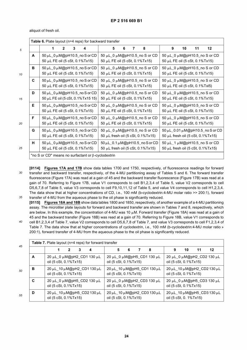
EP 2 516 669 B1
24
5
10
15
20
25
30
35
40
45
50
55
aliquot of fresh oil.
[0114] Figures 17A and 17B show data tables 1700 and 1750, respectively, of fluorescence readings for forwardtransfer and backward transfer, respectively, of the 4-MU partitioning assay of Tables 5 and 6. The forward transferfluorescence (Figure 17A) was read at a gain of 45 and the backward transfer fluorescence (Figure 17B) was read at again of 70. Referring to Figure 17B, value V1 corresponds to cell B1,2,3,4 of Table 5, value V2 corresponds to cellD5,6,7,8 of Table 5, value V3 corresponds to cell F9,10,11,12 of Table 5, and value V4 corresponds to cell H1,2,3,4.The data show that at higher concentrations of CD, i.e., 100 mM (b-cyclodextrin:4-MU molar ratio >> 200:1), forwardtransfer of 4-MU from the aqueous phase to the oil phase is significantly reduced.[0115] Figures 18A and 18B show data tables 1800 and 1850, respectively, of another example of a 4-MU partitioningassay. The microtiter plate layouts for forward and backward transfer are shown in Tables 7 and 8, respectively, whichare below. In this example, the concentration of 4-MU was 10 mM. Forward transfer (Figure 18A) was read at a gain of45 and the backward transfer (Figure 18B) was read at a gain of 70. Referring to Figure 18B, value V1 corresponds tocell B1,2,3,4 of Table 7, value V2 corresponds to cell D5,6,7,8 of Table 7, and value V3 corresponds to cell F1,2,3,4 ofTable 7. The data show that at higher concentrations of cyclodextrin, i.e., 100 mM (b-cyclodextrin:4-MU molar ratio »200:1), forward transfer of 4-MU from the aqueous phase to the oil phase is significantly reduced.
Table 6. Plate layout (n=4 reps) for backward transfer
1 2 3 4 5 6 7 8 9 10 11 12
A 50 mL_0 [email protected]_no S or CD 50 mL_0 [email protected]_no S or CD 50 mL_0 [email protected]_no S or CD50 mL FE oil (5 cSt, 0.1%Tx15) 50 mL FE oil (5 cSt, 0.1%Tx15) 50 mL FE oil (5 cSt, 0.1%Tx15)
B 50 mL_0 [email protected]_no S or CD 50 mL_0 [email protected]_no S or CD 50 mL_0 [email protected]_no S or CD50 mL FE oil (5 cSt, 0.1%Tx15) 50 mL FE oil (5 cSt, 0.1%Tx15) 50 mL FE oil (5 cSt, 0.1%Tx15)
C 50 mL_0 [email protected]_no S or CD 50 mL_0 [email protected]_no S or CD 50 mL_0 [email protected]_no S or CD50 mL FE oil (5 cSt, 0.1%Tx15) 50 mL FE oil (5 cSt, 0.1%Tx15) 50 mL FE oil (5 cSt, 0.1%Txl5)
D 50 mL_0 [email protected]_no S or CD 50 mL_0 [email protected]_no S or CD 50 mL_0 [email protected]_no S or CD50 mL FE oil (5 cSt, 0.1%Tx15 15) 50 mL FE oil (5 cSt, 0.1%Tx15) 50 mL FE oil (5 cSt, 0.1%Tx15)
E 50 mL_0 [email protected]_no S or CD 50 mL_0 [email protected]_no S or CD 50 mL_0 [email protected]_no S or CD50 mL FE oil (5 cSt, 0.1%Tx15) 50 mL FE oil (5 cSt, 0.1%Tx15) 50 mL FE oil (5 cSt, 0.1%Tx15)
F 50 mL_0 [email protected]_no S or CD 50 mL_0 [email protected]_no S or CD 50 mL_0 [email protected]_no S or CD50 mL FE oil (5 cSt, 0.1%Tx15) 50 mL FE oil (5 cSt, 0.1%Tx15) 50 mL FE oil (5 cSt, 0.1%Tx15)
G 50 mL_0 [email protected]_no S or CD 50 mL_0 [email protected]_no S or CD 50 mL_0.01 [email protected]_no S or CD50 mL FE oil (5 cSt, 0.1%Tx15) 50 mL fresh oil (5 cSt, 0.1%Tx15) 50 mL fresh oil (5 cSt, 0.1%Tx15)
H 50 mL_0 [email protected]_no S or CD 50 mL_0.1 [email protected]_no S or CD 50 mL_1 [email protected]_no S or CD50 mL FE oil (5 cSt, 0.1%Tx15) 50 mL fresh oil (5 cSt, 0.1%Tx15) 50 mL fresh oil (5 cSt, 0.1%Tx15)
"no S or CD" means no surfactant or β-cyclodextrin
Table 7. Plate layout (n=4 reps) for forward transfer
1 2 3 4 5 6 7 8 9 10 11 12
A 20 mL_0 mM@pH2_CD1 130 mL oil (5 cSt, 0.1%Tx15)
20 mL_0 mM@pH5_CD1 130 mL oil (5 cSt, 0.1%Tx15)
20 mL_0 mM@pH2_CD2 130 mL oil (5 cSt, 0.1%Tx15)
B 20 mL_10 mM@pH2_CD1 130 mL oil (5 cSt, 0.1%Tx15)
20 mL_10 mM@pH5_CD1 130 mL oil (5 cSt, 0.1%Tx15)
20 mL_10 mM@pH2_CD2 130 mL oil (5 cSt, 0.1%Tx15)
C 20 mL_0 mM@pH5_CD2 130 mL oil (5 cSt, 0.1%Tx15)
20 mL_0 mM@pH2_CD3 130 mL oil (5 cSt, 0.1%Tx15)
20 mL_0 mM@pH5_CD3 130 mL oil (5 cSt, 0.1%Tx15)
D 20 mL_10 mM@pH5_CD2 130 mL oil (5 cSt, 0.1%Tx15)
20 mL_10 mM@pH2_CD3 130 mL oil (5 cSt, 0.1%Tx15)
20 mL_10 mM@pH5_CD3 130 mL oil (5 cSt, 0. 1%Tx15)

EP 2 516 669 B1
25
5
10
15
20
25
30
35
40
45
50
55
[0116] In the droplet operations environment of a droplet actuator, formation of an inclusion complex between 4-MUand cyclodextrins is used to substantially minimize droplet cross-contamination by preventing 4-MU from leaking fromthe aqueous phase into the filler fluid (e.g., silicone oil) phase at low pH. Cyclodextrin is used as an additive to a reagentdroplet that includes the 4-MU-containing substrate. A sample droplet (e.g., a dried blood extract droplet) is combinedand mixed using droplet operations with the reagent droplet. Because cyclodextrin is included in the reagent droplet,the enzymatic product, i.e., 4-MU, is sequestered within an inclusion complex as soon as it is released by the enzymepresent in the sample droplet.[0117] As described herein surfactants may be used to retain 4-MU (or derivatives) within an aqueous phase (e.g.,an aqueous phase droplet). The efficacy of different surfactants in containing 4-MU (or derivatives) within an aqueousphase may, for example, be evaluated and selected using a partitioning assay, such as the 4-MU partitioning assaydescribed in reference to Figure 13. In this example, parameters that may be varied in the assay for evaluation ofsurfactants in aqueous containment of 4-MU (or derivatives) include, but are not limited to, the pH of the aqueous phasesolution (e.g. pH 2 to pH 10.5 range), and the critical micellar concentration of the surfactant.
(continued)
Table 7. Plate layout (n=4 reps) for forward transfer
1 2 3 4 5 6 7 8 9 10 11 12
E 20 mL_0 mM@pH2_CD4 130 mL oil (5 cSt, 0.1%Tx15)
20 mL_0 mM@pH5_CD4 130 mL oil (5 cSt, 0.1%Tx15)
20 mL_0 mM@pH2_CD5 130 mL oil (5 cSt, 0.1%taxi5)
F 20 mL_10 mM@pH2_CD4 130 mL oil (5 cSt, 0.1%Tx15)
20 mL_10 mM@pH5_CD4 130 mL oil (5 cSt, 0.1%Tx15)
20 mL_10 mM@pH2-CD5 130 mL oil (5 cSt, 0.1%Tx15 15)
G 20 mL_0 mM@pH5_CD5 130 mL oil (5 cSt, 0.1%Tx15)
EMPTY EMPTY
H 20 mL_10 mM@pH5_CD5 130 mL oil (5 cSt, 0.1%Tx15)
EMPTY EMPTY
*CD1=10 mM methyl-β-cyclodextrin; CD2=1 mM methyl-β-cyclodextrin; CD3=10 mM methyl-β-cyclodextrin; CD4=100mM methyl-β-cyclodextrin; CD5=100 mM hydroxytpropyl-β-cyclodextrin
Table 8. Plate layout (n=4 reps) for backward transfer
1 2 3 4 5 6 7 8 9 10 11 12
A 50 mL_0 [email protected]_no S or CD 50 mL_0 [email protected]_no S or CD 50 mL_0 [email protected]_no S or CD50 mL FE oil (5 cSt, 0.1%Tx15 15) 50 mL FE oil (5 cSt, 0.1%Tx15) 50 mL FE oil (5 cSt, 0.1%Tx15)
B 50 mL_0 [email protected]_no S or CD 50 mL_0 [email protected]_no S or CD 50 mL_0 [email protected]_no S or CD50 mL FE oil (5 cSt, 0.1%Tx15) 50 mL FE oil (5 cSt, 0.1%Tx15) 50 mL FE oil (5 cSt, 0.1%Tx15)
C 50 mL_0 [email protected]_no S or CD 50 mL_0 [email protected]_no S or CD 50 mL_0 [email protected]_no S or CD50 mL FE oil (5 cSt, 0.1 % Tx 15) 50 mL FE oil (5 cSt, 0.1%Tx15) 50 mL FE oil (5 cSt, 0.1%Tx15)
D 50 mL_0 [email protected]_no S or CD 50 mL_0 [email protected]_no S or CD 50 mL_0 [email protected]_no S or CD50 mL FE oil (5 cSt, 0.1%Tx15) 50 mL FE oil (5 cSt, 0.1%Tx15) 50 mL FE oil (5 cSt, 0.1%Tx15)
E 50 mL_0 [email protected]_no S or CD 50 mL_0 [email protected]_no S or CD 50 mL_0 [email protected]_no S or CD50 mL FE oil (5 cSt, 0.1%Tx15) 50 mL FE oil (5 cSt, 0.1%Tx15) 50 mL FE oil (5 cSt, 0.1%Tx15)
F 50 mL_0 [email protected]_no S or CD 50 mL_0 [email protected]_no S or CD 50 mL_0 [email protected]_no S or CD50 mL FE oil (5 cSt, 0.1%Tx15) 50 mL FE oil (5 cSt, 0.1%Tx15) 50 mL FE oil (5 cSt, 0.1%Tx15)
G 50 mL_0 [email protected]_no S or CD 50 mL_0 [email protected]_no S or CD 50 mL_0.01 [email protected]_no S or CD50 mL FE oil (5 cSt, 0.1%Tx15 15) 50 mL fresh oil (5 cSt, 0.1%Tx15) 50 mL fresh oil (5 cSt, 0.1%Tx15)
H 50 mL_0 [email protected]_no S or CD 50 mL_0.1 [email protected]_no S or CD 50 mL_1 [email protected]_no S or CD50mL FE oil (5 cSt, 0.1%Txl5) 50 mL fresh oil (5 cSt, 0.1%Tx15) 50 mL fresh oil (5 cSt, 0.1%Tx15 15)
"no S or CD" means no surfactant or β-cyclodextrin
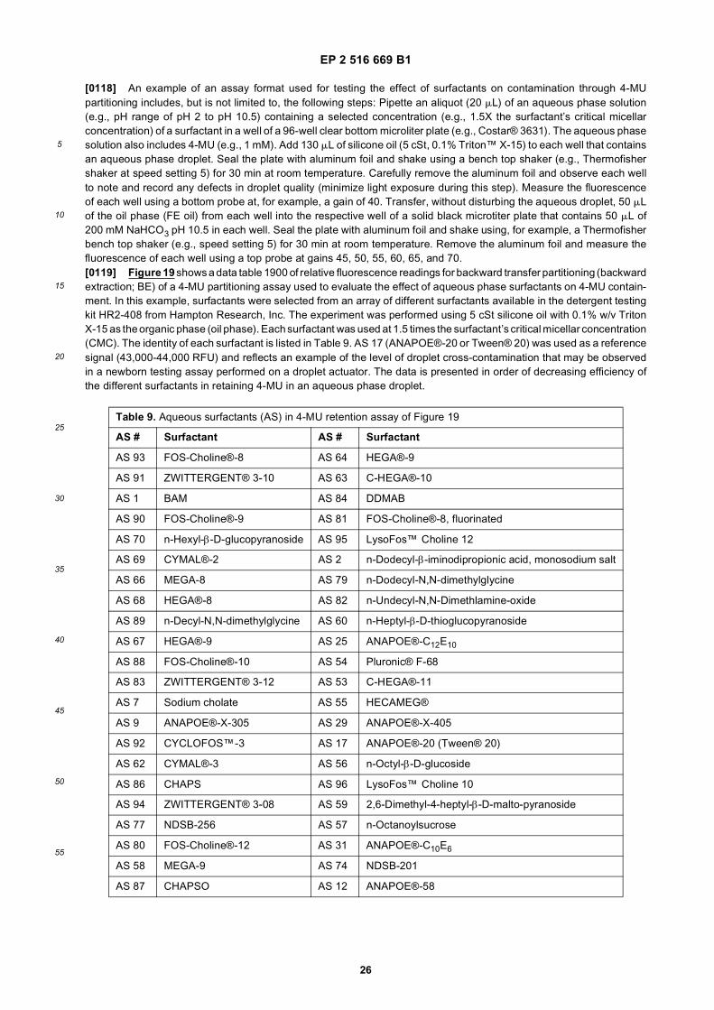
EP 2 516 669 B1
26
5
10
15
20
25
30
35
40
45
50
55
[0118] An example of an assay format used for testing the effect of surfactants on contamination through 4-MUpartitioning includes, but is not limited to, the following steps: Pipette an aliquot (20 mL) of an aqueous phase solution(e.g., pH range of pH 2 to pH 10.5) containing a selected concentration (e.g., 1.5X the surfactant’s critical micellarconcentration) of a surfactant in a well of a 96-well clear bottom microliter plate (e.g., Costar® 3631). The aqueous phasesolution also includes 4-MU (e.g., 1 mM). Add 130 mL of silicone oil (5 cSt, 0.1% Triton™ X-15) to each well that containsan aqueous phase droplet. Seal the plate with aluminum foil and shake using a bench top shaker (e.g., Thermofishershaker at speed setting 5) for 30 min at room temperature. Carefully remove the aluminum foil and observe each wellto note and record any defects in droplet quality (minimize light exposure during this step). Measure the fluorescenceof each well using a bottom probe at, for example, a gain of 40. Transfer, without disturbing the aqueous droplet, 50 mLof the oil phase (FE oil) from each well into the respective well of a solid black microtiter plate that contains 50 mL of200 mM NaHCO3 pH 10.5 in each well. Seal the plate with aluminum foil and shake using, for example, a Thermofisherbench top shaker (e.g., speed setting 5) for 30 min at room temperature. Remove the aluminum foil and measure thefluorescence of each well using a top probe at gains 45, 50, 55, 60, 65, and 70.[0119] Figure 19 shows a data table 1900 of relative fluorescence readings for backward transfer partitioning (backwardextraction; BE) of a 4-MU partitioning assay used to evaluate the effect of aqueous phase surfactants on 4-MU contain-ment. In this example, surfactants were selected from an array of different surfactants available in the detergent testingkit HR2-408 from Hampton Research, Inc. The experiment was performed using 5 cSt silicone oil with 0.1% w/v TritonX-15 as the organic phase (oil phase). Each surfactant was used at 1.5 times the surfactant’s critical micellar concentration(CMC). The identity of each surfactant is listed in Table 9. AS 17 (ANAPOE®-20 or Tween® 20) was used as a referencesignal (43,000-44,000 RFU) and reflects an example of the level of droplet cross-contamination that may be observedin a newborn testing assay performed on a droplet actuator. The data is presented in order of decreasing efficiency ofthe different surfactants in retaining 4-MU in an aqueous phase droplet.
Table 9. Aqueous surfactants (AS) in 4-MU retention assay of Figure 19
AS # Surfactant AS # Surfactant
AS 93 FOS-Choline®-8 AS 64 HEGA®-9
AS 91 ZWITTERGENT® 3-10 AS 63 C-HEGA®-10
AS 1 BAM AS 84 DDMAB
AS 90 FOS-Choline®-9 AS 81 FOS-Choline®-8, fluorinated
AS 70 n-Hexyl-β-D-glucopyranoside AS 95 LysoFos™ Choline 12
AS 69 CYMAL®-2 AS 2 n-Dodecyl-β-iminodipropionic acid, monosodium salt
AS 66 MEGA-8 AS 79 n-Dodecyl-N,N-dimethylglycine
AS 68 HEGA®-8 AS 82 n-Undecyl-N,N-Dimethlamine-oxide
AS 89 n-Decyl-N,N-dimethylglycine AS 60 n-Heptyl-β-D-thioglucopyranoside
AS 67 HEGA®-9 AS 25 ANAPOE®-C12E10
AS 88 FOS-Choline®-10 AS 54 Pluronic® F-68
AS 83 ZWITTERGENT® 3-12 AS 53 C-HEGA®-11
AS 7 Sodium cholate AS 55 HECAMEG®
AS 9 ANAPOE®-X-305 AS 29 ANAPOE®-X-405
AS 92 CYCLOFOS™-3 AS 17 ANAPOE®-20 (Tween® 20)
AS 62 CYMAL®-3 AS 56 n-Octyl-β-D-glucoside
AS 86 CHAPS AS 96 LysoFos™ Choline 10
AS 94 ZWITTERGENT® 3-08 AS 59 2,6-Dimethyl-4-heptyl-β-D-malto-pyranoside
AS 77 NDSB-256 AS 57 n-Octanoylsucrose
AS 80 FOS-Choline®-12 AS 31 ANAPOE®-C10E6
AS 58 MEGA-9 AS 74 NDSB-201
AS 87 CHAPSO AS 12 ANAPOE®-58

EP 2 516 669 B1
27
5
10
15
20
25
30
35
40
45
50
55
7.10.2 Modification of 4-MU and HMU Substrates for NBS
[0120] Droplet actuator-based lysosomal enzyme tests used in newborn testing assays (NBS) are fluorescent basedtests which measure the release of 4-methylumbelliferone (4-MU) or 6-hexadecanoylamido-4-methylumbelliferone(HMU) after enzymatic hydrolysis of the substrates. The enzymatic assays may be performed in aqueous droplets withinthe oil filled gap of the droplet actuator. In one example, the oil filler fluid may be polydimethylsiloxane silicon oil (PDMS).The lysosomal enzyme tests are performed at acidic pH values ranging between pH 2.8 and pH 5.6. After incubation,the enzymatic reactions may be terminated by the addition of sodium bicarbonate, pH 10.1 which also dissociates theproton from the umbelliferone hydroxyl leading to a substantial increase in fluorescent signal. At the acid pH values usedfor testing lysosomal enzymes, the products of the assays, 4-MU and HMU, are protonated and may partition from theaqueous droplet into the PDMS filler fluid. Partitioning of 4-MU and HMU into the oil phase may result in a reduction inthe assay signal and potential contamination of neighboring samples.[0121] Described are methods to substantially reduce or eliminate the oil solubility of umbelliferyl derivatives (e.g., 4-MU and HMU) during fluorescent based assays (e.g., NBS samples, assays, glycosidase, protease, and immunoassays)on a droplet actuator. Chemical modification of 4-MU and HMU may be used to form derivatives with substantiallyreduced solubility in the oil filler fluid (e.g., PDMS). Examples of chemical modifications that may be used to reduce theoil solubility of umbelliferyl derivatives may include addition of amino groups, carbon chain length, tethering 4-MU orHMU with molecules such as cellulose or polylysine. Chemical modification(s) may be selected such that activity and/orspecificity of the enzymatic reaction (e.g., various lysosomal enzyme assays) are not substantially altered (disrupted).Chemical modification(s) may also be selected such that the fluorescent signal of 4-MU or HMU is not eliminated.Preferably, retaining enough fluorescent signal of 4-MU or HMU to distinguish a positive or negative in the test. Chemicalmodification(s) may also be selected such that the umbelliferyl derivatives are compatible with downstream syntheticreactions used to prepare individual assay substrates. Chemical modification(s) may also be selected such that theumbelliferyl derivatives have sufficient water solubility suitable for specific NBS assays. In one example, the watersolubility of umbelliferyl derivatives may be selected for assay conditions ranging between pH 3.5 and pH 6.0. Suitableumbelliferyl derivatives may, for example, be selected using the Log D values at pH 3.5 and pH 6.0.[0122] In one example, the 4-methyl group of 4-MU and HMU may be modified with a chemical structure that increasesthe hydrophilicity of 4-MU and HMU. Because the 4-MU and HMU derivatives are hydrophilic, retention of the fluorescentderivatives in the aqueous phase reaction droplet is substantially increased. Examples of suitable chemical structuresthat may be used to modify 4-MU and HMU to increase their hydrophilicity include, but are not limited to, polymersincluding linear or branched polyalkalene glycols (PAG), such as polyethylene glycols (PEG), and/or polypropyleneglycols (PPG), or combinations of PEG and PPG; sulfonic acids; and amino groups. The efficacy of chemical modificationin promoting retention of 4-MU and HMU in the aqueous phase may, for example, be evaluated using an on-chippartitioning assay.[0123] The compounds described herein are analogs of 4-methylumbilleferone (4-MU) and 6-hexadecanoyl-4-meth-ylumbelliferone.[0124] The 4-MU analogs have one of the following general formulas:
(continued)
Table 9. Aqueous surfactants (AS) in 4-MU retention assay of Figure 19
AS # Surfactant AS # Surfactant
AS 85 FOS-MEA®-10 AS 34 ANAPOE®-C10E9
AS 78 ZWITTERGENT® 3-14 AS 19 ANAPOE®-35

EP 2 516 669 B1
28
5
10
15
20
25
30
35
40
45
50
55
wherein:
X is, independently, selected from the group consisting of O, S, and NH,
R1 is selected from the group consisting of H, C1-15 oxyalkyl, -C1-15 aminoalkyl, -C1-15 alkylamino, (CH2CH2O)n-CH3,-(CH2CH2O)n-SO3H, C1-6-alkyl-C(O)NH2, -(CH2CH2NH)n-H, -SO3H, -C1-6-alkyl-C(O)OH, -SO3Me, C1-6-alkyl-hete-rocyclyl, monosaccharides, and disaccharides
R2 is H, a monosaccharide, a disaccharide, or a polyhydric alcohol;
R3 is a hydrophilic oligomer or polymer, which can be selected from the group consisting of polyalkylene glycols,such as polyethylene glycol, wherein the polyalkylene glycols can optionally further include NH or S moieties;polycationic polymers such as polylysine; carbohydrates, including mono-saccharides and disaccharides, C1-15oxyalkyl, -C1-15 aminoalkyl,-C1-15 alkylamino, -(CH2CH2O)n-CH3, -(CH2CH2O)n-SO3H, C1-6-alkyl-C(O)NH2,-(CH2CH2NH)n-H, -SO3H, -C1-6-alkyl-C(O)OH, -SO3Me, C1-6-alkyl-heterocyclyl, monosaccharides, and disaccha-rides,
and n is an integer of from 1 to 100, or from 1 to 50, or from 1 to 25, or from 1 to 10.[0125] The hydrophilic polymer or oligomer can be any of a variety of oligomers comprising a polyalkalene glycolmoiety, as will be understood by those skilled in the art. Preferably, the polyalkalene glycol moiety of the oligomer has1-100 polyalkalene glycol subunits. More preferably, the polyalkalene glycol moiety has 1-50 polyalkalene glycol subunitsand, most preferably, the polyalkalene glycol moiety has 1-25 polyalkalene glycol subunits.[0126] The hydrophilic polymer or oligomer can be any of a variety of oligomers comprising a polyethylene glycolmoiety, as will be understood by those skilled in the art. Preferably, the polyethylene glycol moiety of the oligomer has1-100 polyethylene glycol subunits. More preferably, the polyethylene glycol moiety has 1-50 polyethylene glycol subunitsand, most preferably, the polyethylene glycol moiety has 1-25 polyethylene glycol subunits.[0127] The hydrophilic polymer or oligomer can be any of a variety of oligomers comprising a polypropylene glycolmoiety, as will be understood by those skilled in the art. Preferably, the polypropylene glycol moiety of the oligomer has1-100 polypropylene glycol subunits. More preferably, the polypropylene glycol moiety has 1-50 polypropylene glycolsubunits and, most preferably, the polypropylene glycol moiety has 1-25 polypropylene glycol subunits.[0128] Additional hydrophilic oligomers and polymers include those containing polar or charged functional groups,rendering them soluble in water. For example, acrylics include acrylic acid, acrylamide, and maleic anhydride polymersand copolymers. Amine-functional polymers include allylamine, ethyleneimine, oxazoline, and other polymers containing

EP 2 516 669 B1
29
5
10
15
20
25
30
35
40
45
50
55
amine groups in their main- or side-chains, such as polylysine. These may be combined with PAGs.[0129] The oligomer may comprise one or more other moieties as will be understood by those skilled in the art including,but not limited to, additional hydrophilic moieties, lipophilic moieties, spacer moieties, linker moieties, and terminatingmoieties. The various moieties in the oligomer are covalently coupled to one another by either hydrolyzable or non-hydrolyzable bonds.[0130] The or more additional hydrophilic moieties (i.e., moieties in addition to the polyethylene glycol moiety) caninclude, but are not limited to, sugars, polyhydric alcohols, polyalkylene oxides, and polyamine/PEG copolymers. Aspolyethylene glycol is a polyalkylene oxide, the additional hydrophilic moiety may be a polyethylene glycol moiety.Adjacent polyethylene glycol moieties will be considered to be the same moiety if they are coupled by an ether bond.For example, the moiety --O--C2H4--O-- C2H4-O--C2H4--O-- C2H4--O-- C2H4-O-- C2H4 - is a single polyethylene glycolmoiety having six polyethylene glycol subunits. Adjacent polyethylene glycol moieties will be considered to be differentmoieties if they are coupled by a bond other than an ether bond.[0131] The oligomer can further comprise one or more spacer moieties as will be understood by those skilled in theart. Spacer moieties are preferably selected from the group consisting of sugar, cholesterol and glycerine moieties.[0132] The oligomer can further comprise one or more terminating moieties at the one or more ends of the oligomer,which are not coupled to the 4-HU or 6HMU backbone. The terminating moiety is preferably an alkyl or alkoxy moiety,and is more preferably a lower alkyl or lower alkoxy moiety (where lower means C1-6). Most preferably, the terminatingmoiety is methyl or methoxy. While the terminating moiety is preferably an alkyl or alkoxy moiety, it is to be understoodthat the terminating moiety may be various moieties as will be understood by those skilled in the art including, but notlimited to, sugars, cholesterol, alcohols, and fatty acids. The oligomer or polymer may include one or more anionic orself-forming moietes, such as tertiary anion.[0133] A subset of Formulas 1-5 is shown below, in which each R2 is H.
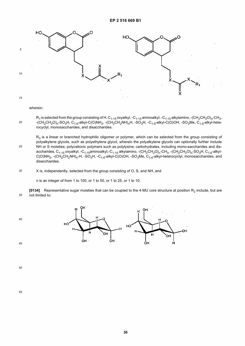
EP 2 516 669 B1
30
5
10
15
20
25
30
35
40
45
50
55
wherein:
R1 is selected from the group consisting of H, C1-15 oxyalkyl, -C1-15 aminoalkyl, -C1-15 alkylamino, -(CH2CH2O)n-CH3,-(CH2CH2O)n-SO3H, C1-6-alkyl-C(O)NH2, -(CH2CH2NH)nH, -SO3H, -C1-6-alkyl-C(O)OH, -SO3Me, C1-6-alkyl-hete-rocyclyl, monosaccharides, and disaccharides.
R3 is a linear or branched hydrophilic oligomer or polymer, which can be selected from the group consisting ofpolyalkylene glycols, such as polyethylene glycol, wherein the polyalkylene glycols can optionally further includeNH or S moieties; polycationic polymers such as polylysine; carbohydrates, including mono-saccharides and dis-accharides, C1-15 oxyalkyl, -C1-15aminoalkyl,-C1-15 alkylamino, -(CH2CH2O)n-CH3, -(CH2CH2O)n-SO3H, C1-6-alkyl-C(O)NH2, -(CH2CH2NH)n-H, -SO3H, -C1-6-alkyl-C(O)OH, -SO3Me, C1-6-alkyl-heterocyclyl, monosaccharides, anddisaccharides.
X is, independently, selected from the group consisting of O, S, and NH, and
n is an integer of from 1 to 100, or 1 to 50, or 1 to 25, or 1 to 10.
[0134] Representative sugar moieties that can be coupled to the 4-MU core structure at position R2 include, but arenot limited to:

EP 2 516 669 B1
31
5
10
15
20
25
30
35
40
45
50
55

EP 2 516 669 B1
32
5
10
15
20
25
30
35
40
45
50
55

EP 2 516 669 B1
33
5
10
15
20
25
30
35
40
45
50
55
[0135] The sugar moieties shown are a-pyranose, b-pyranose, a-furanose, and b-furanose forms of allose, altrose,glucose, mannose, gulose, idose, galactose and talose, respectively. The sugar moeities can be attached to the phenolmoiety to form an ether linkage using etherification techniques, ideally those in which the hydroxy groups not involvedin the coupling chemistry are protected during the etherification step, and deprotected at a later time in the overallsynthesis. Suitable protecting groups are described, for example, in Greene and Wuts, Protective Groups in OrganicSynthesis, 3rd Ed., John Wiley &Sons, 1999.[0136] Where glucose is the sugar, and reacts with the phenol at R2, the resulting compound is known as a glucop-yranoside. For formation of glucopyranosides is well known to those of skill in the art.[0137] Ribose and deoxyribose sugars can also be coupled to the phenol at position R2:
[0138] Representative polyhydric alcohols include glycol (2-carbon), glycerol (3-carbon), erythritol (4-carbon), threitol(4-carbon), arabitol (5-carbon), xylitol (5-carbon), ribifol (5-carbon), mannitol (6-carbon), sorbitol (6-carbon), dulcitol (6-carbon), iditol (6-carbon), isomalt (12-carbon), maltitol (12-carbon), lactitol (12-carbon), and polyglycitol.[0139] Examples of individual 4-MU analogs include the following:

EP 2 516 669 B1
34
5
10
15
20
25
30
35
40
45
50
55
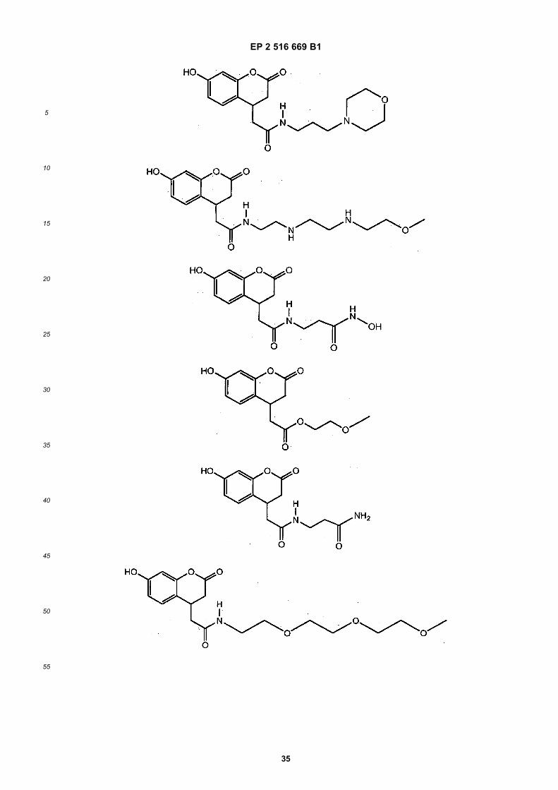
EP 2 516 669 B1
35
5
10
15
20
25
30
35
40
45
50
55

EP 2 516 669 B1
36
5
10
15
20
25
30
35
40
45
50
55

EP 2 516 669 B1
37
5
10
15
20
25
30
35
40
45
50
55
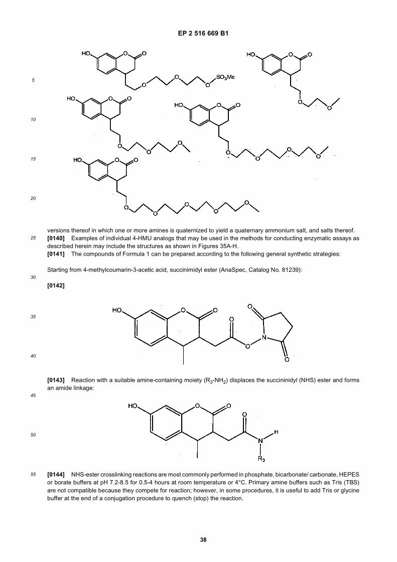
EP 2 516 669 B1
38
5
10
15
20
25
30
35
40
45
50
55
versions thereof in which one or more amines is quaternized to yield a quaternary ammonium salt, and salts thereof.[0140] Examples of individual 4-HMU analogs that may be used in the methods for conducting enzymatic assays asdescribed herein may include the structures as shown in Figures 35A-H.[0141] The compounds of Formula 1 can be prepared according to the following general synthetic strategies:
Starting from 4-methylcoumarin-3-acetic acid, succinimidyl ester (AnaSpec, Catalog No. 81239):
[0142]
[0143] Reaction with a suitable amine-containing moiety (R3-NH2) displaces the succininidyl (NHS) ester and formsan amide linkage:
[0144] NHS-ester crosslinking reactions are most commonly performed in phosphate, bicarbonate/ carbonate, HEPESor borate buffers at pH 7.2-8.5 for 0.5-4 hours at room temperature or 4°C. Primary amine buffers such as Tris (TBS)are not compatible because they compete for reaction; however, in some procedures, it is useful to add Tris or glycinebuffer at the end of a conjugation procedure to quench (stop) the reaction.

EP 2 516 669 B1
39
5
10
15
20
25
30
35
40
45
50
55
[0145] To prepare compounds in which a thioamide or C(NH)-NHR3 moiety is present, one can start with an analogin which the carbonyl in the succinimidyl ester is replaced with a C(S) or a C(NH) moiety.[0146] To prepare compounds of Formula 2, one can use the following reactions:
[0147] In these reactions, the phenol, if present at position R2, should be protected until after the coupling chemistryis completed.[0148] Suitable C1-15 oxyalkyl, -C1-15 aminoalkyl, -C1-15 alkylamino, -(CH2CH2O)n-CH3, -(CH2CH2O)n-SO3H,C1-6-alkyl-C(O)NH2, -(CH2CH2NH)n-H, -SO3H, -C1-6-alkyl-C(O)OH, -SO3Me, C1-6-alkyl-heterocyclyl, monosaccharides,disaccharides, and other moieties are well-known to those of skill in the art, and can readily be prepared.[0149] In the case of ester formation, one can reflux the desired alcohol with the carboxylic acid (where X is O) moietyand, using a Dean-Stark trap, remove water as it is formed to drive the equilibrium towards products.[0150] To prepare compounds of Formula 3, one can use the following reactions:
where the carbon on R1 bonded to the halogen (preferably Cl, Br, or I), or, alternatively, another suitable leaving group,is a primary or secondary carbon.[0151] In these reactions, the phenol, if present at position R2, should be protected until after the coupling chemistryis completed.

EP 2 516 669 B1
40
5
10
15
20
25
30
35
40
45
50
55
where X is an alkoxy or thioalkoxy moiety. When X is an amine, the amine can be used to displace the leaving group,without needing to deprotonate the amine (i.e., without needing to form R1-NH-).[0152] To prepare compounds of Formula 4, one can use the following reaction:
where X is an alkoxy or thioalkoxy moiety. When X is an amine, the amine can be used to displace the leaving group,without needing to deprotonate the amine (i.e., without needing to form R1-NH-).[0153] Alternatively, the chemistry can be performed in a stepwise manner, where a moiety HX-CH2-C(O)-XH isdeprotonated, and the more basic X- anion (i.e., the one attached to the CH2 moiety rather than the C(O) moiety) actsas a nucleophile to displace the leaving group. Then, the less basic X- anion can be protonated, and converted to asuitable leaving group (i.e., an acid halide, an anhydride, and the like), and an R1CH moiety reacted with the leavinggroup to form the final product.[0154] In these reactions, the phenol, if present at position R2, should be protected until after the coupling chemistryis completed.[0155] To prepare compounds of Formula 4, one can use the following reaction:
[0156] In this reaction, the phenol, if present at position R2, should be protected until after the coupling chemistry iscompleted. Similarly, any functional groups on R1 that would otherwise react with the isocyanate should be protected.[0157] Examples of the 6-HMU analogs have the following general formulas:

EP 2 516 669 B1
41
5
10
15
20
25
30
35
40
45
50
55
wherein:
X is, independently, selected from the group consisting of O, S, and NH,
R1 is selected from the group consisting of H, C1-15 oxyalkyl, -C1-15 aminoalkyl, -C1-15 alkylamino, -(CH2CH2O)n-CH3,-(CH2CH2O)n-SO3H, C1-6-alkyl-C(O)NH2, -(CH2CH2NH)n-H, -SO3H, -C1-6-alkyl-C(O)OH, -SO3Me, C1-6-alkyl-hete-rocyclyl, monosaccharides, and disaccharides.
R2 is H, a monosaccharide, a disaccharide, or a polyhydric alcohol;
R3 is a hydrophilic oligomer or polymer, which can be selected from the group consisting of polyalkylene glycols,such as polyethylene glycol, wherein the polyalkylene glycols can optionally further include NH or S moieties;polycationic polymers such as polylysine; carbohydrates, including mono-saccharides and disaccharides, C1-16oxyalkyl, -C1-15 aminoalkyl, - C1-15 alkylamino, -(CH2CH2O)n-CH3, -(CH2CH2O)n-SO3H, C1-6-alkyl-C(O)NH2,-(CH2CH2NH)n-H, -SO3H, -C1-6-alkyl-C(O)OH, -SO3Me, C1-6-alkyl-heterocyclyl, monosacchacides, and disaccha-rides.
R4 is a C5-16 alkyl, oxyalkyl, or amino-oxyalkyl, moiety, and
n is an integer of from 1 to 100, or 1 to 50, or 1 to 25, or 1 to 10 and
Versions thereof in which one or more amines is quaternized to yield a quaternary ammonium salt, and salts thereof.[0158] Examples of individual 6-HMU analogs include the following:

EP 2 516 669 B1
42
5
10
15
20
25
30
35
40
45
50
55
Examples of individual 6-HMU analogs may further include the structures as shown in Figures 36A-I.
[0159] The compounds of Formulas 6-10 can be prepared using the same general reactions used to prepare thecompounds of Formulas 1-5, except that the starting materials further include a -NH-C(X)-R4 moiety. The amide, thio-amide, or -C(NH)-NH- moiety is non-reactive under the coupling conditions used to prepare the compounds, so can bepresent at the time the coupling chemistry is performed. Alternatively, a protected aniline moiety (next to the phenolmoiety) can be present, and can be deprotected at an appropriate time after the coupling chemistry is complete. Fromthere, the aniline group can be reacted with a suitable acid chloride (i.e., R4-C(O)-halo), or acid anhydride (R4-C(O)-O-C(O)-R4) to form an amine.
7.11 Enhanced Hydrolysis of Enzymatic Substrates
[0160] Fluorescent substrates used in enzymatic NBS assays may have varying degrees of hydrophobicity. Becausethe fluorescent substrates may be hydrophobic, their interaction with more hydrophilic enzymes, such as the enzymesin dried blood extracts, may be reduced. The invention provides methods to enhance the hydrolysis of enzymatic sub-

EP 2 516 669 B1
43
5
10
15
20
25
30
35
40
45
50
55
strates such as fluorescent substrates used in NBS assays. According to the invention, the hydrolysis of enzymaticsubstrates (i.e. 4-MU- or HMU-containing substrates) is enhanced by formation of an inclusion complex that stabilizesthe substrate within an aqueous environment. Cyclodextrins (e.g., hydroxypropyl-β-cyclodextrin, methyl-β-cyclodextrin)are used to form an inclusion complex containing a hydrophobic substrate. Negatively charged cyclodextrins, may beused to further enhance substrate hydrolysis. The methods of the invention provide for improved separation between alower signal and a higher signal in enzyme-substrate based bioassay such as NBS assays for Fabry, Gaucher, Pompe,Krabbe, and Niemann-Pick diseases.[0161] To evaluate the efficacy of cyclodextrins in enhancing the hydrolysis of enzymatic substrates (4-MU substrates)used in NBS assays, DBS extracts were prepared from quality control (QC) dried blood spot samples (i.e., base pool(BP), low (L), medium (M) and high (H) activity samples). The QC samples were obtained from CDC, Atlanta GA. Thebase pool (BP) QC sample was prepared from a pool of leukoreduced human red blood cells that was adjusted withserum to a hematocrit of 50%. The High (H) activity QC sample was prepared from pooled cord blood that was adjustedwith serum to a hematocrit of 50%. The medium (M) activity QC sample was prepared by using 50% base pool (BP)and 50% high (H) activity sample. The low (L) activity QC sample was prepared by using 95% base pool (BP) and 5%high (H) activity sample. The BP sample is used as a control for hydrolysis non-specific to white blood cell lysosomalenzymes. The DBS extracts were analyzed using on-bench assay protocols for Fabry, Gaucher, Krabbe, and Pompediseases. For each assay, on-chip reagent formulations were scaled up to bench volumes. Hydroxypropyl-β-cyclodextrinor methyl-β-cyclodextrin reagent stocks (300 mM) were prepared in appropriate assay buffers. For each reaction, 10 mLof the appropriate substrate was mixed with 10 mL of DBS extract. After an incubation period (20 hrs at 37 °C), 50 mLof stop buffer (0.2 M NaCO3, pH 10.1) was added to the reaction. Fluorescence was read at 360/460 nm at a gain of75. For each assay, QC samples (BP, L, M and H) were analyzed in duplicate.[0162] Figures 20A and 20B show bar graphs 2000 and 2050 of the effect of hydroxypropyl-β-cyclodextrin and methyl-β-cyclodextrin, respectively, on substrate hydrolysis in a Fabry assay. The 4-MU substrate in the Fabry assay was β-D-galactopyranoside. The concentrations of β-cyclodextrins in the reaction mixture (substrate + DBS extract) were 10 mM,30 mM and 100 mM. The data show that low concentrations (10 mM) of methyl-β-cyclodextrin increase the hydrolysisof the 4-MU substrate in the Fabry assay and improve the separation between low, medium and high QC-dried bloodspot samples. The effect of hydroxypropyl-β-cyclodextrin was less effective.[0163] Figures 21A and 21B show bar graphs 2100 and 2150 of the effect of hydroxypropyl-β-cyclodextrin and methyl-β-cyclodextrin, respectively, on substrate hydrolysis in a Gaucher assay. The 4-MU substrate in the Gaucher assay wasβ-D-glucopyranoside. The concentrations of β-cyclodextrins in the reaction mixture (substrate + DBS extract) were 10mM, 30 mM, and 100 mM. The data show that low concentrations (10 mM) of hydroxypropyl- and methyl-β-cyclodextrinincrease the hydrolysis of the 4-MU substrate in the Gaucher assay and improve the separation between low, mediumand high QC-dried blood spot samples.[0164] To further evaluate the efficacy of lower concentrations of cyclodextrins in enhancing the hydrolysis of 4-MUsubstrates, Fabry and Gaucher assays were repeated using cyclodextrins concentrations ranging from 1 mM to 25 mM.Figures 22A and 22B show bar graphs 2200 and 2250, respectively, of the effect of lower concentrations of hydroxy-propyl-β-cyclodextrin and methyl-β-cyclodextrin, respectively, on substrate hydrolysis in a Fabry assay. The 4-MU sub-strate in the Fabry assay was β-D-galactopyranoside. The concentrations of β-cyclodextrins in the reaction mixture(substrate + DBS extract) were 1, 5, 10, 15, 20, and 25 mM. The data show that methyl-β-cyclodextrin (10 mM) has apositive effect on the Fabry assay increasing the QC-H signal by 37%. The preferred concentration of methyl-β-cyclo-dextrin is from about 1 mM. to about 20 mM, or from about 5 mM to about 15 mM, or from about 7 mM to about 13 mM,or from about 9 mM to about 11 mM, or from about 9.5 mM to about 11.5 mM. The effect of hydroxypropyl-β-cyclodextrinon substrate hydrolysis in a Fabry assay was less effective.[0165] Figures 23A and 23B show bar graphs 2300 and 2350, respectively, of an experimental result of the effect oflower concentrations of hydroxypropyl-β-cyclodextrin and methyl-β-cyclodextrin, respectively, on substrate hydrolysisin a Gaucher assay. The 4-MU substrate in the Gaucher assay was β-D-glucopyranoside. The concentrations of β-cyclodextrins in the reaction mixture (substrate + DBS extract) were 1, 5, 10, 15, 20, and 25 mM. The data in oneexperiment suggested that both hydroxypropyl-β- and methyl-β-cyclodextrin may affect the fluorescence signal (in atleast one case showing an increase), in the Gaucher assay. The β-cyclodextrin used may be hydroxypropyl-β-cyclodextrinat a concentration from about 5 mM to about 15 mM, or from about 7 mM to about 13 mM, or from about 9 mM to about11 mM, or from about 9.5 mM to about 11.5 mM.[0166] The effect of hydroxypropyl-β-cyclodextrin and methyl-β-cyclodextrin on substrate hydrolysis in a Pompe assaywas less effective (data not shown). The effect of hydroxypropyl-β-cyclodextrin and methyl-β-cyclodextrin on substratehydrolysis in a Krabbe assay was inhibitory (data not shown).
7.12 Clotting in Fabry Assay
[0167] The buffer system used to prepare substrate-inhibitor formulation for Fabry disease may include 0.1M sodium

EP 2 516 669 B1
44
5
10
15
20
25
30
35
40
45
50
55
citrate, a pH from about 4-5, or from about 4.2-4.8, or from about 4.5-4.7, or about 4.6, containing 1% sodium taurocholate.Although this formulation produces statistically significant separation between the normal and Fabry affected dried bloodspot extracts, the inventors have found that it results in clot formation within the droplets. This clotting may adverselyaffect the reproducibility of the assay because enzyme may be trapped within the clots where it is unavailable to thesubstrate. The clots seemed to appear very early in the assay (30 minutes from the inception). The inventors have foundthat a buffer system using 0.04M Sodium Citrate, a pH from about 4-5, or from about 4.2-4.8, or from about 4.5-4.7, orabout 4.6, with 0.01% Tween® 20 does not affect the signal at all, and the clot formation was eliminated. In anotherexample, 0.04M Sodium Acetate, pH 4.6 with 0.01% Tween® 20 was used in preparing substrate-inhibitor formulationwhich produced the same results as the Sodium citrate buffer. Thus, provided is an enzymatic assay making use ofblood, a blood component, or a reconstituted blood spot, and further makes use of a substrate inhibitor formulation ata pH from about 4-5, or from about 4.2-4.8, or from about 4.5-4.7, or about 4.6, but which lacks sodium taurocholate.Also described is an enzymatic assay making use of blood, a blood component, or a reconstituted blood spot, and furthermakes use of a substrate inhibitor formulation at a pH from about 4-5, or from about 4.2-4.8, or from about 4.5-4.7, orabout 4.6, and comprises sodium citrate. Also described is an enzymatic assay making use of blood, a blood component,or a reconstituted blood spot, and further makes use of a substrate inhibitor formulation at a pH from about 4-5, or fromabout 4.2-4.8, or from about 4.5-4.7, or about 4.6, and comprises sodium acetate. Also described is an enzymatic assaymaking use of blood, a blood component, or a reconstituted blood spot, and further makes use of a substrate inhibitorformulation at a pH from about 4-5, or from about 4.2-4.8, or from about 4.5-4.7, or about 4.6, and comprises sodiumcitrate and lacks sodium acetate. Also described is an enzymatic assay making use of blood, a blood component, or areconstituted blood spot, and further makes use of a substrate inhibitor formulation at a pH from about 4-5, or from about4.2-4.8, or from about 4.5-4.7, or about 4.6, and comprises sodium acetate and lacks sodium acetate. Also describedis an enzymatic assay making use of blood, a blood component, or a reconstituted blood spot, and further makes useof a substrate inhibitor formulation at a pH from about 4-5, or from about 4.2-4.8, or from about 4.5-4.7, or about 4.6 andcomprises sodium citrate and a surfactant. Also described is an enzymatic assay making use of blood, a blood component,or a reconstituted blood spot, and further makes use of a substrate inhibitor formulation a pH from about 4-5, or fromabout 4.2-4.8, or from about 4.5-4.7, or about 4.6, and comprises sodium acetate and a surfactant.
7.13 Alternative Assay Protocols
[0168] In one example, an assay involves the following: One set of dried blood spot extract droplets is combined usingdroplet operations with one set of substrate fluid droplets. The resulting set of 2X droplets is split using droplet operationsto form two sets of 1X droplets. Each droplet in the first set of 1X droplets is combined using droplet operations with astop buffer droplet. Endpoint fluorescence of the combined droplet measured. The second set of 1X reaction dropletsis incubated for a predetermined time and then each reaction droplet is combined with a stop buffer droplet. Again,endpoint fluorescence is measured. The difference between time t-END hrs and t-0 hrs is reported. However, in someprotocols there is no time t-0 measurement, which reduces the number of droplet handling operations by half.[0169] In one example, a droplet actuator of the invention is configured to perform 5-plex assays on 48 samples (240assays) which means 240 2X droplets have to be stored for 20 hours in the limited real estate on the chip. Since 2Xdroplets cannot be stored on a 3 phase bus, the 2X droplets may be split using droplet operations, and 1X may betransported to waste while the remaining 1X droplet is incubated. In one example, all the dispensing electrodes (reagentand sample) are smaller than the electrodes on the rest of the chip (900 microns or 750 microns; gap height 285 microns).In such a droplet actuator, a 2X droplet on the smaller electrodes would actually be close to a 1X droplet on the largerelectrodes. This configuration would avoid the need for splitting the droplet, and would simplify droplet actuator designand work flow easier.[0170] As already noted, a disadvantage performing detection at time t=0 and t=20 hours is the number of dropletsthat should be handled. Performing "m" plex assays on "n" number of samples, requires the total number of droplets tobe "m*n*2", which limits the total number of samples that can be screened on a single droplet actuator. To fit as manysamples as possible on a single cartridge, it is helpful to avoid the time t=0 measurement. The main reasons to performa time t=0 measurement are to assess the quality of the substrates; to determine non-enzymatic hydrolysis; and toaccount for the effect of hemoglobin quench of fluorescence.[0171] However, as already discussed, the inventors have surprisingly found that DBS quench on fluorescence is veryminimal on chip due to the low path lengths. So it can safely be assumed that that hemoglobin does not affect fluorescence.By detecting the background fluorescence of all the substrates before the start of the actual assay, the QC of all thesubstrates can be obtained. A blank extract may be incubated with the substrate as a control for non-enzymatic hydrolysis.By taking these steps, the time t=0 measurement can be eliminated, thereby reducing the total number of droplets tohandle by half. A droplet of known concentration of 4-MU sodium salt may be dispensed using droplet operations andits fluorescence may be detected to provide a calibrant. One droplet each of substrate may be dispensed and mergedusing droplet operations with a droplet of stop buffer. Endpoint fluorescence of this combination may be detected to

EP 2 516 669 B1
45
5
10
15
20
25
30
35
40
45
50
55
assess the QC of the substrates and provide time t=0 measurement. Next, one droplet of each substrate may be mergedwith a corresponding droplet of DBS extract, yielding a set of reaction droplets. At the end of the incubation period, thereaction droplet may be combined with a stop, buffer droplet, and endpoint fluorescence may be detected.
7.13.1 Hemoglobin Quench Normalization for NBS Assays
[0172] Dried blood spots (DBS) from newborns for testing of lysosomal storage disease (LSD) enzymes are inherentlyvariable because of the variation in hematocrit between individuals and the irregular deposition of the blood across thefilter paper. This variation leads to differences in the amount of hemoglobin in the final assay for a LSD target enzyme.In some cases, hemoglobin (or other quenching material) in the DBS sample may quench the fluorescence emitted fromthe reaction product (e.g., 4-MU or HMU). In the event of hemoglobin quench on fluorescence, enzyme activity in asample may be underestimated.[0173] Described are assay methods to measure fluorescence (e.g. 4-MU or HMU) quench in a reconstituted DBSsample in a droplet actuator-based NBS assay. In one example of a droplet actuator-based NBS assay, a droplet ofknown concentration of 4-MU (4-MU standard droplet) is combined using droplet operations with a droplet of reactiontermination buffer (stop buffer) and fluorescence detected on-chip to provide a calibration value for the enzyme assays.This same 4-MU standard droplet may also be used to determine a quench coefficient for each DBS sample droplet tobe tested. An example of an assay format for measuring fluorescence quench in a reconstituted DBS sample includes,but is not limited to, the following steps: At the start of an assay, a DBS sample droplet is dispensed and combined usingdroplet operations with a droplet of the 4-MU standard in termination buffer and the fluorescence read (no incubationperiod required). If no hemoglobin or quenching material is present in the DBS sample droplet, the fluorescence of thecombined DBS sample/4-MU droplet will be equivalent to the fluorescence of the 4-MU standard droplet. The quenchcoefficient will be 1.0 or no quench observed. If hemoglobin or quenching material is present in the DBS sample droplet,a lower fluorescent signal will be detected in the combined DBS sample/4-MU droplet relative to the fluorescence of the4-MU standard droplet. The fluorescence of the combined DBS sample/4-MU droplet divided by the fluorescence of the4-MU standard droplet is a measure of the fluorescence quench (quench coefficient) in the sample. The fluorescencequench coefficient for each sample may be used to normalize all of the fluorescent output signals from all of the DBSassay samples and eliminate the differences in signal outputs because of differences in hemoglobin content (or otherquench material) in each sample.
7.14 Multiplexed Pompe, Fabry and Hunter’s Assays on a Droplet Actuator
[0174] Described are a droplet actuator device and methods for multiplexed testing (3-plex assay on 12 samples) forPompe, Fabry and Hunter’s lysosomal storage disorders (LSDs) on a single droplet actuator. Samples used for multi-plexed testing for LSDs may be prepared from dried blood spots (DBS) using an on-bench protocol prior to loading ona droplet actuator. Reagent preparation (e.g., stop buffer, extraction buffer, substrate formulations and calibrant) mayalso be prepared using on-bench protocols prior to loading on a droplet actuator. Reagent and/or samples may beprepared in resevoirs on the droplet actuator then flowed to different operations gaps, or prepared in the droplet operationsgap.
7.14.1 Substrate Formulations and Reagent Preparation
[0175] All reagents and formulations required for Pompe, Fabry and Hunter’s assays may be prepared on-bench andsubsequently loaded into fluid dispensing reservoirs of a droplet actuator. Reagents common to all three assays include:stop buffer (STB; 0.2M Sodium bicarbonate pH 10.0 with 0.01% Tween® 20) and calibrant (CAL; 0.15 mM 4-Methylumbelliferone prepared in stop buffer) used to calibrate fluorescence detection. Specific substrate formulations for Pompe,Fabry and Hunter’s assays are summarized in Table 10. Reagent and/or samples may be prepared in resevoirs on thedroplet actuator then flowed to different operations gaps, or prepared in the droplet operations gap.
Table 10. Summary of substrate formulations
Assay Buffer (mL) Inhibitor or Enzyme (mL) Substrate (mL) Total volume (mL)
PI-DIL 197 (PB) 3 (PI) N/A 200
POM 41.4 (PB) 5.0 (PI-DIL) 3.6 (PS) 50.0
FAB 39.3 (FB) 10.0 (FI) 0.7 (FS) 50.0
HUN N/A 2.0 (HE) 18.0 (HS) 20.0

EP 2 516 669 B1
46
5
10
15
20
25
30
35
40
45
50
55
[0176] Reagents to prepare stock solutions and buffers for testing for lysosomal storage disorders (LSDs), such asPompe, Fabry, Gaucher, Hurler and Hunter assays on a droplet actuater may include any one or more of the following:Fabry substrate (FS), e.g., 4-Methylumbelliferyl α-D-Galactopyranoside (4-MU-α Gal); Fabry inhibitor (FI), e.g., N-Acetyl-D-Galactosamine (GalNaC); Sodium Acetate (99% purity); Acetic Acid, 99.5% (17.4N); Pompe substrate (PS), e.g., 4-MU-α-D-glucopyranoside (4-MU-α-Gluc); Pompe Inhibitor (PI), e.g., Acarbose; Hunter Substrate (HS), e.g., 4-Methyl-umbelliferyl-α-L-Iduronide-2-Sulphate (4 MU-αIdoA-2S); Lead (II) Acetate Trihydrate, reagent grade; Molecular biologygrade BSA; Recombinant Iduronidase from R&D Systems (HE), e.g., Recombinant α-L-Iduronidase supplied at 0.5mg/mL(20mL) in 0.05M Sodium Acetate pH 5.0 with 0.5M NaCl; Gaucher Substrate (GS), e.g., 4-Methyumbelliferyl β-D-Glu-copyranoside (4-MU-β-Gluc); Hurler Substrate (MS), e.g., 4-Methylumbelliferyl-α-LIduronide Sodium Salt (4-MU-α-Idu);Hurler Inhibitor (Ml), e.g., D-Saccharic acid 1,4-lactone monohydrate (D-Sac); Methyl- β-Cyclodexcrin (MBCD); Dimethylsulphoxide (DMSO); Molecular grade water; Tween® 20; Sodium bicarbonate; 4-Methyl Umbelliferone Sodium salt;Sodium chloride (NaCl), Molecular Biology Grade; Sodium Phosphate, monohydrate; Citric Acid, monohydrate; Tauro-cholic acid, sodium salt; Sodium Hydroxide (NaOH); Polydimethlysiloxane, Trimethlysiloxy Terminated (OIL); and Triton™
X15.[0177] Stock solutions of Pompe substrate (PS; 70 mM 4-MU-α-D-glucopyranoside (4-MU gluc) in DMSO) and Pompeinhibitor (PI; 8mM Acarbose prepared in 0.04 M Sodium Acetate, pH 3.8) may be prepared, aliquoted and stored at -80°C until use.
Preparation of the substrate formulation for the Pompe assay includes the following steps:
[0178]
1. Remove aliquots of PS and PI from the freezer and thaw at room temperature for 5 min in the dark (e.g., coveredwith aluminum foil);
2. Prepare a working stock of PI-DIL (120 mM Acarbose in 0.04 M Sodium Acetate, pH 3.8) by adding 3 mL of PI (8mM Acarbose stock) to 197 mL of Pompe assay buffer (PB; 0.04 M Sodium Acetate, pH 3.8); and
3. Add 41.4 mL of PB (0.04 M Sodium Acetate pH 3.8), 5 mL of PI-DIL (120 mM Acarbose solution) and 3.6 mL ofPS (70 mM substrate stock) in that order to yield 50 mL of reagent formulation. Vortex thoroughly. The final substrateconcentration in the reagent formulation is 5 mM. The final Acarbose (inhibitor) concentration in the reagent formu-lation is 12 mM.
[0179] In an embodiment a method of preparing a stock solution of about 1800mL Pompe substrate (PS), with amolecular weight of about 4-MU -α-Gluc- 338.3 g/mol, at a concentration of about 70mum), may include the followingprotocol. Weigh about 43 mg of 4-MU -α-Gluc and dissolve in about 1800mL of DMSO; and vortex thoroughly until thesubstrate completely dissolves in the solution.[0180] A method of preparing about 7.2mLs Pompe Inhibitor (PI) with a molecular weight of Acarbose of about 645.6g/mol, with a concentration of about 8mM, may include the following protocol. Weigh about 37mgs of Acarbose anddissolve in about 7.2mL of 0.04M sodium acetate pH 3.8; vortex thoroughly until it dissolves substantially completely inthe solution; and aliquote.[0181] According to the invention a method of preparing Pompe Buffer with about 21.5 mM Methyl-β-Cyclodextrin(PB) may include the following steps. Weigh about 0.656 g of Sodium Acetate and dissolve in about 200 mL of moleculargrade water to result in about 0.04 M sodium acetate; vortex thoroughly until it is substantially completely dissolved;adjust the pH by titrating it with about 0.04M Acetic acid, measure the final pH using a pH meter until the final pH of thesolution is about 3.8; place about 200 mL of the titrated buffer solution into about a 250-ml plastic bottle, add about 200mL of 10% Tween® 20, cap and swirl for about 1 minute; in about a 250-mL plastic bottle, weigh about 2.537 g (+/- 2%)of Methyl-β-Cyclodextrin; add about 90 mL of the buffer (0.04M Acetic acid pH 3.8, 0.01% Tween® 20); cap and swirl(if plastic bottle) or vortex (if centrifuge tube) until substantially complete dissolution of the solid is achieved; dispenseappropriate aliquots in centrifuge tubes; and store at about 4° C:[0182] Stock solutions of Fabry substrate (FS; 4-Methyumbelliferyl α-D-Galactopyranoside (4-MUGal)) and Fabryinhibitor (FI; 750 mM of N-Acetyl-D-Galactosamine (GalNaC)) may be prepared in molecular grade water, aliquoted andstored at -80 °C until use.
Preparation of the substrate formulation for the Fabry assay includes the following steps:
[0183]

EP 2 516 669 B1
47
5
10
15
20
25
30
35
40
45
50
55
1. Remove aliquots of FS and FI from the freezer and thaw at room temperature for 5 min in the dark (e.g., coveredwith aluminum foil); and
2. Add 39.3 mL of Fabry buffer (FB; 0.04M Sodium Acetate pH 4.6 with 0.01% Tween® 20), 10 mL of FI (750 mMN-Acetyl Galactosamine) and 0.7 mL of FS (700 mM substrate stock) in that order to obtain 50 mL of reagentformulation. Vortex thoroughly. The final substrate concentration in the reagent formulation is 10 mM. The finalinhibitor concentration in the reagent formulation is 150 mM.
[0184] A method of preparing a stock solution of about 1080mL Fabry substrate (FS) of a molecular weight of 4-MU-α-Gal- 338.3 g/mol, at a concentration of 0.7 moI/L (700mum), with an aliquot size of about 3mL (each aliquot enoughfor about 4 cartridges including some overage), may include the following protocol. Weighing 256 mg of 4-MU-α-Galand dissolve in 1080mL of DMSO; vortex thoroughly until the substrate is completely dissolved in the solution; and aliquote.[0185] A method of preparing about 18mL of Fabry Inhibitor (FI) of a molecular weight of about GalNac 221.21 g/mol,with a concentration of about 750mM, and an aliquot size of about 50mL, may include the following protocol. Weighingabout 10mg of GalNac and dissolve in about 60.24mL of molecular grade water; Vortex thoroughly until it is substantiallycompletely dissolved in solution to obtain about 750mM concentrated GalNac solution; aliquote; and store at about -80° C.[0186] A method of preparing Tween® 20, 10%w/v (10%Tween20), may include the steps of weighing 1 g of Tween®20 into about a 15-mL centrifuge tube; use molecular biology grade water and bring the volume up to 10mL; and vortex(5 x 10s).[0187] According to the invention a method of preparing a Fabry buffer with about 25.5 mM Methyl-p-Cyclodextrin(FB), includes the steps of: weighing about 0.656 g of sodium acetate and dissolve in about 200 mL of molecular gradewater; vortex thoroughly until completely dissolved; prepare about 0.04 M acetic acid by measuring out about 0.46 mLabout 17.4N acetic acid, then use molecular biology grade water and bring the solution to a final volume of about 200mL; adjust the pH by titrating it with about 0.04 M Acetic acid; measure the final pH using a pH meter until the final pHof the solution is about 4.6; place about 200 mL of the titrated buffer solution into about a 250-ml plastic bottle; add about200 mL of 10% Tween® 20; cap and swirl for about 1 minute; in about a 50-mL centrifuge tube, weigh 1.10 +/- 0.02 g(+/- 2%) of Methyl-β-Cyclodextrin; add about 33.0 mL of the buffer; cap and swirl (if plastic bottle) or vortex (if centrifugetube) until substantially complete dissolution of the solid is achieved; aliquote; and store at about 4° C.[0188] Stock solutions of Hunter substrate (HS; 1.25 mM 4-Methylumbelliferyl-α-L-Iduronide-2-Sulfate prepared in 0.1M Sodium Acetate pH 5.0 containing 10 mM Lead Acetate; 0.01% Tween® 20) and Recombinant Iduronidase (HE; 10mg/mL of Iduronidase in Hunter assay buffer (HB; 0.05 M Sodium Acetate pH 5.0 with 0.01% Tween® 20 and 1 mg/mL,BSA)) may be prepared, aliquoted (10 mL) and stored at -80 °C until use.
Preparation of the substrate formulation for the Hunter assay includes the following steps:
[0189]
1. Remove aliquots of HS and HE from the freezer and thaw at room temperature for 5 minutes in the dark (e.g.,covered with aluminum foil); and
2. Add 2 mL of HE (10 mg/mL Iduronidase) to 18 mL of HS (1.25 mM 4-MU-a-L= Iduronide-2-Sulfate). The finalsubstrate concentration in the reagent formulation is 1.125 mM. The final Iduronidase concentration in the reagentformulation is 1.0 mg/mL.
[0190] According to the invention a method of preparing 1 L of Hunter Substrate buffer with Methyl-β-Cyclodextrin(HSB) (0.1M Sodium Acetate/0.1M Acetic acid buffer containing 10mM lead acetate, 0.01% Tween® 20, 22.2 mM Me-b-CD), includes the following. Tare a 2-L beaker containing about a 3" stirbar; weigh about 800 g of molecular biologygrade water (max = about 994.25 g) into the beaker, and add about 5.75 mL of acetic acid (17.4 N) measured with aserological plastic pipette; stir for about 10 min with a magnetic stirrer at medium speed; bring the final volume to about1L with molecular biology grade water, this makes about 0.1M acetic acid. Then weigh about 8.2 g of sodium acetatein a weighing boat, and add the solid quantitatively to the beaker, (i.e. rinsing the boat out several times with molecularbiology grade water and adding the rinses to the beaker); stir until all solids are substantially dissolved (∼ 10 minutes);bring the final volume up to about 1L with molecular biology grade water, this makes about 0.1M sodium acetate. Nextweigh 0.285 g of lead II acetate (Pb(OAc)2-3H2O) into a weighing boat, and transfer quantitatively into a 250-ml plasticbottle fitted with about a 1 "-1.5" magnetic stir bar; Add about 75 mL of the plain 0.1 M Sodium acetate buffer (pH about5.0) to the bottle; Add about 75 uL of 10%Tween® 20 to the same bottle; Stir magnetically for about 10 min while avoidingthe formation of excessive foam; In a about 50-mL centrifuge tube, weigh about 0.655 g (+/- 2%) of Methyl-β-Cyclodextrinrequired for the total HSB preparation (prep is available in multiples of about 8.33 mL); Add about 22.5 mL of the buffer

EP 2 516 669 B1
48
5
10
15
20
25
30
35
40
45
50
55
solution; cap and vortex until substantially complete dissolution of the solid is achieved; and store at 4°C.[0191] A method of preparing 12mLs of a Hunter Substrate (HS) with a Molecular weight of about 4 MU-αIdoA-2S -477 g/mol and a concentration of about 1.25mM, may include the following protocol. Add about 3mL of substrate buffer(HSB) to a vial containing about 5mg of 4 MU-αIdoA-2S, substantially dissolve the substrate and transfer the solutionto about a 10mL tube; rinse vial with about 3mL substrate buffer (HSB) and add this solution to the 10 mL tube; addabout 2.33mL substrate buffer (HSB) to the 10 mL tube which now contains about 8.33mL of 1.25mM substrate solution(HS); repeat the process to produce about 8.33mL of 1.25mM substrate solution (HS) using another 5mg vial; and aliquote.[0192] A method of preparing Hunter Enzyme Buffer (HEB) may include the following. Preparing about 100 mg/mLBSA in molecular biology grade water, by adding about 1mL molecular biology grade water to about 100mg BSA;Preparing about 10 mL of HEB (= 0.05 M sodium acetate, pH about 5.0; 0.01% Tween® 20, 1 mg/ml BSA), by placingabout 5 ml of plain 0.1 M acetate buffer (pH about 5.0) (HB) in a about 15-mL centrifuge tube; Add about 5 mL molecularbiology grade water; Add about 10 ul of 10%Tween® 20; Add about 100 ul of 100 mg/ml BSA in molecular biology gradewater; cap and swirl for about 1 minute, trying to avoid foam formation; and store at about 4°C.[0193] A method of preparing about 1.8mLs of a Hunter Enzyme (HE) at a concentration of 10mg/mL, may include thefollowing protocol. Obtain about 10mg vial of Iduronidase enzyme, measure the volume of enzyme in the vial, subtractthe volume of enzyme from 1000 and add the difference of HEB to produce about 1000mL of HE; vortex gently until itdissolves substantially completely in the solution, preferably avoiding creating foam; repeat the process using one morevial of Iduronidase enzyme to produce about 1000mL (total volume = 2000 mL); aliquote; and store at -80° C.[0194] A method of preparing about 1080mL Gaucher Substrate (GS) with a molecular weight of about 4-MU-β-Gluc-338.3 g/mol, and a concentration of about 0.7 mol/L (700mM), may include the following protocol. Weigh 256 mg of 4-MU-β-Gluc and dissolve in about 1080mL of DMSO; vortex thoroughly until the substrate is substantially completelydissolved in the solution; and aliquote.[0195] A method of preparing a Gaucher Buffer (GB) may include the following steps. Tare an empty glass containinga magnetic stir bar and record the tare; in a tared weighing boat, weigh about 2.10 g (+/- 0.05 g) of citric acid monohydrateand transfer quantitatively into the tared beaker containing the stirbar (rinse the boat into the bottle, using about 3 x ~5mL of molecular biology grade water); in another tared weighing boat, weigh about 2.76 g (+/- 0.05 g) of sodium phosphatemonohydrate, and transfer quantitatively into the beaker, using about 3 x ~5 mL to rinse the boat; Add about 50 mL ofmolecular biology grade water into the beaker. (total volume in bottle at this point preferably about 75-85 mL); magneticallystir until the solids are substantially completely dissolved (about 10 min); tare the beaker with its contents, and, whilestirring, add NaOH (2M) until the pH reaches a value of about 5.20 (+/-0.05), as measured with a pH meter freshlycalibrated between pH about 4.00 and 7.00; once the desired pH is reached, measure the weight of the beaker with itscontents again, determine the weight of NaOH (2M) added, record this value in the batch traveler; to determine the totalweight of chemicals added to the beaker, substract the initial tare weight (= empty bottle + stirbar) from the final totalweight, determine the difference between this weight and about 100 g, add this amount of molecular biology grade waterto the beaker; stir for about 2 min, measure the final pH and record the value in the batch traveler (this constitutes plaincitrate/phosphate buffer (about pH 5.2)); in about a tared 50-mL centrifuge tube, weigh directly about 0.540 g (+/- 0.005g) of sodium taurocholate, add about 36.0 g (+/- 0.3 g) of plain citrate/phosphate buffer (pH 5.2); cap and vortex forabout 4 x 10 s (this yields a final taurocholate concentration of about 1.5 % w/v); measure the final pH with a pH metercalibrated between pH of about 4.00 and 7.00, record the final value in the batch traveler (it preferably is at a pH of about5.20 (+/- 0.05)); add about 100mL of 10% Tween® 20 to the final solution and mix; and aliquote.[0196] A method of preparing an extraction buffer (EXT) may include the steps of, obtaining about 5mL of 10% Tween®20 solution from a previous preparation; and mixing about 5mL of 10% Tween® 20 solution into about 495mL of moleculargrade water to result in about 0.1% Tween® 20 solution (FXT).[0197] A method of preparing a stop buffer (STB), may include the following steps. Tare about a 250-ml plastic bottlefitted with about a 1" magnetic stirbar; Weigh about 1.058 g of sodium bicarbonate (MW 84.01) into the bottle; bring thevolume to about the 40-45 mL range by adding molecular biology grade water; magnetically stir thoroughly until thepowder dissolves substantially completely; using a pH meter freshly calibrated between pH of about 7.00 and 10.00,measure the pH while adding NaOH (1 M) to the solution under agitation until the pH reaches about 10.00 (+/- 0.05);bring the final volume of bottle contents to about 63.0 mL by adding molecular biology grade water; add about 63 mL of10% Tween® 20 to the bottle, stir for about 5 minutes; measure the final pH, and record it (it should be at pH of about10.00 +/- 0.05); and store at about room temperature.[0198] A method of preparing a calibrant (CAL), may include the following steps. Dissolve about 60 mg of 4-MU sodiumsalt in about 10mL of molecular grade DMSO; vortex thoroughly until it dissolves substantially completely into solutionto obtain about 30mM of 4-MU solution in DMSO; make a dilution by mixing about 100mL, of 30mM stock solution withabout 900mL of DMSO, repeat this about 3 times resulting in about 30mM 4-MU solution in STB; mix about 100 mL of30mM 4-MU solution with about 19.9mL of STB to result in about 0.15 mM of 4-MU solution in STB which is used as theCAL; aliquote; and store at about -80° C.
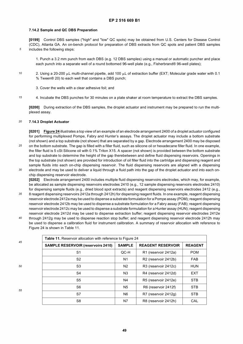
EP 2 516 669 B1
49
5
10
15
20
25
30
35
40
45
50
55
7.14.2 Sample and QC DBS Preparation
[0199] Control DBS samples ("high" and "low" QC spots) may be obtained from U.S. Centers for Disease Control(CDC), Atlanta GA. An on-bench protocol for preparation of DBS extracts from QC spots and patient DBS samplesincludes the following steps:
1. Punch a 3.2 mm punch from each DBS (e.g. 12 DBS samples) using a manual or automatic puncher and placeeach punch into a separate well of a round bottomed 96-well plate (e.g., Fisherbrand® 96-well plates);
2. Using a 20-200 mL multi-channel pipette, add 100 mL of extraction buffer (EXT; Molecular grade water with 0.1% Tween® 20) to each well that contains a DBS punch;
3. Cover the wells with a clear adhesive foil; and
4. Incubate the DBS punches for 30 minutes on a plate shaker at room temperature to extract the DBS samples.
[0200] During extraction of the DBS samples, the droplet actuator and instrument may be prepared to run the multi-plexed assay.
7.14.3 Droplet Actuator
[0201] Figure 24 illustrates a top view of an example of an electrode arrangement 2400 of a droplet actuator configuredfor performing multiplexed Pompe, Fabry and Hunter’s assays. The droplet actuator may include a bottom substrate(not shown) and a top substrate (not shown) that are separated by a gap. Electrode arrangement 2400 may be disposedon the bottom substrate. The gap is filled with a filler fluid, such as silicone oil or hexadecane filler fluid. In one example,the filler fluid is 5 cSt Silicone oil with 0.1% Triton X15. A spacer (not shown) is provided between the bottom substrateand top substrate to determine the height of the gap therebetween and define fluid dispensing reservoirs. Openings inthe top substrate (not shown) are provided for introduction of oil filler fluid into the cartridge and dispensing reagent andsample fluids into each on-chip dispensing reservoir. The fluid dispensing reservoirs are aligned with a dispensingelectrode and may be used to deliver a liquid through a fluid path into the gap of the droplet actuator and into each on-chip dispensing reservoir electrode.[0202] Electrode arrangement 2400 includes multiple fluid dispensing reservoirs electrodes, which may, for example,be allocated as sample dispensing reservoirs electrodes 2410 (e.g., 12 sample dispensing reservoirs electrodes 2410)for dispensing sample fluids (e.g., dried blood spot extracts) and reagent dispensing reservoirs electrodes 2412 (e.g.,8 reagent dispensing reservoirs 2412a through 2412h) for dispensing reagent fluids. In one example, reagent dispensingreservoir electrode 2412a may be used to dispense a substrate formulation for a Pompe assay (POM); reagent dispensingreservoir electrode 2412b may be used to dispense a substrate formulation for a Fabry assay (FAB); reagent dispensingreservoir electrode 2412c may be used to dispense a substrate formulation for a Hunter assay (HUN); reagent dispensingreservoir electrode 2412d may be used to dispense extraction buffer; reagent dispensing reservoir electrodes 2412ethrough 2412g may be used to dispense reaction stop buffer; and reagent dispensing reservoir electrode 2412h maybe used to dispense a calibration fluid for instrument calibration. A summary of reservoir allocation with reference toFigure 24 is shown in Table 11.
Table 11. Reservoir allocation with reference to Figure 24
SAMPLE RESERVOIR (reservoirs 2410) SAMPLE REAGENT RESERVOIR REAGENT
S1 QC-H R1 (reservoir 2412a) POM
S2 N1 R2 (reservoir 2412b) FAB
S3 N2 R3 (reservoir 2412c) HUN
S4 N3 R4 (reservoir 2412d) EXT
S5 N4 R5 (reservoir 2412e) STB
S6 N5 R6 (reservoir 2412f) STB
S7 N6 R7 (reservoir 2412g) STB
S8 N7 R8 (reservoir 2412h) CAL

EP 2 516 669 B1
50
5
10
15
20
25
30
35
40
45
50
55
[0203] Sample dispensing reservoir electrodes 2410 and reagent dispensing reservoir electrodes 2412 are intercon-nected through an arrangement, such as a path or array, of droplet operations electrodes 2414 (e.g., electrowettingelectrodes). Droplet operations are conducted atop droplet operations electrodes 2414 on a droplet operations surface.Electrode arrangement 2400 includes a reaction zone 2416. Electrode arrangement 2400 also includes a detection spot2418.[0204] The droplet actuator that includes electrode arrangement 2400 is designed to fit onto an instrument deck thathouses extra-droplet actuator features such as one or more thermal controllers (heater assemblies) for controlling thetemperature within certain processing zones, and a fluorimeter for fluorescence detection. In one example, the dropletactuator is deigned to fit into an NBS-LSD100 analyzer instrument that is connected to a desktop computer with SpotLogicsoftware installed. The droplet actuator may be provided and stored in a vacuum sealed aluminum pouch prior to use.
7.14.4 Assay Protocol
[0205] A high level overview of an example of the steps used to prepare a droplet actuator and run a digital microfluidicprotocol for multiplexed Pompe, Fabry and Hunter’s assays is shown in Table 12 below. In this example, the instrumentis an NBS-LSD100 analyzer that is connected to a desktop computer with SpotLogic software installed.
[0206] Initialization of the instrument (referring to Step 0 of Table 12) includes the following steps:
(continued)
Table 11. Reservoir allocation with reference to Figure 24
SAMPLE RESERVOIR (reservoirs 2410) SAMPLE REAGENT RESERVOIR REAGENT
S9 N8
S10 N9
S11 N10
S12 QC-L
Table 12. Overview of multiplexed Pompe, Fabry and Hunter assay protocol
SNo. Step Description
0 InitializationHardware components initialized. Thermal control set to heat the
cartridge to 37°C. Wait for setpoint to be reached. Read and verify actual voltage and temperature.
1 Oil loading Add 1.9mL of OIL into the cartridge.
2Sample and reagent
loadingAdd 3.4 mL of sample and 6.6 mL of reagents
3 Calibration Low Detect background fluorescence of stop buffer
4 Calibration High Detect background fluorescence of 0.15 mM of CAL
5 Substrate QCDetect fluorescence of (EXT + POM/FAB/HUN + STB) to determine
quality of substrate formulations
6up reactions Set up
reactionsMerge unit droplet of each DBS extract with unit droplet of POM/FAB/HUN
7 Incubate Incubate the reaction droplets for 1 hours at 37°C
8Detection of Pompe
assay dropletsMerge each Pompe assay droplet with unit droplet of STB and detect
end point fluorescence
9Detection of Fabry assay
dropletsMerge each Fabry assay droplet with unit droplet of STB and detect end
point fluorescence
10Detection of Hunter
assay dropletsMerge each Hunter assay droplet with unit droplet of STB and detect end
point fluorescence

EP 2 516 669 B1
51
5
10
15
20
25
30
35
40
45
50
55
1. Prior to starting each run, insert a "test cartridge" provided with the instrument into the instrument deck as specifiedin the instrument manual;
2. Engage the test cartridge to the voltage I/O pins by lowering the blue lever on the NBS-LSD1000 instrument; and
3. Open the SpotLogic™ software application and click "Test" to test for the electrowetting effector, fluorimeter andthe thermal controller.
[0207] Loading filler fluid in the droplet actuator (referring to Step 1 of Table 12) includes the following steps:
1. Remove a droplet actuator from its vacuum sealed aluminum pouch and place it flat on a bench surface;
2. Using a 1000 mL pipette (e.g., Fisherbrand® aerosol pipette tip) dispense 930 mL of filler fluid (e.g., 5 cSt Siliconeoil with 0.1% Triton® X15) into the droplet actuator through the 2 mM oil-input opening in the top substrate. Proceedslowly in order to avoid formation of air bubbles. This process is repeated twice so that the total volume of oil in thecartridge is 1.86 mL;
3. Remove any air bubbles that are formed in the reservoirs by carefully sucking out air and some oil from thereservoirs using a 20 mL pipette;
4. Top the remaining 2 mm openings with 30-50 mL of oil so that there is excess oil in all the openings. The oil-filledcartridge is left flat on the bench surface for 3 minutes to test for any oil leaks through the bond line between thetop substrate and the bottom substrate (e.g., a PCB). If oil is leaking from the bond line between the top substrateand bottom substrate, reject the droplet actuator and discard it into a biohazard bag. Obtain a new droplet actuatorand load the filler fluid; and
5. After loading the filler fluid in the droplet actuator and testing all the main components of the NBS-LSD100 analyzer(Step 0 of Table 12), select the instrument serial number in the software where intend to perform the testing andclick "Start". Follow the prompts on the software and install the droplet actuator in the instrument deck by lifting theblue lever. Stop when the software prompts "Load Samples".
[0208] Loading reagents and sample fluids in the droplet actuator (referring to Step 2 of Table 12) includes the followingsteps:
1. Following the instructions on the software, load the samples and reagents into the designated reservoirs usinga 20 mL pipette. Reservoir allocation as set-up in SpotLogic™ software is described in reference to Figure 24 andTable 11.
2. Load 3.4 mL of DBS extracts in the sample reservoirs as defined in the template in the SpotLogic™ software;
3. Load 6.6 mL of reagent fluids (POM, FAB, HUN, EXT, CAL, and STB) into their respective reagent dispensingreservoirs as specified by the SpotLogic™ software. Referring to Table 11, the order of reagent loading is STB inreservoirs R5, R6 and R7, CAL in reservoir R8, EXT in reservoir R4, POM in reservoir R1, FAB in reservoir R2 andHUN in reservoir R3;
4. A loading check (e.g., presence of air bubbles, volume) is performed visually during the loading of each reservoir.During reagent and sample loading, filler fluid (oil) may seep out of the reservoir openings in the top substrate.Seepage of oil may cause the formation of air bubbles in the reservoirs. To maintain the level of filler fluid and avoidthe formation of air bubbles, about 30 mL to 50 mL of oil may be added to all reservoir openings. Expertise in fluidloading and visual inspection may be acquired arid assessed, for example, during a training session on the instru-mentation and NBS assay protocol;
5. After loading all the samples and reagents in their respective reservoirs, check the oil level again and confirmthat all information that was entered is correct; and
6. Start the assay by clicking the "Start" button in the software.
[0209] Running the assay (referring to Steps 3 through 10 of Table 12) includes the following steps:
EP 2 516 669 B1
52
5
10
15
20
25
30
35
40
45
50
55
1. The initial steps of the assay, calibration (Steps 3 and 4) and substrate QC (Step 5) are monitored to identify any"flags". A flag may be generated due to malfunctioning of either the droplet actuator or the instrument. If any flagsare observed, stop the assay, remove the cartridge and initiate a new run;
2. If no flags are observed, close the instrument lid and allow about 2.5 hours to obtain 36 data points (3-plex assayson 12 samples); and
3. After completion of the run, lift the blue lever to disengage the droplet actuator from the I/O connector pins andcarefully remove the droplet actuator from the instrument deck. Discard the droplet actuator into a bio-hazard bagfor disposal.
7.15 Hunter’s Assay Using DBS Samples on a Droplet Actuator
[0210] Described are a digital microfluidic platform and assay methods using dried blood spot (DBS) samples fordetection of the lysosomal storage disorder Hunter’s syndrome (mucopolysaccharidosis II). Hunter’s syndrome is causedby deficient activity of the lysosomal enzyme iduronate-2-sulfate sulphatase (IDS). The substrate fluid for IDS in anenzymatic assay may, for example, be the fluorogenic substrate 4-methylumbelliferyl-α-L-iduronide-2-sulfate (MU-αIdoA-2S). The use of MU-αIdoA-2S as a fluorescent substrate to measure the activity of IDS requires the sequential actionof a second enzyme, α-L iduronidase, to convert the product of the sulphatase, 4-methylumbeliferyl-α-L-iduronic acid,into iduronic acid and 4-MU as described in reference to Figure 1. The microfluidic protocol is a single-step homogenousassay using purified recombinant iduronidase that is performed at a single pH (i.e., pH from about 4.5-5.5, or from about4.8-5.2, or from about 4.9-5.1, or about 5.0), with a time to result of 8 hours or less. The microfluidic protocol is performedwith a time to result (from extraction of DBS to result) of about 2 hours or about 90 minutes or less. Other examples maymake use of the alternative substrates described herein.[0211] The digital microfluidics platform may, for example, include a disposable, self-contained droplet actuator inwhich the enzymatic reaction is performed in aqueous droplets within an oil filled gap of the droplet actuator. Samplesand assay reagents (e.g., substrate, stop buffer) are manipulated as discrete droplets upon an electrode array (digitalelectrowetting). The electrode array may, for example, be fabricated on a printed-circuit-board (PCB) and enclosed inan oil (e.g., polydimethylsiloxane 2 cst, Gelest, Inc.) filled droplet actuator. The droplet actuator may be designed to fitinto the deck of, or otherwise electronically coupled to, an instrument that incorporates all control and detection capa-bilities. The interface between the droplet actuator and instrument device may, for example, be provided by spring loadedconnector pins between the device and contact pads on the droplet actuator. Samples and reagents are introduced intothe droplet actuator through fluid loading ports and on-chip reservoirs. The droplet actuator may be inserted into thedevice which automatically performs all assay manipulations including dispensing required volumes of samples andreagents, mixing, incubation, reaction termination and detection. A droplet may be selectively dispensed from any res-ervoir, transported, combined with other droplets and divided thereby replicating all of the required liquid manipulationsto perform an enzymatic assay. The volume of a single droplet on the droplet actuator may, for example, be 300 nL.Detection of the fluorescent sodium umbelliferone product (4-MU; 360 nm excitation, 460 nm emission) may, for example,be achieved using a fluorimeter module in an epi-illumination configuration mounted directly above a droplet actuatordetection electrode. Filtered excitation light from an ultraviolet LED is directed at the sample and collected back alongthe same optical path. Fluorescent light greater than 427 nm wavelength passes through a dichroic beam splitter andan emission filter to a photodiode. The LED excitation and collection electronics may be in a lock-in configuration toreduce background noise. Specifically, the LED oscillates at 1 kHz and the photodiode signal is sampled at 32 kHz forone second. The reported fluorescent signal is obtained by examining the magnitude of the collected signal at 1 kHzusing a fast Fourier transform.[0212] Figure 25 shows a plot 2500 of IDS activity in a Hunter’s assay performed on the digital microfluidic platformusing extracts from DBS samples. A stock solution of recombinant human α-L-iduronidase (>7.5 nmoles/min/mg; 10mg/ml iduronidase in 0.05 M sodium acetate, 0.5 M sodium chloride, 1 mg/ml BSA, 0.01 % Tween® 20, pH 5.0 buffer)was prepared and stored in 10 mL aliquots at -80°C. Iduronidase-2-sulfate sulfatase (IDS) assay solution was preparedfresh by thawing an aliquot of iduronidase. Unused iduronidase was discarded and not refrozen. Dried blood spots (3mm punches) from presumed normal individuals and from Hunter affected individuals were reconstituted on-bench bygentle mixing in 100 mL 0.1% Tween® 20 in microfuge tubes for 30 minutes at room temperature. Prepared reagentsand DBS samples were loaded onto fluid dispensing reservoirs of a droplet actuator.[0213] The digital microfluidic protocol for measuring IDS activity included the following steps: One droplet of recon-stituted DBS extract was combined using droplet operations with one droplet of assay solution (1.125 mM 4-methylum-belliferyl-α-L-iduronate-2-sulfate, 1 mg/ml recombinant human α-L-iduronidase, 0.1 M sodium acetate, and 0.01 M leadacetate, pH 5.0) to yield a 2X reaction droplet. The 2X reaction droplet was split using droplet operations to yield two1X reaction droplets. One 1X reaction droplet was immediately combined using droplet operations with one 1X droplet

EP 2 516 669 B1
53
5
10
15
20
25
30
35
40
45
50
55
of termination buffer (0.2 M sodium bicarbonate, 0.01% Tween® 20, pH 10.0) and transported to the detector electrodeto measure the fluorescent signal at zero time. The second 1X reaction droplet was incubated on the droplet actuatorfor one hour at 37°C. After the incubation period, the 1X reaction droplet was combined with a 1X droplet of terminationbuffer and transported to the detector electrode to measure the fluorescent signal at time t=1 hour. The activity of IDSis expressed in micromoles product formed per hour per liter blood. The IDS activity was calculated based on a 3 mmDBS punch containing 3.1 mL of blood. Assay calibration was performed on the droplet actuator using sodium 4-meth-ylumbelliferone in 0.2 M sodium bicarbonate, 0.01% Tween® 20, pH 10.0 termination buffer as a standard.[0214] In this example, an iduronidase concentration of 1 mg/ml in the IDS assay was sufficient to liberate 4-methyl-umbelliferone (4-MU) from 4-methylumbelliferyl-α-L-iduronic acid after IDS removal of the sulfate moiety from 4-meth-ylumbelliferyl-α-L-iduronide-2-sulfate (data not shown). Dilution (31-fold) of blood during extraction of the DBS and thepresence of 10 mM lead acetate in the substrate buffer as in section 7.14.1 was sufficient to substantially prevent inhibitionof iduronidase-2-sulfate sulfatase activity by anions such as chloride, phosphate and sulfate. in the reaction droplet.[0215] The IDS activity in extracts of DBS from random newborns (n=105 random newborn DBS samples) and Hunterpatients (n=6 affected DBS samples) was measured by two operators using two different instruments and droplet actu-ators. The data show the IDS activity in the Hunter patients (range, 0-2.7; mean, 1.1; median, 1.0 mmol/h/L blood witha standard deviation of 1.48) was well below and clearly separated from the IDS activity found in presumed normalnewborns (range, 10.4-56.9; mean, 22.9; median 20.2 mmole/h/L blood with a standard deviation of 9.7). It is not knownif any of the DBS from the random newborn pool were from carriers of the Hunter trait.
7.16 Single-Step Assay for Hunter’s Syndrome
[0216] Described is an enymatic assay for Hunter’s Syndrome. The microfluidic protocol is a single-step homogenousassay using purified recombinant iduronidase that is performed at a single pH (i.e., pH 5.0) with a time to result of 8hours or less. On-chip reaction volumes i.e., 2 mL Hunter enzyme and 18 mL Hunter substrate may be scaled to on-bench volumes, i.e., 10 mL Hunter enzyme and 90 mL of Hunter substrate. The incubation time for sufficient enzymatichydrolysis of substrate and time to result may be selected for applications. The assay may, for example, use extractsprepared from dried blood spot (DBS) samples to test for Hunter’s disease. A stock solution of Hunter substrate (HS;4-methylumbelliferyl-α-L-iduronide-2sulfate Na2; Moscerdam Substrates) may be prepared in Hunter substrate buffer(HSB; 0.1 M Sodium Acetate, 10 mM lead (II) acetate (Sigma Aldrich), 0.01% (w/v) Tween® 20). The pH of the Huntersubstrate buffer (HSB) may from about 4 to about 6, preferably from about 4.5 to about 5.5, ideally about 5. A stocksolution of Hunter enzyme formulation (HE; recombinant human α-L-iduronidase; 10 mg/20 mL; R&D Systems) may beprepared in Hunter enzyme buffer (HEB; 0.05 M Sodium Acetate, 0.01% (w/v) Tween® 20, 1 mg/mL bovine serumalbumin (BSA); Sigma Aldrich). The pH of the Hunter enzyme buffer (HEB) may from about 4 to about 6, preferably fromabout 4.5 to about 5.5, ideally about 5. Stock solutions may be prepared, aliquoted, and stored at -80 °C until use.
Preparation of the Hunter substrate formulation (HS) for an on-bench Hunter assay includes the following steps:
[0217]
1. Add 3 mL of Hunter substrate buffer (HSB; 0.1 M Sodium Acetate, pH 5.0, 10 mM lead (II) acetate (Sigma Aldrich),0.01% (w/v) Tween® 20) to a 5 mg vial of 4-methylumbelliferyl-α-L-iduronide-2sulfate Na2. Dissolve and transferthe solution to a 10 mL conical, polystyrene tube wrapped in foil;
2. Rinse the vial by adding a second 3 mL of HSB to the vial and mix vigorously. Transfer this solution to the same10 mL conical tube;
3. Add 2.33 mL of HSB to the same 10 mL conical tube to yield a total volume of 8.33 mL.
4. Aliquot 105 mL of the solution to individual microfuge tubes; and
5. Store at -80 °C protected from light.
Preparation of the Hunter enzyme (HE) formulation for the Hunter assay includes the following steps:
[0218]
1. Add 20 mL of Iduronidase to 980 mL of Hunter enzyme buffer (HEB; 0.05 M Sodium Acetate, pH 5.0, 0.01% (w/v)Tween® 20, 1 mg/mL bovine serum albumin (BSA; Sigma Aldrich));

EP 2 516 669 B1
54
5
10
15
20
25
30
35
40
45
50
55
2. Gently vortex until completely dissolved;
3. Aliquot 13 mL of the solution to individual microfuge tubes; and
4. Store at -80 °C.
[0219] An assay protocol for Hunter’s syndrome includes, but is not limited to, the following steps:
1. Extract one 3 mm DBS sample punch in 100 mL of extraction buffer (0.1%. w/v Tween® 20 in molecular gradewater) for 30 min at room temperature on a shaking platform at 1400 rpm;
2. At the end of the extraction, transfer the liquid to a 1.5 mL microfuge tube;
3. Test each sample in duplicate. Each aliquot of HS and HE is sufficient for testing 5 samples in duplicate, 10reactions total;
4. Prepare Hunter working substrate solution (HWSS) by combining 99 mL of HS with 11 mL of HE. Mix well andprotect from light;
5. In a Costar black, half-area microtiter plate, combine 10 mL of DBS extract and 10 mL HWSS. Avoid using thewells on the edge of the plate (i.e., rows A and H and columns 1 and 12);
6. Seal the plate with adhesive plate sealer and wrap the plate in aluminum foil;
7. Incubate the reactions at 37 °C for 20 hrs;
8. At the end of the incubation period, stop the reaction with 50 mL of stop buffer (0.2 M sodium bicarbonate, pH10.1) and mix well; and
9. Read fluorescence at 360/460 excitation/emission at a gain of 60.
[0220] Figure 26 shows a bar graph 2600 of fluorescence data of a single-step on-bench assay for Hunter’s syndrome.DBS extracts were prepared from quality control (QC) samples, i.e., base pool (BP), low (L), medium (M) and high (H)activity samples. The base pool (BP) QC sample was prepared from a pool of leukoreduced human red blood cells thatwas adjusted with heat-treated serum to a hematocrit of 50%. The High (H) activity QC sample was prepared frompooled cord blood that was adjusted with heat-treated serum to a hematocrit of 50%. The medium (M) activity QC samplewas prepared by using 50% base pool (BP) and 50% high (H) activity sample. The low (L) activity QC sample wasprepared by using 95% base pool (BP) and 5% high (H) activity sample. The BP sample is used as a control for hydrolysisnon-specific to white blood cell lysosomal enzymes. The data show a strong signal and clear separation between L, Mand H samples.[0221] Figure 27 shows a bar graph 2700 of another example of fluorescence data of a single-step on-bench assayfor Hunter’s syndrome. In this example, DBS extracts were prepared from BP, L, M, and H QC samples, 10 normal (N;unaffected) DBS samples and 2 Hunter’s (MPS-II; affected) DBS samples. The data show a strong signal and clearseparation between normal and affected (MPS-II) samples. Higher levels of enzyme activity in the normal samplesrelative to QC samples may be due to the presence of enzyme activity in the non-heat treated normal samples. Thelevel of enzyme activity in the non-heated treated Hunter’s samples is essentially reduced to BP levels.
7.17 Assay for Fabry Disease
[0222] Described is an enzymatic assay for Fabry disease (described in reference to Tables 10 and 12 and Figure24) may be adapted to an on-bench protocol. The assay uses extracts prepared from dried blood spot (DBS) samplesto test for Fabry disease. Stock solutions for Fabry substrate, 4-Methylumbelliferyl α-D-galactopyranoside (40MUGal;MW=338.3 g/mol; Sigma) may be prepared in Dimethly Sulfoxide (DMSO, 99.9%) at a concentration of 700 mM. Stocksolutions of Fabry inhibitor (750 mM of N-Acetyl-D-Galactosamine; Sigma) may be prepared in molecular biology gradewater. Fabry substrate and inhibitor stock solutions may be prepared, aliquoted and stored at -80 °C until use. The on-bench assay may, for example, be performed in multi-well microtiter plates (e.g., Costar black half-area plates). Amicrotiter plate reader (e.g., Biotek KC4 plate reader) may be used for fluorescence detection.[0223] An on-bench assay protocol for Fabry disease includes, but is not limited to, the following steps:

EP 2 516 669 B1
55
5
10
15
20
25
30
35
40
45
50
55
1. Extract one 3 mm DBS sample punch in 100 mL of extraction buffer (0.1% w/v Tween® 20 in molecular gradewater) for 30 min at room temperature on a shaking platform at 1400 rpm;
2. At the end of the extraction, transfer the liquid to a 1.5 mL microfuge tube;
3. Prepare a Fabry working substrate solution (FWS) by combining 78.6 mL Fabry enzyme buffer (0.04 M SodiumAcetate with 0.01% (w/v) Tween® 20, pH4.6) with 20 mL Fabry inhibitor stock solution and 1.4 mL Fabry substratestock solution;
4. In a Costar black, half-area microtiter plate, combine 10 mL of DBS extract and 10 mL Fabry working substratesolution (FWS). Run each sample in duplicate;
5. Seal the microtiter plate with adhesive plate sealer and wrap the plate in aluminum foil;
6. Incubate the reactions at 37 °C for 20 hrs;
7. At the end of the incubation period, stop the reaction with 50 mL of stop buffer (0.2 M sodium bicarbonate, pH10.1) and mix well; and
8. Read fluorescence at 360/460 excitation/emission at a gain of 75.
7.18 Enzyme-Mediated Release of Nucleic Acids from DBS
[0224] The use of dried blood spots (DBS) collected on filter paper for nucleic acid-based testing of infectious diseases,newborn testing and SCIDS is widespread in the public health community. In nucleic acid-based testing protocols, suchas PCR, the entire DBS sample is inserted into a PCR tube and amplified. For nucleic acid testing on a droplet actuator,the DBS sample may be extracted from the filter paper prior to analysis. However, the release of nucleic acids (e.g.,DNA and RNA) from the DBS samples is difficult and inefficient when using chemical extraction methods and may resultin gross underestimations of target nucleic acid concentrations by up to several orders of magnitude. The inefficiencyof nucleic acid extraction from DBS samples has been attributed to the physical interaction (e.g., intertwined) of thenucleic acid molecules with the fibers of the filter paper.[0225] The use of DBS collected on filter paper for enzyme-based testing of infectious diseases, newborn testing andSCIDS is widespread in the public health community. For enzyme-based testing on a droplet actuator, the DBS samplemay be extracted from the filter paper prior to analysis. However, the release of enzymes from the DBS samples isdifficult and inefficient when using chemical extraction methods and may result in gross underestimations of targetenzyme acid concentrations by up to several orders of magnitude. The inefficiency of enzyme extraction from DBSsamples has been attributed to the physical interaction (e.g., intertwined) of the enzyme molecules with the fibers of thefilter paper.[0226] Described are methods for using enzymes to release nucleic acids in DBS samples collected on filter paper.A cellulose degrading enzyme, such as cellulase, may used to partially or totally solubilize the filter paper matrix torelease substantially all of the nucleic acid in the sample into an extraction buffer. In one example, the cellulase complexfrom Trichoderma reesei (formerly T. viride) may be used to release nucleic acids in DBS samples collected on filterpaper. The cellulase complex from T. reesei quantitatively converts cellulose to glucose. An example of an on-benchprotocol to release nucleic acids from a DBS collected on filter paper may include the following: The DBS sample maybe washed (e.g., two or three times) with an aqueous buffer to remove soluble PCR inhibitory components from theDBS. The washed DBS sample may be incubated with a cellulose degrading enzyme, such as cellulase from T. reesei,to release nucleic acids in the DBS. Nucleic acids in the sample may be concentrated using a magnetic bead-basedprotocol to reduce the sample volume prior to loading the extracted blood sample onto a droplet actuator. The concentratedDBS extract may be loaded onto a sample dispensing reservoir of a droplet actuator and dispensed for quantitative PCRanalysis using a digital microfluidic protocol.[0227] The method may be used for the enzymatic assays described herein. A cellulose degrading enzyme, such ascellulase, may used to partially or totally solubilize the filter paper matrix to release substantially all of the enzyme in thesample into an extraction buffer.
7.19 Digital Microfluidic Platform for Bilirubin, G6PD and CH Newborn Testing
[0228] Described are a digital microfluidic platform and assay methods for multiplexed testing of newborns. The digitalmicrofluidic platform includes a multi-well droplet actuator that may be configured for one or more molecular assays for

EP 2 516 669 B1
56
5
10
15
20
25
30
35
40
45
50
55
newborn testing. Because of the software programmability of digital microfluidics, most of the parameters varied betweenand within protocols, such as incubation times, sequences of reagent additions, and thermal programs, may be readilyadapted to different assay protocols.[0229] The digital microfluidic platform may be configured for multiplexed testing of newborns that are at risk forhyperbilirubinemia and its most common underlying pathological causes, glucose-6-phosphate dehydrogenase (G6PD)deficiency, and congenital hypothyroidism (CH). On-bench assays for determination of total bilirubin, G6PD activity andCH (i.e., TSH assay) may be described and implemented on a droplet actuator as discrete step-by-step droplet-basedprotocols. Assay protocol parameters may, for example, be selected for linearity, increased sensitivity (limit of detection),specificity, droplet carryover, and rapid time-to-result.[0230] The droplet actuator may be designed to fit into, or otherwise be electrically coupled to, an instrument deckthat that houses other components that are external to the droplet actuator. Examples of external components include,but are not limited to, one or more magnets for immobilization of magnetically responsive beads, one or more heaterassemblies for controlling the temperature within certain processing zones, and a detection system.
7.19.1 Total Bilirubin Assay
[0231] Assays for determination of total bilirubin (serum or plasma) levels may be adapted for use on a droplet actuator.In one example, the total bilirubin assay kit (B576-480) available from Teco Diagnostics may be adapted for use on adroplet actuator. The total bilirubin assay is a colorimetric assay based on the reaction between bilirubin and diazotizedsufanilic acid to produce azobilirubin which has an absorbance maximum at 560 nm wavelength in the presence ofdimethylsulfoxide (DMSO) solvent. The kit includes the following reagents: Total Bilirubin Reagent (sulfanilic acid 32mM, hydrochloric acid 165 mM, DMSO 7 M (55% w/v)); and Bilirubin Nitrite Reagent (sodium nitrite 60 mM); BilirubinCalibrator (20 mg/dL N-1-naphthylethylenediamine dihydrochloride salt). Working reagents and calibration solutionswere prepared on-bench as follows: Total bilirubin working reagent was prepared on-bench before each experiment byadding 50 mL of sodium nitrite reagent to 1.0 mL of total bilirubin reagent. Bilirubin calibrator solutions were preparedon-bench at 3, 7, 10, 14, and 20 mg/dL by diluting the 20 mg/dL calibrator stock using deionized water.[0232] Figures 28A and 28B show plots 2800 and 2850 of bilirubin calibration curves obtained on-chip and a scatterplot of on-bench values, respectively. Discarded and deidentified pediatric serum samples were obtained from DukeUniversity (Durham, NC) along with the total bilirubin values. The on-bench reference assay (Duke Method) was per-formed before (e.g., a day before) the on-chip assay. Prepared reagents, calibrators and serum samples were loadedonto fluid dispensing reservoirs of a droplet actuator. The on-chip assay protocol included the following steps: Forcalibration, a 1X droplet of calibrator was combined using droplet operations with two 1X droplets of working reagent toyield a 3X droplet. The 3X droplet was split using droplet operations into a 1X droplet and a 2X droplet. The 1X dropletwas combined with two 1X droplets of working reagent. For serum blank, a 1X droplet of serum was combined usingdroplet operations with two 1X droplets of total bilirubin reagent to yield a 3X droplet. The 3X droplet was split usingdroplet operations into a 1X droplet and a 2X droplet. The 1X droplet was combined with two 1X droplets of total bilirubinreagent. For unknown serum bilirubin, a 1X droplet of serum was combined using droplet operations with two 1X dropletsof working reagent to yield a 3X droplet. The combined droplet was incubated at 37 °C for 5 minutes. The 3X dropletwas split using droplet operations into a 1X droplet and a 2X droplet. The 1X droplet was combined with two 1X dropletsof working reagent. The combined droplet was incubated at about 37 °C for about 5 minutes. Absorbance was read at560 nm wavelength using a LED-photodiode colorimeter. All experiments were performed in triplicate on three differentdroplet actuators. For both the on-bench and on-chip experiments, the calibration curve was obtained by linear regressionanalysis in Microsoft Excel®. The absorbance of the serum blank is subtracted from serum absorbance. The blankcorrected serum absorbance value is used to obtain the unknown bilirubin value using the regression equation. Theexperiments were performed using eleccrowetting mediated droplet operations in silicon oil.[0233] The data shows the calibration curve is linear up to 20 mg/dL. Examination of the scatter plot indicates thatthere is a small proportional bias in the assay on cartridge when compared to the on-bench reference assay. This maybe expected while using diazo methods for bilirubin assay. The correlation is good and is may be improved by performingthe reference assay and the on-chip assay at the same time. In this example, there was about a one day delay betweenthe reference test and on-chip assay. Bilirubin is a very labile analyte and the method comparison should ideally beperformed at the same time. In another example, a direct bilirubin test as a marker for hyperbilirubinemia may be adaptedfor use on a droplet actuator.
7.19.2 G6PD Assay
[0234] Neonatal testing test kits for glucose-6-phosphate dehydrogenase (G6PD) deficiency are commercially avail-able and may be adapted for use on a droplet actuator. In one example, the G6PD deficiency neonatal testing test kitavailable from Interscientific Inc. may be adapted for use on a droplet actuator. The assay is based on the oxidation of

EP 2 516 669 B1
57
5
10
15
20
25
30
35
40
45
50
55
glucose-6-phosphate to 6-phosphogluconate, and reduction of NADP to NADPH, in the presence of glucose-6-phos-phate-dehydrogenase. The NADPH produced reduces tetrazolium dye (MTT) in the presence of phenazine methosulfateto produce a colored product with an absorbance peak at 565nm. The kit includes the following reagents: R1 (Elution/LysisBuffer), R2 (Work Reagent), R3 (Color Reagent), R4 (CRB; Color Reagent Buffer). To prepare the Working ColorReagent, 1 part of CRB (R4) and 10 parts of Color Reagent (R3) are mixed together on-bench. The kit also includesassay controls representing normal, intermediate and deficient G6PD values.[0235] Figure 29 shows a plot 2900 of the G6PD assay performed on-chip. Pediatric whole blood samples (n=8presumed normal samples) were obtained from Duke University. Prepared reagents, assay controls (normal, intermediateand deficient) and whole blood samples (n=8 presumed normal) were loaded onto fluid dispensing reservoirs of a dropletactuator. The on-chip assay protocol for whole blood samples and controls included the following steps: A 1X dropletof whole blood (or control) was combined using droplet operations with three 1X droplets of elution/lysis buffer (R1) andincubated for 1 minute. The combined 4X droplet was split using droplet operations to yield a 1X droplet and a 3X droplet.The 1X droplet was combined using droplet operations with a 1X droplet of work reagent (R2) and incubated for 30seconds. The combined 2X droplet was split using droplet operations to yield two 1X droplets. One 1X droplet wascombined using droplet operations with two 1X droplets of work reagent (R2) and incubated for 30 seconds. The combined3X droplet was split using droplet operations into a 1X droplet and a 2X droplet. The 2X droplet was transported usingdroplet operations to a detection spot and absorbance read at 405 nm wavelength for hemoglobin normalization. Theremaining 1X droplet was combined using droplet operations with a 1X working color reagent droplet and absorbanceread at 560 nm wavelength in kinetic mode. Sample concentration may be expressed using the following formula:
[0236] The data shows good separation between deficient, intermediate and normal samples in the on-chip G6PDassay.
7.19.3 Congenital Hypothyroidism (CH) Assay
[0237] Congenital hypothyroidism (CH) is currently screened for in some newborn testing programs using a primarythyroid stimulating hormone (TSH) assay. Assays for determination of TSH levels may be adapted for use on a dropletactuator. In one example, the Access HYPERsensitive hTSH kit (Cat # 33820) available from Beckman Coulter may beadapted for use on a droplet actuator. The assay is a paramagnetic particle, chemiluminescent assay for the quantitativedetermination of human thyroid-stimulating hormone (hTSH). The kit includes the following reagents: paramagneticbeads coated with primary capture antibody, blocking solution and alkaline phosphatase (ALP) labeled secondary an-tibody. Attoglow (Michigan Diagnostics) may be used as the chemiluminescent substrate.[0238] Figure 30 shows a plot 3000 of a TSH calibration curve generated on-chip. A reagent mixture was preparedon-bench by mixing equal volumes of primary antibody bound to magnetically responsive beads, blocking solution andsecondary antibody labeled with ALP. Prepared reagent mixture, TSH standards (at different concentrations), washbuffer and chemiluminescent substrate were loaded onto fluid dispensing reservoirs of a droplet actuator. The on-chipassay protocol included the following steps: A 1X droplet (300 nL) of reagent mixture was combined using dropletoperations with two 1X droplets of a TSH standard and incubated for 4 minutes at room temperature. After the incubationperiod, the magnetically responsive beads were washed to remove any unbound secondary antibody by immobilizingthe beads to the edge of the 4X reaction droplet using an external permanent magnet and removing the excess unboundmaterial by activating the adjacent electrodes and deactivating the intermediate electrode. One 1X droplet of chemilu-minescent substrate was combined with the bead containing droplet. After 2 minute incubation, end point chemilumins-cence was measured using a photon counting photomultiplier tube. A 4-parameter logistic fit was used to fit the data.The error bars represent standard deviation from four different assays. Cut-off concentration for TSH that is used bymost testing laboratories (US) is 20 mL U/mL.[0239] In another example, the chemiluminescent TSH assay may be adapted for use on a droplet actuator as acolorimetric assay. In this example, an alkaline phosphatase (ALP) labeled secondary antibody may be used with acolorimetric substrate, such as p-nitrophenol phosphate (detection wavelength 405 nm), for detection of ALP. Assayparameters may be selected for increased sensitivity in the colorimetric assay. For example, the sample volume that isincubated with the primary capture beads may be increased from about 0.3 mL to about 25 mL. The detection reactiontime may, for example, be increased from about 2 minutes to about 15 minutes. The assay temperature may, for example,be increased from room temperature to 37 °C. The path length at the detection window may, for example, be increasedfrom about 0.3 mm to about 1 mm.

EP 2 516 669 B1
58
5
10
15
20
25
30
35
40
45
50
55
7.19.4 Multiplexed Bilirubin, G6PD and TSH Assays on a Droplet Actuator
[0240] Figure 31 illustrates a flow diagram of an example of a protocol 3100 for multiplexed newborn testing for totalbilirubin, G6PD and TSH on a droplet actuator. Protocol 3100 may include, but is not limited to, the following steps: Inone step, a sample of whole blood (e.g., about 100 mL) is collected from a heel stick using a 100 mL capillary bloodcollection tube (e.g., SAFE-T-FILL® collection tubes from Ram Scientific). In another step, an aliquot of the whole blood(e.g., about 10 mL) is transferred to a fluid reservoir of a droplet actuator for the G6PD assay. In another step, theremaining whole blood sample (e.g., about 90 mL) is centrifuged to obtain serum. In one embodiment, a bench-topcentrifuge may be used to separate serum from whole blood. In another embodiment, an on-chip separation method(e.g., lateral flow filters) may be used to prepare serum from whole blood. In another step, an aliquot of the serum (e.g.,about 30 mL) is transferred to another fluid reservoir of the droplet actuator for dispensing for total bilirubin and TSHassays. For the bilirubin assay, about 0.5 mL of serum is used. For the TSH assay, about 25 mL of serum is used.[0241] Figure 32 shows a top view of an example of an electrode arrangement 3200 of a droplet actuator configuredfor performing multiplexed total bilirubin, G6PD and TSH assays on a droplet actuator. In this example, the dropletactuator is configured for performing a 3-plex assay on 6 samples. The droplet actuator may include a bottom substrate(not shown) and a top substrate (not shown) that are separated by a gap. The bottom substrate may, for example, bea printed circuit board (PCB). Electrode arrangement 3200 may be disposed on the bottom substrate. The gap is filledwith a filler fluid, such as silicone oil. Openings in the top substrate (not shown) are provided for introduction of oil fillerfluid into the cartridge and dispensing reagent and sample fluids into each on-chip dispensing reservoir. The fluid dis-pensing reservoirs are aligned with a dispensing electrode and may be used to deliver a liquid through a fluid path intothe gap of the droplet actuator and into each on-chip dispensing reservoir electrode. In one embodiment, bulk liquidreagents including filler fluid (e.g., oil) may be provided in dropper bottles to facilitate reagent loading and minimizepipetting steps, which may be done robotically. Because precision aliquoting of reagents is performed inside the dropletactuator using electrowetting-based droplet operations, variations in fluid input volume are readily tolerated. Dry reagentsmay be provided in metered containers in which they can be reconstituted and frozen if necessary.[0242] Electrode arrangement 3200 includes multiple fluid dispensing reservoirs electrodes, which may, for example,be allocated as sample dispensing reservoirs electrodes 3210 (e.g., 12 sample dispensing reservoirs electrodes 3210athrough 32101) for dispensing sample fluids (e.g., whole blood, plasma or serum), reagent dispensing reservoirs elec-trodes 3212 (e.g., 12 reagent dispensing reservoirs 3212a through 32121) for dispensing reagent fluids, a wash bufferdispensing reservoir 3214, a substrate dispensing reservoir 3216, and a waste collection site 3218. In one example,reagent dispensing reservoir electrodes 3212a through 3212c may be used to dispense reagents for the TSH assay(e.g., primary and secondary antibodies, calibration adjustor); reagent dispensing reservoir electrodes 3212d through3212f may be used do dispense reagents for the total bilirubin assay (e.g., reagents and calibrator); reagent dispensingreservoir electrodes 3212g and 3212h may be used dispense dilution buffer; reagent dispensing reservoir electrodes3212i through 32121 may be used to dispense reagents for the G6PD assay (e.g., lysis buffer, reagents, standard).Sample dispensing reservoirs 3210 may be used to dispense plasma and whole blood samples from six different samplesets. For example, sample dispensing reservoir electrodes 3210a and 3210b may be used to dispense plasma andwhole blood, respectively, from a first sample; sample dispensing reservoir electrodes 3210c and 3210d may be usedto dispense plasma and whole blood, respectively, from a second sample; sample dispensing reservoir electrodes 3210eand 3210f may be used to dispense plasma and whole blood, respectively, from a third sample; sample dispensingreservoir electrodes 3210g and 3210h may be used to dispense plasma and whole blood, respectively, from a fourthsample; sample dispensing reservoir electrodes 3210i and 3210j may be used to dispense plasma and whole blood,respectively, from a fifth sample; sample dispensing reservoir electrodes 3210k and 32101 may be used to dispenseplasma and whole blood, respectively, from a sixth sample.[0243] Sample dispensing reservoir electrodes 3210, reagent dispensing reservoir electrodes 3212, wash buffer dis-pensing reservoir electrode 3214, substrate dispensing reservoir electrode 3214 and waste collection site 3218 areinterconnected through an arrangement, such as a path or array, of droplet operations electrodes 3220 (e.g., electrowet-ting electrodes). Droplet operations are conducted atop droplet operations electrodes 3220 on a droplet operationssurface. A path of droplet operations electrodes 3220 extending from each sample dispensing electrode forms dedicatedelectrode lanes 3222, i.e., 12 dedicated electrode lanes 3222a through 32221. Dedicated electrode lanes 3222 proveindividual reaction zones for processing different samples and sample types (i.e., plasma and whole blood). The use ofdedicated lanes for sample droplets minimizes cross-contamination among samples.[0244] One or more magnets 3224 (e.g., six magnets 3224a through 3224f) may be positioned in proximity to certaindroplet operations electrodes 3220 for retaining a quantity of magnetically responsive beads. Each magnet 3224 may,for example, be a permanent magnet or an electromagnet. Each magnet 3224 is positioned in a manner which ensuresspatial immobilization of magnetically responsive beads during washing steps. Mixing and incubations may be performedon certain droplet operations electrodes 3220 away from the magnet.[0245] Electrode arrangement 3200 includes multiple detection electrodes 3226 (e.g., 12 detection electrodes 3226a

EP 2 516 669 B1
59
5
10
15
20
25
30
35
40
45
50
55
through 32261). Detection electrodes 3226 are positioned in proximity to certain droplet operations electrodes in eachdedicated electrode lanes 3222. The use of independent detection electrodes for sample droplets minimizes cross-contamination among samples. Detection electrodes 3226 may be fabricated as optically transparent electrodes asdescribed in reference to Figure 33. Detection electrodes 3226 may be aligned with certain optical detection channels(e.g., 560 nm channel or 405 nm channel) as described in reference to Figure 34.[0246] Because of the flexibility and programmability of a droplet actuator, the architecture of the droplet actuator maybe readily configured to accommodate fewer or more samples.[0247] For total bilirubin and G6PD assays, a single point calibration curve may be generated on-chip as the assaysare substantially linear over the required range. For the TSH assay, a master calibration curve for every droplet actuatorlot number or reagent batch may be generated. A calibration adjustor may be run on-chip to make adjustments to thecalibration curve. This approach is commonly used in immunoassay analyzers. Positive and negative controls may berun to identify systematic errors such as a defective droplet actuator or bad reagent lot. A detailed Design Failure Modesand Effects Analysis (FMEA) may be conducted to identify both external and internal quality control needs.[0248] Figure 33 illustrates a top view of a portion of a droplet actuator 3300 that includes optically transparent detectionelectrodes suitable for detection of colorimetric reaction products. Droplet actuator 3300 may include a bottom substrate3310. Bottom substrate 3310 may, for example, be a PCB. An arrangement, such as a path or array, of droplet operationselectrodes 3312 (e.g., electrowetting electrodes) may be disposed on the bottom substrate 3310. Droplet operationsare conducted atop droplet operations electrodes 3312 on a droplet operations surface. Droplet actuator 3300 mayinclude a detection electrode 3314. A plated through hole 3316 may be fabricated in detection electrode 3314. Platedthrough hole 3316 may, for example, be fabricated as unfilled via holes. Plated through hole 3316 may, for example, beabout 250 mm in diameter. Plated through hole 3316 provides for transmission of light (e.g., from an LED) through bottomsubstrate 3310. Because plated through hole 3316 is sufficiently small relative to the size of detection electrode 3314(e.g., about 1000 mm), electrowetting performance is substantially unaffected.
7.19.5 Instrument Platform
[0249] Described is an instrument platform that is suitable for use in limited resource settings. The instrument may bea small, bench-top instrument that is light weight and portable. The instrument may be a small handheld device (e.g.,about 10"x5"x5") that is battery powered (e.g., 8 AA batteries). The instrument houses components that are external tothe droplet actuator. Examples of external components include, but are not limited to, one or more magnets for immo-bilization of magnetically responsive beads, one or more heater assemblies for controlling the temperature within certainprocessing zones, and a detection system. A droplet actuator positioned in the instrument deck may be controlled usingan electrical controller, which, for example, has a microprocessor and switching circuitry to control 108 high-voltageelectrical I/Os. The electrical interface may, for example, use spring-loaded connector pins to make electrical contactwith the droplet actuator. In this example, controllers and software provide switching of 108 high voltage channelsindependently. A high level user-friendly software package with varying levels of control for the switching circuitry maybe selected to meet requirements of the end user.[0250] One or more flexible heater circuits and passive cooling within the instrument may be used to control thetemperature within certain processing zones on the droplet actuator. Because only certain processing zones are heated,power consumption during instrument use is substantially reduced. The relationship between heater temperature, heaterpower, and droplet temperature may depend on the thermal contact between the heater assembly(s) and the dropletactuator. Heater assembly materials may be selected to provide reproducible, low-thermal-resistance contact at theheater assembly/droplet actuator interface. For example, thermally conductive elastomers may be used to facilitateconformation of foil heaters to droplet actuator features. In another example, the thermal subsystem may be providedon the droplet actuator. For example, heating elements may be fabricated (e.g., screen printed) directly on the dropletactuator. Because the heating elements are provided on the droplet actuator, power consumptions during instrumentuse may be further reduced.[0251] Figure 34 illustrates a perspective view of an example of a detection system 3400 for detection of colorimetricreaction products. Detection system 3400 may include multiple excitation LEDs 3410 that are aligned with multipledetectors 3412. Detectors 3412 may, for example, be photodiodes. In one example, LEDs 3410 are positioned belowan instrument deck 3414 and aligned with detectors 3412 positioned atop instrument deck 3414. By way of example,detection system 3400 may include four LEDs (e.g., LEDs 3410a through 3410d) that are aligned with four detectors3412 (e.g., detectors 3412a through 3412d). LEDs 3410 and detectors 3412 may be selected to provide optical channelsfor one or more wavelengths of light. For example, LEDs 3410 and detectors 3412 may be selected to provide opticalchannels for the 560 nm wavelength required for bilirubin and G6PD newborn testing assays and for the 405 nm wave-length for hemoglobin measurements and TSH assay.[0252] A droplet actuator 3416 may be positioned in instrument deck 3414 in proximity to imaging system 3400. Inparticular, droplet actuator 3416 may be positioned such that one or more detection electrodes (e.g., 4 detection elec-

EP 2 516 669 B1
60
5
10
15
20
25
30
35
40
45
50
55
trodes; not shown) on droplet actuator 3416 are substantially aligned with detection system 3400.[0253] The newborn testing instrument platform is a low cost, portable and low maintenance instrument. The instrumentplatform is suitable for use in limited resource settings (e.g., developing countries such as China and India) and newborntesting environments such as a maternity hospital or birthing center laboratory.
7.20 Enzyme Assays for Sanfilippo A (MPS IIIA) and B (MPS IIIB) Syndromes on a Droplet Actuator
[0254] Described are assay methods for detection of Sanfilippo A (MPS IIIA) and B (MPS IIIB) syndromes on a dropletactuator. Sanfilippo A syndrome and Sanfilippo B syndrome are caused by deficient activity of the lysosomal enzymesheparan sulfate sulfamidase (SGSH) and alpha-N-acetylglucosaminidase (NAGLU), respectively. Described are meth-ods for a droplet-based enzymatic assay for heparan sulfate sulfamidase activity in a biological sample. The sample forthe enzymatic assay may, for example, be a dried blood extract droplet. The droplet-based enzymatic assay for heparansulfate sulfamidase activity may, for example, be performed at a pH of about 6. The substrate fluid for heparan sulfatesulfamidase in the enzymatic assay may, for example, be the fluorogenic substrate 4-methylumbelliferyl-α-N-sulpho-D-glucosaniinide (MU-αGlcNS; Moscerdam Substrates). Generation of a fluorescent signal from the MU-αGlcNS substraterequires the activity of two enzymes, heparan sulfate sulfamidase and a supplemented second enzyme α-N-acetylglu-cosaminidase. In this assay, heparan sulfate sulfamidase first acts on the MU-αGlcNS substrate fluid to yield a 4-MU-αGlcNH2 intermediate. The second enzyme α-N-acetylglucosaminidase acts on the 4-MU-αGlcNH2 intermediate torelease 4-methylumbelliferyl (4-MU) generating a fluorescent signal. In the absence of heparan sulfate sulfamidase, the4-MU-αGlcNH2 intermediate is not formed and no fluorescent signal is produced. In one example, the supplemented α-N-acetylglucosaminidase activity may be provided by using yeast α-glucosidase. In another example, the supplementedα-N-acetylglucosaminidase activity may be provided by using recombinant α-N-acetylglucosaminidase.[0255] Described are methods for a droplet-based one-step enzymatic assay for alpha-N-acetylglucosaminidase (NA-GLU) activity in a biological sample. The sample for the enzymatic assay may, for example, be a dried blood extractdroplet. The substrate fluid for alpha-N-acetylglucosaminidase in the enzymatic assay may, for example, be the fluoro-genic substrate 4-methylumbelliferyl-α-D-N-acetylglucosamine (Moscerdam Substrates).[0256] Other examples may make use of the modified umbelliferyl substrates described herein.
7.21 Enzyme Assays for Metachromatic Leukodystrophy (MLD) and Maroteaux-Lamy Syndrome (MPS VI) on a Droplet Actuator
[0257] Described are assay methods for detection of metachromatic leukodystrophy (MLD) and Maroteaux-Lamysyndrome on a droplet actuator. MLD and MPS VI are caused by deficient activity of the lysosomal enzymes arylsulfataseA and arylsulfatase B, respectively. Current bench-based assays for determination of arylsulfatase A and arylsulfataseB activities may be described and implemented on a droplet actuator as discrete step-by-step droplet-based protocols.Protocols that use the colorimetric substrate p-nitrocatechol sulfate (PNCS; Sigma) may be adapted for use on a dropletactuator. In one example, a bench-based protocol that uses PNCS for detection of arylsulfatase A activity may be adaptedfor use on a droplet actuator. In the bench-based assay, the assay buffer is 50 mM NaOAc, 0.5 M NaCl, pH 4.5. At areaction temperature of 37 °C, PNCS may be used as a substrate for detection of both arylsulfatase A and arylsulfataseB activities. For specific detection of arylsulfatase A activity (i.e., MLD), the enzymatic assay is performed at 0 °C.Absorbance is read at 516 nm. Translation of the bench-based protocol to a droplet-based protocol may, for example,include modifications in reaction components (e.g., assay buffer, pH, and reaction volumes) and incubation time. Thedroplet actuator may, for example, be configured for absorbance (colorimetric) detection. The droplet actuator may bedesigned to fit onto an instrument deck that houses extra-droplet actuator features such as a cooling assembly (e.g., 0°C) for performing arylsulfatase A-specific reactions (i.e., MLD) and a detection system for detection of colorimetricreaction products.[0258] In another example, a bench-based protocol that uses PNCS for detection of arylsulfatase B activity may beadapted for use on a droplet actuator. In the bench-based assay, the assay buffer is 50 mM MES, pH 6.5. Absorbanceis read at 516 nm. Translation of the bench-based protocol to a droplet-based protocol may, for example, includemodifications in reaction components (e.g., assay buffer, pH, and reaction volumes) and incubation time. The dropletactuator may, for example, be configured for absorbance (colorimetric) detection. The droplet actuator may be designedto fit onto an instrument deck that houses extra-droplet actuator features such as a detection system for detection ofcolorimetric reaction products.[0259] As described herein a fluorogenic substrate such as 3-O-sulfate-β-D-galactosyl-4-methylumbelliferyl may beused for detection of arylsulfatase A activity (i.e., MLD). Generation of a fluorescent signal from the 3-O-sulfate-β-D-galactosyl-4-methylumbelliferyl substrate requires the activity of two enzymes, arylsulfatase A and a supplementedsecond enzyme 8-galactosidase. In this assay, arylsulfatase A first acts on the 3-O-sulfate-β-D-galactosyl-4-methylum-belliferyl substrate fluid to yield a 4-methylumbelliferyl-β-D-galactose intermediate. The second enzyme ß-galactosidase
EP 2 516 669 B1
61
5
10
15
20
25
30
35
40
45
50
55
acts on 4-methylumbelliferyl-β-D-galactose to release 4-methylumbelliferyl (4-MU) generating a fluorescent signal. Inthe absence of arylsulfatase A, the 4-methylumbelliferyl-β-D-galactose intermediate is not formed and no fluorescentsignal is produced. Supplemental ß-galactosidase may be selected such that the enzyme has substantially no activityon 3-O-sulfate-β-D-galactosyl-4-methylumbelliferyl. In one example, the supplemented ß-galactosidase activity may beprovided by using bovine testis ß-galactosidase. In another example, the supplemented ß-galactosidase activity maybe provided by using human galactocerebrosidase.
7.22 Sample
[0260] The enzyme assays of the invention make use of sample droplets and substrate droplets. Sample droplets areblood or blood-derived samples, such as plasma, serum, tissue, cell fractions, and treated, fractionated, concentratedand/or diluted forms of the foregoing. For example, diagnosis for Pompe disease is performed on fibroblasts. Otherbiological fluids may be used as samples; nonlimiting examples include tears, semen, urine, saliva, amniotic liquid andcerebrospinal fluid. For example, in the testing to diagnose Fabry disease, tears may be used as the input sample droplet.Still other examples of biological fluids are listed hereinbelow. Biological fluids may be treated as necessary to preparethem for being subjected to the protocols of the invention. For example, samples may be diluted or buffered, heated orcooled; pH may be adjusted; and/or blood samples may be treated with one or more anticoagulants. Samples may beloaded into a reservoir associated with a droplet actuator, and may be dispensed into one or more subsamples. In somecases, the subsamples are unit-sized subsamples. The subsamples may be in contact with or surrounded with one ormore filler fluids.[0261] In one embodiment, the sample includes a reconstituted dried blood spot. Typically the subject’s skin is prickedusing a sterile puncture device, such as a lancet. Droplets of blood are spotted onto filter paper and allowed to dry. Thefilter paper may, for example, be a Whatman Neonatal Screening Card, such as the Whatman 903 Neonatal BloodCollection Card (available from GE Healthcare, Inc.). To reconstitute the dried blood spots, a small disc is punched fromthe filter paper and placed in solution to yield a solution of reconstituted blood. The disc typically has a diameter of about3.2 mm, though other sizes may be used. The reconstituted blood solution may be loaded onto a droplet actuator whereit is subject to droplet operations for conducting one or more assays.[0262] In some embodiments, the disc may be punched directly into a droplet actuator reservoir, such as a reservoirsituated in a droplet operations gap and/or a reservoir which is external to the droplet operations gap. The externalreservoir may be associated with a fluid passage suitable for flowing reconstituted blood sample into the droplet operationsgap. Fluid input reservoirs may be sized to accommodate a punch and reconstitution solution. In one embodiment, thewell-to-well pitch is 4.5 mm which is sufficient to fit a 3 mm DBS punch. Reservoirs may thus be arranged to permit useof existing punchers, such as the Perkin-Elmer DBS Puncher™. Since the inner surfaces of the droplet operations gapare hydrophobic, the reconstitution solution, when added in to the reservoir, will remain in the reservoir. Liquid from thereservoir containing the punch can be pulled into the droplet actuator through electric field to form droplets for subsequentenzymatic assays. Reservoirs may be associated with agitators or sonicators to effect mixing of the reconstituted samples.Any tendency of reconstituted sample to flow into the droplet operations gap, e.g., during moving or shaking of the dropletactuator, may be reduced or minimized by lowering the pressure of the liquid by configuring the reservoir to reduce theheight of the liquid column in the reservoir.[0263] In some embodiments, a disc having a diameter of less than about 8 mm is reconstituted in less than about1000 mL of solution, the sample is dispensed into at least 10 sub-droplets, and each sub-droplet is used to conduct adifferent enzyme assay. In other embodiments, a disc having a diameter of less than about 4 mm is reconstituted in lessthan about 1000 mL of solution, the sample is dispensed into at least 10 sub-droplets, and each sub-droplet is used toconduct a different enzyme assay. In some embodiments, a disc having a diameter of less than about 8 mm is reconstitutedin less than about 500 mL of solution, the sample is dispensed into at least 10 sub-droplets, and each sub-droplet isused to conduct a different enzyme assay. In some embodiments, a disc having a diameter of less than about 4 mm isreconstituted in less than about 500 mL of solution, the sample is dispensed into at least 10 sub-droplets, and each sub-droplet is used to conduct a different enzyme assay. In other embodiments, a disc having a diameter of less than about8 mm is reconstituted in less than about 1000 mL of solution, the sample is dispensed into at least 20 sub-droplets, andeach sub-droplet is used to conduct a different enzyme assay. In other embodiments, a disc having a diameter of lessthan about 4 mm is reconstituted in less than about 1000 mL of solution, the sample is dispensed into at least 20 sub-droplets, and each sub-droplet is used to conduct a different enzyme assay. In some embodiments, a disc having adiameter of less than about 8 mm is reconstituted in less than about 500 mL of solution, the sample is dispensed into atleast 20 sub-droplets, and each sub-droplet is used to conduct a different enzyme assay. In some embodiments, a dischaving a diameter of less than about 4 mm is reconstituted in less than about 500 mL of solution, the sample is dispensedinto at least 20 sub-droplets, and each sub-droplet is used to conduct a different enzyme assay. In some embodiments,a disc having a diameter of less than about 8 mm is reconstituted in less than about 1000 mL of solution, the sample isdispensed into at least 100 sub-droplets, and each sub-droplet is used to conduct a different enzyme assay. In other

EP 2 516 669 B1
62
5
10
15
20
25
30
35
40
45
50
55
embodiments, a disc having a diameter of less than about 4 mm is reconstituted in less than about 1000 mL of solution,the sample is dispensed into at least 100 sub-droplets, and each sub-droplet is used to conduct a different enzymeassay. In some embodiments, a disc having a diameter of less than about 8 mm is reconstituted in less than about 500mL of solution, the sample is dispensed into at least 100 sub-droplets, and each sub-droplet is used to conduct a differentenzyme assay. In some embodiments, a disc having a diameter of less than about 4 mm is reconstituted in less thanabout 500 mL of solution, the sample is dispensed into at least 100 sub-droplets, and each sub-droplet is used to conducta different enzyme assay. In some embodiments, a disc having a diameter of less than about 8 mm is reconstituted inless than about 100 mL of solution, the sample is dispensed into at least 20 sub-droplets, and each sub-droplet is usedto conduct a different enzyme assay. In some embodiments, a disc having a diameter of less than about 4 mm isreconstituted in less than about 100 mL of solution, the sample is dispensed into at least 20 sub-droplets, and each sub-droplet is used to conduct a different enzyme assay. In some embodiments, a disc having a diameter of less than about8 mm is reconstituted in less than about 100 mL of solution, the sample is dispensed into at least 100 sub-droplets, andeach sub-droplet is used to conduct a different enzyme assay. In some embodiments, a disc having a diameter of lessthan about 4 mm is reconstituted in less than about 100 mL of solution, the sample is dispensed into at least 100 sub-droplets, and each sub-droplet is used to conduct a different enzyme assay.[0264] In some cases, the droplet including an enzyme of interest is prepared by reconstituting a dried blood spot dischaving a diameter of less than about 3 mm in less than about 200 mL of solution. The sample may, for example, bedispensed into at least 10 sample droplets, and each sample droplet is used to conduct a different enzyme assay. Inanother embodiment, the disc has a diameter of less than about 3 mm; the disk is reconstituted in less than about 200mL of solution; the sample is dispensed into at least 5 sub-droplets; and at least 5 sub-droplets are each used to conducta different enzyme assay. In another embodiment, the disc has a diameter of less than about 6 mm; the disc is reconstitutedin less than about 800 mL of solution; the sample is dispensed into at least 5 sub-droplets; and at least 5 sub-dropletsare each used to conduct a different enzyme assay. In another embodiment, the disc has a diameter of less than about6 mm; the disc is reconstituted in less than about 800 mL of solution; the sample is dispensed into at least 5 sub-droplets;and at least 5 sub-droplets are each used to conduct a different enzyme assay. In another embodiment, the disc has adiameter of less than about 3 mm; the disk is reconstituted in less than about 200 mL of solution; the sample is dispensedinto at least 10 sub-droplets; and at least 10 sub-droplets are each used to conduct a different enzyme assay. In anotherembodiment, the disc has a diameter of less than about 6 mm; the disc is reconstituted in less than about 800 mL ofsolution; the sample is dispensed into at least 10 sub-droplets; and at least 10 sub-droplets are each used to conducta different enzyme assay. In another embodiment, the disc has a diameter of less than about 6 mm; the disc is reconstitutedin less than about 800 mL of solution; the sample is dispensed into at least 10 sub-droplets; and at least 10 sub-dropletsare each used to conduct a different enzyme assay. In another embodiment, the disc has a diameter ranging from about1 mm to about 6 mm; the disc is reconstituted in solution ranging from about 22 mL to about 800 mL; the sample isdispensed into at least 5 sub-droplets; and at least 5 sub-droplets are each used to conduct a different enzyme assay.[0265] In some cases, the volume of each of the sample and substrate droplets used to conduct the enzyme assaysof the invention may range from about 1 nL to about 1000 mL; or about 1 nL to about 1000 nL; or about 1 nL to about500 nL; or about 1 nL to about 250 nL. Where a dried blood spot is used, the sample droplet is prepared by reconstitutinga dried blood spot disc. In some cases, the disc has a diameter of less than about 10 mm and is reconstituted in lessthan about 1000 mL of solution; or less than about 750 mL of solution; or less than about 500 mL of solution; or less thanabout 250 mL of solution; or ranging from about 25 mL to about 750 mL; or ranging from about 25 mL to about 500 mL;or ranging from about 25 mL to about 250 mL; or ranging from about 25 mL to about 150 mL. In some cases, the sampleis dispensed into at least 5 sample droplets; or at least 10 sample droplets; or at least 25 sample droplets; or at least40 sample droplets. In some cases, the dried blood spot disc has a diameter ranging from about 1 mm to about 10 mm;or from about 1 mm to about 8 mm; or from about 1 mm to about 6 mm; or from about 1 mm to about 4 mm.[0266] In another aspect of the invention, fresh blood from a subject is used to conduct the assays of the invention.In one aspect, less than about 1.0 mL of blood is removed from a newborn. In another aspect, less than about 0.1 mLof blood is removed from a newborn. In another aspect, less than about 0.05 mL of blood is removed from a newborn.In another aspect, less than about 0.01 mL of blood is removed from a newborn. The removed blood may be depositedinto a reservoir on a droplet actuator. In some cases a diluents and/or buffer droplet may be combined with the freshblood sample. In some cases a droplet comprising an anticoagulant may be combined with the fresh blood sample oran anticoagulant may be mixed with the sample droplet.
7.23 Systems
[0267] Fluorescence and absorbance detection may be performed on a single droplet actuator, such as the dropletactuator device described in reference to Figure 11. The path lengths for fluorescence detection on a droplet actuatormay, for example, be about 300 mm. Because of the small path lengths, it may be useful to reduce interference ofhemoglobin in the DBS extract. For example, in an LSD testing assay, hemoglobin in a DBS extract does not substantially

EP 2 516 669 B1
63
5
10
15
20
25
30
35
40
45
50
55
affect fluorescence at 365 nm. The droplet actuator device may be further adapted for increased detection sensitivity.In one example, filler fluids may be selected to substantially minimize partitioning of reaction products into the filler fluid.In another example, substrate coatings on bottom and/or top substrates of the droplet actuator may be selected tosubstantially minimize background fluorescence. In addition, appropriate filters in a photomultiplier-based detectionsystem may be used to increase detection sensitivity. In yet another example, because the assays are enzymatic, thesubstrate fluid may be added in excess to yield a better signal over a more prolonged period of time. In yet anotherexample, additives such as DMSO and other ionic surfactants may be added to increase the solubility of the reactionproducts within the aqueous droplet.[0268] Any suitable computer useable medium may be utilized for software aspects. The computer-usable or computer-readable medium may be, for example but not limited to, an electronic, magnetic, optical, electromagnetic, infrared, orsemiconductor system, apparatus, device, or propagation medium. The computer readable medium may include tran-sitory and/or non-transitory examples. More specific examples (a non-exhaustive list) of the computer-readable mediumwould include some or all of the following: an electrical connection having one or more wires, a portable computerdiskette, a hard disk, a random access memory (RAM), a read-only memory (ROM), an erasable programmable read-only memory (EPROM or Flash memory), an optical fiber, a portable compact disc read-only memory (CD-ROM), anoptical storage device, a transmission medium such as those supporting the Internet or an intranet, or a magnetic storagedevice. Note that the computer-usable or computer-readable medium could even be paper or another suitable mediumupon which the program is printed, as the program can be electronically captured, via, for instance, optical scanning ofthe paper or other medium, then compiled, interpreted, or otherwise processed in a suitable manner, if necessary, andthen stored in a computer memory. In the context of this document, a computer-usable or computer-readable mediummay be any medium that can contain, store, communicate, propagate, or transport the program for use by or in connectionwith the instruction execution system, apparatus, or device.[0269] Program code for carrying out operations may be written in an object oriented programming language such asJava, Smalltalk, C++ or the like. However, the program code for carrying out operations may also be written in proceduralprogramming languages, such as the "C" programming language or similar programming languages. The program codemay be executed by a processor, application specific integrated circuit (ASIC), or other component that executes theprogram code. The program code may be simply referred to as a software application that is stored in memory (suchas the computer readable medium discussed above). The program code may cause the processor (or any processor-controlled device) to produce a graphical user interface ("GUI"). The graphical user interface may be visually producedon a display device, yet the graphical user interface may also have audible features. The program code, however, mayoperate in any processor-controlled device, such as a computer, server, personal digital assistant, phone, television, orany processor-controlled device utilizing the processor and/or a digital signal processor.[0270] The program code may locally and/or remotely execute. The program code, for example, may be entirely orpartially stored in local memory of the processor-controlled device. The program code, however, may also be at leastpartially remotely stored, accessed, and downloaded to the processor-controlled device. A user’s computer, for example,may entirely execute the program code or only partly execute the program code. The program code may be a stand-alone software package that is at least partly on the user’s computer and/or partly executed on a remote computer orentirely on a remote computer or server. In the latter scenario, the remote computer may be connected to the user’scomputer through a communications network.[0271] The communications network may be a cable network operating in the radio-frequency domain and/or theInternet Protocol (IP) domain. The communications network, however, may also include a distributed computing network,such as the Internet (sometimes alternatively known as the "World Wide Web"), an intranet, a local-area network (LAN),and/or a wide-area network (WAN). The communications network may include coaxial cables, copper wires, fiber opticlines, and/or hybrid-coaxial lines. The communications network may even include wireless portions utilizing any portionof the electromagnetic spectrum and any signaling standard (such as the IEEE 802 family of standards, GSM/CDMA/TD-MA or any cellular standard, and/or the ISM band). The communications network may even include powerline portions,in which signals are communicated via electrical wiring.[0272] Certain aspects are described with reference to various methods and method steps. It will be understood thateach method step can be implemented by the program code and/or by machine instructions. The program code and/orthe machine instructions may create means for implementing the functions/acts specified in the methods.[0273] The program code may also be stored in a computer-readable memory that can direct the processor, computer,or other programmable data processing apparatus to function in a particular manner, such that the program code storedin the computer-readable memory produce or transform an article of manufacture including instruction means whichimplement various aspects of the method steps.[0274] The program code may also be loaded onto a computer or other programmable data processing apparatus tocause a series of operational steps to be performed to produce a processor/computer implemented process such thatthe program code provides steps for implementing various functions/acts specified in the methods.
EP 2 516 669 B1
64
5
10
15
20
25
30
35
40
45
50
55
Claims
1. A method of conducting an enzymatic assay, the method comprising incubating an aqueous droplet in oil, saiddroplet comprising a 4-methylumbelliferone (4-MU) or hexadecanoylamino methylumbelliferone (HMU) comprisingenzymatic substrate, a sample potentially comprising an enzyme, and a cyclodextrin compound; and detecting afluorescence signal.
2. The method of claim 1 wherein the cyclodextrin compound is selected from the group consisting of α-cyclodextrins,β-cyclodextrins, and γ-cyclodextrins, and analogs and derivatives of the foregoing.
3. The method of claim 1 wherein the 4-MU or HMU comprising enzymatic substrate is selected from the groupconsisting of, 4-methylumbelliferyl-α-L-iduronide, 4-methylumbelliferyl-α-L-iduronide-2-sulfate, 4-methylumbellifer-yl-α-L-idopyranosiduronic acid, 4-methylumbelliferyl-α-L-fucoside, 4-methylumbelliferyl-α-L-mannoside, 4-methyl-umbelliferyl-β-D-mannoside, 4-methylumbelliferyl-β-D-N-acetylglucosaminide, 4-methylumbelliferyl-β-D-N-acetylglucosaminide sulfate, 4-methylumbelliferyl-α-D-galactoside, 4-methylumbelliferyl-β-D-galactoside, 4-methy-lumbelliferyl-β-D-glucouronic acid, 4-methylumbelliferylchitotrioside, 4-methylumbelliferyl-β-galactose,, 4-methyl-umbelliferrone phosphate, 6-alkanoylamido-4-methylumbelliferones, substrates comprising a 4-methyllumbelliferylgroup, 6-hexadecanoylamido-4-methylumbelliferone, 4-methyllumbelliferyl-β-D-glucosaminide, 4-methylumbellifer-yl-α-neuraminic acid, 4-methylumbelliferyl-α-D-N-acetylgalactosaminide, and their functional analogs and deriva-tives.
4. The method of claim 1 wherein said droplet comprises a volume of liquid on a droplet actuator.
5. The method of claim 4 wherein manipulation of said droplet by the droplet actuator is electrode mediated.
6. The method of claim 5 wherein manipulation of said droplet by the droplet actuator is electrowetting mediated.
7. The method of claim 5 wherein manipulation of said droplet by the droplet actuator is dielectrophoresis mediated.
8. A method of conducting an enzymatic assay as in any of the previous claims wherein the sample is dried blood extract.
Patentansprüche
1. Ein Verfahren zur Durchführung eines enzymatischen Assays, wobei das Verfahren Folgendes beinhaltet: Inkubiereneines wässrigen Tropfens in Öl, wobei der Tropfen ein 4-Methylumbelliferon (4-MU) oder Hexadecanoylaminome-thylumbelliferon (HMU) beinhaltendes enzymatisches Substrat, eine Probe, die potenziell ein Enzym beinhaltet,und eine Cyclodextrin-Verbindung beinhaltet; und Nachweisen eines Fluoreszenzsignals.
2. Verfahren gemäß Anspruch 1, wobei die Cyclodextrin-Verbindung ausgewählt ist aus der Gruppe, bestehend ausα-Cyclodextrinen, β-Cyclodextrinen und γ-Cyclodextrinen und Analogen und Derivaten der oben Angegebenen.
3. Verfahren gemäß Anspruch 1, wobei das 4-MU oder HMU beinhaltende enzymatische Substrat ausgewählt ist ausder Gruppe, bestehend aus 4-Methylumbelliferyl-α-L-iduronid, 4-Methylumbelliferyl-α-L-iduronid-2-sulfat, 4-Methy-lumbelliferyl-α-L-idopyranosiduronsäure, 4-Methylumbelliferyl-α-L-fucosid, 4-Methylumbelliferyl-α-L-mannosid, 4-Methylumbelliferyl-β-D-mannosid, 4-Methylumbelliferyl-β-D-N-acetylglucosaminid, 4-Methylumbelliferyl-β-D-N-acetylglucosaminidsulfat, 4-Methylumbelliferyl-α-D-galactosid, 4-Methylumbelliferyl-β-D-galactosid, 4-Methylum-belliferyl-β-D-glucuronsäure, 4-Methylumbelliferylchitotriosid, 4-Methylumbelliferyl-β-galactose, 4-Methylumbellife-ronphosphat, 6-Alkanoylamido-4-methylumbelliferonen, Substraten, beinhaltend eine 4-Methyllumbelliferylgruppe,6-Hexadecanoylamido-4-methylumbelliferon, 4-Methyllumbelliferyl-β-D-glucosaminid, 4-Methylumbelliferyl-α-neu-raminsäure, 4-Methylumbelliferyl-α-D-N-acetylgalactosaminid und ihren funktionellen Analogen und Derivaten.
4. Verfahren gemäß Anspruch 1, wobei der Tropfen ein Volumen von Flüssigkeit auf einem Tropfenaktuator beinhaltet.
5. Verfahren gemäß Anspruch 4, wobei die Manipulation des Tropfens durch den Tropfenaktuator elektrodenvermitteltist.
6. Verfahren gemäß Anspruch 5, wobei die Manipulation des Tropfens durch den Tropfenaktuator elektrobenetzungs-

EP 2 516 669 B1
65
5
10
15
20
25
30
35
40
45
50
55
vermittelt ist.
7. Verfahren gemäß Anspruch 5, wobei die Manipulation des Tropfens durch den Tropfenaktuator dielektrophorese-vermittelt ist.
8. Verfahren zur Durchführung eines enzymatischen Assays wie in einem der vorhergehenden Ansprüche, wobei dieProbe ein Trockenblutextrakt ist.
Revendications
1. Une méthode de réalisation d’une épreuve enzymatique, la méthode comprenant l’incubation d’une goutteletteaqueuse dans une huile, ladite gouttelette comprenant un substrat enzymatique comprenant la 4-méthylumbellifé-rone (4-MU) ou l’hexadécanoylamino-méthylumbelliférone (HMU), un échantillon susceptible de comprendre uneenzyme et un composé de la cyclodextrine ; et la détection d’un signal de fluorescence.
2. La méthode de la revendication 1, le composé de la cyclodextrine étant sélectionné dans le groupe consistant endes α-cyclodextrines, des β-cyclodextrines et des γ-cyclodextrines et des analogues et dérivés de celles-ci.
3. La méthode de la revendication 1, le substrat enzymatique comprenant la 4-MU ou l’HMU étant sélectionné dansle groupe consistant en les suivants :
4-méthylumbelliféryl-α-L-iduronide, 4-méthylumbelliféryl-α-L-iduronide-2-sulfate, acide 4-méthylumbelliféryl-α-L-idopyranosiduronique, 4-méthylumbelliféryl-α-L-fucoside, 4-méthylumbelliféryl-α-L-mannoside, 4-méthylum-belliféryl-β-D-mannoside, 4-méthylumbelliféryl-β-D-N-acétylglucosaminide, 4-méthylumbelliféryl-β-D-N-acétyl-glucosaminide-sulfate, 4-méthylumbelliféryl-α-D-galactoside, 4-méthylumbelliféryl-β-D-gaiactoside, acide 4-méthylumbelliféryl-β-D-glucouronique, 4-méthylumbelliférylchitotrioside, 4-méthylumbelliféryl-β-galactose, 4-méthylumbelliférone-phosphate, 6-alkanoylamido-4-méthylumbelliférones, substrats comprenant un groupe-ment 4-méthyllumbelliféryle, 6-hexadécanoylamido-4-méthylumbelliférone, 4-méthyllumbelliféryl-β-D-glucosa-minide, acide 4-méthylumbelliféryl-α-neuraminique, 4-méthylumbelliféryl-α-D-N-acétylgalactosaminide et leursanalogues et dérivés fonctionnels.
4. La méthode de la revendication 1, ladite gouttelette comprenant un volume de liquide sur un diffuseur à gouttelettes.
5. La méthode de la revendication 4, la manipulation de ladite gouttelette par le diffuseur à gouttelettes faisant intervenirune électrode.
6. La méthode de la revendication 5, la manipulation de ladite gouttelette par le diffuseur à gouttelettes faisant intervenirun électromouillage.
7. La méthode de la revendication 5, la manipulation de ladite gouttelette par le diffuseur à gouttelettes faisant intervenirune diélectrophorèse.
8. Une méthode de réalisation d’une épreuve enzymatique telle que dans n’importe lesquelles des revendicationsprécédentes, l’échantillon étant un extrait de sang séché.

EP 2 516 669 B1
66

EP 2 516 669 B1
67

EP 2 516 669 B1
68

EP 2 516 669 B1
69

EP 2 516 669 B1
70

EP 2 516 669 B1
71

EP 2 516 669 B1
72

EP 2 516 669 B1
73

EP 2 516 669 B1
74

EP 2 516 669 B1
75

EP 2 516 669 B1
76

EP 2 516 669 B1
77

EP 2 516 669 B1
78
EP 2 516 669 B1
79

EP 2 516 669 B1
80

EP 2 516 669 B1
81

EP 2 516 669 B1
82
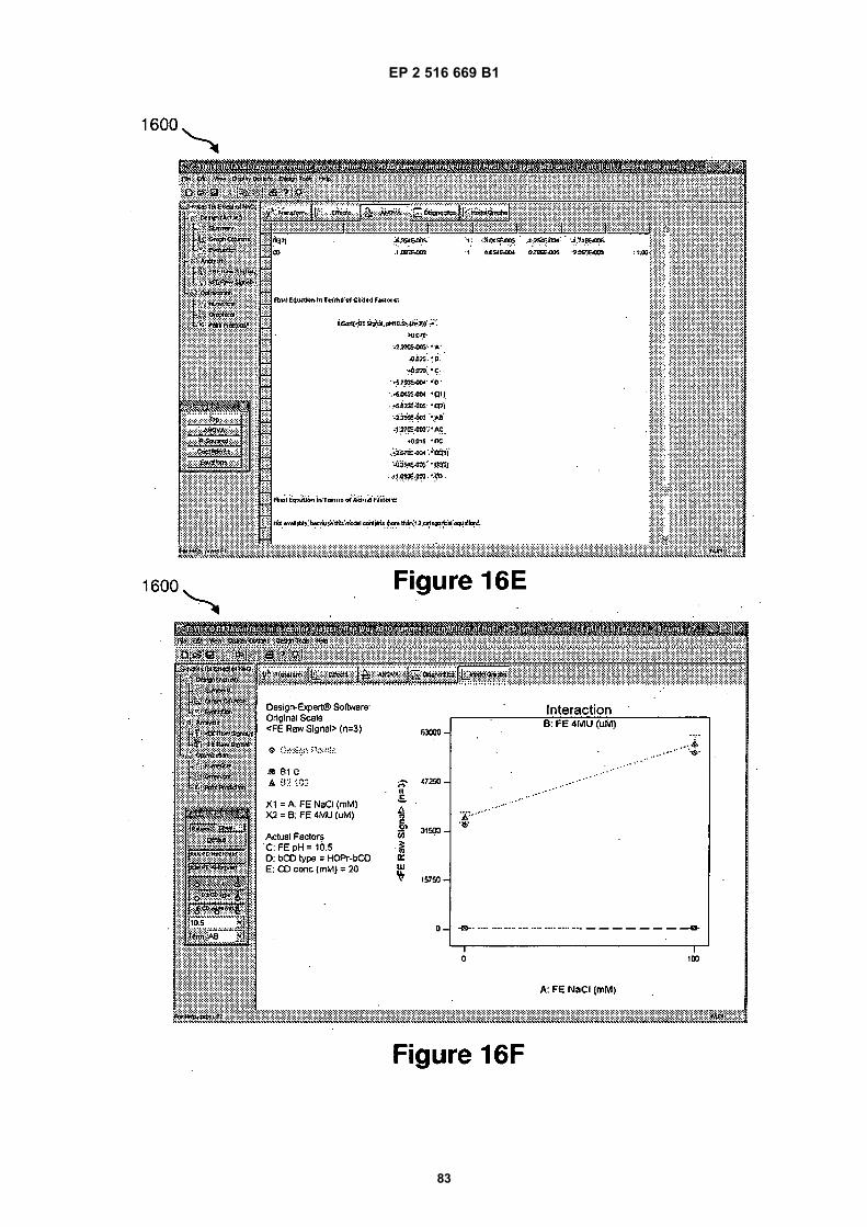
EP 2 516 669 B1
83
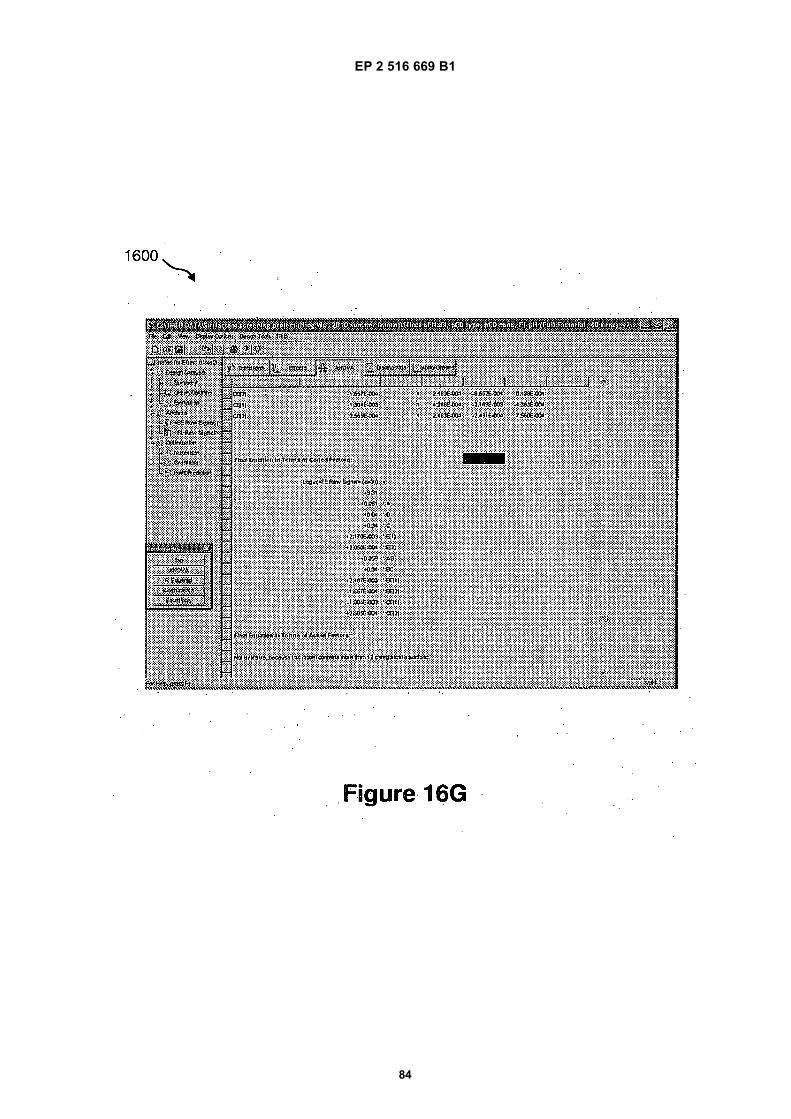
EP 2 516 669 B1
84

EP 2 516 669 B1
85

EP 2 516 669 B1
86

EP 2 516 669 B1
87

EP 2 516 669 B1
88

EP 2 516 669 B1
89
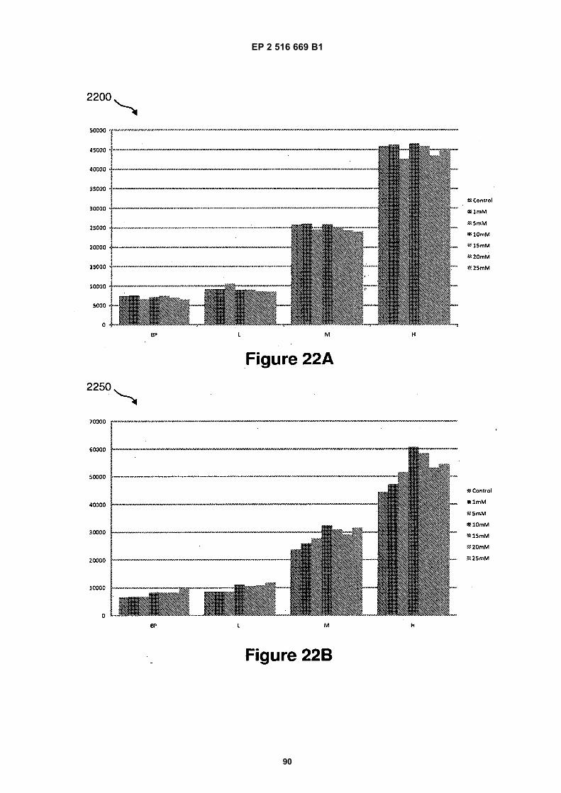
EP 2 516 669 B1
90

EP 2 516 669 B1
91

EP 2 516 669 B1
92

EP 2 516 669 B1
93

EP 2 516 669 B1
94

EP 2 516 669 B1
95

EP 2 516 669 B1
96

EP 2 516 669 B1
97
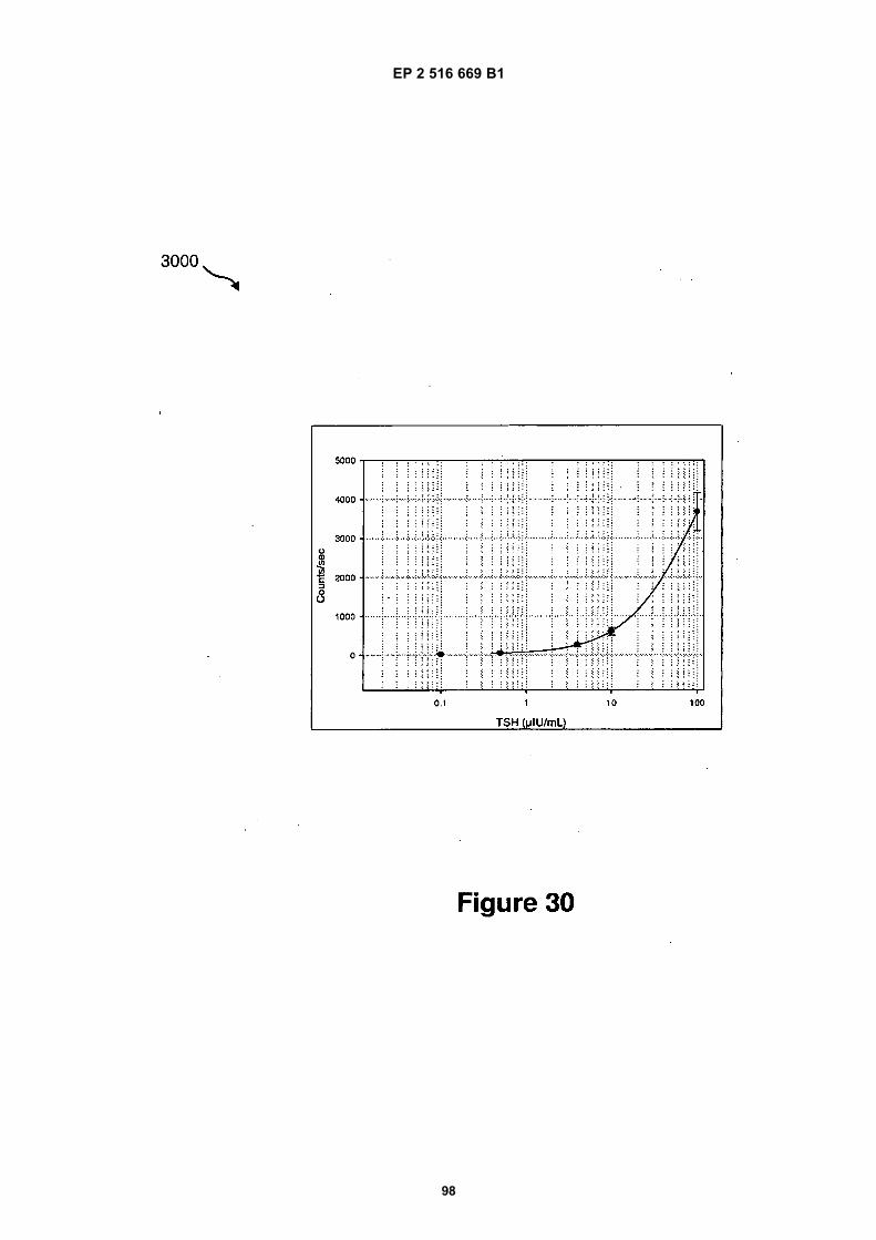
EP 2 516 669 B1
98
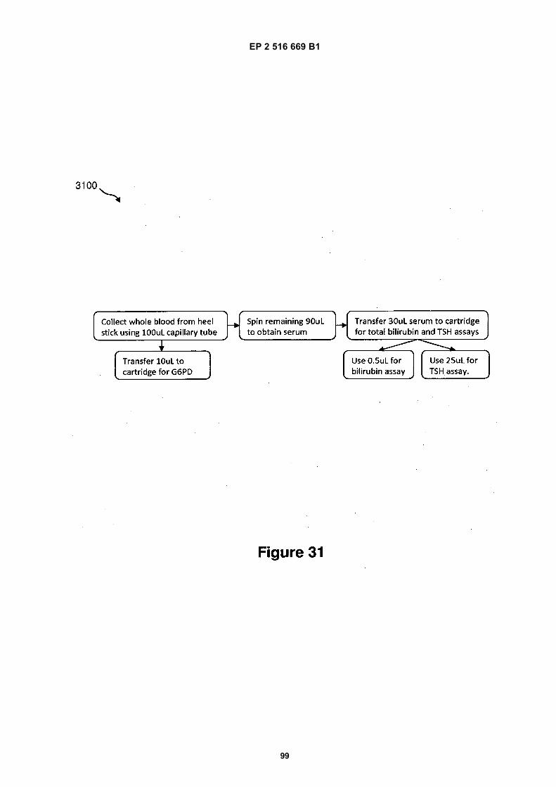
EP 2 516 669 B1
99

EP 2 516 669 B1
100

EP 2 516 669 B1
101
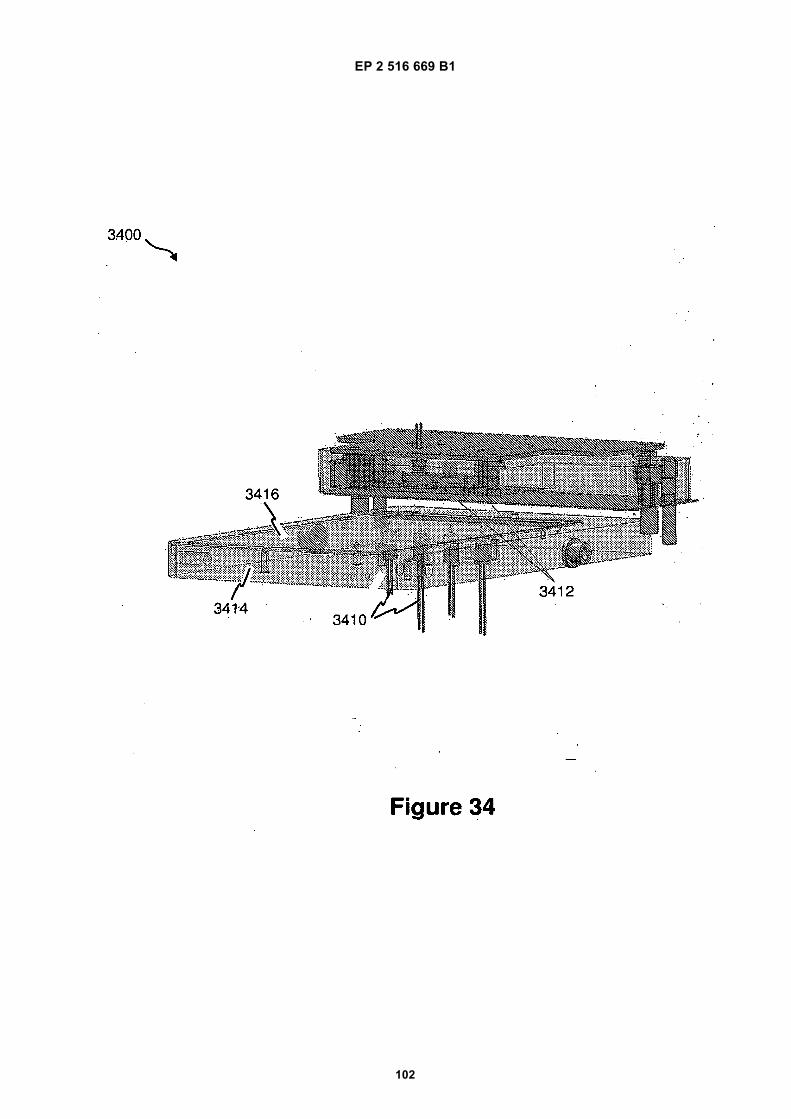
EP 2 516 669 B1
102
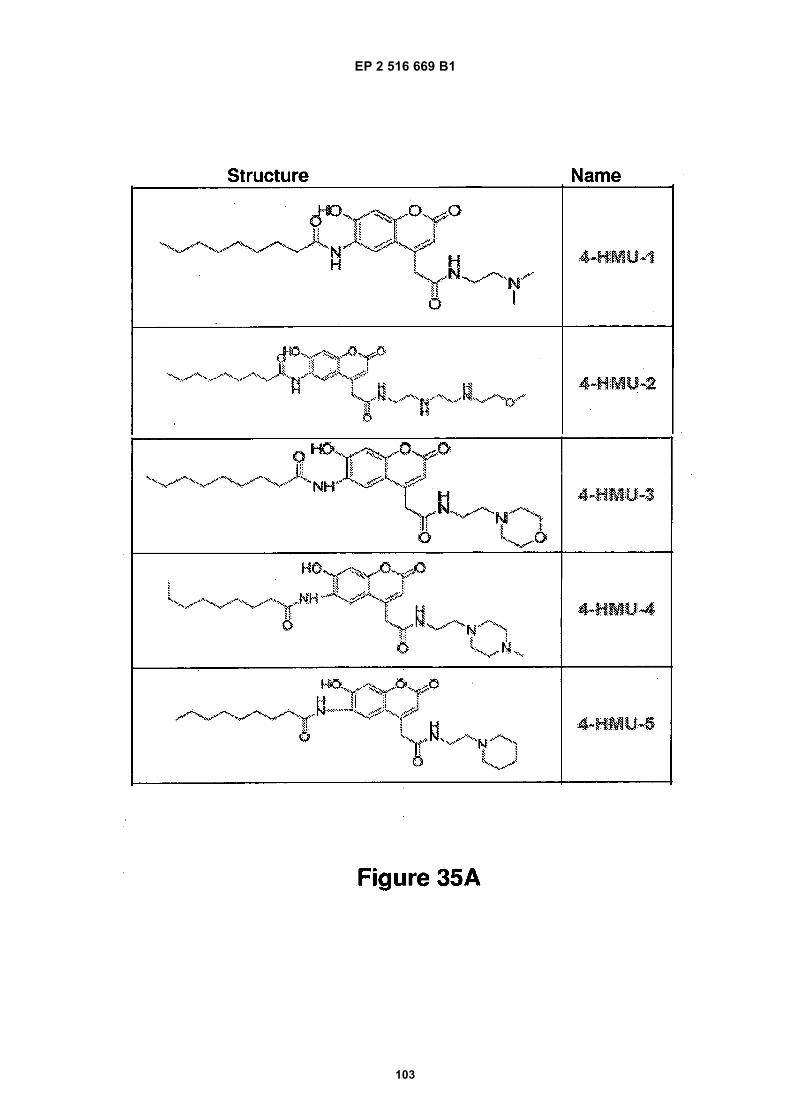
EP 2 516 669 B1
103

EP 2 516 669 B1
104

EP 2 516 669 B1
105

EP 2 516 669 B1
106

EP 2 516 669 B1
107

EP 2 516 669 B1
108

EP 2 516 669 B1
109

EP 2 516 669 B1
110

EP 2 516 669 B1
111

EP 2 516 669 B1
112

EP 2 516 669 B1
113

EP 2 516 669 B1
114

EP 2 516 669 B1
115

EP 2 516 669 B1
116

EP 2 516 669 B1
117

EP 2 516 669 B1
118

EP 2 516 669 B1
119
REFERENCES CITED IN THE DESCRIPTION
This list of references cited by the applicant is for the reader’s convenience only. It does not form part of the Europeanpatent document. Even though great care has been taken in compiling the references, errors or omissions cannot beexcluded and the EPO disclaims all liability in this regard.
Patent documents cited in the description
• WO 2008116209 A [0003]• US 20050260686 A [0014]• US 20030132538 A [0014]• US 20050118574 A [0014]• US 20050277197 A [0014]• US 20060159962 A [0014]• US 63956606 A [0014]• US 61039183 A [0014]• US 61047789 A [0014]• US 61086183 A [0014]• US 2008053545 W [0014]• US 2008058018 W [0014]• US 2008058047 W [0014]• US 2006047486 W [0014] [0017]• US 20080305481 A [0014]• US 20080151240 A [0014]• US 20070207513 A [0014]• US 20070064990 A [0014]• US 0647486 W [0016]• US 6911132 B, Pamula [0017]• US 34328406 A, Pamula [0017]• US 6773566 B, Shenderov [0017]• US 6565727 B [0017]• US 34326103 A, Kim and/or Shah [0017]• US 11275668 B [0017]
• US 11460188 B [0017]• US 12465935 B [0017]• US 12513157 B [0017]• US 7547380 B, Velev [0017]• US 7163612 B, Sterling [0017]• US 7641779 B, Becker and Gascoyne [0017]• US 6977033 B [0017]• US 7328979 B, Decre [0017]• US 20060039823 A, Yamakawa [0017]• WO 2009003184 A [0017]• US 20090192044 A, Fouillet [0017]• US 7052244 B, Fouillet [0017]• US 20080124252 A, Marchand [0017]• US 20090321262 A, Adachi [0017]• US 20050179746 A, Roux [0017]• US 2010040705 W [0017]• WO 2008101194 A [0018]• WO 2010027894 A [0019]• WO 2009021173 A [0019]• WO 2008098236 A [0019]• US 20080283414 A, Monroe [0019]• US 7439014 B, Pamula [0022]• US 7815871 B, Pamula [0087]• US 0943774 W [0103]
Non-patent literature cited in the description
• DHINDSA et al. Virtual Electrowetting Channels:Electronic Liquid Transport with Continuous ChannelFunctionality. Lab Chip, 2010, vol. 10, 832-836[0017]
• Merck Manual. 07 April 2006 [0030]• Greene and Wuts, Protective Groups in Organic Syn-
thesis. John Wiley &Sons, 1999 [0135]