(12) united states patent (10) patent no.: us … · ... u.s. cl. uspc ..... 435/6.1; 435/6.11: ......
TRANSCRIPT

(12) United States Patent Xiao et al.
USOO8628919B2
(10) Patent No.: US 8,628,919 B2 (45) Date of Patent: Jan. 14, 2014
(54) METHODS AND DEVICES FOR SINGLE-MOLECULE WHOLE GENOME ANALYSIS
(75) Inventors: Ming Xiao, Huntingdon Valley, PA (US); Parikshit A. Deshpande, Princeton, NJ (US); Han Cao, Philadelphia, PA (US); Michael Austin, Philadelphia, PA (US); Kandaswamy Vijayan, San Diego, CA (US); Alexey Y. Sharonov, Hamden, CT (US); Michael Boyce-Jacino, Titusville, NJ (US)
(73) Assignee: Bionano Genomics, Inc., Philadelphia, PA (US)
(*) Notice: Subject to any disclaimer, the term of this patent is extended or adjusted under 35 U.S.C. 154(b) by 111 days.
(21) Appl. No.: 13/001,697
(22) PCT Filed: Jun. 30, 2009
(86). PCT No.: PCT/US2O09/049244
S371 (c)(1), (2), (4) Date: Mar. 22, 2011
(87) PCT Pub. No.: WO2010/002883 PCT Pub. Date: Jan. 7, 2010
(65) Prior Publication Data
US 2011 FO171634 A1 Jul. 14, 2011
Related U.S. Application Data (60) Provisional application No. 61/076,785, filed on Jun.
30, 2008.
(51) Int. Cl. CI2O I/68 (2006.01) CI2M I/34 (2006.01) CI2M 3/00 (2006.01) C7H 2L/02 (2006.01)
(52) U.S. Cl. USPC ....... 435/6.1; 435/6.11: 435/287.2:536/23.1;
977/704
(58) Field of Classification Search USPC ........ 435/6.1, 6.11, 287.2:536/23.1977/704 See application file for complete search history.
(56) References Cited
U.S. PATENT DOCUMENTS
5,079,169 A * 1/1992 Chu et al. ...................... 436,174 5,356,776 A * 10/1994 Kambara et al. ............. 435/6.12 6,117,634 A 9/2000 Langmore 6,197.557 B1 3, 2001 Makarov et al. 7,771,944 B2 * 8/2010 Xiao et al. ................... 435/6.11
2003/0219792 A1 11/2003 Armes et al. 2006, O199202 A1 9/2006 Lyamichev et al. 2008/0242556 A1* 10, 2008 Cao et al. .......................... 506.9 2011/0306504 A1* 12/2011 Xiao et al. ........................ 506.6
FOREIGN PATENT DOCUMENTS
EP O 497 272 A1 10, 1992 WO WO 98/39485 9, 1997 WO WO 2011/05O147 4/2011
OTHER PUBLICATIONS
Xiao et al. Rapid DNA mapping by fluorescent single molecule detection, 2007, published on Dec. 14, 2006, Nucleic Acids Research, 35, e16, pp. 1-12.* Das et al. Single molecule linear analysis of DNA in nano-channel labeled with sequence specific fluorescent probes, 2010, Nucleic Acids Research, 38, e177, pp. 1-8.* Cai et al., "High-resolution restriction maps of bacterial artificial chromosomes constructed by optical mapping.” Proc. Natl. Acad. Sci. USA, vol. 95, No. 7, pp. 3390-3395 (1998). Cao et al., “Fabrication of 10nm enclosed nanofluidic channels.” Appl. Phys. Lett., 81:174-176 (2002). Cao et al., “Gradient nanostructures for interfacing microfluidics and nanofluidics.” Appl. Phys. Lett., 81:3058-3060 (2002). Castro et al., “Single-molecule detection of specific nucleic acid sequences in unamplified genomic DNA. Analytical Chemistry, 69(19):3915-3920 (1997). Olivier et al., "High-throughput genotyping of single nucleotide polymorphisms using new biplex invader technology.” Nucleic Acids Research, vol. 30, No. 12, p. E53 (2002). Piepenburg et al., “DNA detection using recombination proteins.” PLOS Biology, vol. 4, No. 7, e204 (2006). Reccius et al., “Compression and free expansion of single DNA molecules in nanochannels.” Physical Review Letters, 95:268101-1 (2005). Tegenfeldt et al., “From the Cover: The dynamics of genomic-length DNA molecules in 100-nm channels.” Proc. Natl. Acad. Sci. USA, 101(30): 10979-83 (2004). International Search Report dated Apr. 7, 2011 for PCT Application No. PCT/US2010/053513 filed Oct. 21, 2010. Written Opinion of International Search Authority dated Apr. 21. 2011 for PCT Application No. PCT/US2010/053513 filed Oct. 21, 2010.
International Search Report and Written Opinion of International Search Authority dated Oct. 9, 2010 for PCT Application No. PCT/ US2009/049244 filed Jun. 30, 2009. Fu D-Jet al., “Sequencing Double-Stranded DNA by Strand Dis placement'. Nucleic Acids Research, Information Retrieval Ltd., vol. 25, No. 3, pp. 677-679 (Jan. 1997).
(Continued)
Primary Examiner — Dave Nguyen Assistant Examiner — Narayan Bhat (74) Attorney, Agent, or Firm — Knobbe Martens Olson & Bear LLP
(57) ABSTRACT
Provided are methods and devices for single-molecule genomic analysis. In one embodiment, the methods entail processing a double-stranded nucleic acid and characterizing said nucleic acid. These methods are useful in, e.g., determin ing structural variations and copy number variations between individuals.
46 Claims, 11 Drawing Sheets

US 8,628,919 B2 Page 2
(56) References Cited
OTHER PUBLICATIONS Cai et al., “High-resolution restriction maps of bacterial artificial chromosomes constructed by optical mapping.” Proceedings of the National Academy of Sciences—PNAS, National Academy of Sci ences, US vol. 95, No. 7, pp. 3390-3395 (Mar. 1998). Kuhn et al., "Labeling of unique sequences in double-stranded DNA at sites of vicinal nicks generated by nicking endonucleases.” Nucleic Acids Research, 36(7):e40: 1-10 (2008).
Jo et al., “A single-molecule barcoding system using nanoslits for DNA analysis.” Proc. Natl. Acad. Sci., 104(8):2673-2678 (2007). Chan et al., “DNA mapping using microfluidic stretching and single molecule detection of fluorescent site-specific tags.” Genome Research, 14(6): 1137-1146 (2004). Phillips et al., “Application of single molecule technology to rapidly map long DNA and study the conformation of stretched DNA.” Nucleic Acids Research, 33(18):5829-5837 (2005).
* cited by examiner

U.S. Patent Jan. 14, 2014 Sheet 1 of 11 US 8,628,919 B2
Fig 1a. First, Generate Flap Sequences by nicking and displacing downstream strand
GAG G Gc, CCGG Flapsequences
GIG CC G
Nicking endonuclease 4Gr s : CGCra 4Aa
Polymerase extension GC4c. ra
CACCCTGCTTGCTGAGGTTTGCACCGGTGTGGCTCCGGAAGTAACGCAAAGCAC GGCCTGGGGAAAACAGTACG GTGGGACGAACGACTCCAAACGTGGCCACACCGAGGCCTTCAATTGCGATTTCGTGACCGGACCCCT TTTGTCAGC
Fig 1b 12.7 kb 17.6 kb 30.3 kb
5' - - - 5 3 ?h ? ?
16.7 kb 17.3 kb 30 kb 40.5 kb
Flap generation
Hybridization of fluorescent sequence specific probe at one site
S’ "...is NS
3' -- - 8 5'

U.S. Patent Jan. 14, 2014 Sheet 2 of 11 US 8,628,919 B2
. . . . . . . . . .
A tagging agent A, B or Care associated with the extended DNA during incorporation or hybridization; a co-localization event could be detected by a specific Fluorescent Resonance Energy Transfer (FRET) signal.

U.S. Patent Jan. 14, 2014 Sheet 3 of 11 US 8,628,919 B2
Fig 3a
5.GCTGAGG.3' nick labeling 5.GCTGAGG.3' y -G- W 3. CGACTCC.5 3...CGACTCC.5
In silico Nicking sites on lambda ds-DNA (48.5 kbp total length)
12.7 kb 17.6 kb 30.3 kb 5' v 3'
3' M ?h 5’ ? 16.7 kb 17.3 kb 30 kb 40.5 kb
Observed Nicking sites on lambda ds-DNA (48.5 kbp total length)
Fig 3b
Similar barcode results shown on linearized human BAC clone DNAS with complete stretching( 170Kb); over 17 labeled sites (in fluorescent color) are shown here.
U.S. Patent Jan. 14, 2014 Sheet 4 of 11 US 8,628,919 B2
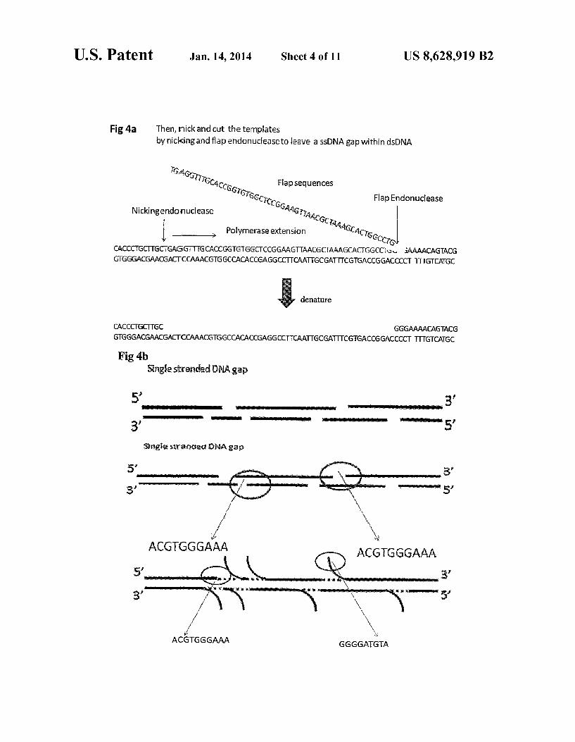
Fig. 4a Then, nick and cut the templates by nicking and flap endonuclease to leave a SSDNA gap within dsDNA
G.4s Gir irGCA CCGs Flapsequences
Si 66cc Flap Endonuclease
Nicking endonuclease *Gr P 446 olymerase extension Acis --> Ccro
CACCCTGCTTGCGAGGTTTGCACCGGTGGGCTCCGGAAG AACGCAAAGCAC GGCC, a AAAACAGTACG GTGGGACGAACGACCCAAACGGGCCACACCGAGGCCTTCAATTGCGATTCGTGACCGGACCCCT TITGTCAGC
denature
CACCCGCTGC GGGAAAACAGTACG GTGGGACGAACGACTCCAAACGGGCCACACCGAGGCC CAATGCGATTCGGACCGGACCCCT TTTGTCAGC
Fig 4b Singles:tranded DNA gap
5’ www.www.
uwuwe i unio 5'
Single staracted Nagap
w
y
ACGTGGGAAA GGGGATGTA

U.S. Patent Jan. 14, 2014 Sheet 5 of 11 US 8,628,919 B2
FIG Sa
distance
GCTGACGG, .... GCTGAGCGG, ... GCTGAACGT.... GCTGACCCC....
Sb
distance
5'-GCGGCCGC-3' 5'-GCGGCCGC-3' - CpG-islands CGCGCGCG BF: Binding Factor
5c Epigenetic patterns overlay with genomic location patterns in real time
Signal from methylation sites Signal from Flap or Gap genomic probes 71
Y YY Open reading Frame CpG islands CGCGCGCG
TF Consensus binding site 5'- ACTN{A} -3' TF:Transcription Factor

U.S. Patent Jan. 14, 2014 Sheet 6 of 11 US 8,628,919 B2
NanoAnalyzer Genome Assem bly Scaffolding Application &
Fig 6a Chromosome or genome sample
Labeled Large Fragments streaming in Nanochannel array chip of NanoAnalyzer
Non-virtual Genome Assembly
: ... ::::ss.xxxx-ax&ism-ww.
: & Raw images of OBSERVED BARCODE are generated
OBSERVED BARCODE of large fragments 50 kb 100 mb Extracted
ow-wow womanowo-powwowww.wow.wowowow.
... . . . . . . . . . . . . .
assembled into even longer continuous region - Scaffolds
Hell). --H -----Execuu -at weviewNawwamimyomasvienner ?aram
Contig's in silico barcodes aligned and mapped onto OBSERVED scaffold for actual positions
f s F. 3 : S: as w . is .: ... Fig 6b it is 3.
. E"...i. . . . . g" s' s
in Silico BARCODE computationally generated on these Contigs based on Same-SequenceSpecific motifs. www.www.wrwr. ww.www.s. www.www.ww.www
i
8
8 :
8
& : 3. :
Computationally assembled into discrete regions contigs of 10100 kb in length
Generate Millions of Random short reads of 35 850 bps
Current sequencing Methods ?h Map back to ref ap back to reference
database virtual Genome
ASSembly-resequencing
8 : Fragmentation of genome sample into 50-1000's bps

U.S. Patent Jan. 14, 2014 Sheet 7 of 11 US 8,628,919 B2
Fig 7 lmaging Analysis automatic DNA detection and sizing
U.S. Patent Jan. 14, 2014 Sheet 8 of 11
Ca
SSWO % 2 Preservoir
nanogate gate labeled DNA linearized in to-o-or
Label (Flap) nanochannel
nanochannel
Flap

U.S. Patent Jan. 14, 2014 Sheet 9 of 11 US 8,628,919 B2
Fig 9
Nanochannel or nanotrack
R1=nucleic acid (SS-DNA, ds-DNA, RNA) R2=sequence being analyzed with analysis proceeding at multiple points R3=modification to permittethering

U.S. Patent Jan. 14, 2014 Sheet 10 of 11 US 8,628,919 B2
Fig 10 a Tethering at entrance to channel
bead
Fig 10 b Tethering at entrance to channel
Tethering within nanochannel Nucleic acid
R=chemical modification A=region of attachment which may have surface chemical modification

U.S. Patent Jan. 14, 2014 Sheet 11 of 11 US 8,628,919 B2
Fig 11 a : Magnetic modification for tethering nucleic acid Tethering at entrance to channel
Nucleic acid B
Tethering within nanochannel
B
M=magnetic modification A=on-chip magnetic field for tethering B=externally applied magnetic field for tethering
Fig 11 b : Optical trapping for tethering nucleic acid Tethering at entrance to channel
Nucleic acid
Tethering within nanochannel
A=dielectric modification B=field of optical trap

US 8,628,919 B2 1.
METHODS AND DEVICES FOR SINGLE-MOLECULE WHOLE GENOME
ANALYSIS
CROSS-REFERENCE TO RELATED APPLICATIONS
This application is the National Stage of International Application No. PCT/US2009/049244, filed Jun. 30, 2009, which claims the benefit of U.S. application No. 61/076,785, “Single Molecule Whole Genome Analysis filed Jun. 30. 2008, the entireties of which are incorporated herein by ref erence for all purposes.
TECHNICAL FIELD
The present invention relates to the field of nanofluidics and to the field of DNA sequencing.
BACKGROUND
Macromolecules are long polymer chains composed of many chemical units bonded to one another. Polynucleotides area class of macromolecules that include, for example, DNA and RNA. Polynucleotides are composed of long sequences of nucleotides. The sequence of nucleotides is directly related to the
genomic and post-genomic gene expression information of the organism. Direct sequencing and mapping of sequence regions, motifs, and functional units such as open reading frames (ORFs), untranslated regions (UTRs), exons, introns, protein factor binding sites, epigenomic sites such as CpG clusters, microRNA sites, Small interfering RNA (SiRNA) sites, large intervening non-coding RNA (linckNA) sitesand other functional units are all important in assessing the genomic composition of individuals.
In many cases, complex rearrangement of these nucle otides sequence. Such as insertions, deletions, inversions and translocations, during an individual’s life span leads to dis ease states Such as genetic abnormalities or cell malignancy. In other cases, sequence differences as in Copy Number Variations (CNVs) among individuals reflects the diversity of the genetic makeup of the population and their differential responses to environmental stimuli and signals such as drug treatments. Instill other cases, processes Such as DNA methy lation, histone modification, chromatin folding or other changes that modify DNA or DNA-protein interactions influ ence gene regulations, expressions and ultimately cellular functions resulting in diseases and cancer.
It has been found that genomic structural variations (SVs) are much more widespread than previously thought, even among healthy individuals. The importance of understanding genome sequence with structural variations information to human health and common genetic disease has thus become increasingly apparent.
Functional units and common structural variations are thought to encompass from tens of bases to more than mega bases. Accordingly, a method that is direct, inexpensive and yet flexible of revealing sequence information and SVs across the resolution scale from Sub-kilobase to megabase along large native genomic molecules is highly desirable in sequencing and fine-scale mapping projects of more indi viduals in order to catalog previously uncharacterized genomic features.
Furthermore, phenotypical polymorphism or disease States of biological systems, particularly in multiploidy organism Such as humans, are consequence of the interplay between the
10
15
25
30
35
40
45
50
55
60
65
2 two haploid genomes inherited from maternal and paternal lineage. Cancer, in particular, is often the result of the loss of heterozygosity among diploid chromosomal lesions.
Conventional cytogenetic methods such as karyotyping, FISH (Fluorescent in situ Hybridization) provided a global view of the genomic composition in as few as a single cell, they are effective in revealing gross changes of the genome Such as aneuploidy, gain, loss or rearrangements of large fragments of thousands and millions bases pairs. These meth ods, however, suffer from relatively low sensitivity and reso lution in detecting medium to Small sequence motifs or lesions. The methods are also laborious, which limits speed and inconsistency. More recent methods for detecting sequence regions,
sequence motifs of interests and SVs, such as acGH (array Comparative Genomic Hybridization), fiberFISH or massive pair-end sequencing have improved in the aspects of resolu tion and throughput. These methods are nonetheless indirect, laborious, expensive and rely on existing reference databases. Further, the methods may have limited fixed resolution, and provide either inferred positional information relying on mapping back to a reference genome for reassembly or com parative intensity ratio information. Such methods are thus unable to reveal balanced lesion events such as inversions or translocations.
Current sequencing analysis approaches are limited by available technology and are largely based on Samples derived from an averaged multiploidy genomic materials with very limited haplotype information. The front end sample preparation methods currently employed to extract the mixed diploid genomic material from a heterogeneous cell population effectively shred the material into smaller pieces, which results in the destruction of native the crucially important structural information of the diploid genome.
Even the more recently developed second-generation methods, though having improved throughput, further com plicate the delineation of complex genomic information because of more difficult assembly from much shorter sequencing reads.
In general, short reads are more difficult to align uniquely within complex genomes, and additional sequence informa tion are needed to decipher the linear order of the short target region. An order of 25-fold improvement in sequencing coverage
is needed to reach similar assembly confidence instead of 8-10 fold coverage needed in conventional BAC and so-called shotgun Sanger sequencing (WendlMC, Wilson RK Aspects of coverage in medical DNA sequencing, BMC Bioinformat ics, 16 May 2008; 9:239). This multi-fold sequencing cover age imposes high costs, effectively defeating the overarching goal in the field of reducing sequencing cost below the S1,000 mark.
Single molecule level analysis of large intact genomic mol ecules thus provides the possibility of preserving the accurate native genomic structures by fine mapping the sequence motifs in situ without cloning process or amplification. The larger the genomic fragments are, the less complex of sample population in genomic samples, for example, in ideal sce nario, only 46 chromosomal length of fragments need to be analyzed at single molecule level to cover the entire normal diploid human genome and the sequence derived from Such approach has intact haplotype information by nature. Further, megabase-scale genomic fragments can be extracted from cells and preserved for direct analysis, which dramatically reduces the burden of complex algorithm and assembly, also
US 8,628,919 B2 3
co-relates genomic and/or epigenomic information in its original context more directly to individual cellular pheno types.
In addition to genomics, the field of epigenomics has been increasingly recognized in the past 20 years or so as being of 5 singular importance for its roles in human diseases such as cancer. With the accumulation of knowledge in both genom ics and epigenomics, a major challenge is to understand how genomic and epigenomic factors correlate directly or indi rectly to develop the polymorphism or pathophysiological conditions in human diseases and malignancies. Whole genome analysis concept has evolved from a compartmental ized approach in which areas of genomic sequencing, epige netic methylation analysis and functional genomics were studied largely in isolation, to a more and more multi-faceted holistic approach. DNA sequencing, structural variations mapping, CpG island methylation patterns, histone modifi cations, nucleosomal remodeling, microRNA function and transcription profiling have been increasingly viewed more closely in Systematical way, however, technologies examin ing each of above aspects of the molecular state of the cells are often isolated, tedious and non-compatible which severely circumvent the holistic analysis with coherent experiment data results.
Accordingly, there is a need in the art for methods and devices that enable single molecule level analysis of large intact native biological samples so as to enable determination of genomic and epigenomic information of a target sample. Such methods and devices would provide a very powerful tool to researchers and clinicians alike.
SUMMARY
In meeting the described challenges, the claimed invention first provides methods of characterizing DNA, comprising: processing a double-stranded DNA comprising a first DNA strand and a second DNA strand to give rise to an unhybrid ized flap of the first DNA strand and a corresponding region on the second DNA strand, the unhybridized flap comprising from about 1 to about 1000 bases; extending the first DNA Strand along the corresponding region of the second DNA Strand; and labeling at least a portion of the unhybridized flap, a portion of the extended first DNA strand, or both.
Also provided are methods of identifying structural varia tions between DNAs, comprising: labeling, on a first double Stranded DNA, two or more sequence-specific locations on the first DNA; labeling, on a second double-stranded DNA, the two or more corresponding sequence-specific locations on the second DNA; linearizing at least a portion of the first double-stranded DNA; linearizing at least a portion of the first double-stranded DNA; and comparing the distance between two or more labels on the first, linearized double-stranded DNA to the distance between the corresponding labels on the second, linearized linearized double-stranded DNA.
Further disclosed are methods of obtaining structural infor mation from DNA, comprising: labeling, on a first double Stranded DNA, one or more sequence-specific locations on the first DNA; labeling, on a second double-stranded DNA, the corresponding one or more sequence-specific locations on the second double-stranded DNA; linearizing at least a por tion of the first double-stranded DNA; linearizing at least a portion of the first double-stranded DNA; and comparing the intensity of a signal of the at least one label of the first, linearized double-stranded DNA to the intensity of the signal of the at least one label of the second, linearized double stranded DNA.
10
15
25
30
35
40
45
50
55
60
65
4 Additionally provided are methods of obtaining structural
information from a macromolecule, comprising: translocat ing a macromolecule comprising at least one flap extending therefrom along a channel having at least one constriction disposed therein; and detecting at least one signal corre sponding to the passage of the at least one flap of the macro molecule through the at least one constriction of the channel.
Provided also are methods of obtaining structural informa tion from a macromolecule, comprising: labeling at least a portion of a macromolecule; immobilizing the macromol ecule; disposing at least a portion of the macromolecule within a channel Such that at least a portion of the macromol ecule is linearized within the channel; and detecting at least one signal related to the labeled portion of the macromol ecule.
Also disclosed are analysis systems, comprising: a Sub strate comprising at least one channel having a width in the range of from about 1 to about 100 nanometers; the substrate comprising at least one immobilization region.
Further provided are methods of characterizing a nucleic acid polymer, comprising: labeling one or more regions of a nucleic acid polymer with one or more sequence-specific motif labels; correlating one or more signals from one or more of the sequence-specific motif labels to the position of the one or more sequence-specific motif labels of the nucleic acid polymer; sequencing one or more segments of the nucleic acid polymer, the one or more segments including one or more of the sequence specific motif labels of the nucleic acid polymer; and comparing one or more signals of one or more sequenced segments to one or more corresponding signals of the labeled nucleic acid polymer so as to develop the relative locations within the nucleic acid polymer, of two of more sequenced segments.
BRIEF DESCRIPTION OF THE DRAWINGS
The Summary, as well as the following detailed description, is further understood when read in conjunction with the appended drawings. For the purpose of illustrating the inven tion, there are shown in the drawings exemplary embodi ments of the invention; however, the invention is not limited to the specific methods, compositions, and devices disclosed. In addition, the drawings are not necessarily drawn to scale. In the drawings:
FIG. 1 (SEQID NOS: 1-3) depicts a schematic view of the claimed flap-labeling methods;
FIG.2 depicts labeled probes hybridized to a flap generated from a first DNA strand and a label residing in the region of the first strand corresponding to the flap;
FIG.3 depicts an alternative embodiment of placing DNA “barcodes' on polynucleic acids;
FIG. 4 (SEQ ID NO: 1-6) depicts sequencing along a genomic region;
FIG. 5 depicts concurrent parallel sequencing and spatial assembly;
FIG. 6 depicts obtaining genome assembly information from a nucleic acid polymer;
FIG. 7 is a software image of labeled DNA polymers undergoing image analysis;
FIG. 8 depicts optical and non-optical detection schemes according to the claimed invention;
FIG. 9 depicts a labeled nucleic acid polymer linearized within a nanochannel or nanotrack;
FIG. 10 depicts nucleic acid polymers immobilized adja cent to or within nanochannels, by various means; and

US 8,628,919 B2 5
FIG. 11 depicts magnetic and optical trapping of nucleic acid polymers disposed within nanochannels or nanotracks.
DETAILED DESCRIPTION OF ILLUSTRATIVE EMBODIMENTS
The present invention may be understood more readily by reference to the following detailed description taken in con nection with the accompanying figures and examples, which form a part of this disclosure. It is to be understood that this invention is not limited to the specific devices, methods, applications, conditions or parameters described and/or shown herein, and that the terminology used herein is for the purpose of describing particular embodiments by way of example only and is not intended to be limiting of the claimed invention. Also, as used in the specification including the appended claims, the singular forms “a,” “an and “the include the plural, and reference to a particular numerical value includes at least that particular value, unless the context clearly dictates otherwise. The term “plurality', as used herein, means more than one. When a range of values is expressed, another embodiment includes from the one par ticular value and/or to the other particular value. Similarly, when values are expressed as approximations, by use of the antecedent “about it will be understood that the particular value forms another embodiment. All ranges are inclusive and combinable.
It is to be appreciated that certain features of the invention which are, for clarity, described herein in the context of sepa rate embodiments, may also be provided in combination in a single embodiment. Conversely, various features of the invention that are, for brevity, described in the context of a single embodiment, may also be provided separately or in any Subcombination. Further, reference to values stated in ranges include each and every value within that range.
In a first aspect, the present invention provides of charac terizing DNA, comprising processing a double-stranded DNA comprising a first DNA strand and a second DNA strand to give rise to an unhybridized flap of the first DNA strand and a corresponding region on the second DNA strand, the unhy bridized flap comprising from about 1 to about 1000 bases: extending the first DNA strand along the corresponding region of the second DNA strand; and labeling at least a portion of the unhybridized flap, a portion of the extended first DNA strand, or both. The flap is suitably from about 1 to about 1000 bases in
length. A flap is suitably from about 20 to about 100 bases in length, or even in the range of from about 30 to about 50 bases.
The methods also include incorporating one or more replacement bases into the first strand of double-stranded DNA so as to extend the first DNA strand (from which the flap is peeled) to fill-in and eliminate the gap (i.e., the now-corre sponding region of the second DNA strand) left by formation of the flap. The user may label at least a portion of the processed double-stranded DNA (the first DNA strand, the second DNA strand, the flap, or any combination thereof) with one or more tags. The filled-in gap left by the flap can include one or more labeled portions. In some embodiments (not shown), the flap may be excised using a flap-removing enzyme, leaving behind a dsDNA having one or more nucle otides incorporated therein. The processing is Suitably accomplished by nicking the
first strand of double-stranded DNA. This nicking is suitably effected at one or more sequence-specific locations, although the nicking can be effected at one or more non-specific loca tions, including random or non-specific locations.
10
15
25
30
35
40
45
50
55
60
65
6 Nicking is Suitably accomplished by exposing the double
Stranded DNA polymerto a nicking endonuclease, or nickase. Nickases are Suitably highly sequence-specific, meaning that they bind to a particular sequence of bases (motif) with a high degree of specificity. Nickases are available, e.g., from New England BioLabs (www.neb.com). The nicking may also be accomplished by other enzymes
that effect a break or cut in a strand of DNA. Such breaks or nicks can also be accomplished by exposure to electromag netic radiation (e.g., UV light), one or more free radicals, and the like. Nicks may be effected by one or more of these techniques.
Incorporation of replacement bases into the first strand (i.e., the nicked strand) of double-stranded DNA suitably comprises contacting DNA with a polymerase, one or more nucleotides, a ligase, or any combination thereof. Other meth ods for replacing the "peeled-away’ bases present in the flap will also be knownto those of ordinary skill in the art. The first DNA strand is Suitably extended along the corresponding region of the second DNA, which region is left behind/ex posed by the formation of the flap. In some embodiments, the polymerase acts concurrent with a nickase that gives rise to a flap. The incorporation of these replacement bases can be con
ceptualized as filling-in the gap left behind by the formation and "peeling-up' of the flap. By filling in the gap, the position formerly occupied by the flap is occupied by a set of bases that Suitably has the same sequence as the bases located in the flap. The filling can prevent re-hybridization of the flap to the second stand of DNA to which the flap was formerly bound.
Labeling is suitably accomplished by (a) binding at least one complementary probe to at least a portion of the flap, the probe comprising one or more tags, (b) utilizing, as a replace ment base that is part of the first DNA strand extended along the corresponding region of the second DNA strand, a nucle otide comprising one or more tags, or any combination of (a) and (b). In this way, the flap, the bases that fill-in the gap, or both may be labeled.
Probes are suitably nucleic acids (single or multiple) that include a tag, as described elsewhere herein. A probe may be sequence specific (e.g., AGGCTA, or some other particular base sequence), although probes may be randomly generated. As described elsewhere herein, a probe may be selected or constructed based on the user's desire to have the probe bind to a sequence of interest or, in one alternative, bind to a sequence that up- or downstream from a sequence or other region of interest on a particular DNA polymer (i.e., probes that bind so as to flank or bracket a region of interest). A probe may be as long as a flap (i.e., up to 1000 bases). A probe is suitably in the range of from 1 to about 100 bases in length, or from about 3 to 50 bases, or even in the range of from about 5 to about 20 bases in length. A schematic view of these methods is shown in FIG.1. In
that figure, the creation of a flap and the back-filling of the resulting gap is shown. The back-filling may be with so-called “hot” or labeled bases, and the flap may be contacted with one or more probes that are complementary to at least a portion of the flap. A sequence specific nicking endonuclease, or nick ase, creates a single strand cut gap on double stranded DNA, and a polymerase binds to the nicked site and starts strand extension while generating a displaced Strand or so-called "peeled flap' simultaneously. The peeled flap then creates an available region (i.e., an unhybridized, corresponding region on the second DNA strand of the nucleic acid polymer) for sequencing specific hybridization with labeled probes togen erate detectable and identifiable signals.

US 8,628,919 B2 7
FIG. 1b shows a labeled large genomic DNA being unfolded linearly within a nanochannel. As shown at the bottom of the figure, a fluorescently labeled flap enables the user to visualize the location of the probe within the larger context of the macromolecule. As shown, a nicked-labeled macromolecule may be linearized within a nanochannel. The spatial distance between signals from tags is consistent and can then be quantified, which in turn provides for a unique “barcoding signature pattern that reflects specific genomic sequence information about the region under analysis. Mul tiple nicking sites on a lambda dsDNA (48.5 kbp totallength) were shown as an example created by a specific enzyme, include but not limited to Nb.BbvCI; Nb.BSmI; Nb.BsrDI: Nb.BtsI; NtAlwI: Nt.BbvCI; Nt.BspOI: Nt.BstNBI; Nt.CviPII and the combination digestion of any of above. A linearized single lambda DNA image is included to show
a fluorescently labeled oligonucleotide probe hybridized to an expected nickase created location. Such recorded actual barcodes along long biopolymers are described elsewhere herein as observed barcodes. By linearizing a macromolecule having labeled flaps,
labeled gaps, or both, the user can determine the relative positions of the labels to one another. As described elsewhere herein, Such relative distance information is useful in diag nostic applications and in characterizing the nucleic acid polymer.
In some embodiments, the methods further include obtain ing sequence information derived from one or more replace ment bases incorporated into the first DNA strand of the double-stranded DNA, from one or more probes binding to a flap, or both. This sequence information may be obtained in a variety of ways.
In one example, a labeled probe complementary to a spe cific base sequence is introduced to the flap, and the user determines whether that sequence-specific probe binds to the flap. This process may be repeated several times, using probes having different sequence specificities, ultimately enabling the user to determine the sequence of bases residing in the flap.
In another example, the sequence information is obtained by determining the sequence of bases that fill-in the gap left behind by the flap. This may be accomplished by labeling one or more of the bases with the same or different labels and assaying the signals emitted by bases as they are incorporated into the gap or after they are incorporated into the gap. In other embodiments, the user may monitor one or more signals evolved from a polymerase that incorporates bases into the gap so as to determine the sequence of the bases.
Determination of sequence information can be performed in free solution or can be performed in nanochannels, so as to allow for high-resolution analysis of a single DNA polymer. A flap could also be excised via an appropriate enzyme and then the excised flap itself could also be sequenced.
The sequence information may be obtained from a single flap, a single gap, or both. In some embodiments, however, the sequence information is obtained from two or more flaps or gaps, thus enabling faster sequencing of a given target. Sequencing information can also be determined by using sequence-specific probes and determining where (and whether) such probes bind to a portion of the nucleic acid polymer.
FIG. 4 depicts sequencing along a comparatively long genomic region. In that figure, single strand flaps are gener ated after the “parent nucleic acid polymer is digested by sequence specific nicking endonuclease and polymerase extension in the first strand of the polymer. This structure can be digested again by a nicking endonuclease and a flap endo
10
15
25
30
35
40
45
50
55
60
65
8 nuclease, which cuts where flap joins the first strand (shown by arrows), and the resulting dsDNA can be denatured under appropriate conditions so as to generate a single Stranded gap that spans the nicking site and the flap endonuclease cutting site. This gap can then be exposed to sequencing reactions using polymerase extension or hybridization and ligation with specific probes and enzymes
FIG. 4b depicts a schematic showing multiple nicking sites, single Stranded flap sites, and single stranded gap sites created along a long dsDNA. Sequencing reactions are then initiated at one or more nicking, flap sequence sites or single Stranded gap sites, with the sequencing effected by poly merase extension or sequencing by hybridization or ligation. A variety of species can serve as tags for the present meth
ods. A tag can include, for example, a fluorophore, a quantum dot, a dendrimer, a nanowire, a bead, a peptide, a protein, a magnetic bead, a methyl group, a methyltransferase, a non cutting restriction enzyme, a zinc-finger protein, an antibody, a transcription factor, a DNA binding protein, a hairpin polya mide, a triplex-forming oligodeoxynucleotide, a peptide nucleic acid, and the like. The methods may include the use of two or more different tags, and a single molecule may accord ingly include multiple tags. The methods also include detecting one or more signals
from one or more tags. Such signals can include a fluorescent signal, a chemoluminescent signal, an electromagnetic sig nal, an electrical signal, a potential difference, and the like. The signal may be related to a physical size difference between two bodies, which may be, for example, the signal evolved when a bead attached to a DNA target is entrapped in a constriction that is Smaller in cross-section than is the bead. Fluorescent signals are considered especially suitable, par ticularly in embodiments where a fluorescent molecule is attached to a base, a probe, or both.
In some embodiments, the signal may derive from energy transferred (e.g., fluorescence energy transfer, "FRET) between a tag on a replacement base and a tag on a probe residing on a flap, by fluorescence resonance energy transfer between two or more tags on a probe residing on a flap, or by any combination thereof.
FIG. 2 illustrates exemplary positions for labels and probes on nucleic acid polymers prepared according to the claimed invention. That figure depicts probes (shown as A and B) disposed on a flap and a probe (shown as C) along a DNA stranded extended so as to fill-in the gap left behind by the formation and peeling of the flap. The probes include, for example, organic fluorophore,
quantum dot, dendrimer, nanowires, bead, Au beads, para magnetic beads, magnetic bead, polystyrene bead, polyeth ylene bead, peptide, protein, haptens, antibodies, antigens, streptavidin, avidin, neutravidin, biotin, nucleotide, oligo nucleotide, sequence specific binding factors such as engi neered restriction enzymes, methyltransferases, Zinc finger binding proteins, and the like. As shown, more than one probe may be disposed on a flap. In a sample embodiment, a tag (or tags) within a gap are excited by an excitation radiation. The excited gap-tag then transfers energy to a tab disposed on a probe that is itself disposed on the flap. One or both of the gap-and flap-tags may emit a signal that
is detectable by the user. In some embodiments, the gap tag, the first flap tag, or both may excite a second flap tag. In this way, the user may configure a detection system that is highly specific by choosing tags that are excited only by specific wavelengths or types of radiation, thus creating a system in which the tag that is detected by the user is only excited if one or more precursor tags are in proper position. Thus, a co localization event can be detected (e.g., visualized) by energy

US 8,628,919 B2
transfer between two or more labels, which enhances the specificity of the binding event assay. The flap region is, in Some cases, selected because the flap,
gap, or both includes at least a portion of a specific sequence of interest on the double-stranded DNA. Such sequences of interest may include, for example, a sequence known to code for a particular protein or a particular condition.
In some embodiments, the flap, gap, or both, includes at least a portion of the double-stranded DNA that flanks the sequence of interest on the double-stranded DNA. This is useful where, for example, the user seeks to label regions on a DNA that bracket the location of a particular gene or other area of interest So as to highlight that area. The claimed methods also include at least partially linear
izing (e.g., untangling) at least a portion of the double Stranded DNA comprising at least one flap, one gap, or both. The user may also at least partially linearize at least a portion of the double-stranded DNA comprising at least two flaps, two gaps, or any combination thereof. Such linearization may be accomplished, for example, by translocating a DNA through a channel or other structure of Such dimensions that the DNA is linearized by way of physical confinement within the channel or other structure.
The user may also, in some embodiments, measure the distance between two flaps, between two or more tags dis posed adjacent to two or more flaps, two or more tags dis posed within two or more gaps, or any combination thereof. This distance is then suitably correlated to structure, a sequence assembly, a genetic or cytogenetic map, a methyla tion pattern, a location of acpG island, an epigenomic pattern, a physiological characteristic, or any combination thereof of the DNA. Because the claimed invention enables investiga tion of structure and of other epigenomic factors (e.g., methy lation patterns, location of cpG islands, and the like), the user can overlay results relating to structure and epigenomic pat terns to arrive at a complete genomic picture. One aspect of the claimed invention is its ability to provide
both genomic (sequence) and epigenomic (Supra-sequence) information about a nucleic acid or other genetic material. More specifically, the claimed invention allows the user to determine, by way of sequencing, whetheraparticular gene is present and also, by way of obtaining epigenomic informa tion, the activity of that gene.
In one non-limiting example, a user may obtain genomic information (via the labeling methods described elsewhere herein) about a nucleic acid polymer, Such as whether a par ticular gene is present. The user can then also obtain epige nomic information about the nucleic acid polymers methy lation patterns (which are indicative of the activity of those gene loci located proximate to the methylation) by using, for example, a labeled methyl-binding protein so as to identify the positions of methyls along the nucleic acid polymer. Such methyls may reside on cytosines and within so-called cpG island clusters, which may be correlated to the regulation of functional gene loci. Other binding molecules (such as mol ecules that bind to transcription factor binding sites and the like) are also suitable for obtaining epigenomic information.
Thus, a user can determine—simultaneously, in some embodiments—the presence of one or more functional genes and, via methyl-based epigenomic information, whether Such genes are active. In one example, the user might label the genes sequence information with label of a first color and label the methylation regions with a label of a second color, thus enabling observation of gene location/sequence and gene activity (i.e., methylation patterns) simultaneously. The epigenomic information may also include locations where transcription enzymes can—or cannot bind.
5
10
15
25
30
35
40
45
50
55
60
65
10 The utility of epigenomic information is apparent. As
described elsewhere herein, the utility of genomic informa tion is that an oligomer-based probe (or set of probes com prising a barcode) provides “static' information regarding the sequence of the nucleic acid polymer under study. Epige nomic information (e.g., information regarding methylation or transcription factor binding) provides dynamic informa tion about agene sequence, effectively providing on/off infor mation about the gene. The present invention thus enables simultaneous collection of both genomic and epigenomic information. As one illustrative, non-limiting example, a user may label
locations (i.e., flaps, filled-in gaps, or some combination of the two) on DNA from a first patient, the locations being chosen such that they are up- and down-stream from (i.e., flank) the location of a particular gene, e.g., a breast cancer gene, on the DNA. After linearizing the labeled DNA, the user may compare the distance between these labels to the dis tance between corresponding labels on a DNA from a control subject known to have a “proper number of copies of the breast cancer gene. If the distance between the labels for the first patient is greater than the distance between the labels for the control subject, it is then known that the patient has additional or extra copies of the breast cancer gene, and a treatment regimen can be designed accordingly. The technique can also be used to determine copy number
variations between two or more individuals, none of which is a “control” or even copy number variations within a single patient (i.e., by comparing DNA taken from the patient at two different times). In this way, the present methods facilitate rapid analysis and characterization of DNA or other macro molecules from a single subject or from a larger population Segment. The user may also measure the intensity of at least one
signal from at least one tag disposed adjacent to a flap, a tag disposed within the gap, or both. The user may then correlate the intensity of the at least one signal to a sequence assembly, a genetic or cytogenetic map, a physiological characteristic, or other features (e.g., epigenomic patterns) described else where herein. This enables the user to develop a complete picture of the pathophysiological state of the Source of the nucleic acid polymer.
This is shown by non-limiting FIG.5c. That figure shows, schematically, the use of a labeled binding factor (BF), such as a anti-methyl-antobody or a methyl-binding protein (MBP) to locate one or more epigenomic sites of interest along a genomic region to generate an epigenomic barcode pattern. As shown, the user also—simultaneously, in some cases—uses the disclosed methods to “barcode” the same region (using, e.g., sequence-specific probes) to determine the genomic region's structure. The genomic probes may emit or excite at a different different wavelength or with a signal distinguishable from any labels associated with the epigenomic analysis. In one embodiment, the epigenomic barcodes include (but are not limited to) patterns derived from transcription factor binding sites or siRNA or LincRNA bind ing sites. This demonstrates the capability of the claimed invention to correlate static genomic sequence and structure information with dynamic regulatory and functional informa tion simultaneously, in real time, and in the same field of view with direct imaging at the single molecule level. As another non-limiting example, a user may label one or
more flaps (or filled-in gaps) corresponding to regions of DNA from a first patient that are within a gene (e.g., breast cancer) of interest. The user then measures the intensity of one or more signals evolved from these labels. The user then measures the intensity of one or more signals evolved from
US 8,628,919 B2 11
corresponding labels on DNA from a “control’ or second subject. If the intensity of the signal(s) from the first patient differs from the intensity of the signal(s) from the control, the user will have some indication that the two subjects have different copy numbers of the gene. Intensity signals may also be correlated to the prevalence of a single base or a particular sequence of bases in a given polymer. The intensity of a signal may also provide information regarding the spatial density of sequences complementary to the probe bearing the label emitting the signal.
FIG. 7 illustrates image analysis performed on nucleic acid polymers according to the claimed invention. More specifi cally, the figure shows “raw' DNA images captured, with end-to-end contour length and intensity information being extracted and measured in real-time. A histogram of the size distribution is shown so as to demonstrate the readings that result from a heterogeneous mixture of DNA. The claimed invention also provides methods of character
izing multiple DNAs. These methods include labeling, on a first double-stranded DNA, two or more locations (sequence specific, random, or both) on the first DNA; labeling, on a second double-stranded DNA, the two or more corresponding sequence-specific locations on the second DNA; linearizing at least a portion of the first double-stranded DNA; linearizing at least a portion of the first double-stranded DNA; and com paring the distance between two or more labels on the first, linearized double-stranded DNA to the distance between the corresponding labels on the second, linearized double stranded DNA.
In some embodiments, the labeling is accomplished—as described elsewhere herein by nicking a first strand of a double-stranded DNA so as to give rise to (a) flap of the first strand being separated from the double-stranded DNA, and (b) a gap in the first strand of the double-stranded DNA defined by the site of the nicking and the site of the flap's junction with the first strand of the double-stranded DNA.
The methods may further include exposing the flap to a labeled probe complementary to at least a portion of the probe, inserting into the gap one or more labeled bases, or both. As described elsewhere, the labeling is suitably accom plished by exposing the first and second double-stranded DNAS to a non-cutting restriction enzyme, a methyltrans ferase, a zinc-finger protein, an antibody, a transcription fac tor, a DNA binding protein, a hairpin polyamide, a triplex forming oligodeoxynucleotide, a peptide nucleic acid, and the like. The non-cutting restriction enzyme may include a tag. The distance is then Suitably correlated to, as described elsewhere herein, a structure, a sequence assembly, a genetic or cytogenetic map, a methylation pattern, a physiological characteristic, a location of a cpG island, an epigenomic pattern, or any combination thereof, of the DNA. One embodiment of these methods is shown in FIG. 5,
which is a schematic illustration showing parallel sequencing and spatial assembly at the same time. Many sequence initia tion sites along long genomic region can be created in a sequence motif specific fashion, in this case, GCTGAXXXX, and the physical locations of these sites are detected and registered on a physical map. Subsequent reads are recorded by a sequencing chemistry, either by sequencing with poly merase extension or hybridization and ligation with specific probes.
In addition to the sequencing reads, the corresponding linear order, and spatial distance and locations of these mul tiple sequencing reads are recorded and assembled onto a physical map simultaneously. Such a map-based sequencing scheme ultimately provides better assembly accuracy, effi ciency and cost reduction over existing methods.
10
15
25
30
35
40
45
50
55
60
65
12 FIG. 5b is a schematic illustration, showing the use of a
DNA binding factor (BF), including genetic engineered non functional restriction enzymes that retain only the binding domain of a restriction enzyme but lack the DNA cutting function of such enzymes. DNA methyltransferases that rec ognize and bind to DNA in a sequence specific fashion are also useful, as are other enzymes, Zinc finger proteins, tran Scription factors bind to DNA in a sequence motif specific, methylbinding proteins oranti-methyl antibodies that bind to methylation specific sites, other DNA associated factor spe cific (secondary binding) fashion. For example, DNA meth yltransferases (MTase) include but are not limited to M.BseCI(methylates adenine at N6 within the 5'-ATCGAT-3' sequence), M.TaqI (methylates adenine at N6 within the 5'-TCGA-3' sequence) and M.Hhal (methylates the first cytosine at C5 within the 5'-GCGC-3' sequence).
In general, this listing of Suitably binding bodies includes those bodies that bind (e.g., in a sequence-specific fashion) to double-stranded DNA without also cutting that same dsDNA. In the figure, the various stars represent different labeling tags, such as QD (quantum dots), fluorescent labels, and the like. The spatial distance between these tags and the intensity of these “dots on a string barcode patterns can be used to study other biological functions such as active transcription sites, ORFs (open reading frames), hypo and hyper-methy lated sites, and the like.
In another aspect, the claimed invention provides methods of obtaining structural information from DNA. These meth ods include labeling, on a first double-stranded DNA, one or more sequence-specific locations on the first DNA. The meth ods also include labeling, on a second double-stranded DNA, the corresponding one or more sequence-specific locations on the second double-stranded DNA; linearizing at least a por tion of the first double-stranded DNA, linearizing at least a portion of the first double-stranded DNA; and comparing the intensity of a signal of the at least one label of the first, linearized double-stranded DNA to the intensity of the signal of the at least one label of the second, linearized double stranded DNA. As described elsewhere herein, the labeling is suitably
accomplished by nicking a first strand of a double-stranded DNA so as to give rise to (a) flap of the first strand being separated from the double-stranded DNA, and (b) a gap in the first strand of the double-stranded DNA corresponding to the flap, the gap defined by the site of the nicking and the site of the flap's junction with the first strand of the double-stranded DNA. The flap is suitably exposed to a labeled probe comple mentary to at least a portion of the probe, inserting into the gap one or more labeled bases, or both, so as to extend the first Strand along the corresponding region of the second DNA Strand. The signal intensities are then correlated to at least one physiological characteristic of a donor of the nucleic acid polymer. The intensity may also be related to a structural characteristic of the nucleic acid polymer, an epigenomic pattern, or both. The present invention provides the user the ability to obtain
and analyze both structural and epgenomic information from a given polymer. As described elsewhere herein, the claimed invention provides a “barcoding technique by which a region of nucleic acid polymer is given a unique signature. This barcode can be applied (as described elsewhere herein) so as to provide information regarding structure (by way of e.g., labels with sequence specific motifs, first barcodes) and epigenomic patterns (by way of labels specific to an epige nomic indicator, Such as a methylation site, a cpG island, and the like, second barcodes). By utilizing information gleaned

US 8,628,919 B2 13
from both first and second barcodes, the user can obtain structural and epigenomic information regarding a given nucleic acid polymer.
Also provided are methods of obtaining structural infor mation from a macromolecule. Such as double-stranded DNA. These methods include translocating a macromolecule comprising at least one flap extending therefrom along a channel having at least one constriction disposed therein; and detecting at least one signal corresponding to the passage of the at least one flap of the macromolecule through the at least one constriction of the channel. In some embodiments, the flap is labeled, in others, it is not, and the signal is related to the passage of the “bare’ flap past the constriction.
Suitable channels are known in the art, e.g., the channels described in U.S. application Ser. No. 10/484.293, which is incorporated herein in its entirety. In some embodiments, the flap—or a region of the macromolecule adjacent to the flap— comprises a label. In some embodiments, a label is disposed within the filled-in gap left when the flap was formed, as described elsewhere herein. The signal is suitably an optical signal, an electrical signal,
an electromagnetic signal, or even Some combination thereof. The signal may be related to the passage of the flap through the constriction, or may be related to the passage of the label through the constriction. The flap may be translocated through a constriction more than once.
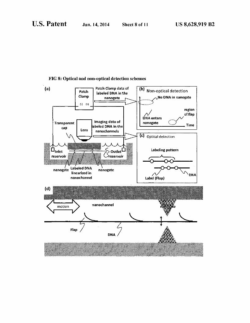
Exemplary, non-limiting embodiments of these methods are shown in FIG. 8. That figure first (FIG. 8a) depicts a system for obtaining labeled barcode information from a nucleic acid polymer, utilizing both optical and non-optical detection methods. As shown, a labeled long nucleic acid molecule is shown
stretched and linearized within a nanochannel having one or more narrow constrictive points (known as nanogates or nanonozzles; see U.S. application Ser. No. 12/374,141, the entirety of which is incorporated herein by reference).
In some embodiments, DNA movement and current mea Surement are controlled by an electrical circuit in connection with fluidic devices and external reservoirs. Optical images of the barcodes patterns and non-optical recording of the labels (i.e., electrical recording of physical "bumps' along the uni form polymers) are shown in, are schematically shown in FIG. 8b and FIG. 8c. The optical and non-optical results may be correlated or compared against one another for better data accuracy.
FIG. 8d depicts a nanogate-comprising fluidic device. Shown here is a series of “flaps' generated by methods pre viously described, which flaps may include additional label ing tags. The flaps, their tags, or both are detected directly during passage through the nanogates, during which the flaps, tags, or both generate detectable electronic signals such as an ionic current signatures reflecting the target genomic region. Labeled bases may—as described elsewhere herein—also be present in the nucleic acid polymer in the region vacated by the flap. Such bases may also be detected as they pass by a nanogate.
Also provided are methods of obtaining structural infor mation from a macromolecule. These methods include label ing at least a portion of a macromolecule; immobilizing the macromolecule; disposing at least a portion of the macromol ecule within a channel Such that at least a portion of the macromolecule is linearized within the channel; and detect ing at least one signal related to the labeled portion of the macromolecule.
FIG. 9 depicts a tethered nucleic acid at one end or both ends inside a nanochannel or nanotrack on the Surface of a Substrate for sequence imaging analysis. As shown in the
10
15
25
30
35
40
45
50
55
60
65
14 figure, a region of the nucleic acid polymer is modified to enable tethering, the nucleic acid polymer having a sequence (R2) that is labeled or other wise being analyzed at multiple locations. As a non-limiting example, R2 may be known to reside
within a gene for a particular disease, and the presence of multiple R2 sequences within the polymer may demonstrate an abnormal (or normal) number of copies of that sequence. The polymer may be translocated along the channel from one reservoir to another, and may be stopped or immobilized at any point along its translocation path. The immobilization may be accomplished in a number of
ways. In one embodiment, as shown in FIG. 10a, the macro molecule is bound to at least one bead, the molecule being immobilized by the at least one bead being caught by a con striction Smaller in cross-section than the bead. Immobiliza tion may also be accomplished by chemically tethering the macromolecule to a Surface, by magnetically immobilizing the macromolecule, by optically trapping the macromolecule, or any combination thereof.
In embodiments including a bead, the bead is chosen such that its effective diameter is larger than at least one of the cross-sectional dimensions of the nanochannel. As the modi fied nucleic molecule is flowed into the nanochannel, its flow is impeded because the modifying bead is larger than at least a portion of the nanochannel. The unmodified portions of the nucleic acid molecule can then be linearized and are available for sequence analysis. The bead can be polymeric, magnetic, semi-conducting, dielectric, metallic or any combination thereof and modification of the nucleic acid molecule can be based on a covalent bond or non-covalent interaction includ ing protein interactions and can involve an intermediary link age. In all modes of tethering or immobilization, an applied flow or gradient field may be modulated so as to enable or disengage the tethering. The modifying species for tethering can be chosen Such
that the nature of binding of the nucleic acid molecule within the nanochannel is magnetic, electrical, optical, chemical, frictional, flow-based, physical obstruction or any combina tion thereof.
In another embodiment, a nucleic acid molecule is chemi cally modified at or near one end of the molecule, as shown in non-limiting FIG. 10b. The chemical modification is chosen Such that a covalent or non-covalent interaction occurs between the modifying species and the nanochannel material of sufficient strength to tether the nucleic acid molecule and prevent its flow through the nanochannel.
Examples of chemical modifiers include thiol groups, silane groups, carboxy groups, amine groups, alkyl chains, phosphate groups, photocleavable groups, proteins, biotin, amino acid residues, metallic groups, or any combination thereof. In some cases, the nanochannel Surface may include some chemical modification to facilitate the interaction with the modifying species.
In another embodiment, a nucleic acid molecule is mag netically modified at or near one end of the molecule, as shown in FIG. 11a. The magnetic modification can be a magnetic bead, paramagnetic particle, Superparamagnetic particle, or other moiety capable of Sustaining a magnetic dipole for the duration of the sequence analysis. In Such a case, the magnetic force can be integrated into or near the nanochannel device or, alternatively, can be the consequence of an externally applied magnetic field, also as shown in FIG. 11a.
In another embodiment, a nucleic acid is modified at or near one end of the molecule with a particle or moiety capable

US 8,628,919 B2 15
of experiencing a dielectric force gradient in the presence of optical tweezers. This is shown in non-limiting FIG.11b. As shown, optical tweezers are used to trap the particle
within confines of the beam when the particle is flowing through the nanochannel thus allowing the attached nucleic acid molecule to be linearized within the nanochannel. The optical tweezers can be used to move a target as well as immobilize it.
In another embodiment, multiple forces are employed to immobilize or tether the DNA. For example, an opposing fluid flow and an electric field can be employed concurrently to keep the molecule stretched and stationary within the area of analysis.
Linearization is suitably accomplished by a channel that is suitably sized so as to effect linearization of the macromol ecule, Suitably by physical-entropic confinement.
Also provided are analysis systems. Systems according to the claimed invention include a Substrate comprising at least one channel having a width in the range of from about 1 to about 500 nanometers; the Substrate comprising at least one immobilization region. The channels suitably have a width in the range of from about 10 to about 200 nm, or from about 20 to about 100 nm, or even about 50 nm. The channels depth may be in the same range, although the width and depth of a particular channel need not be the same. Channels can be of virtually any length, from 10 nm up to centimeters. Such channels suitably have a length in the millimeter range, although the optimal length for a given application will be apparent to the user of ordinary skill in the art. The immobilization region is capable of immobilizing a
macromolecule. Macromolecules may include one or more modifications, which can include flaps, beads, dielectric modifications, magnetic particles, and the like. The systems and macromolecular modifications may be chosen in concert and on the basis of their affinity for one another. Exemplary immobilization regions include magnetic regions, chemically active regions, constrictions, and the like, as shown in FIG. 10 and FIG. 11.
In some embodiments, the polymer is immobilized, and a gradient is applied so as to disposed at least a portion of the polymer in the channel, as shown in FIG. 10 and FIG. 11. In this way, a polymer—which can be labeled, as described elsewhere herein may be linearized and, by virtue of its confinement within the channel, may remain in linear form.
While not shown in the figures, the present invention also include embodiments in which a labeled polymer is immobi lized or tethered and then linearized by application of a gra dient (pressure, electrical, and the like) in order that one or more labels (or flaps) disposed on the polymer can be detected and correlated to a characteristic of the polymer. The polymer can be maintained in a linear form by continued application of the gradient or by being adhered to a Substrate once it has been linearized by the gradient (i.e., the polymer is linearized and then adhered down the substrate in its linearized form).
Also provided are methods of characterizing a nucleic acid polymer. These methods include labeling one or more regions of a nucleic acid polymer with one or more sequence-specific motif labels; correlating one or more signals from one or more of the sequence-specific motif labels to the position of the one or more sequence-specific motif labels of the nucleic acid polymer; sequencing one or more segments of the nucleic acid polymer, the one or more segments including one or more of the sequence specific motif labels of the nucleic acid polymer; and comparing one or more signals of one or more sequenced segments to one or more corresponding signals of
10
15
25
30
35
40
45
50
55
60
65
16 the labeled nucleic acid polymer so as to develop the relative locations within the nucleic acid polymer, of two of more sequenced segments. The labeling aspect of the claimed methods is suitably
accomplished by labeling methods described elsewhere herein, i.e., forming a flap in the nucleic acid polymer and labeling the flap, the region vacated by the flap, or any com bination thereof. Suitable labels and tags are described else where herein.
Correlating Suitably entails linearizing at least one labeled portion of the nucleic acid polymer. The linearization may be accomplished by linearizing the labeled portion of the poly mer in a Suitably sized nanochannel, by applying a gradient (fluid, electrical, for example) to the polymer, and the like. In other embodiments, the polymer is tethered or otherwise immobilized and linearized by application of a gradient (pres Sure, electrical, and the like). Segments may be generated by random or sequence-specific cleaving of the nucleic acid polymer. The correlating may include, for example, determining the
distance between two or more labels, comparing the intensity of signals evolved from two or more labels, and the like. Sequencing of the segments of the polymer—known, in some instances, as "contigs”, may be accomplished by a variety of techniques known in the art. These techniques include, for example, Sanger sequencing, Maxam-Gilbert sequencing, dye terminator sequencing, in vitro clonal amplification, sequencing by hybridization, and the like. Segments are Suit ably up to 30 kb or even 50kb in length, but are suitably in the kb length range. Comparing the signal or signals of a labeled segment to the
corresponding signal of the labeled nucleic acid polymer is accomplished, for example, by aligning one or more labeled, sequenced segments against the labeled nucleic acid polymer such that a sequence-specific motif label of the labeled, sequenced segment is placed in register with the correspond ing sequence-specific motif label of the labeled nucleic acid polymer. This effectively allows the user to utilize the labels on the segments as “barcodes' that allow for identification of individual segments. Thus, by matching a barcoded contig against the corresponding barcode on the parent nucleic acid polymer, the user may determine the position (and ori entation) of the barcoded contig within the “parent nucleic acid polymer.
In this way, by aligning one or more signals from labels on the segment with the corresponding labels on the “mother polymer, the user can determine the proper alignment of the segment. By repeating this process for multiple segments, the user can then determine the proper order—and orientation— of the segments, allowing for massively parallel sequencing of nucleic acid polymers.
This process is further depicted in FIG. 6, which depicts the claimed methods of obtaining genome scaffolding (e.g., sequence) assembly information from a nucleic acid polymer. As shown in the figure, the user extracts comparatively
long genomic DNA molecules from a polymer (from 1 kb up to 100 mb or more) and labels the molecules, e.g., according to the labeling methods described elsewhere herein so as to give rise to create sequence specific signals that are detected and recorded along the linearized long polymers to generate a signature “observed barcode” (shown as “Raw Images of OBSERVED BARCODE') that represents particular regions of the molecule genome; the molecules can represent a genome. The observed barcodes from individual molecules can then be assembled into comparatively long scaffolds, which scaffolds can be up to the size of an intact genome.

US 8,628,919 B2 17
Discrete segments (“contigs’, in Some embodiments; from about 5 to about 30 kb) may be computationally assembled based on partial overlapping short base reads generated by current sequencing sequencing technology. Such contigs can be random or be generated on the basis of sequence specific ity. As shown in FIG. 6, a genome may be fragmented into contigs of 50 bp up to 1000 bp, for example. The user can then generate many (millions) of short reads, of about 35 to about 850 bps. One or more of the contigs is suitably labeled with a
sequence specific motif (such as a Nb.BbvCI site, GCT GAGG) identical to the sequence specific motif used to label the "parent nucleic acid polymer to generate a series of barcodes. Where the contigs are virtually labeled (i.e., via computer), the barcodes are considered in silico barcodes. The user then aligns the barcodes of the contigs (segments)
against the corresponding, observed barcodes of the experi mentally constructed Scaffolds, which alignment then pro vides the user with the physical locations of the contigs within the scaffold, along with the proper orientation of a contig within the scaffold. This in turn yields information about the scaffold (and the corresponding genome). Such as copy num bers of sequences within the scaffold, structural information (e.g., translation), and the like. Thus, individual contigs are mapped precisely onto the genome so as to generate true, accurate genomic sequencing information of a specific poly mer under analysis.
These methods have numerous advantages over existing sequencing techniques, including the ability to provide infor mation regarding copy number and the ability to place contigs in the proper position/order relative to one another. This in turn provides true sequencing information; without the bar coding techniques described herein, the linear order of con tigs along the analyzed genome would be unknown, espe cially if there is no prior reference database to compare against to (de novo sequencing). Due to the high complexity of large genomes having copy number variations (CNVs) and structural variations (SVs), independent assembly directly from random shorter reads, especially for denovo sequencing or highly scrambled cancer genome, has become increasingly difficult and prone to errors. As one non-limiting example, a first segment (of known
sequence) might include barcodes A, B, and C, each of which barcodes correspond to the position of a sequence-specific label on the segment, the intensity of the sequence-specific label, or both. The labeled segment thus presents a unique profile based on the A, B, and C barcodes. A second labeled segment (of known sequence) may include barcodes C, D, and E. By aligning the first and second segments against the “mother polymer from which the segments were cleaved, the user can determine that the two segments overlap at barcode C and—by combining the sequences of the two segments (without double-counting the sequence corresponding to bar code C)—can determine the sequence of the “mother poly mer from which the two segments were derived. By scaling this process up to address multiple segments simultaneously, the present methods thus enable determination of sequence information for long nucleic acid polymers. One similar embodiment is shown in FIG. 3. This figure
illustrates an example using Lambda DNA, predicted nicking sites by the nickase Nb.BbvC I are shown in sequence motif and indicated by arrows along the long DNA molecule. The nicking sites are labeled with fluorescent (Alexa) nucleotides T that are incorporated at the nicking sites (shown in green color), as the native T base is displaced and replaced.
In this model system, the observed signature “barcode' patterns of the labeling agree with the predicted sequence
10
15
25
30
35
40
45
50
55
60
65
18 motif map of the genome generated with nicking enzyme digestion in silico, designated here as in silico BARCODE, based on 100% stretched lambda DNA in low salt conditions within 80 nm by 80 nm wide channels, as shown by FIG.3b. Similar barcode results shown on linearized human BAC clone DNAs with complete stretching (~170 Kbp); over 17 labeled sites (in fluorescent color) are also shown.
ADDITIONALEXAMPLES AND EMBODIMENTS
Additional Embodiments
As described elsewhere herein, the claimed invention pro vides, inter alia, methods relating to DNA mapping and sequencing, including methods for making long genomic DNA, methods of sequence specific tagging and a DNA bar coding strategy based on direct imaging of individual DNA molecules and localization of multiple sequence motifs or polymorphic sites on a single DNA molecule inside the nanochannel (<500 nm in diameter). The methods also pro vide continuous base by base sequencing information, within the context of the DNA map. Compared with prior methods, the claimed method of DNA mapping provides improved labeling efficiency, more stable labeling, high sensitivity and better resolution; our method of DNA sequencing provide base reads in the long template context, easy to assemble and information not available from other sequencing technolo gies, such as haplotype, and structural variations.
In DNA mapping applications, individual genomic DNA molecules or long-range PCR fragments are labeled with fluorescent dyes at specific sequence motifs. The labeled DNA molecules are stretched into linear form inside nanochannels (described elsewhere herein) and are imaged using fluorescence microscopy. By determining the positions and, in some cases, the colors of the fluorescent labels with respect to the DNA backbone, the distribution of the sequence motifs can be established with accuracy, akin to barcode on a package. This DNA barcoding method is applied to the iden tification of lambda phage DNA molecules and to human bac-clones. One embodiment utilizing nicks at specific sequence sites
on dsDNA comprises the steps of: a) nicking one strand of a long (e.g., more than 2 kb) double
stranded genomic DNA molecule with one or more nick ing endonucleases to introduce nicks at specific sequence motifs;
b) incorporating fluorescent dye-labeled nucleotides at the nicks with a DNA polymerase:
c) stretching the labeled DNA molecule into linear form inside nanochannels, the molecules either flowing through the channels or a portion of the molecule being immobilized such that one end of the DNA is then dis posed within the channel;
d) determining the positions of the fluorescent labels with respect to the DNA backbone using fluorescence microscopy to obtain a map or barcode of the DNA.
Another embodiment with flap sequences at sequence spe cific nicking sites comprises the steps of
a) nicking one strand of a long (>2. Kb) double stranded genomic DNA molecule with a nicking endonucleases to introduce nicks at specific sequence motifs;
b) incorporating fluorescent dye-labeled nucleotides or none fluorescent dye-labeled nucleotides at the nicks with a DNA polymerase, displacing the downstream strand to generate a flap sequences;

US 8,628,919 B2 19
c) labeling the flap sequences by polymerase incorporation of labeled nucleotides; or direct hybridization of a fluo rescent probe; or ligation of the fluorescent probes with ligases:
d) stretching the labeled DNA molecule into linear form as 5 described elsewhere herein;
e) determining the positions of the fluorescent labels with respect to the DNA backbone using fluorescence microscopy so as to obtain a map or barcode of the DNA.
Another embodiment utilizing a ssDNA gap at sequence 10 specific nicking sites comprises the steps of:
a) nicking one strand of a long (>2. Kb) double stranded genomic DNA molecule with a nicking endonucleases to introduce nicks at specific sequence motifs;
b) incorporating fluorescent dye-labeled nucleotide probes 15 or non-fluorescent dye-labeled nucleotides at the nicks with a DNA polymerase, displacing downstream strand to generate one or more flap sequences;
c) employing a nicking endonuclease to nick the newly extended Strand and cut the newly formed flap sequences 20 with flap endonucleases. The detached ssDNA can be removed by, for example, increasing the temperature so as to release their bonds.
d) labeling the ssDNA gap (evolved by the nicking and Subsequent formation of the flaps) via incorporation of 25 labeled nucleotides; or direct hybridization of the fluo rescent probes; or ligation of the fluorescent probes with ligases.
e) stretching the labeled DNA molecule into linear form as described elsewhere herein; 30
f) determining the positions of the fluorescent labels with respect to the DNA backbone using fluorescence microscopy to obtain a map or barcode of the DNA.
In other DNA sequencing applications, individual genomic DNA molecules or long-range PCR fragments are 35 labeled with fluorescent dyes at specific sequence motifs. The labeled DNA molecules are then linearized within nanochannels and are then imaged using fluores cence microscopy. By determining the positions and colors of the fluorescent labels with respect to the DNA 40 backbone, the distribution of the sequence motifs can be established with accuracy, in a manner similar to reading a barcode. Single or multiple bases information were obtained in the context of the DNA map.
One embodiment of this sequencing method applicable to 45 genomic DNA comprises the steps of:
a) nicking one strand of a long (>2. Kb) double stranded genomic DNA molecule with a nicking endonucleases to introduce nicks at specific sequence motifs;
b) tagging the nicking sites with fluorescent dye molecules 50 through nick-incorporation; flap labeling, ssDNA gap labeling, or some combination thereof;
c) stretching the labeled DNA molecule into linear form as described elsewhere herein;
d) determining the positions of the fluorescent labels with 55 respect to the DNA backbone using fluorescence microscopy to obtain a map or barcode of the DNA;
e) using the nicking sites as the initialization points of sequencing reactions. Different DNA structures includ ing but not limited to the following, are useful in DNA 60 sequencing.
In one sequencing embodiment, a polymerase incorporates fluorescent nucleotides at the 3' end of the nicking sites, sequentially detecting the incorporated labels at each nicking site to obtain the sequence information. This process is 65 repeated/cycled to sequentially obtain “reads' on many bases.
20 In another embodiment, a sequencing primer is hybridized
to a flap sequence and is extended with a polymerase to incorporate a fluorescent nucleotide. By reading the colors of these various incorporated fluorescent nucleotides, sequence information is then inferred. This process is repeated/cycled to obtain many base reads sequentially.
In another embodiment, one short fluorescent oligonucle otide is directly hybridized to the flap sequences, the sequence information can be inferred from the presence of the hybridized oligos. This process is cycled/repeated to obtain many base reads sequentially.
In another embodiment, two short oligonucleotides are hybridized to flap sequences next to each other and are then ligated together with, e.g., ligases, The sequence information can be inferred from the ligation products. This process is repeated/cycled to obtain many base reads sequentially.
In another embodiment, one short fluorescent oligonucle otide is directly hybridized next to the 3' end of the nicking sites and ligated. The sequence information is then inferred from the presence of the ligated oligonucleotides. This pro cess is repeated/cycled to obtain many base read sequentially. The methods may be performed in conjunction with
nanochannel arrays. Such arrays Suitably have a plurality of channels in the material of the Surface, the channels having a trench width of less than about 500 nanometers and a trench depth of less than 500 nanometers. At least some of the channels are suitably Surmounted by sealing material to ren der Such channels at least Substantially enclosed.
In some embodiments, the claimed invention includes car tridges or other modular devices. Such cartridges may include, for example, a including a nanofluidic chip in accor dance with this invention are also disclosed herein. Such cartridges are capable of being inserted into, used and removed. Cartridges useful with analytical systems other than the systems of the present invention are also within the scope of the present invention.
Nanochannels, in Some embodiments, are capable of trans porting a macromolecule across their length. Devices of the claimed invention may include one or more components use ful in effecting macromolecular transport, which transport may be effected by pressure or vacuum gradients across a channel, electroësmosis, and electrokinesis. The surface material of the nanochannels can be formed
from almost any Substrate material. Such as a conductive material, a semiconductor material, or a non-conductive material. Examples of conductive materials include metals Such as aluminum, gold, silver, and chromium. Examples of semiconductive materials include doped silicon dioxide and gallium arsenide. Examples of non-conductive materials include fused silica, silicon dioxide, silicon nitride, glass, ceramics, and synthetic polymers. The foregoing is exem plary only.
In some embodiments, a nucleic acid molecule is modified at or near one end and is then disposed into a nanochannel or nanotrack (a region defined by borders that restrain fluid passage. Such as hydrophobic borders). The modification Suitably permits tethering of the nucleic acid at the entrance of the nanochannel or within the nanochannel. The nucleic acid is then constrained to adopt a linearized
form due to the nanochannel The nucleic acid is suitably DNA or RNA, e.g., dsDNA. The nanochannel is preferably <500 nm, more preferably <300 nm and most preferably <150 nm with a length capable of accommodating a linearized nucleic acid with more than 2000 bases. The following embodiments also apply to nanotracks,
which are linear regions defined on chemically or topologi cally predefined surface patterns.

US 8,628,919 B2 21
Fluids that can be analyzed by the system includes fluids from a mammal (e.g., DNA, cells, blood, biopsy tissues), synthetic macromolecules Such as polymers, and materials found in nature (e.g., materials derived from plants, animals, and other life forms). Such fluids can be managed, loaded, and injected using automated or manual loading apparatus of the present invention.
EXAMPLES
Example 1
Generating Single Stranded DNA Flaps on Double Stranded DNA Molecules
Genomic DNA samples were diluted to 50 ng for use in the nicking reaction. 10 uI of Lambda DNA (50 ng/uIL) were added to a 0.2 mL PCR centrifuge tube followed by 2 ul of 10x NE Buffer #2 and 3 ul of nicking endonucleases, includ ing but not limited to Nb.BbvCI: Nb.BsmI: Nb.BsrDI: Nb.BtsI; NtAlwI: Nt.BbvCI; Nt.BspOI: Nt.BstNBI; Nt.CviPII. The mixture was incubated at 37 degrees C. for one hour.
After the nicking reaction completes, the experiment pro ceeded with limited polymerase extension at the nicking sites to displace the 3' down stream Strand and form a single Stranded flap. The flap generation reaction mix consisted of 15ul of nicking product and 5 ul of incorporation mix con taining 2 ul of 10x buffer, 0.5ul of polymerase including but not limited to vent(exon-), Bstand Phi29 polymerase and 1 ul nucleotides at various concentration from 1 uM to 1 mM. The flap generation reaction mixture was incubated at 55 degrees. The length of the flap was controlled by the incubation time, the polymerases employed and the amount of nucleotides used.
Example 2
Generating Single Stranded DNA Gaps on Double Stranded DNA Molecules
After flap generation, the original nicking endounuclease was used to nick the filled double stranded DNA and Flap endonulceases including but not limited to FEN1 was used to cut the flap sequences. By increasing the temperature, the nicked single stranded DNA molecules were removed from the double stranded DNA molecules to generate a single stranded DNA gap on double stranded DNA molecules.
Example 3
Generating Long Single Stranded DNA Molecules
After the nicking reaction completes, the experiment pro ceeded with complete polymerase extension including but not limited to Phi29, Bst polymerase at the nicking sites to displace the 3' down stream Strand and generate single stranded DNA molecules.
Example 4
The Method of Fluorescently Labeling Sequence Specific Nicks on Double Stranded DNA Molecules
Genomic DNA samples were diluted to 50 ng for use in the nicking reaction. 10 uI of Lambda DNA (50 ng/uIL) were added to a 0.2 mL PCR centrifuge tube followed by 2 ul of
10
15
25
30
35
40
45
50
55
60
65
22 10x NE Buffer #2 (New England BioLabs, www.neb.com), and 3 ul of nicking endonucleases, including but not limited to Nb.BbvCI; Nb.BsmI: Nb.BsrDI; Nb.BtSI: Nt. AlwI: Nt. B bvCI: Nt.BspQI: Nt.BstNBI: Nt.CviPII. The mixture was incubated at 37 degrees C. for one hour.
After the nicking reaction completes, the experiment pro ceeds with polymerase extension to incorporate dye nucle otides onto the nicking sites. In one embodiment, a single fluorescent nucleotide terminator was incorporated. In another embodiment, multiple fluorescent nucleotides were incorporated. The incorporation mix consisted of 15 Jul of nicking prod
uct and 5 ul of incorporation mix containing 2 ul of 10x buffer, 0.5 ul of polymerase including but not limited to Vent(exon-), 1 Jul fluorescent dye nucleotides or nucleotide terminators including but not limited to cy3, alexa labeled nucleotides. The incorporation mixture was incubated at 55 degrees C. for about 30 minutes.
Example 5
The Method of Sequence Specific Labeling Single Stranded DNA Flaps on Double Stranded DNA
Molecules
Once the flap sequence was generated, the flap can be labeled with fluorescent dye molecules including but not limited to the following methods, hybridization of probe, incorporation of fluorescent nucleotide with polymerase and ligation of fluorescent probes.
Example 6
The Method of Sequence Specific Labeling Single Stranded DNA Gaps on Double Stranded DNA
Molecules
A nanofluidic chip having a width,depth, or both of 500 nm or less is filled using capillary action with a buffer solution containing stained genomic DNA to draw the DNA macro molecules into the channels with an electric field. Bacteria phage DNA molecules Lambda (48.5 kb) and Human BAC clone (170 kb) were stained with the dye YOYO-1. This solution of stained DNA is diluted to 0.5ug/mL into 0.5xTBE containing 0.1 M dithiothreatol as an anti-oxidant and 0.1% of a linear acrylamide used as an anti-sticking agent. An Olympus IX-71 inverted microscope with a 100x
(N.A. 1.35) oil immersion objective is used with a solid-state laser (e.g., diode pumped solid State laser), which can have different excitation wavelengths (e.g., 473 nm for YOYO-1 dye). Other lasers (e.g., for Alexa series of dyes, Cy3, Cy5, etc.) include a 532 nm DPSS laser, a 635 Laser Diode laser, a 543 nm gas laser, a 591 nm DPSS laser, and a 633 nm gas laser. An ANDOR cooled-EMCCD camera with a 512x512 pixel array and 16 bits digital output is used to image the molecules. Digital images are analyzed using a data processor by J-image and other analysis Software.
Example 7
Detection Schemes
In one example of a detection scheme, video images of DNA moving in flow mode are captured by a time delay and integration (TDI) camera. In such an embodiment, the move ment of the DNA is synchronized with the TDI.

US 8,628,919 B2 23
In another example of a detection scheme, video images of a DNA moving in flow mode are capture by a CCD or CMOS camera, and the frames are integrated by soft ware or hardware to identify and reconstruct the image of the DNA.
In another example of a detection scheme, video images of a DNA are collected by simultaneously capturing different wavelengths on a separate set of sensors. This is accom plished by using one camera and a dual or multi-view splitter,
SEQUENCE LISTING
<16 Os NUMBER OF SEO ID NOS: 6
<21 Os SEQ ID NO 1 &211s LENGTH: 51 &212s. TYPE: DNA
<213> ORGANISM: Artificial Sequence 22 Os. FEATURE:
<223> OTHER INFORMATION: Synthetic oligonucleotide
<4 OOs SEQUENCE: 1
24 or using by filters and multiple cameras. The camera can be a TDI, CCD or CMOS detection system.
In another example, using simultaneous multiple wave length video detection, a backbone dye is used to identify a unique DNA fragment, and the labels are used as markers to follow the DNA movement. This is useful in cases where the DNA’s length is greater than the field of view of the camera, and the markers can serve to help map a reconstructed image of the DNA.
tgaggtttgc accggtgtgg ct coggaagt taacgct aaa gCactggcct g 51
<21 Os SEQ ID NO 2 211s. LENGTH: 77
&212s. TYPE: DNA
<213> ORGANISM: Artificial Sequence 22 Os. FEATURE:
<223> OTHER INFORMATION: Synthetic oligonucleotide
<4 OOs SEQUENCE: 2
Caccctgctt gctgaggttt gcaccggtgt ggct Coggala gttaacgcta aagc actggc 60
Ctggggaaaa cagtacg
<21 Os SEQ ID NO 3
<4 OOs SEQUENCE: 3
OOO
<21 Os SEQ ID NO 4 &211s LENGTH: 12
&212s. TYPE: DNA
<213> ORGANISM: Artificial Sequence 22 Os. FEATURE:
<223> OTHER INFORMATION: Synthetic oligonucleotide
<4 OOs SEQUENCE: 4
caccctgctt gc
<21 Os SEQ ID NO 5 &211s LENGTH: 14
&212s. TYPE: DNA
<213> ORGANISM: Artificial Sequence 22 Os. FEATURE:
<223> OTHER INFORMATION: Synthetic oligonucleotide
<4 OOs SEQUENCE: 5
gggaaaacag tacg
<21 Os SEQ ID NO 6 &211s LENGTH: 10
&212s. TYPE: DNA
<213> ORGANISM: Artificial Sequence
77
12
14
US 8,628,919 B2 25 26
- Continued
22 Os. FEATURE:
<223> OTHER INFORMATION: Synthetic oligonucleotide
<4 OOs SEQUENCE: 6
acgtgggaaa
What is claimed is: 1. A method of characterizing DNA, comprising: processing a double-stranded DNA comprising a first DNA
strand and a second DNA strand to give rise to an unhy bridized flap of the first DNA strand and a corresponding region on the second DNA strand,
the unhybridized flap comprising about 10 to about 1000 bases;
extending the first DNA strand along the corresponding region of the second DNA strand; and
labeling at least a portion of the unhybridized flap, a por tion of the extended first DNA strand, or both.
2. The method of claim 1, wherein the processing com prises nicking the first DNA strand.
3. The method of claim 2, wherein the nicking is effected at one or more sequence specific locations.
4. The method of claim 2, wherein the nicking is effected at one or more non-specific locations.
5. The method of claim 1, wherein the labeling is accom plished by (a) binding at least one complementary probe to at least a portion of the unhybridized flap, the probe comprising one or more tags, (b) extending the first DNA strand along the corresponding region of the second DNA strand with one or more nucleotides comprising one or more tags, or any com bination of (a) and (b).
6. The method of claim 5, further comprising obtaining sequence information derived from one or more tags.
7. The method of claim 5, wherein labeling comprises attaching a tag comprising a fluorophore, a quantum dot, a dendrimer, a nanowire, a bead, a peptide, a protein, a mag netic bead, a methyl group, a methyltransferase, a non-cutting restriction enzyme, a zinc-finger protein, an antibody, a tran Scription factor, a DNA binding protein, a hairpin polyamide, a triplex-forming oligodeoxynucleotide, a peptide nucleic acid, or any combination thereof.
8. The method of claim 7, further comprising at least par tially linearizing at least a portion of the double-stranded DNA comprising an unhybridized flap, a portion of the extended region of the first DNA strand, or both.
9. The method of claim 8, further comprising measuring the distance between two or more tags.
10. The method of claim 9, further comprising correlating the distance to a structure, a sequence assembly, a genetic or cytogenetic map, a methylation pattern, a location of a cpG island, an epigenomic pattern, a physiological characteristic, or any combination thereof.
11. The method of claim 8, wherein the linearization is effected by confinement of the DNA in a channel.
12. The method of claim 7, further comprising measuring the intensity of at least one signal from at least one tag.
13. The method of claim 12, further comprising correlating the intensity of the at least one signal to a structure, a sequence assembly, a genetic or cytogenetic map, a methylation pat tern, a physiological characteristic, a location of a cpG island, an epigenomic pattern, or any combination thereof.
14. The method of claim 5, further comprising detecting one or more signals from one or more tags.
10
15
25
30
35
40
45
50
55
60
65
1O
15. The method of claim 1, wherein the flap comprises at least a portion of a base sequence of interest on the double stranded DNA.
16. The method of claim 1, wherein the unhybridized flap of the first DNA strand, the corresponding region of the second DNA strand, or both, comprises at least a portion of the double-stranded DNA that flanks a base sequence of inter est on the double-stranded DNA.
17. The method of claim 1, further comprising obtaining epigenomic information from the DNA.
18. A method of characterizing DNAs, comprising: labeling, on a first double-stranded DNA, two or more
sequence-specific locations on the first DNA; labeling, on a second double-stranded DNA, the two or more corresponding sequence-specific locations on the second DNA;
linearizing at least a portion of the first double-stranded DNA;
linearizing at least a portion of the second double-stranded DNA; and
comparing the distance between two or more labels on the first, linearized double-stranded DNA to the distance between the corresponding labels on the second, linear ized double-stranded DNA, wherein the labeling is accomplished by nicking a first strand of a double stranded DNA so as to give rise to (a) an unhybridized flap of the first strand and (b) a corresponding region in the second strand of the double-stranded DNA, wherein the unhybridized flap comprises about 10 to about 1000 bases.
19. The method of claim 18, further comprising exposing the flap to a labeled probe complementary to at least a portion of the probe, extending the first strand of the DNA along the corresponding region of the second strand with one or more labeled bases, or both.
20. The method of claim 18, wherein the labeling is accom plished by exposing the first and second double-stranded DNAS to a non-cutting restriction enzyme, a methyltrans ferase, a Zinc finger protein, an antibody, a transcription fac tor, a DNA binding protein, a hairpin polyamide, a triplex forming oligodeoxynucleotide, a peptide nucleic acid, or any combination thereof.
21. The method of claim 20, wherein the non-cutting restriction enzyme comprises a tag.
22. The method of claim 18, further comprising correlating the distance to a structure, a sequence assembly, a genetic or cytogenetic map, a methylation pattern, a physiological char acteristic, a location of a cpG island, an epigenomic pattern, or any combination thereof, of the DNA.
23. The method of claim 18, further comprising obtaining epigenomic information from a DNA.
24. A method of characterizing DNA, comprising: labeling, on a first double-stranded DNA, one or more
sequence-specific locations on the first double-stranded DNA;
labeling, on a second double-stranded DNA, the corre sponding one or more sequence-specific locations on the second double-stranded DNA;
US 8,628,919 B2 27
linearizing at least a portion of the first double-stranded DNA;
linearizing at least a portion of the second double-stranded DNA; and
comparing the intensity of a signal of a label of the first, linearized double-stranded DNA to the intensity of the signal of a label of the second, linearized double stranded DNA, wherein the labeling is accomplished by nicking a first strand of each of the first and second double-stranded DNAs so as to give rise to (a) an unhy bridized flap of the first strand and (b) a corresponding region in the second strand of the corresponding double stranded DNA, wherein the unhybridized flap comprises about 10 to about 1000 bases.
25. The method of claim 24, further comprising exposing the flap to a labeled probe complementary to at least a portion of the probe, extending the first strand of the DNA along the corresponding region of the second DNA strand with one or more labeled bases, or both.
26. The method of claim 24, further comprising correlating the signal intensities to a structure, a sequence assembly, a genetic or cytogenetic map, a methylation pattern, a physi ological characteristic, a location of a cpG island, an epige nomic pattern, or any combination thereof, of the DNA.
27. The method of claim 24, further comprising obtaining epigenomic information from the DNA.
28. A method of characterizing a macromolecule, compris 1ng:
translocating a macromolecule comprising at least one flap extending therefrom along a channel having at least one constriction disposed therein, wherein the flap com prises about 10 to about 1000 bases; and
detecting at least one signal corresponding to the passage of the at least one flap of the macromolecule through the at least one constriction of the channel.
29. The method of claim 28, wherein the flap comprises a label.
30. The method of claim 28, wherein the macromolecule comprises a label.
31. The method of claim 28, wherein the macromolecule comprises double-stranded DNA or chromatin fibers.
32. The method of claim 28, further comprising translocat ing the at least one flap through the at least one constriction more than once.
33. A method of characterizing a macromolecule, compris 1ng:
labeling at least a portion of a macromolecule, wherein the labeled macromolecule comprises at least a flap com prising about 10 to about 1000 bases:
immobilizing the macromolecule: disposing at least a portion of the macromolecule within a
channel such that at least a portion of the macromolecule is linearized within the channel; and
detecting at least one signal related to the labeled portion of the macromolecule.
34. The method of claim33, wherein the macromolecule is bound to at least one bead, the molecule being immobilized by the at least one bead being caught by a constriction smaller in cross-section than the bead.
35. The method of claim 33, wherein the immobilization is accomplished by chemically tethering the macromolecule to
10
15
25
30
35
40
45
50
55
60
28 a surface, by magnetically immobilizing the macromolecule, by optically trapping the macromolecule, or any combination thereof.
36. The method of claim 33, wherein the macromolecule comprises at least one dielectric modification and is immo bilized by an optical trap.
37. The method of claim 33, wherein the channel is sized so as to effect linearization of the macromolecule.
38. A method of characterizing a nucleic acid polymer, comprising:
labeling one or more regions of a nucleic acid polymer with one or more sequence-specific motif labels, wherein the labeled nucleic acid polymer comprises at least a flap comprising about 10 to about 1000 bases;
correlating one or more signals from one or more of the sequence-specific motif labels to the position of the one or more sequence-specific motif labels of the nucleic acid polymer;
sequencing one or more segments of the nucleic acid poly mer, the one or more segments including one or more of the sequence specific motif labels of the nucleic acid polymer; and
comparing one or more signals of one or more sequenced segments to one or more corresponding signals of the labeled nucleic acid polymer so as to develop relative locations within the nucleic acid polymer, of two of more sequenced segments.
39. The method of claim38, wherein the labeling is accom plished by forming a flap in the nucleic acid polymer and labeling the flap, the region vacated by the flap, or any com bination thereof.
40. The method of claim 38, wherein the correlating com prises linearizing at least one labeled portion of the nucleic acid polymer.
41. The method of claim 40, wherein the linearizing com prises disposing the at least one labeled portion of the nucleic acid polymer in a channel capable of linearizing the at least one labeled portion of the nucleic acid polymer.
42. The method of claim38, further comprising application of a gradient to the at least one labeled portion of the nucleic acid polymer so as to linearize the at least one labeled portion of the nucleic acid polymer, to maintain the at least one labeled portion of the nucleic acid polymer in linear form, or any combination thereof.
43. The method of claim 38, wherein the correlating com prises determining the distance between two or more labels, comparing the intensity of signals evolved from two or more labels, or any combination thereof.
44. The method of claim 38, wherein the sequencing com prises Sanger sequencing, Maxam-Gilbert sequencing, dye terminator sequencing, in vitro clonal amplification, sequencing by synthesis, sequencing by hybridization, sequencing by ligation or any combination thereof.
45. The method of claim38, wherein a segment comprises from about 5 to about 30 kilobases.
46. The method of claim 38, wherein the comparing com prises aligning one or more labeled, sequenced segments against the labeled nucleic acid polymer such that a sequence specific motif label of the labeled, sequenced segment is in register with the corresponding sequence-specific motif label of the labeled nucleic acid polymer.
ck ck ck ck ck