1 introduction dna damage and types of repair dna is subject
TRANSCRIPT

1
INTRODUCTION
DNA damage and types of repair
DNA is subject to various types of damage that can impair cellular function
leading to cell death or carcinogenesis. DNA damage blocks normal cellular processes
such as replication and transcription [1, 2]. Should DNA damage persist it can have
catastrophic consequences for the cell and for the organism including mutagenesis,
cancer, and cell death (i.e. in neurons). DNA damage may come from internal as well as
external sources and is associated with various types of genetic diseases and cancers.
Therefore DNA repair provides a mechanism to restore DNA nucleotide sequences to the
native state. Several DNA repair pathways have been identified including base excision
repair (BER), mismatch repair (MMR) and nucleotide excision repair (NER). The type
of lesion will dictate which pathway is utilized for repair. For example an N7-methyl
guanine residue can only be removed by BER, while a benzopyrene guanine adduct
residue can only be removed by NER [3].
Nucleotide Excision Repair
Nucleotide Excision Repair (NER) is a versatile DNA repair pathway because of
its ability to repair a wide range of nucleotide adducts [4]. NER is responsible for the
repair of bulky DNA damage lesions including cyclobutane pyrimidine dimers (CPDs)
and 6-4 pyrimidine pyrimidone photoproducts (6-4PPs) caused by UV radiation [3, 5].
NER repairs platinum DNA adducts that are formed by cancer chemotherapeutic drugs
such as cisplatin and carboplatin [1, 6, 7]. UV radiation and platinum compounds cause
covalent linkage of adjacent thymine or guanine residues respectively. Aromatic

2
hydrocarbons, as found in cigarette smoke, cause guanine adducts that also require NER
for their removal. The steps involved in NER include recognition of DNA damage,
excising the lesion as a 25-30 single stranded oligomer, resynthesizing the excised region
of DNA, and finally ligating the newly repaired DNA strand [5, 8]. NER is divided into
two distinct sub-pathways: transcription-coupled repair (TC-NER), which affects the
transcribed strand in genes that are expressed, and global genomic repair (G-NER), which
is responsible for removing DNA damage from the overall genome. These pathways
differ in the early recognition step of DNA damage, but utilize the same proteins in
physically excising the lesion and restoring the DNA sequence.
TC-NER is the repair of lesions on DNA that is simultaneously undergoing
transcription. While TC-NER only occurs in a small percent of the genome it is
nonetheless critical to cell survival. DNA damage recognition in TC-NER occurs when
transcription is stalled by the arrest of RNA polymerase II as it encounters the site of
DNA damage [9, 10]. The stalled polymerase is thought to trigger the recruitment of
repair proteins including Cockayne syndrome complementation group A and B (CSA and
CSB) [11]. Defects in TC-NER result in genetic diseases such as Cockayne Syndrome
(CS). CS is a rare autosomal recessive genetic disorder that is characterized by severe
sunlight (UV) sensitivity, growth and developmental defects, and neurological disease
[12-14]. Patients with CS are not cancer prone but typically die at an early age due to
neurological problems [15]. Mutations in either the CSA or CSB gene are seen in over
90% of CS patients and result in defective TC-NER [12, 16]. CS cells are defective in
TC-NER and highly sensitive to UV-radiation and typically undergo apoptosis if exposed
to UV-radiation.

3
G-NER repairs damaged DNA throughout the entire genome. Patients that are
defective in G-NER suffer from a rare autosomal recessive disease known as xeroderma
pigmentosum (XP). Patients with XP are unable to carry out G-NER resulting in sunlight
sensitivity, predisposition to skin, lung, liver, and bladder cancers and in some cases
neurological disorders [17-19]. Seven human genetic complementation groups (XPA
through XPG) have been identified for xeroderma pigmentosum, all of which are
defective in NER. Sugasawa et al. have proposed a multistep damage recognition
mechanism for G-NER that is currently accepted as the mechanism for G-NER [20]. G-
NER is initiated by the recognition of the DNA lesion by XPC. XPC then recruits XPA,
RPA and XPG to the site of damage. The XPA and XPG proteins help to form a pre-
incision complex which is stabilized by RPA. TFIIH, a helicase, unwinds the DNA near
the site of DNA damage. XPF and XPG are endonucleases that cut the damaged DNA
strand 5′ and 3′ of the lesion ends respectively. The excision releases a fragment that is
about 25-30 nucleotides in length. DNA polymerase δ or pol ε fills in the excised gap.
The new fragment is then ligated by DNA ligase I completing the repair [20, 21]. Figure
1 depicts the general mechanism for global genomic nucleotide excision repair (G-NER).

4
Figure 1. Basic mechanism for nucleotide excision DNA repair. In G-NER XPC is
required for early lesion recognition (in TC-NER CSA and CSB substitute for XPC). In
either subpathway, after XPA and TFIIH are recruited, the damaged DNA strand is
excised by XPF and XPG releasing a 25-30-mer oligonucleotide. The excised gap is
filled in by DNA polymerase or and ligated by DNA Ligase I to complete DNA
repair. The diagram shows a guanine diadduct such as produced by the chemotherapy
drug cisplatin.

5
XPC
One of the most common complementation groups of XP seen in patients is that
of XPC [22]. XPC patients show defective G-NER but normal TC-NER. XPC patients
exhibit sensitivity to UV radiation and a dramatically increased risk of skin cancer [19].
In addition, somatically-acquired mutations in XPC have been associated with the poor
prognosis of patients with Nonsmall Cell Lung Carcinoma (NSCLC) [23]. Specific
allelic variants of XPC are associated with increased risk of colorectal cancer [11].
Decreased XPC expression has also been shown to correlate with bladder cancer
malignancy and its resistance to cisplatin treatment [24].
As mentioned, the XPC protein is required for early DNA damage recognition
and has been shown to be the initiator of G-NER [25, 26]. The XPC step is the rate
limiting step in G-NER. XPC has been shown to exhibit a strong binding affinity for
damaged DNA with affinity for UV-damaged DNA and lesions that cause helical
distortions [20, 27, 28]. Cells that lack XPC exhibit little or no NER [29, 30] (Figure 2).
Studies involving XPC knockout mice showed that these mice are viable and develop
normally; however, they exhibit increased sensitivity to UV light and are highly
susceptible to skin and lung cancer similar to XPC patients [30-35].

6
Figure 2. XPC is rate-limiting and is required for NER DNA repair. The purpose of this
experiment was to verify the NER defective phenotype of the mice obtained from
commercial sources. The mice originated from Allen Bradley’s laboratory prior to their
commercial distribution [30]. Bone marrow pooled from three untreated 10-week-old
wild type (WT) or knockout Xpc (–/–) mice were used to illustrate that XPC is required
for DNA repair. XPC protein was below detection by western blot in Xpc–/– mice bone
marrow (inset). Removal of DNA damage was determined by culturing WT and Xpc–/–
cells in cytokine-containing medium for 15 hours. Cells were irradiated with 20 J/m2 254
nm UV-radiation in 1mm depth of phosphate-buffered saline and returned to tissue
culture. Time points were taken at 0, 4, 8 and 16 hours and assayed using an antibody to

7
UV-induced 6-4 photoproducts. Xpc–/– bone marrow did not remove DNA lesions after
16 hours, whereas, WT cells removed 80% of the lesions by 16 hours (P < 0.02 by t-test).
The data confirms the known rate-limiting and required role of XPC in G-NER. The
assay measures mostly G-NER [36].

8
Regulation of XPC
Studies have shown that XPC is required for the recognition of DNA damage in
G-NER and for the recruitment of DNA repair factors to the site of damage. Given the
importance of XPC, relatively little is known about how XPC is regulated. At least one
level of regulation occurs at the transcriptional level by transcription factor/tumor
suppressor p53 [37]. Studies conducted by Ford et al. revealed that Xpc is inducible in a
time and dose dependent manner following UV irradiation in cells that expressed WT
p53, while induction was not seen in cells that lacked p53. Further investigations
revealed a p53 binding element in the Xpc promoter region and showed evidence for
transcription regulation of Xpc by p53 [37, 38].
XPC protein regulation was explored in studies by Wang et al. where they
investigated post translational modifications of XPC. Previous studies showed evidence
of higher molecular weight forms of XPC after UV irradiation by western blot. Later
XPC was shown to be modified by sumoylation and by ubiquitination [39]. The studies
by Wang et al. involved mutating XPC at lysine 655 to alanine (K655A). The K655A
mutation was shown to block sumoylation, ubiquitination, and degradation of XPC [40].
The data also showed that XPC degradation occurs prior to the recruitment of XPG in the
multistep NER process. Other studies provided evidence for the regulation of XPC by
XPE (also known as DDB2) which is an ubiquitin ligase. Studies by El-Mahdy et al.
showed that the E3 ubiquitin ligase cullin 4a (Cul4a) mediates the degradation of DDB2
and that this event is associated with the regulation of XPC damage recognition in NER
[41].

9
Regulation of XPC by ubiquitination
The ubiquitin polypeptide has been linked to the regulation of many proteins.
One or more ubiquitin peptides are covalently attached to target proteins typically leading
to proteosomal degradation. Ubiquitin is covalently attached to target proteins by
activating an E1 ubiquitin-activating enzyme, an E2 ubiquitin-conjugating enzyme, and
an E3 ubiquitin-protein ligase [42, 43]. The ubiquitin pathway has been implicated in
DNA repair and has been found to be required for optimal NER [44-46]. The ubiquitin-
proteosome pathway and the NER pathway combine to promote DNA repair in UV
damaged cells [46-52]. It is commonly thought that the cell cycle suspends its activity to
allow time for DNA repair to occur and that DNA repair machinery is in some way
removed prior to entering S-phase. It is feasible to think that upon RB phosphorylation
the cyclin E protein is activated and thus acts as a signal for the ubiquination of XPC.
Previous studies involving the CDT1 checkpoint and cyclin E may provide insight into
the mechanism of XPC ubiquitination. The CDT1 cell cycle checkpoint is first
phosporylated by cyclin E and then ubiquitinated by Cul4a [53-56]. Studies have also
shown that XPC is post-translationally modified by ubiquitination [39]. While CUL4a
has been identified as an E3 ubiquitin ligase used to target XPC [41], other ubiquitin
ligases may be involved in this process. The MDM2 protein is a known E3 ubiquitin
ligase and promotes the ubiquitination of both p53 and RB, however, its role and
involvement in DNA repair has not been established.

10
DNA Repair and the Cell Cycle
It has long been thought that p53 in its native wildtype (WT) form protects cells
from DNA damage in part by trans-activating downstream genes whose products are
mediators of cell cycle arrest responses. The p21/waf1/cip1 gene (p21) is a prominent
example [57]. After DNA damage, p53 induces p21 which is a cyclin dependent kinase
inhibitor. Inhibition of cyclin-dependent kinase(s) causes cell cycle arrest. It is widely
held that cell cycle arrest(s) protect cells by preventing their S phase entry and thereby
prevents replication of damaged DNA templates. However, the findings that Xpc was a
p53 regulated gene suggested an active role for repair and not merely a passive protective
role that is cell cycle arrest. It would seem therefore, that the relationship between DNA
repair and cell cycle arrest would be important to study.
Early studies showed that p53 could mediate protection from UV -radiation at the
cell survival level and that p53, via its downstream genes, promoted DNA repair [58,
59]. However as p53 regulates at least 100 genes, the finding by Ford that XPC was p53
regulated was a key finding. Other p53 regulated proteins i.e., Gadd45 also contribute to
NER [60, 61]. There are probably additional factors besides p53 that regulate XPC
expression and/or function. It is with this in mind that we further investigated XPC’s
regulation and involvement with the cell cycle.
In line with the idea that p21 and cell cycle arrest could promote DNA repair, a
tetracycline-inducible p21 promoted DNA repair of a UV-damage reporter gene [62].
Moreover, cells null for both p21 alleles were defective in repair of UV-damage using
similar experimental approaches [62, 63]. The cell lines used in these studies lacked
functional p53, thus, p21 was able to bypass a requirement for p53 in promoting DNA

11
repair. However, as no known biochemical contribution by p21 to DNA repair has been
forthcoming, it is likely that any contribution by p21 to DNA repair and protection from
DNA damage is a cellular effect and not one that can be demonstrated biochemically.
Attempts to show an effect of p21 on NER in vitro showed either no effect [64] or an
inhibitory effect [65] in contrast to the cell based studies cited above.
Because of the conflicting studies of the possible role of p21 in promoting NER,
and because XPC is a required biochemical constituent of NER, we focused on XPC
regulation in relation to NER and in relation to the RB-mediated cell cycle checkpoint,
independent of p53 and p21. The latter factors have been studied in detail elsewhere and
the contrasting nature of these studies suggests that a simplified approach is needed [58,
60, 62, 63].
DNA Repair and Cell Cycle Checkpoints
The cell cycle is composed of four main phases G0, G1, S, M and G2 which are
essential to the ordered proliferation of the cell. The process is tightly regulated. Many
studies show a delay in the G1 phase of the cell cycle after DNA damage that allows time
for DNA repair prior to S phase where DNA replication occurs [66-71]. DNA damage
can cause the cell cycle to arrest or may result in cell death. It is thought that in most
instances as DNA damage is encountered, the cell cycle is stalled to complete the repair
process before continuing into DNA replication and cell division. Otherwise, continued
proliferation of cells with damaged DNA could lead to mutations and carcinogenesis.
Therefore, cell cycle checkpoints are critical to responding to DNA damage and to
repairing DNA.

12
Cell cycle checkpoints are crucial in preventing the replication of damaged DNA.
In addition to allowing time for DNA repair to take place, cell cycle checkpoints ensure
that the cell has carried out the biochemical events needed to proceed to the next stage in
the cycle [2, 72-74]. The G1/S cell cycle checkpoint (Figure 3) is particularly important
because it governs S phase entry [75]. Studies conducted by Marini et al. showed that
checkpoint activation in G0/G1 was dependent on the recognition and processing of
damaged lesions by G-NER [76]. Known checkpoints occur to facilitate the transition
into G1/S, S, and G2/M; once the cells pass a checkpoint they are restricted from
returning to the previous cell cycle phase.
The regulation of the G1/S restriction point involves several proteins. One of the
main proteins involved in G1/S is the retinoblastoma protein (RB). RB exists in an
under-phosphorylated state for most of G1, however, as the cycle transitions from G1 to
S phase RB is phosphorylated at multiple residues allowing the release of E2F and
progression of the cell cycle into S phase. RB acts as a mediator of the G1 to S phase
transition and is required to for G1/S arrest response to DNA damage [77]. Cyclin-
dependent kinases (CDKs) specifically phosphorylate RB and promote S phase entry. In
addition, microarray data exploring the role of XPC in cisplatin treated fibroblasts and
XPC defective human cell lines revealed that genes involved in the cell cycle and cell
proliferation were induced when cells were treated with cisplatin [78]. These studies
revealed that cisplatin DNA damage resulted in cell cycle arrest and in the activation of
the p53 pathway.

13
Regulation of the cell cycle and XPC by p53
DNA damage-induced G1 arrest has been associated with the p53 tumor
suppressor protein [79]. In normal cells, the p53 pathway is activated by DNA damage to
cause cell cycle arrest. However, most cancer cells are either p53-null or carry p53
mutations meaning that they are defective in mounting a response to DNA damage. As
stated before, XPC is also now known to be regulated by p53 [37]. Therefore
theoretically a large percentage of human cancers, by lacking a DNA damage response
and Xpc induction may be sensitive to DNA damaging agents commonly used in
chemotherapy like cisplatin and carboplatin [63]. However, current studies conducted
herein indicate that even p53 null cells can regulate XPC in concert with a G1/S RB-
mediated checkpoint response.
The finding that p53 regulates the DNA repair protein XPC [37] suggests a more
active role of the G1 checkpoint in promoting DNA repair, not merely allowing time for
repair but actually increasing the availability of the rate-limiting DNA repair protein
XPC. Our data indicates a role for RB in stabilizing XPC even in the absence of p53.
MDM2 on the other hand, seems to destabilize XPC, which makes sense after repair is
complete (usually 3-4 hours). These data add a new level of regulation of the G1/S
checkpoint in relation to DNA repair.
14
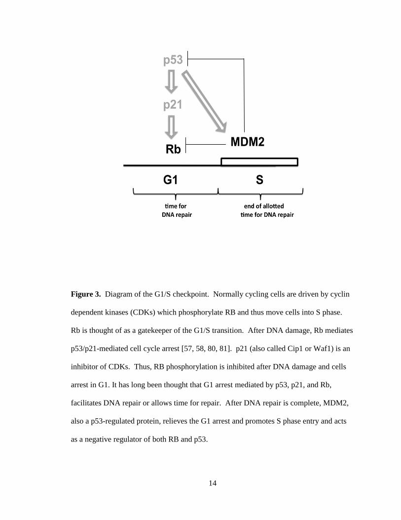
Figure 3. Diagram of the G1/S checkpoint. Normally cycling cells are driven by cyclin
dependent kinases (CDKs) which phosphorylate RB and thus move cells into S phase.
Rb is thought of as a gatekeeper of the G1/S transition. After DNA damage, Rb mediates
p53/p21-mediated cell cycle arrest [57, 58, 80, 81]. p21 (also called Cip1 or Waf1) is an
inhibitor of CDKs. Thus, RB phosphorylation is inhibited after DNA damage and cells
arrest in G1. It has long been thought that G1 arrest mediated by p53, p21, and Rb,
facilitates DNA repair or allows time for repair. After DNA repair is complete, MDM2,
also a p53-regulated protein, relieves the G1 arrest and promotes S phase entry and acts
as a negative regulator of both RB and p53.

15
RB and DNA repair
Although the relationship between the RB protein and the cell cycle is known,
little is known about RB and DNA repair. In studies conducted by Harrington et al.,
Rb -/- mouse embryos were defective in cell cycle arrest after treatment with
chemotherapeutic drugs and γ and UV-irradiation, implicating a role for RB in DNA
damage response [77]. Studies also suggest that loss or inactivation of RB should
sensitize cells to DNA damage and that RB plays a role in determining cell fate after
DNA damage [82]. Other investigations have suggested other roles for RB in response to
UV damage [83]. Recent studies of the E2F1 transcription factor, a downstream effector
of RB, showed that E2F1 accumulates at sites of DNA damage and binds to the XPC
gene promoter after UV irradiation [84, 85]. E2F1 may transcriptionally or post-
transcriptionally regulate XPC although the effects on XPC may be positive or negative.
Further exploration of the relationship between RB and DNA repair is needed to
determine its role in the repair process. It is with this in mind that studies herein
investigate the DNA repair protein XPC and the involvement of RB and G1/S cyclins in
XPC regulation. We hypothesize that key G1/S proteins, including RB and G1/S cyclins,
are involved in the regulation of XPC in the context of the G1/S cell cycle checkpoint.

16
MATERIALS AND METHODS
Cell lines
H1299 human lung carcinoma cells and Saos2 human osteosarcoma cells were
cultured in DMEM 4500mg/L glucose supplemented with 10% fetal bovine serum (FBS)
and 1% L-glutamine and gentamicin. Cells were plated for transfection as described
below at sub-confluency. H1299 and Saos2 carry deletions in both p53 alleles and do not
express p53 protein [86]. Extensive characterization as part of the National Cancer
Institute (NCI) anticancer drug screen showed absence of functional p53 and absence of
the G1/S checkpoint in these cell lines [86].
Cell transfection and plasmids
Cells were evenly seeded at 50% confluency onto cell culture dishes and
incubated at 37°C overnight to allow adhesion. Cells were transiently transfected using
either Fugene HD transfection reagent (Roche Applied Science, Indianapolis, IN, USA)
or Turbofect transfection reagent (Fermentas Life Science, Ontario, Canada) per
manufacturer’s protocols. Transfection mixtures were added to each plate drop-wise.
The plate was then gently rocked back and forth by hand to achieve even distribution.
The cells were incubated for 48 hours at 37°C before collection. All plasmids, with the
exception of shRNA plasmids, were pCMV3 derivatives and encoded full-length cDNAs
driven by the CMV promoter. Plasmids used for shRNA studies were pRS derivatives
driven by the U6 promoter. Empty pCMV3 or pCMV6 (Origene, Rockville, MD)
plasmids were used as vector controls. Full-length cDNA encoding Xpc, Rb, or cyclin E

17
were in pCMV6 (Origene). RB mutants driven by CMV plasmids were a gift from Dr.
Nick Dyson, Massachusetts General Hospital. Additional plasmids used throughout are
listed in Table 1.
PLASMID
DETAILS
SOURCE
pCMV6-XL5 Empty Vector Origene (Rockville, MD)
Xpc Xpc cDNA in pCMV6-XL5 Origene
Mdm2 Mdm2 cDNA in pCMV6-XL5 Origene
Rb Rb cDNA in pCMV6-XL5 Origene
pCMV3 Empty Vector Dr. M.B. Kastan
cyclin E cyclin E ORF in pCMV3 Dr. M.L. Smith [87]
cyclin D1 cyclin D ORF in pCMV3 Dr. Phil Hinds
pRS Empty Vector Origene
Cul4a shRNA Constructs against Cul4A
in pRS plasmid
Origene
Cul4a DN Cul4a dominant negative in
pCMV3
Addgene
(Cambridge, MA)
Mdm2 shRNA Constructs against Mdm2
in pRS plasmid
Origene
Table 1. Plasmids used in H1299 and Saos cell transfection experiments. The pCMV3
and pCMV6-XL5 plasmids were used as vector controls. The Xpc cDNA in pCMV6-
XL5 and Rb cDNA in pCMV6-XL5 were used to carry out XPC, RB co-transfections

18
and throughout most experiments. The amount of plasmid used was optimized for each
experiment ranging from 0.1-1µg.
Immunoblotting
Samples were lysed by directly adding 2x SDS loading dye (1.5g SDS, 2%
bromophenol blue, 1.23g DTT, dissolved in 10ml of water) and collected by scraping and
transferred into 1.5ml eppendorf tubes. The samples were boiled for approximately 30
minutes and 15-20µl of sample were added to precast 4-20% Tris-Glycine gels
(Invitrogen, Carlsbad, CA) and run at 100-150 volts. Proteins were transferred to
nitrocellulose membranes at 5 volts (<10) amps overnight. Nitrocellulose membranes
were blocked with 4% nonfat milk buffer prior to probing with primary and secondary
antibodies. Primary antibodies to XPC A-5 mouse monoclonal or XPC H-300 rabbit
polyclonal (Santa Cruz Biotechnology, Santa Cruz, CA, USA) were used at
concentrations of 0.1-1µg/ml in most experiments. Anti-β actin rabbit polyclonal
(Thermo Scientific, Waltham, MA, USA), anti-β galactosidase mouse monoclonal BG-02
(Abcam, Cambridge, MA, USA), and other additional antibodies were used as indicated
in figure legends and in Table 2. Secondary antibodies were peroxidase conjugates from
Santa Cruz. Immunoblots were detected by ECL (Pierce Thermo Scientific, Rockville,
IL, USA) and exposed to Kodak Biomax XAR film (Sigma Aldrich, St. Louis, MO,
USA).

19
Western blot antibodies
PRIMARY ANTIBODY
CLONE
SOURCE
XPC A-5 mouse monoclonal Santa Cruz Biotechnology
XPC H-300 rabbit polyclonal Santa Cruz Biotechnology
XPC D10 mouse monoclonal Santa Cruz Biotechnology
XPC 3.26 mouse monoclonal Santa Cruz Biotechnology
RB XZ55 and X2104 mouse
monoclonals
Calbiochem
RB AF11 mouse monoclonal Calbiochem
cyclin E HE12 mouse monoclonal Calbiochem
cullin 4a Rabbit polyclonal Abcam
hHR23b Rabbit polyclonal Abcam
biotin BN-34 mouse monoclonal Sigma-Aldrich
ubiquitin PD41 mouse monoclonal Santa Cruz Biotechnology
β-Galactosidase BG-02 mouse monoclonal Abcam
β-Actin Rabbit polyclonal Thermo Scientific
PCNA PC10 mouse monoclonal Calbiochem
Table 2. Western blot antibodies used in immunoblotting H1299 and Saos2 cellular
extracts. Primary antibodies in Table 2 were used at various concentrations, generally
0.1-1µg/ml to ensure the optimization of Western blots to detect their respective proteins.

20
Recombinant proteins
The following recombinant proteins were used as controls in several western blot
and immunoprecipitation experiments:
RECOMBINANT
PROTEIN
DETAILS
SOURCE
XPC partial protein with GST tag Abnova
XPC full length baculovirus
construct
Dr. Suk-Hee Lee
MDM Full length recombinant protein
with GST
Novus Biological
MDM2 Ring domain Mdm2 C-terminal RING
domain
Biomol International
Table 3. Recombinant proteins were used as controls for immunoblots and
immunoprecipitation experiments. Immunoblots and immunprecipitation experiments
were conducted as described herein.
Immunoprecipitation (IP)
Cells were harvested and lysed in immunoprecipitation (IP) lysis buffer
containing 1% Triton X-100 and protease inhibitors. Samples were then transferred to a
1.5ml microcentrifuge tube and incubated on ice 30 minutes and pelleted via
microcentrifugation. The supernatant was transferred to a new tube containing 40-50µl
of protein A plus protein G agarose beads and 1µl/µg of XPC clone D-10 antibody (Santa
Cruz). Rb antibody XZ155 was used in some experiments as indicated. The mixture was
then allowed to incubate at 4°C on a rotator overnight. After incubation the samples were

21
microcentrifuged for 30 seconds to pellet the beads. Beads were washed 4 times with
lysis buffer. Beads were resuspended in 50µl of 2X SDS loading buffer and boiled prior
to loading immunoprecipitated protein onto 4-20% Tris-Glycine gels and
immunoblotting.
Host Cell Reactivation
The host cell reactivation assay (HCR) is used to measure DNA repair in cells
(Figure 4). HCR is an established assay in use in several laboratories [58, 88, 89]. A
plasmid encoding a chloramphenicol acetyltransferase (CAT) reporter gene was damaged
by cisplatin to produce 1 to 5 adducted bases per plasmid molecule. NER-defective
fibroblasts from XPC patients are defective in “reactivating” and thereby expressing the
reporter gene. NER-proficient cells repair the gene and thereby express the reporter [36].
The ability of the host cells to repair the damaged plasmid is reflected in reporter gene
expression. Activity encoded by the damaged reporter gene is determined by ELISA, and
is a reflection of the DNA repair capacity of the host cell. One advantage of the assay is
that only the damaged plasmid and not the cells receive DNA damage. Thus, cellular
effects e.g. apoptosis are not an issue.

22
Figure 4. Diagram of host cell reactivation assay. The HCR assay measures DNA repair
by restoring a plasmid that encodes a reporter gene (CAT). The reporter gene has been
inactivated due to induced damage to the plasmid caused by platinum adducts (indicated
by X). The ability of the host cell to repair the damaged plasmid is determined by
reporter gene expression. Cells that are defective in NER are unable to repair the
damaged plasmid and therefore unable to express the reporter gene. However, NER
competent cells are capable of repairing the damage and express the reporter CAT
reporter gene indicating the host cells ability to repair DNA damage.

23
CAT ELISA
The chloramphinicol acetyltransferase enzyme-linked immunosorbent assay
(CAT ELISA) immunoassay (Roche Applied Science, Indianapolis, IN, USA) was used
for the quantitative determination of the CAT enzyme. Briefly, equivalent protein
concentrations of lysed cell extracts were added to an anti-CAT-coated 96-well
microplate and refrigerated overnight. Microplate wells were then washed and a
digoxigenin-labeled antibody to CAT (anti-CAT-DIG) was added to wells at room
temperature for at least 1 hour. Wells were then washed and an antibody to digoxigenin
conjugated to peroxidase (anti-DIG-POD) was added for 1 hour. The peroxidase
substrate ABTS was added after washing. The plate was then incubated and read at 405
nm in a Tecan Spectra plate reader at multiple time intervals.
Pulse Chase Analysis
After H1299 cells were transfected with Xpc and Rb plasmids, cellular proteins
were labeled for 4 hours with an L-methionine analog that can be subsequently labeled
with biotin (Invitrogen, Carlsbad, CA, USA). The free label was removed by placing the
cells in complete medium and cells were harvested after 0, 4, and 8 hours. XPC was
immunoprecipitated as described above and then labeled by biotinylation (Invitrogen).
The L-methionine analog has a reactive azide group that is then covalently modified by
an alkyne-biotin reagent (Invitrogen) and detected using a mouse monoclonal anti-biotin
antibody (Sigma, St. Louis, MO, USA).

24
Statistical Analysis
All data are presented as mean or relative units + standard error. Data were
analyzed as indicated in figure legends using one way ANOVA or paired t-test with
Microsoft Office Excel 2007 (Seattle, WA) or Graph Pad Prism (San Diego, CA).

25
RESULTS
XPC protein expression and DNA repair activity in the presence of RB
In an effort to investigate the relationship between the G1/S cell cycle checkpoint
and DNA repair, our study focused on the retinoblastoma protein RB, a known mediator
of the G1/S checkpoint, and its association with the XPC DNA repair protein. In order
to determine the effects of RB on XPC, we chose H1299 human lung cancer cells to
carry out most experiments. H1299 cells were chosen because they have low
endogenous XPC, thereby making it easier to detect transfected XPC in transient
expression experiments. Additionally, these cells are p53 null [86]. To determine the
effectiveness of our plasmids and the efficiency of our transfections H1299 cells were
transfected with pCMV-Rb, pCMV-Xpc, or empty plasmid and protein expression was
observed. The pCMV-Rb and pCMV-Xpc plasmids showed appreciable expression of
their respective proteins when introduced to H1299 cells compared to empty CMV
plasmid (Figure 5A).
We then combined pCMV-Rb and pCMV-Xpc and transfected them into H1299
cells. Empty pCMV plasmid was used to keep the amount of plasmid DNA constant
across all the dishes, while pCMV-Rb and pCMV-Xpc plasmids alone were transfected
as controls. When combined with pCMV-Rb, the abundance of XPC protein was
markedly increased (Figure 5B, lane 8) when compared to empty plasmid or the Xpc or
Rb plasmid alone. Most importantly, combining pCMV-Rb with pCMV-Xpc markedly
enhanced DNA repair of a cisplatin-damaged reporter plasmid, pCMV-CAT (Figure 5C,
lane 8). Thus, the co-transfection of Rb together with Xpc significantly (p= 0.0005)

26
enhanced the DNA repair activity of XPC as measured by host cell reactivation assay.
Interestingly, increasing the amount of XPC plasmid alone did not affect the DNA repair
activity of the cells (Figure 5A, lane 4 compared to Figure 5C, lane 4), indicating that
XPC exists in an inactive form that can be activated in combination with RB. Data
herein (Figure 5A and C control lanes) provide evidence that, despite increases in XPC
expression, its DNA repair capacity does not increase. Our data shows DNA repair is
increased with the addition of RB (Figure 5C) implicating a level of regulation that does
not rely on transcription.

27
C
120 kDa XPC -
105 kDa Rb -

28
Figure 5. RB enhances XPC-mediated host cell reactivation of a cisplatin-damaged
reporter gene. A) Plasmids were transfected at indicated amounts. The total amount of
plasmid DNA was kept constant by adding empty CMV plasmid where necessary.
H1299 cells have low endogenous XPC. RB and XPC were encoded by the respective
plasmids and were readily detected by western blot (lanes 2 and 4). B) Mixing RB and
XPC plasmids resulted in a clear increase in XPC protein (western blot, lane 8). XPC
was detected with antibody A5, while RB was detected with antibody AF-11. Actin
served as a loading control. C) Host cell reactivation (HCR) corresponding to the same
lanes as in A and B. Neither RB alone, nor XPC alone (bar graph, lanes 2 and 4
respectively) resulted in increased HCR. When combined (bar graph, lane 8) a
significant increase in HCR was observed (P= 0.0005). CAT activity was measured by
ELISA as previously described in materials and methods.

29
RB activation of XPC does not require ongoing protein synthesis
It is likely that the observed enhancement of XPC-mediated DNA repair is due to
1) interaction of XPC with RB and/or 2) activating modifications of XPC protein that are
triggered by interaction with RB. A third possibility is that RB enhanced the translation
of XPC protein. However, if this were the case, it would result in increased activation of
the XPC protein observed in Figure 5C lane 8. As can be seen in Figure 5A, simply
increasing the amount of XPC to 1µg did not lead to a functional increase in XPC DNA
repair activity as indicated and measured by host-cell reactivation (Figure 5A, lane 4
compared to Figure 5C, lane 4). We conducted experiments with cycloheximide to
block protein synthesis to address this possibility and observed that co-transfection with
RB led to increased XPC levels in the presence of cycloheximide (Figure 6A).
Therefore, the observed enhancement of XPC by RB does not require ongoing protein
synthesis. To determine whether Rb affected the half-life of XPC we conducted pulse-
chase experiments, in which cells co-transfected with Rb and Xpc were labeled with an
L-methionine analog to label methionine residues during a 4-hour “pulse” according to
manufacturer’s instructions (Invitrogen). Cells were returned to complete medium with
excess unlabeled L-methionine (“chase”) and were harvested 0, 4, or 8 hrs later. The co-
transfection of Rb extended the half-life of XPC beyond 8 hrs, compared to <4 hrs in the
control transfectants (Figure 6B). Thus, RB appeared to increase the half-life of XPC.

30
XPC and Rb Co-Immunoprecipitation
Given these results, we hypothesized a possible protein-protein interaction
between RB and XPC. We used XPC and RB recombinant proteins to detect a possible
interaction between RB and XPC, but failed to detect an interaction (results not shown).
One possibility was that the interaction required protein folding concurrent with
translation. If this were the case, the two proteins would interact when co-expressed in
the same cell. When mixed as full-length purified proteins, they would not show an
apparent interaction. To investigate this likelihood, H1299 cells were co-transfected with
XPC and Rb and immune complexes containing RB were probed for the presence of XPC
and vice-versa. XPC was detected in RB immune complexes, and RB was detected in
XPC immune complexes (Figure 7). Thus, there is a putative interaction between Rb
and XPC in H1299 cells.
RB deletion mutants
We attempted to “map” the interaction domain of RB with XPC using a series of
RB deletion mutants for co-transfection experiments. The idea was that we could map
the domain by determining which mutants did or did not stabilize XPC. RB deletion
mutants were transfected into p53null/Rb null Saos2 cells for these experiments (Figure
8). Mutant 1 contains a deletion in the N terminus domain of the RB protein from amino
acid 303-392. Mutant 2 (Δ773-909) contains deletions involving the E2F and MDM2
binding domains. While mutant 3 contains a deletion beginning with the cyclin/cdk
docking site KXLKXL deleting amino acids 870-909. All mutants were compared to full
length RB protein [90].

31
Figure 6. RB stabilizes XPC in the presence of cycloheximide or in pulse-chase
experiments. A) H1299 cells were transfected as in Figure 5 with plasmids encoding
XPC and RB. After 48 hrs in culture, cycloheximide (CHX; 10µM) was added for 15
hrs. Western blots show accumulation of XPC protein where RB was co-transfected with
XPC (lanes 4 and 5). Labled XPC was detected by anti-XPC monoclonal antibody A5.
Actin served as a loading control because of its long half life. B) Metabolic labeling of
XPC with an L-methionine analog. Cells were transfected with XPC and RB plasmids as
indicated, then labeled (“pulsed”) for 4 hours after a 48 hr culture incubation. The label
-anti-Biotin
Labeled XPC

32
was removed and cells returned to complete medium and “chased” for 0, 4, or 8 hrs.
Immunoprecipitated XPC was detected using an anti-biotin antibody. Accumulated XPC
is evident in lanes that were co-transfected with RB (lanes 5 and 6).

33
RB deletion mutants
We attempted to “map” the interaction domain of RB with XPC using a series of
RB deletion mutants for co-transfection experiments. The idea was that we could map
the domain by determining which mutants did or did not stabilize XPC. RB deletion
mutants were transfected into p53null/Rb null Saos2 cells for these experiments (Figure
8). Mutant 1 contains a deletion in the N terminus domain of the RB protein from amino
acid 303-392. Mutant 2 (Δ773-909) contains deletions involving the E2F and MDM2
binding domains. While mutant 3 contains a deletion beginning with the cyclin/cdk
docking site KXLKXL deleting amino acids 870-909. All mutants were compared to full
length RB protein [90].
Surprisingly, each of the three deletion mutants failed to stabilize XPC (Figure
9). Each of the mutants are partially defective in G1 cell cycle arrest, thus, perhaps a G1
arrest is required. Alternatively, each of the mutants may be improperly folded such that
they cannot interact with XPC. Each of the mutants is expressed similar to wildtype pRb
(Figure 9) but we cannot exclude a defect in protein folding.
XPC and G1/S cyclins
As RB is a key mediator of the G1/S transition in cells, we tested whether the
effect of stabilizing XPC was linked to the cell cycle role of RB, or whether it could be a
novel function of RB. To explore this prospect we co-transfected a CMV plasmid
encoding the G1/S cyclin, cyclin E, together with RB and XPC-encoding plasmids in to
Saos2 cells. Cyclin E serves as the signal for S-phase entry by phosphorylating RB at the
G1/S transition. The addition of Cyclin E completely abolished the stabilization of XPC

34
by RB (Figure 10, lane 8). Thus, it is likely that RB in its underphosphorylated G1 form
interacts with and stabilizes XPC, and that RB in its cyclin E-phosphorylated form does
not contribute to the stabilization of XPC.
The simplest hypothesis to explain the data is that RB protects XPC from
degradation and that cyclin E reverses that protection from degradation. The proteosome
inhibitor MG132 rescued XPC from cyclin E-mediated turnover in H1299 cells
transfected with cyclin E (Figure 11). Cyclin E, as expected, decreased DNA repair in
host-cell reactivation assays again suggesting that cyclin E plays a negative role in
regulating XPC (Figure 11). Thus, it is likely that XPC is tightly linked to the G1/S cell
cycle checkpoint by proteosome-mediated stabilization and destabilization mechanisms.

35
Figure 7. Putative interaction between RB and XPC. H1299 cells were transfected with
Rb and Xpc plasmids. Immune complexes containing RB or XPC were probed on
Western blots to detect possible interaction. Left panel, immune complexes obtained
using a negative control (C), MOPC21 IgG, or anti-RB monoclonal antibodies XZ104
and XZ155; right panel, immune complexes obtained using a negative control,
120- 105-
120- 105- -blot
RB
-blot
XPC

36
nonimmune rabbit IgG, or anti-XPC rabbit polyclonal antibody H300. The upper left
panel was probed by western blot with polyclonal rabbit anti-XPC. The upper right panel
was probed by western blot with monoclonal anti-RB clone AF-11. Lower left and right
panels were probed with RB and XPC respectively to serve as additional controls.

37
Figure 8. Structure of RB and Rb deletion mutants. Wildtype Rb contains 928 amino
acid residues. The LXCXE-binding (pocket) domain begins at amino acid 394 and
contains two subdomains (A and B), and an intervening spacer (S). The C-terminus also
interacts with E2F, a downstream effector of the RB family, and MDM2, a p53 regulated
protein and ubiquitin ligase. A cyclin/cdk docking site (KXLKXL) is also present in the
C terminus beginning at amino acid residue 870. Three deletion mutants (a gift from
Dr. Nick Dyson) were used in co-transfection experiments. Mutant 1 contains a deletion
in the N terminus from amino acid 303-392. Deletion mutant 2 (Δ773-909) contains
deletions involving the E2F and MDM2 binding domains. Mutant 3 contains a deletion
beginning with the cyclin/cdk docking site KXLKXL deleting amino acids 870-909.
RB

38
Figure 9. RB mutants fail to stabilize XPC. Wildtype RB or three deletion mutants were
co-transfected together with XPC as previously described. Saos2 cells were used. Robust
stabilization of XPC was observed by wildtype RB (lane 1). Each of the mutants failed
to stabilize XPC (lanes 2, 3, 4). Beta galactosidase served as a co-transfection control.
Actin served as a loading control. XPC was detected by anti-XPC monoclonal antibody
A5.
120-

39
Figure 10. Cyclin E counteracts (wild type) RB stabilization of XPC suggesting that
stabilization of XPC is related to the cell cycle role of RB. Saos2 cells were transfected
as previously with the indicated plasmids, again keeping the total amount of plasmid
DNA constant. Co-transfection of cyclin E alone or in the presence of RB and/or XPC
strongly decreased XPC protein expression suggesting that cyclin E counteracts any
stabilizing effect of RB on XPC. Beta galactosidase served as a co-transfection control.
48-
58-

40
Actin served as a loading control. XPC was detected by anti-XPC monoclonal antibody
A5. RB was detected by anti-RB monoclonal anti-RB clone AF-11 (data not shown).
Cyclin E was detected using monoclonal antibody HE-12 (Calbiochem).

41
Figure 11. Ubiquitin ligase inhibitor MG-132 rescues XPC from cyclin E-triggered
degradation. Cyclin E inhibited XPC expression (left panel) and host-cell reactivation
(HCR; right panel; p=0.004 comparing lanes 1 and 2 via paired t-test). MG-132 was
added (10µM) 15 hrs prior to cell lysis. MG-132 restored XPC and HCR to control
levels. H1299 cells were utilized for both panels.
*
*

42
XPC ubiquitination
Previous studies have shown evidence for the regulation of XPC by post-
translational modifications including ubiquitination [34]. To determine whether Cyclin E
is involved in the negative regulation of XPC by serving as a signal for ubiquitination, we
transfected H1299 cells with Xpc, Cul4a-dominant negative (DN), cyclin D1 and
cyclin E plasmids. Immune complexes were probed on Western blots with anti-XPC and
anti-ubiquitin to detect ubiquitination of XPC. Ubiquitinated Xpc is detected in the
complex containing cyclin E (Figure 12, lane 4). Ubiquitinated Xpc was not observed in
lane 2 where Cul4a-DN was used as a control as studies have shown that XPC is
regulated by E3 ubiquitin ligase or lane 3 (an additional control) containing the G1/S
mediator cyclin D1. These data indicate that Cyclin E may be involved in the negative
regulation of XPC and may act as a signal for the ubiquitination and subsequent
degradation of XPC upon S-phase entry. It is possible that the phosphorylation of RB by
cyclin E upon s-phase entry in turn signals the disassociation of XPC from DNA.

43
Figure 12. Cyclin E may act as a signal for the ubiquitination of XPC. Immune
complexes of transfected cell lysates were probed on Western blots using an anti-
ubiquitin mouse monoclonal antibody PD41 (Santa Cruz Biotechnology) to detect
possible XPC ubiquitination. Ubiquitinated Xpc is detected in lane 4 in the presence of
cyclin E, however, ubiquitinated Xpc was not detected in control lanes.
XPC +
44
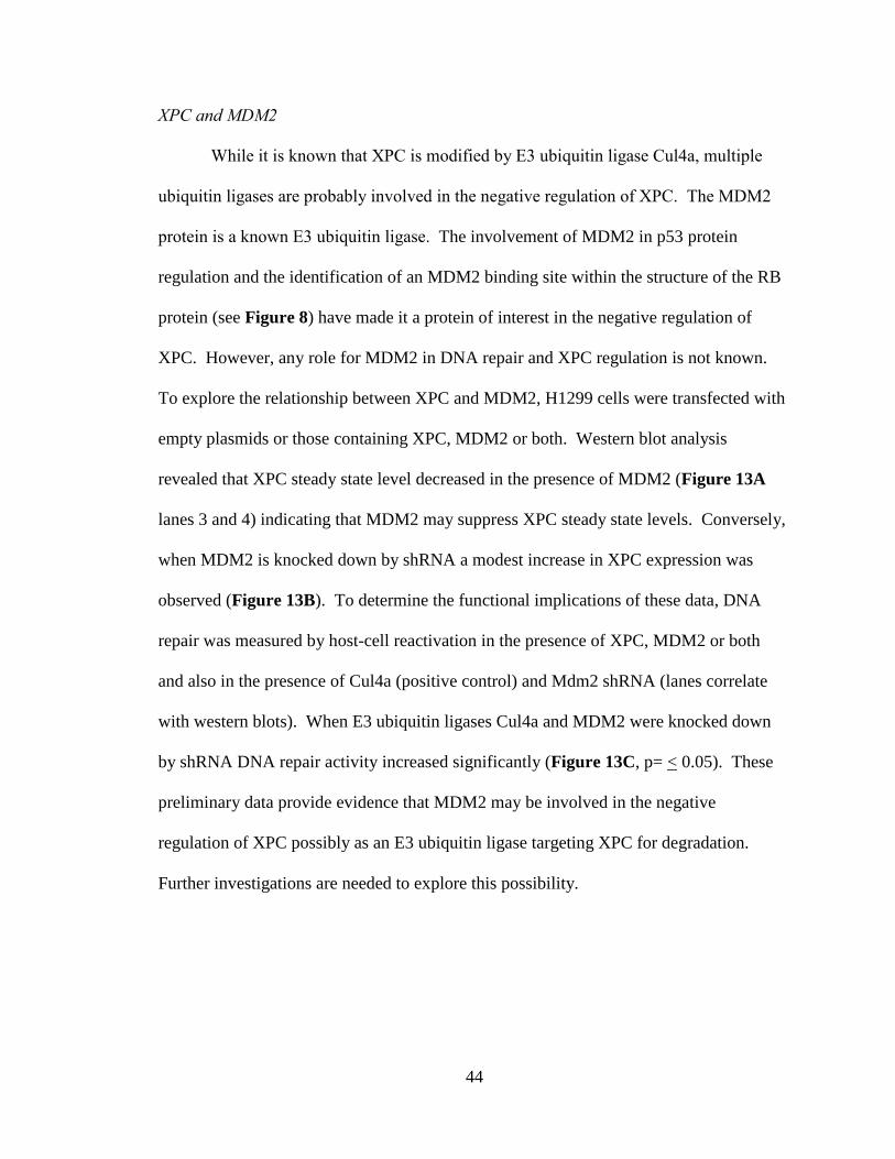
XPC and MDM2
While it is known that XPC is modified by E3 ubiquitin ligase Cul4a, multiple
ubiquitin ligases are probably involved in the negative regulation of XPC. The MDM2
protein is a known E3 ubiquitin ligase. The involvement of MDM2 in p53 protein
regulation and the identification of an MDM2 binding site within the structure of the RB
protein (see Figure 8) have made it a protein of interest in the negative regulation of
XPC. However, any role for MDM2 in DNA repair and XPC regulation is not known.
To explore the relationship between XPC and MDM2, H1299 cells were transfected with
empty plasmids or those containing XPC, MDM2 or both. Western blot analysis
revealed that XPC steady state level decreased in the presence of MDM2 (Figure 13A
lanes 3 and 4) indicating that MDM2 may suppress XPC steady state levels. Conversely,
when MDM2 is knocked down by shRNA a modest increase in XPC expression was
observed (Figure 13B). To determine the functional implications of these data, DNA
repair was measured by host-cell reactivation in the presence of XPC, MDM2 or both
and also in the presence of Cul4a (positive control) and Mdm2 shRNA (lanes correlate
with western blots). When E3 ubiquitin ligases Cul4a and MDM2 were knocked down
by shRNA DNA repair activity increased significantly (Figure 13C, p= < 0.05). These
preliminary data provide evidence that MDM2 may be involved in the negative
regulation of XPC possibly as an E3 ubiquitin ligase targeting XPC for degradation.
Further investigations are needed to explore this possibility.

45
A B
A B
shRNA_____
shRNA_____

46
Figure 13. MDM2 may be involved in the negative regulation of XPC. A) H1299
human lung carcinoma cells were cultured and transfected with either an empty plasmid
or plasmids containing XPC or MDM2 cDNA. Western blots of co-transfected cells
reveal that XPC expression decreased in the presence of MDM2. B) p53 null H1299
cells were cultured and transfected with either an empty plasmid or a plasmid containing
Cul4a or MDM2 shRNA. Western blot analysis showed that XPC expression increased
when MDM2 is knocked down. C) DNA repair activity is increased when MDM2 is
knocked down. DNA repair was measured by chloramphinicol acetyltransferase (CAT)
assay in the presence of XPC, MDM2 or both, and also when Cul4a (positive control)
and Mdm2 were knocked down by shRNA. When either Cul4a or MDM2 were modestly
knocked down by shRNA DNA repair increased significantly p= < 0.05. This increase in
XPC expression and DNA repair activity indicate that MDM2 may be involved in the
negative regulation of XPC.

47
DISCUSSION
One important aspect of the G1/S checkpoint is that p53 transcriptionally
activates Xpc [37]. This finding suggested that p53 and XPC could actively promote
NER, in cells that carry functional p53. However, the data herein suggests an additional
level of regulation that does not rely upon p53. We used human lung cancer H1299 cells,
which carry deletions in both p53 alleles and have low endogenous levels of XPC and we
still observed a significant increase in the cell’s DNA repair capacity as measured by
host-cell reactivation (HCR) when combined with RB (Figure 5B and C, lanes 8). One
often ignored feature of the HCR assays that we and others have used extensively is that
it measures NER of a damaged reporter gene. The cells do not receive direct DNA
damage. In this assay, we detected a latent XPC protein that did not promote HCR
(Figure 5A and C, lanes 4). When combined with RB, XPC was in some way activated
for HCR (Figure 5B and C, lanes 8). While most human tumors have defects in the p53
or Rb pathways¸ the data implies that if either p53 or Rb is intact, NER mechanisms are
capable of repairing damaged DNA. Given evidence that RB may interact with XPC
(Figure 7), it is likely that the interaction of XPC with RB promotes NER at least as
measured by HCR. Post-translational modification(s) of XPC in the presence of RB is in
evidence (Figure 5B, lane 8).
Several studies showed that p21/Waf1/Cip1 promoted NER in p53-defective cell
lines although the mechanistic basis was not known [62, 63, 91-93]. The view was that
simply arresting the cell cycle by p21 promoted NER. Our data provide a mechanistic
insight, that p21 would lead to accumulation of underphosphorylated RB, which would in

48
turn stabilize XPC in a form that is active for NER (Figure 5B and C, lanes 8). Our
finding that cyclin E destabilized XPC and led to a decrease in HCR is also consistent
with the published studies on p21 (Figure 11).
The data represented herein suggests a model for both positive and negative
regulation of XPC (Figure 14). Our data shows that XPC exists in functionally active
and inactive forms. The data shows that upon DNA damage XPC is then stabilized by
the underphosphorylated form of RB and correlates with DNA repair events of the G1/S
phase of the cell cycle. The data also show that G1/S cyclins abrogate XPC expression
and function which likely corresponds to RB phosphorylation and S-phase entry and that
cyclin E may provide a signal for XPC ubiquitination. Perhaps a parallel is suggested by
the Cdt1 cell cycle checkpoint protein which is first phosporylated by cyclin E and then
ubiquitinated by Cul4a.
The cellular model used for the experiments herein is atypical of DNA repair
studies. Most studies involving DNA repair mechanisms center around biochemical and
cell free systems. Our use of a cellular model of DNA repair is likely more reflective of
the signaling mechanisms that actually occur in the cell. The majority of XPC and Rb
experiments used an over expression model, whereas, preliminary data involving MDM2
utilized overexpression and the knockdown of MDM2 as a cellular model. Additional
studies utilizing a knock down or inducible system of expression may give added insight
into the mechanism of action of XPC regulation in the context of the cell cycle.
In response to DNA damage, XPC protein is phosphorylated, ubiquitinated, and
SUMOylated, however, the amino acid residues involved and the net effects on XPC
function are not well-known [37, 39, 94]. Presumably, some post-translational

49
modifications promote XPC-mediated NER while others inhibit XPC-mediated NER.
The data presented in this study supports the long acknowledged theory that NER occurs
readily in G1 but cannot occur in S-phase. Therefore, protein modifications and protein-
protein interactions likely restrict XPC function to G1 while inactivating XPC upon S-
phase entry.
The XPC protein exists in a heterotrimeric complex that includes the hHR23b and
centrin 2 proteins. The hHR23b protein acts to stimulate and stabilize XPC, while the
centrin 2 protein plays an important role in centrosome duplication [95-97]. The yeast
homologue for hHR23b, Rad23, provided a link between DNA repair and the ubiquitin
pathway that led to the discovery that XPC is modified by ubiquitination [48]. Western
blot analysis collected from this study (data not shown) indicated that transfecting H1299
cells with the hHR23b protein does not have a noticeable effect on XPC expression.
While the XPC protein has been shown to be modified by the ubiquitin ligase Cul4a,
most proteins can be poly-ubiquitinated and modified by several ubiquitin ligases [98-
99]. The relationship between MDM2 and G1/S mediators along with its involvement in
the regulation of the p53 and RB tumor suppressor proteins have made it a likely player
in the negative regulation of XPC. MDM2 is an ubiquitin ligase for both RB and p53,
and may also act as an ubiquitin ligase for XPC. Our data indicates that MDM2 affects
XPC expression and that knocking down MDM2 increased the DNA repair capacity of
XPC as measured by HCR (Figure 13) thereby suggesting that MDM2 may play a role in
the negative regulation of XPC and possibly act as an E3 ubiquitin ligase for XPC.

50
Figure 14. Schematic of a possible mechanism of XPC regulation in the context of the
cell cycle. Data herein reveals a functionally inactive (rectangle) and active (oval) form
of XPC DNA repair protein. These data suggest that upon DNA damage XPC is then
stabilized by RB and that occurrence correlates with the DNA repair events of the G1
phase of the cell cycle. These data also show that G1/S cyclins, primarily cyclin E
(cyclin D data not shown) abrogate XPC expression which likely corresponds to RB
phosphorylation and S-phase entry. Data also reveals that MDM2 may signal
ubiquitination of XPC in some way.
Cyclin
E

51
There is evidence that XPC is positively regulated by additional factors including
damaged DNA-binding protein 2 (DDB2). DDB2 is a subunit of the damaged DNA-
binding protein DDB1/DDB2 and is responsible for the phenotypic features of XP-E
patients [98-101]. DDB2 has been shown to positively regulate XPC by promoting the
accumulation and activating the recruitment of XPC to damaged DNA [98].
Additionally, the mRNA and protein levels for both XPC and DDB2 increase in response
to DNA damage. While the exact role of the DDB1/DDB2 complex is still unclear, it is
possible that the complex may also be regulated at the G1/S checkpoint. One reason why
the role of DDB1/DDB2 is unknown is that it is not required in purified systems that
carry out NER in vitro. Such systems have formed the foundation for mechanistic studies
of DNA repair proteins [102]. Like XPC, DDB2 is positively regulated at the level of
transcription by p53 and is postranslationally regulated by poly-ubiquitination.
Therefore, it is not unreasonable to think that other NER proteins, eg. XPA, may likewise
be tightly regulated by post-translational mechanisms including ubiquitination.
The NER pathway is considered to be an error-free pathway. NER utilizes the
nucleotide sequence from intact complementary DNA in order to fill in the 25-30mer gap
excised during repair (Figure 1). XPC has also been shown to be involved other DNA
repair pathways including base excision repair (BER) and error prone DNA repair
mechanisms [103-107]. Studies conducted by Uehara et al. showed that XPC may be
involved in the suppression of multiple mutations generated by error-prone trans-lesional
DNA synthesis [104]. XPC deficient mice contain much higher numbers of unrepaired
lesions that result in multiple mutations. These mutations are suspected to be generated
by trans-lesional DNA polymerases such as pol zeta and pol eta. Pol eta is also involved

52
in the NER pathway and in error prone DNA repair mechanisms. The regulation of XPC
in the context of the G1/S cell cycle may be of particular importance in regards to error
prone mechanisms and preventing mutations caused by trans-lesional bypass
polymerases.
The studies herein were conducted in a p53-null cancer cell line, H1299, which
carries deletions in both p53 alleles [86]. Although it is expected that p53 will contribute
to XPC expression and cellular NER in cells with functional p53, our study raises the
possibility that p53-defective cancer cells might still retain some degree of XPC
regulation and the capacity to regulate cellular NER. We used a cisplatin-damaged
reporter gene for the HCR assays in cells that lack functional p53. However, our data
indicate that substantial DNA repair occurs; indicating NER DNA repair mechanisms
were intact (Figure 5, lane 8). These data suggest it may be possible for cancer cells
lacking functional p53 to evade or resist cisplatin therapy by repairing the cisplatin DNA
damage [108]. From a chemotherapeutic perspective, it may be necessary to target
additional components of the G1/S checkpoint, such as RB, in order to maximize cancer
cell killing by cisplatin.
Platinum-based chemotherapy is used as the preferred treatment method for
patients with advanced NSCLC [109]. Platinum agents (i.e. cisplatin and carboplatin) are
widely used in the clinic to treat various types of cancers including lung, bladder, liver,
breast, testicular, myeloma, genitourinary and gastrointestinal [110]. Platinum
chemotherapy agents cause DNA damage to the cells and inhibit DNA synthesis [111].
The platinum compound forms covalent bonds with purine bases in the DNA strands and
primarily forms 1,2 or 1,3-intrastrand crosslinks, as well as, a small percentage of

53
interstrand crosslinks [111-113]. These crosslinks result in a helical distortion of the
DNA which blocks DNA replication and transcription [111]. While initial platinum
responsiveness is high, the majority of cancer patients will eventually become cisplatin-
resistant [108, 114]. One mechanism of cisplatin resistance that has been proposed
involves increased DNA repair [114]. NER typically repairs the DNA damage caused by
platinum-based chemotherapy drugs. Studies herein suggest that even in cancer cells that
are p53 null XPC is still capable of repairing DNA damage caused by platinum
chemotherapy drugs when RB is intact. The data herein suggest that the addition of DNA
repair inhibitors may be necessary for the effective treatment of certain cancers that are
treated using platinum chemotherapy drugs [115, 116].

54
FUTURE DIRECTIONS
The Xpc complex
Data herein involving the regulation of Xpc DNA repair protein reveal an
additional level of regulation that involve post-translational modification of XPC,
however, the extent of these modifications is not known and the involvement of other
factors within the XPC complex have not been clearly defined. XPC exists in a
heterotrimeric complex including hHR23b and centrin-2. While the XPC component is
known to be required for NER, specific components may have additional functions
within the complex. Particularly, the hHR23b component has been shown to stimulate
and stabilize XPC. Little is known of the relationship between hHR23b and the cell
cycle. However, it may be that hHR23b is modified upon the ubquitination of XPC and
is involved in the destabilization of the XPC complex. Also it may be that knocking
down the hHR23b protein may adversely affect the stability of XPC and its ability to bind
to the site of DNA damage, thus affecting the function of XPC in NER. The possibility
of this occurrence should be addressed in order to determine the effects of individual
components of the XPC complex on NER and the regulation of XPC in the context of the
cell cycle.
NER Co-factors
Nucleotide excision repair occurs in a multistep complex that involves additional
co-factors. Other co-factors, i.e. DDB1/DDB2 and XPA, are involved in the regulation
of XPC and have been shown to be required for NER in certain cell types. It is possible

55
that downstream NER factors involved in repairing DNA damage signal the
ubiquitination of XPC and its dissociation from the NER complex. The possibility of
additional NER co-factors being involved is likely and should be explored.
Chemotherapeutic Implications
Most aspects of cancer chemotherapy involve causing damage to the cellular
DNA. High levels of DNA damage may cause cell-cycle arrest or cell death as is the
goal of cancer chemotherapy drugs. Platinum chemotherapeutic agents are commonly
used to treat various types of cancers but the majority of cancer patients typically develop
a resistance to chemotherapeutic drugs. Intrinsic cellular NER DNA repair mechanisms
may continue to repair the damage caused by platinum chemotherapy drugs and likely
contribute to chemotherapeutic drug resistance and decrease the effectiveness of these
treatments. The data herein suggests that XPC and NER cofactors are capable of
repairing DNA damage from platinum agents (i.e. cisplatin) commonly used in cancer
therapeutics regardless of whether the p53 and pRB proteins are expressed. These data
suggest that the addition of DNA repair inhibitors may be necessary for the effective
treatment of certain cancers. Several studies have proposed the use of drugs targeting
specific DNA repair proteins in addition to traditional chemotherapy [115-117]. The
combination of chemotherapy drugs like temozolomide and platinum chemotherapy
drugs like cisplatin and carboplatin along with inhibitors of poly(ADP-ribose)
polymerase 1 (PARP1) is currently under investigation and involved in several clinical
trials. The decreased repair of intermediate DNA lesions induced by temozolomide in the
presence of PARP inhibitors, and observations showing that PARP inhibitors

56
preferentially kill neoplastic cells make it a significant protein to explore [118-120]. Data
herein suggests that it is also worthwhile to investigate the effects of inhibiting additional
DNA repair proteins (i.e. XPC) to increase the effectiveness of current patient
chemotherapy. Should studies persist it would be interesting to investigate the effects of
XPC activity in the presence of cisplatin damage and DNA repair inhibitors.

57
REFERENCES
1. Altieri, F., et al., DNA damage and repair: from molecular mechanisms to health
implications.(Comprehensive Invited Review). Antioxidants & Redox Signaling,
2008. 10(5): p. 891(47).
2. Kaufmann, W. and R. Paules, DNA damage and cell cycle checkpoints. FASEB
J., 1996. 10(2): p. 238-247.
3. Wood, R.D., DNA repair in eukaryotes. Annual Review of Biochemistry, 1996.
65(1): p. 135-167.
4. de Laat, W.L., N.G.J. Jaspers, and J.H.J. Hoeijmakers, Molecular mechanism of
nucleotide excision repair. Genes & Development, 1999. 13(7): p. 768-785.
5. Hanawalt, P.C., J.M. Ford, and D.R. Lloyd, Functional characterization of global
genomic DNA repair and its implications for cancer. Mutation Research/Reviews
in Mutation Research, 2003. 544(2-3): p. 107-114.
6. Reed, E., Platinum-DNA adduct, nucleotide excision repair and platinum based
anti-cancer chemotherapy. Cancer Treatment Reviews, 1998. 24(5): p. 331-344.
7. Dreij, K., A. Seidel, and B. Jernstrom, Differential removal of DNA adducts
derived from anti-diol epoxides of dibenzo[a,l]pyrene and benzo[a]pyrene in
human cells. Chemical Research in Toxicology, 2005. 18(4): p. 655-664.
8. Nouspikel, T., DNA repair in mammalian cells. Cellular and Molecular Life
Sciences (CMLS), 2009. 66(6): p. 994-1009.
9. Tornaletti, S., DNA repair in mammalian cells. Cellular and Molecular Life
Sciences (CMLS), 2009. 66(6): p. 1010-1020.
10. Nouspikel, T.P., N. Hyka-Nouspikel, and P.C. Hanawalt, Transcription domain-
associated repair in human cells. Mol. Cell. Biol., 2006. 26(23): p. 8722-8730.

58
11. Berndt, S.I., et al., Genetic variation in the nucleotide excision repair pathway
and colorectal cancer risk. Cancer Epidemiol Biomarkers Prev, 2006. 15(11): p.
2263-2269.
12. Hanawalt, P.C., DNA repair: The bases for Cockayne syndrome. Nature, 2000.
405(6785): p. 415-415.
13. Spivak, G., The many faces of Cockayne syndrome. Proceedings of the National
Academy of Sciences of the United States of America, 2004. 101(43): p. 15273-
15274.
14. Lehmann, A.R., DNA repair-deficient diseases, xeroderma pigmentosum,
Cockayne syndrome and trichothiodystrophy. Biochimie, 2003. 85(11): p. 1101-
1111.
15. Cleaver, J.E., and K.H.Kraemer. and eds. Xeroderma pigmentosum and Cockayne
syndrome. In The Metabolic and Molecular Bases of Inherited Disease, ed. A.L.B.
C.R.Scriver, W.S.Sly, and D.Valle. Vol. Vol. III. 1995, McGraw-Hill: New York
4393-4419.
16. Hanawalt, P.C. and G. Spivak, Transcription-coupled DNA repair: two decades
of progress and surprises. Nat Rev Mol Cell Biol, 2008. 9(12): p. 958-970.
17. Cleaver, J.E., Defective repair replication of DNA in xeroderma pigmentosum.
Nature, 1968. 218(5142): p. 652-656.
18. Batty, D.P. and R.D. Wood, Damage recognition in nucleotide excision repair of
DNA. Gene, 2000. 241(2): p. 193-204.
19. Kraemer, K.H., M.M. Lee, and J. Scotto, Xeroderma pigmentosum: cutaneous,
ocular, and neurologic abnormalities in 830 published cases. Arch Dermatol,
1987. 123(2): p. 241-250.
20. Sugasawa, K., et al., A multistep damage recognition mechanism for global
genomic nucleotide excision repair. Genes & Development, 2001. 15(5): p. 507-
521.
21. Friedberg, E.C., How nucleotide excision repair protects against cancer. Nat Rev
Cancer, 2001. 1(1): p. 22-33.

59
22. Legerski, R., and Peterson, C., Expression cloning of a human DNA repair gene
involved in xeroderma pigmentosum group C. Nature, 1992. 359: p. 70-73.
23. Yi-Hui, W., et.al., Reduced XPC messenger RNA level may predict a poor
outcome of patients with nonsmall cell lung cancer. Cancer, 2007. 110(1): p. 215-
223.
24. Chen, Z., et al., Attenuated expression of xeroderma pigmentosum group C is
associated with critical events in human bladder cancer carcinogenesis and
Progression. Cancer Res, 2007. 67(10): p. 4578-4585.
25. Sugasawa, K., et al., Xeroderma pigmentosum group C protein complex is the
initiator of global genome nucleotide excision repair. Molecular Cell, 1998. 2(2):
p. 223-232.
26. You, J.-S., M. Wang, and S.-H. Lee, Biochemical analysis of the damage
recognition process in nucleotide excision repair. J. Biol. Chem., 2003. 278(9): p.
7476-7485.
27. Reardon, J.T., D. Mu, and A. Sancar, Overproduction, purification, and
characterization of the XPC subunit of the human DNA repair excision nuclease.
J. Biol. Chem., 1996. 271(32): p. 19451-19456.
28. Sugasawa, K., et al., A molecular mechanism for DNA damage recognition by the
xeroderma pigmentosum group C protein complex. DNA Repair, 2002. 1(1): p.
95-107.
29. Fischer, J.L., et al., The Xpc gene markedly affects cell survival in mouse bone
marrow. Mutagenesis, 2009: p. gep011.
30. Sands, A.T., et al., High susceptibility to ultraviolet-induced carcinogenesis in
mice lacking XPC. Nature, 1995. 377(6545): p. 162-165.
31. Friedberg, E.C. and L.B. Meira, Database of mouse strains carrying targeted
mutations in genes affecting cellular responses to DNA damage: version 3.
Mutation Research/DNA Repair, 1999. 433(2): p. 69-87.

60
32. Cheo, D.L., et al., Characterization of defective nucleotide excision repair in XPC
mutant mice. Mutation Research/Fundamental and Molecular Mechanisms of
Mutagenesis, 1997. 374(1): p. 1-9.
33. Cheo, D.L., et al., Synergistic interactions between XPC and p53 mutations in
double-mutant mice: neural tube abnormalities and accelerated UV radiation-
induced skin cancer. Current Biology, 1996. 6(12): p. 1691-1694.
34. Hollander, M.C., et al., Deletion of XPC leads to lung tumors in mice and is
associated with early events in human lung carcinogenesis. Proceedings of the
National Academy of Sciences of the United States of America, 2005. 102(37): p.
13200-13205.
35. Melis, J.P.M., et al., Mouse Models for Xeroderma Pigmentosum Group A and
Group C Show Divergent Cancer Phenotypes. Cancer Res, 2008. 68(5): p. 1347-
1353.
36. Ganesan, A.K., Hunt, J., Hanawalt, P.C., Expression and nucleotide excision
repair of a UV-irradiated reporter gene in unirradiated human cells. Mutation
Research, 1999. 433(2): p. 117-26.
37. Adimoolam, S. and J.M. Ford, p53 and DNA damage-inducible expression of the
xeroderma pigmentosum group C gene. Proceedings of the National Academy of
Sciences of the United States of America, 2002. 99(20): p. 12985-12990.
38. Adimoolam, S. and J.M. Ford, p53 and regulation of DNA damage recognition
during nucleotide excision repair. DNA Repair, 2003. 2(9): p. 947-954.
39. Wang, Q.-E., et al., DNA repair factor XPC is modified by SUMO-1 and ubiquitin
following UV irradiation. Nucl. Acids Res., 2005. 33(13): p. 4023-4034.
40. Wang, Q.-E., et al., Ubiquitylation-independent degradation of Xeroderma
pigmentosum group C protein is required for efficient nucleotide excision repair.
Nucl. Acids Res., 2007. 35(16): p. 5338-5350.
41. El-Mahdy, M.A., et al., Cullin 4A-mediated Proteolysis of DDB2 Protein at DNA
Damage Sites Regulates in Vivo Lesion Recognition by XPC. J. Biol. Chem.,
2006. 281(19): p. 13404-13411.

61
42. Pickart, C.M., Back to the Future with Ubiquitin. Cell, 2004. 116(2): p. 181-190.
43. O'Connell, B.C. and J.W. Harper, Ubiquitin proteasome system (UPS): what can
chromatin do for you? Current Opinion in Cell Biology, 2007. 19(2): p. 206-214.
44. Watkins, J.F., et al., The Saccharomyces cerevisiae DNA repair gene RAD23
encodes a nuclear protein containing a ubiquitin-like domain required for
biological function. Mol. Cell. Biol., 1993. 13(12): p. 7757-7765.
45. Wang, Q.-E., et al., Cellular ubiquitination and proteasomal functions positively
modulate mammalian nucleotide excision repair. Molecular Carcinogenesis,
2005. 42(1): p. 53-64.
46. Gillette, T.G., et al., Distinct functions of the ubiquitin-proteasome pathway
influence nucleotide excision repair. EMBO J, 2006. 25(11): p. 2529-2538.
47. Lommel, L., C. Carswell-Crumpton, and P.C. Hanawalt, Preferential repair of the
transcribed DNA strand in the dihydrofolate reductase gene throughout the cell
cycle in UV-irradiated human cells. Mutation Research/DNA Repair, 1995.
336(2): p. 181-192.
48. Schauber, C., et al., Rad23 links DNA repair to the ubiquitin/proteasome
pathway. Nature, 1998. 391(6668): p. 715-718.
49. Russell, S.J., et al., The 19S Regulatory Complex of the Proteasome Functions
Independently of Proteolysis in Nucleotide Excision Repair. Molecular Cell, 1999.
3(6): p. 687-695.
50. Bertolaet, B.L., et al., UBA domains mediate protein-protein interactions between
two DNA damage-inducible proteins. Journal of Molecular Biology, 2001. 313(5):
p. 955-963.
51. Gillette, T.G., et al., The 19S complex of the proteasome regulates nucleotide
excision repair in yeast. Genes & Development, 2001. 15(12): p. 1528-1539.
52. Ng, J.M.Y., et al., A novel regulation mechanism of DNA repair by damage-
induced and RAD23-dependent stabilization of xeroderma pigmentosum group C
protein. Genes & Development, 2003. 17(13): p. 1630-1645.

62
53. Nishitani, H., et al., Two E3 ubiquitin ligases, SCF-Skp2 and DDB1-Cul4, target
human Cdt1 for proteolysis. EMBO J, 2006. 25(5): p. 1126-1136.
54. Liu, E., et al., Cyclin-dependent Kinases Phosphorylate Human Cdt1 and Induce
Its Degradation. Journal of Biological Chemistry, 2004. 279:: p. 17283-17288.
55. Thomer, M., et al., Drosophila double-parked is sufficient to induce re-replication
during development and is regulated by cyclin E/CDK2. Development, 2004.
131(19): p. 4807-4818.
56. Senga, T., et al., PCNA Is a Cofactor for Cdt1 Degradation by CUL4/DDB1-
mediated N-terminal Ubiquitination. Journal of Biological Chemistry, 2006. 281::
p. 6246-6252.
57. El-Deiry, W.S., et al., WAF1/CIP1 Is Induced in p53-mediated G1 Arrest and
Apoptosis. Cancer Res, 1994. 54(5): p. 1169-1174.
58. Smith ML, C.I., Zhan Q, O'Connor PM, Fornace AJ Jr, Involvement of the p53
tumor suppressor in repair of u.v.-type DNA damage. Oncogene, 1995. 10(6): p.
1053-9.
59. Ford, J.M. and P.C. Hanawalt, Li-Fraumeni syndrome fibroblasts homozygous for
p53 mutations are deficient in global DNA repair but exhibit normal
transcription-coupled repair and enhanced UV resistance. Proceedings of the
National Academy of Sciences of the United States of America, 1995. 92(19): p.
8876-8880.
60. Smith, M.L. and A.J. Fornace, p53-mediated protective responses to
UV irradiation. Proceedings of the National Academy of Sciences of the
United States of America, 1997. 94(23): p. 12255-12257.
61. Smith, M.L., et al., The p53-Regulated Cyclin G Gene Promotes Cell Growth:
p53 Downstream Effectors Cyclin G and Gadd45 Exert Different Effects on
Cisplatin Chemosensitivity. Experimental Cell Research, 1997. 230(1): p. 61-68.
62. Sheikh MS, C.Y., Smith ML, Fornace AJ Jr., Role of p21Waf1/Cip1/Sdi1 in cell
death and DNA repair as studied using a tetracycline-inducible system in p53-
deficient cells. Oncogene, 1997. 14(15): p. 1875-1882.

63
63. Fan, S., Chang, J K, Smith, M L, Duba D, Fornace Jr., A. J. and O'Connor, P M,
Cells lacking CIP1/WAF1 genes exhibit preferential sensitivity to cisplatin and
nitrogen mustard. Oncogene, 1997. 14(18): p. 2127-2136.
64. Rong Li, S.W., Gregory J. Hannon, David Beach & Bruce Stillman, Differential
effects by the p21 CDK inhibitor on PCNA-dependent DNA replication and
repair. Nature, 1994. 371: p. 534 - 537.
65. Pan, Z.-Q., et al., Inhibition of Nucleotide Excision Repair by the Cyclin-
dependent Kinase Inhibitor p21. J. Biol. Chem., 1995. 270(37): p. 22008-22016.
66. Little, J.B., Delayed Initiation of DNA Synthesis in Irradiated Human Diploid
Cells. Nature, 1968. 218(5146): p. 1064-1065.
67. Bovie W., H.D., The effect of quartz ultra-violet light on the rate of division of
paramecium caudatum. Journal of Medical Research, 1918. 39: p. 223-231.
68. Han A., S.W., Yu CK., Ultraviolet light-induced division delayed in synchronized
chinese hamster cells. Biophysical Journal, 1971. 11: p. 540-549.
69. Leeper, D.B., M.H. Schneiderman, and W.C. Dewey, Radiation-Induced Division
Delay in Synchronized Chinese Hamster Ovary Cells in Monolayer Culture.
Radiation Research, 1972. 50(2): p. 401-417.
70. Collins, A.R.S., Downes, C. S. , Johnson,R. T., Cell cycle-related variations in
UV damage and repair capacity in chinese hamster (CHO-K1) cells. Journal of
Cellular Physiology, 1980. 103(2): p. 179-191.
71. Bootsma, D. and R.M. Humphrey, The progression of mammalian cells through
the division cycle following ultraviolet irradiation. Mutation
Research/Fundamental and Molecular Mechanisms of Mutagenesis, 1968. 5(2): p.
289-298.
72. Kaufmann, W.K., Cell cycle checkpoints and DNA repair preserve the stability of
the human genome. Cancer and Metastasis Reviews, 1995. 14(1): p. 31-41.
73. Callegari, A.J., Kelly, J. Thomas, Shedding Light on th DNA Damage Checkpoint.
Cell Cycle, 2007. 6 (6): p. 660-666.

64
74. Murnane, J.P., Cell cycle regulation in response to DNA damage in mammalian
cells: A historical perspective. Cancer and Metastasis Reviews, 1995. 14(1): p.
17-29.
75. Dasika, K.G., Lina ,J Suh-Chin, Zhaoa, Song, Sung, Patrick, Tomkinson, Alan
and Lee, Y-H P Eva DNA damage-induced cell cycle checkpoints and DNA
strand break repair in development and tumorigenesis. Oncogene, 1999. 18: p.
7883-7899.
76. Marini, F., et al., DNA nucleotide excision repair-dependent signaling to
checkpoint activation. Proceedings of the National Academy of Sciences, 2006.
103(46): p. 17325-17330.
77. Harrington, E.A., et al., pRB plays an essential role in cell cycle arrest induced by
DNA damage. Proceedings of the National Academy of Sciences of the United
States of America, 1998. 95(20): p. 11945-11950.
78. Wang, G., et al., The initiative role of XPC protein in cisplatin DNA damaging
treatment-mediated cell cycle regulation. Nucl. Acids Res., 2004. 32(7): p. 2231-
2240.
79. Bartek, J. and J. Lukas, Mammalian G1- and S-phase checkpoints in response to
DNA damage. Current Opinion in Cell Biology, 2001. 13(6): p. 738-747.
80. Nelson, W.G. and M.B. Kastan, DNA strand breaks: the DNA template
alterations that trigger p53-dependent DNA damage response pathways. Mol.
Cell. Biol., 1994. 14(3): p. 1815-1823.
81. Slebos, R.J., et al., p53-dependent G1 arrest involves pRB-related proteins and is
disrupted by the human papillomavirus 16 E7 oncoprotein. Proceedings of the
National Academy of Sciences of the United States of America, 1994. 91(12): p.
5320-5324.
82. Wang, J.Y.J., Naderi, Soheil, and Chen, Tung-Ti Role of retinoblastoma tumor
suppressor protein in DNA damage response. Acta Oncologica, 2001. 40(6): p.
689-695.
83. Bosco, E.E. and E.S. Knudsen, Differential role of RB in response to UV and IR
damage. Nucl. Acids Res., 2005. 33(5): p. 1581-1592.

65
84. Lin, P.S., et al., The role of the retinoblastoma/E2F1 tumor suppressor pathway
in the lesion recognition step of nucleotide excision repair. DNA Repair, 2009.
8(7): p. 795-802.
85. Guo, R., et al., E2F1 localizes to sites of UV-induced DNA damage to enhance
nucleotide excision repair. Journal of Biological Chemistry. In press.
86. O'Connor, P.M., et al., Characterization of the p53 Tumor Suppressor Pathway in
Cell Lines of the National Cancer Institute Anticancer Drug Screen and
Correlations with the Growth-Inhibitory Potency of 123 Anticancer Agents.
Cancer Res, 1997. 57(19): p. 4285-4300.
87. Smith, M.L., Seo, Y.R., Sensitivity of cyclin E-overexpressing cells to
cisplatin/taxol combinations. Anticancer Research, 2000. 20(4): p. 2537-9.
88. Seo, Y.R., M.R. Kelley, and M.L. Smith, Selenomethionine regulation of p53 by a
ref1-dependent redox mechanism. Proceedings of the National Academy of
Sciences of the United States of America, 2002. 99(22): p. 14548-14553.
89. Seo, Y.R., Sweeney, C., Smith M. L.,, Selenomethionine induction of DNA repair
response in human fibroblasts. Oncogene, 2002. 21(23): p. 3663-9.
90. Kaelin, W.G., Functions of the retinoblastoma protein. BioEssays, 1999. 21(11):
p. 950-958.
91. Stivala, L.A., Riva, F., Cazalini, O., Savio, M., and Prosperi, E. , p21waf1/cip1-
null human fibroblasts are deficient in nucleotide excision repair downstream the
recruitment of PCNA to DNA repair sites. Oncogene, 2001. 14: p. 2127-2136.
92. Wang, Q.-e., et al., Tumor suppressor p53 dependent recruitment of nucleotide
excision repair factors XPC and TFIIH to DNA damage. DNA Repair, 2003. 2(5):
p. 483-499.
93. Bendjennat, M., et al., UV Irradiation Triggers Ubiquitin-Dependent Degradation
of p21WAF1 to Promote DNA Repair. 2003. 114(5): p. 599-610.
94. Sugasawa, K., et al., UV-Induced Ubiquitylation of XPC Protein Mediated by UV-
DDB-Ubiquitin Ligase Complex. Cell, 2005. 121(3): p. 387-400.

66
95. Sugasawa, K., et al., HHR23B, a human Rad23 homolog, stimulates XPC protein
in nucleotide excision repair in vitro. Mol. Cell. Biol., 1996. 16(9): p. 4852-4861.
96. Araki, M., et al., Centrosome Protein Centrin 2/Caltractin 1 Is Part of the
Xeroderma Pigmentosum Group C Complex That Initiates Global Genome
Nucleotide Excision Repair. J. Biol. Chem., 2001. 276(22): p. 18665-18672.
97. Masutani, C., Sugasawa, K., Yanagisawa,J., Sonoyama, T., Ui, M., Enomoto,
T.,Takio, K., Tanaka, K., van der Spek, P J, Bootsma, D., Purification and
cloning of a nucleotide excision repair complex involving the xeroderma
pigmentosum group C protein and a human homologue of yeast RAD23.
. EMBO J., 1994 13(8): p. 1831-1843.
98. Takedachi, A., M. Saijo, and K. Tanaka, DDB2 Complex-Mediated Ubiquitylation
around DNA Damage Is Oppositely Regulated by XPC and Ku and Contributes to
the Recruitment of XPA. Mol. Cell. Biol. 30(11): p. 2708-2723.
99. Brun, J., et al., Regulation of PCNA polyubiquitination in human cells. BMC
Research Notes. 3(1): p. 85.
100. Moser, J., et al., The UV-damaged DNA binding protein mediates efficient
targeting of the nucleotide excision repair complex to UV-induced photo lesions.
DNA Repair, 2005. 4(5): p. 571-582.
101. Wang, Q.-E., et al., UV radiation-induced XPC translocation within chromatin is
mediated by damaged-DNA binding protein, DDB2. Carcinogenesis, 2004. 25(6):
p. 1033-1043.
102. Fitch, M.E., et al., In Vivo Recruitment of XPC to UV-induced Cyclobutane
Pyrimidine Dimers by the DDB2 Gene Product. Journal of Biological Chemistry,
2003. 278(47): p. 46906-46910.
103. Mu, D., et al., Reconstitution of Human DNA Repair Excision Nuclease in a
Highly Defined System. J. Biol. Chem., 1995. 270(6): p. 2415-2418.
104. Aboussekhra, A., et al., Mammalian DNA nucleotide excision repair reconstituted
with purified protein components. Cell, 1995. 80(6): p. 859-868.

67
105. Saffran, W.A., et al., DNA Repair Defects Channel Interstrand DNA Cross-links
into Alternate Recombinational and Error-prone Repair Pathways. Journal of
Biological Chemistry, 2004. 279(35): p. 36462-36469.
106. Uehara, Y., et al., XPC is involved in genome maintenance through multiple
pathways in different tissues. Mutation Research/Fundamental and Molecular
Mechanisms of Mutagenesis, 2009. 670(1-2): p. 24-31.
107. Ikehata, H., et al., Frequent recovery of triplet mutations in UVB-exposed skin
epidermis of Xpc-knockout mice. DNA Repair, 2007. 6(1): p. 82-93.
108. Sarasin, A., The molecular pathways of ultraviolet-induced carcinogenesis.
Mutation Research/Fundamental and Molecular Mechanisms of Mutagenesis,
1999. 428(1-2): p. 5-10.
109. Cleaver, J.E., Common pathways for ultraviolet skin carcinogenesis in the repair
and replication defective groups of xeroderma pigmentosum. Journal of
Dermatological Science, 2000. 23(1): p. 1-11.
110. Martin, L.P., T.C. Hamilton, and R.J. Schilder, Platinum Resistance: The Role of
DNA Repair Pathways. Clinical Cancer Research, 2008. 14(5): p. 1291-1295.
111. Ramalingam, S.S., et al., Carboplatin and Paclitaxel in Combination With Either
Vorinostat or Placebo for First-Line Therapy of Advanced Non-Small-Cell Lung
Cancer. J Clin Oncol, 2010. 28(1): p. 56-62.
112. Belani, C.P., Recent updates in the clinical use of platinum compounds for the
treatment of lung, breast, and genitourinary tumors and myeloma. Seminars in
Oncology, 2004. 31(Supplement 14): p. 25-33.
113. Wang, D. and S.J. Lippard, Cellular processing of platinum anticancer drugs. Nat
Rev Drug Discov, 2005. 4(4): p. 307-320.
114. Kartalou, M. and J.M. Essigmann, Recognition of cisplatin adducts by cellular
proteins. Mutation Research/Fundamental and Molecular Mechanisms of
Mutagenesis, 2001. 478(1-2): p. 1-21.

68
115. Ahmad, S., Platinum-DNA Interactions and Subsequent Cellular Processes
Controlling Sensitivity to Anticancer Platinum Complexes. Chemistry &
Biodiversity. 7(3): p. 543-566.
116. Stordal, B. and M. Davey, Understanding cisplatin resistance using cellular
models. IUBMB Life, 2007. 59(11): p. 696-699.
117. Hosoya, N. and K. Miyagawa, Clinical importance of DNA repair inhibitors in
cancer therapy. memo - Magazine of European Medical Oncology, 2009. 2(1): p.
9-14.
118. Sánchez-Pérez, I., DNA repair inhibitors in cancer treatment. Clinical and
Translational Oncology, 2006. 8(9): p. 642-646.
119. Helleday, T., et al., DNA repair pathways as targets for cancer therapy. Nat Rev
Cancer, 2008. 8(3): p. 193-204.
120. Kubo, S., et al., Participation of poly(ADP-robose) polymerase in the drug
sensitivity in human lung cancer cell lines. Journal of Cancer Research and
Clinical Oncology, 1992. 118(4): p. 244-248.
121. Robins, H.I., et al., Phase I Trial of Intravenous Thymidine and Carboplatin in
Patients With Advanced Cancer. J Clin Oncol, 1999. 17(9): p. 2922-.
122. Miknyoczki, S.J., et al., Chemopotentiation of Temozolomide, Irinotecan, and
Cisplatin Activity by CEP-6800, a Poly(ADP-Ribose) Polymerase Inhibitor.
Molecular Cancer Therapeutics, 2003. 2(4): p. 371-382.