skin (cutaneous membrane) skin derivatives sweat glands oil glands hair nails
TRANSCRIPT

INTEGUMENTARY SYSTEM Skin (cutaneous membrane) Skin derivatives
Sweat glandsOil glandsHairNails

SKIN FUNCTIONS
Table 4.1 (1 of 2)

SKIN FUNCTIONS
Table 4.1 (2 of 2)
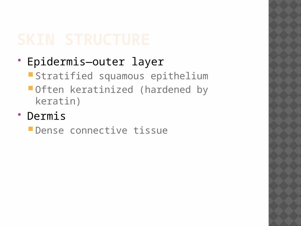
SKIN STRUCTURE Epidermis—outer layer
Stratified squamous epitheliumOften keratinized (hardened by keratin)
DermisDense connective tissue

SKIN STRUCTURE
Figure 4.3

SKIN STRUCTURE Subcutaneous tissue (hypodermis) is
deep to dermisNot part of the skinAnchors skin to underlying organsComposed mostly of adipose tissue

LAYERS OF THE EPIDERMIS Stratum basale (stratum germinativum)
Deepest layer of epidermisLies next to dermisCells undergoing mitosisDaughter cells are pushed upward to
become the more superficial layers Stratum spinosum Stratum granulosum

LAYERS OF THE EPIDERMIS Stratum lucidum
Formed from dead cells of the deeper strataOccurs only in thick, hairless skin of the
palms of hands and soles of feet Stratum corneum
Outermost layer of epidermisShingle-like dead cells are filled with keratin
(protective protein prevents water loss from skin)

LAYERS OF THE EPIDERMIS Summary of layers from deepest to
most superficialStratum basaleStratum spinosumStratum granulosumStratum lucidum (thick, hairless skin only)Stratum corneum

MELANIN Pigment (melanin) produced by
melanocytes Melanocytes are mostly in the stratum
basale Color is yellow to brown to black Amount of melanin produced depends
upon genetics and exposure to sunlight

DERMIS Two layers
Papillary layer (upper dermal region) Projections called dermal papillae
Some contain capillary loopsOther house pain receptors and touch
receptorsReticular layer (deepest skin layer)
Blood vessels Sweat and oil glands Deep pressure receptors

DERMIS Overall dermis structure
Collagen and elastic fibers located throughout the dermis Collagen fibers give skin its toughness Elastic fibers give skin elasticity
Blood vessels play a role in body temperature regulation
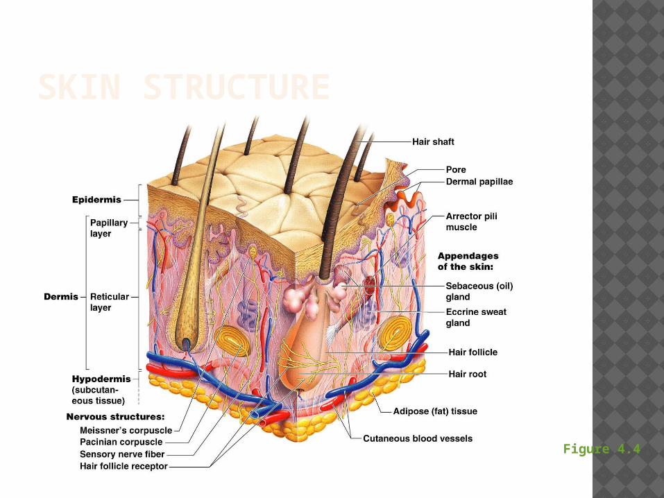
SKIN STRUCTURE
Figure 4.4

NORMAL SKIN COLOR DETERMINANTS Melanin
Yellow, brown, or black pigments Carotene
Orange-yellow pigment from some vegetables
HemoglobinRed coloring from blood cells in dermal
capillariesOxygen content determines the extent of
red coloring

SKIN APPENDAGES Cutaneous glands are all exocrine
glandsSebaceous glandsSweat glands
Hair Hair follicles Nails
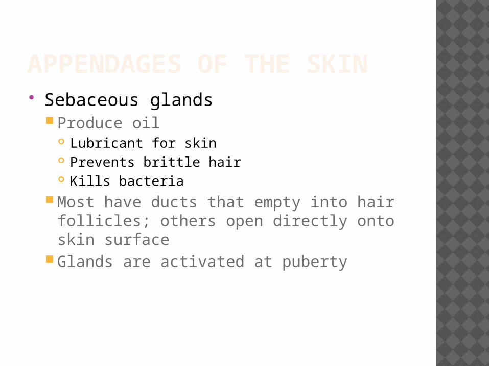
APPENDAGES OF THE SKIN Sebaceous glands
Produce oil Lubricant for skin Prevents brittle hair Kills bacteria
Most have ducts that empty into hair follicles; others open directly onto skin surface
Glands are activated at puberty

APPENDAGES OF THE SKIN
Figure 4.6a

APPENDAGES OF THE SKIN Sweat glands
Produce sweat Widely distributed in skinTwo types
EccrineOpen via duct to pore on skin surface
ApocrineDucts empty into hair follicles

APPENDAGES OF THE SKIN
Figure 4.6b

SWEAT AND ITS FUNCTION Composition
Mostly waterSalts and vitamin CSome metabolic wasteFatty acids and proteins (apocrine only)
FunctionHelps dissipate excess heatExcretes waste productsAcidic nature inhibits bacteria growth
Odor is from associated bacteria

APPENDAGES OF THE SKIN Hair
Produced by hair follicleConsists of hard keratinized epithelial cellsMelanocytes provide pigment for hair color

APPENDAGES OF THE SKIN
Figure 4.7c

APPENDAGES OF THE SKIN
Hair anatomyCentral medullaCortex surrounds
medullaCuticle on outside of
cortex Most heavily
keratinized
Figure 4.7b

APPENDAGES OF THE SKIN Associated hair structures
Hair follicle Dermal and epidermal sheath surround hair root
Arrector pili muscle Smooth muscle Pulls hairs upright when cold or frightened
Sebaceous glandSweat gland

APPENDAGES OF THE SKIN
Figure 4.7a

APPENDAGES OF THE SKIN
Figure 4.8

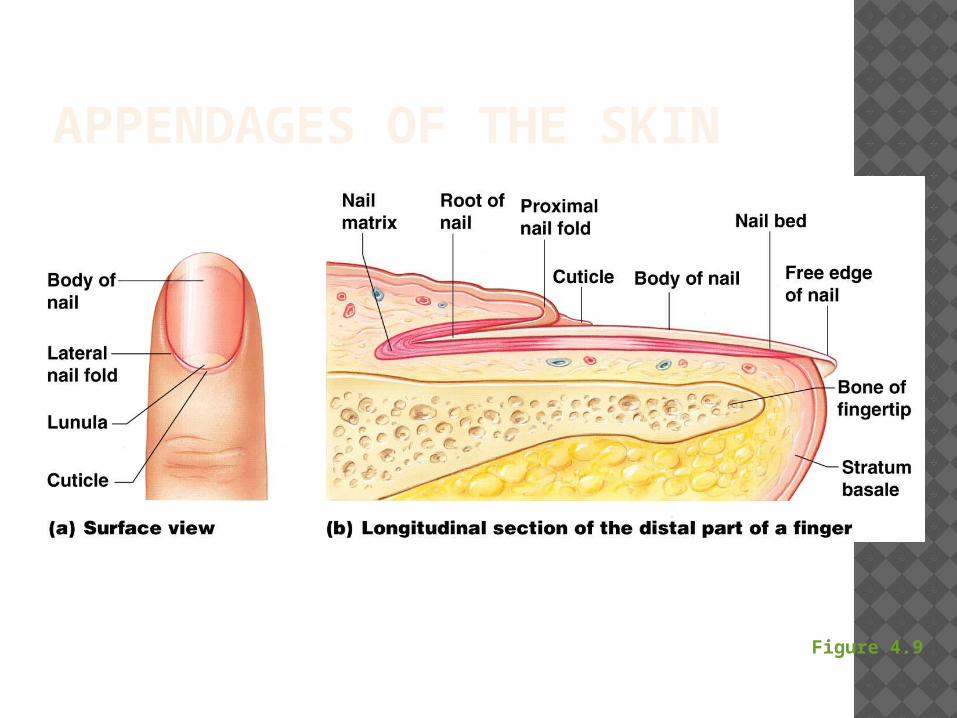
APPENDAGES OF THE SKIN Nails
Scale-like modifications of the epidermis Heavily keratinized
Stratum basale extends beneath the nail bed Responsible for growth
Lack of pigment makes them colorless

APPENDAGES OF THE SKIN Nail structures
Free edgeBody is the visible attached portionRoot of nail embedded in skinCuticle is the proximal nail fold that projects
onto the nail body
APPENDAGES OF THE SKIN
Figure 4.9