kub ivp: intravenous pyelography ultrasonography ct scan & mri studies renal scan topics
TRANSCRIPT

Imaging Studies Of Renal System
Dr.Ibrahim Abujarad9-3-2013

KUB IVP: intravenous pyelography Ultrasonography CT scan & MRI studies Renal scan
Topics

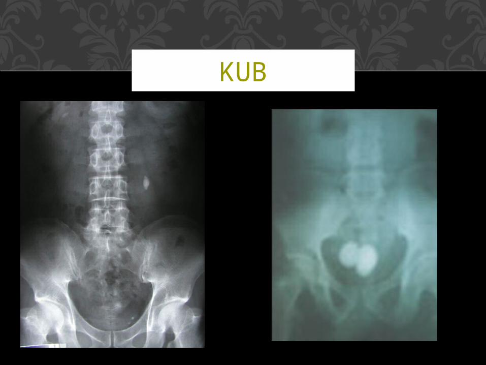
KUB stands for: kidneys, ureter & bladder.
No contrast material is used, it is the same for plain abdominal X-ray to show the different abdominal & pelvic organs as soft tissue shadows.
It is useful also to show radio-opaque renal stones.
KUB
KUB

**What is an Intravenous Pyelogram (IVP)?
An intravenous pyelogram (IVP) is an x-ray examination of the kidneys, ureters and urinary bladder
that uses contrast material.
IVP

How IVP will help in renal cases ??

IVP**How does the procedure work? In the IVP exam, iodine injected through a vein in the arm collects in the kidneys, ureters and bladder, giving these areas a bright white and sharply defined appearance on the x-ray images.**How is the procedure performed?• outpatient basis.• The patient is positioned on the
table. • The contrast material is then
injected, usually in a vein in the patient's arm, followed by additional still images.
• hold very still.• As the contrast material is
processed by the kidneys a series of images is taken.
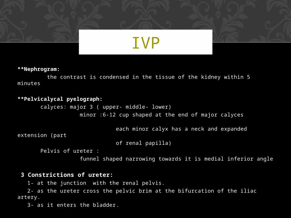
**Nephrogram:
the contrast is condensed in the tissue of the kidney within 5 minutes
**Pelvicalycal pyelograph:
calyces: major 3 ( upper- middle- lower)
minor :6-12 cup shaped at the end of major calyces
each minor calyx has a neck and expanded extension (part
of renal papilla)
Pelvis of ureter :
funnel shaped narrowing towards it is medial inferior angle
3 Constrictions of ureter: 1- at the junction with the renal pelvis.
2- as the ureter cross the pelvic brim at the bifurcation of the iliac artery.
3- as it enters the bladder.
IVP

AT 5 MIN

AT 10 MIN

AT 15 MIN


Ultrasonography
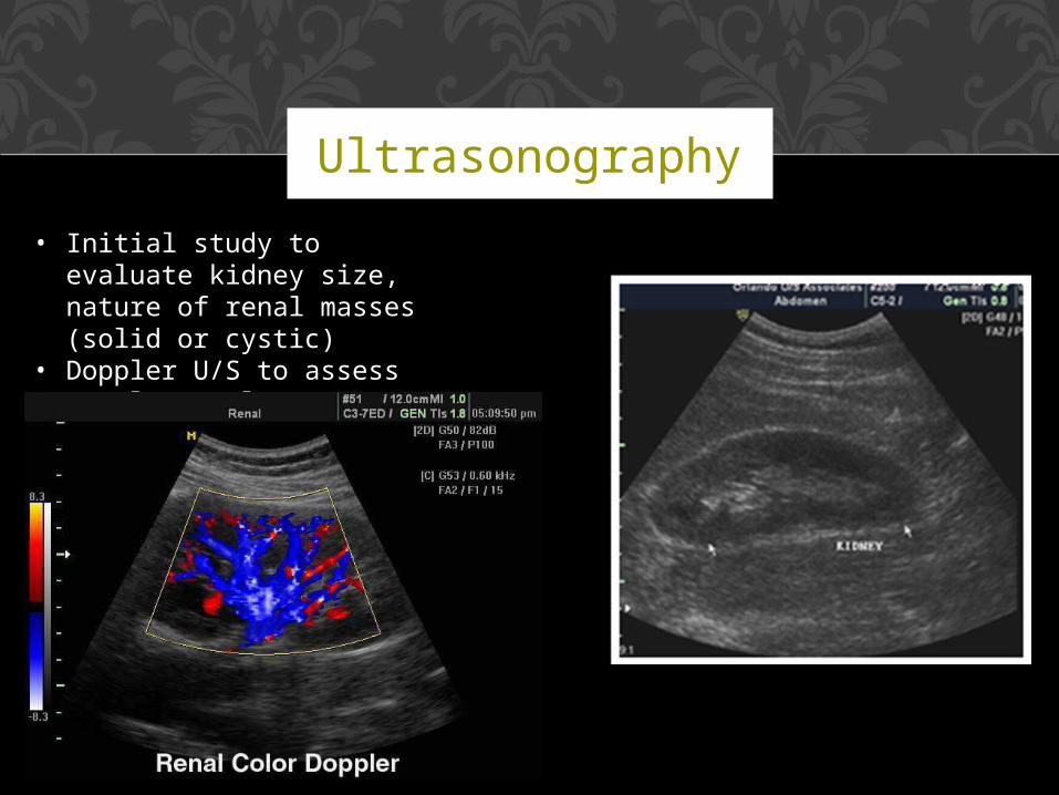
• Initial study to evaluate kidney size, nature of renal masses (solid or cystic)
• Doppler U/S to assess renal vasculature.

Ultrasonography

Ultrasonography

CT scan


3D CT SCAN

MRI


MRI Angiography

A renal scan is a nuclear medicine exam in which a small amount of radioactive material (radioisotope) is used to
measure the function of the kidneys.
Renal scan


Thank You