informationarizona.openrepository.com/arizona/bitstream/10150/188160/1/azu_td... ·...
TRANSCRIPT

VITELLOGENIN OF THE TOBACCO HORNWORM,MANDUCA SEXTA: PROPERTIES AND
ENDOCYTOTIC INCORPORATION INTO FOLLICLES.
Item Type text; Dissertation-Reproduction (electronic)
Authors OSIR, ELLIE ONYANGO.
Publisher The University of Arizona.
Rights Copyright © is held by the author. Digital access to this materialis made possible by the University Libraries, University of Arizona.Further transmission, reproduction or presentation (such aspublic display or performance) of protected items is prohibitedexcept with permission of the author.
Download date 16/07/2018 07:10:45
Link to Item http://hdl.handle.net/10150/188160


INFORMATION TO USERS
This reproduction was made from a copy of a manuscript sent to us for publication and microfilming. While the most advanced technology has been used to photograph and reproduce this manuscript. the quality of the reproduction Is heavily dependent upon the quality of the material submitted. Pages In any manuscript may have Indistinct print. In all cases the best available copy has been filmed.·
The following explanation of techniques Is provided to help clarify notations which may appear on this reproduction.
1. Manuscripts may not always be complete. When it is not possible to obtain . missing pages. a note appears to indicate this.
2. When copyrighted materials are removed from the manuscript. a note appears to indicate this.
3. Oversize materials (maps. drawings. and charts) are photographed by sectioning the original. beginning at the upper left hand comer and continuing from left to right In equal sections with small overlaps. Each oversize page is also filmed as one exposure and is available. for an additional charge. as a standard 35mm slide or in black and white paper format. •
4. Most photographs reproduce acceptably on positive microfilm or microfiche but lack clarity on xerographic copies made from the microfilm. For an additional charge. all photographs are available in black and white standard 35mm slide format. •
·For more information about black and white slides or enlarged paper reproductions. please contact the Dissertations Customer Services Department.


8613444
Osir, Ellie Onyango
VITELLOGENIN OF THE TOBACCO HORNWORM, MANDUCA SEXT A: PROPERTIES AND ENDOCY":"OTIC INCORPORATION INTO FOLLICLES
The University of Arizona
University Microfilms
International 300 N. Zeeb Road, Ann Arbor, MI48106
PH.D. 1986


PLEASE NOTE:
In all cases this material has been filmed in the best possible way from the available copy. Problems encountered with this document have been identified here with a check mark_-I_.
1. Glossy photographs or pages-L
2. Colored illustrations, paper or print ---3. Photographs with dark background __
4. Illustrations are poor copy __ _
5. Pages with black marks, not original copy __
6. Print shows through as there is t(:!xt on both sides of page __ _
7. Indistinct, broken or small print on several pages_.tL-
8. Print exceeds margin requirements __
9. Tightly bound copy with print lost in spine __ _
10. Computer printout pages with indistinct print __ _
11. Page(s) lacking when material received, and not available from school or author.
12. Page(s) seem to be missing in numbering only as text follows.
13. Two pages numbered . Text follows.
14. Curling and wrinkled pages __
15. Dissertation contains pages with print at a slant, filmed as received ___ _
16. Other ____________________________________________ _
University Microfilms
International


VITELLOGENIN OF THE TOBACCO HORNWORM, MANDUCA SEXTA: PROPERTIES AND
ENDOCYTOTIC INCORPORATION INTO FOLLICLES
by
Ellie Onyango Osir
A Dissertation submitted to the Faculty of the
DEPARTMENT OF BIOCHEMISTRY
In Partial Fulfillment of the Requirements For the Degree of
DOCTOR OF PHILOSOPHY
In the Graduate College
THE UNIVERSITY OF ARIZONA
1 986

THE UNIVERSITY OF ARIZONA GRADUATE COLLEGE
As members of the Final Examination Committee, we certify that we have read
the dissertation prepared by Ellie Onyango Osir --------~~-------------------------------
entitled Vitellogenin of the tobacco hornworm, Manduca sexta: Properties
and endocytotic incorporation into follicles
and recommend that it be accepted as fulfilling the dissertation requirement
for the Degree of Doctor of Philosophy
IL(I Fir c Date
Date J
Final approval and acceptance of this dissertation is contingent upon the candidate's submission of the final copy of the dissertation to the Graduate College.
I hereby certify that I have read this dissertation prepared under my direction and recommend that it be accepted as fulfilling the dissertation requirement. ,
~or Date l

STATEMENT BY THE AUTHOR
This dissertation has been submitted in partial fulfillment of requirements for an advanced degree at the University of Arizona and is deposited in the University Library to be made available to borrowers under rules of the Library.
Brief quotations from this dissertation are allowable without permission, provided that accurate acknowledgment of the source is made. Requests for permission for extended quotation from or reproduction of this manuscript in whole or in part may be be granted by the head of the major department or the Dean of the Graduate College when in his or her judgement the proposed use of the material is in the interests of scholarship. In all other instances, however, permission must be obtained from the author.
SIGNED: _---=~~~~:...:....;.,)r:-. ____ _

DEDICATION
This dissertation is dedicated to my parents, Gilbert M. Osir
and lydia A. Osir for their love, encouragement and prayers during the
entire period of my education.
Also to my sisters, Clara, Eve and brothers, Alfred and George
for their love, patience, support.
iii
ACKNOWLEDGMENTS
My most sincere thanks are to my research director, Professor
John H. Law, who not only provided me with the opportunity to pursue
further studies but also offered constant advice and constructive
criticisms during my entire graduate studies.
I wish also to thank the members of my study committee, Drs.
Michael Wells, William Grimes, Marc Tischler, James Berry, and Ralph
Price for their useful suggestions and encouragement during the period
of my research.
Through its founder and director, Professor Thomas Odhiambo, I
wish to offer very special appreciation to the "International Center
of Insect Physiology and Ecology" (I.C.I.P.E) for financing part of my
training.
Last, but by no means least, I wish to thank everyone in lithe
Law laboratory", Dr. Robert Ryan, Dr. John Kawooya, Pam Keirn, Xiao-yu
Wang, Ellie Moreland, and Mary Gonzales for providing constant support
throughout my studies. In addition, Drs. Darrell Anderson and Nobert
Haunerland were of much help. Finally, my stay in Tucson was made
especially pleasant by friends among whom Poovi Abaglo, Christa Sitz
and RoseAnn Jizmejian deserve very special thanks.
This work was supported by a grant from the U. S. Public
Health Service, No. GM 29238, awarded to Dr. John H. Law.
iv

TABLE OF CONTENTS
LIST OF ILLUSTRATIONS •
LIST OF TABLES
LIST OF ABBREVIATIONS
ABSTRACT •
CHAPTER 1. GENERAL INTRODUCTION AND LITERATURE REVIEW
Life cycle of Manduca sexta ••••••••• Female reproductive system ••••••••••• Insect yolk proteins: synthesis and structure Uptake of vitellogenin into follicles ••••• Rationale of the study ••••••••••••
CHAPTER 2. CHEMICAL AND IMMUNOLOGICAL STUDIES ON MANDUCA SEXTA VITELLOGENIN •••• • • •• --
Introduction • • • • • • • • • •••• Materials and methods • • •• • • • • • .••
Experimental animals • • • • • • • • • . ••• Collection of hemolymph and preparative
ultracentrifugation •••••••••••• Gel permeation and cation exchange chromatography. Density measurement • ~ • • • . .•• Protein determination •••• Electrophoresis ••.••.•. Analytical ultracentifugation ••• " •••••••••• Lipid analysis ••••.••• Limited trypsin digestion ••• Isolation of apoproteins •••••••••••• High performance liquid chromatography Scanning densitometry •••• Phosphoprotein staining •• Amino acid composition Phosphorus assay • • • • • Phosphoamino acid analysis Immunological studies •••••. In vitro synthesis of vitellogenin ••••• Labeling of vitellogenin ~ vivo ••••
v
Page
viii
x
xi
xii
1
2 '3 4 9
10
13
13 15 15
15 16 16 1.7 17 17 18 18 19 20 20 20 21 21 21 22 24 25

vi
TABLE OF CONTENTS--Continued Page
Studies on the carbohydrate moiety • • 25 Results • • • . . . . . . • • • • • • • 29
Isolation, physical and chemical properties. • • • •• 29 Limited trypsin digestion. • • • . . • • • . • • • ~9 Isolation and properties of apoproteins • • 41 Immunological properties of apoproteins • • • • • • 45 Synthesis of vitellogenin in vitro • • • • • • • • 48 Phosphoryl ation ••••• -•• -. -.-. • • • 48 Carbohydrate composition ••••••••••••••• 54 Location of the carbohydrate moiety • • • • • • • • •• 54 Gel permeation chromatography of pronase glycopeptides 56 Lectin binding properties. • • • • • • • • 56 Proton NMR spectroscopy of glycopeptides • • • • • •• 58
Discussion . • • • • • • • . • . • • • . • • • . . . . • •• 67 Physical and chemical properties ••••• • • •• 67 Synthesis of vitellogenin by the fat body. • • • • •• 68 Phosphorylation. • • • • • • • • • • • • • • • • • •• 68 Immunological properties ••••••••••••••• 69 Carbohydrate moiety • • • • • • • • • • • • • • • • 70
CHAPTER 3. STUDIES ON BINDING AND UPTAKE OF VITELLOGENIN • • 73
Introduction • • • • • • • • 73 Materials and methods • • • • • •• 76
Isolation of vitellin. • • • • • • • • • •• 76 Deglycosylation • • • • • • • • • • • • • 76 Iodination of protei~~ ..•• • • .••• •• 77 Incubation of follicles. • • • • • • • • • • • • • 77 Trypsin treatment. • • • • • • • • • • . • • • • • 78 Preparation of follicle membranes. • • • • • • • • •• 78 Binding of labeled vitellogenin to follicle membranes. 79
Resul ts ........................ 81 Isolation of vitellin. • • • • • • • • • • • • • • 81 In vitro uptake of labeled vitellogenin by isolated - --:roT1 icles . . . . . . . . . . . . . . . . . . 81 Distribution of labeled vitellogenin in isolated
follicles ••••••••••••• • • • • • 85 Characterization of specific binding to follicle
membranes ••• • • • • • • • • 89 Competition studies • • • • • • • • • • • • • • • 92

TABLE OF CONTENTS--Continued
Discussion ••••••••••••• In vitro uptake of vitellogenin Bin~to follicle membran~~ Competition for binding sites
SUMMARY AND FUTURE PROJECTIONS
REFERENCES •••••••••••
vii
Page
98 98 99
100
101
103

LIST OF ILLUSTRATIONS Figure Page
1. SOS-polyacrylamide gel electrophoresis of adult male and female hemolymph and egg homogenate of ~. sexta • •• 30
2. Gel permeation chromatography of subphase from preparative ultracentrifugation ••••••••••••••••• 31
3. SOS-polyacrylamide gel of fractions from Bio-Gel A 1.5 m
4.
5.
6.
7.
8.
9.
10.
11.
12.
13.
14.
15.
16.
17.
18.
19.
gel permeation column • • • ~3
Cation-exchange chromatography .•
Purification stages of vitellogenin •
Polyacrylamide gel electrophoresis of vitellogenin
Molecular weight determination of vitellogenin
Limited trypsin treatment of vitellogenin ••
Isolatiorl of vitellogenin apoproteins ••••
SOS-polyacrylamide gel electrophoresis of apoproteins •
Immunological reactions of vitellogenin apoproteins •
Oouble radial immunodiffusion •.•
Synthesis of vitellogenin in vitro
Phosphorylation of vitellogenin and vitellin apoproteins
2-dimensional TlC of phosphoamino acids •
FITC-Con A staining of vitellogenin .••
Bio-Gel P-6 gel permeation chromatography of vitellogenin pronase glycopeptide~ •••••••••.•
Profiles of labeled glycopeptides on lentil lectin and and concanavalin-A columns ••••••••••••
250 MHz proton NMR spectrum of the major glycopeptide
viii
34
~5
36
37
40
42
43
46
47
49
50
S3
55
57
59
60

20.
21.
22.
23.
24.
25.
26.
27.
28.
29.
30.
31.
LIST OF ILLUSTRATIONS---Continued
The major structure identified from the NMR spectrum
Schematic diagram of the lipid-linked intermediate
NMR spectrum of endo-H treated glycopeptide •
Elution profile of vitellin from OEAE Bio-Gel A column
SOS-polyacrylamide gel of vitellin and vitellogenin •
Uptake of vitellogenin by isolated follicles
Uptake of endo-H and non endo-H treated vitellogenin by isolated follicles ••••••••••
Time course of vitellogenin binding to follicle membranes ••••••••••••••••
Concentration-dependent binding of vitellogenin to follicle membranes •••
Scatchard analysis of binding data •••••••
pH dependency of vitellogenin binding to follicle membranes • • • • • • • . . . • •
Calcium dependency of vitellogenin binding to follicle membranes • • • • • • • • • • • • • • • • •
ix
Page
62
63
66
82
83
86
87
90
91
93
94
95

LIST OF TABLES
Tables
1. Composition of ~. sexta vitellogenin •
2. Amino acid composition of vitellogenin apoproteins •
3. Phosphorus contents of apoprotei~s •.
4. 1H Chemical shifts of structural reporter groups of
P~e
38
44
52
constituent monosaccharides for the carbohydrate moiety • 61
5. Relationship between follicle length and vitellogenin uptake ~ vitro •..•••••••••••• 84
6. Distribution of labeled vitellogenin in isolated follicles. 88
7. Effects of various proteins on binding of vitellogenin to follicle membranes. • • • • • • •• • • • • • • • • • • •• 97
x

Vg • • •
ApoVg-I
ApoVg-II •
PBS
SOS
PAGE •
Con A
FIn: •
PAS
TLC •••••
LOL
Endo-H •
MES
BSA
NMR
Man
GlcNAc ••
LIST OF ABBREVIATIONS
. . .
. . . . . .
. . ••• Vitellogenin
Apovitellogenin-I
•• Apovitellogenin-II
Phosphate buffered saline
• ••• Sodium dodecyl sulfate
• Polyacrylamide gel electrophoresis
• ••••••.. Concanavalin A
• Fluorescein isothiocyanate
• Periodate-Schiff reagent
Thin layer chromatography
•• Low density lipoprotein
• • Endo-a-N-acetylglucosaminidase H
2-(N Morpholino) Ethane Sulfonic Acid
Bovine serum albumin
Nuclear magnetic resonance
• • • • • •• Mannose
N-acetylglucosamine
xi

ABSTRACT
Manduca sexta vitellogenin is a phosphoglycolipoprotein
(Mr - 500,000) that contains two copies of the apoproteins
(apovitellogenin-I, Mr 180,000 and apovitellogenin-II, Mr 45,000),
13 percent lipids, 3 percent carbohydrates and 0.6 percent
phosphorus. The two apoproteins are immunologically distinct
polypeptides and apovitellogenin-II is not completely accessible to
the aqueous environment in the intact molecule. The carbohydrate
moiety located on apovitellogenin-I has a high mannose structure
(Mang GlcNAc2)·
Sonicated follicle membranes bind 125I_labeled
vitellogenin with high affinity and specificity (KD = 1.3 x 10⁻⁸ M). Total binding sites were estimated at 4 x 10¹⁴
sites/ 9 of follicle membrane protein. The binding was sensitive to
pH and calcium. Competition studies showed that binding of
vitellogenin was blocked by vitellin and deglycosylated vitellogenin
but not by lipophorin, microvitellogenin or apovitellogenin-II. These
results suggest that the uptake of vitellogenin involves binding to
specific receptors on follicle membranes and the carbohydrate moiety
and apovitellogenin-II are not involved in the interaction with the
receptors.
xii

CHAPTER 1
INTRODUCTION AND LITERATURE REVIEW
~ All insects begin their life as an egg. After hatching of
the egg comes a series of complex morphological and physiological
transformations that culminate in the mature reproducing form of the
insect. These transformations, referred to as "metamorphosis", are
typical of all insects except in the most primitive members of the
group (Chapman, 1969). In general, growth and development in insects
occurs by periodic shedding of the hard outer exoskeleton or cuticle.
This process includes formation of a new cuticle, removal of the old
one, and the actual change in form (Chapman, 1969). Depending on how
an insect accomplishes its transformation into the adult form, two
basic modes of development are recognized (Wigglesworth, 1972). In
the first type, referred to as being complete or holometabolous, the
caterpillars (larvae) molt into pupae which subsequently undergo a
second molt into adults. In this two step development, juvenile forms
differ remarkably from adults not only in structure but also in the
types of food consumed, thus enabling the invasion of different
habitats. This group of insects is exemplified by Lepidoptera,
Coleoptera and Diptera. In the second mode of development,
hemimetabolous, juvenile forms closely resemble adults and growth is
achieved by a series of molts without the pupal stage. Examples of
1

2
insects in this group are Orthoptera, Hemiptera, and Homoptera. The
process of molting in insects is under the control of two hormones,
namely juvenile hormone (JH) and ecdysone (or growth and
differentiation hormone) (Wigglesworth, 1972). Ecdysone is a steroid
hormone produced by the prothoracic gland upon stimulation by
prothoracicotrophic hormone, a small peptide hormone released by the
brain neurosecretory cells and stored in corpora allata (Steele and
Davey, 1985). In peripheral tissues, ecdysone is converted into
20-hydroxyecdysone (a-ecdysone) which is the active form of the
hormone (Gilbert et ~., 1980). On the other hand, JH (sesquiterpene
derivatives) are produced by the corpora allata, small organs situated
behind the brain. Between molts, JH exerts a "status quo" effect by
inhibiting the formation of adult structures but at metamorphosis, JH
titers decline thus allowing ecdysone to exert its molting effect.
Life cycle of Manduca sexta
The tobacco hornworm, ~. sexta, is a holometabolous moth in
the order Lepidoptera and family Sphingidae. The larvae of this
insect are pests of tobacco and tomato plants but will also feed on
other solanaceous plants (Hoofmann et ~., 1966). After hatching of
the eggs, the first instar larvae undergo four molts that give rise to
the fifth and last larval instar. The larvae have powerful jaws which
enable them to feed voraciously. Between the first and fifth larval
instars, the larvae increase in weight 4000 fold in only about ten
days (Shapiro and Law, unpublished observation). At the end of the

3
fifth instar, the larvae prepare to molt into the "resting stage"
(pupae). But prior to pupation, a characteristic behavior called
"wandering" ensues. The larvae first cease to feed, void their gut
and then wander around as if they were burrowing in the soil.
Although the pupae remain essentially immobile, they undergo internal
morphological and physiological changes as they prepare for the next
molt into adults after eighteen days. This final molt, termed adult
ec10sion, is generally preceded by darkening and softening of the
cuticle. The entire development from the egg to the adult stage takes
about thirty three days. The adult moths usually feed on nectar and
live for 4-5 days. Mating between adult moths usually occurs at night
and males must fly to find their female mating partners.
In many ways ~. sexta can be considered as an ideal
laboratory experimental animal. The insect can easily be cultured in
the laboratory on an artificial diet that consists of agar, wheat
germ, casein, vitamins and salt mix (Reinecke et ~., 1980).
Generation times are fairly short thus ensuring availability of
different life stages at all times. Furthermore, the large size of
insects permits single animal experiments.
Female reproductive system
The female reproductive system of insects consists of a pair
of ovaries. Each ovary is composed of a series of egg tubes or
ovario1es (Ross, 1965). The number of ovario1es vary from insect to
insect, and in~. sexta the number is eight. In~. sexta, each

ovariole is approximately 6 cm long at the time of adult eclosion and
consists of more than 200 follicles at different stages of development
(Nijhout and Riddiford, 1979). The smallest and youngest follicles
are found at the anterior tip of the ovariole and the follicles
increase in size and age towards the posterior tip. Within the
follicles are oocytes and a special group of cells referred to as
Iinurse cells". The whole complex is surrounded by follicle cells.
The primary function of the nurse cells is to supply oocytes with
ribosomes which serve as the machinery for protein synthesis during
the early stages of embryogenesis or perhaps even for protein yolk
synthesis in some insects such as Drosophila melanogaster (King and
Buning, 1985).
Insect yolk proteins: synthesis and structure
In the cleidoic (closed system) eggs of insects, yolk serves
as the most important source of nutrient during embryonic
development. The predominant components of yolk are proteins, lipids
and glycogen. Yolk proteins are primarily stored in storage vesicles
called yolk granules (Anderson, 1970). Because yolk is an important
component of insect eggs, understanding the processes involved in its
formation is important from the standpoint of insect reproduction. In
many insects, exogenous supply by the hemolymph is the most important
source of yolk proteins. The major proteins which constitute up to 90
percent of all soluble yolk proteins are termed vitellins (Engelmann,
1979). Vitellins are derived from hemolymph precursors,
4

vitellogenins. Insect vitellogenins were first discovered in
hemolymph of the silkmoth, Hyalophora cecropia (Telfer, 1954). Since
then, these proteins have been isolated and characterized from a wide
variety of insects (Engelmann, 1979; Hagedorn and Kunkel, 1979; Kunkel
and Nordin, 1985). Vite110genins are usually distinguished from other
hemolymph proteins by three main criteria: (1) they are largely
female-specific (ii) they constitute between 60 to 90 percent of
soluble yolk proteins and (iii) with a few exceptions, they are
synthesized only in the fat body tissue. In many cases, the
differences between vitel10genins and vite11ins are very subtle. For
example, lipid analysis of vitellin and vite110genin in the cockroach,
B1ate11a germanica showed only minor differences which suggested loss
of lipids upon uptake (Oie et !l., 1975). Heterogeneity of
vite110genins and vite11ins has been reported for a few insects, for
example, in~. sexta (Imboden and Law, 1983). ~. sexta vitel10genin
and vitellin each show three distinct bands when hemolymph or egg
homogenate is subjected to electrophoresis under non-denaturing
conditions. However, isolated vite110genin or vitellin show only
broad bands. Neither the differences between the three distinct
vite110genins nor the cause for their disappearance during
purification is well understood. The most striking difference between
vite110genins and vite11ins has been reported for Locusta migratoria
and Leucophaea maderae (Chino et !l., 1976; Koeppe and Of eng and ,
1976). In both these cases, a substantial change in subunit structure
apparently accompanies vitel10genin uptake into eggs. For example, in
5

~. maderae, vitellogenin is composed of three subunits while vitellin
has four subunits. Proteolytic conversion of one or more of the
vitellogenin subunits within the egg has been proposed as a possible
cause for this observation. It would then appear that at least in a
few well described cases, the use of the terms, vitellogenin and
vitellin, is justi1ied. In those insects for which no difference is
apparent, the two terms are probably not appropriate.
In a wide variety of insects, vitellogenins are synthesized,
processed, and secreted by the fat body tissue. However, Dipteran
vitellogenins are also synthesized by the ovary (Brennan et 21., 1982). For example, in Q. melanogaster, ovarian cells supplement
vitellogenin made by the fat body, and they secrete it into the space
between follicle cells and oocyte (Jowett and Postlethwait, 1980). In
~. sexta, cultured fat body tissue has been shown to secrete
vitellogenin in vitro (Osir and law, unpublished observation). There
was no synthesis of vitellogenin by isolated follicles. In a wide
variety of insects, vitellogenin synthesis is controlled by JH and
ecdysone. For example, in Orthoptera, JH has been shown to have a
direct effect on vitellogenin synthesis ~ vitro (Wyatt et 21., 1976)
while in the dipteran, Q. melanogaster, synthesis is apparently
controlled by both JH and ecdysone (Hagedorn and Kunkel, 1979). For
many other species (including ~. sexta), the control of vitellogenin
synthesis is still not clear (Nijhout and Riddiford, 1974). As in
other transported proteins, a number of post-translational
modifications occur prior to secretion. Among these are
6

glycosylation, phosphorylation, sulfation and the addition of lipids.
The resulting particles are usually complex proteins with molecular
weights as high as Mr 600,000 (Harnish and White, 1982; Kunkel and
Nordin, 1985).
Although most vitellogenins isolated contain lipids and are
glycosylated, the presence of phosphate groups have been shown only in
a few insects including h. maderae (Engelmann and Friedel, 1974), Q.
melanogaster (Brennan and Mahowald, 1982), Bombyx mori (Takahashi,
1983), Aedes aegypti (Borovsky and Van Handel, 1980), Culex pipiens
fatigans (Atlas et ~., 1978), Rynchosciara americana (Pereira and
Bianchi, 1983), and~. sexta (Osir et ~., 1986). In M. sexta - ----vitellogenin, both apoproteins, apoVg-I and apoVg-II, contain
covalently bound phosphate groups (Osir et ~., 1986). As in other
insects examined so far, all protein-bound phosphate groups in ~.
sexta vitellogenin are attached to serine. In general, insect
vitellogenins contain lower amounts of phosphorus compared to
vertebrate lipovitellins. For instance, avian or amphibian phosvitins
contain approximately 10 percent serine-linked phosphorus compared to
vitellogenins of h. maderae (0.14 percent) (Engelmann and Friedel,
1974) or ~ sexta (0.6 percent) (Osir et ~., 1986). Lipids are an
integral part of insect vitellogenins constituting between 7-16
percent with the major components being phospholipids and
diacylglycerol (Engelmann, 1979). A possible role of transporting
diacylglycerols into eggs has been suggested for vitellogenin (Chino
et ~., 1977) although the major lipid transporting lipoprotein,
7

lipophorin, may be more involved in this function. For example, ~.
sexta egg lipophorin probably contains less than 10 percent lipids
"(Osir, Kawooya and Law, unpublished observation) compared to about 40
percent in hemolymph lipophorin (Pattnaik et ~., 1979). Most of the
lipids of lipophorin are probably released after uptake into the
eggs. The function of lipids in vitellogenin is not well understood
although they could be used to construct oocyte membranes or as an
energy source during oocyte development.
All vitellogenins so far characterized contain
covalently-bound carbohydrate moieties (Hagedorn and Kunkel, 1979).
Indeed, many secretory proteins are also glycosylated (Hubbard and
Ivatt, 1983). Interest in the study of carbohydrates in general is
mainly due to the diverse biological functions which they perform. In
many cases, the physiological and biological properties of
glycoproteins have been directly related to the carbohydrate
prosthetic groups that they contain (Ashwell and Morell, 1974).
Although a few reports have assigned carbohydrate structures of insect
vitellogenins based on chemical, enzymatic and lectin binding methods
(Kunkel et ~., 1980, Nordin et ~., 1984), NMR spectroscopy has never
been used to study these structures. In this dissertation, the
primary structure of the carbohydrate moiety of ~. sexta vitellogenin
has been determined by high field proton NMR spectroscopy. NMR has an
advantage over chemical methods in that it is non-destructive and
complete information on primary structures can be obtained in a
relatively short time. Furthermore, sequential modifications can be
8

made on the same sample which is recovered after each analysis.
Current knowledge on insect hemolymph glycoproteins suggests that a
similar glycosylation pathway is probably used throughout plant and
animal kingdoms. The only difference is that the carbohydrate
moieties of insect hemolymph proteins do not appear to be processed
into more complex structures. In contrast to insect vitellogenin
oligosaccharides, which are simple N~linked structures, vertebrate
vitellogenins have complex carbohydrates. For example, the
carbohydrate moiety of K. laevis vitellogenin is an N-linked complex
structure containing GlcNAc, Man, galactose and sialic acid (Gottlieb
and Wallace, 1982). The exact role(s) of these oligosaccharides (if
any) with regard to vitellogenin synthesis, secretion and uptake still
remains unresolved.
Uptake of vitellogenin into follicles
Different animal cells have complex transport mechanisms
which enable them to sort out specific molecules from heterogeneous
mixtures in their surroundings. As a result of these selective
transport phenomena, the tissues are able to concentrate certain
molecules. Vitellogenic blood protein sequestration by ovarian
follicles of amphibians (Wallace and Jared, 1969) and insects (Telfer,
1960) are good examples of selective transport. In insect ovaries,
the transport mechanism favors the female-specific protein,
vitellogenin, which becomes concentrated by as much as thirty times
the hemolymph concentration (Pan, 1971). Incorporation of
9

10
vitellogenin into oocytes has been demonstrated in vivo (Kunkel and
Pan, 1976; Ferenz, 1978) and ~ vitro (Ferenz et Al., 1981; Osir and
Law, 1984; Telfer and Kulakosky, 1984). Vitellogenin uptake involves
permeation of the intercellular spaces of the follicle (Telfer et ~.,
1981) followed by endocytosis at the oocyte surface. It should be
noted that coated pits, the progenitors of coated vesicles, were first
demonstrated in ovaries of the mosquito, ~. aegypti, involved in
vitellogenin uptake (Roth and Porter, 1964). Although the route of
entry of vitellogenin appears to be well understood, the actual
mechanism by which the protein is initially sorted out prior to
enclosure within endocytotic vesicles is not entirely clear. An
interesting question is whether vitellogenin binding sites should be
called receptors. It is generally accepted that the key property of a
binding site which defines it as a receptor is its association with a
specific physiological response or function. Until any such function
is found for vitellogenin, it might be safer to retain the term
"specific binding site(s)~ without overemphasizing receptor function.
Rationale of the study
In conducting these studies, the female insect has been
chosen as the animal model. Specifically, some important aspects
involved in egg production in insects have been studied. The
processes involved in egg formation are interesting in that they could
be used as models for studying other more complex systems.

11
Furthermore, a thorough understanding of some of these processes may
eventually be useful in finding selective ways to control insect pests.
This dissertation is basically divided into two distinct
sections. The first section (Chapter 2) presents detailed chemical
and immunological studies of ~. sexta vitellogenin. In particular,
the immunological relationships between vitellogenin apoproteins have
been explored. The primary structure of the carbohydrate moiety has
also been determined. The information presented is deemed important
in understanding how this class of insect lipoproteins are synthesized
and later utilized by the developing embryo.
The second section deals with the first step in the
utilization of vitellogenins, namely the uptake process. Although
vitellogenin uptake is by and large a storage process, the mechanisms
involved appear to be very complex. The process is highly selective
for vitellogenin which becomes concentrated within the follicles.
This suggests that there are specific binding sites on the oocyte
surface that regulate the uptake process. There are three basic
challenges in the search for vitellogenin binding sites. The first
one is to design a binding assay using follicle membranes and the
second is to find a method for solubilizing the receptor in an active
state. The 'final part would be to identify and isolate the receptor
itself. In this section, an ~ vitro system for the uptake of
vitellogenin is presented. In addition, an assay for characterizing
specific binding sites on the follicle membranes is described. This
assay is potentially useful in understanding the structural basis for

12
the selectivity in vitellogenin uptake.

CHAPTER 2
CHEMICAL AND IMMUNOLOGICAL STUDIES ON MANDUCA SEXTA VITELLOGENIN
Introduction
Insect vitellogenins are glycolipoproteins that are
synthesized by the fat body tissue, secreted into hemolymph and
finally sequestered by the developing eggs or follicles (Engelmann,
1979; Hagedorn and Kunkel, 1979). The insect fat body which is
generally diffuse and distributed allover the abdominal region may be
compared to vertebrate liver and adipose tissue combined (Riddiford,
1980). The fat body stores lipids, synthesizes and secretes many
hemolymph proteins. Although the follicles are constantly bathed by
hemolymph that contains many proteins, only vitellogenin is
selectively and rapidly taken up. Other hemolymph proteins are also
taken up, albeit in only small amounts (Telfer 1960; Telfer, 1981).
From these observations, two basic questions can be asked. First,
what makes vitellogenin the preferred storage protein in the eggs
Second, what structural features of vitellogenin are responsible for
its selective uptake In order to address these questions, it is
necessary to obtain complete information on the chemical structure of
vitellogenins. Once this information is available, the next step is
to search for any unique features that may confer selectivity in the
uptake process. Although vitellogen1ns and vitellins have been
13

14
described from a number of insects, there is still very little
detailed information on their native structures and on the arrangement
of the apoproteins within the molecule.
This section of my dissertation describes detailed
characterization of ~. sexta vitellogenin. Vitellogenin apoproteins
have been isolated and characterized. Rabbit antibodies against the
isolated apoproteins were used to obtain structural information on the
native vitellogenin molecule. The primary structure of the
carbohydrate moiety was determined by high field proton NMR
spectroscopy. It was also shown that vitellogenin apoproteins contain
covalently-bound phosphate and that these groups are present only as
phosphoserine.

15
Materials anm Methods
Experimental animals
Adult female !:h. sexta \Jsed lin all experiments were obtained
from eggs supplied by Drs. J. P. Reinlecke and J. S. Buckner, U. S.
Department of Agriculture, Fargo, North Dakota. After hatching, the
larvae were raised individually ~s previously described by Bell and
Joachim (1976) on a high wheat g~rm diet (Reinecke et ~., 1980).
Collection of hemolymph and prep~ratiye ultracentrifugation
Twenty to thirty adult inselcts, 1-2 days old, were bled as
follows. The legs, wings, and h~ads ,~ere removed from the insects and
the carcasses centrifuged head dQwn into a solution (50 ~l) of
phosphate buffered saline (PBS: Q.2 M: NaCl, 0.02 M sodium phosphate,
pH 6.7) containing 50 mM glutathione and 20 mM of the protease
inhibitor, diisopropylphosphorofluoridate (DFP). The hemolymph was
centrifuged (10,000 x g, 10 min, 4°C)1 in order to remove hemocytes.
Potassium bromide was then added to the supernatant to give a final
concentration of 44 percent (w/v). This solution was placed in a
quick seal centrifuge tube (Beck~an), overlayered with 0.9 percent
NaCl and centrifuged (206,000 x ~, 4 fu, 10°C) using a vertical rotor
(type VTi-50, Beckman) in a Beck~an L8-70 ultracentifuge. This
centrifugation isolated lipophorin from the rest of the hemolymph
proteins.
Gel permeation and cation-exchange chromatography
Subphase (15 ml) from the preparative ultracentrifugation
tube was applied to Bio-Gel A 1.5m column (2.5 x 70 cm) equilibrated
with PBS. Fractions (2.9 ml) were collected and absorbances measured
at 280 nm. Tobacco mosaic virus and vitamin B12 were used to obtain
the void volume (VO = 140 ml) and total volume (Vt = 430 ml),
respectively. Protein standards used for calibrating the column were
thyroglobulin (Mr 670,000), y-globulin (Mr 158,000), ovalbumin
(M 45,000), and myoglobin (M 17,000) (Bio-Rad). Fractions that r r
contained vitellogenin were concentrated by ultrafiltration, dialyzed
(12 h, 4°C) against 46 mM sodium succinate buffer, pH 5.9 and applied
to Cm Bio-Gel A column (1.5 x 14 cm) equilibrated in the same buffer.
The column was then washed with 30 ml of starting buffer to remove
non-adsorbed proteins and vitellogenin was eluted using 0.2 M NaCl.
Isolated vitellogenin was stored at 4°C under nitrogen.
Density measurement
Potassium bromide was added to isolated vitellogenin to give
a final concentration of 44 percent (w/v). The solution was
overlayered with KBr solution (33 percent, w/v) and centrifuged for 20
h (Haunerland and Bowers, personal communication). Fractions (1 ml)
were collected from the tube and refractive indices (R. I) measured.
The absorbances of each fraction was measured at 280 nm. Densities
(D) were calculated from the equation: 0 = (R.I - 1.777)1 0.1561.
16

Protein determination
Protein concentrations were determined using either the
Folin reagent (Lowry et !l., 1951) or the BCA protein assay reagent
(Pierce) with BSA as protein standard.
Electrophoresis
Electrophoresis on SDS-polyacrylamide slab gels in 25 mM
Tris, 192 mM glycine buffer, pH 8.3 was conducted as described by
Laemm1i (1970). Gradient slab gels were cast using a gradient maker
(BRL). Gels were stained for protein with Coomassie Brilliant Blue
(Pierce). High and low molecular weight markers for SDS-PAGE were
obtained from Bio-Rad. Native electrophoresis was carried out on 4-20
percent gels at 4°C with a constant 140 V for 24 h. Protein standards
used were: thyroglobulin (Mr 670,000); ferritin (Mr 440,000);
catalase (Mr 232,000); lactate dehydrogenase (Mr 140,000) and BSA
(Mr 67,000). Molecular weights were determined from the plot of log
molecular weight versus relative migration of the protein standards.
Analytical ultracentrifugation
Sedimentation equilibrium was carried out at 6,000 rpm
(20°C) using type AN-H rotor in a Spinco model E ultracentrifuge
equipped with electronic speed control and a U.V. scanner.
Vite110genin was dialyzed against a buffer containing 0.1 M sodium
chloride, 10 mM sodium phosphate, pH 7.0. Molecular weight was
calculated using a partial specific volume (v) of 0.766 (Pan and
17

Wallace, 1974). The plot of ln (A280) versus r2 (radial position)
had a correlation coefficient of 0.921.
Lipid analysis
Vitellogenin (9 mg) was extracted with chloroform-methanol
according to Bligh and Dyer (1959). The extract was assayed for
phosphate (Bartlett, 1959) and an amount of sample containing 100
nmoles of phosphate was mixed with 10 ~g each of ditridecanoin,
tritridecanoin, stigmasterol, tetracosane and tetradecanoic acid as
internal standards for diacylglycerols, triacylglycerols, cholesterol,
hydrocarbons and free fatty acids, respectively. The lipids were
separated by TLC and then quantitated by gas liquid chromatography
(Fernando-Warnakulasuriya et al., 1981), except that hydrocarbons were
separated using a column' (26 mm x 1.2 m) containing 2.5 percent SE-30
on Gas-chrom Q (Applied Sciences) and a temperature program of
130-300° at 4°/min. The injector port temperature was 235°C.
Limited trypsin digestion
Isolated vitellogenin (100 ~g) was treated with trypsin
(Worthington) for various times at 37°C in a water bath. A control
incubation with no trypsin was included. The ratio of vitellogenin to
trypsin was 100: 1. The reaction was stopped by boiling for 5 min and
aliquots from each incubation (27 ~g protein) were subjected to
SOS-PAGE.
18

Isolation of apoproteins
Initial attempts to isolate vitellogenin apoproteins were
carried out·by the electroelution method after SOS-PAGE. However,
this method was not successful since apoVg-II was always contaminated
by apoVg-I. Furthermore, antibodies raised against these apoproteins
showed cross-reactivity. For this reason, column chromatography was
used to separate the apoproteins after dissociation in a suitable
chaotrope. The apoproteins could easily be dissociated in 6 M
guanidine HC1, 50 mM sodium phosphate, pH 7.0 and separated on a
Sepharose CL-6B column (1.5 x 91 cm). Briefly, isolated vitellogenin
(40 mg) was mixed with 1.1 g of guanidine HCl and incubated for 1 h at
50°C. After cooling, the sample was applied to the column which was
eluted with the same buffer at 12 ml/h. Pooled apoproteins were
dialyzed with four changes against deionized water. ApoVg-I which
precipitated during dialysis, was collected by centrifugation (15,000
x g, 10 min) in a microcentrifuge (Fisher). The low solubility of
apoVg-I compared to apoVg-II provided an alternative method for
isolating the apoproteins. In this procedure, vitellogenin was first
dissociated in guanidine HCl by boiling as described above and then
dialyzed against water. After two changes, apoVg-II was still in
solution while most of apoVg-I had precipitated out. The solution was
centrifuged to separate the apoproteins. The remaining apoVg-I
contaminating apoVg-II preparation was removed by affinity
chromatography on Con A column since only apoVg-I is glycosylated.
19

High performance liquid chromatography
Gel permeation HPLC was carried out on a Varian 5000 liquid
chromatograph with a UV-50 detector (set at 280 nm) using a TSK 250
column (Bio·-Rad) in 0.15 M Na2S04, 0.02 M Na2P04, 0.02 percent
. NaN3, pH 7.0. Sample (50 ~l) was injected and the flow rate
maintained at 0.5 ml/min. Protein standards used were, thyroglobulin
(Mr 670,000), IgG (Mr 158,000), ovalbumin (Mr 45,000) and
cytochrome C (M 14,000) (Bio-Rad). r
Scanning densitometry
In order to compare the staining intensity of the two
apoproteins, the following experiment was conducted. Varying amounts
of apoVg-I (7-30 ~g) and apoVg-II (6-30 ~g) were subjected to SDS-PAGE
as described above. After staining with Coomassie Brilliant Blue for
protein, the gel was scanned using model OU 8 Spectrodensitometer
(Beckman) and the area corresponding to each concentration of protein
determined. For each apoprotein, a plot of staining intensity versus
the amount of protein applied to the gel was linear and the slopes of
the two plots were also identical.
Phosphoprotein staining
Vitellogenin or vitellin was first delipidated (Bligh and
Dyer, 1959) and then separated by SOS-PAGE as described above. The
gel was fixed in 10 percent aqueous sulfosalicylic acid solution and
stained with methyl green as described by Cutting and Roth (1973).
20

21
Amino acid composition
The amino acid composition of vitellogenin apoproteins were
determined in duplicate samples hydrolyzed for 24, 48 and 72 h using 6
N HCl ~ vacuo at 110°C. The hydrolyzates were lyophilized, dissolved
in 100 ~l 0.2 M sodium citrate, pH 2.2 and analyzed with a Dionex
0-300 amino acid analyzer using a column supplied by the
manufacturer. The buffer system was that described by Hirs (1983).
Cysteine and cystine residues were oxidized to cysteic acid with
performic acid using the method of Hirs (1967), hydrolyzed for 24 h ~
vacuo at 110°C in 6 N HCl and analyzed as above. Tryptophan was
determined by amino acid analysis preceded by hydrolysis in 3 N
mercaptoethanesulfonic acid (22 h, 110°C) (Penke et ~., 1974). Amino
acid peaks were integrated with a Hewlett-Packard 3388A integrator.
Phosphorus assay
Phosphorus determination on known quantities of isolated
apoVg-I and apoVg-II was performed as described by Bartlett (1959).
Prior to this determination, the samples were digested in perchloric
acid according to Dittmer and Wells (1969).
Phosphoamino acid analysis
32P-labeled vitellogenin was delipidated twice according
to Bligh and Dyer (1959) and then dissolved in 6 M HC1. After
evacuation, the sample tube was sealed under nitrogen and hydrolysis
carried out at 100°C for 60 min (Sefton et ~., 1981). The sample was

22
lyophilized and redissolved in 0.5 ml of 0.01 N HC1. Phosphoamino
acids were separated from the rest of the amino acids by a Dowex 50
column equilibrated with 0.01 N HC1. Fractions were collected and
radioactivity monitored by liquid scintillation counting. Under these
conditions, most of the amino acids were retained by the column while
phosphoamino acids came through. Separation of phosphoamino acids was
achieved by 2-dimensional TLC using silica gel G 50 (Niederwieser,
1972). Solvents used were chloroform/methanol/17 percent w/w ammonia
(2:2:1) and phenol/water (75:25, w/v) in the first and second
dimensions, respectively. The following standards were used:
phosphotyrosine, phospho-D-threonine, and phospho-L-serine
(Calbiochem-Behring). Amino acids were located by spraying plates
with ninhydrin in acetone (Offord, 1969). Radioactivity in each color
spot was measured by scraping the silica gel from plates followed by
liquid scintillation counting.
Immunological studies
Vitellogenin (1.5 mg), isolated apoVg-I (1.3 mg) and apoVg-II
(1.0 mg) were completely emulsified in Freund's complete adjuvant
(Miles) and injected intramuscularly into the limbs of New Zealand
white rabbiti (0.5 ml per limb). Four weeks later, booster injections
(0.5 mg of each protein emulsified in Freund's incomplete adjuvant)
were administered and after a further two weeks, the animals were bled
through the main ear artery. Blood was allowed to clot in capped
plastic tubes for 3 h at room temperature and then stored in the cold

23
(4°C) overnight. Serum was separated from the clots by centrifugation
(7,000 x g, 15 min). After adding sodium azide (0.04 percent), the
serum was stored at _70°C.
To test the binding properties of the antibodies, protein
samples were separated by SDS-PAGE (4-15 percent) and
electrophoretically transferred to nitrocellulose paper (Millipore).
The papers were incubated with different antisera (anti-Vg,
anti-apoVg-I and anti-apoVg-II) (Towbin et !l., 1979) and finally with
125I-StaphylocOcCus aureus protein A (Sigma) (Burnette, 1981).
Protein bands were visualized by autoradiography as described
previously (Shapiro et !l., 1984). Antisera to Vg, apoVg-I and
apoVg-II were also tested for their abilities to cross-react with
vitellogenin of the giant silkmoth (~. cecropia) using the
immunoblotting technique. Female~. cecropia pupae and vitellogenin
were the gifts of Dr. William Telfer (Department of Biology,
University of Pennsylvania ).
Double radial immunodiffusion (Ouchterlony, 1968) was
performed on polystyrene plates (Miles) using 1 percent agarose
(Seakem, ME) in PBS. Samples were allowed to diffuse for 12 h at room
temperature and the plates soaked in PBS and then in water. After
drying, the plates were stained with Coomassie Brilliant Blue. The
amounts of samples used are detailed in the appropriate figure legends.

~ vitro synthesis of vitellogenin
Fat body was dissected from two 1 day old female insects in
lepidopteran saline (110 mM KC1, 4 mM NaCl, 15 mM MgC1 2, 4 mM
CaC1 2, 5 mM KP04, pH 6.5) (Jungreis et ~., 1973). The fat body
tissue was then rinsed in saline and preincubated for 30 min in the
same saline containing 0.1 percent (w/v) of sucrose. After this step,
the tissue was transferred into a fresh medium (200 ~l) containing 100
~Ci of [35S]methionine (specific activity = 800 Ci/mmol, Amersham).
After incubation (3 h, 27°C), the fat body was separated from the
incubation medium by centrifugation (10,000 x g, 10 min) in a
microcentrifuge (model 235 A, Fischer). After two washing steps with
saline, a protease inhibitor, OFP, was added to the fat body which was
homogenized. The homogenate was centifuged as described above to
remove the membranes and the supernatant solution saved.
Immunoprecipitation of the fat body homogenate and incubation medium
was carried out as follows. The samples were incubated with rabbit
antibodies against vitellogenin at 37°C. After 1 h, 20 ~l of
Staphylococcus aureus cells (Pansorbin, Calbiochem-Behring) was added
and incubation continued for another 1 h. Immunoprecipitates were
then collected b,Y centrifugation as described above and analyzed by
SOS-PAGE. A similar experiment was also carried out using follicles
isolated from 1 day old female insects except that immunoprecipitation
was not carried out.
24

25
Labeling of vitellogenin in vivo
In order to study ~ vivo phosphorylation of vitellogenin,
10 pCi of [32P]-inorganic phosphate (Amersham) was inject~d into the
abdominal cavity of 1 day old female insects. The insects were left
for about 12 hand vitellogenin was isolated from hemolymph as
described above. Eggs dissected from the same insects were used to
isolate vitellin as described (Chapter 3). Samples were delipidated
(Bligh and Dyer, 1959) and separated by SOS-PAGE. The samples were
then transferred to nitrocellulose paper as described above and
autoradiography was carried out using a Kodak X-Om at AP film (4 h,
_70°C).
Another group of adult female insects were each injected
with 20 pCi of [3H]Man (New England Nuclear) in PBS. After 12 h,
vitellogenin in hemolymph was then isolated as described.
fH]-labeled vitellogenin sample was analyzed by SOS-PAGE. After
drying, autoradiography was carried out for three days at _70°C.
Studies on the carbohydrate moiety
Isolated vitellogenin (20 pg) was dialyzed against endo-H
buffer (0.1 M sodium citrate, pH 5.5) and then incubated with 42
milliunits of endo-H at 37°C for 6 h (Tarentino et ~., 1978). The
reaction was stopped by boiling for 3 min. A control sample was
treated in exactly the same manner except that no enzyme was added.
Aliquots from both samples were separated by SOS-PAGE (4-15 percent)

and stained with FITC-Con A (Furlan et ~., 1979) and PAS (Kapitany et
~., 1973).
Lyophilized delipidated vitellogenin (- 100 mg) was
suspended in 1.5 ml pronase buffer (0.1 M Tris-HC1, pH 8.0) and
digested for three days with 0.05 percent solution of predigested
pronase (Calbiochem-Behring) at 37°C. A few drops of toluene were
added to prevent microbial growth. The digests were then centrifuged
to remove particulate matter and the supernatant filtered using a 0.22
micron Millipore filter. The pronase digest was chromatographed on a
Bio-Gel P-6 gel permeation column (1.5 x 175 cm). Elution was carried
out using 0.2 M ammonium bicarbonate, pH 8.3 at the rate of 2.5 ml/h
and fractions (2.5 ml) were collected. The amount of carbohydrate in
each fraction was determined in 0.4 ml aliquots by the phenol-sulfuric
acid assay (Dubois et ~., 1956).
Glycopeptides isolated from Bio-Gel P-6 gel permeation
chromatography were lyophilized and resuspended in 100 ~l of endo-H
buffer. The glycopeptide solution was digested by addition of 164
milliunits of endo-H (Tarentino et ~., 1978) for 4 h at 37°C. The
digest was immediately rechromatographed over the same Bio-Gel P-6
column as above. Two fractions were isolated after detection by the
phenol-sulfuric acid assay (Dubois et ~., 1956).
26

[3H]Mannose-labeled vitellogenin was digested with pronase
as described above and the digest separated on a Bio-Gel P-6 column.
Fractions in the major peak were pooled, lyophilized and
chromatographed on Con A-Sepharose and lentil lectin-Sepharose columns
(bed volume = 8 ml) equilibrated with Tris buffered saline containing
0.15 M NaCl, 0.01 M Tris, pH 8.0, 1 mM CaC1 2 and 1 mM MgC1 2• The
columns were washed to remove unbound material with the same buffer.
Bound glycopeptides were eluted using 0.5 M a-methylmannoside and
fractions (2.5 ml) were collected. Radioactivity was determined in
500 ~l aliquots from each fraction by liquid scintillation.
The carbohydrate composition of vitellogenin was determined
using alditol acetates (Grimes and Greegor, 1976). Gas-liquid
chromatography was performed on a Hewlett Packard model 5700 A gas
chromatograph equipped with a flame ionization detector. A glass
column (6 ft x 1/8 in) containing 3 percent OV 225 on Supelcoport
(Supelco Inc.) was used and programmed for 8 min at 1700 followed by a
2°/min rise to 220° for 8 min. Carbohydrate standards of known
retention times were used and the samples contained 2-deoxyglucose as
the internal standard. Response and retention times were determined
with a Hewlett-Packard 3370b integrator.
Fractions of the peak containing the major Bio-Gel P-6
vitellogenin pronase glycopeptide were pooled and lyophilized to
remove ammonium bicarbonate. The sample was then exchanged in
deuterium oxide for a period of five hours and relyophilized for a
total of three exchanges. After the third exchange and prior to the
27

last drying, the sample was filtered with a 0.22 micron Millipore
filter. The sample was placed in a plastic conical centrifuge tube in
a desiccator containing phosphorus pentoxide and stored under vacuum
for about 12 h. Prior to analysis, the sample was redissolved in 0.4
ml containing equimolar amounts of 99.996 atom percent deuterium oxide
and acetone (which was used as the internal standard). The final
glycopeptide concentration was approximately 20 mM. Proton magnetic
resonance spectroscopy was performed on a Brucker 250 MHz instrument
in the Department of Chemistry, University of Arizona. The spectrum
was obtained by Fourier-transformation (FT mode) of accumulated free
induction decay (FlO) signals after 512 pulses, 16K data points and a
delay between pulses of 4-12 sec. Spectral widths of 4000 Hz were
examined at room temperature (300 0 K) (Atkinson et !l., 1981).
Chemical shifts were calculated by setting the resonance from internal
acetone to 2.225 (relative to internal sodium 2, 2-dimethyl-2-sila
-pentane-5-sulfonate). Resonance peaks were quantitated directly by
computer integration.
28

Results
Isolation, physical and chemical properties
A comparison between adult male and female hemolymph and egg
homogenate proteins on SOS-polyacrylamide gel is shown in Figure 1.
The main difference observed is the presence of two proteins, namely
vitellogenin (apoproteins represented by i and ii) and
microvitellogenin (A) in the female hemolymph (lane 1) and ~gg
homogenate (lane 3) and their absence in the male hemolymph (lane 2).
Both proteins have been shown to be female-specific by immunological
methods (Mundall and Law, 1979; Osir et ~., 1986; Kawooya et ~.,
1986). On the other hand, lipophorin (apoproteins represented by I
and II), is present in both males and females. In addition, a small
amount of lipophorin is found in the eggs.
Vitellogenin and other hemolymph proteins were separated
from lipophorin (the major hemolymph lipoprotein) by preparative
ultracentrifugation in a KBr density gradient (1.03 to 1.30 g/ml)
(Shapiro and Law, 1983). Under these conditions, lipophorin (density
= 1.11 g/ml) floated in the upper part of the tube, while vitellogenin
(density = 1.29 g/ml) and other hemolymph proteins remained in the
lower half of the tube. Gel permeation chromatography of the subphase
from ultracentrifugation yielded a major peak (A) of vitellogenin and
a second peak (B) that contained a blue protein, insecticyanin (Riley
et al., 1984), as well as other proteins (Fig. 2). Fractions that
29

~O
Figure 1. SOS-polyacrylamide gel electrophoresis of adult male and female hemolymph and egg homogenate of M. sexta--Samples were separated by SOS-PAGE (4-15 percent). 1. Female-hemolymph (1:10 dil. 2 ~l); 2. Male hemolymph (1:10 dil. 1 ~l); 3. Egg homogenate (1:10 dil. 1 ~l); 4. High and 5. Low molecular weight standards (Bio-Rad). The gel was stained with Coomassie Brilliant Blue •
. I = apolipophorin-I; II = apolipohorin-II; i = apovitellogenin-I; ii = apovitellogenin-II; A = microvitellogenin.

E 0.5 c c GO 0.4 S! a c 0.3
I 0.2 c(
0.1 Vo
! 140 170 200
Elution Volume (ml) 230
3-1
260
Figure 2. Gel permeation chromatography of subphase from preparative ultracentrifugation--The subphase (15 ml) from KBr density gradient ultracentrifugation was applied to Bio-Gel A 1.5m column (2.5 x 70 cm). The column was eluted using PBS at 9 ml/h and fractions (2.9 ml) were collected. The absorbances were measured at 280 nm.

contained vite110genin (represented by A, Fig. 3) were pooled and
concentrated. At this stage of purification, vite110genin was still
contaminated by ary1phorin, a storage protein (native Mr - 450,000)
(Kramer et ~., 1980). Although it is generally considered that
ary1phorin is not found in the adult stage (Kramer et ~., 1980),
appreciable amounts have been found in the hemolymph of adult female
insects. Cation exchange chromatography on Cm Bio-Ge1 A was used to
remove this contaminant (Fig. 4). This isolation procedure
(summarized in Fig. 5) yielded a pure sample as judged by both native
and SOS PAGE.
A previous study had reported the molecular weight of ~
sexta vite110genin to be Mr - 260,000 based on gel permeation
chromatography (Mundall and Law, 1979). However, as has been
previously noted (Telfer et ~., 1983), gel permeation often yields
deceptively low molecular weight estimates for some insect proteins.
In this study the molecular weight of vite110genin was determined by
two methods. A 4-20 percent PAGE conducted under non-denaturing
conditions was standardized using molecular weight markers (Fig. 6).
From a curve of log molecular weight versus relative mobility, the
molecular weight was calculated as Mr 5 x 10⁵ (Fig. 7). In order
to confirm this value, attempts were made to measure sedimentation
velocity at 60,000 rpm. This technique was not successful since large
aggregates were formed due to pressure effects. For this reason, low
speed (6,000 rpm) sedimentation equilibrium was used. By this method,
a molecular weight value of Mr 491,000 = 9,000 was obtained. This
32

33
A
200 ..• ." -
Figure 3. 50S-polyacrylamide gel of fractions from Bio-Gel A 1.5 m gel permeation column--Aliquots from each fraction were separated by 50S-PAGE (4-15 percent). Peak fractions (A) containing vitellogenin were pooled and concentrated (Fig. 2).
34
0.6 -E Vgr; c
0 eo ~ CD 0.4 Co) c 11 .... S .a C 0.2
2 6 10
Fraction Number
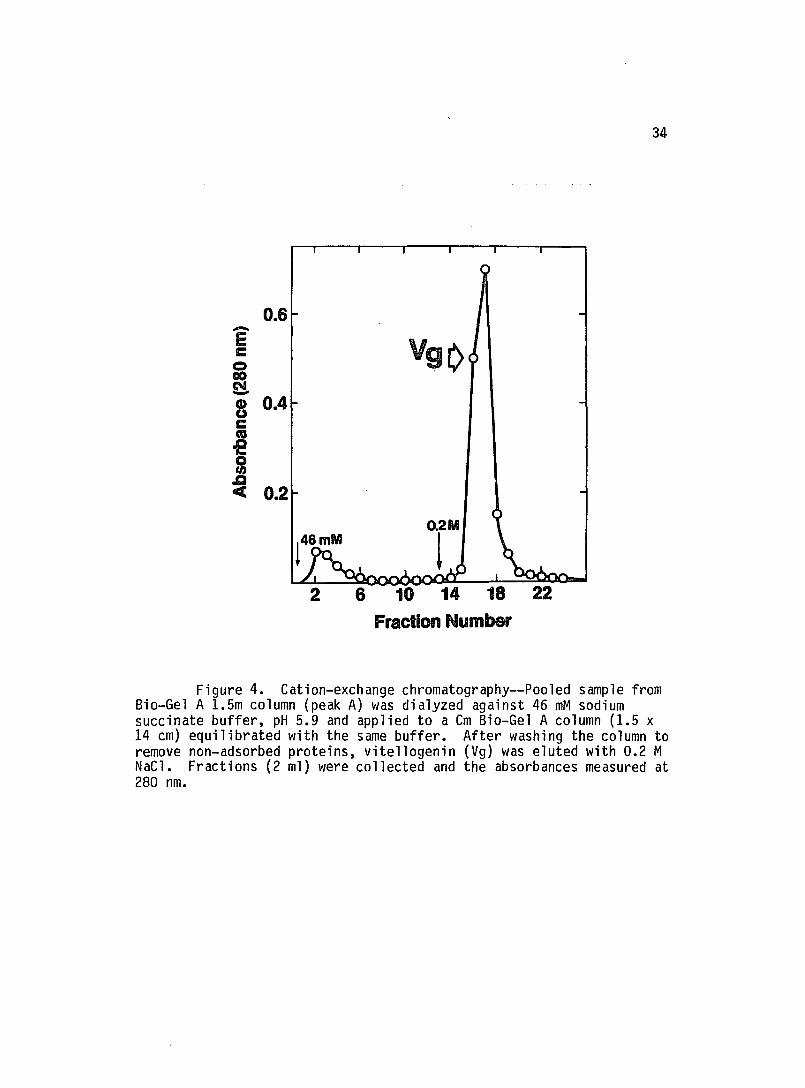
Figure 4. Cation-exchange chromatography--Pooled sample from Bio-Gel A 1.5m column (peak A) was dialyzed against 46 mM sodium succinate buffer, pH 5.9 and applied to a Cm Bio-Gel A column (1.5 x 14 cm) equilibrated with the same buffer. After washing the column to remove non-adsorbed proteins, vitellogenin (Vg) was eluted with 0.2 M NaCl. Fractions (2 ml) were collected and the absorbances measured at 280 nm.

35
-4 5 6
--200.0K
--116.3 K -92.SK
--66.2K
-45.0K
K
K
Figure 5. Purification stages of vitellogenin-- Samples were separated by SOS-PAGE (4-15 percent). 1. Adult female hemolymph; 2. Sample after gel permeation; 3. Arylphorin; 4. Vitellogenin (- 15 ~g protein); 5. Low and 6. High molecular weight standards (Bio-Rad).

1 2
669K-
440
232K-
140
Figure 6. Polyacrylamide gel electrophoresis of vitellogenin--Isolated vitellogenin was separated by native PAGE (4-20 percent) for 24 h at a constant 140 V (4°C) 1. Molecular weight standards (Pharmacia) (see Materials and Methods); 2. Vitellogenin (- 40 IJg).

~
'0 X
70
50
t- 30 :I: (!)
\, VG
~ o
W 0 3: a: « ..J :::> u w ..J o 10 ::E
o \
5~----~----~----~~----~~ o 0.2 0.4 0.6 0.8 RELATIVE MOBILITY
37
Figure 7. Molecular weight determination of vitellogenin--The gel was standardized using proteins of known molecular weights (Fig. 6).

Table 1. Composition of ~. sexta Vitellogenin
Component
Proteins
Carbohydrates
Lipids
Phospholipids
Neutral lipids
Phospholipids
Diacylglycerol
Cholesterol
Triacylglycerol
Free fatty acids
Hydrocarbons
Percent (by weight)a
Percent
84.5
3.0b
12.5
7.2
5.3
(total
62.6
15.8
3.7
6.9
4.3
1.1
lipids) a
(a) Data represents average of two determinations (maximum range 2
percent).
(b) Determined by gas liquid chromatography as described under
Materials and Methods.
38

value compares favorably with the molecular weight of ~. cecropia
vitellogenin (Pan and Wallace, 1974), a protein which has many similar
characteristics with ~. sexta vitellogenin, including identical
chromatographic properties on gel permeation columns.
Isolated vitellogenin was also analyzed for lipid and
carbohydrate compositions and the results are presented in Table 1.
The molecule contains 85 percent protein, 13 percent lipids and 3
percent carbohydrates. The principal lipid components in vitellogenin
are phospholipids (63 percent of total lipids) and diacylglycerol (16
percent of total lipids). The other lipid components were present
only in small amounts. These observations are comparable to the
results obtained with other vitellogenins (Peled and Tietz, 1975,
Chino et !l., 1977).
Limited trypsin digestion
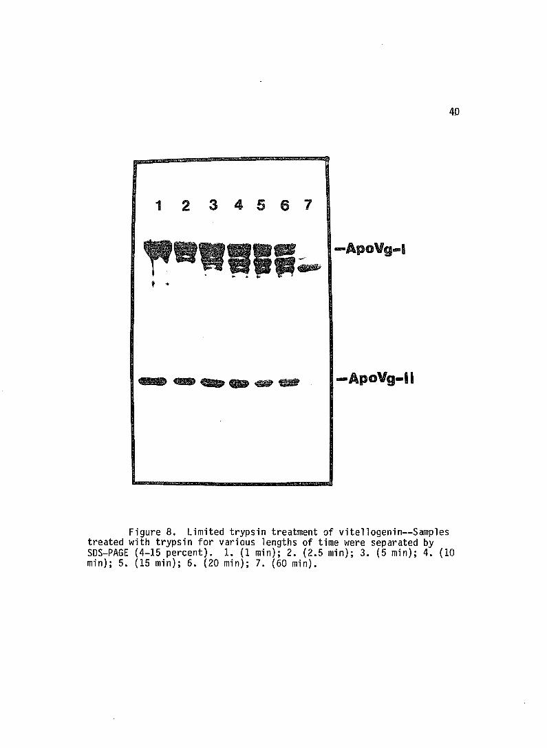
Limited trypsin digestion of vitellogenin showed that
apoVg-II is not accessible to the enzyme during the first 10 min of
incubation (Fig. 8, lane 4). But as apoVg-I was progressively
digested, apoVg-II became accessible to the enzyme (lanes 4-7). FITC
Con A staining of the gel showed that the bands appearing below
apoVg-II are degradation products of apoVg-I since they were also
glycosylated.
39
40
1234567
, .
-ApoVg-11
Figure 8. Limited trypsin treatment of vitellogenin--Samples treated with trypsin for various lengths of time were separated by SDS-PAGE (4-15 percent). 1. (1 min); 2. (2.5 min); 3. (5 min); 4. (10 min); 5. (15 min); 6. (20 min); 7. (60 min).

Isolation and properties of apoproteins
Vitellogenin was dissociated using 6 M guanidine Hel and the
apoproteins separated by gel permeation chromatography on 5epharose
CL-6B column (Fig. 9). After dialysis against deionized water,
apoVg-I precipitated while apoVg-II remained in solution. Purity of
the apoproteins was verified by 50S-PAGE (Fig. 10). The isolated
apoproteins (apoVg-I and apoVg-II) were analyzed for amino acid
composition and the results are presented (Table 2). The larger
apoprotein (apoVg-I) contained 10 mol percent of proline compared to
only 4 mol percent for apoVg-II. Tryptophan, glutamate and cysteine
contents also showed some differences. By comparing the amino acid
compositions, apoVg-II contained more basic residues than apoVg-I.
Earlier work reported the presence of one large (Mr 180,000) and one small (M 45,000) apoprotein and a native molecular
r
weight of Mr 260,000 (Mundall and Law, 1979). However, the presence
of two large apoproteins (apoVg-I a, I b) has been reported recently
(Imboden and Law, 1983). Although two or more protein bands have
occasionally been observed in this study, lack of reproducibility
suggested that these extra bands may arise from proteolytic breakdown
of apoVg-I. Furthermore, the extra apoVg-I was not seen on freshly
prepared protein samples. It was noted that the degradation of most
of the hemolymph proteins could be minimized if the insects were
injected with OFP prior to bleeding. On the other hand, apoVg-II is
very stable and appears on 50S-PAGE as a single protein band (Mr 45,000 = 5,000). This molecular weight value was confirmed by gel
41

42
-E 0.4 C 0 CO N ---• 0.2 u c CO .c ... 0 0.1 en .c C
o 62 74 86 98 Elution Volume (ml)
Figure 9. Isolation of vitellogenin apoproteins-Vitellogenin was dissociated using 6 M guanidine HC1, 50 mM sodium phosphate, pH 7.0 and apoproteins separated on 5epharose CL-6B (1.5 x 91 em). Fractions (2 ml) were collected and the absorbances measured at 280 nm. The apoproteins were identified by 50S-PAGE: A = apoVg-I; B = apoVg-II. Vt = 180 ml; Va = 50 ml.

4.3
345
-200.0K
-116.3K -92.5K
-66.2K
-45.0K
-31.0K
-21.5K
-14.4 K
Figure 10. SOS-polyacrylamide gel electrophoresis of apoproteins--Samples were separated by SOS-PAGE (4-15 percent). 1. Isolated vitellogenin (15 ~g); 2. ApoVg-II (10 ~g); 3. ApoVg-II (13 ~g); 4. Low molecular weight standards; 5. High molecular weight standards. The gel was stained with Coomassie Brilliant Blue.

Table 2. Amino acid composition of vitellogenin apoproteins
Amino Acid ApoVg-I ApoVg-II Mole percent Residues/mol Mole percent Residues/mol
Aspartatea Threonine Serine
Glutamatea Proline Glycine
Alanine Valine Methionine
Isoleucine Leucine Tyrosine
Phenylalanine Histidine Lysine
Arginine Cysteineb Tryptophanc
Total
9.94 3.76 7.68
17.22 9.95 5.17
8.21 5.75 2.09
4.24 5.56 3.94
2.58 4.03 6.86
2.47 0.27 0.27
99.99
(a) Includes acid + amide
160 60
124
277 160 83
132 93 34
68 89 63
42 65
110
40 4 4
1608
10.35 4.39 7.69
12.99 4.37 6.24
8.96 6.45 2.35
4.33 6.13 4.49
2.97 4.87 8.13
3.86 0.90 0.43
99.90
42 18 31
53 18 26
37 26 10
18 25 18
12 20 33
16 4 2
408
(b) Determined as cysteic acid after performic acid oxidation
(Hirs, 1967)
(c) Determined directly after hydrolysis with 3 N
mercaptoethanesulfonic acid (Penke et ~., 1974).
44

permeation HPLC. The specific absorbances for apoVg-I and apoVg-II at
280 nm in 6 M guanidine HCl were 0.422 mg-1ml-1 and 0.624
-1 1-1 mg m , respectively.
The relationship between apoprotein concentrations and
staining intensity was established by densitometric scanning. The
results showed no significant difference in the staining behavior of
the apoproteins. The ratio apoVg-I/apoVg-II was then determined by
the scanning procedure to be 3.6:1.0 by weight or a molar ratio of
approximately 0.90:1.0. The minimum molecular weight of intact
vitellogenin assuming only one apoVg-I and apoVg-II per molecule is
approximately 266,000. Since the actual molecular weight is Mr -
500,000, each vitellogenin molecule contains two copies of each
apoprotein.
Immunological properties of the apoproteins
Immunoblotting experiments showed that rabbit antisera
against apoVg-I and apoVg-II were specific for each apoprotein and
antiserum against intact vitellogenin reacted only with apoVg-I (Fig.
11, lanes 3-8). These antisera also reacted similarly with the
apoproteins of vitellin. In double radial immunodiffusion experiments
(Fig. 12), anti-apoVg-I reacted with intact vitellogenin (B) while
anti-apoVg-II did not (C).
Antisera against intact vitellogenin, apoVg-I and apoVg-II
were used to examine immunological similarities between ~. sexta and
H. cecropia vitellogenins. H. cecropia vitellogenin has two kinds of
45

200K-
116·3 92.5
46
ApoV9_11
Figure 11. Immunological reactions of vitellogenin apoproteins--Adult female hemolymph samples (lanes 3, 5, 7) ; vitellogenin (lanes 4, 6, 8) and high molecular weight standards (Bio-Rad) (lane 1) were separated by SOS-PAGE (4-15 percent). Samples in lanes 3-8 were then transferred to nitrocellulose paper for 24 h at 16°C. Following the transfer, samples in lanes 3 and 4 were incubated with antibodies against intact vitellogenin; lanes 5 and 6 with anti-apoVg-I and lanes 7 and 8 with anti-apoVg-II. After several washing steps, the blots were then incubated with 125I-Staphylococcus aureus protein A and the bands located by autoradiography. Vitellogenin (lane 2) and molecular weight standards (lane 1) were stained with Coomassie Brilliant Blue. Approximately 1 ~g protein per lane was used in lanes 3-8 and 15 ~g in lane 2.

47
A
Figure 12. Double radial immunodiffusion--The center wells in A, Band Chad 10 ~l each of anti-Vg, anti-apoVg-I and anti-apoVg-II, respectively. Peripheral wells had: (A) 3. Male M. sexta hemolymph (1:10 dil., 10 Ill); 4. Female hemolymph (1:10 dil:, IOi;T); 5. M. sexta vitellogenin (- 10 ~g); (B) 1. Female hemolymph (1:10 dil~, 10 ~2. M. sexta vitellogenin (- 10 ~g); 3. Male M. sexta hemolymph (10-fold-dilution, 10 ~l). (C) 1. Female~. sexta-hemolymph (1:10 dil., 10 ~l); 2. M. sexta vitellogenin (- 10 ~ Diffusion was carried out for 24 h-ar-room temperature. The plates were then dried and stained with Coomassie Brilliant Blue as described (Materials and Methods).

apoproteins, with molecular weights of Mr 180,000 and Mr 47,000,
respectively (Harnish and White, 1982). Antiserum against~. sexta
vitellogenin cross-reacted only with the larger apoprotein of ~.
cecropia vitellogenin. Antisera against apoVg-I and apoVg-II also
cross-reacted specifically with the large and small apoproteins of ~.
cecropia vitellogenin, respectively.
Synthesis of vitellogenin in vitro
In order to define the site of synthesis of vitellogenin,
the fat body tissue was incubated with [35S]methionine in vitro.
The incubation medium as well as the fat body tissue were then
analyzed by SDS-PAGE and autoradiography. As shown in Figure 13
(lanes 1-4), vitellogenin apoproteins (represented by I and II) were
labeled both in the fat body homogenate and the incubation medium.
Identification of the apoproteins as those of vitellogenin was made by
immunoprecipitation with anti-vitellogenin (Fig. 13, lanes 2 and 4).
Labeled apoproteins were not detected in follicles that had been
incubated with [35S]methionine (lane 5).
Phosphorylation
Following injection of [32P]-inorganic phosphate into one
day old adult insects, hemolymph was collected and vitellogenin
isolated. Vitellin was also isolated from eggs dissected from the
same insects. The apoproteins were analyzed by SDS-PAGE and
autoradiography. As shown (Fig. 14), apoVg-I and apoVg-II of both
48

49
-200K
1-
OK
Figure 13. Synthesis of vitellogenin in vitro--The synthesis products were separated by SDS-PAGE, transferred onto nitrocellulose paper and the labeled proteins visualized by autoradiography as described under Materials and Methods. 1. Fat body homogenate; 2. Immunoprecipitate of fat body homogenate; 3. Secreted protein; 4. Immunoprecipitate of secreted protein; 5. Follicle homogenate; 6. [14C]methylated protein standards (Amersham). I = apovitellogeini I ; II = apovitellogenin I; arrow represents microvitellogenin.

5,0
1 2
ApoV9-1
-ApoVg-11
Figure 14. Phosphorylation of vitellogenin and vitellin apoproteins--Samples were separated by SDS-PAGE (3-8 percent) and transferred onto nitrocellulose paper as described (Materials and Methods). The bands were located by autoradiography at _70De. 1. Vitellogenin (- 20 ~g); 2. Egg homogenate (- 50 ~g).

vitellogenin and vitellin contain labeled phosphate. Approximately 6
percent of the 32p label in both vitellogenin and vitellin was
51
removed by delipidation. Furthermore, vitellogenin apoproteins were
found to be the only proteins of whole hemolymph that were
significantly phosphorylated. Apoproteins prepared by gel permeation
chromatography in guanidine HCl were used for measurement of
phosphorus contents. There were no significant differences in the
phosphorus contents of vitellogenin and vitellin apoproteins (Table
3). It was also determined whether some of the phosphate groups on
apoVg-I were located on the carbohydrate moiety. The results showed
that deglycosylation of apoVg-I with endo-H removed 5 percent of the
phosphate label suggesting that the carbohydrate moiety may be partly
phosphorylated. However, the possibility of contaminating
phosphatases in the endo-H preparation could not be ruled out since
only a small amount of 32p label was removed. Vitellogenin was
briefly hydrolyzed with 6 M HCl at 110u C and the hydrolyzates
separated by 2 dimensional .TLC (Fig. 15). Identification of the
phosphorylated amino acid as serine was corroborated by comigration of
the phosphate label with authentic phosphoserine. No phosphate label
was found in either phosphotyrosine or phosphothreonine. Similar
results were also obtained by exhaustive pronase digestion of .
vitellogenin instead of acid hydrolysis showing that the occurrence of
Ser-P is not an artifact of acid hydrolysis.
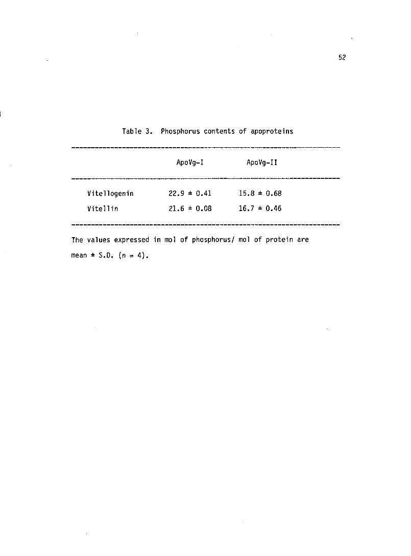
Table 3. Phosphorus contents of apoproteins
Vitellogenin
Vitell in
ApoVg-I
22.9 :!: 0.41
21.6 :!: 0.08
ApoVg-II
15.8 :!: 0.68
16.7 :!: 0.46
The values expressed in mol of phosphorusl mol of protein are
mean:!: S.D. (n = 4).
52

53
o B
Figure 15. 2-dimensional TLC of phosphoamino acids-Phosphoamino acids were separated in the first (A) and second (8) dimensions by TLC as described under Materials and Methods. Amino acids were located using ninhydrin. 1. Phospho-D-serine; 2. Phospho-D-threonine; 3. Tyrosine

54
Carbohydrate composition
The carbohydrate composition of isolated vitellogenin as
determined by gas liquid chromatography consisted mainly of Man and
GlcNAc in addition to trace amounts of glucose. The ratio Man: GlcNAc
was calculated as 11.6:2.0. This result is consistent with the
previously determined values (Mundall and Law, 1979). From the
composition data, it was calculated that each large apoprotein
(apoVg-I) contains 4 Man9GlcNAc2 chains.
Location of the carbohydrate moiety
After separation of the apoproteins by SDS-PAGE, only
apoVg-I reacted with both FITC-Con A (Fig. 16, lane 3) and PAS
(results not shown). FITC-Con A binding was eliminated after
deglycosylation of vitellogenin using endo-H (Fig. 16, lane 4). There
was no detectable difference in the electrophoretic mobility between
endo-H and non endo-H treated samples. Vitellogenin was also labeled
~ vivo to very high specific activity using [3HJMan. Analysis of
this sample by SDS-PAGE followed by autoradiography showed that all
the labeled Man present in vitellogenin was incorporated into apoVg-I
thus confirming that only this apoprotein is glycosylated (results not
shown). ~. cecropia vitellogenin also has an endo-H sensitive
carbohydrate moiety located on the larger apoprotein.

200K-
116.3K-92.5
66.2
55
ApoV9_1
~ApoV9-11
Figure 16. FITC Con A staining of vitellogenin-- Samples were separated by SOS-PAGE (4-15 percent) 1. Molecular weight standards (Bio-Rad); 2 and 3. Isolated vitellogenin; 4. Endo-H treated vitellogenin. After electrophoresis, samples in lanes 1 and 2 were stained with Coomassie Brilliant Blue and samples in 3 and 4 with FITC-Con A as described (Materials and Methods).

Gel permeation chromatography of pronase glycopeptides
Isolated vitellogenin was first digested with pronase and
then chromatographed on a Bio-Gel P-6 gel permeation column. The
digest contained only one significant peak of carbohydrate as judged
by the phenol-sulfuric acid assay (Fig. 17 A). A small amount of
undigested material eluted in the void volume. Fractions of the major
glycopeptide peak were pooled and lyophilized for analysis by high
field NMR. The amounts of carbohydrate in the minor peaks were
insufficient to permit analysis by NMR spectroscopy. After the
initial spectrum was taken, the sample was digested with endo-H and
rechromatographed over the same Bio-Gel P-6 column. The results
showed that the entire glycopeptide was sensitive to the enzyme (Fig.
17 B). The digestion resulted in a shift of the peak by about
thirteen fractions in the elution profile. In addition, a shoulder
(arrow, Fig. 17 B) was partially resolved eluting with a slightly
higher apparent molecular weight. This peak is roughly equivalent to
the amounts quantitated for the unassigned signals in the NMR spectrum
(see spectrum, page 60). The endo-H treated peak was isolated as two
fractions (I and II, Fig. 17 B) in order to evaluate the contents of
the minor peak. Both fractions were subsequently analyzed by high
field NMR (see spectrum, page 66).
Lectin binding properties
[3H]Man-vitellogenin pronase glycopeptides were bound to
Con A-Sepharose column and eluted using 0.5 M a-methylmannoside (Fig.
56

.57
A 21
E 1.8 c 0 00 1.5 q-Q)
1.2 u c 0 .0 0 0.9 If) .0 <t 0.6
0.3
0 O.B
0.7
~ 0.6
0 ~ 0.5
~ 0.4 c 0 .0 S 0.3 If) .0 <t 0.2
0.1
0
30 40 50 60 70 80 90 Fraction number
Figure 17. Bio-Gel P-6 profile of vitellogenin pronase glycopeptides--(A): Pronase digest of vitellogenin was applied to the column. Elution was carried out using 0.2 M ammonium bicarbonate buffer, pH 8.3 and fractions (2.5 ml) collected. Aliquots (0.4 ml) from each fraction were assayed for carbohydrate content by the phenol-sulfuric acid method and the absorbances measured at 480 nm. The major peak (51-60) was isolated and analyzed by NMR spectroscopy. (B): Elution profile of the same glycopeptide on Bio-Gel P-6 column after endo-H treatment. The column was eluted as in (A). Two fractions I (65-69) and II (70-76) were analyzed by NMR spectroscopy.

18 B). Conversely, the glycopeptides did not bind to a lentil
lectin-Sepharose column (Fig. 18 A). Con A has been shown to bind
mainly high Man oligosaccharides in addition to some biantennary
complex and hybrid-type Asn-linked oligosaccharides. In contrast,
lentil lectin does not bind high Man oligosaccharides (Kornfeld et
~., 1981).
Proton NMR spectroscopy of glycopeptides
The 250 MHz NMR spectrum of vitellogenin glycopeptide,
recorded in D?O at 300 u K, is presented (Fig. 19). Definitive ~
features are shown (Table 4). Examination of the anomeric region of
the spectrum revealed the presence of a relatively homogeneous high
Man class glycopeptide. The spectrum is consistent with 9 Man and 2
GlcNAc residues (Fig. 20; see also Fig. 21). The chemical shifts of
the CI-H protons of GlcNAc 1 and 2 together with the CI-H and C2-H
protons of Man units 3, 4 and 5 indicate the presence of the core
pentasaccharide Man 3 a(1~4) G1CNac a(I~N)Asn substituted at C-3 and
C-6 of Man 3 by a -linked Man units 4 and 5 (Vliegenthart et ~.,
1981, 1983).
In addition to the a-anomeric signals, there are 8 a-linked
CI-H proton signals belonging to Man units. Three of these protons at
6 ~ 5.05 ppm are assigned to 3 terminal a (1~2)-linked Man residues
located at the terminus of three branches in the glycan structure.
The existence of these units is confirmed by the presence of the a
(1~3)-linked core Man chemical shift at 6 = 5.337 ppm, the a
58

59
A
15
10
a-ms1hyI~
~ 5 ! G-O ,. 0
020 )(
'l: e.. IS
to
S
! 0
0 2 4 6 8 to 12 Fraction number
14 16 18 20
Figure 18. Profiles of labeled glycopeptides on lentil lectin and concanavalin-A columns--[3H]Man-vitellogenin pronase glycopeptides were chromatographed on lentil lectin-Sepharose (A) and Con A-Sepharose (B) columns equilibrated with Tris buffered saline containing 0.15 M NaCl, 0.01 M Tris, pH 8.0, 1 mM CaC12 and 1 mM MgC12. Bound material was eluted with 0.5 M a-methyl mannoside and fractions (2.5 ml) were collected. Radioactivity was determined using 500 ~l aliquots from each fraction.

5.0 4.0 r d PPM
3.0 2.0
Figure 19. 250 MHz proton NMR spectrum of the major glycopeptide--The major identifiable reporter groups present in the H-1 and H-2 regions as well as the N-acetyl regions are designated by numbers as corresponding to known resonance peaks reported for previously defined structures. The numbers represent the monosaccharide units shown (Fig. 20).

Table 4. IH Chemical shifts of structural reporter groups
of constituent monosaccharides for the carbohydrate moiety
Monosacchari~e
Reporter groups
Unit
No. a
Chemical
Shifts (ppm)b
Anomeric
Linkage
61
--------------------------------------------------------------------
CI-H 1 5.088 B CI-H 2 4.610 B NAc-CH3 1 2.007 B NAc-CH3 2 2.064 B
CI-H 3 4.773c B CI-H 4 4.875c a CI-H 5 5.337 a
CI-H 6 5.145 a CI-H 7 5.408 a .CI-H 8 5.312 a
CI-H 9 5.052 a CI-H 10 5.052 a CI-H 11 5.060 a
C2-H 3 4.223 B C2-H 4 4.158 a C2-H 5 4.083 a
C2-H 6 4.025 a C2-H 7 4.110 a C2-H 8 4.110 a
C2-H 9 4.074 a C2-H 10 4.068 a C2-H 11 4.063 a
--------------------------------------------------------------------( a) Refer to Fig. 20.
(b) Data obtained at 250 MHz, T = 300oK.
(c) Partially obscured by HOD at 300o K.

9 6
Mana(1+2)Mana(1+6)
"4 Mana( 1+6)
11 7 / \3 2 1 Mana(1+2)Mana(1+3) ManB(1+4)GlcNAcB(1+4)GlcNAcB(1+N)Asn
Mana(1+2)Mana(1+2)Mana(1+3)~ 10 8 5
Figure 20. The major structure identified from the NMR spectrum--Reporter groups identified as structural unit 1-11 were obtained from evaluation of the spectrum shown (Fig. 19).
62

Gte
Gt: '.,1 Gic ,,,' Man Man IVbn ~.2 F',2 'cr-I,2
Man Man Man
:--tfa;:.········~n-f .. • ; '''',I / i 'Ma,('"c-", ! GIc~(4) ~ , : GlcNAc : ............ ~ .. ~ ................ .
·O-P=O m · 9 .
·O-p=O • o •
63
Figure 21. Schematic diagram of the lipid-linked intermediate--The diagram shows the structure that is transferred to an asparagine residue in the sequence: Asn--X--Serine or Threonine.

(l~3)-linked arm Man at 0 = 5.408 ppm, and the internal a (l~6)-linked
arm Man at 0 = 5.146 ppm (units 5, 7 and 6, respectively) all of which
reflect resonances of a (l~2)-linked substituents (Vliegenthart et
~., 1981; 1983). In a similar manner unit 4 is assigned to the
resonance peak at 0 = 4.875 which indicates a substitution on this
residue at C-3 and C-6 by unit 7 and 6, respectively (Cohen et ~.,
1980; van Halbeek et ~., 1981). The final H-1 residue located at 0 =
5.312 ppm is assigned to the internal a (1~2)-linked Man (unit 8)
located on the a (1~3) arm and is present in equimolar amounts to all
other H-1 resonances (Dorland et ~., 1981). The assignment of the
reporter groups found in the H-1 region follow very closely to those
elements observed in the H-2 region. In particular, the presence of
the B (1~4) linked core Man proton signal at 0 = 4.233 ppm confirms
the presence of the H-1 proton which is partially obscured by HOD at
300 u K. Integration of this peak yields an area almost identical with
the molar yields found from evaluation of other single unit protons.
A total of 8 a-linked Man protons can be accounted for in this
region. The combined expression of chemical shift values from these
reporter groups given (Table 4) produce a glycopeptide structure that
is almost identical to that reported for soybean agglutinin (Dorland
et ~., 1981).
Close examination of the spectrum showed the existence of
two sets of resonance peaks which could not be assigned to the above
mentioned structure. The resonances located at 0 = 5.263, 5.248, and
o = 4.430 to 4.490 are not observed in structures of this type.
64

Quantitation by peak integration indicated that these unassigned
resonances contribute a signal equal to about 5-10 percent of the
total signals contributed by the other single unit protons. In order
to find the cause of these unusual peaks, the original sample was
treated with endo-H and reanalyzed by NMR. A partial spectrum of the
region 6 = 5.0-5.5 is presented (Fig 22). Fraction I which was
isolated as the shoulder from Bio-Gel P-6 chromatography (fig. 17 B)
contained a significant enrichment of the unassigned resonance seen at
6 = 5.255 and 5.257 (Fig. 22 A). On the other hand, very little of
this signal is present in the main glycopeptide fraction II (Fig. 22
B). The other resonance peaks observed in the region of 6 = 4.4-4.5
ppm were present at approximately equal amounts in both glycopeptide
fractions and thus appear to be independent of the peaks observed in
the H-1 region.
65

66
A
B
5.5 5.0 4.5 4.0 8 ppm
Figure 22. NMR spectrum of endo-H treated glycopeptide--250 MHz proton NMR spectroscopy of vitellogenin oligosaccharides isolated from Bio-Gel P-6 gel permeation after endo-H treatment. The arrows indicate the presence of unidentifiable resonance peaks located in the H-l and H-2 regions of the spectrum 0 ~ 4.0 to 5.5 ppm. The presence of additional doublet peaks at 0 = 5.22 ppm not observed in the original spectrum are due to the removal of the single Asn-linked GlcNAc cleaved by the enzyme.

67
Discussion
Physical and chemical properties
As in the case of other Lepidoptera, ~. sexta vitellogenin
is composed of two apoproteins, namely apoVg-I (Mr 180,000) and
apoVg-II (Mr 45,000). The protein falls into group 1 vitellogenins
proposed by Harnish and White (1982). This group is distinguished by
having two distinct apoprotein sizes (large and ~mall). Also
classified under this group are the orthopterans, ephemeropterans, and
dermapterans. The numbers of large and small apoproteins vary in the
different orders of insects in this group. Lepidopteran vitellogenins
differ in this regard from a variety of other insects. For example,
1. migratoria vitellogenin is composed of about eight separate
polypeptides with molecular weights in the range Mr 52,000-140,000
(Chen et ~., 1978) while Q. melanogaster vitellogenin (group 3) has
only three closely related small polypeptides (Mr 44,000, 45,000,
46,000) (Warren and Mahowald, 1979).
Using isolated~. sexta vitellogenin apoproteins, a complete
amino acid analyis was carried out (Table 2). As in other insect
proteins in general, both vitellogenin apoproteins contained
relatively high percentages of aspartate and glutamate. The lower
specific absorbance of apoVg-I at 280 nm compared to apoVg-II can be
accounted for by the differences in tryptophan content of the
apoproteins (0.27 mol percent in apoVg-I compared to 0.43 mol percent

in apoVg-II). However, the amino acid composition of the apoproteins
did not differ significantly from the average values for other
proteins in general (Dayhoff, 1973).
Synthesis of vitellogenin by the fat body
68
Vitellogenin was shown to be synthesized and secreted by the
fat body ~ vitro. This tissue is the main site of vitellogenin
synthesis in most insects so far examined (Hagedorn and Kunkel,
1979). Exceptions to this are found in Q. melanogaster (Bownes, 1980)
and Glossina austeni (Heubner et ~., 1975) where vitellogenin
synthesis also occurs within the ovary. In addition to vitellogenin,
the fat body of adult females synthesizes many other other proteins.
Notable among these-is-the newly discovered female specific protein,
microvitellogenin (Mr = 31,000) (Fig. 12, arrow). A parallel
experiment with follicles showed that no radioactivity was
incorporated either into the apoproteins of vitellogeni~ or
microvitellogenin. This indicated that in~. sexta, the follicles do
not synthesize these proteins.
Phosphorylation
Both apoVg-I and apoVg-II are phosphoproteins as shown by
methyl green staining (result not shown) and in vivo labeling with
32p. The phosphate was found to be attached only to serine
residues. The location of phosphate groups on the polypeptide
backbone and their functions are questions that need to be addressed.

69
Since apoVg-I is also glycosylated, the possibility that some of the
phosphate groups on this apoprotein may be located on the carbohydrate
moiety was examined. The results showed that although most of the
phosphate groups on apoVg-I are on the protein, a small amount was
probably associated with the carbohydrate moiety. The phosphorus
contents of ~. sexta vitellogenin (0.6 percent) is considerably lower
than that reported for B. americana vitellogenin (1.33 percent)
(Pereira and Bianchi, 1983) but higher than in h. migratoria vitellin
(0.3 percent) (Chen et ~., 1978) or ~ maderae vitellin (0.14
percent) (Engelmann and Friedel, 1974).
Immunological properties
Rabbit antisera against isolated vitellogenin apoproteins
were used to show that apoVg-I and apoVg-II are distinct polypeptides
since no immunological cross-reactivity was observed. Immunodiffusion
studies using the same antisera provided some information about the
arrangement of the apoproteins in the intact molecule. The fact that
antiserum against vitellogenin and apoVg-I formed precipitin lines
with vitellogenin while anti-apoVg-II did not, suggested that apoVg-II
is not accessible to the aqueous environment in the intact molecule.
Indeed, limited trypsinization of vitellogenin showed that apoVg-II
was initially not accessible to the enzyme (Fig. 8). However, as more
of apoVg-I was broken down, apoVg-II was degraded. The structural
organization of ~. sexta vitellogenin and lipophorin, the major lipid

70
transport protein, show some interesting parallels. As in
vitellogenin, lipophorin apoproteins (apoLp-I and apoLp-II) are
immunologically distinct from each other (Shapiro et ~., 1984) and
only the larger apoprotein (apoLp-I) is readily degraded by limited
trypsin digestion (Pattnaik et ~., 1979). More studies are needed to
find out if this arrangement of the apoproteins is common to all
insect lipoproteins. The similarities between vitellogenins of ~.
sexta and ~. cecropia were examined by the immunoblotting technique.
Antisera to~. sexta vitellogenin and its apoproteins showed the same
binding specificities in ~. cecropia vitellogenin as in~. sexta
vitellogenin. It is evident from these results that vitellogenins of
H. cecropia and~. sexta have similar structural features.
Carbohydrate moiety
This work has confirmed a previous report on the composition
of the carbohydrate moiety of ~ sexta (Mundall and Law, 1979).
FITC-Con A and PAS staining was used to show that the carbohydrate
mciety is located only on apoVg-I. Furthermore, all the [3HJMan in
vitellogenin was found on apoVg-I. The ability of endo-H to remove
the carbohydrate moiety provided evidence for presence of the
chitobiosyl-Asn linkage (Yamashita et ~., 1978). Since all the
carbohydrate moiety was completely removed by endo-H, it is unlikely
that any O-glycosidic linked carbohydrate structures exist in~. sexta
vitellogenin.

71
This study reports the first complete primary structure of
the carbohydrate moiety of an insect vitellogenin using high field
proton NMR spectroscopy (see also Osir et ~., 1986). The structure
is essentially identical in all respects to that of N-glycosidically
linked Man9GlcNAc2 oligosaccharide found in soybean agglutinin
glycopeptide (Dorland, 1981) and in other mammalian glycoproteins such
as porcine thyroglobulin (van Halbeek et ~., 1980 a, b). The
existence of the same structure in insect glycoproteins suggests that
the method for synthesizing N-glycosidically linked oligosaccharides
occurs in a general manner throughout the plant and animal kingdoms.
The mechanism by which glycosylation occurs in insects appears to be
similar and involves the transfer of the carbohydrate unit,
(Glu)3(Man)9(GlcNAc)2' from a lipid intermediate to an Asn in
the polypeptide backbone followed by a rapid removal of the terminal
glucose residues on the a (1~3) arm (Montreuil et al., 1980) (Fig.
21). This assumption is supported by the discovery of the lipid
intermediate, dolichol phosphate, in the microsomes of the
mediterranean fruit fly, yeratitis capitata (Allue, 1980). The
existence of two sets of unrecognizable chemical shifts in minor
amounts suggests the presence of a small amount of heterogeneity in
the structure. This finding contrasts with results reported in a
recent paper on cockroach vitellins (Nordin et ~., 1984). It was
observed that the carbohydrate moieties of these vitellins showed a
great deal of heterogeneity indicating extensive processing. In this
dissertation, the minor peaks shown in the Bio-Gel P-6 profile of

72
pronase glycopeptides (Fig. 17 A) were not analyzed since their
carbohydrate contents were very small. The primary structure
presented is the Ilmain structure" present. The minor peaks which were
not analyzed may well represent some processed structures. It is also
likely that some variations exist among different insects. Until the
carbohydrate structures of vitellogenins from many insects are
determined, attempts to make broad generalizations on the different
structures that may exist should be avoided.
The minor chemical shifts in the area of 0 = 4.4 to 4.5 ppm
are as yet unidentified. The second set of unidentifiable shifts at 0
= 5.263 and 5.248 are enriched in the higher molecular subfraction of
the glycopeptide. This subfraction is only slightly resolved from the
bulk of the major structure. It is present after release from the
peptide portion by treatment with endo-H and is thus unlikely to be a
result of interaction with polypeptide or other environmental
effects. The presence of additional sugars such as glucose is
unlikely because the difference in molecular weight would be expected
to cause a more prominent shift in in the Bio-Gel P-6 column profile.
Another possibility is the presence of phosphate which would account
for the slight difference in molecular weight. A phosphate group
would not in itself have a proton resonance signature but could cause
an environmental effect on other protons of contributing sugars.
Additional modification studies will be required to find the cause of
these unusual peaks.

CHAPTER 3
STUDIES ON BINDING AND UPTAKE OF VITELLOGENIN.
Introduction
The major component of yolk in insect eggs is called
vitellin. In most insects groups, vitellin is derived from a
hemolymph precursor, vitellogenin (Engelmann, 1979). However, in a
few cases, the ovary itself has the capacity to synthesize
vitellogenin. For example, in Q. melanogaster and other related
species, follicle cells supplement vitellogenin made by the fat body
(Postlewait et ~., 1980). In another dipteran, Glossina austeni,
yolk is presumably assembled primarily from follicle cell products
(Heubner et ~., 1975). In addition to vitellin, yolk contains other
extra-ovarian proteins. An important example is the famale specific
protein, microvitellogenin (Kawooya and Law, 1983; Telfer and
Kulakosky, 1984). Like vitellogenin, microvitellogenin is synthesized
by the fat body, secreted into hemolymph and then taken up by the
follicles. The hemolymph contains many other proteins that are not
concentrated by the ovaries. For instance, the major hemolymph
lipoprotein, lipophorin, is found only in trace amounts within the
yolk (Chino et ~., 1981; Kawooya, Osir and Law, unpublished
observation). From the examples given above, it is apparent that
insect eggs have a specific mechanism(s) for selectively extracting
73

74
proteins from the hemolymph. A likely candidate for such a mechanism
is a receptor-mediated endocytotic process. The discovery of coated
pits in mosquito oocytes by Roth and Porter (1964) was the first
indication that vite1logenin uptake is indeed a receptor mediated
process. Since this discovery, other endocytotic processes have been
extensively investigated (Goldstein et ~., 1979 a; Pastan and
Willingham, 1981), but the insect ovary has been largely neglected as
an appropriate object for investigations at the molecular level. The
insect ovary is more complicated than simple systems like the
fibroblast LDL system. Eggs develop in long strands or ovario1es with
the oldest at the posterior end. In Lepidoptera, each egg is
surrounded by a mass of follicle cells, which ultimately secrete the
chorion, and nurse cells, which provide some proteins and RNA (Koch
and King, 1966). A single follicle, at mid-development, has a
monolayer of mixed follicle and nurse cells surrounding it
Wigglesworth, 1972). It is generally believed that the follicle cells
separate to reveal the oocyte membrane, in which specific receptors
bind vitellogenin from the hemolymph (Telfer et ~., 1981). The very
active endocytotic process necessarily entraps small amounts of other
hemolymph or foreign proteins which are thereby coincidentally taken
into the oocyte (Telfer, 1960).
Vitellogenin uptake may be conveniently divided into three
intricately interrelated processes. The first step involves specific
recognition and binding of vitellogenin to putative receptors on the
oocyte surface. This is then followed by the actual endocytosis.

The transfer of vitellogenin, now called vitellin, into yolk granules
might constitute the final process. Although some of the processes
mentioned above have been examined independently, the overall picture
of what happens during vitellogenin uptake cannot become clear until
all the information becomes available.
75
Isolated labeled proteins have been used as physiological
probes for studying the uptake process in vitro (Anderson, 1971;
Ferenz et ~., 1981; Kunkel and Pan, 1976; Lange and Loughton, 1980;
Osir and Law, 1984; Rohrkasten and Ferenz, 1985 and Telfer and
Kulakosky, 1984). However, no serious attempts have so far been made
to use labeled vitellogenin as a ligand for identifying and
characterizing specific binding sites on the egg membranes. In this
chapter, an in vitro system for the uptake of 125I_labeled
vitellogenin into isolated follicles is described. Furthermore, a
membrane binding assay for identifying specific binding sites on
follicle membranes is presented. It is proposed that these binding
sites are responsible for specifically recognizing vitellogenin in the
hemolymph during uptake.

76
Materials and Methods
Isolation of vitellin
Eggs were dissected from freshly eclosed insects and
homogenized in lepidopteran saline containing 50 mM glutathione and 20
mM of DFP. The egg homogenate was centrifuged (14,000 x g, 10 min) in
a microcentrifuge (model 235 A, Fisher) in order to remove egg
membranes. The homogenate was then dialyzed against 0.08 M sodium
phosphate buffer, pH 7.0 and applied to a DEAE Bio-Gel A column (1.5 x
12 cm) equilibrated with the same buffer. After washing with two
column volumes to remove non-adsorbed proteins, vitellin was eluted
with a continuous NaCl gradient (0 to 0.2 M) using a gradient maker
(BRL). Fractions (2.5 ml) were collected and the absorbance monitored
at 280 nm. Fractions that contained purified vitellin were
concentrated by ultrafiltration using Amicon YM 10 membranes (Mr cut
off 10,000) and stored under nitrogen at 4°C. Freshly prep'ared
samples were used for all experiments.
Deglycosylation
Vitellogenin (100 ~g) was incubated with endo-H (42 unit/ml)
in endo-H buffer containing 10 mM of DFP for 30 min at room
temperature as described in chapter 2. After incubation, the sample
was chromatographed on Con A Sepharose column in order to separate the
deglycosylated protein from un reacted protein. Complete

deglycosylation was confirmed by FITC Con A binding as described in
chapter 2.
Iodination of proteins
77
Vitellogenin, deglycosylated vitellogenin and vitellin were
radioiodinated using Iodobeads (Pierce) as described by Markwell
(1982). Approximately 50 ~g of each protein in PBS was mixed with 0.3
mCi of Na[125 I] (Amersham, specific activity = 15 ~Ci/~g) and
incubated with three Iodobeads for 15 min at room temperature. The
reactions were stopped by pipetting the ·sample away from the
Iodobeads. Free iodide was removed by passing the samples through
Bio-Gel P-6DG desalting columns (1 x 21 cm) equilibrated and eluted
with PBS. Prior to this, the column was washed with PBS containing 2
mg/ml BSA to reduce any binding of labeled proteins to the column.
The specific activity of labeled vitellogenin or vitellin prepared in
this way was approximately 5 x 10⁵ cpm/~g protein. These proteins
had the same chromatographic and electrophoretic properties as the
unlabeled samples. Lipid extraction by the method of Bligh and Dyer
(1959) showed that approximately 4 percent of the radioactivity in
either vitellogenin or vitellin was incorporated into lipids.
Incubation of follicles
Ovarioles containing developing follicles were carefully
dissected from 1 day old female insects in lepidopteran saline. Under
a dissecting microscope the muscular sheath covering the follicles was

78
carefully removed and the lengths of individual follicles measured
using an ocular micrometer. Undamaged follicles (- 0.8 mm in length)
were transferred to plastic pipette tips sealed at one end. The
incubation media had 200 pl of lepidopteran saline containing 2 mg/ml
BSA and either vitellogenin or vitellin (specific activity - 2 x 10⁴
cpm/ pg). After incubation (27°C or 4°C), the follicles were washed
three times with 50 pl of lepidopteran saline. Radioactivity
associated with the follicles was determined in a gamma counter (model
4000 counting system, Beckman).
Trypsin treatment
Follicles were incubated with labeled vitellogenin as
described above either at 4°C or 27°C. After incubation for 1 h, the
follicles were washed with saline and transferred into a medium
containing trypsin (1 mg/ml, Worthington). After further incubation
for 1 h (4°C or 27°C), the follicles were washed and radioactivity
determined. A control incubation with no trypsin was included.
Preparation of follicle membranes
Follicles of approximately 0.8 mm in length dissected from
one day old female insects were used as the source of follicle
membranes. The membrane complex used in this experiment consisted of
follicle basement membrane, oocyte membranes and follicular cells.
The follicles were first punctured to release yolk and membrane
complex was washed 3-4 times with ice cold lepidopteran saline to

remove any adhering yolk. The membranes were thoroughly homogenized
in a glass homogenizer and centrifuged (15,000 x g, 10 min, 4°C) in a
microcentrifuge (model 235 A, Fisher). To obtain a uniform suspension
the membrane pellet was resuspended in an ice cold buffer (20 mM MES ,
pH 8.0, 5 mg/ml BSA, for 30 min) and sonicated briefly (microprobe,
setting No.6, 8 sec.) on a sonifier cell disruptor (model 200,
Branson Sonic Power Co.) on ice. Only freshly prepared membranes were
used for the binding studies.
Binding of labeled vitellogenin to follicle membranes
Binding assays were carried out in polyethylene Eppendorf
tubes (1.5 ml). In order to reduce non-specific binding of labeled
protein to walls of the tubes, they were coated by soaking in
polyethylene glycol (1 percent, w/v). The reaction mixture contained
70 pl of the binding buffer (20 mM MES, pH 6.8, 5 mg/ml BSA, 5 mM
CaC1 2), approximately 20 pg of the homogenized follicle membrane
preparation (protein content measured by the Pierce BCA protein assay
reagent) and different amounts of l25I_labeled vitellogenin, as
indicated in the appropriate figure legends. Non-specific binding of
labeled vitellogenin to the membranes was determined by carrying out
binding assays in the presence of ISO-fold molar excess of unlabeled
vitellogenin. The specific activity of vitellogenin used in binding
studies was approximately 3 x 10⁵ cpm/ pg protein. After incubation
(70 min, 4°C), 300 pl of the binding buffer was added to the reaction
mixture and the tubes centrifuged (10,000 x g, 5 min, 4°C) in a
79

80
microcentrifuge (model 235 A, Fischer). The supernatant solutions
were carefully removed with a Pasteur pipette and the pellets washed
three times with the same buffer by recentrifugation as described
above. Preliminary experiments showed that little or no radioactivity
was released after three washing steps. The lower tip of the tubes
were sliced in half and placed in gamma counting vials. Radioactivity
was determined in a gamma counter. Since approximately 10 percent of
the radiolabeled protein was found to bind to Eppendorf tubes, blank
tubes were run exactly as in the experimental tubes except that
membranes were omitted. The counts in the blanks were subtracted from
the the total counts bound in each experimental tube.
For competition studies, excess unlabeled vitellin,
deglycosylated vitellogenin, apoVg-II, microvitellogenin (gift of Dr.
John Kawooya, Department Biochemistry, University of Arizona) and
lipophorin were each tested for their abilities to compete with
labeled vitellogenin for binding sites on the follicle membranes.

81
Results
Isolation of vitellin
Vitellin purification utilized a single step of
anion-exchange chromatography on OEAE-Bio-Gel A column. The elution
profile is shown in Figure 23. Vitellin (fraction II) was identified
on the basis of its similarities with vitellogenin on 50S-PAGE.
Purity of the samples was verified by 50S-PAGE (Fig. 24).
~ vitro uptake of labeled vitellogenin by isolated follicles
Each M. sexta ovariole contains more than 200 follicles of
varying lengths. The oldest follicle (- 1.0 mm in length at the time
of adult emergence, and reaching a maximum length of 1.9 mm when
mature) is found at the posterior end of the ovariole and the lengths
decrease towards the anterior tip (Nijhout and Riddiford, 1979).
Insect egg follicles must achieve a certain critical size and age
before becoming competent to incorporate vitellogenin ~ vitro (Telfer
and Anderson, 1968). On the other hand, they lose this ability once
they attain a larger size. Competent follicles were found by
investigating the abilities of follicles with varying lengths to
incorporate vitellogenin ~ vitro. The ability to incorporate labeled
vitellogenin varied with the follicle length (Table 5). The greatest
uptake was achieved with follicles between approximately 0.7 mm and
1.3 mm in length. Follicles less than 0.6 mm or larger than 1.6 mm

82
-E / c / 0 / ~ 0.8 / 0.08 - / Q)
, u / U c / c c / Z .0 0.4 V 0.04 I ~
0 / I (/)
..c I / IT <{ /
I /
0 16 32 48 64 80 96
Elution volume (m I)
Figure 23. Elution profile of vitellin from DEAE-Bio-Gel A column--Egg homogenate was dialyzed against 0.08 M sodium phosphate buffer, pH 7.0 and applied to DEAE-Bio-Gel column (1.5 x 12 cm) equilibrated with the same buffer. After washing the column to remove non-adsorbed proteins, vitellin was eluted using a continous gradient, 0.0-0.2 M NaCl (----). Samples containing vitellin (peak II) were pooled.

83
200K
I 45K
Figure 24. SOS-polyacrylamide gel of vitellin and vitellogenin--1. Vitellogenin (25 ~g), 2. Vitellin (30 ~g, ) and 3. High molecular weight standards (lane 3) (Bio-Rad) were separated by SOS-PAGE (4-15 percent). The gel was stained using Coomassie Brilliant Blue. I = apovitellogenin-I; II = apovitellogenin-II.

Table 5. Relationship between follicle length and vitellogenin
uptake in vitro
Follicle Length 125I_VG Taken Up
(mm) (~g protein follicle-1 h-1)
0.50 0.115 :I: 0.010
0.62 0.350 :I: 0.130
0.70 0.621 :I: 0.110
0.80 1.218 :I: 0.014
1.02 0.937 :I: 0.042
1.60 0.465 :I: 0.015
1. 70 0.258 :I: 0.014
Follicles (0.5-1.7 mm) were incubated·in lepidopteran saline (200
~l) containing 125I_VG (ISO ~g/ml, specific activity = 2 x 10⁴
cpm/~g) •. After incubation (I h, 27°C), the follicles were washed
and radioactivity determined using a gamma counter as described
(Materials and Methods). The values are mean :I: S.D (n = 4).
84

were less able to take up labeled vitellogenin. The uptake of labeled
vitellogenin was also examined as function of the incubation time. As
shown in Figure 25, uptake increased linearly during the entire
incubation period at 27°C (- 1.13 ~g/follicle/h). Under these
experimental conditions, uptake usually decreased after about 4 h.
The uptake of 125I_vitellin was found to be identical to that of
vitellogenin under the same experimental conditions.
In order to assess the possible role of the carbohydrate
moiety on uptake, the uptake of 125I_endo_H treated vitellogenin was
examined (Fig. 26). The results showed that there was no significant
difference in uptake between the endo-H treated and the control
sample, suggesting that the carbohydrate moiety is not required for
the incorporation of vitellogenin into isolated follicles.
Distribution of labeled vitellogenin in isolated follicles
The distribution of labeled vitellogenin between the follicle
membranes and yolk after incubation at 4°C and 27°C is shown in Table
6. Most of the labeled protein was localized on the follicle
membranes after incubation for 1 h at 4°C. On the other hand, the
yolk had most of the labeled protein at 27°C. Since most of the
labeled vitellogenin was on the follicle membranes at 4°C, the effect
of trypsin on membrane-bound vitellogenin was investigated. Trypsin
treatment reduced the radioactive protein associated with the
follicles at 4°C by 70 percent compared to a control incubation in
85

I I I • i I
2.S t .
2.4 t- o
Q)
"0 o 2.0~ . -, c
'Q) 1.6 . -0 "-Q.
1.2 ~ -0'1 ~
O.S ~
0.4 i i I I I I I I
0 30 60 90 120 150 ISO 210 Incubation time (min.)
Figure 25 •. Uptake of vitellogenin by isolated follicles-Pairs of follicles were incubated at 27°C with 125I-vitellogenin (- 150 ~g/ml, 2 x 10⁴ cpm/~g) At the end of each incubation time, the follicles were washed and radioactivity determined as described (Material and Methods). The data shown are mean % S.D (n = 4).
86

u.I .... o
2.4-
:::i -.... o ~ %' 1.6--w I-o ia: a.
" ~ 0.8-
I
r - 1
I
o
I
I 1
I 60
I
I 1
I
I
I
I
I 1.
I I
120
T I
T
1 T 1
I I
180
INCUBATION TI~jE(Min)
87
-
-
-
-
--
-
Figure 26. Uptake of endo-H (e) and non endo-H treated ((» vitellogenin by isolated follicles--Pairs of follicles were incubated with 125I-VG samples (- 75 ~g/ml) for up to 3 h at 27°C. After incubation, the follicles were washed and radioactivity determined in a gamma counter as described (Materials and Methods). Each data point represents mean % S.D (n = 4).

Table 6. Distribution of labeled vitellogenin in
isolated follicles
Temperature Radioactivity (cpm) Percent
(OC) Total Membranes Yolk
10,552
26,888
9,692
2,150
830
24,466
Membranes Yolk
92.1
7.9
7.8
90.0
Follicles were incubated with 125 I_VG for 1 h at 4°C or 27°C as
described in Table 5. After incubation, follicles were washed and
then punctured to release yolk contents. The follicle membranes
were washed to remove any adhering yolk. Radioactivity associated
with the follicle membranes and yolk including the washes were
separately determined as described (Materials and Methods). Data
are the averages of three incubations.
88

89
which trypsin was not added. On the other hand, most of the labeled
vitellogenin was taken up into the follicles at 27°C and therefore not
accessible to trypsin.
Characterization of specific binding to follicle membranes
Since vitellogenin is selectively incorporated by isolated
follicles i~ vitro, we used 125I_labeled vitellogenin to assay for
specific binding sites on the homogenized follicle membranes prepared
as described under Materials and Methods. Since receptor-mediated
binding is a dynamic process, an experiment was carried out to
determine the length of incubation time required for the binding
reaction to reach equilibrium. The results showed that specific
binding of 125I-vitellogenin to the follicle membranes reached
equilibrium after about 60 min of incubation at 4°C (Fig. 27) or 27°C
(results not shown). All subsequent incubations were carried out for
70 min, sufficient time for the binding reactions to reach
equilibrium. Incubations were carried out at 4°C because isolated
vitellogenin is more stable at this temperature.
Binding of increasing amounts of vitellogenin to a fixed
amount of follicle membrane preparation was studied at equilibrium.
The results showed that specific binding increased to a saturation
level (Fig. 28). In most experiments, non-specific binding was
between 17 and 25 percent of specific binding. These results
suggested the presence of specific saturable binding sites for
vitellogenin on the follicle membranes. Analysis of the binding data

-rf) b ->'!
::rE a.. u -""0 c: :::J o
.Q
C!)
> I -It)
N -
90
40
o~ ____ ~ __ ~~ __ ~ ____ ~ __ o 20 40 60 80
Time (min.)
Figure 27. Time course of vitellogenin binding to follicle membranes--Membrane preparation (- 20 ~g protein) was incubated with -30 ~g/ml of 125I-VG at 4°C from 0-70 min. After incubation, bound vitellogenin was determined as described (Materials and Methods). In this experiment, incubation was considered to end when centrifugation began. All values were corrected for non-specific binding to the incubation tubes. Each point is the average of two determinations.

0.3 -l c::J' ::l.
"0 0.2 c ~
.8 c.!)
>
.. cf"""
O---er-----o------------0 0 0 -0
_------0
20 30 40 50 VG Added (p.g/mt)
9.1
60
Figure 28. Concentration-dependent binding of vitellogenin to follicle membranes--A fixed amount of membranes (- 20 ug protein) was incubated with increasing amounts of 125I-VG in the presence or absence of excess unlabeled VG. After incubation (70 min, 4°C), bound VG was determined in duplicate aliqouts as described (Materials and Methods). Specific binding (4P) was calculated as the difference between total and non-specific binding (C». All values were also corrected for non-specific binding to the incubation tubes.

92
by the method of Scatchard (1949) yielded a linear plot indicating the
presence of only one class of binding sites (Fig. 29). The apparent
equilibrium dissociation constant (KO) as calculated from the slope
of the Scatchard plot (l/KO) was - 1.3 x 10⁻⁸ M. The maximum
amount of vitellogenin bound was deduced from the x-intercept of the
same plot to be 0.36 = 0.001 ~g/mg of membrane protein. Molar
concentrations were based on a native molecular weight of M 5 x r
105 for !:!. sexta vitellogenin (Chapter 2).
Binding of vitellogenin was found to be specific to the
follicle membranes since the fat body and gut tissue membranes did not
show any specific binding activity. The influence of pH on binding of
labeled vitellogenin to the follicle membrane preparation was examined
by varying the pH of the binding buffer from pH 3 to 8 with
Tris-Malate. The apparent pH optimum was found to between 6 and 7
(Fig. 30). The pH of !:!. sexta adult female hemolymph is approximately
7. As shown in Figure 31, binding to follicle membranes required
calcium. The amount of protein bound increased as the amount of
calcium in the incubation medium was increased from 0 to 6 mM.
Further increase in the cation did not result in any increase in the
amount of ligand bound. Under the same conditions, the influence of
magnesium on vitellogenin binding" was identical to that of calcium.
Competition studies
In order to determine the specificity of vitellogenin
binding, the abilities of various hemolymph proteins to compete with

-'0 -)C -
93
1
40 o~ __ ~~ __ ~~ __ ~~~~ o 10 20
Bound
Figure 29. Scatchard analysis of binding data--The ordinate represents amount of vitellogenin {VG} bound {~g/~g membrane protein} and the abscissa the amount of VG bound divided by VG free. The line was drawn by least squares analysis of the binding data. The slope {l/KO} = 6.41 ± 0.004 ~g/ml {correlation coefficient = -0.997}. The x-intercept (Bmax) = 0.36 ± 0.001 ~g/mg of membrane protein.

-g 20 ~ o
..Q .
(!)
> I -It)
'" ...
94
Figure 30. pH dependency of vitellogenin binding to follicle membranes--Binding of 125I-VG (30 ~g/ml) to a fixed amount of membranes (- 20 ~g protein) was measured under different buffer pH conditions. The buffer pH was adjusted using Tris-Malate and bound VG was determined as described (Materials and Methods). The data was corrected for non-specific binding to the incubation tubes. Each point represents the average of two determinations.

96
-It)
b ..... )C
:E 0... U -"C C :s 0
.Q
C!)
> I ~
It) N -
0 0 8
Figure 31. Calcium dependency of vitellogenin binding to follicle membranes--125 I-VG (30 ~g/ml) was added to membranes (- 20 ~g protein) prepared as described (Materials and Methods) except that calcium was omitted from all buffers. To different reaction mixtures, increasing amounts of calcium (0-6 mM) was added and incubations carried out for 70 min at 4°C. Bound VG was determined as described (Materials and Methods). The data was corrected for non-specific binding to the incubation tubes. Each point is the average of two determinations.

labeled vitellogenin for binding to follicle membrane binding sites
was examined. If these proteins bound to the same site as
vitellogenin, then competition would result in a decrease in the
labeled vitellogenin bound to follicle membranes. Lipophorin is the
96 .
.major insect hemolymph lipoprotein and a known constituent of insect
eggs (Chino et ~., 1977), while microvitellogenin is a 31,000 dalton
female-specific protein found in both hemolymph and eggs (Kawooya and
Law, 1983; Telfer and Kulakosky, 1984). The results showed that
binding could be significantly reduced by addition of unlabeled excess
vitellin but not by apoVg-II, lipophorin or microvitellogenin (Table
7). Binding was also inhibited by deglycosylated vitellogenin
suggesting that the carbohydrate moiety is not involved in the
interaction with the binding sites.
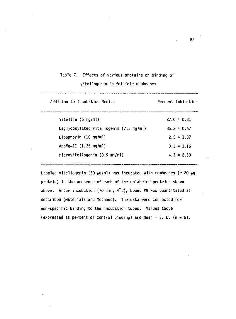
97
Table 7. Effects of various proteins on binding of
vitellogenin to follicle membranes
Addition t~ Incubation Medium
Vitellin (6 mg/ml)
Deglycosylated vitellogenin (7.5 mg/ml)
Lipophorin (10 mg/ml)
ApoVg-II (1.35 mg/ml)
Microvitellogenin (O.g mg/ml)
Percent Inhibition
87.0 : 0.31
85.3 = 0.67
2.5 = 1.37
3.1 = 1.16
4.3 = 2.60
Labeled vitellogenin (30 ~g/ml) was incubated with membranes (- 20 ~g
protein) in the presence of each of the unlabeled proteins shown
above. After incubation (70 min, 4°C), bound VG was quantitated as
described (Materials and Methods). The data were corrected for
non-specific binding to the incubation tubes. Values above
(expressed as percent of control binding) are mean: S. D. (n = 5).

98
Discussion
~ vitro uptake of vitellogenin
Incorporation of insect hemolymph proteins into eggs has been.
demonstrated in vivo (Kunkel and Pan, 1976; Ferenz, 1978) and ~ vitro
(Lange and Loughton, 1980; Ferenz et ~., 1981; Osir and Law, 1984;
Telfer and Kulakosky, 1984). Foreign egg proteins have been tested
for ~ vitro uptake and shown to be taken up by the host ovaries at
lower rates than those of host proteins ·(Kunkel and Pan, 1976).
Foreign non-egg proteins are also taken up at lower rates, probably by
non-receptor mediated endocytosis. In approaching the study of
vitellogenin uptake, we first established an ~ vitro system using
isolated follicles. Individual follicles were isolated by carefully
removing the muscular sheath around them. Since follicle size was
found to be an important factor in vitellogenin uptake, all
experiments were carried out using follicles of about 0.8 mm in
length. The large size of the follicles permitted gross dissection,
and thus enabled the assessment of the distribution of labeled
vitellogenin between the yolk and follicle membranes after incubation
at 4°C and 27°C. At 4°C, labeled vitellogenin was associated with the
follicle membranes. This observation is consistent with the fact
that, although ligands can bind to receptors at 4°C, only limited
internalization occurs (Goldstein et ~, 1979 a; Carpenter, 1979).
Furthermore, approximately 70 percent of labeled vitellogenin bound at

99
4°C was removed by trypsin treatment. On the other hand, most of
labeled vitellogenin was internalized at 27°C and therefore
inaccessible to trypsin. These results are in close agreement with
those reported for macrophages in which trypsin treatment removed two
thirds of 125I_lOl bound at 4°C (Goldstein et ~., 1979 b).
Binding to follicle membranes
Binding of 125I-vitellogenin to homogenized follicle
membrane preparation showed the presence of specific binding sites.
This binding not only exhibited high affinity and specificity for
labeled vitellogenin but was found to be dependent on calcium and pH.
The requirement for calcium makes the binding similar to that of the
lOl receptor in human fibroblasts (Goldstein and Brown, 1977) or the
storage protein receptor from Sarcophaga per'egrina (Ueno et ~.,
1983). It is still unclear how calcium is involved in the interaction
between vitellogenin and its binding sit~. The equilibrium
dissociation constant (KO) for vitellogenin-receptor complex was
estimated to be 1.3 x 10⁻⁸ M. The linear plot obtained by Scatchard
analysis indicated homogeneity of the follicle membrane binding
sites. The fact that only follicle membranes showed specific binding
suggested that only this tissue serves as the target tissue for
vitellogenin. Once synthesized and released by the fat body,
vitellogenin is not taken up again by the same tissue. This

100
observation is in contrast with ~. sexta storage protein (SP-2) which
is synthesized and sequestered by the fat body tissue (Ryan et ~.,
1984).
Competition for binding sites
Competition experiments with other proteins demonstrated that
the binding sites for vitel10genin are indeed very specific; only
vitellin and deglycosylated vitellogenin competed with labeled
vitellogenin for binding. This result is in accord with the fact that
vitellin is very similar to vitellogenin in ~ sexta and that labeled
vitellin is taken up by isolated follicle just as well as vitellogenin
in vitro. Two other ~. sexta hemolymph proteins, microvitellogenin
and lipophorin were found unable to compete with labeled vitellogenin
for binding sites, indicating that, if they are taken up by a receptor
mediated process, then they must have different binding sites from
those of vitellogenin. The ability of deglycosylated vitellogenin to
compete for binding indicated that the carbohydrate moiety is not
involved in the interaction with the hinding site. Indeed,
deglycosylated vitellogenin was taken up just as well as the untreated
protein. ApoVg-II did not compete with vitellogenin for binding.
ApoVg-I could not be tested in the competition studies since it was
insoluble under the conditions used in the binding studies. Attempts
to identify vitellogenin receptors by the ligand blotting technique
have been unsuccessful.

101 .
SUMMARY AND FUTURE PROJECTIONS
This dissertation has presented detailed physicochemical and
immunological studies on vitellogenin from the tobacco hornworm, ~.
sexta. The apoproteins of vitellogenin have been analyzed both for
carbohydrate and phosphorus contents. The apoproteins have been shown
to be immunologically distinct. Immunological techniques and limited
trypsin digestion gave some insight into the arrangement of the
apoproteins in the native molecule. The elucidation of the primary
structure of the oligosaccharide is an important step towards
understanding the role of this moiety in insect vitellogenins. I hope
that NMR spectroscopic analysis of oligosaccharides of other
vitellogeins or vitellins will provide a better understanding of how
synthesis and processing of the carbohydrate moiety occurs in
insects. Finding the exact role of the carbohydrate moiety will be an
interesting area for future research. This work has also described an
~ vitro assay for identifying specific binding sites for vitellogenin
on the follicle membranes. The results suggest that these sites
indeed meet the requirements for a receptor. This system is very
useful in trying to understand the structural basis for selective
transport of vitellogenin into the eggs. This aim might be achieved
by carrying out various chemical modifications of vitellogenin. An
interesting question is whether separate follicle receptors are
involved in transport of the different proteins that are localized in

102
the eggs. Future efforts will undoubtedly focus on the isolation and
characterization of the receptor for vitellogenin.

LITERATURE CITED
Allue, L. A. Q. (1980) Mol. Cellular Biochem. 33, 149-155.
Anderson, L. M. (1970) J. Microsc. ~, 721-738.
Anderson, L. M. (1971) J. Cell Sci. ~, 735-750.
Anderson, L. M. and Telfer, W. H. (1970) J. Embryol. Exp. Morphol. 23, 35-52.
Aswell, G. and Morell, A. G. (1974) Adv. Enymol. Relat. Areas Mol. Biol. 41, 99-105.
Atkinson, P. H., Grey, A. A., Carver, J. P., Hakimi, J., and Ceccarini, C. (1980) Biochemistry 20, 3979-3986.
Atlas, S. J., Roth, T. F. and Falcone, A. J. (1978) Insect Biochem. ~, 111-115.
Bartlett, G. R. (1959) J. Biol. Chem. 234, 466-468.
Bell, R. A., and Joachim, F. G. (1976) Ann. Entomol. Soc. 69, 365-373.
Bligh, E. G. and Dyer, W. J. (1959) Can. J. Biochem. Physiol. ~, 911-917.
Bownes, M. (1980) Differentiation ~, 109-116.
Borovsky, D. and Van Handel, E. (1980) Int. J. Invertebr. Reprod. ~, 153-164.
Brennan, M. D., Weiner, A. J., Goralski, T. J. and Mahowald. A. P. (1982) Dev. Biol. 89, 225-236.
Brennan, M. D. and Mahowald, A. P. (1982) Insect Biochem. 1£, 669-673.
Burnette, W. N. (1981) Anal. Biochem. 112, 195-203.
Carpenter, G. (1979) Ann. Rev. Biochem. 48, 193-216.
Chapman, R. F. (1969) in The insects: Structure and Function (1969), pp 403-424, American Elsevier Publishing Co. New York.
Chen, T. T., Strahlendorf, P. W., and Wyatt, G. R. (1978) J. Biol. Chem. 253, 5325-5331.
103

'104
Chino, H., Downer, R. G. H., and Takahashi, S. V. (1977) Biochim. Biophys.Acta 487, 508-516.
Chinzei, V., Chino, H. and Wyatt, G. R. (1981) Insect Biochem. 11, 1-7.
Cohen, R. E, and Ballou, C. E. (1980) Biochemistry~, 4345-4358.
Cutting, J. A. and Roth, T. F. (1973) Anal Biochem. 54, 386-394.
Dayhoff, M. O. (1973) in Atlas of Protein Sequence and Structure, 5, Supple 2, p 301, National Biomedical Research Foundation, Washington, DC.
Dittmer, J. C. and Wells, M. A. (1969) Methods Enzymol. li, 482-530.
Dorland, L., van Halbeek, H., Vliegenthart, J. F. G., Lis, H., and Sheron, N. (1981) J. Biol. Chern. 256, 7708-7711.
Dubois, M., Gilles, K. A., Hamilton, J. K., Rebers, P. A. and Smith, F. (1956) Anal. Biochem. 28, 350-352.
Engelmann, F. (1979) Adv. Insect Physiol. 14, 49-108.
Engelmann, F. and Friedel, T. (1974) Life Sciences 14, 587-594.
Ferenz, H. J. (1978) J. Insect Physiol. 24, 273-278.
Ferenz, H. J., Lubzens, E. W., and Glass, H. (1981) J. Insect Physiol. !J..., 869-875.
Fernando-Wanakulasuriya, G. J. P., Staggers, J. E., Frost, S. C., and Wells, M. A. (1981) J. Lipid Res. 22, 668-674.
Furlan, M., Perret, B. A., and Beck, E. A. (1979) Anal. Biochem. 96, 208-214.
Gilbert, L. I. Bollenbacher, W. E., Granger, N. A. (1980) Ann. Rev. Physiol. 42, 493-510.
Goldstein, J. L., Anderson, R. G. W., and Brown, M. S. (1979, a) Nature 279, 679-684.
Goldstein, J. L. and Brown, M. S. (1977) Ann. Rev. Biochem. 46, 897-930.
Goldstein, J. L., Ho, V. K., Basu, S. K. and Brown, M. S. (1979, b) Proc. Natl. Acad. Sci. USA~, 333-337.
Gottlieb, T. A. and Wallace, R. A. (1982) J. Biol. Chern. 257, 95-103.

Grimes, W. J. and Greegor, S. (1976) Cancer Res. 36, 3905-3910.
Hagedorn, H. H. and Kunkel, J. G. (1979) Ann. Rev. Entomol. 24, 475-505. -
Harnish, D. G. and White, B. N. (1982) J. Ex!:!. Zool. 220, 1-10.
Heubner, E., Tobe, S. S. and Davey, K. G. (1975) Cell, i, 535-558.
Hirs, C. H. W. (1967) Methods Enzymol. 11., 59-62.
Hirs, C. H. W. (1983) Methods Enzymo 1. 91, 59-62.
105
Hoofmann, J. D. and Lawson, F. R. and Yamamoto, R. T. (1966) in Insect Colonization and Mass production (Smith, C. N. ed), pp 479-486, Academic Press, New York.
Hubbard, S. C. and Ivatt, R. J. (1983) Ann. Rev. Biochem. 50, 555-583.
Imboden H. and Law, J. H. (1983) Insect Biochem. 11, 151-162.
Jowett, T. and Postlethwait, J. H. (1980) Dev. Biol. 80, 225-234.
Jungreis, A. M. Jatlow, P. and Wyatt, G. R. (1973) J. Insect Physiol. 19, 225-233.
Kapitany, R. A. and Zabrowski, E. J. (1973) Anal. Biochem. 56, 361-369
Kawooya J. K. and Law, J. H. (1983) Biochem. Bio!:!hys. Res. Commun. 11.7, 643-650.
Kawooya, J. K., Osir, E. o. and Law, J. H. (1986) (manuscript in preparation).
King, R. C. and Buning, J. in Com rehensive Insect Ph sio10 , Biochemistr~, and Pharmacology Kerkut, G. A., Gilbert, L. I., eds) pp 37- 2, Pergamon Press, London.
Koch, E. A. and King, R. C. (1966) J. Mor!:!hol. 119, 283-304.
Koeppe, J. and Of eng and , J. (1976) in Invertebrate Tissue Culture (Kurstak, E. and Maramorosch, K., eds) pp 185-94, Academic Press, New York.
Kornfeld, K., Reitman, M. L. and Kornfeld, R. (1981) J. Biol Chern. 256, 6633-6640.
Kramer, S. J., Munda11, E. C. and Law, J. H. (1980) Insect Biochem. 10, 279-288.

Laemmli, U. K. (1970) Nature (Lond.) 277, 680-685.
Lange, A. B. and Loughton, B. G. (1980) Experientia 37, 273-274.
Lowry, O. H., Rosenbrough, N. J., Farr, A. L. and Randall, R. J. (1951) J. Biol. Chern. 193, 265-275.
Markwell, M. A. K. (1982) Anal. Biochem. 125, 427-432.
and
Montreuil, J. (1980) Adv. Carbohydr. Chern. Biochem. 37, 157-223.
Mundall, E. C. and Law, J. H. (1979) Compo Biochem. Physiol. 63 B, 459-468.
Nierderwieser, A. (1972) Methods Enzymol. 25, 60-99.
Nijhout, M. M. and Riddiford, L. M. (1974) Biol. Bull. 146, 377-392.
Nijhout, M. M. and Riddiford, L. M. (1979) Int. J. Invert. Reprod. 1, 209-219.
Nordin, J. H., Gochoco, C. H., Wojchowski, D. M. and Kunkel, J. G. (1984) Compo Biochem. Physiol. 79 B, 379-390.
Offord, R. E. (1969) in Data for Biochemical Research (Dawson, R. M. C., Elliot, W. H. and Jones, K. M., eds) pp 562-527, Oxford University Press, Oxford.
Oie, M., Takahashi, S. Y., Ishizaki, H. (1975) Deve. Growth Diff. 1i, 237-246.
Osir, E. O. and Law, J. H. (1984) Fed. Proceedings 43, 2047.
Osir, E. 0., Wells, M. A. and Law, J. H. (1986) Arch. Insect Biochem. Physiol. (in Press).
Osir, E. 0., Anderson, D. R., Grimes, W. J. and Law, J. H. (1986) Insect Biochem. (in press).
Ouchterlony, o. (1968) in Handbook of Immunodiffusion and ImmunoelectrophoreSis, Ann Arbor publlshers, Ann. Arbor, Michigan.
106

Pan, M. L. (1971) J. Insect Physio1. 1I, 677-689.
Pan, M. L. and Wallace, R. A. (1974) Amer. Zool. 14, 1239-1242.
Pastan, I. H. and Willingham, M. C. (1981) Science, 214, 504-509.
Pattnaik, N. M., Mundall, E. C., Trambusti, B. G., Law, J. H., Kezdy, F. J. (1979) Compo Biochem. Physiol. 63 B, 469-476.
Peled, Y. and Tietz, A. (1975) Insect Biochem. ~, 61-72.
Penke, B., Ferenczi, R., and Kovacs, K. (1974) Anal. Biochem. 60, 45-50.
Pereira, S. D. and Bianchi, A. G. (1983) Insect Biochem. 11, 323-332.
Reinecke, J. P., Buckner, J. S. and Grugel, S. R. (1980) Bio1. Bull. 158, 129-140.
Riddiford, L. M. (1980) Ann. Rev. Biochem. 42, 511-528.
107
Riley, C. T., Barbeau, B. K., Keirn, P. S., Kezdy, F. J., Henriksen, R. L. and Law, J. H. (1984) J. Biol. Chern. 259, 13159-13165.
Rohrkasten, A. and Ferenz, H. J. (1985) Roux's Arch. Dev. Biol. 194, 411-416.
Ross, H. H. (1965) in Textbook of Entomology, pp 104-107, John Wiley and sons, Inc. New York.
Roth, T. F. and Porter, K. R. (1964) J. Cell Biol. 20, 313-332.
Ryan, R., Keirn, P. S., Wells, M. A. and Law, J. H. (1984) J. Biol. Chern. 260, 782-787.
Scatchard, G. (1949) Ann N. Y. Acad. Sci. El, 660-672.
Sefton, B. M., Hunter, T., Ball, E. H. and Singer, S. J. (1981) Cell 24, 156-174.
Shapiro, J. P., and Law, J.H. (1983) Biochem. Biophys. Res. Commun. 115, 924-931.
Shapiro, J. P. Keirn., P. S. and Law, J. H. (1984) J. Biol. Chern. 259, 3680-3685.
Steele, C. G. H. and Davey, K. G. (1985) in Com rehensive Insect PhrSi010gy and Biochemistr~ and Pharmaco ogy Ker ut, G. A., Gi bert, L. I., eds) pp 1- 5, Pergamon Press, London.

. Takahashi, S. Y. (1983) Int. J. Invert. Reprod. £, 65-76.
Tarentino, A. L., Trimble, R. B. and Maley, F. (1978) Methods Enzymol. 50, 574-580.
Telfer, W. H. (1954) J. Gen. Physiol. 37, 539-558.
Telfer, W. H. (1960) Biol. Bull (Woods Hole) 118, 338-351.
Telfer, W. H. and Anderson, L. M. (1968) Dev. Biol. 11, 512-535.
Telfer, W. H., Heubner, E. and Smith, D. S. (1982) in Insect Ultrastructure (King, R. C. and Akai, H., eds) pp 118-149, Plenum Press, London.
Telfer, W. H., Keirn, P. S. and Law, J. H. (1983) Insect Biochem. 11, 601-613.
Telfer, W. H. and Kulakosky, P. C. (1984) Adv. Invertebr. Reprod. 3 (Engels, W., ed) pp 81-86, Elsevier, Amsterdam.
Telfer, W. H., Rubenstein, E., and Pan, M. L. (1981) in Regulation of Insect Develo ment and Behavior (Sehnal, F., Zabza, A., Menn,
• an ym orows 1, B., e s pp 637-654, Wroclaw Technical University Press, Wroclaw.
Towbin, H., Staehelin, T. and Gordon, J. (1979) Proc. Natl. Acad. Sci. USA 76, 4350-4354.
Ueno, K., Ohsawa, F. and Natori, S (1983) J. Biol. Chern. 253, 12210-12214.
van Halbeek, H. Dorland, L., Veldink, G. A., Vliegenthart, J. F. G., Strecker, G., Michalski, J. G., Montreuil, J. and Hull, W. E. (1980, a) FEBS Lett. 121, 65-70.
van Halbeek, H., Dorland, L., Veldink, G. A., Vliegenthart, J. F. G., Strecker, G., Michalski, J. G., Montreuil, J. and Hull, W. E. (1980, b) FEBS Lett. 121, 71-77.
van Halbeek, H., Dorland, L., Vliegenthart, J. F. G., Spik, G., Cheron, A. and Montreuil, J. (1981) Biochim. Biophys. Acta 675, 293-296.
Vliegenthart, J. F, G., van Halbeek, H., and Dorland, L. (1981) Pure Appl. Chern. 53. 45-77.
Vliegenthart, J. F. G., Dorland, L. and van Halbeek, H. (1983) Adv. Carbohydr. Chern. Biochem. il, 209-374.
108
109
Wallace, R. A. and Jared, D. W. (1969) Dev. 8iol. 12, 498-526.
Warren, T. G. and Mahowald, A. P. (1979) Dev. Biol. 68, 130-139.
Wigglesworth, V. B. (1965) in Principles of Insect Physiology, pp 1-26, Chapman and Hill, london.
Wigglesworth, V. B. (1972) in The Life of Insects, pp 86-107, Redwood Press, Trowbridge, London.
Wyatt,
Yamashita, K., Tachibana, Y. and Kobata, A. (1978) J. Biol. Chern. 253, 3862-3869.