p={'t':'3', 'i':'670887841'}; d=''; var...
TRANSCRIPT

Te c h n i c a l B u l l e t i n
INTE
GR
ATE
DSO
LUT
ION
S world-c lassSERVICE& SUPPORT
INTE
GR
ATE
DSO
LUT
ION
S
use me withGLOMAX ®
INSTRUMENTS
CellTiter-Glo® LuminescentCell Viability AssayINSTRUCTIONS FOR USE OF PRODUCTS G7570, G7571, G7572 ANDG7573.
PRINTED IN USA.Revised 12/12 Part# TB288
tb288.1212:EIVD_TM.qxd 12/17/2012 4:41 PM Page 1
Promega Corporation · 2800 Woods Hollow Road · Madison, WI 53711-5399 USA Toll Free in USA 800-356-9526 · Phone 608-274-4330 · Fax 608-277-2516 · www.promega.comPrinted in USA. Part# TB288Revised 12/12 Page 1
1. Description ..........................................................................................................1
2. Product Components and Storage Conditions ............................................4
3. Performing the CellTiter-Glo® Assay ............................................................5A. Reagent Preparation.............................................................................................5B. Protocol for the Cell Viability Assay .................................................................6C. Protocol for Generating an ATP Standard Curve (optional).........................7
4. Appendix .............................................................................................................7A. Overview of the CellTiter-Glo® Assay ..............................................................7B. Additional Considerations..................................................................................8C. References ............................................................................................................11D. Related Products.................................................................................................12
1. Description
The CellTiter-Glo® Luminescent Cell Viability Assay(a–e) is a homogeneousmethod to determine the number of viable cells in culture based on quantitationof the ATP present, which signals the presence of metabolically active cells. TheCellTiter-Glo® Assay is designed for use with multiwell-plate formats, makingit ideal for automated high-throughput screening (HTS) and cell proliferationand cytotoxicity assays. The homogeneous assay procedure (Figure 1) involvesadding a single reagent (CellTiter-Glo® Reagent) directly to cells cultured inserum-supplemented medium. Cell washing, removal of medium or multiplepipetting steps are not required.
The homogeneous “add-mix-measure” format results in cell lysis and generationof a luminescent signal proportional to the amount of ATP present (Figure 2).The amount of ATP is directly proportional to the number of cells present inculture in agreement with previous reports (1). The CellTiter-Glo® Assay relies onthe properties of a proprietary thermostable luciferase (Ultra-Glo™ RecombinantLuciferase), which generates a stable “glow-type” luminescent signal andimproves performance across a wide range of assay conditions. The luciferasereaction for this assay is shown in Figure 3. The half-life of the luminescent signalresulting from this reaction is greater than five hours (Figure 4). This extendedhalf-life eliminates the need for reagent injectors and provides flexibility forcontinuous or batch-mode processing of multiple plates. The uniquehomogeneous format reduces pipetting errors that may be introduced duringthe multiple steps required by other ATP-measurement methods.
CellTiter-Glo® Luminescent Cell Viability Assay
All technical literature is available on the Internet at: www.promega.com/protocols/ Please visit the web site to verify that you are using the most current version of this
Technical Bulletin. Please contact Promega Technical Services if you have questions on useof this system. E-mail: [email protected]
tb288.1212:EIVD_TM.qxd 12/17/2012 4:41 PM Page 1

Figure 1. Flow diagram showing preparation and use of CellTiter-Glo® Reagent.
Promega Corporation · 2800 Woods Hollow Road · Madison, WI 53711-5399 USA Toll Free in USA 800-356-9526 · Phone 608-274-4330 · Fax 608-277-2516 · www.promega.comPart# TB288 Printed in USA.Page 2 Revised 12/12
3170MA12_0A
CellTiter-Glo®
Substrate
CellTiter-Glo®
Reagent
Mixer
Luminometer
CellTiter-Glo®
Buffer
tb288.1212:EIVD_TM.qxd 12/17/2012 4:41 PM Page 2
System Advantages
• Homogeneous: “Add-mix-measure” format reduces the number of plate-handling steps to fewer than that required for similar ATP assays.
• Fast: Data can be recorded 10 minutes after adding reagent.• Sensitive: Measures cells at numbers below the detection limits of
standard colorimetric and fluorometric assays.• Flexible: Can be used with various multiwell formats. Data can be
recorded by luminometer or CCD camera or imaging device.• Robust: Luminescent signal is very stable, with a half-life >5 hours,
depending on cell type and culture medium used.• Able to Multiplex: Can be used with reporter gene assays or other
cell-based assays from Promega (2,3).
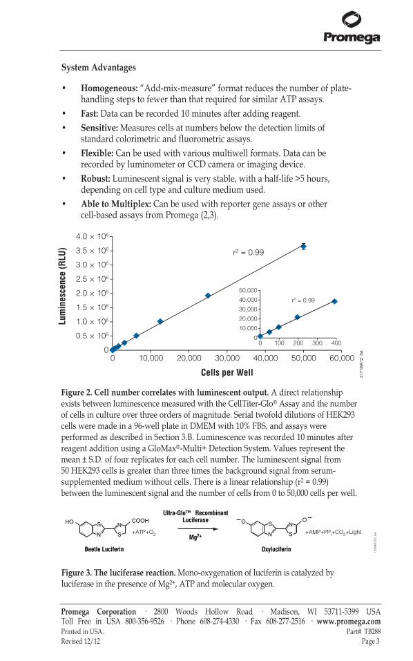
Figure 3. The luciferase reaction. Mono-oxygenation of luciferin is catalyzed byluciferase in the presence of Mg2+, ATP and molecular oxygen.
Promega Corporation · 2800 Woods Hollow Road · Madison, WI 53711-5399 USA Toll Free in USA 800-356-9526 · Phone 608-274-4330 · Fax 608-277-2516 · www.promega.comPrinted in USA. Part# TB288Revised 12/12 Page 3
3171MA12_0A
Lum
ines
cenc
e (R
LU)
Cells per Well
10,000 60,00020,000 30,000 40,000 50,0000
R² = 0.999
0.5 × 10⁶
1.0 × 10⁶
1.5 × 10⁶
2.0 × 10⁶
2.5 × 10⁶
3.0 × 10⁶
3.5 × 10⁶
4.0 × 10⁶
r² = 0.99
0
20,000
10,000
30,000
40,000
50,000
r² = 0.99
00 100 200 300 400
HO S
N S
NO S
N S
NOCOOH
+ATP+O2
Ultra-Glo™ RecombinantLuciferase
+AMP+PPi+CO2+Light
Beetle Luciferin Oxyluciferin
Mg2+
13
99
MD
03
_6A
Figure 2. Cell number correlates with luminescent output. A direct relationshipexists between luminescence measured with the CellTiter-Glo® Assay and the numberof cells in culture over three orders of magnitude. Serial twofold dilutions of HEK293cells were made in a 96-well plate in DMEM with 10% FBS, and assays wereperformed as described in Section 3.B. Luminescence was recorded 10 minutes afterreagent addition using a GloMax®-Multi+ Detection System. Values represent themean ± S.D. of four replicates for each cell number. The luminescent signal from50 HEK293 cells is greater than three times the background signal from serum-supplemented medium without cells. There is a linear relationship (r2 = 0.99)between the luminescent signal and the number of cells from 0 to 50,000 cells per well.
tb288.1212:EIVD_TM.qxd 12/17/2012 4:41 PM Page 3

Figure 4. Extended luminescent half-life allows high-throughput batchprocessing. Signal stability is shown for three common cell lines. HepG2 and BHK-21cells were grown and assayed in MEM containing 10% FBS, while CHO-K1 cellswere grown and assayed in DME/F-12 containing 10% FBS. CHO-K1, BHK-21 andHepG2 cells, at 25,000 cells per well, were added to a 96-well plate. After an equalvolume of CellTiter-Glo® Reagent was added, plates were shaken and luminescencemonitored over time with the plates held at 22°C. The half-lives of the luminescentsignals for the CHO-K1, BHK-21 and HepG2 cells were approximately 5.4, 5.2 and5.8 hours, respectively.
2. Product Components and Storage Conditions
Product Size Cat.#CellTiter-Glo® Luminescent Cell Viability Assay 10ml G7570Substrate is sufficient for 100 assays at 100µl/assay in 96-well plates or 400 assays at25µl/assay in 384-well plates. Includes:
• 1 × 10ml CellTiter-Glo® Buffer• 1 vial CellTiter-Glo® Substrate (lyophilized)
Product Size Cat.#CellTiter-Glo® Luminescent Cell Viability Assay 10 × 10ml G7571Each vial of substrate is sufficient for 100 assays at 100µl/assay in 96-well plates or400 assays at 25µl/assay in 384-well plates (1,000 to 4,000 total assays). Includes:
• 10 × 10ml CellTiter-Glo® Buffer• 10 vials CellTiter-Glo® Substrate (lyophilized)
Promega Corporation · 2800 Woods Hollow Road · Madison, WI 53711-5399 USA Toll Free in USA 800-356-9526 · Phone 608-274-4330 · Fax 608-277-2516 · www.promega.comPart# TB288 Printed in USA.Page 4 Revised 12/12
Rela
tive
Lum
ines
cenc
e (%
)
Time (minutes)
CHO-K1BHK-21HepG2
0102030405060708090
100
0 50 100 150 200 250 300
3173MA12_0A
tb288.1212:EIVD_TM.qxd 12/17/2012 4:41 PM Page 4

Product Size Cat.#CellTiter-Glo® Luminescent Cell Viability Assay 100ml G7572Substrate is sufficient for 1,000 assays at 100µl/assay in 96-well plates or 4,000assays at 25µl/assay in 384-well plates. Includes:
• 1 × 100ml CellTiter-Glo® Buffer• 1 vial CellTiter-Glo® Substrate (lyophilized)
Product Size Cat.#CellTiter-Glo® Luminescent Cell Viability Assay 10 × 100ml G7573Each vial of substrate is sufficient for 1,000 assays at 100µl/assay in 96-well plates or4,000 assays at 25µl/assay in 384-well plates (10,000 to 40,000 total assays). Includes:
•10 × 100ml CellTiter-Glo® Buffer• 10 vials CellTiter-Glo® Substrate (lyophilized)
Storage Conditions: For long-term storage, store the lyophilized CellTiter-Glo®
Substrate and CellTiter-Glo® Buffer at –20°C. For frequent use, the CellTiter-Glo®
Buffer can be stored at 4°C or room temperature for 48 hours without loss ofactivity. See product label for expiration date information. ReconstitutedCellTiter-Glo® Reagent (Buffer plus Substrate) can be stored at room temperaturefor up to 8 hours with <10% loss of activity, at 4°C for 48 hours with ~5% lossof activity, at 4°C for 4 days with ~20% loss of activity or at –20°C for 21 weekswith ~3% loss of activity. The reagent is stable for up to ten freeze-thaw cycles,with less than 10% loss of activity.
3. Performing the CellTiter-Glo® Assay
Materials to Be Supplied by the User• opaque-walled multiwell plates adequate for cell culture• multichannel pipette or automated pipetting station for reagent delivery• device (plate shaker) for mixing multiwell plates• luminometer, CCD camera or imaging device capable of reading multiwell plates• optional: ATP for use in generating a standard curve (Section 3.C)
3.A. Reagent Preparation
1. Thaw the CellTiter-Glo® Buffer, and equilibrate to room temperature priorto use. For convenience the CellTiter-Glo® Buffer may be thawed andstored at room temperature for up to 48 hours prior to use.
2. Equilibrate the lyophilized CellTiter-Glo® Substrate to room temperatureprior to use.
Promega Corporation · 2800 Woods Hollow Road · Madison, WI 53711-5399 USA Toll Free in USA 800-356-9526 · Phone 608-274-4330 · Fax 608-277-2516 · www.promega.comPrinted in USA. Part# TB288Revised 12/12 Page 5
tb288.1212:EIVD_TM.qxd 12/17/2012 4:41 PM Page 5

3.A. Reagent Preparation (continued)
3. Transfer the appropriate volume (10ml for Cat.# G7570 and G7571, or 100mlfor Cat.# G7572 and G7573) of CellTiter-Glo® Buffer into the amber bottlecontaining CellTiter-Glo® Substrate to reconstitute the lyophilizedenzyme/substrate mixture. This forms the CellTiter-Glo® Reagent.
4. Mix by gently vortexing, swirling or inverting the contents to obtain ahomogeneous solution. The CellTiter-Glo® Substrate should go intosolution easily in less than 1 minute.
3.B. Protocol for the Cell Viability Assay
We recommend that you perform a titration of your particular cells todetermine the optimal number and ensure that you are working within thelinear range of the CellTiter-Glo® Assay. Figure 2 provides an example of sucha titration of HEK293 cells using 0 to 50,000 cells per well in a 96-well format.
1. Prepare opaque-walled multiwell plates with mammalian cells in culturemedium, 100µl per well for 96-well plates or 25µl per well for 384-wellplates.Multiwell plates must be compatible with the luminometer used.
2. Prepare control wells containing medium without cells to obtain a value forbackground luminescence.
3. Add the test compound to experimental wells, and incubate according toculture protocol.
4. Equilibrate the plate and its contents at room temperature forapproximately 30 minutes.
5. Add a volume of CellTiter-Glo® Reagent equal to the volume of cell culturemedium present in each well (e.g., add 100µl of reagent to 100µl of mediumcontaining cells for a 96-well plate, or add 25µl of reagent to 25µl ofmedium containing cells for a 384-well plate).
6. Mix contents for 2 minutes on an orbital shaker to induce cell lysis.
7. Allow the plate to incubate at room temperature for 10 minutes to stabilizeluminescent signal.Note: Uneven luminescent signal within standard plates can be caused bytemperature gradients, uneven seeding of cells or edge effects in multiwellplates.
8. Record luminescence.Note: Instrument settings depend on the manufacturer. An integration timeof 0.25–1 second per well should serve as a guideline.
Promega Corporation · 2800 Woods Hollow Road · Madison, WI 53711-5399 USA Toll Free in USA 800-356-9526 · Phone 608-274-4330 · Fax 608-277-2516 · www.promega.comPart# TB288 Printed in USA.Page 6 Revised 12/12
!
tb288.1212:EIVD_TM.qxd 12/17/2012 4:41 PM Page 6
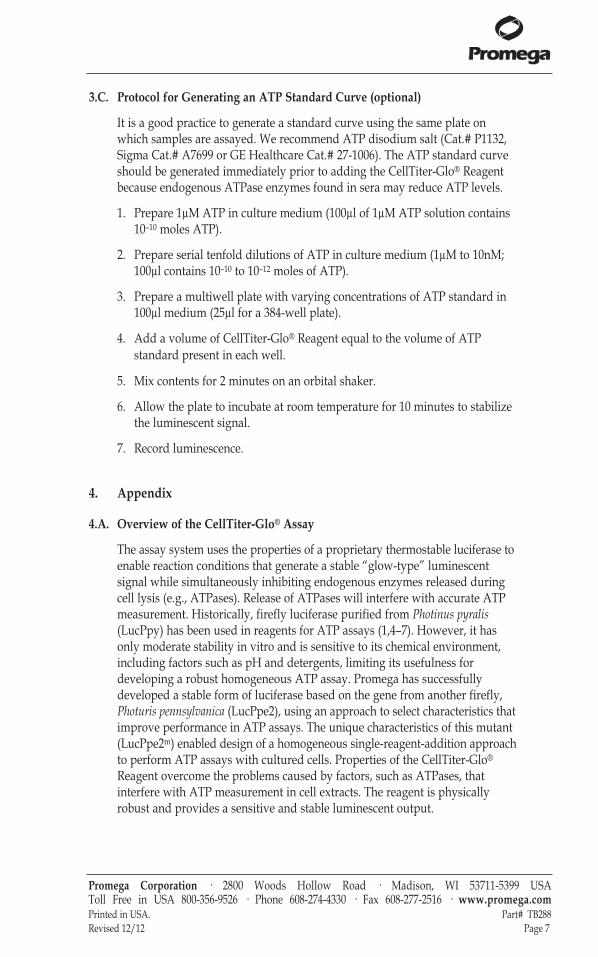
3.C. Protocol for Generating an ATP Standard Curve (optional)
It is a good practice to generate a standard curve using the same plate onwhich samples are assayed. We recommend ATP disodium salt (Cat.# P1132,Sigma Cat.# A7699 or GE Healthcare Cat.# 27-1006). The ATP standard curveshould be generated immediately prior to adding the CellTiter-Glo® Reagentbecause endogenous ATPase enzymes found in sera may reduce ATP levels.
1. Prepare 1µM ATP in culture medium (100µl of 1µM ATP solution contains10–10 moles ATP).
2. Prepare serial tenfold dilutions of ATP in culture medium (1µM to 10nM;100µl contains 10–10 to 10–12 moles of ATP).
3. Prepare a multiwell plate with varying concentrations of ATP standard in100µl medium (25µl for a 384-well plate).
4. Add a volume of CellTiter-Glo® Reagent equal to the volume of ATPstandard present in each well.
5. Mix contents for 2 minutes on an orbital shaker.
6. Allow the plate to incubate at room temperature for 10 minutes to stabilizethe luminescent signal.
7. Record luminescence.
4. Appendix
4.A. Overview of the CellTiter-Glo® Assay
The assay system uses the properties of a proprietary thermostable luciferase toenable reaction conditions that generate a stable “glow-type” luminescentsignal while simultaneously inhibiting endogenous enzymes released duringcell lysis (e.g., ATPases). Release of ATPases will interfere with accurate ATPmeasurement. Historically, firefly luciferase purified from Photinus pyralis(LucPpy) has been used in reagents for ATP assays (1,4–7). However, it hasonly moderate stability in vitro and is sensitive to its chemical environment,including factors such as pH and detergents, limiting its usefulness fordeveloping a robust homogeneous ATP assay. Promega has successfullydeveloped a stable form of luciferase based on the gene from another firefly,Photuris pennsylvanica (LucPpe2), using an approach to select characteristics thatimprove performance in ATP assays. The unique characteristics of this mutant(LucPpe2m) enabled design of a homogeneous single-reagent-addition approachto perform ATP assays with cultured cells. Properties of the CellTiter-Glo®
Reagent overcome the problems caused by factors, such as ATPases, thatinterfere with ATP measurement in cell extracts. The reagent is physicallyrobust and provides a sensitive and stable luminescent output.
Promega Corporation · 2800 Woods Hollow Road · Madison, WI 53711-5399 USA Toll Free in USA 800-356-9526 · Phone 608-274-4330 · Fax 608-277-2516 · www.promega.comPrinted in USA. Part# TB288Revised 12/12 Page 7
tb288.1212:EIVD_TM.qxd 12/17/2012 4:41 PM Page 7

4.A. Overview of the CellTiter-Glo® Assay (continued)
Sensitivity and Linearity: The ATP-based detection of cells is more sensitivethan other methods (8–10). In experiments performed by Promega scientists,the luminescent signal from 50 HEK293 cells is greater than three standarddeviations above the background signal from serum-supplemented mediumwithout cells. There is a linear relationship (r2 = 0.99) between the luminescentsignal and the number of cells from 0 to 50,000 cells per well in the 96-wellformat. The luminescence values in Figure 2 were recorded after 10 minutes ofincubation at room temperature to stabilize the luminescent signal as describedin Section 3.B. Incubation of the same 96-well plate used in the experimentshown in Figure 2 for 360 minutes at room temperature had little effect on therelationship between luminescent signal and number of cells (r2 = 0.99).
Speed: The homogeneous procedure to measure ATP using the CellTiter-Glo®
Assay is quicker than other ATP assay methods that require multiple steps toextract ATP and measure luminescence. The CellTiter-Glo® Assay also is fasterthan other commonly used methods to measure the number of viable cells(such as MTT, alamarBlue® or Calcein-AM) that require prolonged incubationsteps to enable the cells’ metabolic machinery to convert indicator moleculesinto a detectable signal.
4.B. Additional Considerations
Temperature: The intensity and decay rate of the luminescent signal from theCellTiter-Glo® Assay depends on the luciferase reaction rate. Environmentalfactors that affect the luciferase reaction rate will change the intensity andstability of the luminescent signal. Temperature is one factor that affects therate of this enzymatic assay and thus the light output. For consistent results,equilibrate assay plates to a constant temperature before performing the assay.Transferring eukaryotic cells from 37°C to room temperature has little effect onATP content (5). We have demonstrated that removing cultured cells from a37°C incubator and allowing them to equilibrate to 22°C for 1–2 hours hadlittle effect on ATP content. For batch-mode processing of multiple assayplates, take precautions to ensure complete temperature equilibration. Platesremoved from a 37°C incubator and placed in tall stacks at room temperaturewill require longer equilibration than plates arranged in a single layer.Insufficient equilibration may result in a temperature gradient effect betweenwells in the center and at the edge of the plates. The temperature gradientpattern also may depend on the position of the plate in the stack.
Promega Corporation · 2800 Woods Hollow Road · Madison, WI 53711-5399 USA Toll Free in USA 800-356-9526 · Phone 608-274-4330 · Fax 608-277-2516 · www.promega.comPart# TB288 Printed in USA.Page 8 Revised 12/12
tb288.1212:EIVD_TM.qxd 12/17/2012 4:41 PM Page 8
Chemicals: The chemical environment of the luciferase reaction affects theenzymatic rate and thus luminescence intensity. Differences in luminescenceintensity have been observed using different types of culture media and sera.The presence of phenol red in culture medium should have little impact onluminescence output. Assaying 0.1µM ATP in RPMI medium without phenolred resulted in ~5% increase in luminescence output (in relative light units[RLU]) compared to assays in RPMI containing the standard concentration ofphenol red, whereas assays in RPMI medium containing twice the normalconcentration of phenol red showed a ~2% decrease in luminescence.
Solvents for the various test compounds may interfere with the luciferasereaction and thus the light output from the assay. Interference with theluciferase reaction can be detected by assaying a parallel set of control wellscontaining medium without cells. Dimethylsulfoxide (DMSO), commonly usedas a vehicle to solubilize organic chemicals, has been tested at finalconcentrations of up to 2% in the assay and only minimally affects light output.
Plate Recommendations: We recommend using standard opaque-walledmultiwell plates suitable for luminescence measurements. Opaque-walledplates with clear bottoms to allow microscopic visualization of cells also maybe used; however, these plates will have diminished signal intensity andgreater cross talk between wells. Opaque white tape may be used to decreaseluminescence loss and cross talk.
Cellular ATP Content: Different cell types have different amounts of ATP,and values reported for the ATP level in cells vary considerably (1,4,11–13).Factors that affect the ATP content of cells may affect the relationship betweencell number and luminescence. Anchorage-dependent cells that undergocontact inhibition at high densities may show a change in ATP content per cellat high densities, resulting in a nonlinear relationship between cell numberand luminescence. Factors that affect the cytoplasmic volume or physiology ofcells also will affect ATP content. For example, oxygen depletion is one factorknown to cause a rapid decrease in ATP (1).
Promega Corporation · 2800 Woods Hollow Road · Madison, WI 53711-5399 USA Toll Free in USA 800-356-9526 · Phone 608-274-4330 · Fax 608-277-2516 · www.promega.comPrinted in USA. Part# TB288Revised 12/12 Page 9
tb288.1212:EIVD_TM.qxd 12/17/2012 4:41 PM Page 9

4.B. Additional Considerations (continued)
Mixing: Optimal assay performance is achieved when the CellTiter-Glo®
Reagent is mixed completely with the cultured cells. Suspension cell lines (e.g.,Jurkat cells) generally require less mixing to achieve lysis and extract ATP thanadherent cells (e.g., L929 cells). Tests were done to evaluate the effect ofshaking the plate after adding the CellTiter-Glo® Reagent. Suspension cellscultured in multiwell plates showed only minor differences in light outputwhether or not the plates were shaken after adding the CellTiter-Glo® Reagent.Adherent cells are more difficult to lyse and show a substantial differencebetween shaken and nonshaken plates.
Several additional parameters related to reagent mixing include the force ofdelivery of CellTiter-Glo® Reagent, sample volume and dimensions of the well.All of these factors may affect assay performance. The degree of reagent mixingrequired may be affected by the method used to add the CellTiter-Glo® Reagentto the assay plates. Automated pipetting devices using a greater or lesser forceof fluid delivery may affect the degree of subsequent mixing required.Complete reagent mixing in 96-well plates should be achieved using orbitalplate shaking devices built into many luminometers and the recommended 2-minute shaking time. Special electromagnetic shaking devices that use aradius smaller than the well diameter may be required to efficiently mixcontents of 384-well plates. The depth of medium and geometry of themultiwell plates may have an effect on mixing efficiency. We recommend thatyou take these factors into consideration when performing the assay andempirically determine whether a mixing step is necessary for the individualapplication.
Luminometers
For highly sensitive luminometric assays, the luminometer model and settingsgreatly affect the quality of data obtained. Luminometers from differentmanufacturers will vary in sensitivities and dynamic ranges. We recommendthe GloMax® products because these instruments do not require gainadjustments to achieve optimal sensitivity and dynamic range. Additionally,GloMax® instruments are preloaded with Promega protocols for ease of use.
If you are not using a GloMax® luminometer, consult the operating manual foryour luminometer to determine the optimal settings. The limits should beverified on each instrument before analysis of experimental samples. The assayshould be linear in some portion of the detection range of the instrument used.For an individual luminometer there may be different gain settings. Werecommend that you optimize the gain settings.
Promega Corporation · 2800 Woods Hollow Road · Madison, WI 53711-5399 USA Toll Free in USA 800-356-9526 · Phone 608-274-4330 · Fax 608-277-2516 · www.promega.comPart# TB288 Printed in USA.Page 10 Revised 12/12
tb288.1212:EIVD_TM.qxd 12/17/2012 4:41 PM Page 10

4.C. References
1. Crouch, S.P. et al. (1993) The use of ATP bioluminescence as a measure of cellproliferation and cytotoxicity. J. Immunol. Methods 160, 81–8.
2. Farfan, A. et al. (2004) Multiplexing homogeneous cell-based assays. Cell Notes 10, 2–5.
3. Riss, T., Moravec, R. and Niles, A. (2005) Selecting cell-based assays for drugdiscovery screening. Cell Notes 13, 16–21.
4. Kangas, L., Grönroos, M. and Nieminen, A.L. (1984) Bioluminescence of cellular ATP:A new method for evaluating cytotoxic agents in vitro. Med. Biol. 62, 338–43.
5. Lundin, A. et al. (1986) Estimation of biomass in growing cell lines by adenosinetriphosphate assay. Methods Enzymol. 133, 27–42.
6. Sevin, B.U. et al. (1988) Application of an ATP-bioluminescence assay in human tumorchemosensitivity testing. Gynecol. Oncol. 31, 191–204.
7. Gerhardt, R.T. et al. (1991) Characterization of in vitro chemosensitivity ofperioperative human ovarian malignancies by adenosine triphosphatechemosensitivity assay. Am. J. Obstet. Gynecol. 165, 245–55.
8. Petty, R.D. et al. (1995) Comparison of MTT and ATP-based assays for themeasurement of viable cell number. J. Biolumin. Chemilumin. 10, 29–34.
9. Cree, I.A. et al. (1995) Methotrexate chemosensitivity by ATP luminescence in humanleukemia cell lines and in breast cancer primary cultures: Comparison of the TCA-100assay with a clonogenic assay. AntiCancer Drugs 6, 398–404.
10. Maehara, Y. et al. (1987) The ATP assay is more sensitive than the succinatedehydrogenase inhibition test for predicting cell viability. Eur. J. Cancer Clin. Oncol. 23,273–6.
11. Stanley, P.E. (1986) Extraction of adenosine triphosphate from microbial and somaticcells. Methods Enzymol. 133, 14–22.
12. Beckers, B. et al. (1986) Application of intracellular ATP determination in lymphocytesfor HLA-typing. J. Biolumin. Chemilumin. 1, 47–51.
13. Andreotti, P.E. et al. (1995) Chemosensitivity testing of human tumors using amicroplate adenosine triphosphate luminescence assay: Clinical correlation forcisplatin resistance of ovarian carcinoma. Cancer Res. 55, 5276–82.
Promega Corporation · 2800 Woods Hollow Road · Madison, WI 53711-5399 USA Toll Free in USA 800-356-9526 · Phone 608-274-4330 · Fax 608-277-2516 · www.promega.comPrinted in USA. Part# TB288Revised 12/12 Page 11
tb288.1212:EIVD_TM.qxd 12/17/2012 4:41 PM Page 11

4.D. Related Products
Cell Proliferation Products
Product Size Cat.#ApoLive-Glo™ Multiplex Assay 10ml G6410ApoTox-Glo™ Triplex Assay 10ml G6320CellTiter-Fluor™ Cell Viability Assay (fluorescent) 10ml G6080CellTiter-Blue® Cell Viability Assay (resazurin) 20ml G8080CellTiter 96® AQueous One Solution Cell Proliferation Assay (MTS, colorimetric) 200 assays G3582CellTiter 96® AQueous Non-RadioactiveCell Proliferation Assay (MTS, colorimetric) 1,000 assays G5421CellTiter 96® AQueous MTS Reagent Powder 1g G1111CellTiter 96® Non-Radioactive Cell Proliferation Assay (MTT, colorimetric) 1,000 assays G4000 Additional sizes available.
Cytotoxicity Assays
Product Size Cat.#CytoTox-Glo™ Cytotoxicity Assay (luminescent)* 10ml G9290Mitochondrial ToxGlo™ Assay* 10ml G8000MultiTox-Glo Multiplex Cytotoxicity Assay (luminescent, fluorescent)* 10ml G9270MultiTox-Fluor Multiplex Cytotoxicity Assay(fluorescent)* 10ml G9200CytoTox-Fluor™ Cytotoxicity Assay (fluorescent)* 10ml G9260CytoTox-ONE™ Homogeneous MembraneIntegrity Assay (LDH, fluorometric)* 200–800 assays G7890CytoTox-ONE™ Homogeneous MembraneIntegrity Assay, HTP 1,000–4,000 assays G7892CytoTox 96® Non-Radioactive Cytotoxicity Assay 1,000 assays G1780(LDH, colorimetric)*GSH-Glo™ Glutathione Assay 10ml V6911
50ml V6912GSH/GSSG-Glo™ Assay 10ml V6611
50ml V6612*Additional sizes available.
Promega Corporation · 2800 Woods Hollow Road · Madison, WI 53711-5399 USA Toll Free in USA 800-356-9526 · Phone 608-274-4330 · Fax 608-277-2516 · www.promega.comPart# TB288 Printed in USA.Page 12 Revised 12/12
tb288.1212:EIVD_TM.qxd 12/17/2012 4:41 PM Page 12

Luminometers
Product Size Cat.#GloMax®-Multi+ Detection System with Instinct™ Software: Base Instrument with Shaking 1 each E8032GloMax®-Multi+ Detection System with Instinct™ Software: Base Instrument with Heating and Shaking 1 each E9032GloMax®-Multi+ Luminescence Module 1 each E8041
Apoptosis Products
Product Size Cat.#Caspase-Glo® 2 Assay* 10ml G0940Caspase-Glo® 6 Assay* 10ml G0970Caspase-Glo® 3/7 Assay* 2.5ml G8090Caspase-Glo® 8 Assay* 2.5ml G8200Caspase-Glo® 9 Assay* 2.5ml G8210Apo-ONE® Homogeneous Caspase-3/7 Assay 1ml G7792DeadEnd™ Fluorometric TUNEL System 60 reactions G3250DeadEnd™ Colorimetric TUNEL System 20 reactions G7360Anti-ACTIVE® Caspase-3 pAb 50µl G7481 Anti-PARP p85 Fragment pAb 50µl G7341Anti-pS473 Akt pAb 40µl G7441Caspase Inhibitor Z-VAD-FMK, 20mM 50µl G7231
125µl G7232*Additional sizes available.
Promega Corporation · 2800 Woods Hollow Road · Madison, WI 53711-5399 USA Toll Free in USA 800-356-9526 · Phone 608-274-4330 · Fax 608-277-2516 · www.promega.comPrinted in USA. Part# TB288Revised 12/12 Page 13
(a)U.S. Pat. Nos. 6,602,677 and 7,241,584, European Pat. No. 1131441, Japanese Pat. Nos. 4537573 and 4520084 and other patentspending(b)U.S. Pat. No. 7,741,067, Japanese Pat. No. 4485470 and other patents pending.(c)U.S. Pat. No. 7,700,310, European Pat. No. 1546374 and other patents pending.(d)U.S. Pat. Nos 7,083,911, 7,452,663 and 7,732,128, European Pat. No. 1383914 and Japanese Pat. Nos. 4125600 and 4275715.(e)The method of recombinant expression of Coleoptera luciferase is covered by U.S. Pat. Nos. 5,583,024, 5,674,713 and 5,700,673.© 2001–2012 Promega Corporation. All Rights Reserved.Anti-ACTIVE, Apo-ONE, Caspase-Glo, CellTiter 96, CellTiter-Blue, CellTiter-Glo, CytoTox 96 and GloMax are registeredtrademarks of Promega Corporation. ApoTox-Glo, ApoLive-Glo, CellTiter-Fluor, CytoTox-Fluor, CytoTox-Glo, CytoTox-ONE,DeadEnd, GSH-Glo, GSH/GSSG-Glo, Instinct, Mitochondrial ToxGlo and Ultra-Glo are trademarks of Promega Corporation.alamarBlue is a registered trademark of Trek Diagnostic Ssystems, Inc.Products may be covered by pending or issued patents or may have certain limitations. Please visit our Web site for moreinformation.All prices and specifications are subject to change without prior notice.Product claims are subject to change. Please contact Promega Technical Services or access the Promega online catalog for themost up-to-date information on Promega products.
tb288.1212:EIVD_TM.qxd 12/17/2012 4:41 PM Page 13