zlarge enough room veng/eng testing z - dizziness … 1 veng/eng testing the test suite zlarge...
TRANSCRIPT

9/28/2008
1
VENG/ENG Testing
The test suiteLarge enough roomExam tableSinkENG equipment
LARGE monitorComputerAir/water irrigator
Examiner’s stoolComputer display screen
Video GogglesVideo is preferred to electrodes. Clean, accurate don’t drift, low noiseTwo types of goggles:
Binocular goggles record from both eyesMonocular goggles record from one eye while viewing from other.
ENG Electrodes – not recommended
Bad aspectsNoisy and driftExpensive disposable suppliesHarder to set up
Good:Can wear glasses so can see the targetMay work when goggles don’t (e.g. false eye one side with monocular goggles)
We suggest using an external eye monitor- the bigger, the better !
Pre test instructions for the patient
No tranquilizers, sedatives, or vestibular suppressants for at least 48 hours before the test. Ondansetron is OK (for nausea)No alcoholic beverages for at least 24 hours before the testNo food before the test (breakfast only is OK if test is after lunch)No make up around eyesPatient should bring/wear usual glasses or contactsPatient should have someone available to bring them home, if necessary
9/28/2008
2
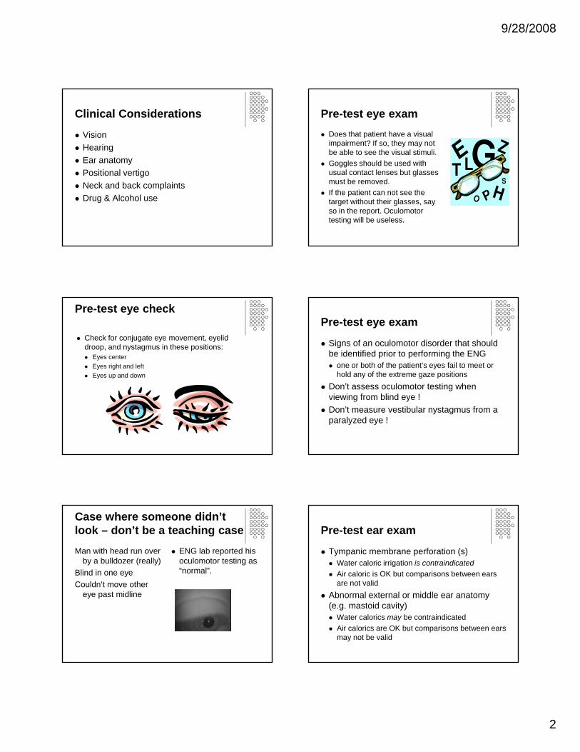
Clinical Considerations
VisionHearingEar anatomyPositional vertigoNeck and back complaintsDrug & Alcohol use
Pre-test eye examDoes that patient have a visual impairment? If so, they may not be able to see the visual stimuli.Goggles should be used with usual contact lenses but glasses must be removed.If the patient can not see the target without their glasses, say so in the report. Oculomotor testing will be useless.
Pre-test eye check
Check for conjugate eye movement, eyelid droop, and nystagmus in these positions:
Eyes centerEyes right and leftEyes up and down
Pre-test eye exam
Signs of an oculomotor disorder that should be identified prior to performing the ENG
one or both of the patient’s eyes fail to meet or hold any of the extreme gaze positions
Don’t assess oculomotor testing when viewing from blind eye !Don’t measure vestibular nystagmus from a paralyzed eye !
Case where someone didn’t look – don’t be a teaching caseMan with head run over
by a bulldozer (really)Blind in one eyeCouldn’t move other
eye past midline
ENG lab reported his oculomotor testing as “normal”.
Pre-test ear exam
Tympanic membrane perforation (s) Water caloric irrigation is contraindicatedAir caloric is OK but comparisons between ears are not valid
Abnormal external or middle ear anatomy (e.g. mastoid cavity)
Water calorics may be contraindicatedAir calorics are OK but comparisons between ears may not be valid

9/28/2008
3
Pre-test ear examExcessive cerumen in the ear canal
Cerumen may block the irrigationShould be removed prior to testing
Narrow ear canalAdequate irrigation may be impossibleCaloric responses may then appear weakRotatory chair testing is needed here
Ear wax error example
Patient seen at prominent institutionReduced caloric response on leftExam 1 month later revealed near impaction of left ear canal.Repeat ENG after impaction is removed normal.
Drugs and medications
Has the patient taken vestibular suppressants, tranquilizers or sedatives or consumed alcohol in the last 48 hours? If so, there may be a suppression of vestibular nystagmus or other eye movement abnormalities (such as slowing).Best usually to proceed with test and note in interpretation
Test instructions to the patient
Talk to the patient, establish rapport.Ask how they feel today – are they dizzy ?Ask about vision, ear/hearing, back and neck problems prior to testingDo they vomit easily ? (consider pretreatingwith ondansetron)
Test Instructions to the patientThe test is divided into 3 main segments:
Looking at light on the wall (oculomotor)Lying in various positions (positional)Running cool and warm water (or air) in each ear, one at a time (caloric)
Breaks may be given if necessary but it is easiest to do this between segments.Ask the patient to report spinning or dizziness during the test.
Test instructions to the patient
Let the patient know that:They will feel some spinning or dizziness during parts of the test.You are trying to measure the eye movements that occur when you feel this way.It will not persistYou are in control of the situation and you will be here the whole time.

9/28/2008
4
Patient preparation
Place goggles on the patient and center the pupil. (Clean the skin and apply the electrodes if not using VNG goggles.)
Patient preparation
Darken the roomA darker room will dilate the pupil and make it easier to track for the vision allowed portions.Center pupil on the video screen. Adjust “tracking” so only pupil is white.For electrodes, impedance should be under 5kohms.
The Basic ENG Test BatteryOculomotor:
SpontaneousGazeSaccadesPursuitOptokinetic
Positional/PositioningCalorics
The optional ENG testsCervical testingHead ShakeHyperventilationFistulaRebound nystagmusTullio’sValsalvaVibration
(Suggest always doing those in bold)
Suggested order for testingCalibrate (H/V)Saccades (H/V)Pursuit (H/V)Spontaneous nystagmusGaze and Rebound (H)Head-shake (H)Vibration
Optional special testsFistula, Valsalva, HVT
Cervical testDix-HallpikeCaloric Tests
Oculomotor testsCalibrateSaccadesSpontaneousGazePursuitOKN
For VENG - -do an “intrinsically calibrated” test like saccades and pursuit right away – and analyze it !

9/28/2008
5
Calibration
Calibration should be performed at start for VNG, and if goggles are repositioned. VNG calibrations do not change unless goggles are repositioned. The patient will be instructed to follow a moving dot. The examiner will “lock in” the calibration when the patient is accurately calibrated.
Calibration
Poor calibration will result in major errors ! Everything is wrong by the % error in the calibration. It is important to recognize when the patient was doing fine, but the calibration was off.
Awful Calibration SaccadeTest
Purpose: To detect central disorders of saccadesConfirm your calibration is good
ProceduresVision allowedFollow target jumping on screenHorizontal and vertical
ResultVelocity, accuracy, latency
Saccade disordersSaccadic disorders are rareToo slow
Velocity – medication effect or brainstemLatency – lack of cooperation or attention
AsymmetricalOculomotor palsyINO
Inaccurate (overshoot)Cerebellar disorder
VNG can’t record – saccadic oscillations
CNS- intranuclear opthalmoplegia
Caused by lesion of the MLF between CN3 & CN6 nucleiBilateral > demyelination (MS)

9/28/2008
6
Watch Out!Note any drugs the patient has taken and possible effect on the testAlerting, especially if the patient is medicatedBlink artifact
Ocular dysmetria
Cerebellum controls smooth integration of body muscles in agonist/antagonist relationshipCerebellar ( or its connections) disease causes defects of limb movementsThe ocular component is dysmetria
Saccades – overshoots (hypermetric)
Saccades – undershoots (hypometric)
Saccadic slowingThe eyes can accurately reach the target but do so much slower than
normal. This can be symmetric or asymmetric. CALIBRATION errors and drug use must be ruled out for accurate interpretation.
Caution!Superimposed gaze nystagmusSuperimposed congenital nystagmusDrugs (usually dysmetria)Inattention/poor visionEye blinksHead movement

9/28/2008
7
Oculomotor - Tracking
Tracking (pursuit) test:The patient follows a visual target moving in the horizontal plane.The recording is examined for abnormalities
Normal Pursuit
Abnormal – saccadic pursuitAka “Cogwheeling” - Eyes fall behind target
Abnormal – saccadic pursuitDisorganized and disconjugate
Reduced horizontal gaze capacityDisconjugate movement is possibleBrainstem/Cerebellar localization
Pursuit PitfallsDrugs disrupt pursuitInattentionHead movementSuperimposed gaze nystagmusSuperimposed congenital nystagmus(cross check with other subtests)
Spontaneous Nystagmus Test
Purpose: To detect vestibular and central nystagmus
Procedures20 seconds vision allowed20 seconds vision denied
ResultNystagmus or notEffect of vision on nystagmus

9/28/2008
8
Effect of Vision
Nystagmus with vision allowed which is enhanced with vision denied is usually PERIPHERAL (but can be anything)Nystagmus unaffected by vision is usually CENTRAL.Horizontal nystagmus which is enhanced with visual fixation (vision allowed) and is reduced or abolished with vision denied is always due to CONGENITAL NYSTAGMUS
Congenital nystagmusFeatures: 1/1000 people
Fixed, genetic developmental brain defectOccurs at birth or soon after in an otherwise healthy person so will be obvious and known to patientGenerally horizontal (rarely torsional or vertical)Worse in light when trying to fixate, reduced or abolished in the darkConvergence = reduction or abolitionNull Point – orbital position where nystagmus is minimal
Congenital nystagmus in light with fixation vs. dark
Gaze Test
Purpose: To detect gaze evoked nystagmusTo document ocular range is full
Vision is allowedProcedures
Horizontal left and rightVertical up and down
ResultGaze-evoked nystagmus or not.
Gaze testing – method30 degrees in each directionRecorded for 20 seconds in each direction vision is allowedCompare nystagmus in each condition
Optional Test – Gaze Rebound
MethodVision denied.Eyes to far right for 10 secondsReturn to center for 10 seconds or moreEyes to far left for 10 secondsReturn to center for 10 seconds or more
OutputRebound nystagmus is always central

9/28/2008
9
Gaze- Rebound
Nystagmus is present upon returning to center gaze from an eccentric gaze position that is held for 10 seconds.The nystagmus beats in the opposite direction of the previously held gaze.This is a central finding associated with cerebellar disease.
Gaze - Rebound
Optokinetic
Optokinetic test:The patient follows a series of visual targets moving to the right and then to the left. This provokes optokinetic nystagmus.The recordings are examined for weak nystagmus in one or both directions of the moving target.
Normal variationsSpeed of the eyes should match speed of the stimulus up to 30 deg/sec and then may fall behind stimulus (but should still increase)Responses should be symmetric
Abnormal- asymmetryOKN asymmetry is rarely encounteredCongenital nystagmusOccasionally indicates CNS abnormality
Look Out!DrugsInattentionInstructions

9/28/2008
10
Recommended Special TestsHigh Frequency Headshake Test – goggles recommended
Shake the patients head side to side with eyes wide open but vision denied for 10-20 secondsImmediately after stopping, a nystagmus may be seen that beats away from the pathologic ear
Vibration Test – goggles recommendedApply vibration to each SCM muscle for 10-20 seconds with vision denied but eyes wide openImmediately after vibration, a nystagmus that beats away from the pathologic ear may be seen from both sides.
Headshake
VibrationHeadshake and VibrationCross-checks on calorics
HeadshakeShould match up with caloric asymmetryBeats away from lesion
VibrationShould match up with caloric asymmetryBeats away from lesion
Positional tests
The patient is moved into these sets of positions to diagnose positional vertigo
Dix Hallpike (head right and left)Supine, head right, head left
Eye movements are examined in each of these positions for nystagmus.
Special Tests – CervicalSuggest do as a routine
MethodVision denied, sitting uprightHead to end of Left range for 20 secondsHead center for 20 secondsHead to end of Right range for 20 secondsHead center for 20 seconds
Pitfall – make sure eyes are in center.Output – nystagmus related to head on neck

9/28/2008
11
Cervical Source
May be result of torsion on the neckCan be from VBI – rare but dangerousCan be from neck pain “spilling over” into the vestibular systemCan be from a herniated disk (variation – Arnold Chiari malformation)
Cervical Source
Old School “Laterals”If nystagmus is present in head right or head left position, roll patient onto the offending side. If it goes away, it was not really based on the head position but rather, the torsion of the neck.
Cervical SourceNew School AKA “Vertebral artery Test”
Video Goggles are an invaluable resource for this testHave patient sit upright so that there is no change of orientation of the head relative to gravity.Keep the body perfectly still and rotate the head as far as you can to one side – keeping the eyes in the center of the orbit.Nystagmus may be small but is significant if present.
BPPV Nystagmus Patterns• PC or AC BPPV
• Vertical and torsional nystagmus
• Latency• Burst• Fatigues (and is
inconsistent)
LC BPPVDirection changing nystagmusGeotrophic (beats towards ground)Ageotrophic (beats towards sky)Depends on previous head position
Central PositionalThis is rare – you shouldn’t be making this call very
often !• Doesn’t fit the BPPV patterns
• Too consistent (doesn’t fatigue)• Doesn’t go away• Wrong direction (no torsional component)
Positional PitfallsGaze L or R during positionsConvergence (can be a problem for monocular goggles)Alcohol ingestion (causes positional nystagmus)

9/28/2008
12
Positional/positioning
Dix-HallpikePatient’s head is rotated to the side and body is brought back from sitting to laying with head hanging down and to the side.This is done to the right and then to the left.Eyes are observed for nystagmus induced by the change in body position.
Procedure
Frenzel lenses or video goggles in darkened room - big monitor is nice for this.Is typically torsional but may also be horizontal and vertical in rare formsRecord anyway!
Traditional Dix-HallpikePatient sits on exam table and examiner stands to the side.The patient’s head is turned 45 deg. To the left (or right)The patient is brought down to a supine position with the head hanging off the edge of the table still to the left (or right)
Dix HallpikeThe head is supported by the examiners hand and is held in this position for at least 40 seconds.If nystagmus appears, hold this position for at least one minute or until the response subsides.At this point you may return the patient to a sitting point, if nystagmus is present you may treat the patient with a CRP.OR, you may return the patient to a sitting position and look for a possible brief reversal of the nystagmusRepeat this maneuver with the head turned to the other direction.It is a good idea to start with the suspected uninvolved side.
Normals AbnormalNormals may have a few beats of nystagmus during the downward motion but none otherwise.
Benign Paroxsymal positioning vertigo
Delayed onsetShort durationTorsional beats toward undermost earFatigable upon repeat trialsMay reverse direction upon return to sittingUsually accompanied by vertigo
Pick a canal, any canal
Generally, posterior canal is involvedRPC= upward & rightward torsionalLPC= upward and leftward torsional
Less commonly, anterior canal is involvedRAC= downward and rightward torsionalLAC downward and leftward torsional
Very rarely, horizontal canal is involvedRHC= horizontal geotropic fast phase stronger right ear downLHC= horizontal geotropic fast phase stronger left ear down

9/28/2008
13
Typical PC BPPV
Typical LC BPPV CaloricsEach of the patient’s ears is irrigated twice
Once with cold stimulusOnce with warm stimulus
Use water unless there is a contraindicationThese stimuli provoke caloric nystagmus
COWS: cold-opposite, warm-sameThe eye movement recordings are examined for a weak response in one or both ears.
The procedureThe head is elevated 30 degreesEach canal is stimulated one at a time with a cool and a warm stimulus
Water:Warm: 44 degrees CCold: 30 degrees C
Air:Warm: 50 degrees CCold: 24 degrees C
The patient has their vision denied and is alerted during the procedure
Patient InstructionsInform patient of what is about to happenThere will be some noise (from irrigator)Let them know that the cool stimulus is not cold and the warm stimulus may feel hot but is not really hot enough to burn or hurt themTell them it is common to feel a turning or spinning sensation but it won’t lastGive them good alerting tasks (name vegetables for example)

9/28/2008
14
Pre-stimulus StimulusCalibrateLet the recorder run to make sure you don’t have any preexisting nystagmusGive the patient alerting tasks after calibration
Place the irrigator tip deeply into the canal. Warn the patient it is about to begin and not to pull awayBegin the irrigation
The stimulusNystagmus and vertigo will commence near the end of the stimulationIt will build to a crescendo about 30 seconds after stimulation and then taper offKEEP ALERTING THE WHOLE TIME!!
Normal AbnormalAll four caloric responses should be roughly equalNormals (and peripheral) lesions should have normal fixation suppression
Unilateral weaknessDirectional preponderanceBilateral weaknessHyperactive responsesFailure of fixation suppressionCaloric inversion and perversion
CalculationsDuration of nystagmus response
From beginning of irrigation to end of responsePeak nystagmus frequency
Average frequency of nystagmus beats during 10 second interval when response is most intense
Maximum SPV during 10 second interval when response is most intense
Most widely usedDetermined by inspection of record
Calculation
Each response is quantified by the maximum SPV – this is done by the software but it’s good to understand where the numbers are coming from to verify accuracy of results.All four responses are compared for the following calculations:
Unilateral weaknessDirectional preponderanceFixation index
Unilateral weakness
The amount by which 2 responses provoked by right ear stimulation differ in intensity from those provoked by left ear stimulation.Significant finding is >22 % difference between ears (Jacobson)Indicates peripheral problem on the weaker sideBe careful of asymmetric canal anatomy!
Unilateral weakness formula:(RW+RC) – (LW+LC)/(RW+RC+LW+LC) x 100

9/28/2008
15
Unilateral Weakness
Patient with left sided acoustic neuroma
Traces for unilateral weakness
Bilateral weakness
Caloric response of both ears is very weak or absentIf all four caloric responses total less than 22 deg/sec (Jacobson), its BWIs indicative of CNS or bilateral peripheral lesionsIce calorics should be considered.Rotatory chair if available
Bilateral Weakness -- Gentamicin
Bilateral Weakness -- Gentamicin Ice caloricsAlways do for “dead Ear”
Patient closes eyes, turns head and recording is started.2ml of ice water (or more) is squirted into the ear using a big syringe (no needle of course). “Toomey” is goodWater remains for 20 seconds and then head is turned to empty the ear.Head is brought back to center position.This may provoke an opposite beating nystagmus not seen before. (cold=opposite), because much stronger stimulus than air or 7 deg centigrade irrigator

9/28/2008
16
Ice Calorics -- ProneIf you get a responseHave the patient quickly flip over to see if the response changed direction.If the response does not change direction, this indicates that it is not a gravity dependant response and is probably just a latent spontaneous nystagmus - not a true caloric response.This is important for case management decisions regarding possible surgical intervention vs. VRT!!
Ice caloric Bilateral Weakness --Gentamicin
Directional preponderance
The difference in intensity between the 2 right beating and the 2 left beating responsesGenerally, this is seen with a pre-existing nystagmusSometimes, can be seen without one.Useless for clinical diagnosisHowever, it is conventionally reported and is an abnormal finding if >28% (Jacobson)
Directional preponderance formula(RW+LC)-(LW+RC)/(RW+LC+LW+RC) x 100
RB directional preponderance
Barber & Stockwell
Directional Preponderance and UWDP technical error

9/28/2008
17
FixationAs the response begins to decline (about 90 seconds after beginning the irrigation) ask the patient to look at the fixation light (if using electrodes, have them open their eyes & look at your finger or a spot on the ceiling)Keep looking for about 10 seconds then the light will shut off (or have them close the eyes again if using electrodes) The recording should be continued until the nystagmus has tapered off.Wait 3-5 minutes before starting the next irrigation.
Fixation IndexMeasure of the effectiveness of visual fixation in suppressing caloric nystagmus.Should be calculated for at lease one right beating and one left beating caloric response
Fixation index formula:SPV (fixating)/SPV (not fixating)
Fixation Suppression Examples
Failure of fixation suppression (CNS)
Good fixation suppression (peripheral)
Failure of fixation suppressionNystagmus intensity with eyes open nearly equals matches or exceeds that with eyes closedFixation suppression will occur in normalsand peripheral lesionsFI greater than .6 is abnormal and indicative of CNS involvementCalculated at peak or near end of peakRotatory chair does it better
Hyperactive responsesDefined as water responses that exceed 50deg/sec for cools and 80deg/sec for warms (Jacobson: 99 deg/sec for cools, 146 deg/sec for warms, 221 deg/sec total)Usually is due to excellent inner ear function in a nervous young person.Rarely can be due to abnormal anatomy (tm perf, mastoidectomy, etc.)In absence of this, very rarely it is due to CNS problem (cerebellar).
Hyperactive responses (central)

9/28/2008
18
Hyperactive response (central)
Normal response
Caloric inversion and perversion – usually your error
Inversion: entire caloric response beats in the wrong direction (anatomically impossible to explain)Perversion: vertical or oblique nystagmus in response to caloric stimulation (some physiology here)Caloric inversion/perversion is usually caused by a technical error
Wrong temperature or head positionAlerting bringing out spontaneous nystagmusToo short wait between irrigations
Rarely, perversion is brainstemShould have LOTS of other brainstem findings.
The optional ENG testsCervical testingHead Shake HyperventilationFistulaRebound nystagmusTullio’sValsalvaVibration
We suggest always doing bolded tests and have already discussed in earlier material
Special Tests – HVT(suggest - - on demand)
MethodRecord baselineHVT for 30 secondsRecord post-HVT
OutputNystagmus induced, enhanced or reversed by HVTReversal suggests vest nerve irritability
PitfallsNone
Special Tests – Fistula(suggest - -on demand only)
MethodApply positive pressure into the ear canal
OutputNystagmus associated with pressure
PitfallHard to calibrate pressureVery insensitive
Special Tests – Tullio’sSuggest - -on demand only
MethodVision deniedLoud noise close to one ear
OutcomeNystagmus correlated with loud noiseSuggests SCD
PitfallsInsensitiveDifficult to calibrate loud noise.

9/28/2008
19
Special Tests – ValsalvaOn demand or always
MethodVision deniedPatient takes a deep breath and “strains” –increasing intrathoracic pressure. 5 sec is enough.
OutputNystagmus provoked by strainingSuggests SCD or fistula
PitfallsSome people don’t cooperate leading to false negatives
SummaryENG consists of 4 groups of studies
OculomotorPositionalCaloricSpecial maneuvers
Powerful method to detect many ear disordersComplex interpretation process