zif-1 - development -
TRANSCRIPT

3373DEVELOPMENT AND STEM CELLS RESEARCH ARTICLE
INTRODUCTIONPrimordial germ cells, the source of gametes in the adult animaland the genetic link between two generations, are specified earlyduring embryogenesis in most animals. Recent studies in worms,flies and mice show that specification of primordial germ cellsrequires global repression of transcription, usually throughinhibition of initiation or elongation, and chromatin remodeling(Extavour and Akam, 2003; Nakamura and Seydoux, 2008).Defective transcriptional repression in primordial germ cells resultsin sterility.
In C. elegans, the single founder blastomere for the entiregermline, P4, is specified through a series of four asymmetricdivisions, beginning with the zygote, P0 (Strome, 2005). Each ofthese divisions results in a smaller germline precursor (P1 throughP4, termed the P lineage) and a larger somatic sister cell (Fig. 1).P4 divides symmetrically to generate Z2 and Z3, which generatethe entire germline postembryonically. All germline blastomeresare transcriptionally repressed, whereas their somatic sistersundergo rapid transcriptional activation and lineage-specificdifferentiation, requiring ready reversibility for any repressive
mechanism operating in the P lineage (Seydoux and Dunn, 1997;Seydoux et al., 1996). Epigenetic marks characteristic oftranscriptionally competent chromatin are found in C. elegansgermline blastomeres (Schaner et al., 2003), consistent with thembeing transcriptionally competent but being actively restrainedfrom differentiation-promoting transcription.
Transcriptional repression in the P lineage in C. elegans requiresat least two groups of maternally supplied proteins. In P0 and P1,two closely related and functionally redundant cytoplasmicproteins, OMA-1 and OMA-2, globally repress transcriptioninitiation by binding to TAF-4, a crucial component of the RNApolymerase II pre-initiation complex (Guven-Ozkan et al., 2008).In P2-P4, PIE-1 globally represses transcription elongation byinhibiting P-TEFb, the kinase which phosphorylates serine 2 (Ser2)residues within heptapeptide repeats of the RNA polymerase II C-terminal domain (Batchelder et al., 1999; Seydoux and Dunn,1997; Zhang et al., 2003). Ser2 phosphorylation (Ser2P) is requiredfor transcriptional elongation (Komarnitsky et al., 2000; Shim etal., 2002).
OMA-1, OMA-2 and PIE-1 proteins are all expressed in oocytesfrom maternally supplied mRNAs. OMA-1 and OMA-2 aredegraded soon after the first mitotic division and are not detectedin subsequent P-lineage blastomeres (Fig. 1) (Detwiler et al., 2001;Lin, 2003). Degradation requires that OMA proteins bephosphorylated by at least two kinases, one of which, the DYRK2-type kinase MBK-2, is developmentally activated in newlyfertilized embryos (Cheng et al., 2009; Nishi and Lin, 2005;Shirayama et al., 2006; Stitzel et al., 2006). PIE-1 is segregatedasymmetrically to the germline blastomere at each P-lineageblastomere division. In addition, the minor amount of PIE-1
Development 137, 3373-3382 (2010) doi:10.1242/dev.055327© 2010. Published by The Company of Biologists Ltd
Department of Molecular Biology, University of Texas Southwestern Medical Center,Dallas, TX 75390, USA.
*These authors contributed equally to this work†Present address: Department of Molecular and Cellular Biology, Harvard University,Cambridge, MA 02138, USA‡Author for correspondence ([email protected])
Accepted 6 August 2010
SUMMARYSpecification of primordial germ cells requires global repression of transcription. In C. elegans, primordial germ cells aregenerated through four rounds of asymmetric divisions, starting from the zygote P0, each producing a transcriptionally repressedgermline blastomere (P1-P4). Repression in P2-P4 requires PIE-1, which is provided maternally in oocytes and segregated to allgermline blastomeres. We have shown previously that OMA-1 and OMA-2 repress global transcription in P0 and P1 bysequestering TAF-4, an essential component of TFIID. Soon after the first mitotic cycle, OMA proteins undergo developmentallyregulated degradation. Here, we show that OMA proteins also repress transcription in P2-P4 indirectly, through a completelydifferent mechanism that operates in oocytes. OMA proteins bind to both the 3� UTR of the zif-1 transcript and the eIF4E-bindingprotein, SPN-2, repressing translation of zif-1 mRNA in oocytes. zif-1 encodes the substrate-binding subunit of the E3 ligase forPIE-1 degradation. Inhibition of zif-1 translation in oocytes ensures high PIE-1 levels in oocytes and germline blastomeres. Thetwo OMA protein functions are strictly regulated in both space and time by MBK-2, a kinase activated following fertilization.Phosphorylation by MBK-2 facilitates the binding of OMA proteins to TAF-4 and simultaneously inactivates their function inrepressing zif-1 translation. Phosphorylation of OMA proteins displaces SPN-2 from the zif-1 3� UTR, releasing translationalrepression. We propose that MBK-2 phosphorylation serves as a developmental switch, converting OMA proteins from specifictranslational repressors in oocytes to global transcriptional repressors in embryos, together effectively repressing transcription inall germline blastomeres.
KEY WORDS: OMA proteins, zif-1, Translational repression, C. elegans, Germline, Oocyte-to-embryo transition, MBK-2
zif-1 translational repression defines a second, mutuallyexclusive OMA function in germline transcriptionalrepressionTugba Guven-Ozkan*, Scott M. Robertson*, Yuichi Nishi† and Rueyling Lin‡
DEVELO
PMENT
3374
segregated to the somatic sister is rapidly degraded (Mello et al.,1996; Reese et al., 2000). Repression by both OMA and PIE-1provide a robust, but readily reversible, way to repress transcriptionin the P-lineage while maintaining the chromatin primed fortranscriptional activation in the somatic sisters.
OMA-1, OMA-2 and PIE-1 have additional functions beyondrepressing transcription in germline blastomeres. All three proteinscontain tandem CCCH zinc fingers, a domain usually associatedwith RNA binding (Detwiler et al., 2001; Lai et al., 1999; Mello etal., 1996; Pagano et al., 2007). However, the CCCH zinc fingersare not required for PIE-1 to repress transcription (Tenenhaus et al.,2001) or for the OMA proteins to bind to and sequester TAF-4(Guven-Ozkan et al., 2008). OMA-1 and OMA-2 activity arerequired for oocyte maturation, although the molecular basis forthis requirement is unknown (Detwiler et al., 2001; Shimada et al.,2002). All three proteins contribute to the restricted expressionpattern of a Nanos-related protein, NOS-2, to the P4 germlineblastomere. OMA proteins have been shown to bind to the nos-23� UTR and repress translation in oocytes, whereas PIE-1 has beenshown to maintain the expression level of NOS-2 through anunknown mechanism (Jadhav et al., 2008; Tenenhaus et al., 2001).Recently, OMA proteins have also been implicated in thetranslational repression of mei-1 in embryos (Li et al., 2009). Oneintriguing unanswered question is how the multiple functions ofOMA proteins or PIE-1 intersect in vivo. We have shownpreviously that phosphorylation of OMA-1 by MBK-2, at the sameamino acid that triggers its degradation, facilitates OMA-1 bindingto TAF-4 (Guven-Ozkan et al., 2008), suggesting coordinatedregulation.
Degradation of PIE-1 in somatic cells is carried out by a CUL-2-containing E3 ligase (DeRenzo et al., 2003). The substrate-binding subunit of this E3 ligase, ZIF-1, binds to PIE-1 via its firstCCCH zinc finger (DeRenzo et al., 2003). ZIF-1 also binds to andpromotes the degradation of tandem CCCH zinc finger proteinsMEX-1, POS-1, MEX-5, and MEX-6 in somatic blastomeres(DeRenzo et al., 2003). How the degradation of these ZIF-1substrates is restricted to somatic blastomeres, and not in germlineblastomeres or oocytes, remains unknown. ZIF-1 is not responsiblefor OMA protein degradation (DeRenzo et al., 2003).
Increasing evidence supports an important role for translationalrepression during germ cell differentiation in both flies and worms(for a review, see Nakamura and Seydoux, 2008). In Drosophila,two RNA-binding proteins, Nanos and Pumilio, repress translationof differentiation-promoting proteins in germline stem cells,
maintaining their stem cell character (Parisi and Lin, 2000). In C.elegans, spatiotemporal expression of most proteins expressed inthe germline is regulated by their 3� UTR sequences (Merritt et al.,2008). Indeed, many key regulators of germline development areRNA-binding proteins (Kimble and Crittenden, 2005). Forexample, depletion of two KH-domain RNA-binding proteinsexpressed in the C. elegans gonad, MEX-3 and GLD-1, drivesgerm cells into precocious somatic differentiation within the gonad(Ciosk et al., 2006).
We show here that OMA proteins have an essential function inmaintaining a high level of PIE-1 protein in oocytes and embryos.OMA proteins do so by binding to and repressing translation of zif-1 mRNA in oocytes, thereby preventing PIE-1 degradation. Bypreventing PIE-1 degradation, OMA proteins indirectly represstranscription in P2-P4. The two mechanisms by which OMAproteins repress primordial germ cell transcription, namely bydirect sequestration of TAF-4 and indirect repression of zif-1translation, are distinct. They take place at different developmentalstages, require distinct OMA protein domains, can be disruptedindependently of each other and are differentially regulated byMBK-2 phosphorylation. We propose that phosphorylation byMBK-2, itself a developmentally regulated kinase, serves as adevelopmental switch that converts OMA proteins fromtranslational repressors of specific maternal transcripts in thegermline to global transcriptional repressors in the early embryo,together ensuring transcriptional repression in germlineblastomeres.
MATERIALS AND METHODSStrainsN2 was used as the wild-type strain. Genetic markers were: LGI, gld-1(q485); LGIII, unc-119(ed3); LGIV, oma-1(zu405), oma-1(te33), oma-1(te21), mbk-2(ne992); LGV, oma-2(te50);oma-2(te51). Plasmids used,strain names and transgenes were as follows: TX1246 (teIs113 [Ppie-1gfp::h2b::UTRzif-1,771bp]), TX1248 (teIs114 [Ppie-1gfp::h2b::UTRzif-1,771bp]),TX1240 (teEx602 [Ppie-1gfp::h2b::UTRzif-1,304bp]),TX1251 (teEx604 [Ppie-1gfp::h2b::UTRzif-1,771bp, 4-63]), TX1409 (teEx656 [Ppie-1gfp::h2b::UTRzif-1,771bp, 64-123]),TX1272 (teEx606 [Ppie-1gfp::h2b::UTRzif-1,771bp, 124-183]), TX1298 (teEx607 [Ppie-1gfp::h2b::UTRzif-1,771bp, 184-243]), TX1410 (teEx657 [Ppie-1gfp::h2b::UTRzif-1,771bp, 244-303]), TX1311 (teEx610 [Ppie-1gfp::h2b::UTRzif-1,771bp, 64-183]), TX1315 (teEx611 [Ppie-1gfp::h2b::UTRzif-1,771bp, 64-183]), TX1375 (teIs126 [Ppie-1gfp::zif-1::UTRzif-1,771bp]).
RESEARCH ARTICLE Development 137 (20)
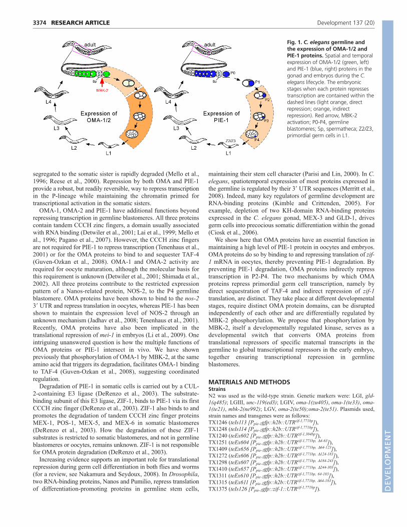
Fig. 1. C. elegans germline andthe expression of OMA-1/2 andPIE-1 proteins. Spatial and temporalexpression of OMA-1/2 (green, left)and PIE-1 (blue, right) proteins in thegonad and embryos during the C.elegans lifecycle. The embryonicstages when each protein repressestranscription are contained within thedashed lines (light orange, directrepression; orange, indirectrepression). Red arrow, MBK-2activation; P0-P4, germlineblastomeres; Sp, spermatheca; Z2/Z3,primordial germ cells in L1.
DEVELO
PMENT
JH1436, JH227, AZ212 and TX1162 contain Ppie-1gfp::pie-1 zf1::UTRpie-1,Ppie-1gfp::pie-1::UTRpie-1, Ppie-1gfp::H2B::UTRpie-1 and Poma-1 oma-1�46-80::gfp::UTRoma-1 transgenes, respectively, as described (Guven-Ozkan etal., 2008; Praitis et al., 2001; Reese et al., 2000).
Plasmid constructionMost plasmids were constructed with the Gateway cloning technology. Thezif-1 3� UTR is annotated in Wormbase as being 304 nucleotides long. Inorder to ensure proper expression in vivo, a longer piece of genomic DNA(771 nucleotides downstream of the stop codon) was used to generate thezif-1 translational reporter. The 771 nucleotide sequence was cloneddownstream of pie-1 promoter-driven GFP::H2B in the germlineexpression vector pID3.01B (Reese et al., 2000), which contains the pie-13� UTR now downstream of the 771 nucleotide zif-1 3� sequence. Alldeletion constructs were derived from the 771 nucleotide sequence.
C. elegans transformationAll integrated lines were generated by microparticle bombardment (Praitiset al., 2001), whereas other transgenic lines were generated by complexarray injection (Kelly et al., 1997). For each construct, expression wasanalyzed and found to be consistent in at least two independent lines.Germline expression in some of the non-integrated lines became silencedafter 4-6 generations.
RNA interferenceFeeding RNAi was performed as described (Timmons and Fire, 1998)using HT115 bacteria seeded on NGM plates containing 1 mM IPTG. L1larvae were fed for 2 days at 25°C or 3 days at 20°C. oma-1/2(RNAi);gld-1(q485) animals were incubated at 25°C.
ImmunofluorescenceImmunofluorescence for C. elegans gonads and embryos was carried outas described previously for anti-PIE-1 (1/50) (Mello et al., 1996), anti-OMA-1a (1/100) (Shimada et al., 2006), anti-GFP (1/250, rabbit,Invitrogen), anti-MEX-1 (Guedes and Priess, 1997) and anti-Ser2P (1/300,MMS-129R, Covance) (Seydoux and Dunn, 1997). Secondary antibodiesused were Alexa488- or Alexa568-conjugated goat anti-rabbit or goat anti-mouse (Invitrogen, 1/250).
Lysate preparation and RNA binding assayWorm lysates were prepared from 1-day-old gravid adults as describedexcept the following modifications (Lee and Schedl, 2001). Worms wereharvested, washed and then disrupted in a Dounce homogenizer by 4-5strokes with a loose-fitting pestle followed by 15-20 strokes with a tight-fitting pestle. Lysates were spun at 16,000 g for 20 minutes and thesupernatants were used for binding. Protein concentrations averaged 6-8mg/ml. Synchronized mbk-2(ne992ts) (Pang et al., 2004) L1 larvae werecultured at 16°C until L4, then shifted to 25°C for one day before beingharvested for lysate.
RNA pulldowns were performed as in Lee and Schedl (Lee and Schedl,2001). After pulldown and washing, beads were then boiled in SDS samplebuffer. Supernatants were loaded on a 10% SDS-PAGE gel and subjectedto western blot analysis with the indicated antibodies: anti-OMA-1 (1:50)(Detwiler et al., 2001), anti-SPN-2N (1:2000) (Li et al., 2009) or anti-MBP(1:1000; NEB). More detailed protocols for lysate preparation, RNAsynthesis and RNA pulldown are available upon request.
Analysis of embryos, imaging and quantificationImages of immunofluorescence and live embryos were acquired andprocessed as described (Guven-Ozkan et al., 2008).
RESULTSOMA-1/2 are required to prevent PIE-1degradationDouble loss-of-function mutants for oma-1 and oma-2 are sterileowing to a defect in oocyte maturation (Detwiler et al., 2001).Embryos with reduced levels of OMA-1 and -2 can be obtained byRNAi [oma-1(RNAi);oma-2(RNAi)]. These RNAi embryos have
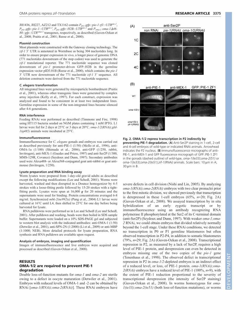
severe defects in cell division (Nishi and Lin, 2005). By analyzingoma-1(RNAi);oma-2(RNAi) embryos with two clear pronuclei priorto the first mitotic division, we showed previously that transcriptionis derepressed in these 1-cell embryos (65%, n20; Fig. 2A)(Guven-Ozkan et al., 2008). We assayed transcription by in situhybridization of an early zygotic transcript or byimmunofluorescence using an antibody recognizing RNApolymerase II phosphorylated at the Ser2 of its C-terminal domain(anti-Ser2P) (Seydoux and Dunn, 1997). With weaker oma-1;oma-2 RNAi, we could obtain embryos that divided relatively normallybeyond the 1-cell stage. Under these RNAi conditions, we detectedno transcription in P0 or P1 germline blastomeres but oftenobserved transcription in P2-P4, in addition to somatic blastomeres(79%, n29; Fig. 2A) (Guven-Ozkan et al., 2008). Transcriptionalrepression in P2, as measured by a lack of Ser2P, requires a highlevel of PIE-1 protein, and derepression can even be detected inembryos missing one of the two copies of the pie-1 gene(Tenenhaus et al., 1998). The observed defect in transcriptionalrepression in P2 in oma-1/2-depleted embryos is an indirect effectof a reduced level, or loss, of PIE-1 protein. oma-1(RNAi);oma-2(RNAi) embryos have a reduced level of PIE-1 (100%, n9), withthe extent of PIE-1 reduction proportional to the severity oftranscriptional derepression (the intensity of Ser2P staining)(Guven-Ozkan et al., 2008). In worms homozygous for oma-1(te33);oma-2(te51) (both loss-of-function mutations), or worms
3375RESEARCH ARTICLEOMA proteins repress zif-1translation
Fig. 2. OMA-1/2 repress transcription in P2 indirectly bypreventing PIE-1 degradation. (A)Anti-Ser2P staining in 1-cell, 2-celland 4-cell embryos of wild-type or indicated RNAi animals. Arrowheadindicates the P2 nucleus. (B)Immunofluorescence micrographs of anti-PIE-1, anti-MEX-1 and GFP fluorescence micrograph of GFP::PIE-1 ZF1in the gonads (dashed outline) of wild-type, oma-1(te33);oma-2(51) oroma-1(te33);oma-2(te51);zif-1(RNAi) animals. Scale bars: 10m in A;30m in B.
DEVELO
PMENT

3376
that have been strongly depleted of oma-1 and oma-2 by RNAi, wedetected no, or a dramatically reduced level of, PIE-1 in the gonad(94%, n17; Fig. 2B).
The level of many transgenic proteins regulated by the pie-1promoter and the pie-1 3� UTR are not affected in oma-1(RNAi);oma-2(RNAi) animals (Guven-Ozkan et al., 2008).Therefore, it is unlikely that OMA-1/2 regulate transcription ortranslation of PIE-1 protein. Three lines of evidence suggest,instead, that OMA-1/2 regulate PIE-1 stability. First, depletion ofzif-1 by RNAi in oma-1(–);oma-2(–) animals blocks PIE-1degradation and suppresses the loss of PIE-1 phenotype (100%,n9; Fig. 2). Second, the first zinc finger of PIE-1 is both necessaryand sufficient for its degradation by the ZIF-1-containing E3 ligase(Reese et al., 2000). We show that expression of a reporter GFPprotein carrying only the first zinc finger of PIE-1 (GFP::PIE-1ZF1) (Reese et al., 2000) is dramatically reduced in the oocytes ofoma-1(–);oma-2(–) animals (100%, n15; Fig. 2). Third, levels ofMEX-1, another tandem CCCH zinc-finger protein whosedegradation is also mediated by ZIF-1 (DeRenzo et al., 2003), aredecreased or abolished in oma-1(–);oma-2(–) oocytes (100%,n10; Fig. 2). Together, these results support the model that OMAproteins prevent PIE-1 degradation by inhibiting the ZIF-1-containing E3 ligase activity.
OMA proteins maintain PIE-1 stability andsequester TAF-4 via distinct domainsSequestration of TAF-4 by OMA-1 requires a 35-amino-acidregion (residues 46-80) that resembles the histone-fold domain ofTAF-12, the normal binding partner for TAF-4 (Guven-Ozkan etal., 2008). Worms expressing only the mutant OMA-1 �46-80, inan otherwise oma-1(te33);oma-2(te51) background, do not have anOma phenotype. Instead, these worms are fertile and their embryosare transcriptionally derepressed in P0 and P1 (89%, n57; Fig.3A-C) (Guven-Ozkan et al., 2008). PIE-1 protein levels aresimilarly high, or slightly reduced, in oocytes of worms expressingonly OMA-1 �46-80, compared with wild-type worms (89%, n9).This is in dramatic contrast to the complete absence of PIE-1 inoma-1(te33);oma-2(te51) animals (Fig. 2B).
We now show that maintenance of PIE-1 levels in oocytesrequires one or both OMA zinc fingers. oma-1(te21) is amissense mutation resulting in the substitution of lysine for aconserved aspartate residue in the first zinc finger of OMA-1(E141K) (Fig. 3A) (Detwiler et al., 2001). Similarly, oma-2(te50) is a missense mutation that substitutes tyrosine for thesecond cysteine in the second zinc finger of OMA-2 (C162Y)(Detwiler et al., 2001). We showed previously that both OMA-1(E141K) and OMA-2 (C162Y) were expressed at wild-typelevels in oma-1(te21) and oma-2(te50) animals, respectively(Detwiler et al., 2001). PIE-1 protein levels are greatly reducedor abolished in oma-1(te21);oma-2(RNAi) and oma-1(te21);oma-2(te50) animals, similar to those observed in animals with nodetectable OMA proteins [oma-1(te33);oma-2(te51) or oma-1(RNAi);oma-2(RNAi)] (Fig. 2B; Fig. 3B; see Fig. S1 in thesupplementary material). Although only the first finger of OMA-1 and the second finger of OMA-2 were tested, their highsequence similarity and redundant in vivo function suggest thatboth zinc fingers are probably required for OMA-1 and OMA-2to maintain PIE-1 stability.
Neither the first finger of OMA-1 nor the second finger ofOMA-2 is required for OMA-dependent transcriptional repressionin the P0 or P1 blastomeres. We detected no ectopic Ser2P stainingin the majority of 1-cell and 2-cell embryos derived from oma-
1(te21);oma-2(te50) animals (88%, n25; Fig. 3D). However,100% of 4-cell embryos (n30) derived from oma-1(te21);oma-2(te50) animals demonstrated ectopic Ser2P in the P2 blastomere,supporting our notion above that the defect in transcriptionalrepression in P2 is not a consequence of a defect in P0, but ratherdue to the absence of PIE-1 protein.
Taken together, these results suggest that OMA proteins maintainPIE-1 protein levels through a zinc-finger-dependent mechanismthat is independent of TAF-4 binding.
OMA proteins prevent PIE-1 degradation byrepressing zif-1 translationOur results above suggest that OMA proteins prevent PIE-1degradation by inhibiting the ZIF-1-containing E3 ligase activity.In addition to ZIF-1, this E3 ligase contains the cullin CUL-2,the ubiquitin-like protein elongin B (ELB-1), the adaptor protein
RESEARCH ARTICLE Development 137 (20)
Fig. 3. Translational repression and transcriptional repressionrequire distinct domains of OMA proteins. (A)Schematic of OMA-1�46-80, OMA-1 E141K [oma-1(te21)] and OMA-2 C162Y [oma-2(te50)]. White box, zinc finger. (B)Immunofluorescence micrographsof anti-PIE-1 staining in the gonad of oma-1(te33) animals expressingeither wild-type OMA-1::GFP or OMA-1 �46-80::GFP with or withoutoma-2 depletion by RNAi (left column), and animals of the indicatedgenotypes (right column). (C)Anti-Ser2P staining of 1-cell embryos fromoma-1(te33) animals expressing the indicated version of OMA-1::GFPwith or without oma-2(RNAi). (D)Anti-Ser2P staining in oma-1(te21);oma-2(te50) embryos. Note that Ser2P staining is only detectedin P2 (arrow) but not P0 or P1. Scale bars: 30m in B; 10m in C,D.
DEVELO
PMENT

elongin C (ELC-1), and the ring finger protein RBX-1 (DeRenzoet al., 2003; Vasudevan et al., 2007). Depletion of cul-2 or othercomponents in this E3 ligase complex results in a large varietyof phenotypes in the germline and early embryos that were notobserved in zif-1(RNAi) animals (Liu et al., 2004; Sonneville andGonczy, 2004). Genetic data are consistent with another E3ligase containing CUL-2, ELB-1, ELC-1 and RBX-1 beingactive in wild-type gonads and newly fertilized embryos, whereit regulates degradation of other proteins in a ZIF-1-independentmanner (Vasudevan et al., 2007). Therefore, it is unlikely thatOMA-1 and OMA-2 inhibit the ZIF-1-containing E3 ligase inthe germline by inhibiting any of the common components ofthese complexes. The best candidate whose expression oractivity might be repressed by OMA-1 and OMA-2 was ZIF-1(DeRenzo et al., 2003).
zif-1 mRNA is maternally supplied and is detected at a high levelthroughout the gonad and early embryo (C. elegans expressiondatabase), although the in vivo spatiotemporal localization of ZIF-1 protein has not been determined. We raised an antibody againstthe ZIF-1 protein; however, this ZIF-1 antibody either did not workin our immunofluorescence analyses or failed to detect ZIF-1protein owing to low abundance. Recent studies have shown thatexpression of the majority of maternally supplied proteins in C.elegans is regulated by the corresponding 3� UTR sequence(Merritt et al., 2008). We therefore generated a reporter constructthat would express nuclear GFP under the control of the zif-1 3�UTR (Ppie-1-gfp::h2b-UTRzif) as a means to indirectly monitor zif-1 translation. For the purpose of this paper, we will refer toGFP::H2B expressed from this transgene as GFP::H2Bzif-1.
GFP::H2Bzif-1 recapitulates the temporal and spatial localizationof the known ZIF-1 activity in the gonad and embryo (Fig. 4). Thatis, ZIF-1 activity and GFP::H2Bzif-1 expression are repressed inoocytes and germline blastomeres and both are present in somaticblastomeres. We observed nuclear GFP::H2Bzif-1 signal in embryosstarting from the 4-cell stage and then only in somatic blastomeres(Fig. 4A). In germline blastomeres, where PIE-1 levels were high,no GFP::H2Bzif-1 was detected. GFP::H2Bzif-1 expression was alsoabsent in 1-cell and 2-cell embryos and oocytes, where PIE-1 levelsare high (Fig. 4). A similar expression pattern was also observed fora reporter in which the sequence-encoding histone H2B was replacedwith the full-length zif-1 coding sequence (GFP::ZIF-1zif-1) (see Fig.S2A in the supplementary material). This is in clear contrast toGFP::H2B expressed under the control of the pie-1 3� UTR(GFP::H2Bpie-1) (Praitis et al., 2001), which is ubiquitous, beingdetected in all nuclei in the gonad and early embryos (Fig. 4B).
Repression of Ppie-1-gfp::h2b-UTRzif in oocytes is dependent onOMA proteins and, more precisely, on the first finger of OMA-1and second finger of OMA-2 (Fig. 4B). We observed a high levelof GFP::H2Bzif-1 in animals homozygous for oma-1(te33);oma-2(te51) or depleted of oma-1 and oma-2 by RNAi (100%, n19 and20, respectively). Similarly, GFP::H2Bzif-1 was detected in oocytesof oma-1(te21);oma-2(RNAi) and oma-1(RNAi);oma-2(te50)animals (100%, n10 and 15, respectively). Repression of Ppie-1-gfp::h2b-UTRzif in the meiotic gonad distal to oocytes is dependenton another RNA-binding protein, GLD-1. GLD-1 has been shownto repress the translation of several oocyte proteins in the distalmeiotic gonads, including OMA-1 and OMA-2 (Lee and Schedl,2001). We found that animals depleted of oma-1, oma-2 and gld-1exhibit GFP::H2Bzif-1 expression throughout the gonad (67%, n33;see Fig. S2B in the supplementary material). For the remainder ofthis paper, we will characterize repression of Ppie-1-gfp::h2b-UTRzif
only in oocytes where OMA proteins are normally expressed.
OMA-1 and OMA-2 are not global translational repressors inoocytes. We found no evidence that they repress the translation ofanother reporter construct containing the glp-1 3� UTR (Merritt etal., 2008). GLP-1 expression resembles that of GFP::H2Bzif-1 andhas been shown to be regulated through its 3� UTR (Evans et al.,1994; Marin and Evans, 2003; Ogura et al., 2003). However, aGFP::H2B reporter containing the glp-1 3� UTR remains repressedin oma-1(RNAi);oma-2(RNAi) animals (100%, n15; see Fig. S2Cin the supplementary material).
OMA proteins repress zif-1 mRNA translation bydirect binding to its 3� UTRThe fact that zif-1 translational repression requires the CCCH zinc-finger domains supports the notion that the OMA proteins binddirectly to the zif-1 3� UTR. To test whether OMA proteins are
3377RESEARCH ARTICLEOMA proteins repress zif-1translation
Fig. 4. GFP::H2Bzif-1 recapitulates the temporal and spatiallocalization of ZIF-1 activity. (A)Fluorescence micrographs of stagedembryos expressing indicated GFP reporters. Embryo stage is indicatedto the left by the name of its germline blastomere (arrow). (B)GFP::H2Bexpression from reporters containing indicated 3� UTR (upper righthand corner) in gonads of different genetic backgrounds (upper lefthand corner). A wild-type gonad exhibiting GFP::H2Bzif-1 in mitoticgermline stem cells (arrowheads), but repressed in meiotic germ cells, isshown with both DIC (top left) and fluorescence micrographs (topright). GFP::H2B expressed from another reporter construct differingonly in the 3�UTR is expressed in oocytes (bottom right). Additionaldata demonstrating specificity of GFP::H2Bzif-1 is shown in Fig. S3 in thesupplementary material. Scale bars: 10m in A; 30m in B.
DEVELO
PMENT

3378
capable of specifically binding to the zif-1 3� UTR, we performeda series of in vitro RNA-binding assays (Lee and Schedl, 2001;Mootz et al., 2004). The assay combined protein, either purifiedfrom bacteria or endogenous protein in a worm extract, within-vitro-synthesized, single-stranded biotinylated RNAscorresponding to the zif-1 3� UTR. Following pulldown of theRNA using streptavidin-coated magnetic beads, proteins also pulleddown were assayed by SDS-PAGE and western blotting analysesusing specific antibodies. We observed that both MBP-taggedOMA-1 protein generated in bacteria and OMA-1 from wild-typeworm extracts bound to the zif-1 3� UTR in a strand- and sequence-specific manner (Fig. 5C).
We divided the 300 nucleotide zif-1 3� UTR into five regions(I-V) and observed that region III is both necessary and sufficientfor binding by bacterially expressed OMA-1 (Fig. 5A,C). We alsoassayed various deletions within the 3� UTR sequence for theirability to repress the expression of GFP::H2B in vivo (Fig. 5B).Consistent with the in vitro binding result that OMA proteins bindto region III, deletion of region III resulted in the derepression,albeit weak, of GFP::H2B in oocytes, as well as some meioticnuclei preceding cellularization. A stronger derepression wasobserved when both regions II and III were deleted. Deletingregions I, IV or V individually did not result in derepression in thegonad (see Fig. S3 in the supplementary material). Regions II andIII are not only necessary, but also sufficient, for repression ofPpie-1-gfp::h2b-UTRzif in oocytes. This result suggests thatrepression of zif-1 translation in oocytes requires OMA proteinsbinding to region III. However, it also suggests that one or moreadditional RNA-binding proteins that bind to region II function inzif-1 translational repression.
Repression of the zif-1 reporter requires theeIF4E-binding protein, SPN-2We showed previously that OMA-1 binds to SPN-2, an eIF4E-binding protein (4E-BP), and that together these two proteins mightrepress translation of mei-1, the meiotic katanin, in the 1-cellembryo (Li et al., 2009; Srayko et al., 2000). 4E-BP binding toeIF4E, the 5� CAP binding protein, prevents binding of eIF4G to
eIF4E, which is required for 40S ribosomal subunit recruitment(Gingras et al., 1999), thereby inhibiting translation initiation.Although some 4E-BPs repress translation of all mRNAs, others,like Maskin and Cup, are targeted to a small number of mRNAsthrough interactions with specific RNA-binding proteins (Richterand Sonenberg, 2005). The phenotype of spn-2 mutant embryossuggests that SPN-2 has a limited number of RNA targets (Li et al.,2009). SPN-2 does not contain an RNA-binding motif and isbelieved to bind to RNA indirectly through its association witheIF4E or other RNA-binding proteins (Li et al., 2009). Therefore,the specificity of SPN-2 targets is likely to be determined by thesequence-specific RNA-binding proteins with which SPN-2interacts. Because OMA-1 binds to SPN-2, we asked whether SPN-2 is required for translational repression of zif-1 in oocytes. Indeed,we observed a robust derepression of GFP::H2Bzif-1 in oocytes ofspn-2(RNAi) animals (Fig. 5B). This result supports a model inwhich the OMA proteins repress translation of zif-1 by preventingtranslational initiation via its interaction with SPN-2 and, indirectly,the 5� CAP binding protein, eIF4E.
Ectopic OMA-1 protein in embryos can represszif-1 translationWe showed previously that the oma-1(zu405) mutation (P240L)abolishes or greatly reduces MBK-2-mediated phosphorylation andprevents the timely degradation of OMA-1 after the first mitoticcycle (Fig. 6) (Lin, 2003; Nishi and Lin, 2005). In oma-1(zu405)embryos, translation of GFP::H2Bzif-1 in somatic blastomeres isrepressed and degradation of PIE-1 ZF1 is dramatically delayed[94%, n33 and 75%, n76, respectively; Fig. 6] (Lin, 2003). Weobserved a similar defect in translation of GFP::H2Bzif-1 anddelayed degradation of PIE-1 ZF1 in embryos depleted of mbk-2(88%, n18 and 92%, n13, respectively), as well as cul-2 (100%,n12 and 88%, n18, respectively; Fig. 6), which is required forMBK-2 activity (Stitzel et al., 2006). OMA-1 degradation is alsodefective in embryos depleted of cul-1, the likely E3 ligaseresponsible for OMA protein degradation (Shirayama et al., 2006).However, in dramatic contrast to oma-1(zu405), mbk-2(RNAi) andcul-2(RNAi) embryos, degradation of PIE-1 ZF1 and repression of
RESEARCH ARTICLE Development 137 (20)
Fig. 5. Biochemical and functional analyses of the zif-1 3�UTR. (A)Schematic of the zif-1 3� UTR subregions or deletions assayed in transgenicworms and the degree of zif-1 reporter derepression in oocytes (+++, strong; +, weak; –, none). (B)Fluorescence micrographs of these transgenicworms (also see Fig. S3 in the supplementary material). Repression by regions II and III is sensitive to oma-1/2 depletion (third panel). Expression ofGFP::H2Bzif-1 in oocytes was observed in spn-2(RNAi) animals (bottom panel). (C)In vitro RNA pulldowns using MBP::OMA-1 purified from bacteriaand the indicated variant zif-1 3�UTR RNAs. AS, antisense RNA; S, sense RNA; II+III*, middle 60 nucleotides of regions II and III. Scale bar: 30m.
DEVELO
PMENT

GFP::H2Bzif-1 both appear wild-type in cul-1(RNAi) embryos,despite a high level of persisting OMA proteins (100%, n16; Fig.6; data not shown). Whereas MBK-2-dependent OMA-1phosphorylation is compromised in cul-2(RNAi), mbk-2(RNAi) oroma-1(zu405) embryos, it is not affected in cul-1(RNAi) animals(DeRenzo et al., 2003; Guven-Ozkan et al., 2008). Indeed, weshowed previously that ectopic OMA-1 in cul-1(RNAi) embryosexhibits MBK-2-dependent TAF-4 sequestration (Guven-Ozkan etal., 2008). Therefore, ectopic OMA protein in embryos is sufficientto repress translation of the zif-1 reporter and to inhibit PIE-1degradation in somatic cells. These results also suggest thatphosphorylation of OMA-1/2 by MBK-2 inhibits repression of thezif-1 reporter mediated by ectopic OMA proteins.
Phosphorylation of OMA-1 by DYRK2 displacesSPN-2 from the zif-1 3� UTR RNATo elucidate the mechanism by which MBK-2 phosphorylationinterferes with the translational repression of zif-1 mRNA byOMA proteins, we performed the following biochemical analysesusing the commercially available human DYRK2 (hDYRK2)kinase. First, we asked whether phosphorylation by hDYRK2prevented OMA-1 association with, or promoted OMA-1dissociation from, the zif-1 3� UTR. As shown in Fig. 7A-C, pre-phosphorylation of MBP::OMA-1 by hDYRK2 did not interferewith OMA-1 binding to the zif-1 RNA, nor did phosphorylationof OMA-1 that was already bound to the zif-1 3� UTR promote itsdissociation from the RNA. Second, we tested whether MBK-2
3379RESEARCH ARTICLEOMA proteins repress zif-1translation
Fig. 6. Ectopic unphosphorylated OMA-1protein in embryos can repress zif-1translation. Immunofluorescence micrographs of8- to 12-cell embryos stained with anti-OMA-1(top row) and fluorescence micrographs of 4-cellembryos expressing GFP::PIE-1 ZF1 (middle row) orGFP::H2Bzif-1 (bottom row) in wild-type, oma-1(zu405), mbk-2(RNAi), cul-2(RNAi), cul-1(RNAi)and zif-1(RNAi) backgrounds. Scale bar:10m.
Fig. 7. MBK-2 phosphorylation releases SPN-2 but not OMA-1 from the zif-1 3� UTR. (A-C)In vitro RNA pulldown experiments using zif-1 3�UTR regions II+III. Schematic of the pulldown experiments is shown to the left and representative western blot results to the right. Mock reactionswere processed identically to kinase reactions except that no hDYRK2 was present. (A,B)MBP::OMA-1 was phosphorylated by hDYRK2 either priorto RNA binding (A) or following RNA binding and pulldown (B). The primary antibody was anti-MBP. (C)Proteins from worm extracts were bound tozif-1 3� RNA and then phosphorylated by hDYRK2. The primary antibodies were anti-OMA-1 and anti-SPN-2. Input: 1.3% of lysate used in thepulldown. Note that SPN-2 binding to the zif-1 3� UTR is only detectable using the mbk-2 extract (MOCK) and is greatly reduced following hDYRK2incubation. The small amount of SPN-2 that remains bound to the RNA exhibits retarded gel migration, suggesting it could be phosphorylated.mbk, mbk(ne992) extract; WT, wild-type extract. (D)Model: (I) OMA (O), SPN-2 (S), eIF4E (4E) and other RNA-binding protein(s) (X) form a complexlinking the zif-1 3� UTR (blue line adjacent to the poly A tail) and 5� CAP (mGppp), which inhibits translation. (II)Phosphorylation of OMA-1 andother factors dissociates the complex, (III) releasing SPN-2 and allowing translation. (IV)MBK-2 phosphorylation is likely to promote OMA binding toTAF-4 by inducing a conformational change. D
EVELO
PMENT

3380
phosphorylation affected the association of SPN-2 with the zif-13� UTR. We noticed that the abundance of SPN-2 in wild-typeextracts varied dramatically from one sample to another, whereasSPN-2 levels remained similarly abundant in extracts preparedfrom mbk-2(ne992) animals. Interestingly, using extracts withcomparable amounts of SPN-2, we were able to pull down SPN-2 with the zif-1 3� UTR only from mbk-2(ne992) and not wild-typeextracts (Fig. 7C). This result suggests that MBK-2 activity, eitherdirectly or indirectly, affects SPN-2 protein levels as well as SPN-2 binding to the zif-1 3� UTR.
Strikingly, upon hDYRK2 phosphorylation, the majority ofSPN-2 pulled down from the mbk-2(ne992) extracts was displacedfrom the zif-1 3�UTR, whereas bound OMA-1 levels remainedunchanged (Fig. 7C). The small amount of SPN-2 that remainedbound to the zif-1 3� UTR in the hDYRK2-treated sample showedretarded mobility, suggesting that SPN-2 was phosphorylated byhDYRK2 in vitro. Sequence analysis identified one potentialMBK-2 phosphorylation site (T433) in SPN-2. These resultsdemonstrate that phosphorylation of OMA-1/2 (and possibly SPN-2 or other not-yet-identified proteins bound to the zif-1 3� UTR) byhDYRK2 weakens the interaction between SPN-2 and the zif-1 3�UTR-protein complex, releasing SPN-2 from the zif-1 3� UTR (Fig.7D). This provides a molecular mechanism by which MBK-2phosphorylation inhibits translational repression of the ZIF-1reporter in vivo (Fig. 6).
DISCUSSIONWe have shown previously that OMA proteins directly repress globaltranscription in early germline blastomeres in C. elegans bysequestering from the nucleus TAF-4, a TATA binding-protein-associated factor and a key component of TFIID. Here, we show thatOMA proteins also repress transcription in germline blastomeresindirectly, through a completely different mechanism that operatesin oocytes. OMA proteins bind to both the 3� UTR of the zif-1transcript as well as the eIF4E-binding protein, SPN-2, repressingtranslation of zif-1 mRNA in oocytes. Although PIE-1 asymmetry inthe early embryo does not depend on degradation, but rather ondiffusion properties of PIE-1 (Daniels et al., 2009), inhibition of zif-1 translation in oocytes is crucial to ensure maintenance of PIE-1levels in oocytes and, as a consequence, PIE-1 levels in germlineblastomeres. In this way, the OMA proteins, which are degraded atthe end of the first zygotic cell cycle and are absent from the latergermline blastomeres, indirectly promote transcriptional repressionin these cells. We also show that the two OMA protein functions arestrictly regulated in both space and time by MBK-2-dependentphosphorylation, as the MBK-2 kinase itself is only activatedfollowing fertilization. Phosphorylation of OMA (and possibly other)proteins in P0 displaces SPN-2 from the zif-1 3� UTR, releasingtranslational repression. This ensures correct developmental timingfor activation of the E3 ligase that will function to degrade the smallamount of PIE-1 protein that remains in somatic blastomeresfollowing the early embryonic cell divisions. We propose that MBK-2 phosphorylation serves as the developmental switch that convertsOMA proteins from repressors of specific protein translation in thegonad to global transcriptional repressors in the earliest germlineprecursors. Both OMA protein functions, one acting directly in theearliest germline blastomeres and the other acting earlier andindirectly in the oocytes, together help guarantee transcriptionalrepression in all germline blastomeres.
Our results demonstrate that repression of zif-1 translationrequires the sequence-specific RNA-binding proteins, OMA-1/2,and the 4E-BP, SPN-2. 4E-BPs compete with eIF4G for binding to
eIF4F, an essential subunit for binding of 40S ribosomes to mRNA.Through its interaction with OMA proteins at the 3� UTR andeIF4E at the 5� CAP of the zif-1 mRNA, SPN-2 enables theformation of an inhibitory closed loop that precludes the bindingof the translational initiation complex (Fig. 7D) (Jackson et al.,2010). It has been speculated that all C. elegans tandem CCCHzinc-finger proteins bind RNA with less stringent RNA sequencerequirements compared with the vertebrate TIS11 proteins (Paganoet al., 2007). Sequence specificity toward target mRNAs for C.elegans CCCH zinc-finger proteins probably comes fromcooperative binding of multiple RNA-binding proteins. Our in vivoreporter assay suggested that an additional factor(s) binds to regionII of the zif-1 3� UTR and contributes to its translational repression.We believe that this factor(s) probably contributes to the stabilityand/or specificity of zif-1 mRNA binding by the OMA proteins.
OMA proteins are dual functional proteins with distinct domainsperforming distinct functions. One intriguing aspect of their dualfunctionality is how the RNA-binding-dependent functionsintersect with their RNA-binding-independent functions as globaltranscriptional repressors. Multifunctional proteins are well-knownin many different systems. The majority of these proteins displaymultifunctionality within the same biochemical process. Forexample, plant MFP possesses four enzymatic activities involvedin fatty acid -oxidation (Preisig-Muller et al., 1994). Othermultifunctional proteins are involved in completely differentprocesses. For example, in addition to its function in proteinsynthesis, tryptophanyl tRNA synthetase can bind to VE-cadherinand inhibit angiogenesis (Zhou et al., 2010). Examples ofmultifunctional proteins where the mechanism of the switch infunction is known are few. Proteolytic cleavage of tryptophanyltRNA synthetase produces a smaller peptide that can bindVE-cadherin (Zhou et al., 2010). Phosphorylation induces aconformational change that converts phosphofructokinase 2, theenzyme that regulates the rates of glycolysis versusgluconeogenesis, from exhibiting a kinase activity to exhibiting aphosphatase activity (Kurland et al., 1992).
We show here that MBK-2 phosphorylation not only facilitatesOMA-1 and TAF-4 binding in mammalian tissue culture cells(Guven-Ozkan et al., 2008), but also simultaneously inactivatesthe RNA-binding-dependent OMA-1 function required in oocytes.Unlike the allosteric mode of regulation for phosphofructokinase2 or the irreversible cleavage of tryptophanyl tRNA synthetase, thedual functions of OMA proteins regulate completely differentbiochemical processes and are switched by a reversiblemodification. The mutual exclusivity of the two OMA functionsis very different from a modification that simply adds a secondfunction. It suggests that both functions must not overlap withinthe organism, and this is supported by the available geneticevidence. There might be a developmental requirement that thefirst function be completed before the second function initiates orthat the second function must initiate immediately upontermination of the first function. This very stringent functionalswitch is best achieved via a single dual-function protein whosemodification, which is also stringently timed, shuts down the firstfunction while simultaneously activating the second function. Ontop of that, phosphorylation of the OMA proteins by MBK-2results not only in their switch in function, but also marks theproteins for proteasomal degradation, delimiting the second OMAprotein function, sequestration of TAF-4, to the 1-cell embryo. TheOMA proteins, along with MBK-2, play an important role,perhaps the key role, in orchestrating the oocyte-to-embryotransition.
RESEARCH ARTICLE Development 137 (20)
DEVELO
PMENT

We present a molecular mechanism by which the two functionsof OMA proteins can be switched by MBK-2 phosphorylation. Wepropose that OMA proteins, regardless of their phosphorylationstate, can bind to the zif-1 mRNA 3� UTR. The phosphorylationstatus of the OMA proteins, however, dictates the association ofother proteins with the zif-1 mRNA. Phosphorylation of OMA-1 atT239 by MBK-2 results in the dissociation of SPN-2, releasing thetranslational repression. We feel it is unlikely that phosphorylationof OMA-1 at T239 directly interferes with SPN-2 binding, as sucha mechanism would predict that phosphorylation by MBK-2 wouldaffect all targets regulated by both OMA and SPN-2 proteins.Currently, there is no indication that translational repression of mei-1, which also requires OMA-1 and SPN-2, is regulated by MBK-2phosphorylation (Li et al., 2009). Therefore, we believe it morelikely that phosphorylation of OMA proteins interferes with thebinding of OMA protein with another protein that is specific to thezif-1 mRNA/RNP complex, indirectly affecting the binding ofSPN-2. Our in vitro data suggest that SPN-2 might also be asubstrate of MBK-2 in vivo. It has been shown thatphosphorylation of 4E-BPs in response to numerous stimuliweakens their interaction with eIF4E, thereby initiating thetranslation of target mRNAs (Gingras et al., 1999; Jackson et al.,2010; Van Der Kelen et al., 2009). As the in-vitro-synthesized,biotinylated zif-1 3� UTR RNA used in our pulldown assays doesnot possess a 5� CAP, the hDYRK2-induced dissociation of SPN-2 can not be explained by a dissociation from eIF4E. It is, however,possible that in vivo phosphorylation of SPN-2 induces itsdissociation from eIF4E, contributing to the derepression of zif-1translation during the egg-to-embryo transition. TAF-4 bindingcould be the result of a conformational change of the OMAproteins due to MBK-2 phosphorylation, or a consequence ofnewly available binding sites caused by the phosphorylation-promoted dissociation of other factors. Our previous studiesshowed that MBK-2 phosphorylation facilitates OMA-1 and TAF-4 binding in mammalian tissue culture cells, supporting the firstpossibility.
The identification of zif-1 as a target mRNA for OMA proteinsis very significant because translational derepression of zif-1 inoocytes is detrimental to transcriptional repression in germlineblastomeres. The two independent functions of OMA proteins havethe same goal: to guarantee the totipotency of germline blastomeresby keeping them transcriptionally repressed. The multiple levels ofregulation utilized to achieve this aim, including translationalrepression of zif-1 in oocytes and transcriptional repression in P0and P1 by the two functionally redundant OMA proteins, as wellas transcriptional repression in P2-P4 by PIE-1, highlight just howimportant it must be for the animal to ensure transcriptionalrepression in germline blastomeres. Our results suggest that PIE-1function is not sufficient to repress transcription in P0 or P1because animals expressing only �46-80 OMA-1 aretranscriptionally derepressed in P0 and P1, despite a high level ofPIE-1. In light of our findings that OMA proteins exhibit dual,mutually exclusive functions, we suggest that PIE-1 might have afunction in oocytes or 1-cell embryos that is distinct from its laterfunction as a transcriptional repressor.
Not all phenotypes associated with the oma-1(zu405) mutationcan be explained by the persistence of known ZIF-1 target proteins(PIE-1, MEX-1, POS-1, MEX-5 and MEX-6), making it probablethat OMA proteins repress translation of additional proteins aswell (Lin, 2003). Indeed, at least two other target RNAs of OMAproteins, nos-2 and mei-1, have recently been identified (Jadhavet al., 2008; Li et al., 2009). However, unlike what we present here
for ZIF-1 translational repression, repression of MEI-1 by SPN-2and OMA-1 appears to take place in the embryo and not inoocytes (Li et al., 2009). Although the precise timing for OMA-and SPN-2-mediated mei-1 translational repression in embryos hasnot been determined, it appears that the repression is not subjectto MBK-2 regulation. We propose that SPN-2 is an obligatecomponent of many OMA-mediated translational repressions andthat OMA proteins regulate different mRNA targets bycooperating with different accessory RNA-binding proteins,allowing the repression to be subject to different modes ofregulation.
Finally, the C. elegans germline lineage bears some resemblanceto a stem cell lineage, and repression of expression of genesassociated with lineage specific differentiation is a common featureof various types of stem cells. Our studies on OMA-1 and -2, bothon TAF-4 sequestration and translational repression leading to thestabilization of PIE-1, might shed light on an additionalmechanism(s) by which transcription can be repressed in vertebratestem cells.
AcknowledgementsWe thank Lin laboratory members for discussions, Jim Priess for antibodies toMEX-5 and PIE-1, Geraldine Seydoux for the germline expressing vectors, Min-Ho Lee and Tim Schedl for the RNA-binding protocol, Lesilee Rose for the SPN-2 antibody, Yuji Kohara for all yk cDNA clones and Hiroyuki Kawahara for anti-OMA-1a antibody. All other strains used were provided by C. elegans GenomeCenter (CGC). This work was supported by NIH grants HD37933 andGM84198 to R.L. Deposited in PMC for release after 12 months.
Competing interests statementThe authors declare no competing financial interests.
Supplementary materialSupplementary material for this article is available athttp://dev.biologists.org/lookup/suppl/doi:10.1242/dev.055327/-/DC1
ReferencesBatchelder, C., Dunn, M. A., Choy, B., Suh, Y., Cassie, C., Shim, E. Y., Shin, T.
H., Mello, C., Seydoux, G. and Blackwell, T. K. (1999). Transcriptionalrepression by the Caenorhabditis elegans germ-line protein PIE-1. Genes Dev.13, 202-212.
Cheng, K. C., Klancer, R., Singson, A. and Seydoux, G. (2009). Regulation ofMBK-2/DYRK by CDK-1 and the pseudophosphatases EGG-4 and EGG-5 duringthe oocyte-to-embryo transition. Cell 139, 560-572.
Ciosk, R., DePalma, M. and Priess, J. R. (2006). Translational regulatorsmaintain totipotency in the Caenorhabditis elegans germline. Science 311, 851-853.
Daniels, B. R., Perkins, E. M., Dobrowsky, T. M., Sun, S. X. and Wirtz, D.(2009). Asymmetric enrichment of PIE-1 in the Caenorhabditis elegans zygotemediated by binary counterdiffusion. J. Cell Biol. 184, 473-479.
DeRenzo, C., Reese, K. J. and Seydoux, G. (2003). Exclusion of germ plasmproteins from somatic lineages by cullin-dependent degradation. Nature 424,685-689.
Detwiler, M. R., Reuben, M., Li, X., Rogers, E. and Lin, R. (2001). Two zincfinger proteins, OMA-1 and OMA-2, are redundantly required for oocytematuration in C. elegans. Dev. Cell 1, 187-199.
Evans, T. C., Crittenden, S. L., Kodoyianni, V. and Kimble, J. (1994).Translational control of maternal glp-1 mRNA establishes an asymmetry in the C.elegans embryo. Cell 77, 183-194.
Extavour, C. G. and Akam, M. (2003). Mechanisms of germ cell specificationacross the metazoans: epigenesis and preformation. Development 130, 5869-5884.
Gingras, A. C., Raught, B. and Sonenberg, N. (1999). eIF4 initiation factors:effectors of mRNA recruitment to ribosomes and regulators of translation. Annu.Rev. Biochem. 68, 913-963.
Guedes, S. and Priess, J. R. (1997). The C. elegans MEX-1 protein is present ingermline blastomeres and is a P granule component. Development 124, 731-739.
Guven-Ozkan, T., Nishi, Y., Robertson, S. M. and Lin, R. (2008). Globaltranscriptional repression in C. elegans germline precursors by regulatedsequestration of TAF-4. Cell 135, 149-160.
Jackson, R. J., Hellen, C. U. and Pestova, T. V. (2010). The mechanism ofeukaryotic translation initiation and principles of its regulation. Nat. Rev. Mol.Cell Biol. 10, 113-127.
3381RESEARCH ARTICLEOMA proteins repress zif-1translation
DEVELO
PMENT

3382
Jadhav, S., Rana, M. and Subramaniam, K. (2008). Multiple maternal proteinscoordinate to restrict the translation of C. elegans nanos-2 to primordial germcells. Development 135, 1803-1812.
Kelly, W. G., Xu, S., Montgomery, M. K. and Fire, A. (1997). Distinctrequirements for somatic and germline expression of a generally expressedCaernorhabditis elegans gene. Genetics 146, 227-238.
Kimble, J. and Crittenden, S. L. (2005). Germline proliferation and its control. InWormBook (ed. The C. elegans Research Community).doi/10.1895/wormbook.1.13.1, http://www.wormbook.org.
Komarnitsky, P., Cho, E. J. and Buratowski, S. (2000). Different phosphorylatedforms of RNA polymerase II and associated mRNA processing factors duringtranscription. Genes Dev. 14, 2452-2460.
Kurland, I. J., el-Maghrabi, M. R., Correia, J. J. and Pilkis, S. J. (1992). Rat liver6-phosphofructo-2-kinase/fructose-2,6-bisphosphatase. Properties of phospho-and dephospho- forms and of two mutants in which Ser32 has been changedby site-directed mutagenesis. J. Biol. Chem. 267, 4416-4423.
Lai, W. S., Carballo, E., Strum, J. R., Kennington, E. A., Phillips, R. S. andBlackshear, P. J. (1999). Evidence that tristetraprolin binds to AU-rich elementsand promotes the deadenylation and destabilization of tumor necrosis factoralpha mRNA. Mol. Cell. Biol. 19, 4311-4323.
Lee, M. H. and Schedl, T. (2001). Identification of in vivo mRNA targets of GLD-1,a maxi-KH motif containing protein required for C. elegans germ celldevelopment. Genes Dev. 15, 2408-2420.
Li, W., DeBella, L. R., Guven-Ozkan, T., Lin, R. and Rose, L. S. (2009). An eIF4E-binding protein regulates katanin protein levels in C. elegans embryos. J. CellBiol. 187, 33-42.
Lin, R. (2003). A gain-of-function mutation in oma-1, a C. elegans gene requiredfor oocyte maturation, results in delayed degradation of maternal proteins andembryonic lethality. Dev. Biol. 258, 226-239.
Liu, J., Vasudevan, S. and Kipreos, E. T. (2004). CUL-2 and ZYG-11 promotemeiotic anaphase II and the proper placement of the anterior-posterior axis in C.elegans. Development 131, 3513-3525.
Marin, V. A. and Evans, T. C. (2003). Translational repression of a C. elegansNotch mRNA by the STAR/KH domain protein GLD-1. Development 130, 2623-2632.
Mello, C. C., Schubert, C., Draper, B., Zhang, W., Lobel, R. and Priess, J. R.(1996). The PIE-1 protein and germline specification in C. elegans embryos.Nature 382, 710-712.
Merritt, C., Rasoloson, D., Ko, D. and Seydoux, G. (2008). 3� UTRs are theprimary regulators of gene expression in the C. elegans germline. Curr. Biol. 18,1476-1482.
Mootz, D., Ho, D. M. and Hunter, C. P. (2004). The STAR/Maxi-KH domainprotein GLD-1 mediates a developmental switch in the translational control of C.elegans PAL-1. Development 131, 3263-3272.
Nakamura, A. and Seydoux, G. (2008). Less is more: specification of thegermline by transcriptional repression. Development 135, 3817-3827.
Nishi, Y. and Lin, R. (2005). DYRK2 and GSK-3 phosphorylate and promote thetimely degradation of OMA-1, a key regulator of the oocyte-to-embryotransition in C. elegans. Dev. Biol. 288, 139-149.
Ogura, K., Kishimoto, N., Mitani, S., Gengyo-Ando, K. and Kohara, Y. (2003).Translational control of maternal glp-1 mRNA by POS-1 and its interactingprotein SPN-4 in Caenorhabditis elegans. Development 130, 2495-2503.
Pagano, J. M., Farley, B. M., McCoig, L. M. and Ryder, S. P. (2007). Molecularbasis of RNA recognition by the embryonic polarity determinant MEX-5. J. Biol.Chem. 282, 8883-8894.
Pang, K. M., Ishidate, T., Nakamura, K., Shirayama, M., Trzepacz, C.,Schubert, C. M., Priess, J. R. and Mello, C. C. (2004). The minibrain kinasehomolog, mbk-2, is required for spindle positioning and asymmetric cell divisionin early C. elegans embryos. Dev. Biol. 265, 127-139.
Parisi, M. and Lin, H. (2000). Translational repression: a duet of Nanos andPumilio. Curr. Biol. 10, R81-R83.
Praitis, V., Casey, E., Collar, D. and Austin, J. (2001). Creation of low-copyintegrated transgenic lines in Caenorhabditis elegans. Genetics 157, 1217-1226.
Preisig-Muller, R., Guhnemann-Schafer, K. and Kindl, H. (1994). Domains ofthe tetrafunctional protein acting in glyoxysomal fatty acid beta-oxidation.
Demonstration of epimerase and isomerase activities on a peptide lackinghydratase activity. J. Biol. Chem. 269, 20475-20481.
Reese, K. J., Dunn, M. A., Waddle, J. A. and Seydoux, G. (2000). Asymmetricsegregation of PIE-1 in C. elegans is mediated by two complementarymechanisms that act through separate PIE-1 protein domains. Mol. Cell 6, 445-455.
Richter, J. D. and Sonenberg, N. (2005). Regulation of cap-dependenttranslation by eIF4E inhibitory proteins. Nature 433, 477-480.
Schaner, C. E., Deshpande, G., Schedl, P. D. and Kelly, W. G. (2003). Aconserved chromatin architecture marks and maintains the restricted germ celllineage in worms and flies. Dev. Cell 5, 747-757.
Seydoux, G. and Dunn, M. A. (1997). Transcriptionally repressed germ cells lacka subpopulation of phosphorylated RNA polymerase II in early embryos ofCaenorhabditis elegans and Drosophila melanogaster. Development 124, 2191-2201.
Seydoux, G., Mello, C. C., Pettitt, J., Wood, W. B., Priess, J. R. and Fire, A.(1996). Repression of gene expression in the embryonic germ lineage of C.elegans. Nature 382, 713-716.
Shim, E. Y., Walker, A. K., Shi, Y. and Blackwell, T. K. (2002). CDK-9/cyclin T (P-TEFb) is required in two postinitiation pathways for transcription in the C.elegans embryo. Genes Dev. 16, 2135-2146.
Shimada, M., Kawahara, H. and Doi, H. (2002). Novel family of CCCH-typezinc-finger proteins, MOE-1, -2 and -3, participates in C. elegans oocytematuration. Genes Cells 7, 933-947.
Shimada, M., Yokosawa, H. and Kawahara, H. (2006). OMA-1 is a P granules-associated protein that is required for germline specification in Caenorhabditiselegans embryos. Genes Cells 11, 383-396.
Shirayama, M., Soto, M. C., Ishidate, T., Kim, S., Nakamura, K., Bei, Y., vanden Heuvel, S. and Mello, C. C. (2006). The conserved kinases CDK-1, GSK-3,KIN-19, and MBK-2 promote OMA-1 destruction to regulate the oocyte-to-embryo transition in C. elegans. Curr. Biol. 16, 47-55.
Sonneville, R. and Gonczy, P. (2004). zyg-11 and cul-2 regulate progressionthrough meiosis II and polarity establishment in C. elegans. Development 131,3527-3543.
Srayko, M., Buster, D. W., Bazirgan, O. A., McNally, F. J. and Mains, P. E.(2000). MEI-1/MEI-2 katanin-like microtubule severing activity is required forCaenorhabditis elegans meiosis. Genes Dev. 14, 1072-1084.
Stitzel, M. L., Pellettieri, J. and Seydoux, G. (2006). The C. elegans DYRKkinase MBK-2 marks oocyte proteins for degradation in response to meioticmaturation. Curr. Biol. 16, 56-62.
Strome, S. (2005). Specification of the germ line. In WormBook (ed. The C.elegans Research Community). doi/10.1895/wormbook.1.9.1,http://www.wormbook.org.
Tenenhaus, C., Schubert, C. and Seydoux, G. (1998). Genetic requirements forPIE-1 localization and inhibition of gene expression in the embryonic germlineage of Caenorhabditis elegans. Dev. Biol. 200, 212-224.
Tenenhaus, C., Subramaniam, K., Dunn, M. A. and Seydoux, G. (2001). PIE-1is a bifunctional protein that regulates maternal and zygotic gene expression inthe embryonic germ line of Caenorhabditis elegans. Genes Dev. 15, 1031-1040.
Timmons, L. and Fire, A. (1998). Specific interference by ingested dsRNA. Nature395, 854.
Van Der Kelen, K., Beyaert, R., Inze, D. and De Veylder, L. (2009). Translationalcontrol of eukaryotic gene expression. Crit. Rev. Biochem. Mol. Biol. 44, 143-168.
Vasudevan, S., Starostina, N. G. and Kipreos, E. T. (2007). The Caenorhabditiselegans cell-cycle regulator ZYG-11 defines a conserved family of CUL-2complex components. EMBO Rep. 8, 279-286.
Zhang, F., Barboric, M., Blackwell, T. K. and Peterlin, B. M. (2003). A model ofrepression: CTD analogs and PIE-1 inhibit transcriptional elongation by P-TEFb.Genes Dev. 17, 748-758.
Zhou, Q., Kapoor, M., Guo, M., Belani, R., Xu, X., Kiosses, W. B., Hanan, M.,Park, C., Armour, E., Do, M. H. et al. (2010). Orthogonal use of a humantRNA synthetase active site to achieve multifunctionality. Nat. Struct. Mol. Biol.17, 57-61.
RESEARCH ARTICLE Development 137 (20)
DEVELO
PMENT