zebrafish screen identifies novel compound with … · markus mu¨schen,7 tatiana perova,8,9 radia...
TRANSCRIPT
doi:10.1182/blood-2011-12-398818Prepublished online April 9, 2012;2012 119: 5621-5631
Guidos, David A. Jones and Nikolaus S. Trede
J.Matthew Sigman, Markus Müschen, Tatiana Perova, Radia Johnson, Bertrand Montpellier, Cynthia Spangrude, Rodney R. Miles, James E. Cox, J. Kimble Frazer, Michael Deininger, Kaveri Balan,Choudhry, Elizabeth J. Manos, Hossein Sofla, Ali Sanati, Seth Welborn, Archana Agarwal, Gerald J. Suzanne Ridges, Will L. Heaton, Deepa Joshi, Henry Choi, Anna Eiring, Lance Batchelor, Priya against leukemiaZebrafish screen identifies novel compound with selective toxicity
http://bloodjournal.hematologylibrary.org/content/119/24/5621.full.htmlUpdated information and services can be found at:
(326 articles)Plenary Papers � (1130 articles)Lymphoid Neoplasia �
Articles on similar topics can be found in the following Blood collections
http://bloodjournal.hematologylibrary.org/site/misc/rights.xhtml#repub_requestsInformation about reproducing this article in parts or in its entirety may be found online at:
http://bloodjournal.hematologylibrary.org/site/misc/rights.xhtml#reprintsInformation about ordering reprints may be found online at:
http://bloodjournal.hematologylibrary.org/site/subscriptions/index.xhtmlInformation about subscriptions and ASH membership may be found online at:
Copyright 2011 by The American Society of Hematology; all rights reserved.Washington DC 20036.by the American Society of Hematology, 2021 L St, NW, Suite 900, Blood (print ISSN 0006-4971, online ISSN 1528-0020), is published weekly
only.For personal use at UNIV OF CALIFORNIA DAVIS on July 9, 2012. bloodjournal.hematologylibrary.orgFrom

Plenary paper
Zebrafish screen identifies novel compound with selective toxicity againstleukemia*Suzanne Ridges,1 *Will L. Heaton,1 *Deepa Joshi,1 Henry Choi,1 Anna Eiring,1 Lance Batchelor,1 Priya Choudhry,1
Elizabeth J. Manos,1 Hossein Sofla,1 Ali Sanati,1 Seth Welborn,1 Archana Agarwal,2 Gerald J. Spangrude,2
Rodney R. Miles,2 James E. Cox,3 J. Kimble Frazer,1,4 Michael Deininger,1,5 Kaveri Balan,6 Matthew Sigman,6
Markus Muschen,7 Tatiana Perova,8,9 Radia Johnson,8,10 Bertrand Montpellier,8 Cynthia J. Guidos,8-10 David A. Jones,1
and Nikolaus S. Trede1,4
1Department of Oncological Sciences, Huntsman Cancer Institute, 2Department of Pathology, 3Metabolomics Core Facility, and Departments of 4Pediatrics,5Hematology, and 6Chemistry, University of Utah, Salt Lake City, UT; 7Leukemia and Lymphoma Program, Norris Comprehensive Cancer Center, University ofSouthern California, Los Angeles, CA; 8Program in Developmental and Stem Cell Biology, Hospital for Sick Children Research Institute, Toronto, ON; andDepartments of 9Medical Biophysics and 10Immunology, University of Toronto, Toronto, ON
To detect targeted antileukemia agents wehave designed a novel, high-content in vivoscreen using genetically engineered, T-cellreporting zebrafish. We exploited the devel-opmental similarities between normal andmalignant T lymphoblasts to screen a smallmolecule library for activity against imma-ture T cells with a simple visual readout inzebrafish larvae.After screening 26 400 mol-ecules, we identified Lenaldekar (LDK), acompound that eliminates immature T cellsin developing zebrafish without affecting
the cell cycle in other cell types. LDK is welltolerated in vertebrates and induces long-term remission in adult zebrafish with cMYC-induced T-cell acute lymphoblastic leuke-mia (T-ALL). LDK causes dephosphorylationof members of the PI3 kinase/AKT/mTORpathway and delays sensitive cells in latemitosis. Among human cancers, LDK selec-tively affects survival of hematopoieticmalignancy lines and primary leukemias,including therapy-refractory B-ALL andchronic myelogenous leukemia samples,
and inhibits growth of human T-ALL xeno-grafts. This work demonstrates the utility ofour method using zebrafish for antineoplas-tic candidate drug identification and sug-gests a new approach for targeted leukemiatherapy. Although our efforts focused onleukemia therapy, this screening approachhas broad implications as it can be trans-lated to other cancer types involving malig-nant degeneration of developmentally ar-rested cells. (Blood. 2012;119(24):5621-5631)
Introduction
The yearly incidence in the US for all leukemia types, includingacute lymphoblastic leukemia (ALL), acute myeloid leukemia(AML), and chronic myelogenous leukemia (CML), was estimatedat more than 40 000 men and women in 2010, with a yearly deathrate of 50%.1 More than 2000 cases of ALL are diagnosed in USchildren every year, making it the most common childhood cancer.2
T-cell ALL (T-ALL) represents approximately 15% and 25% ofpediatric and adult ALL cases, respectively.3 Although leukemiatreatment has become increasingly efficient over the past 50 years,mortality from ALL is still 20% for children and more than 40% foradults, and T-ALL has been more difficult to treat than B-cell ALL(B-ALL).4 Currently, research efforts are devoted to molecular-based risk stratification of patients and the development of targetedtherapies to limit side effects5-7 and to increase treatment efficacy.
Development of targeted cancer therapies typically requiresknowledge of the molecular target.8 In the absence thereof, analternative approach may use a robust readout designed to screenlarge numbers of compounds for specific effects9 against themalignant cell type in question. More than 50% of patients withT-ALL have deregulated NOTCH1,10 and in a recent study 47%had mutations in the PI3 kinase/AKT/mTOR (P/A/mT) pathway.11
NOTCH1 signaling requires proteolytic cleavage by �-secretase
and other proteases12 to release the intracytoplasmic domain,providing severalpotential targets for therapeutic intervention.Targeted treatment approaches for T-ALL using �-secretase inhibi-tors (GSIs), although appearing a priori promising, have beendisappointing,13 possibly through pre-existing or newly acquiredmutations in phosphatase and tensin homolog (PTEN) that rendermany T-ALL cell lines AKT-addicted.14 However, others found thateven in the absence of PTEN, primary murine and human T-ALLsamples remain sensitive to NOTCH inhibition.15 Overall, gain-of-function mutations in the NOTCH1 and P/A/mT pathways arestrongly selected for in human T-ALL. This has raised interest inclinically useful, nontoxic inhibitors of the P/A/mT pathway13 forleukemia and other cancers,16 and makes combined treatmentapproaches (anti-NOTCH, anti-P/A/mT) attractive.17
Small molecule screens can be carried out in vitro either usingbiochemical assays or cell lines. Although often successful inproviding “hits,” these approaches lack the biologic context of anentire vertebrate organism, and identified active compounds oftenfail when applied in vivo because of poor bioavailability or toxicity.Although mice are an integral component of preclinical drugdevelopment, their use for high-throughput drug screening isfiscally prohibitive. Small animal models are therefore needed. For
Submitted December 19, 2011; accepted March 11, 2012. Prepublished online asBlood First Edition paper, April 9, 2012; DOI 10.1182/blood-2011-12-398818.
*S.R., W.L.H., and D.J. contributed equally to this work.
There is an Inside Blood commentary on this article in this issue.
The online version of this article contains a data supplement.
The publication costs of this article were defrayed in part by page chargepayment. Therefore, and solely to indicate this fact, this article is herebymarked ‘‘advertisement’’ in accordance with 18 USC section 1734.
© 2012 by The American Society of Hematology
5621BLOOD, 14 JUNE 2012 � VOLUME 119, NUMBER 24
only.For personal use at UNIV OF CALIFORNIA DAVIS on July 9, 2012. bloodjournal.hematologylibrary.orgFrom

anti–T-ALL drug development, the zebrafish (Danio rerio) appearsparticularly well-suited because its adaptive immune system issimilar to that of humans,18 T-ALL models have been estab-lished,19,20 and its use for in vivo drug discovery is advantageous(reviewed by Langenau et al21).
We have previously shown that dexamethasone, widely used fortreatment of T-ALL, can eliminate immature T cells from thethymus of zebrafish larvae carrying the T-cell specific p56lck-promoter:enhanced green fluorescence protein (lck:EGFP) trans-gene.21 In a novel zebrafish screen we searched a small moleculelibrary for compounds that similarly eliminate immature T cellsfrom 5 days post fertilization (dpf) lck:EGFP larvae, reasoning thatactive compounds may also eliminate developmentally arrested,immature T-ALL blasts. One compound we identified, with previ-ously unknown biologic activity, is active against immature normaland cMYC-transformed leukemic T cells in adult zebrafish20 andhas selective activity against human leukemia lines. We named thecompound Lenaldekar (LDK; 1H-indole-3-carbaldehyde 8-quinoli-nylhydrazone). LDK is well tolerated in mice, has favorablepharmacokinetics, and slows the in vivo growth of human T-ALLin murine xenografts. Importantly, LDK is active against primaryhuman leukemia cells, including T315I mutated, BCR-ABL posi-tive, therapy-refractory B-ALL and CML samples. We show that inall sensitive cells LDK has 2 independent activities: delay in latemitosis and inhibition of the P/A/mT pathway, probably by anindirect route. LDK-resistant cells tested to date lack mitotic delay,suggesting that both activities of LDK may be required forcytotoxicity and providing a possible mechanistic basis for itsselectivity. We propose that LDK’s unique activity offers a new,targeted approach to leukemia treatment.
Methods
Zebrafish and mouse studies
Animal studies were performed according to the University of Utah animalprotocol guidelines under protocol numbers 11-07006 (zebrafish) and09-11001 and 09-11002 (mouse).
Zebrafish drug screen
The transgenic p56lck:EGFP zebrafish line (lck:EGFP) was previouslydescribed.21 lck:EGFP transgenic zebrafish were bred and eggs collectedand raised in E3 water 5 to 6 dpf. Larvae were then aliquoted 3 per well in a96-well format in 300 �L E3 fish water. Compounds for screening from theChemBridge DIVERSet Library (ChemBridge) were added to each well ofthe duplicate plates at a final concentration of 10�M. Zebrafish larvae wereexamined after 2 days of incubation in compounds and scored for thymusfluorescence via Nikon Eclipse E600 imaging system by 2 operators. Scores(“unchanged,” “moderate,” or “strong reduction” of fluorescence emittedfrom T cells in thymus compared with diluent treated controls) werecompared and reconciled. Initial “hits” were retested and subjected to adose-response assay at 1, 5, 10, and 25�M. Initially, 112 compounds scoredas “hits.” Of these, 21 compounds that reproducibly caused “strongreduction” in thymus fluorescence without obvious larval toxicity � 10�Mwere retained for further evaluation.
Zebrafish embryo cell cycle analysis
Dechorionated embryos were incubated with compounds at 4 hourspostfertilization (hpf). At 24 hpf, larvae were dissociated, stained withpropidium iodide (PI), and analyzed for cell cycle status38 using the ModFitLT Version 3.1 software (Verity Software House).
Leukemic zebrafish experiments
rag2. cMYC-ER zebrafish22 were crossed with lck:EGFP zebrafish andleukemic adults were identified via fluorescence microscopy for T-ALLdissemination. Leukemic adults were treated either with vehicle or LDK ona 2-day-on, 1-day-off treatment regimen with daily water changes. Treat-ment was discontinued after 14 days. Tumor response was monitored twiceweekly using the Olympus MVX10 Imaging System.
Tissue sections and staining
Fish were fixed in paraformaldehyde (4% in PBS), and sequential 4-micron-thick sections were either stained with H&E or with anti-GFP antibody(B-2; Santa Cruz Biotechnology) at 1:400 dilution with hematoxylincounterstaining.
Mouse xenograft experiments
Male nonobese diabetic (NOD)–severe combined immunodeficiency (SCID)mice were injected with 500 000 Matrigel (BD Bioscience) embeddedCCRF-CEM-Luc cells per flank. After 3 days, mice were injected intraperi-toneally twice daily with vehicle only (5% cremophor-EL, 5% N,N-dimethylacetamide, and 0.7% NaCl in ddH2O at pH 6.0) or LDK(16 mg/kg) and monitored weekly for tumor progression using XenogenIVIS 100 Imaging System (Caliper Life Sciences) and Living ImageVersion 2.50 software (Caliper Life Sciences). P values were calculatedusing Wilcoxon rank sum test.
Primary human leukemia samples
De-identified primary human patient samples were obtained under theUniversity of Utah IRB protocol no. 10924. B-ALL samples (see Figure5A-D) were cocultured with OP9 feeder cells. For CML specimens, frozenCD34� cells from peripheral blood (PB) of CML-CP (chronic phase)patients (n � 2) were cultured overnight in Iscove modified Dulbeccomedium (IMDM) plus 30% FBS and 2mM L-glutamine supplemented withIL-3 (20 ng/mL), IL-6 (20 ng/mL), Flt-3 ligand (100 ng/mL), and kit ligand(100 ng/mL; StemCell Technologies). The CD34� fraction was isolatedusing the CD34 MultiSort kit (Miltenyi Biotec). For Ph�ALL specimens,frozen mononuclear patient cells from PB were cultured overnight inIMDM plus 30% FBS and 2mM L-glutamine supplemented IL-7 (10 ng/mL; Peprotech) and treated as indicated.
Results
Zebrafish screen identifies compounds targeted to immatureT cells
Our previous studies revealed that dexamethasone, known for itstoxicity against human lymphoblasts, also eliminates immatureT cells in the thymus of zebrafish larvae.21 We reasoned that amongthe molecules identified in a drug screen for their activity againstimmature T cells in zebrafish, we would find novel compoundswith the potential to eradicate human T-ALL cells. The use of thetransgenic lck:EGFP line that fluorescently labels all T cellsfacilitates rapid assessment of a compound’s effect on T-cellsurvival in a 96-well format by fluorescence microscopy.21 Basedon these considerations, 3 lck:EGFP transgenic zebrafish at 5 dpfwere placed in each well of a 96-well plate and incubated for 2 dayswith compounds at 10�M from the ChemBridge DIVERSet library(Figure 1A-C). On retesting of 112 primary “hits” (among the26 400 compounds screened) with strong reduction of EGFP-positive immature T cells in the thymus of zebrafish larvae, 21 wereretained as candidates (supplemental Table 1, available on theBlood Web site; see the Supplemental Materials link at the top ofthe online article). To ascertain that these “hits,” contrary to
5622 RIDGES et al BLOOD, 14 JUNE 2012 � VOLUME 119, NUMBER 24 only.For personal use at UNIV OF CALIFORNIA DAVIS on July 9, 2012. bloodjournal.hematologylibrary.orgFrom

conventional chemotherapy, did not affect the cell cycle in all celltypes, we incubated each with 20 zebrafish embryos at 4 hourspostfertilization (hpf) and performed cell cycle analysis on cellsisolated from whole embryos at 24 hpf (Figure 1D). Sixteen of the21 compounds caused detectable cell cycle delays in S-phase orG0/G1 (supplemental Table 1) and some caused a sub-G1 peak,indicative of cytotoxicity. The remaining 5 compounds had noeffect on the cell cycle, caused no cytotoxicity (Figure 1E-H) andthus met criteria for drugs targeted to immature zebrafish T cells.
Lenaldekar induces apoptosis selectively in human leukemicblasts
Testing these 5 molecules by thiazolyl blue tetrazolium bromide(MTT) assay in the Jurkat T-ALL line (Figure 2A), one compoundthat we named LDK (Figure 2B) was the most potent with an IC50
consistently between 0.8 and �1.3�M, measured by both MTT(Figure 2A,D-F,H) and trypan blue exclusion test (Figure 2C).LDK was equally effective against the other 4 human T-ALL lines
Figure 1. Zebrafish drug screen identifies anti–T-cell compounds. Zebrafish larvae carrying the T-cell specific lck:EGFP transgene were used for screening of a smallmolecule library for anti–T-cell effect. (A) Dorsal (left) and lateral (right) views of 5 days post fertilization (dpf) normal healthy lck:EGFP larva (yellow arrow indicates eye; andarrowhead, thymus). (B) Three 5-dpf larvae per well in 96-well format were treated with compounds, DMSO (vehicle, yellow box) and dexamethasone (Dex; positive control, redbox). Fluorescence emission was evaluated after 48 hours: no effect/normal fluorescence (dark green), strong effect (light green), toxic effect (black well/red x), and empty well(hatched). (C) Examples of strong (bottom) and no effect (top). Of 26 400 compounds screened, 387 compounds with weak reduction of thymus fluorescence were alsoidentified. (D) Twenty-one compounds with strong effect on survival of immature T cells were tested for cell cycle effects in nonlymphoid cells. Four hours postfertilization (hpf)zebrafish larvae were incubated in candidate compounds for 20 hours, dissociated into single-cell suspensions, stained with propidium iodide (PI) and subjected to flowcytometry. (E) Representative cell cycle profile for DMSO vehicle-treated embryos. (F) Hydroxyurea-treated embryos show S-phase arrest (arrow) as well as sub-G1 peak(arrowhead). (G) Lenaldekar (5�M) shows a cell cycle profile similar to that of control embryos. (H) Compound 4 shows S-phase arrest (arrow) and sub-G1 peak (arrowhead);2n indicates G0/G1 phase; 4n, G2/M phase; and between 2n and 4n, S phase. (A-C) Images were acquired at room temperature using Olympus MVX10 microscope with MVPLAPO 1� lens (Olympus). Camera used was Diagnostic Instruments model 14.2 Color Mosaic Insight FireWire SPOT. Acquisition software used was SPOT Alias Version4.6 software, 2000 ms exposure with no binning, � � 1.0. Fluorescence excitation light source was EXFO X-Cite Series 120. (D) Microscope used was Nikon Eclipse E600 withNikon Plan APO 4� lens at room temperature. Camera used was CRI Nuance multispectral imaging system model N-MSI-420-FL. SPOT Advanced Version 4.6 acquisitionsoftware was used.
NOVEL, SELECTIVE COMPOUND FOR LEUKEMIA TREATMENT 5623BLOOD, 14 JUNE 2012 � VOLUME 119, NUMBER 24 only.For personal use at UNIV OF CALIFORNIA DAVIS on July 9, 2012. bloodjournal.hematologylibrary.orgFrom

we tested (Figure 2D). LDK was more potent than GSI IX atinhibiting growth of 4 primary murine T-ALL lines harboringmutant NOTCH1, derived from Atm-deficient mice,23 regardless ofPTEN status (supplemental Figure 1). However, the IC50 of LDK inIL-2 stimulated peripheral blood T cells (PBTCs), B cells, andmonocytes from healthy donors was 10-fold higher than inlymphoblasts (Figure 2E). AKT is an important enzyme in thesurvival of T-ALL cells and developing, immature thymocytes. Wetherefore chose an AKT inhibitor to test whether such a molecule
would affect the survival of PBTCs. Interestingly, AKT inhibitorIV, equipotent to LDK against lymphoblastic cells, readily killedPBTCs (Figure 2F), which suggests that the lack of LDK activityagainst PBTCs was not based on general resistance to cell death inour assay system, and indicates a superior therapeutic windowcompared with a known AKT inhibitor. LDK was active against3 B-ALL lines with an IC50 of 1 to 2�M, but higher concentrations(IC50 of � 25�M) were required against the RS4;11 line (Figure2G). The IC50s of 8 epithelial cancer cell lines tested ranged from
Figure 2. LDK is active against malignant lymphoblasts. IC50 values (�M) of indicated treatments by MTT assay after 48-hour incubation are shown in parentheses.(A) Among 5 hit compounds without general cell cycle effects, only Lenaldekar (LDK) had low micromolar activity against human Jurkat T-ALL (IC50 � 0.8�M). (B) LDKchemical structure (1H-indole-3-carbaldehyde quinolin-8-yl-hydrazone). (C) Trypan blue exclusion assay for LDK-treated Jurkat cells. (D) LDK dose-response for 5 humanT-ALL lines. (E) LDK dose-response for PBTCs (stimulated with IL-2 30 U/mL), PB B cells (stimulated with IL-10 100 ng/mL), and PB monocytes from healthy human donors,compared with Jurkat. (F) Dose-response for LDK compared with AKT Inhibitor IV for Jurkat, PBTCs, and PB B cells. (G) LDK dose-response for 4 human B-ALL lines.(H) Jurkat T-ALL response to LDK in comparison to glioblastoma (U138), melanoma (Lox), and colon cancer (SW480). (A-H) n � 3, error bars � SEM.
5624 RIDGES et al BLOOD, 14 JUNE 2012 � VOLUME 119, NUMBER 24 only.For personal use at UNIV OF CALIFORNIA DAVIS on July 9, 2012. bloodjournal.hematologylibrary.orgFrom

20 to 100�M, significantly higher than those for lymphoblasticlines, (Figure 2H, data not shown). To interrogate the apparentselectivity for lymphoid lineage malignancies, we tested LDK forefficacy against the NCI60 cell line panel. The 6 hematologicmalignancy lines in this panel, including the multiple myeloma lineRPMI-8226, the AML line HL-60, and the CML-derived lineK-562, had IC50 values of 0.16 to 2.3�M, within the range of theT and B-ALL lines we had previously tested (supplemental Figure2A). By contrast, the majority of the remaining 54 epithelial cancerlines had IC50 values more than 10-fold above the hematologicmalignancy lines.
We next explored whether LDK killed Jurkat cells by apoptosis,the predominant form of cell death after drug treatment. Indeed,LDK at 1�M induced apoptosis in Jurkat cells, measured both byflow cytometry (supplemental Figure 2B), by Western blot (supple-mental Figure 2C), and by activated caspase 3 staining (supplemen-tal Figure 2D). LDK at 10�M caused only minimal visible toxicityin developing zebrafish embryos and larvae (supplemental Table 2).
LDK has in vivo activity in a zebrafish T-ALL model
We next tested whether LDK had anti–T-ALL activity in vivo.Affected adult individuals from the human cMYC-expressing lineof zebrafish, crossed onto the lck:EGFP background (Figure 3A),were incubated with LDK at 250nM for 14 days on a 2-days-on/1-day-off treatment regimen. EGFP emission from T lymphoblastswas captured by fluorescence microscopy. Eighty-five percent oftreated fish responded to LDK with a marked reduction offluorescence extent and intensity (Figure 3B) and the remaining15% of LDK-treated fish showed stable tumor burden (data notshown). To ascertain that fluorescence reduction was because ofelimination of T-ALL cells, treated fish were sectioned andcompared with untreated siblings. H&E staining of sectionsdemonstrated a marked reduction in cells infiltrating nonlymphoidtissues such as skin and was mirrored by a decrease in EGFP-positive cells (Figure 3C) in treated individuals. This indicates thatdecrease in GFP intensity in LDK-treated zebrafish was because ofabsence of T lymphoblasts and not to quenching of the EGFPsignal. By contrast, 100% of vehicle treated fish showed tumorprogression and all succumbed to disease by day 40 (Figure 3B-D).Remarkably, despite the short course of treatment, the majority ofLDK-treated fish maintained long-term remission (Figure 3D).
LDK is active against human T-ALL in murine xenografts
For in vivo efficacy testing in mammals, we intended to useluciferase-transduced Jurkat cells, but found that they did notengraft well into NOD/SCID mice (data not shown), as has beenpreviously reported.24 We therefore turned to luciferase-transducedCCRF-CEM T-ALL cells that had IC50 values (Figure 2D) similarto Jurkat cells, and engrafted better in NOD/SCID mice.24 Othercharacteristics of CCRF-CEM cells were also similar to Jurkat cells(supplemental Figure 6). We embedded 5 � 105 cells in Matrigeland transplanted them into each flank of NOD/SCID mice.25 Threedays after xenografting, and then weekly for 4 weeks, mice wereinjected with luciferin and luminescence was measured. After thefirst measurement, mice were separated into 2 groups, one receiv-ing diluent only, the other LDK at 16 mg/kg intraperitoneally twicedaily. Luminescence was significantly lower in LDK-treated indi-viduals on weeks 2, 3, and 4 (Figure 3E-F), resulting in a greaterthan 4-fold difference at the end of treatment. Furthermore, theaverage weight of the mice in each group was not statistically
different at any time point, indicating lack of gastrointestinaltoxicity of LDK (Figure 3G).
LDK leads to inhibition of the P/A/mT pathway
Next we wanted to identify the biologic pathway modulated byLDK. Our screening rationale predicted a pathway on which bothimmature developing T cells and developmentally arrested, imma-ture T lymphoblasts are critically dependent. An obvious candidateis the P/A/mT pathway that is important for survival of immatureT cells.26 In addition, the recent detection of high-frequencyabnormalities in the P/A/mT pathway in T-ALL11 explains theirexquisite sensitivity to inhibition of this pathway.27 Western blotexperiments showed that LDK, but not dexamethasone (data notshown), led to diminished phosphorylation of AKT on both T308and S473 (Figure 4A). Similarly, LDK led to dephosphorylation ofmTOR and of p70S6 kinase (S6K), a direct mTOR target (Figure4B-C). Decreased AKT and mTOR phosphorylation was firstdetectable 1 hour after incubation with LDK at 2.5 and 5�M,respectively (supplemental Figure 3A), close to the IC50 in Jurkatcells. To determine the effects of LDK on AKT’s phosphorylationdynamics, we examined LDK’s ability to prevent serum-inducedphosphorylation of serum-starved cells. Among T-ALL lines tested,CCRF-CEM showed the most robust decrease in AKT phosphory-lation after 72 hours of serum starvation. The known PI3K inhibitorLy294002 blocked any rephosphorylation of AKT with a 15- or30-minute preincubation time (supplemental Figure 3B). LDK, onthe other hand, did not block serum-induced AKT phosphorylationeven after 30 minutes of preincubation and showed reduction ofAKT phosphorylation first at 15 minutes after serum-stimulation(supplemental Figure 3B). We have also examined phosphorylationdynamics of S473 and have obtained identical results to T308 (datanot shown).
To test whether P/A/mT pathway inhibition was selective and ageneral feature of LDK’s activity in lymphoblastic cells, weassessed murine B-cell lines Ramos and BaF3 by Phosflowanalysis.28 Growth factor–independent Ramos cells have baselinehyperphosphorylated SRC and S6 ribosomal protein (S6; supple-mental Figure 3C). As expected, the tyrosine kinase inhibitordasatinib reduced SRC phosphorylation to background levels butdid not affect S6 phosphorylation. Conversely, LDK did not affectpSRC but reduced S6 phosphorylation close to background levels(supplemental Figure 3C). The IL-3 dependent pro-B cell lineBaF3 shows increased phosphorylation of signal transducer andactivator of transcription (STAT)3 and S6 after IL-3 stimulation(supplemental Figure 3D). As expected, JAK Inhibitor I reducedIL-3–induced phosphorylation of both STAT3 and S6 to back-ground levels. In contrast, LDK prevented IL-3–induced phosphor-ylation of S6 but not of STAT3 (supplemental Figure 3D).Furthermore, LDK did not affect IL-3–induced phosphorylation ofSTAT5, ERK1/2, or p38 in BaF3 cells (data not shown). Thesefindings support selective action of LDK on the P/A/mT signalingpathway in lymphoblastic lines.
Next we wanted to determine whether inhibition of the P/A/mTpathway was required for LDK’s cytotoxic activity. For this wemodified a myristoylated form of AKT (myr-AKT, a kind gift fromDr S. Grant) that targets AKT to the plasma membrane, byintroducing phosphomimetic aspartic acid residues at T308 andS473. We introduced this constitutively active form of AKT(myr-AKT-DD) into Jurkat cells. Expression and activity of theconstruct was demonstrated by Western blot (supplemental Figure4A). Whereas native Jurkat cells responded to LDK treatment withPARP cleavage and marked decrease in viability, constitutively
NOVEL, SELECTIVE COMPOUND FOR LEUKEMIA TREATMENT 5625BLOOD, 14 JUNE 2012 � VOLUME 119, NUMBER 24 only.For personal use at UNIV OF CALIFORNIA DAVIS on July 9, 2012. bloodjournal.hematologylibrary.orgFrom

Figure 3. LDK treatment inhibits tumor progession in 2 in vivo models of T-ALL. (A-D) Adult rag2:cMYC-ER/lck:EGFP transgenic zebrafish with T-cell leukemia infiltration(outline) were treated with DMSO vehicle (n � 10) or 250nM LDK (n � 20) dissolved in E3 fish water over a 2-week period. As continuous exposure to LDK had toxic sideeffects, fish were subjected to a well-tolerated 2-day on drug/1-day off drug treatment regimen before being taken off drug altogether after day 14. (A) Fluorescence (left) andbright field (right) imaging of normal healthy adult rag2:cMYC-ER/lck:EGFP transgenic fish (arrow indicates normal T-cell fluorescence in thymus). (B) Treatment outcome ofleukemic zebrafish incubated in DMSO vehicle only or 250nM LDK. One representative fish shown per treatment. (C) Vehicle control (left panels) and LDK treated (right panels)leukemic fish were sectioned and stained with H&E (top panels) as well as immunohistochemistry staining for GFP (bottom panels). Scale bars, 10 �m. (D) Kaplan-Meiersurvival plot shows superior survival of LDK-treated fish. Although all vehicle-treated fish (10/10) had expired by day 40, 67% of LDK-treated fish (13/20) were alive at day270 (256 days after treatment). Three LDK-treated fish expired of unknown causes. Arrows indicate start and end time points of treatment. (E-G) LDK treatment inhibits tumorgrowth in a mouse xenograft model of T-ALL. Thirty male NOD-SCID mice were injected in each flank with 5 � 105 luciferase-transduced CCRF-CEM T-ALL cells. Cells werethen allowed to engraft for 3 days before first bioluminescence measurement. The mice were then divided into 2 groups of 15 mice each and treated twice daily viaintraperitoneal injection with either LDK (16 mg/kg) or vehicle only. Bioluminescence was assessed weekly with a CCD camera. (E) Representative pictures for tumorprogression in vehicle control (top) versus LDK-treated (bottom) mice. (F) Increase in tumor bioluminescence of vehicle and LDK-treated mice, normalized to baseline.(*P � .0265, **P � .0006, ***P � .0304). (G) Average weight of LDK-treated (red line) versus vehicle only (blue line) treated mice. Error bars � SEM. (A-B) Images wereacquired at room temperature using the Olympus MVX10 microscope with MV PLAPO 1� lens (Olympus). Camera used was Diagnostic Instruments model 14.2 Color MosaicInsight FireWire SPOT. SPOT Alias Version 4.6 acquisition software was used, 2000 ms exposure with no binning, � � 1.0. Fluorescence excitation light source was EXFOX-Cite Series 120. (C) Images were obtained using automated immunostainer (BenchMark XT; Ventana Medical Systems) followed by IView DAB detection (Ventana MedicalSystems). (E) Mouse images and tumor emittance data were collected at 37°C using the XENOGEN IVIS 100 imaging system (Caliper Life Sciences) with Spectral Instruments600 Series camera controller. Mouse images were processed and analyzed for quantitative emittance using Living Image Version 2.50.2 software (Caliper Life Sciences).
5626 RIDGES et al BLOOD, 14 JUNE 2012 � VOLUME 119, NUMBER 24 only.For personal use at UNIV OF CALIFORNIA DAVIS on July 9, 2012. bloodjournal.hematologylibrary.orgFrom

active AKT caused almost complete inhibition of PARP cleavageand increased LDK’s IC50 3-fold (supplemental Figure 4A-C).Although these data suggest that inhibition of the P/A/mT pathwayis at least partially required for LDK’s cytotoxic activity in Jurkatcells it does not address whether LDK’s action on the P/A/mTpathway is direct or indirect.
To answer this question, we sought to identify enzymes whoseactivity LDK may be modulating. Using purified protein substratesfor in vitro kinase assays we tested 33 enzymes, including membersof the P/A/mT pathway and serine/threonine and tyrosine kinasesthat may act on the P/A/mT pathway. Whereas the nonspecifickinase inhibitor staurosporine at 1�M potently inhibited the
Figure 4. LDK down-regulates phosphorylation of targets in the PI3K/AKT/mTOR pathway and causes late mitosis arrest in treated cells. (A-C) Jurkat cells weretreated with 10�M LDK for the indicated durations then probed for phospho as well as total proteins by Western blot. Densitometric ratio of phospho to total protein is indicatedbelow the paired panels. (A) LDK treatment reduces phosphorylation of AKT at Thr308 and Ser473. (B) LDK treatment reduces phosphorylation of mTOR. (C) LDK treatmentreduces serum-induced phosphorylation of mTOR downstream target, p70S6 kinase, at 6 hours of treatment. Note that LDK did not prevent serum-induced phosphorylation ofp70S6K at 3 hours. (cropped, noncontiguous sections of same gels are shown for panels A-C). (D-G) Western blot assessment of LDK-treated Jurkat cells indicates that theG2/M block occurs after anaphase. (D) Schematic of cyclin A and B1 as well as phospho-histone H3 (pH3) temporal expression patterns observed during the mammalian cellcycle. (E-G) Western blot assessment of LDK (10�M) treated cells indicates that neither cyclin A, cyclin B1, nor pH3 accumulate in treated cells. Treatment durations were24 hours for E and 16 hours for panels F and G. Dox indicates100nM doxorubicin; Noc, 1�M nocodazolel; and dox and noc, positive control. (H-I) Western blot assessment ofJurkat cells treated with the inhibitors doxorubicin and nocodazole indicates that these inhibitors do not cause dephosphorylation of AKT. (E-I) Vinc indicates vinculin loadingcontrol, noncontiguous sections of same gel. Western blots were scanned at room temperature using Epson Expression 1680 scanner and software, 16-bit grayscaleacquisition, 300 dpi resolution. Image processing was done using Adobe Photoshop 9.0.2.
NOVEL, SELECTIVE COMPOUND FOR LEUKEMIA TREATMENT 5627BLOOD, 14 JUNE 2012 � VOLUME 119, NUMBER 24 only.For personal use at UNIV OF CALIFORNIA DAVIS on July 9, 2012. bloodjournal.hematologylibrary.orgFrom
majority of these enzymes, high doses of LDK (25�M) wererequired to achieve a mild (20%-30%) reduction in activity of2 tyrosine and 2 serine/threonine kinases (supplemental Table 3),and inhibited the remainder of the tested enzymes less than 20%.To confirm these data we have also tested 451 different kinases,including lipid and atypical kinase families (KINOMEScan).Remarkably, LDK at 1�M did not inhibit any of the kinases to asignificant degree (� 35% of control activity, data not shown).Taken together, these data suggest that unlike staurosporine, LDKis not a nonspecific kinase inhibitor. Rather, it potently inducesinhibition of the P/A/mT pathway in lymphoblasts, probably byacting on an as of yet unidentified upstream target.
LDK induces cell cycle delay in late mitosis
In most cells, conventional P/A/mT inhibitors, such as wortmannin,delay the cell cycle in G0/G1 (supplemental Table 4). Surprisingly,in 4 LDK-sensitive (Ls) lines (Jurkat, CCRF-CEM, 697 andNalm-6) LDK induced G2/M delay, whereas the LDK-resistant (Lr)line RS4;11 was delayed in G0/G1 (supplemental Table 4). Atime-course in Jurkat cells showed that G2/M delay was detectableby 4 hours after LDK treatment (supplemental Figure 5), andsubsequently cells progressively accumulated in G2/M. To deter-mine the specific timing of the LDK induced G2/M delay, weanalyzed the expression of cyclin A, cyclin B1, and phospho(Ser10)-histone H3 (pH3), whose levels are all highly regulated (Figure 4D)within the mammalian cell cycle.29,30 Whereas the relative expres-sion of these 3 proteins in Jurkat cells accurately describeddoxorubicin-induced G2 delay and nocodazole-induced metaphasedelay, LDK treatment showed a very different profile, indicating anaccumulation of cells in late mitosis (Figure 4E-G).
Taken together our results suggest that LDK has 2 activities inLs cells: indirect inhibition of the P/A/mT pathway and cell cycledelay in late mitosis. We asked whether these 2 activities aredependent on each other. As P/A/mT inhibition with wortmannindoes not lead to mitosis delay in the T and B-ALL lines we tested(supplemental Table 4), we probed whether, conversely, G2/Mdelay could cause reduced AKT phosphorylation. We induced G2
delay with doxorubicin and mitosis delay using nocodazole.Neither G2 nor M delay reduced phosphorylation of AKT in Jurkatcells (Figures 4H-I). We therefore conclude that LDK has2 independent activities in Ls cells and that inhibition of theP/A/mT pathway is not a mere consequence of ill health ofLDK-treated cells.
Favorable pharmacokinetics and lack of endorgan toxicity inLDK-treated mice
We tested serum concentrations of LDK after 14 days of twice dailyintraperitoneal application of LDK at 16, 80, and 200 mg/kg.Serum concentrations, measured 24 hours after the last intraperito-neal LDK application, were proportional to the applied doses(supplemental Figure 7A-B). Determination of pharmacokineticproperties of LDK in mice revealed a plasma elimination half-lifeof � 2 hours after both single oral and intravenous administration(supplemental Figure 7C). In toxicology studies, mice were treatedwith LDK by gavage at doses up to 45 mg/kg for 14 days. Miceremained healthy, did not show weight loss even at the highestdose, and serum protein, renal, and liver function tests were notsignificantly influenced by LDK treatment, regardless of dose (datanot shown). Pathologic examination of kidneys and livers ofdiluent and 45 mg/kg LDK-treated animals showed no toxic effects(supplemental Figure 7D). LDK-treated individuals showed no
significant difference compared with diluent-only treated mice inPB counts, spleen, and thymus differential cell counts (supplemen-tal Figure 8A-C). We observed a 10% drop in the percentage ofCD8 single-positive T lymphocytes in the thymus that may be areflection of the particular sensitivity of immature, intermediate,CD8 single-positive thymocytes to AKT inhibition.31 Total num-bers of thymocytes and overall size of thymi were reduced byapproximately 50% (data not shown).
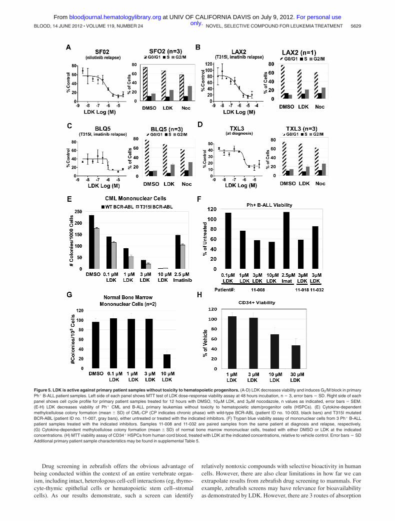
LDK kills the majority of primary human leukemia cells
To interrogate the clinical relevance of our data, we asked whetherLDK had activity against primary human leukemia samples. Wetested several primary B-ALL samples either grown directly inculture (supplemental Figure 9, supplemental Table 6) or on OP9stromal cells (Figure 5A-D, supplemental Table 5) includingtreatment-refractory, relapsed BCR-ABL B-ALL and T315I-mutated BCR-ABL positive B-ALL samples. We found thatproliferation was strongly inhibited in 85% of patient B-ALLsamples and that they exhibited G2/M delay, similar to Jurkat cells(Figure 5). As the NCI60 data indicated that LDK might haveactivity against CML, we also tested 2 primary samples frompatients with imatinib-resistant CML, one with wild-type BCR-ABL, the other with the T315I mutation. Colony assays demon-strated strong activity of LDK against both samples, whereas highdoses of imatinib had only mild effects (Figure 5E). Finally, wetested 3 primary imatinib-resistant Ph� B-ALL samples for viabil-ity in the presence of LDK (Figure 5F). Samples 11-008 and 11-032are paired diagnosis and relapse samples, with the relapse sampleshowing decreased sensitivity to LDK treatment. By contrast, LDKdid not compromise health of normal bone marrow mononuclearcells at concentrations up to 3�M (Figure 5G). Similarly, the IC50
of proliferating human CD34 positive cord blood cells was 8-foldhigher than leukemia cells (Figure 5H), supporting LDK’s therapeu-tic window we had observed with normal PB mononuclear cells(Figure 2E). In sum, LDK was active against the majorityof primary human leukemia samples, and led to G2/M delayin Ls cells.
Discussion
The screen described herein represents the first successful andunbiased approach using zebrafish larvae to identify moleculeswith potency against a human cancer from a small molecule librarycontaining compounds with unknown activity. Zebrafish have beenincreasingly used to screen for bioactive compounds with possibleclinical relevance. For example, a screen of 2000 well-character-ized compounds (Spectrum Library, MicroSource Discovery Sys-tem) revealed PGE2 as a stimulator of hematopoietic stem cellgrowth,32 a finding that is now being translated into clinical trials(L. Zon, personal e-mail communication, December 13, 2011). Inanother screen of the Spectrum Library for active compounds in azebrafish model of acute myeloid leukemia (AML), Yeh et alidentified a new role for COX-2 and -catenin in AML1-ETO–induced AML.33 More recently, a screen of the Spectrum Libraryfor chemical suppressors of neural crest development revealed aclass of dihydroorotate dehydrogenase inhibitors that not onlyalmost completely abrogate neural crest development in zebrafishbut also markedly decrease melanoma growth both in vitro and inmouse xenograft studies.34
5628 RIDGES et al BLOOD, 14 JUNE 2012 � VOLUME 119, NUMBER 24 only.For personal use at UNIV OF CALIFORNIA DAVIS on July 9, 2012. bloodjournal.hematologylibrary.orgFrom
Drug screening in zebrafish offers the obvious advantage ofbeing conducted within the context of an entire vertebrate organ-ism, including intact, heterologous cell-cell interactions (eg, thymo-cyte-thymic epithelial cells or hematopoietic stem cell–stromalcells). As our results demonstrate, such a screen can identify
relatively nontoxic compounds with selective bioactivity in humancells. However, there are also clear limitations in how far we canextrapolate results from zebrafish drug screening to mammals. Forexample, zebrafish screens may have relevance for bioavailabilityas demonstrated by LDK. However, there are 3 routes of absorption
Figure 5. LDK is active against primary patient samples without toxicity to hematopoietic progenitors. (A-D) LDK decreases viability and induces G2/M block in primaryPh� B-ALL patient samples. Left side of each panel shows MTT test of LDK dose-response viability assay at 48 hours incubation, n � 3, error bars � SD. Right side of eachpanel shows cell cycle profile for primary patient samples treated for 12 hours with DMSO, 10�M LDK, and 3�M nocodazole, n values as indicated, error bars � SEM.(E-H) LDK decreases viability of Ph� CML and B-ALL primary leukemias without toxicity to hematopoietic stem/progenitor cells (HSPCs). (E) Cytokine-dependentmethylcellulose colony formation (mean SD) of CML-CP (CP indicates chronic phase) with wild-type BCR-ABL (patient ID no. 10-003, black bars) and T315I mutatedBCR-ABL (patient ID no. 11-007, gray bars), either untreated or treated with the indicated inhibitors. (F) Trypan blue viability assay of mononuclear cells from 3 Ph� B-ALLpatient samples treated with the indicated inhibitors. Samples 11-008 and 11-032 are paired samples from the same patient at diagnosis and relapse, respectively.(G) Cytokine-dependent methylcellulose colony formation (mean SD) of normal bone marrow mononuclear cells, treated with either DMSO or LDK at the indicatedconcentrations. (H) MTT viability assay of CD34� HSPCs from human cord blood, treated with LDK at the indicated concentrations, relative to vehicle control. Error bars � SDAdditional primary patient sample characteristics may be found in supplemental Table 5.
NOVEL, SELECTIVE COMPOUND FOR LEUKEMIA TREATMENT 5629BLOOD, 14 JUNE 2012 � VOLUME 119, NUMBER 24 only.For personal use at UNIV OF CALIFORNIA DAVIS on July 9, 2012. bloodjournal.hematologylibrary.orgFrom
in fishes after immersion in compound (gastrointestinal, gill, andskin), compared with only one after oral application in mammals,and only the gill route is not available in zebrafish larvae at 5 dpfwhen we carried out the screen. In addition, the exposure to drug isconstant and only restricted by stability in water during immersion,a different scenario from the intermittent oral dosing in mammals.Therefore, bioavailability in fishes may not accurately predict thesame in mammals. Finally, targets may have diverged duringevolution so that compounds that are active in zebrafish may notexhibit the desired effect in mammalian cells. This may be the casefor the 4 compounds that had specific activity in zebrafish but didnot kill Jurkat cells in our screen (Figure 2A).
Screening of large numbers of compounds is not practical inadult leukemic zebrafish. However, contrary to the AML model,35
currently no zebrafish T-ALL models exist that manifest early indevelopment to allow screening of thousands of compounds. Wedeveloped a 2-pronged screening strategy to identify targeted,nontoxic, antileukemia compounds in zebrafish larvae from a largechemical library. First, we reasoned that given their similardevelopmental stage, immature T cells and leukemic T lympho-blasts share similar pathways or biologic processes on which bothcritically depend. Consequently, compounds that eliminate theformer may also kill the latter. Our rationale was supported by ourprevious finding that dexamethasone, a mainstay of anti-ALLtreatment, eliminated immature T cells from the thymus ofdeveloping zebrafish larvae.21 Our data that killing of immaturezebrafish T cells and malignant human T-ALL lines by LDKinvolves indirect inhibition of the P/A/mT pathway (see below)bolstered our rationale as both types of cells critically depend onAKT activity for survival.14,26,36 Second, we sought to identifyactive molecules that are devoid of general toxicity. The capacity tointerfere with the cell cycle of dividing cells underpins the efficacyand toxicity of most chemotherapeutic agents. Despite species-specific variations, cell cycle regulation is highly conservedthroughout metazoan evolution37 and modulators of the cell cycleidentified in zebrafish are active in mammals and vice versa.38,39
We therefore reasoned that nontargeted compounds that are gener-ally toxic to mammalian cells could be detected and eliminated byscreening for cell cycle effects in developing zebrafish embryos(Figure 1D-H). Our finding that LDK has little toxicity indeveloping and adult zebrafish, mice, and nonmalignant humancells corroborates the rationale for our screening algorithm.
The P/A/mT pathway has become an attractive target for drugdevelopment16 and molecules have been identified that inhibit 140,41
or 227,42,43 components of the pathway. The targets of theseinhibitors are either the 3 enzymes themselves or kinases thatactivate the pathway, as for example PDK144 and mTORC2.45
These inhibitors act against a wide range of tumor cell lines thatdepend on P/A/mT signaling.46 LDK induces decreased phosphor-ylation state in members of the P/A/mT pathway and has potentactivity against leukemias. Several lines of evidence indicatethat LDK exerts an indirect effect on the P/A/mT pathway. First,extensive in vitro kinase assays clearly show that LDK does notsignificantly inhibit any of the members of the P/A/mT pathway.Second, in our serum starvation experiments, LDK exhibited markedlydelayed activity on AKT phosphorylation compared with the PI3Kinhibitor Ly294002. Third, LDK lacks activity against several AKT-dependent tumor lines, including glioblastoma and melanoma that arereadily killed by AKT inhibitors. We therefore conclude that LDK’saction on the P/A/mT pathway is indirect.
What is the underpinning of LDK’s leukemia selectivity?Although the molecular basis of LDK resistance versus sensitivity
remains the subject of future investigations, selective antileukemiaactivity could be explained by several mechanisms. These include ahematopoietic cell-restricted target (Figure 2, supplemental Figure2), unique sensitivity of lymphocytes47 to LDK’s action, ordifferential drug metabolism by resistant versus sensitive lines.48
Finally, the 2 activities of LDK may have to coincide forcytotoxicity. For example, hematopoietic cells can overcome cellcycle arrest in G2/M through growth factor-stimulated activation ofthe P/A/mT pathway.49 Thus, LDK’s ability to induce G2/M delaywhile simultaneously blocking the P/A/mT pathway, even aftergrowth factor stimulation (supplemental Figure 3D), appearsparticularly significant. A requirement for dual activity is bolsteredby our observation that in RS4;11 cells failure of LDK-mediatedG2/M delay correlates with resistance (Figure 2G, supplementalTable 4). We have shown that LDK’s 2 activities, P/A/mTinhibition and late mitosis delay, are independent of each other(Figure 4H-I), but whether LDK accomplishes selectivity byinterfering with single or multiple targets must be addressed infuture studies.
In our study, LDK was active as monotherapy against all T-ALLlines tested and 85% of primary leukemia samples, regardless ofPTEN status, resistance to glucocorticoids or other antileukemiacompounds that may compromise patient treatment. Furthermore,with our dosing regimen LDK lacks endorgan (supplementalFigure 7D) and hematopoietic toxicity (Figures 2E and 5G-H,supplemental Figure 8). Thus LDK is an attractive, nontoxiccompound that will form the basis of future endeavors to bringleukemia-selective treatments to the bedside.
Acknowledgments
The authors wish to acknowledge Ira Kraft, Jon Beck, RupengZhuo, Kalavathy Ramachandran, and Bradley Demarest for experttechnical assistance. Histology and immunohistochemistry wereperformed at the ARUP Institute for Clinical and ExperimentalPathology with the technical assistance of Sheryl Tripp. Cell linesand fish strains were a kind gift of Joshua Schiffman, RandyJensen, and Doug Grossman (University of Utah, Salt Lake City,UT); Adolfo Ferrando (Columbia University, New York, NY); andAndrew Kung, Alejandro Gutierrez, and Thomas Look (Dana-Farber Cancer Institute, Boston, MA). Dr Steven Grant (VirginiaCommonwealth University Medical Center, Richmond, VA) pro-vided the myr-AKT plasmid.
N.S.T. was supported by The Dana Foundation, The WilliamLawrence-Blanche Hughes Foundation, The Alex’s LemonadeStand Foundation, and the Huntsman Cancer Foundation. C.J.G.was supported by a grant from Genome Canada Competition IIIthrough the Ontario Genomics Institute. Core facilities of theHuntsman Cancer Institute, supported by National Cancer Institute grantP30 CA042014, and the University of Utah, supporting the CZARzebrafish research core facility, also contributed to this work.
Authorship
Contribution: S.R., W.L.H., D.J., and N.S.T. conceived the experi-ments; H.C., P.C., H.S., A.S., E.J.M., and D.A.J. designed andcarried out the zebrafish screen; D.A.J. carried out cell cycleanalysis and toxicity studies on zebrafish embryos and larvae;D.A.J., W.L.H., and S.W. performed MTT assays, cell cycle, andantiactivated caspase 3 assay; K.B. and M.S. synthesized LDK;L.B. and J.K.F. conceived and carried out in vivo testing of LDK in
5630 RIDGES et al BLOOD, 14 JUNE 2012 � VOLUME 119, NUMBER 24 only.For personal use at UNIV OF CALIFORNIA DAVIS on July 9, 2012. bloodjournal.hematologylibrary.orgFrom

adult leukemic zebrafish; A.A., R.R.M., and G.J.S. carried outpathologic and biochemical evaluation of LDK-treated mice andzebrafish; J.E.C. developed the LC/MS assay to measure LDKconcentrations; T.P., R.J., B.M., and C.J.G. conceived and per-formed Phosflow analysis, and efficacy testing of LDK in murineT-ALL lines and in primary human leukemias; A.E., M.M., M.D.,and W.L.H. conceived and performed further experiments onprimary human leukemia cells; and N.S.T. wrote the paper.
Conflict-of-interest disclosure: The authors declare no compet-ing financial interests.
The current affiliation for M.M. is Department of LaboratoryMedicine, University of California, San Francisco, San Francisco,CA 94143.
Correspondence: Nikolaus Sebastian Trede, The HuntsmanCancer Institute, University of Utah, 2000 Circle of Hope, SaltLake City, UT 84112; e-mail: [email protected].
References
1. National Cancer Institute, Surveillance Epidemiol-ogy and End Result. Stat Fact Sheets: Leukemia.Available at http://www.seer.cancer.gov/statfacts/html/leuks.html. Accessed December 10, 2011.
2. Smith MA, Gloeckler Ries LA, Gurney JG, RossJA. Leukemia. SEER Pediatric Monograph. Avail-able at seer.cancer.gov/publications/childhood/leukemia.pdf. National Cancer Institute. Ac-cessed December 10, 2011.
3. Pui CH, Relling MV, Downing JR. Acute lympho-blastic leukemia. N Engl J Med. 2004;350(15):1535-1548.
4. Goldberg JM, Silverman LB, Levy DE, et al.Childhood T-cell acute lymphoblastic leukemia:the Dana-Farber Cancer Institute acute lympho-blastic leukemia consortium experience. J ClinOncol. 2003;21(19):3616-3622.
5. Armstrong SA, Look AT. Molecular genetics ofacute lymphoblastic leukemia. J Clin Oncol.2005;23(26):6306-6315.
6. Ferrando AA, Look AT. Clinical implications of re-curring chromosomal and associated molecularabnormalities in acute lymphoblastic leukemia.Semin Hematol. 2000;37(4):381-395.
7. Yeoh EJ, Ross ME, Shurtleff SA, et al. Classifica-tion, subtype discovery, and prediction of out-come in pediatric acute lymphoblastic leukemiaby gene expression profiling. Cancer Cell. 2002;1(2):133-143.
8. Druker BJ, Tamura S, Buchdunger E, et al. Ef-fects of a selective inhibitor of the Abl tyrosinekinase on the growth of Bcr-Abl positive cells. NatMed. 1996;2(5):561-566.
9. Smith A. Screening for drug discovery: the lead-ing question. Nature. 2002;418(6896):453-459.
10. Weng AP, Ferrando AA, Lee W, et al. Activatingmutations of NOTCH1 in human T cell acute lym-phoblastic leukemia. Science. 2004;306(5694):269-271.
11. Gutierrez A, Sanda T, Grebliunaite R, et al. Highfrequency of PTEN, PI3K, and AKT abnormalitiesin T-cell acute lymphoblastic leukemia. Blood.2009;114(3):647-650.
12. Lai EC. Notch cleavage: Nicastrin helps Preseni-lin make the final cut. Curr Biol. 2002;12(6):R200-202.
13. Palomero T, Dominguez M, Ferrando AA. Therole of the PTEN/AKT Pathway in NOTCH1-in-duced leukemia. Cell Cycle. 2008;7(8):965-970.
14. Palomero T, Sulis ML, Cortina M, et al. Mutationalloss of PTEN induces resistance to NOTCH1 in-hibition in T-cell leukemia. Nat Med. 2007;13(10):1203-1210.
15. Medyouf H, Gao X, Armstrong F, et al. Acute T-cell leukemias remain dependent on Notch sig-naling despite PTEN and INK4A/ARF loss. Blood.2010;115(6):1175-1184.
16. Yap TA, Garrett MD, Walton MI, et al. Targetingthe PI3K-AKT-mTOR pathway: progress, pitfalls,and promises. Curr Opin Pharmacol. 2008;8(4):393-412.
17. Cullion K, Draheim KM, Hermance N, et al. Tar-geting the Notch1 and mTOR pathways in amouse T-ALL model. Blood. 2009;113(24):6172-6181.
18. Meeker ND, Trede NS. Immunology and ze-brafish: spawning new models of human disease.Dev Comp Immunol. 2008;32(7):745-757.
19. Frazer JK, Meeker ND, Rudner L, et al. HeritableT-cell malignancy models established in a ze-brafish phenotypic screen. Leukemia. 2009;23(10):1825-1835.
20. Langenau DM, Traver D, Ferrando AA, et al. Myc-induced T cell leukemia in transgenic zebrafish.Science. 2003;299(5608):887-890.
21. Langenau DM, Ferrando AA, Traver D, et al. Invivo tracking of T cell development, ablation, andengraftment in transgenic zebrafish. Proc NatlAcad Sci U S A. 2004;101(19):7369-7374.
22. Gutierrez A, Grebliunaite R, Feng H, et al. Ptenmediates Myc oncogene dependence in a condi-tional zebrafish model of T cell acute lymphoblas-tic leukemia. J Exp Med. 2011;208(8):1595-1603.
23. Barlow C, Hirotsune S, Paylor R, et al. Atm-deficient mice: a paradigm of ataxia telangiecta-sia. Cell. 1996;86(1):159-171.
24. Fusetti L, Pruneri G, Gobbi A, et al. Human my-eloid and lymphoid malignancies in the non-obese diabetic/severe combined immunodefi-ciency mouse model: frequency of apoptotic cellsin solid tumors and efficiency and speed of en-graftment correlate with vascular endothelialgrowth factor production. Cancer Res. 2000;60(9):2527-2534.
25. Masiero M, Minuzzo S, Pusceddu I, et al. Notch3-mediated regulation of MKP-1 levels promotessurvival of T acute lymphoblastic leukemia cells.Leukemia. 2011;25(4):588-598.
26. Jones RG, Parsons M, Bonnard M, et al. Proteinkinase B regulates T lymphocyte survival, nuclearfactor kappaB activation, and Bcl-X(L) levels invivo. J Exp Med. 2000;191(10):1721-1734.
27. Chiarini F, Fala F, Tazzari PL, et al. Dual inhibitionof class IA phosphatidylinositol 3-kinase andmammalian target of rapamycin as a new thera-peutic option for T-cell acute lymphoblastic leuke-mia. Cancer Res. 2009;69(8):3520-3528.
28. Sachs K, Perez O, Pe’er D, et al. Causal protein-signaling networks derived from multiparametersingle-cell data. Science. 2005;308(5721): 523-529.
29. McManus KJ, Hendzel MJ. The relationship be-tween histone H3 phosphorylation and acetyla-tion throughout the mammalian cell cycle.Biochem Cell Biol. 2006;84(4):640-657.
30. Morgan D. The Cell Cycle. London, United King-dom: Oxford University Press; 2007.
31. Juntilla MM, Koretzky GA. Critical roles of thePI3K/Akt signaling pathway in T cell develop-ment. Immunol Lett. 2008;116(2):104-110.
32. North TE, Goessling W, Walkley CR, et al. Pros-taglandin E2 regulates vertebrate haematopoieticstem cell homeostasis. Nature. 2007;447(7147):1007-1011.
33. Yeh JR, Munson KM, Elagib KE, et al. Discover-ing chemical modifiers of oncogene-regulatedhematopoietic differentiation. Nat Chem Biol.2009;5(4):236-243.
34. White RM, Cech J, Ratanasirintrawoot S, et al.DHODH modulates transcriptional elongation in
the neural crest and melanoma. Nature. 2011;471(7339):518-522.
35. Yeh JR, Munson KM, Chao YL, et al. AML1-ETOreprograms hematopoietic cell fate by downregu-lating scl expression. Development. 2008;135(2):401-410.
36. Huo J, Xu S, Lam KP. Fas apoptosis inhibitorymolecule regulates T cell receptor-mediated apo-ptosis of thymocytes by modulating Akt activationand Nur77 expression. J Biol Chem. 2010;285(16):11827-11835.
37. DePamphilis ML, Blow JJ, Ghosh S, et al. Regu-lating the licensing of DNA replication origins inmetazoa. Curr Opin Cell Biol. 2006;18(3):231-239.
38. Murphey RD, Stern HM, Straub CT, et al. Achemical genetic screen for cell cycle inhibitors inzebrafish embryos. Chem Biol Drug Des. 2006;68(4):213-219.
39. Stern HM, Murphey RD, Shepard JL, et al. Smallmolecules that delay S phase suppress a ze-brafish bmyb mutant. Nat Chem Biol. 2005;1(7):366-370.
40. Feldman RI, Wu JM, Polokoff MA, et al. Novelsmall molecule inhibitors of 3-phosphoinositide-dependent kinase-1. J Biol Chem. 2005;280(20):19867-19874.
41. Martelli AM, Tazzari PL, Tabellini G, et al. A newselective AKT pharmacological inhibitor reducesresistance to chemotherapeutic drugs, TRAIL,all-trans-retinoic acid, and ionizing radiation ofhuman leukemia cells. Leukemia. 2003;17(9):1794-1805.
42. Fan QW, Knight ZA, Goldenberg DD, et al. A dualPI3 kinase/mTOR inhibitor reveals emergent effi-cacy in glioma. Cancer Cell. 2006;9(5):341-349.
43. Janes MR, Limon JJ, So L, et al. Effective andselective targeting of leukemia cells using aTORC1/2 kinase inhibitor. Nat Med. 2010;16(2):205-213.
44. Mora A, Komander D, van Aalten DM, et al.PDK1, the master regulator of AGC kinase signaltransduction. Semin Cell Dev Biol. 2004;15(2):161-170.
45. Sarbassov DD, Guertin DA, Ali SM, et al. Phos-phorylation and regulation of Akt/PKB by the ric-tor-mTOR complex. Science. 2005;307(5712):1098-1101.
46. Serra V, Markman B, Scaltriti M, et al. NVP-BEZ235, a dual PI3K/mTOR inhibitor, preventsPI3K signaling and inhibits the growth of cancercells with activating PI3K mutations. Cancer Res.2008;68(19):8022-8030.
47. Carson DA, Kaye J, Matsumoto S, et al. Bio-chemical basis for the enhanced toxicity of de-oxyribonucleosides toward malignant human Tcell lines. Proc Natl Acad Sci U S A. 1979;76(5):2430-2433.
48. Higgins CF. Multiple molecular mechanisms formultidrug resistance transporters. Nature. 2007;446(7137):749-757.
49. Henry MK, Lynch JT, Eapen AK, et al. DNA dam-age-induced cell-cycle arrest of hematopoieticcells is overridden by activation of the PI-3 ki-nase/Akt signaling pathway. Blood. 2001;98(3):834-841.
NOVEL, SELECTIVE COMPOUND FOR LEUKEMIA TREATMENT 5631BLOOD, 14 JUNE 2012 � VOLUME 119, NUMBER 24 only.For personal use at UNIV OF CALIFORNIA DAVIS on July 9, 2012. bloodjournal.hematologylibrary.orgFrom