york mri facility
TRANSCRIPT
York MRI Facility
Standard Operating Procedures
Index Disclaimer Glossary of Terms Personnel and Emergency Contacts SOP #10-02 Restricted Access Policy SOP #15-01 Maintenance Staff Access and Alarm Response SOP #20-01 New Protocols and Ethics Procedures SOP #30-02 Safety Training Procedures SOP #31-02 General Safety Procedures SOP #40-02 Medical Emergency Procedure SOP #41-01 Emergency Fire Procedure SOP #42-02 Emergency Quench Procedure SOP #50-01 General Experimental Procedures SOP #51-01 MRI Equipment Handling Procedures SOP #52-02 MRI Data Handling SOP #53-01 Incidental Findings SOP #54-01 Drawing Blood SOP #60-02 MRI System Start-Up SOP #61-02 MRI System Shutdown SOP #62-01 MRI Black/Brown Out SOP #63-01 Oxygen Sensor Alarm Response SOP #64-02 Compressor Alarm Response SOP #65-01 QA Testing SOP #70-01 System Billing Guide and Standard Rates SOP #71-01 Cancellation / No Show Policy SOP #80-01 Decontamination Procedure SOP #90-01 MRI Simulator Usage and Booking Policy
York MRI Facility
Standard Operating Procedures
Disclaimer The information contained in the York MRI Facility standard operating procedures (SOPs) are provided by the Facility Management and are intended for the sole use of the management and users of this research facility. Note that these guidelines do not necessarily represent the governing rules and guidelines of other research and/or clinical MRI sites. By downloading any of the information contained in the York MRI Facility standard operating procedures, you agree to the terms and provisions as outlined in this notice. The information contained in these SOPs may not be modified without the prior written permission of the York MRI Facility Management. The management welcomes your feedback regarding all areas of these SOPs. If you want to send us your comments please contact [email protected].
York MRI Facility
Standard Operating Procedures
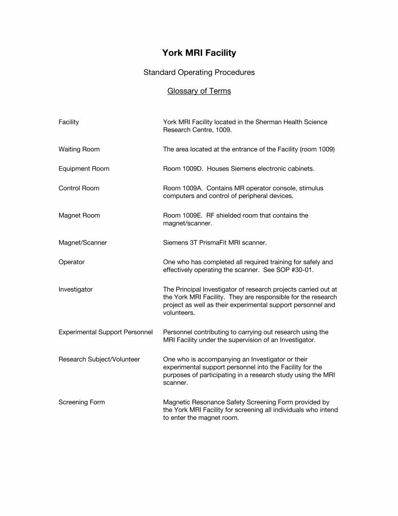
Glossary of Terms Facility York MRI Facility located in the Sherman Health Science
Research Centre, 1009.
Waiting Room The area located at the entrance of the Facility (room 1009)
Equipment Room Room 1009D. Houses Siemens electronic cabinets.
Control Room Room 1009A. Contains MR operator console, stimulus computers and control of peripheral devices.
Magnet Room Room 1009E. RF shielded room that contains the magnet/scanner.
Magnet/Scanner Siemens 3T PrismaFit MRI scanner.
Operator One who has completed all required training for safely and effectively operating the scanner. See SOP #30-01.
Investigator The Principal Investigator of research projects carried out at the York MRI Facility. They are responsible for the research project as well as their experimental support personnel and volunteers.
Experimental Support Personnel Personnel contributing to carrying out research using the MRI Facility under the supervision of an Investigator.
Research Subject/Volunteer One who is accompanying an Investigator or their experimental support personnel into the Facility for the purposes of participating in a research study using the MRI scanner.
Screening Form Magnetic Resonance Safety Screening Form provided by the York MRI Facility for screening all individuals who intend to enter the magnet room.
York MRI Facility
Standard Operating Procedures
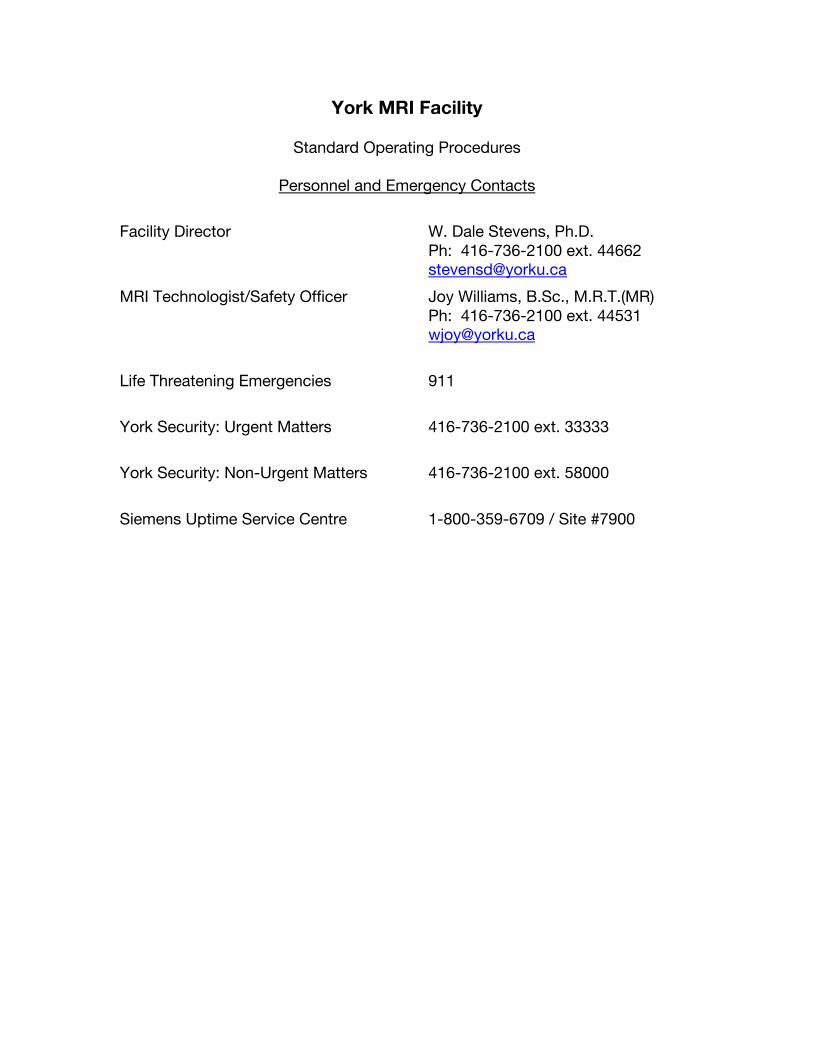
Personnel and Emergency Contacts Facility Director W. Dale Stevens, Ph.D.
Ph: 416-736-2100 ext. 44662 [email protected]
MRI Technologist/Safety Officer Joy Williams, B.Sc., M.R.T.(MR) Ph: 416-736-2100 ext. 44531 [email protected]
Life Threatening Emergencies 911
York Security: Urgent Matters 416-736-2100 ext. 33333
York Security: Non-Urgent Matters 416-736-2100 ext. 58000
Siemens Uptime Service Centre 1-800-359-6709 / Site #7900
York MRI Facility
Standard Operating Procedure #10-02 Last Updated: 16 Dec 2015
Restricted Access Policy
1. Introduction
1.1. The 3T MRI scanner at the York MRI Facility is used primarily for in-vivo studies of
human and animal structure and function. These studies include assessment of cognitive function and vascular dynamics, metabolism and physiology in normal and research patient populations. The facility resources are available to R & D phase medical device development and academic research, with appropriate Review Ethics Board protocols in place, see SOP #20-01 “New Protocols and Ethics Procedures”.
1.2. Research involving Magnetic Resonance Imaging (MRI) at high magnetic field strengths present unique hazards to both research subjects and individuals working within and around the MRI system. Consequently, the potential for serious personal injury is present due to the sheer size and strength of the static magnetic field along with the flexibility of the research system and associated peripheral hardware.
1.3. The static magnetic field in the York MRI Facility is always present. It is important that
all those entering the facility be aware of the presence of the field, as it cannot be detected in any way, i.e. magnetic fields cannot be felt, seen or smelled. Ferromagnetic objects brought into the magnet room could quickly become dangerous projectiles, and the magnetic field can also interfere with the operation of certain medical implants.
2. Zones 2.1. The MRI suite is divided into separate safety zones, by standard convention labeled I –
IV. 2.1.1. Zone I consists of all areas outside the MRI suite, accessible to the general public. 2.1.2. Zone II is located inside the secure access door to the York MRI Facility, Sherman
1009, and includes the MRI waiting room, and the Analysis laboratory. 2.1.3. Zone III is located inside the second secure access door, Sherman 1009A, and
includes the MRI control room, the MRI equipment room and prep rooms. This zone contains several areas where magnetic fields exceed 5 Gauss.
2.1.4. Zone IV is the magnet room itself. This entire room exceeds the 5 Gauss magnetic field safety limit. The door to the magnet room must be kept closed at all times, unless entering or exiting, and must be locked when the scanner is not in use.
3. Entry Regulations
3.1. Zone I 3.1.1. Entry to Zone I is not restricted in any way. The general public has access to
Zone I.
3.2. Zone II 3.2.1. Entry to Zone II, Sherman 1009, is restricted by the secure access door at the
entrance to the waiting room. 3.2.2. There is no training required to obtain security access to the waiting room.
|SOP#10-022
3.2.3. There is open access from the waiting room to the Analysis laboratory.
3.3. Zone III 3.3.1. Entry to Zone III, Sherman 1009A (the control room, equipment room, prep rooms
and washroom) is restricted to personnel who have completed Level 1 or Level 2 safety training, or to those who are under the supervision of Level 2 personnel.
3.3.2. It is very important to monitor the location of those who have not completed safety training as there are areas in Zone III that exceed magnetic field strengths of 5 Gauss.
3.4. Zone IV 3.4.1. Entry to Zone IV (the magnet room) is restricted to personnel who have completed
Level 1 or Level 2 safety training, and to those who are under the supervision of the Level 2 personnel.
3.4.2. All those with intent to enter the magnet room MUST complete a “Magnetic Resonance Safety Screening Form” and have it reviewed and approved by a Level 2 individual before entering the magnet room.
4. Security Access Procedure 4.1. Security access to the waiting room door (Zone II, Sherman 1009)
4.1.1. Present your request in writing by email to the MRI Technologist or the Facility Director and including the following information: your name, access card number and a reason for the request.
4.2. Security access to the control room door (Zone III, Sherman 1009A) 4.2.1. Those wishing to obtain access to the control room door must complete Level 1
Safety Training as described in SOP #30-02 “Safety Training Procedures”. 4.2.2. All operators of the 3T MRI scanner must obtain security access to the control
room door by following the procedures outlined in SOP #30-02 “Safety Training Procedures”. Operators wishing to scan only phantoms are required to complete Level 1 Safety Training. Those who will be scanning human subjects on the 3T MRI scanner must complete Level 2 Safety Training.
4.2.3. Security access to the control room will not be granted to any individuals with medical devices, implants or objects as listed in SOP #31-02 “General Safety Procedures”.
4.2.4. If a person displays inappropriate and/or unsafe behavior in the York MRI Facility he/she may be denied access by the operator, MRI Technologist or Facility Director.
Oct. 31, 2011
York MRI Facility
Standard Operating Procedure #15-01 Last Updated: 16 Dec 2015
Procedure for Maintenance Staff Entering MRI
1. Introduction
Rm. 1009 Sherman Health Science Research Centre contains a Magnetic Resonance Imaging (MRI) scanner in Rm. 1009E that generates a high-strength (3 Tesla) magnetic field. The magnetic field in Rm. 1009E is always present but is shielded so that the field outside the room is reduced to the safety limit of 5 gauss (G). Areas where the field exceeds 5 gauss are marked with a permanent red line on the floor. Rm. 1009 includes: Rm. 1009 Waiting area (below 5G) Rm. 1009A Control room (below 5G) Rm. 1009B,C Change area and washroom (below 5 G) Rm. 1009D Equipment room (3–20G) Rm. 1009E Scanner room (above 20 G)
2. Access Authority and Control Access to Rm.1009 and 1009A are restricted by use of an access card. The provision of the access card is authorized only by the MRI Safety Officer or the York MRI Facility Director. Certain emergency response (e.g., Security Services) and maintenance staff may have access to this room with a master key or card. The contact information for the York MRI Facility Director and the MRI Safety Officer are available in the York MRI Facility Contacts document. (in an emergency, Security Services and DOHS have emergency contact numbers for the Facility
Director and MRI Safety Officer)
3. Procedure for entering Rm. 1009 Sherman by maintenance staff 3.1. No one is allowed to enter the magnet room (Rm. 1009E) without
authorization and special extensive training and screening by the MRI Safety Officer or Facility Director.
3.2. Maintenance persons entering Rm. 1009A, B or C (i.e. not including the
equipment room and magnet room) must:
Oct. 31, 2011
3.2.1. Complete MRI Safety Awareness Training (Training is provided by the MRI Safety Officer).
3.2.2. Inform MRI Safety Officer and/or Facility Director in advance as to the purpose and time of entry.
3.3. Maintenance persons entering Rm.1009D (Equipment room), must:
3.3.1. Inform/email MRI Safety Officer and/or Facility Director in advance as to the person’s name, purpose and time of entry,
3.3.2. Complete MRI Safety Awareness Training (Training is provided by the MRI Safety Officer), and
3.3.3. Complete MRI screening form administered by MRI Safety Officer or Facility Director (this can be done any time in advance of entrance).
In an emergency/urgent situation where immediate access is required before completing 3.3.2 or 3.3.3, the worker must be accompanied by the Facility Director or MRI Safety Officer.
4. Procedure for Responding to a Chiller alarm in the MRI Facility During Off Hours.
4.1. When the chiller alarm is sounding, and it is not supplying chilled water to the
MRI equipment, the water temperature in the chiller loop will increase to temperatures greater than 15 °C quite quickly.
4.2. If the chilled water temperatures are above 15 °C in the equipment room, put the chiller into city bypass. Lift the cover labeled “chiller bypass switch”, and toggle the Dump Valve to the ON position.
4.3. Make note of the error code on the chiller.
4.4. Contact Carrier for service/repair if necessary.
4.5. Contact the Facility Director or MRI Safety Officer to inform them of the status
of the equipment and actions taken.
5. Procedure for Responding to an Oxygen Sensor alarm in the MRI Facility During Off Hours.
5.1. If Maintenance has been contacted by persons in the MRI control room that the
Oxygen sensor is in an error state (no LED illuminated), call Honeywell and request an emergency repair (see http://www.yorku.ca/mri SOP #63-01 “Oxygen Sensor Alarm Response” for reference)
5.2. If the Oxygen sensor is in an alarm state (red LED illuminated and audible alarm sounding), this may indicate that helium levels may have saturated the air.
Oct. 31, 2011
5.2.1. Immediately turn ON the “Magnet Room Exhaust Fan” located to the left of the oxygen sensor monitoring panel. This engages a roof top ventilation fan that will draw the air out of the magnet room.
5.2.2. Evacuate the magnet room.
5.2.3. If any individual is not responding, not breathing and has no pulse, follow
the procedure outlined in SOP #40-02 “Medical Emergency Procedure”.
5.2.4. Once the magnet room has been evacuated, close the magnet room door.
5.2.5. Notify the Facility Director or Safety Officer and York Security Ext. 33333, immediately following the incident.
(see http://www.yorku.ca/mri SOP #63-01 “Oxygen Sensor Alarm Response” for reference)
.
York MRI Facility
Standard Operating Procedure #20-01 Last Updated: 16 Dec 2015
New Protocols and Ethics Procedures
1. Introduction 1.1. The 3T MRI scanner at the York MRI Facility is used primarily for in-vivo studies of
human and animal structure and function. These studies include assessment of cognitive function and vascular dynamics, metabolism and physiology in normal and research patient populations. The facility resources are available to R & D phase medical device development and academic research, with appropriate Review Ethics Board protocols in place, see below.
2. New Protocols 2.1. Any Investigator wishing begin a new study using the 3T MRI scanner at the York MRI
Facility must complete the following: 2.1.1. All studies involving human subjects or animals, must be approved by the Human
Participants Review Committee (HPRC) or the Animal Care Committee (ACC) respectively.
2.1.2. A copy of the Ethics Approval Certificate and the Consent Form must be submitted to the MRI Technologist prior to commencing the study.
2.1.3. The Cost Centre Number to be charged for scan time, or the name and address to be invoiced must also be submitted to the MRI Technologist.
York MRI Facility
Standard Operating Procedure #30-03 Last Updated: 14 Nov 2016
Safety Training Procedures
1. Introduction 1.1. The 3T MRI scanner at the York MRI Facility is used primarily for in-vivo studies of
human and animal structure and function. These studies include assessment of cognitive function and vascular dynamics, metabolism and physiology in normal and research patient populations. The facility resources are available to R & D phase medical device development and academic research, with appropriate Review Ethics Board protocols in place, see SOP #20-01 “New Protocols and Ethics Procedures”.
1.2. Research involving Magnetic Resonance Imaging (MRI) at high magnetic field strengths present unique hazards to both research subjects and individuals working within and around the MRI system. Consequently, the potential for serious personal injury is present due to the sheer size and strength of the static magnetic field along with the flexibility of the research system and associated peripheral hardware.
1.3. The static magnetic field in the York MRI Facility is always present. It is important that
all those entering the facility be aware of the presence of the field, as it cannot be detected in any way, i.e. magnetic fields cannot be felt, seen or smelled. Ferromagnetic objects brought into the magnet room could quickly become dangerous projectiles, and the magnetic field can also interfere with the operation of certain medical implants.
2. MRI Safety Training
2.1. All individuals wishing to complete MRI Safety Training, must complete an MRI Safety
Screening Form and have it reviewed by the MRI Technologist or MRI Safety Officer. If the MRI Technologist or the MRI Safety Officer have found that the individual is not safe to enter the magnet room, the individual may still attend the MRI Safety Training, but may not enter the magnet room during the training.
2.2. MRI Safety Training consists of the following:
2.2.1. Read the Standard Operating Procedures and sign the signature sheet indicating
that you understand and will follow them. 2.2.2. Read “Part B: MR System components” of the Operator Manual (skip section B.3
“In-Room syngo Acquisition Workplace”). 2.2.3. Attend a Safety Instruction Session with the MRI Safety Officer. This session will
include: 2.2.3.1. Watching the first 12 minutes of the Siemens MRI Safety Video. 2.2.3.2. Overview of general safety procedures in the MRI environment. 2.2.3.3. Overview of all Emergency Procedures and locations of Table Stop,
Electrical Stop and Quench buttons. 2.2.3.4. Thorough review of how to make oneself safe to enter the magnet room.
2.2.4. Complete the MRI Safety Training test. A grade of 70% must be obtained to pass the test.
|SOP#30-03
2
2.3. After completion of MRI Safety Training the participant will:
2.3.1. Know how to make him- or herself safe to enter the MR environment. 2.3.2. Understand the dangers of a static magnetic field. 2.3.3. Be familiar with the Standard Operating Procedures. 2.3.4. Know all emergency procedures including the operation of the table/sequence
stop, electrical stop and quench buttons. 2.3.5. Pass the MRI Safety Training test.
2.4. Restrictions of MRI Safety training include:
2.4.1. The individual may not screen others to enter the MR environment. 2.4.2. The individual may not operate the MR scanner on human or animal subjects.
2.5. The individual must then be certified by either the Facility Director or the MRI Safety
Officer.
2.6. Card access to Zone II (the MRI waiting room, Sherman 1009) will be granted after successful completion of MRI Safety Training.
3. Safety Awareness Training 3.1. All staff and contractors working within the York MRI facility, must be trained on the
safety concerns of the MRI environment. This training includes:
3.1.1. Attending an MRI Safety Awareness training session lead by either the Facility Director or the MRI Safety Officer.
3.1.2. Completing an MRI Safety Screening form and having it approved by the MRI Technologist. If the screening form is not approved by the MRI Technologist due to medical reasons, the individual may not cross the 5 Gauss line (indicated by permanent red lines on the floor of the facility and by signs).
York MRI Facility
Standard Operating Procedure #31-02 Last Updated: 17 Dec 2015
General Safety Procedures
1. Introduction
1.1. Research involving Magnetic Resonance Imaging (MRI) at high magnetic field strengths present unique hazards to both research subjects and individuals working within and around the MRI system. Consequently, the potential for serious personal injury is present due to the sheer size and strength of the static magnetic field along with the flexibility of the research system and associated peripheral hardware.
1.2. The static magnetic field in the York MRI Facility is always present. It is important that
all those entering the facility be aware of the presence of the field, as it cannot be detected in any way, i.e. magnetic fields cannot be felt, seen or smelled. Ferromagnetic objects brought into the magnet room could quickly become dangerous projectiles, and the magnetic field can also interfere with the operation of certain medical implants.
1.3. During MRI data acquisition the subject being imaged is also exposed to rapidly
changing magnetic fields due to pulsed magnetic field gradients, and fields oscillating at radiofrequencies (around 128 MHz for 3 T). These time-varying fields are much weaker than the static field (up to 10 mT or 100 gauss) but create additional safety risks and all personnel working with the MRI equipment must be aware of these risks.
1.4. During certain types of MRI data collection, there exists high, and therefore potentially
dangerous, acoustic sound pressure levels (SPL). All those entering the facility must be made aware of this risk and be instructed as to the proper precautionary measures to be taken. Any patients, volunteers and/or research personnel present in the magnet room during an MRI experiment must wear appropriate hearing protection as outlined below.
1.5. As a result of the potential for serious injury, access to the York MRI Facility is
restricted, and requires permission. See SOP #10-02 “Restricted Access Policy”.
1.6. Any person with intent to enter the Magnet Room must complete a “Magnetic Resonance Safety Screening Form” and have it reviewed and approved by a Level 2 individual before entering the magnet room.
2. Incompatible Medical Devices
2.1. There are medical devices, implants and objects that are incompatible with the MR environment. A copy of Frank Shellock’s “Reference Manual for Magnetic Resonance Safety, Implants, and Devices” is available in the Control Room at the York MRI Facility. Any individual with a device listed as Not Safe or not listed in this reference manual may not proceed beyond the 5 Gauss line unless the object can be safely removed. The 5 Gauss line is marked by the red line at the magnet room door and the edge of the counter facing the magnet room window. There are also areas exceeding 5 Gauss in the Equipment room, restricting access to this area as well.
3. Safety Issues – Due to High Static Magnetic Field Strengths
|SOP#31-022
3.1. High static magnetic field strengths are present in the York MRI Facility. These strong
magnetic fields pose potential risks to those working, volunteering, or touring in the environment. Medical safety is very important hence, everyone entering the environment must be aware of the potential dangers.
3.2. All metallic objects have the potential to become projectiles in the MR environment, as
they may contain ferrous components. This could potentially cause serious injury to anyone near the magnet, and damage to the MRI system. As a result, objects entering the magnet room are restricted.
3.2.1. The operator is responsible to screen all objects entering the magnet room for
ferrous components. 3.2.2. All objects not already in the magnet room, should not be brought into the magnet
room unless they are necessary for the successful execution of the experiment, and have been tested using a permanent magnet in the control room, or have been viewed and permitted for entry by the Facility Director or MRI Safety Officer.
3.2.3. There are several metals that are non-ferrous. These metals include titanium, copper, gold, silver, aluminum, brass and lead. It is extremely important to note that all metal objects must be tested or permitted for entry by the Facility Director or MRI Safety Officer, even if they are thought to have no ferrous components.
3.3. It is mandatory to remove all personal metallic objects from your person before
crossing the 5 Gauss line as marked by the door threshold and on signs on the magnet room door. This includes but is not limited to the following list of articles:
Hearing Aids Dentures Pager Cell Phone Keys Eyeglasses Hair Pins Wigs Barrettes Jewellery-including body piercings Watch
Safety Pins Paperclips Magnetic Strip Cards Coins Pens Pocket Knife Nail Clippers Foil backed Medication Patches Steel-toed boots/shoes Tools
3.4. It is extremely important that no large metal objects be brought into or near the magnet
room at any time. All large metal objects must not go beyond the 5 Gauss line as marked on the floor and on signs on the walls, unless specifically directed by the Siemens Service Technician or the Facility Director. A large metal object with ferrous properties, placed to near the magnet, will fly toward the magnet with great force, potentially causing serious injury to anyone near the magnet and damage to the MRI system.
3.4.1. In such an instance, if someone is pinned to the magnet, trapped or potentially in
a life-threatening situation as the result of a large ferrous object coming too near the magnet, the operator, or if the operator is pinned, one of the experimental support personnel, must follow SOP #42-02 “Emergency Quench Procedure”, and apply first responder principles.
4. Safety Issues – Due to Rapidly Changing Magnetic Fields
|SOP#31-023
4.1. Rapidly changing magnetic fields are generated by gradient and RF coils in the MRI scanner during data acquisition. These fields pose potential risks for volunteers being scanned and for personnel working in the bore during the scan. To minimize the risk of burns it is important to prevent skin-to-skin contact points and the formation of “closed-loops” from touching body parts. Contact between skin and the transmitting body coil must also be avoided. This can be achieved by using foam padding available on the shelves in the magnet room.
4.2. Objects with good electrical conductance must not be permitted to rest against the skin
of the subject within the region spanned by the body coil, and should not be permitted within the body coil unless essential for the successful execution of the study.
4.3. During certain types of MRI data collection, there exists high, and therefore potentially
dangerous acoustic sound pressure levels (SPL). It is mandatory for the volunteer/patient, and all others who will be present in the magnet room during the scan session to wear hearing protection either in the form of earplugs or headphones provided by the 3T MRI Facility.
5. Safety Issues – Due to Electrical Power
5.1. There exist dangerous and potentially lethal level of electricity in the 3T MRI system. As such, it is important that all individuals working around the MRI system be aware of the danger and therefore knowledgeable as to the safety issues concerning electricity. There is a risk of electric shock from extremely high voltages, possibly causing severe injury or death, and damage to the MRI system. Only trained personnel should set up hardware in the magnet room and plug in or change the placement of any cables. High currents in wires also pose a risk of fire.
5.1.1. If someone is electrocuted in the 3T MRI Facility, the operator, or if the operator
was electrocuted, one of the experimental support personnel, must follow the procedure outlined in SOP #40-02 “Medical Emergency Procedure”.
5.1.2. In the case of a fire, the operator must follow the procedure outlined in SOP #41-01 “Emergency Fire Procedure”. The operator must keep his/her own safety in mind as a priority while removing the volunteer/patient from the magnet. If at this time the volunteer/patient is not responding, not breathing and has no pulse, the operator must follow the procedure outlined in SOP #40-02 “Medical Emergency Procedure”. After all parties are safe, it is appropriate to seek to minimize damage to the system.
York MRI Facility
Standard Operating Procedure #40-02 Last Updated: 27 Nov 2018
Medical Emergency Procedure
1. Introduction
1.1. Research involving Magnetic Resonance Imaging (MRI) at high magnetic field strengths present unique hazards to both research subjects and individuals working within and around the MRI system. Consequently, the potential for serious personal injury is present due to the sheer size and strength of the static magnetic field along with the flexibility of the research system and associated peripheral hardware.
1.2. The static magnetic field in the York MRI Facility is always present. It is important that
all those entering the facility be aware of the presence of the field, as it cannot be detected in any way, i.e. magnetic fields cannot be felt, seen or smelled. Ferromagnetic objects brought into the magnet room could quickly become dangerous projectiles, and the magnetic field can also interfere with the operation of certain medical implants.
1.3. As a result of the potential for serious injury, access to the York MRI Facility is
restricted, and requires permission. See SOP #10-02 “Restricted Access Policy”.
1.4. Working within and around the high field MRI requires in depth training on safety and Standard Operating Procedures, and documented proof of other necessary training. See SOP #30-02 “Safety Training Procedures”.
1.5. It is imperative that all personnel who are within and around the York MRI Facility
always keep in mind the potential safety risks, and act in accordance with the guidelines set out in the Standard Operating Procedures.
2. Medical Emergency
2.1. A medical emergency is specifically defined as a situation in which a person requires immediate medical attention, because of an acute injury or illness that poses an immediate risk to a person's life or long-term health.
2.2. Medical emergencies encountered in the MRI environment may include stroke, cardiac
arrest, impaled objects, crush wounds or burns.
2.3. If a medical emergency arises, the procedure set out below must be followed. 3. Emergency Procedure
3.1. Immediately STOP the scan (if in progress).
3.2. Remove the volunteer/patient from the scanner.
3.2.1. Either use the “Home” button to advance the bed out of the scanner, or 3.2.2. Pull the Emergency Release located under the support frame of the bed to unlock
the clutch, and pull the bed out manually to the Home Position.
|SOP#40-02 2
3.3. Assess the condition of the injured person.
3.4. If immediate medical assistance is required:
3.4.1. Call 911 (9-911 from a campus phone). 3.4.2. Call York Security x33333 (416-736-5333). 3.4.3. Prop open the Control Room door to facilitate access for the emergency response
team. 3.4.4. Have someone wait at the entrance to the York MRI Facility to let the paramedics
into the facility when they arrive. 3.4.5. The paramedics or other emergency responders must not enter the magnet room
unless they have been fully screened, and have removed all metal.
3.5. Remove the injured person from the magnet room either by aiding them while walking or by undocking the MRI bed as follows:
3.5.1. The bed must be in the Home Position. 3.5.2. Raise the side rails of the MRI bed. 3.5.3. Set the lateral pedal to the middle or upper position (must NOT be in lower
position, or brakes will be engaged). 3.5.4. Step on the left pedal to undock the table. 3.5.5. Use the handle at the foot of the bed and the side rails to move the bed to the
MRI Control Room. 3.5.6. Close the magnet room door.
3.6. Apply first responder principles or help the injured person according to your skills,
knowledge and comfort level. Follow instructions of emergency personnel when they arrive.
3.7. Once the emergency situation is over, report the incident to the Facility Director or the
Safety Officer.
York MRI Facility
Standard Operating Procedure #41-01 Last Updated: 27 Nov 2018
Emergency Fire Procedure
1. Introduction
1.1. Research involving Magnetic Resonance Imaging (MRI) at high magnetic field strengths present unique hazards to both research subjects and individuals working within and around the MRI system. Consequently, the potential for serious personal injury is present due to the sheer size and strength of the static magnetic field along with the flexibility of the research system and associated peripheral hardware.
1.2. The static magnetic field in the York MRI Facility is always present. It is important that
all those entering the facility be aware of the presence of the field, as it cannot be detected in any way, i.e. magnetic fields cannot be felt, seen or smelled. Ferromagnetic objects brought into the magnet room could quickly become dangerous projectiles, and the magnetic field can also interfere with the operation of certain medical implants.
1.3. Working within and around the high field MRI requires in depth training on safety and
Standard Operating Procedures, and documented proof of other necessary training. See SOP #30-02 “Safety Training Procedures”.
1.4. It is imperative that all personnel who are within and around the York MRI Facility
always keep in mind the potential safety risks, and act in accordance with the guidelines set out in the Standard Operating Procedures.
2. First Signs of Potential Fire
2.1. There are signs of a potential fire present before a fire occurs. Operators need to be aware of the signs to prevent injury to the volunteer/patient and other experimental support personnel in the magnet room, equipment room and the control room during a scan.
2.2. The first sign of a potential fire is often an irregular noise, for example a loud popping
sound or a sudden stop of the gradients. It is imperative that the operator determine the cause of the irregular noise before continuing with the scan session.
2.3. The second sign of a potential fire is often a subtle detection of an odour.
2.4. The third sign of a potential fire is small amounts of smoke. There may not be enough
smoke to set off the detector or the automatic fire suppression system, so it is important to always be aware of the possibility of the presence of smoke. If anyone in the control room, equipment room or the magnet room notices smoke, even if the smoke detector alarm is not sounding, the procedure below must be followed.
2.5. The final case is one in which the smoke detector has gone off and an alarm is
sounding in the magnet room.
2.6. In each of the above circumstances the operator must:
|SOP#41-01 2
2.6.1. Immediately STOP the scan (if in progress). 2.6.2. Shut-down the electrical power to the MRI equipment by pressing one of the red
electrical stop buttons labeled “EMERGENCY STOP SIEMENS EQUIPMENT”. They are located:
2.6.2.1. On the wall to the right of the operator console, 2.6.2.2. Immediately to the left after entering the magnet room, 2.6.2.3. Immediately to the right after entering the equipment room.
2.6.3. Remove the volunteer/patient from the scanner. 2.6.4. Investigate the source of the irregular noise, odour or smoke. 2.6.5. Close the magnet room door. 2.6.6. Immediately notify the Facility Director or Safety Officer.
2.7. It is important to keep in mind that any smoke or odour caused by heat can contain
chemicals that are harmful if inhaled. Limit your exposure and close the magnet room door to prevent the noxious fumes from permeating the rest of the building.
3. Emergency Fire Procedure
3.1. Remember to use common sense! There are three basic steps to follow:
3.1.1. Ensure your own safety. 3.1.2. Ensure the safety of others in the facility. 3.1.3. Contain the fire if possible. If it is not possible to contain the fire, follow the
procedure in section 4 “Emergency Fire Procedure for Uncontrollable Fires”.
3.2. Shut-down the electrical power to the MRI equipment by pressing one of the red electrical stop buttons labeled “EMERGENCY STOP SIEMENS EQUIPMENT” (this will NOT quench the magnet). They are located:
3.2.1. In the Control Room, on the wall to the right of the operator console, 3.2.2. Immediately to the left after entering the magnet room, 3.2.3. Immediately to the right after entering the equipment room.
3.3. Remove the volunteer/patient from the scanner.
3.3.1. There will be no power to the bed because the electrical stop button was pushed,
so the bed must be withdrawn manually. 3.3.2. Pull the Emergency Release located under the support frame of the bed to unlock
the clutch, and pull the bed out manually. 3.3.3. If the volunteer/patient is not responding, not breathing or is in respiratory or
cardiac distress, follow the procedure set out in SOP #40-02 “Medical Emergency Procedure”.
3.4. Contain the fire.
3.4.1. The automatic fire suppression system will only activate if there is sufficient heat
and smoke being emitted by the fire. If the sprinklers have not yet activated and it is safe to do so, use the non-magnetic fire extinguisher to put out the fire. The non-magnetic fire extinguisher is located to the left as you enter the control room from the waiting room. Only trained individuals should operate a fire extinguisher.
3.4.2. If there are flames that are not being extinguished by the non-magnetic fire extinguisher, exit the magnet room and pull the fire alarm in the control room to the right of the magnet room door. It reads, “LIFT & PULL TO ACTIVATE PRE-ACTION
|SOP#41-01
3
SPRINKLER SYSTEM”. Activation of this pull station will flood the sprinkler lines with water and will activate the building fire alarm. When there is sufficient heat and smoke the sprinkler heads will begin to spray water.
3.4.3. If the fire has not been contained by the non-magnetic fire extinguisher or the automatic fire suppression system, proceed to Section 4. “Emergency Fire Procedure for Uncontrollable Fires”.
3.5. Close the magnet room door.
3.6. Call York University Security Services at Ext. 33333 (416-736-5333) and explain that
there was a small, controllable fire that has been extinguished in the 3T MRI, York MRI Facility in Sherman Health Science Research Centre.
3.7. Evacuate the building to avoid smoke or if the fire alarm is sounding.
3.8. Report the incident to the Facility Director or the Safety Officer.
4. Emergency Fire Procedure for Uncontrollable Fires
4.1. Always remember to:
4.1.1. Ensure your own safety. 4.1.2. Ensure the safety of the volunteer/patient in the magnet.
4.2. Follow steps 3.2 – 3.5 in section 3 “Emergency Fire Procedure”.
4.3. If the fire cannot be contained using the non-magnetic fire extinguisher and it has not
been extinguished by the automatic fire suppression system, close the magnet room door and Quench the magnet following SOP #42-02 “Emergency Quench Procedure”.
4.4. From a safe place call 911, and then notify York University Security Services at Ext.
33333 (416-736-5333). York Security services will help guide the Fire Department to the correct location.
4.5. Evacuate the building, and pull the fire alarm if it is not already sounding. There is a pull
station located in the Control Room to the right of the magnet room door, as well as at each exit.
4.6. Meet York Security and/or the fire department at the exterior door.
4.6.1. Give them details regarding the incident including the specific location of the fire
and whether or not the magnet has been quenched. 4.6.2. If the magnet has not been quenched the fire fighters must be informed that the
magnet is still at field. The fire fighters must not enter the magnet room with their gear donned; doing so could cause serious injury to themselves or anyone near the magnet at the time.
4.6.3. If the fire fighters deem it necessary to enter the magnet room with their gear donned, the magnet must be quenched following SOP #42-02 “Emergency Quench Procedure”.
4.6.4. Inform the fire department that there are plastic bottles (phantoms) containing nickel sulfate in the southwest corner of the magnet room. The phantom fluids may produce toxic nickelous aerosols.
4.7. Notify the Facility Director or Safety Officer immediately following the incident.
York MRI Facility
Standard Operating Procedure #42-02 Last Updated: 27 Nov 2018
Emergency Quench Procedure
1. Introduction
1.1. Research involving Magnetic Resonance Imaging (MRI) at high magnetic field strengths present unique hazards to both research subjects and individuals working within and around the MRI system. Consequently, the potential for serious personal injury is present due to the sheer size and strength of the static magnetic field along with the flexibility of the research system and associated peripheral hardware.
1.2. The static magnetic field in the York MRI Facility is always present. It is important that
all those entering the facility be aware of the presence of the field, as it cannot be detected in any way, i.e. magnetic fields cannot be felt, seen or smelled. Ferromagnetic objects brought into the magnet room could quickly become dangerous projectiles, and the magnetic field can also interfere with the operation of certain medical implants.
1.3. As a result of the potential for serious injury, access to the York MRI Facility is
restricted, and requires permission. See SOP #10-02 “Restricted Access Policy”.
1.4. Working within and around the high field MRI requires in depth training on safety and Standard Operating Procedures, and documented proof of other necessary training. See SOP #30-02 “Safety Training Procedures”.
1.5. It is imperative that all personnel who are within and around the York MRI Facility
always keep in mind the potential safety risks, and act in accordance with the guidelines set out in the Standard Operating Procedures.
2. Description of a Quench
2.1. A “quench” is an event that occurs only in superconducting magnets. It is caused by a rapid increase in the resistance of the magnet coil windings that causes a loss of superconductivity. This process generates heat, which causes the rapid evaporation, or boil-off, of the magnet’s coolant (liquid helium). This evaporated coolant is extremely hazardous and requires an emergency ventilation system, consisting of a bursting disk and quench pipe through the building’s roof, in order to protect facility staff and subjects. Note that once initiated, a quench cannot be stopped, and may cause complete magnet failure.
2.2. There are two situations in which a quench may occur:
2.2.1. Spontaneously, due to some force or disruption of the magnet system. 2.2.2. Intentionally, when the emergency quench button is pressed.
3. Spontaneous Quench
3.1. In the event of a spontaneous quench:
|SOP#42-022
3.1.1. Immediately abort the current scan. 3.1.2. Turn ON the “MAGNET ROOM EXHAUST FAN”, located on the wall to the right of
the operator console. 3.1.3. Evacuate the magnet room. 3.1.4. Close the door to the magnet room. 3.1.5. Notify the Facility Director or Safety Officer and York University Security Services
Ext. 33333, immediately following the incident. 4. Depressing the Quench Button
4.1. The emergency quench button must be pressed in the following situations:
4.1.1. There is a fire in the magnet room that CANNOT be contained with the non-magnetic fire extinguisher and/or the automatic fire suppression system, and requires the fire department to enter the magnet room. Refer to SOP #41-01 “Emergency Fire Procedure”.
4.1.2. An individual is pinned, impaled, or in a life-threatening situation because of a large ferromagnetic object and no other method can prevent further injury or free the person.
4.1.2.1. Do NOT attempt to pull large magnetic objects away from the magnet. The object may reorient itself to the magnet field lines and become a projectile, potentially causing a serious or fatal injury.
4.1.2.2. If the situation is not life-threatening, it may be possible to have a Siemens Service engineer ramp the magnet down slowly.
4.1.2.3. Always put a person’s immediate well-being first. 5. Emergency Quench Procedure
5.1. Evacuate the magnet room if possible, and close the magnet room door.
5.2. Press one of the emergency quench buttons. They are located:
5.2.1. In the Control Room, to the right of the operator console at the centre of the Siemens Alarm Panel. The button is red and is labeled STOP. It is covered by a plastic protective guard with a yellow sticker and no-magnet symbol.
5.2.2. In the magnet room, immediately to the left as you enter the room. The button is red and is labeled STOP. It is covered by a plastic protective guard with a yellow sticker and no-magnet symbol.
5.3. Turn ON the “Magnet Room Exhaust Fan”, located on the wall to the right of the operator console.
5.4. The alarm will be activated at the alarm box, the WARNING led will be illuminated and
an alarm signal will sound.
5.5. If the magnet was quenched because someone was pinned, and they are injured, the operator must assess the condition of the individual and apply first responder principles. If the victim is not responding, not breathing and has no pulse, follow the procedure outlined in SOP #40-02 “Medical Emergency Procedure”. Once the magnet room has been evacuated, close the magnet room door.
5.6. Notify the Facility Director or Safety Officer and York Security Ext. 33333, immediately
following the incident.
York MRI Facility
Standard Operating Procedure #50-01
General Experimental Procedures
1. Introduction
1.1. Research involving Magnetic Resonance Imaging (MRI) at high magnetic field strengths present unique hazards to both research subjects and individuals working within and around the MRI system. Consequently, the potential for serious personal injury is present due to the sheer size and strength of the static magnetic field along with the flexibility of the research system and associated peripheral hardware.
1.2. The static magnetic field in the York MRI Facility is always present. It is important that
all those entering the facility be aware of the presence of the field, as it cannot be detected in any way, i.e. magnetic fields cannot be felt, seen or smelled. Ferromagnetic objects brought into the magnet room could quickly become dangerous projectiles, and the magnetic field can also interfere with the operation of certain medical implants.
1.3. During MRI data acquisition the subject being imaged is also exposed to rapidly
changing magnetic fields due to pulsed magnetic field gradients, and fields oscillating at radiofrequencies (around 128 MHz for 3 T). These time-varying fields are much weaker than the static field (up to 10 mT or 100 gauss) but create additional safety risks and all personnel working with the MRI equipment must be aware of these risks.
1.4. During certain types of MRI data collection, there exists high, and therefore potentially
dangerous, acoustic sound pressure levels (SPL). All those entering the facility must be made aware of this risk and be instructed as to the proper precautionary measures to be taken. Any patients, volunteers and/or research personnel present in the magnet room during an MRI experiment must wear appropriate hearing protection as outlined in SOP #31-02 “General Safety Procedures”.
1.5. As a result of the potential for serious injury, access to the York MRI Facility is
restricted, and requires permission. See SOP #10-02 “Restricted Access Policy”.
1.6. It is imperative that all personnel who are within and around the York MRI Facility always keep in mind the potential safety risks, and act in accordance with the guidelines set out in the Standard Operating Procedures.
2. General Set Up Procedure
2.1. The Operator must log their time on the “3T MRI Log Sheet” located on the counter to the right of the operator console. See SOP #70-01 “System Billing Guide and Standard Rates”.
2.2. The volunteer/patient, all experimental support personnel, the operator, and anyone
going into the magnet room, must be screened for incompatible medical devices by completing the “Magnetic Resonance Safety Screening Form”, and must remove all metallic objects from their person before crossing the 5 Gauss line as marked on the floor and on signs in the York MRI Facility. For a list of articles see SOP #31-02 “General Safety Procedures”.
|SOP#50-01 2
2.2.1. The operator is responsible to screen all objects entering the magnet room for ferrous components.
2.2.2. All objects not already in the magnet room, should not be brought into the magnet room, unless they are necessary for the successful execution of the experiment, and have been tested using a permanent magnet in the control room, or have been viewed and permitted for entry by either the Facility Director or Safety Officer.
2.3. It is mandatory for the volunteer/patient, and all others who will be present in the
magnet room during the scan session to wear hearing protection either in the form of earplugs or headphones provided by the York MRI Facility.
2.4. It is imperative that all research support personnel present in the magnet room be
aware of the responsibilities and risks associated with equipment as it is operating. This includes areas of high electrical activity and potential mechanical failure points. A safe operating distance from these designated areas must always be maintained. Failure to do so could result in severe injury or death.
2.5. The Operator will advance the volunteer/patient into the magnet at his/her own
discretion. If the operator feels at any time that the volunteer/patient is not comfortable and may panic, s/he may refuse to advance them into the magnet and may cancel the scan session.
3. Responsibilities of the Principal Investigator and Research Personnel
3.1. The principal investigator or research personnel must inform their operator of a cancelation of scan time as soon as possible. The operator will then immediately remove the entry from the online schedule so that another user may book the time.
3.2. It is the responsibility of the principal investigator or research personnel to ensure that
their scanning session ends punctually at the specified time as listed on the online schedule. An experiment will not be allowed to exceed the scheduled time unless:
3.2.1. there is time available on the schedule following the session and the operator
agrees to stay and operate the scanner for the extra time. 3.2.2. OR the researcher and operator scheduled in the subsequent scanning slot both
agree to allow the extra time. 4. Responsibilities of the Operator
4.1. The Operator is responsible for the physical and emotional safety of all research personnel and volunteers/patients within the magnet room. This includes wearing proper hearing protection and being made aware of the critical operating areas.
4.2. The operator is responsible for ensuring that all necessary patient safety devices are
operational for a scan session. It is at the discretion of the operator to cancel the scan session at any time if any or all of the safety devices are not operational. All patient safety devices are listed below. Not all safety devices may be necessary for all experiments.
4.2.1. Emergency squeeze ball 4.2.2. Audio system 4.2.3. Camera 4.2.4. Fire extinguisher 4.2.5. Smoke detector
|SOP#50-01 3
4.2.6. Oxygen Sensor
4.3. The operator is responsible for notifying the Facility Director or Safety Officer of any patient safety device that is not operational.
4.4. The operator is responsible for notifying the Facility Director or MRI Technologist of any
peripheral device that is not operational. Peripheral devices include but are not limited to:
4.4.1. Stimulus Projection Systems 4.4.2. Task Feedback Systems 4.4.3. Control or Stimulus Presentation Computers 4.4.4. Projection Screens 4.4.5. RF Coils
4.5. It is the responsibility of the operator to screen all items entering the magnet room for
ferrous components. A strong hand held magnet is made available for such testing.
4.6. The operator is responsible for placing any soiled linen in the laundry hamper.
4.7. The operator is responsible for returning all peripheral devices including RF Coils and any other items used during the scan session to their original holding places upon completion of the scanning session.
5. Responsibilities of the Facility
5.1. The facility is responsible to check all Primary devices daily. Primary devices are as follows:
5.1.1. The magnet system. 5.1.2. All patient safety devices.
5.2. The facility will inform operators and investigators of malfunctions of Primary devices, if
their scan time will be affected.
5.3. Secondary devices will not be checked daily. Secondary devices are all peripheral devices as listed in section 4.4. If one of these devices fails, the facility may out of courtesy inform operators and investigators. If the facility is aware of failure of a specific secondary device that will affect upcoming scan time, the facility will notify the appropriate operators and investigators.
York MRI Facility
Standard Operating Procedure #51-01
MRI Equipment Handling Procedures
1. Introduction
1.1. Execution of an MRI study often requires the utilization of a number of pieces of equipment aside from the MRI system itself. There are many different stimulus and monitoring devices available of which one or more may be used in any given experiment.
1.2. Many pieces of equipment in the York MRI Facility are supplied by the facility and
shared amongst the investigators. There is also equipment stored in the facility that is specific to a certain fMRI study and is owned by the investigator and/or group performing the study.
2. Equipment Handling and Procedures
2.1. Equipment owned by the facility may be used by all investigators for studies performed at the York MRI Facility. It is the responsibility of the investigator and/or experimental support personnel to ensure that the equipment they use is functional before beginning the experiment and after completion of the experiment.
2.2. If a piece of equipment is found to be not working, it is the responsibility of the
investigator and/or experimental support personnel to inform the facility of the equipment and its current state.
2.3. Equipment that is labeled as belonging to a specific group is to be used by that group
only. Other investigators are not permitted to use labeled items unless they have received consent from the owning group.
2.4. Please use courtesy in handling all equipment in the York MRI Facility. Many of the
items are very expensive and some are irreplaceable. Upon completion of a study, please turn off every piece of equipment that was turned on for use during the study.
3. New Equipment
3.1. A quality assurance test must be performed before and after the installation of any new electronic equipment or equipment that passes through the RF shielding. This test will validate the system performance and ensure the absence of artifacts or increases in noise or geometric distortion.
Written by: Joy Williams, B.Sc.(Hons), M.R.T.(MR) Last updated: 09 Sep 2014
York MRI Facility
Standard Operating Procedure #52-02
MRI Data Handling
1. Introduction
1.1. The 3T MRI scanner at the York MRI Facility is used primarily for in-vivo studies of human and animal structure and function. These studies include assessment of cognitive function and vascular dynamics, metabolism and physiology in normal and research patient populations. The facility resources are available to peer-reviewed grant funded scientific collaborators with appropriate Review Ethics Board protocols in place, see SOP #20-01 “New Protocols and Ethics Procedures”.
1.2. Full-time technical support for scanner operation is provided and included in hourly
rates (see SOP #70-01 “System Billing Guide and Standard Rates”) during regular weekday hours Monday through Friday, 9 am – 5 pm.
2. Data Handling
2.1. All data collected in the York MRI Facility is initially stored in a DICOM database on the MRI console computer. At the end of the study the Operator will transfer the images in DICOM format to the Investigator’s directory on the DICOM server.
2.2. Each Investigator will be given a User Account on the DICOM server, and is responsible
for transferring data off of the DICOM server to their local systems using sFTP.
2.3. Data will remain available on the MRI console for a maximum of two weeks time.
2.4. Data will remain on the DICOM server for 6 months.
2.5. No person shall delete data from the MRI console computer unless it has been uploaded fully to the network DICOM server. This applies to all situations, including, but not limited to, data acquired for a study, a tour, or a presentation, data acquired in the process of developing new protocols or testing hardware/software/pulse sequences.
York MRI Facility
Standard Operating Procedure #53-01
Incidental Findings
1. Introduction
1.1. The 3T MRI scanner at the York MRI Facility is used primarily for in-vivo studies of human and animal structure and function. These studies include assessment of cognitive function and vascular dynamics, metabolism and physiology in normal and research patient populations. The facility resources are available to peer-reviewed grant funded scientific collaborators with appropriate Review Ethics Board protocols in place, see SOP #20-01 “New Protocols and Ethics Procedures”.
1.2. The operators at the York MRI Facility are not trained or qualified to detect or diagnose
pathologies, and a radiologist does not review acquired images. Therefore detecting abnormalities is limited by the training and experience of the MRI operators and study investigators.
1.3. In general, Principal Investigators and Operators should not to make any comments
about a participant’s brain, just as one would not comment on any other body part. An individual’s brain is often intrinsic to their self-image and even the most innocent comments could potentially cause stress or concern and thus ethical harm in some subjects.
2. Incidental Findings
2.1. If a potential anomaly is detected in a participant’s brain, follow the procedure below:
2.1.1. Continue the scan session as usual and do not inform the participant. Informing a
participant would cause ethical harm in the event of a false alarm. 2.1.2. After the completion of the scan session, show the images to the MRI
Technologist or the Facility Director. 2.1.3. The MRI Technologist or the Facility Director will send the images to a
neuroradiologist for review. 2.1.4. The neuroradiologist will determine if additional attention or follow up is required.
2.1.4.1. If additional attention is required, this information will be conveyed to the Principal Investigator who will then inform the participant.
York MRI Facility
Standard Operating Procedure #54-01
Drawing Blood
1. Introduction
1.1. The 3T MRI scanner at the York MRI Facility is used primarily for in-vivo studies of human and animal structure and function. These studies include assessment of cognitive function and vascular dynamics, metabolism and physiology in normal and research patient populations. The facility resources are available to peer-reviewed grant funded scientific collaborators with appropriate Review Ethics Board protocols in place, see SOP #20-01 “New Protocols and Ethics Procedures”.
1.2. Some studies taking place at the York MRI Facility may require blood to be taken for
related tests. In all cases the blood draw requires a requisition from a physician and must be performed by an authorized and certified individual.
2. Procedure for Drawing Blood
2.1. Ensure the requisition is present.
2.2. Confirm the patient’s identity, and ensure it matches the requisition, and the labels on
the blood collection tubes.
2.3. Explain the procedure to the patient and answer any questions.
2.4. Gather all equipment, including the sharps container.
2.5. Wash your hands.
2.6. Don clean gloves.
2.7. Apply tourniquet.
2.8. Select an appropriate vein, and palpate to determine depth, size, length and resiliency.
2.9. Swab the vein with either alcohol or iodophor, using appropriate technique to disinfect the injection site.
2.10. Open the needle device and check the bevel.
2.11. Insert the needle into the vein using aseptic technique.
2.12. Check for flashback.
2.13. Advance catheter or butterfly needle into vein.
2.14. Release the tourniquet.
2.15. Push the tube onto the vacutainer barrel, or other attachment on the line. Blood
should flow into the tube.
2.16. Remove the tube after it has filled completely; insert next tube using proper
order of draw.
2.17. Gently invert all tubes with additive 5 – 10 times to ensure proper mixing.
2.18. Gently remove needle or catheter from patient’s arm using aseptic technique.
2.19. Immediately apply pressure with 2x2 gauze to stop bleeding and prevent bruising.
2.20. Tape gauze to puncture site.
2.21. Dispose of vacutainer/needle in the Sharps Container; dispose of other waste in
appropriate containers.
2.22. Label all tubes with patient information, date and time of blood sample. Record this information on the requisition slip or collection list.
2.23. Insert the blood tubes into the specimen bag and close securely.
York MRI Facility
Standard Operating Procedure #60-02 Last Modified: 26 April 2011
MRI System Start-Up
1. Introduction
1.1. Research involving Magnetic Resonance Imaging (MRI) at high magnetic field strengths present unique hazards to both research subjects and individuals working within and around the MRI system. Consequently, the potential for serious personal injury is present due to the sheer size and strength of the static magnetic field along with the flexibility of the research system and associated peripheral hardware.
1.2. Working within and around the high field MRI requires in depth training on safety and Standard Operating Procedures, and documented proof of other necessary training. See SOP #30-02 “Safety Training Procedures”.
2. Start-up Procedure for the Powered Down or Standby State
2.1. Press the “SYSTEM ON” button located on the Siemens control panel.
2.2. Do not plug in or unplug any RF coils or move the bed while the system is booting. This may cause system errors and may necessitate a system reboot.
2.3. When the system is fully operational:
2.3.1. You will hear 3 knocks from the magnet. 2.3.2. A “System Online” message will appear in the Magnet Status window. 2.3.3. The red line will disappear from the Magnet Status icon.
2.4. The system is now booted and ready to scan.
York MRI Facility
Standard Operating Procedure #61-02 Last Modified: 26 April 2011
MRI System Shutdown/Standby
1. Introduction
1.1. Research involving Magnetic Resonance Imaging (MRI) at high magnetic field strengths present unique hazards to both research subjects and individuals working within and around the MRI system. Consequently, the potential for serious personal injury is present due to the sheer size and strength of the static magnetic field along with the flexibility of the research system and associated peripheral hardware.
1.2. Working within and around the high field MRI requires in depth training on safety and Standard Operating Procedures, and documented proof of other necessary training. See SOP #30-02 “Safety Training Procedures”.
1.3. If you are unsure of any of the steps in any of the following procedures, DO NOT
CONTINUE. Contact the Facility Director or MRI Technologist for further instruction. 2. Shutdown Procedure
2.1. On the operator console:
2.1.1. Select System è End Session 2.1.2. A window will pop up with the following options
2.1.2.1. Log Off 2.1.2.2. Shutdown System 2.1.2.3. Restart System 2.1.2.4. Restart Application
2.1.3. Select “Shutdown System”. 2.1.4. A verification window will pop up. Click “Yes”. 2.1.5. Wait for the message, “It is now safe to turn off your computer”. 2.1.6. Press the “System Off” button on the Siemens control panel. 2.1.7. The system should be OFF at this point.
3. Standby Procedure
3.1. There are 2 ways to put the system into Standby.
3.2. First method: On the operator console:
3.2.1. Select System è Control 3.2.2. The System Manager window will pop up 3.2.3. Select the MR Scanner tab 3.2.4. Click the “Stand By” button 3.2.5. A verification window will pop up. Click “Yes”.
3.3. The second method: press the Standby button on the Siemens Alarm Panel. 3.4. An error window may pop up as the system is shutting down the RF amplifier. This
message can be disregarded.

|SOP#61-02 2
3.5. The operator console will remain on but the gradient and RF amplifiers and magnet
controls will be shut down.
3.6. The system is now in the Standby state.
York MRI Facility
Standard Operating Procedure #62-01
MRI Black/Brown Out
1. Introduction
1.1. Research involving Magnetic Resonance Imaging (MRI) at high magnetic field strengths present unique hazards to both research subjects and individuals working within and around the MRI system. Consequently, the potential for serious personal injury is present due to the sheer size and strength of the static magnetic field along with the flexibility of the research system and associated peripheral hardware.
1.2. Working within and around the high field MRI requires in depth training on safety and Standard Operating Procedures, and documented proof of other necessary training. See SOP #30-02 “Safety Training Procedures”.
2. Black/Brown Out Procedure
2.1. If power is lost to the 3T MRI scanner as a result of a black/brown out, none of the
systems will be functional, and there is no back up power.
2.2. Remove the volunteer/patient from the scanner.
2.2.1. Pull the Emergency Release located under the support frame of the bed to unlock the clutch, and pull the bed out manually.
2.2.2. Communicate with the volunteer/patient to reassure them during the process.
2.3. Cancel the scan session currently in progress.
2.4. The compressor LED will turn red on the Siemens Alarm Panel and an alarm will sound while the power is off. The compressor will restart automatically when the power is resumed.
2.5. If the compressor fails to restart once the power has resumed, follow the procedure
outlined in SOP #64-02 “Compressor Alarm Response”.
2.6. Report the situation to the Facility Director or MRI Technologist.
York MRI Facility
Standard Operating Procedure #63-01
Oxygen Sensor Alarm Response
1. Introduction
1.1. Research involving Magnetic Resonance Imaging (MRI) at high magnetic field strengths present unique hazards to both research subjects and individuals working within and around the MRI system. Consequently, the potential for serious personal injury is present due to the sheer size and strength of the static magnetic field along with the flexibility of the research system and associated peripheral hardware.
1.2. The static magnetic field in the York MRI Facility is always present. It is important that all those entering the facility be aware of the presence of the field, as it cannot be detected in any way, i.e. magnetic fields cannot be felt, seen or smelled. Ferromagnetic objects brought into the magnet room could quickly become dangerous projectiles, and the magnetic field can also interfere with the operation of certain medical implants.
1.3. Working within and around the high field MRI requires in depth training on safety and Standard Operating Procedures, and documented proof of other necessary training. See SOP #30-02 “Safety Training Procedures”.
1.4. As a result of the potential for serious injury, access to the York MRI Facility is
restricted, and requires permission. See SOP #10-02 “Restricted Access Policy”. 2. Oxygen Sensor Monitoring Panel
2.1. There is an oxygen sensor present in the magnet room just below the filter panel
cabinet. This sensor is connected to an alarm box in the equipment room, and to a monitoring panel in the control room. The monitoring panel in the control room is located on the wall to the right of the operator console.
2.2. The monitoring panel contains:
2.2.1. A green light indicating normal levels of oxygen in the magnet room. 2.2.2. A red light indicating low levels of oxygen in the magnet room. 2.2.3. An acknowledge button located on the bottom of the panel, labeled “ACK”.
2.3. Under normal conditions the green light will be illuminated.
2.4. If there are low levels of oxygen in the magnet room the red light will be illuminated and
an audible alarm will sound. 3. Oxygen Sensor Error State
3.1. The oxygen sensor is in an error state, if the green light is NOT illuminated and it is not
in an alarm state, i.e. there are no lights illuminated on the panel.
3.2. If the oxygen sensor is in an error state, immediately call the York University Work Control Centre (PRB Dispatch) at Ext. 22401. Inform them that an emergency repair is
|SOP#63-01 2
required on the oxygen sensor in the MRI suite at Sherman Health Science Research Centre, Room 1009.
3.3. Notify the Facility Director or Safety Officer regarding the incident.
4. Oxygen Sensor Alarm Procedure
4.1. Oxygen levels in the magnet room could decrease as the result of an improperly vented
quench. Helium levels will saturate the air, and will cause the oxygen sensor to go into an alarm mode. If the red light on the oxygen sensor monitoring panel is red and the alarm is sounding, follow the steps below.
4.2. Immediately turn ON the “Magnet Room Exhaust Fan” located to the left of the oxygen sensor monitoring panel. This engages a roof top ventilation fan that will draw the air out of the magnet room.
4.3. Evacuate the magnet room.
4.4. If any individual is not responding, not breathing and has no pulse, follow the procedure outlined in SOP #40-02 “Medical Emergency Procedure”.
4.5. Once the magnet room has been evacuated, close the magnet room door.
4.6. Notify the Facility Director or Safety Officer and York Security Ext. 33333, immediately
following the incident.
York MRI Facility
Standard Operating Procedure #64-02
Compressor Alarm Response
1. Introduction 1.1. Research involving Magnetic Resonance Imaging (MRI) at high magnetic field strengths
present unique hazards to both research subjects and individuals working within and around the MRI system. Consequently, the potential for serious personal injury is present due to the sheer size and strength of the static magnetic field along with the flexibility of the research system and associated peripheral hardware.
1.2. Working within and around the high field MRI requires in depth training on safety and Standard Operating Procedures, and documented proof of other necessary training. See SOP #30-02 “Safety Training Procedures”.
1.3. It is imperative that all personnel who are within and around the York MRI Facility
always keep in mind the potential safety risks, and act in accordance with the guidelines set out in the Standard Operating Procedures.
2. Compressor Alarm
2.1. There is an LED for the Compressor on the Siemens Alarm Panel located on the wall in
front of the operator console. This LED will be green under normal conditions.
2.2. If the compressor goes down and is no longer functioning properly the compressor LED will turn red and an audible alarm will be emitted from the Siemens Alarm Panel.
2.3. The compressor is part of the Cold Head system that refrigerates the liquid helium in
the MRI scanner. The MRI scanner cannot be run if the Cold Head/compressor is not functioning properly.
3. Compressor Alarm Response Procedure
3.1. In the event of a compressor alarm the following actions must be taken.
3.2. Press the “Acknowledge” button on the Siemens Alarm Panel to quiet the audible
alarm.
3.3. In the equipment room, check the temperature of the chilled water in the primary loop (water cooled by the Carrier chiller).
3.3.1. If the chilled water temperatures are below 15 °C, call Siemens Uptime Service at
1-800-359-6709 to inform them of the error. 3.3.2. If the chilled water temperatures are above 15 °C, put the chiller into city bypass.
Lift the cover labeled “chiller bypass switch”, and toggle the Dump Valve to the ON position. Call York University Work Control Centre (PRB Dispatch) at Ext. 22401, and inform them that the dedicated MRI chiller in the Sherman Health Science Research Centre is down and requires immediate attention.
York MRI Facility
Standard Operating Procedure #65-01
Quality Assurance Testing
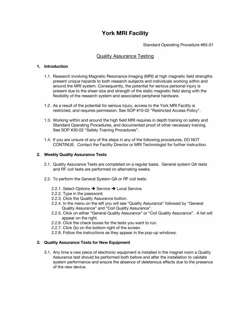
1. Introduction 1.1. Research involving Magnetic Resonance Imaging (MRI) at high magnetic field strengths
present unique hazards to both research subjects and individuals working within and around the MRI system. Consequently, the potential for serious personal injury is present due to the sheer size and strength of the static magnetic field along with the flexibility of the research system and associated peripheral hardware.
1.2. As a result of the potential for serious injury, access to the York MRI Facility is restricted, and requires permission. See SOP #10-02 “Restricted Access Policy”.
1.3. Working within and around the high field MRI requires in depth training on safety and
Standard Operating Procedures, and documented proof of other necessary training. See SOP #30-02 “Safety Training Procedures”.
1.4. If you are unsure of any of the steps in any of the following procedures, DO NOT
CONTINUE. Contact the Facility Director or MRI Technologist for further instruction. 2. Weekly Quality Assurance Tests
2.1. Quality Assurance Tests are completed on a regular basis. General system QA tests
and RF coil tests are performed on alternating weeks.
2.2. To perform the General System QA or RF coil tests:
2.2.1. Select Options è Service è Local Service. 2.2.2. Type in the password. 2.2.3. Click the Quality Assurance button. 2.2.4. In the menu on the left you will see “Quality Assurance” followed by “General
Quality Assurance” and “Coil Quality Assurance”. 2.2.5. Click on either “General Quality Assurance” or “Coil Quality Assurance”. A list will
appear on the right. 2.2.6. Click the check boxes for the tests you want to run. 2.2.7. Click Go on the bottom right of the screen. 2.2.8. Follow the instructions as they appear in the pop-up windows.
3. Quality Assurance Tests for New Equipment
3.1. Any time a new piece of electronic equipment is installed in the magnet room a Quality
Assurance test should be performed both before and after the installation to validate system performance and ensure the absence of deleterious effects due to the presence of the new device.
| SOP #65-01 2
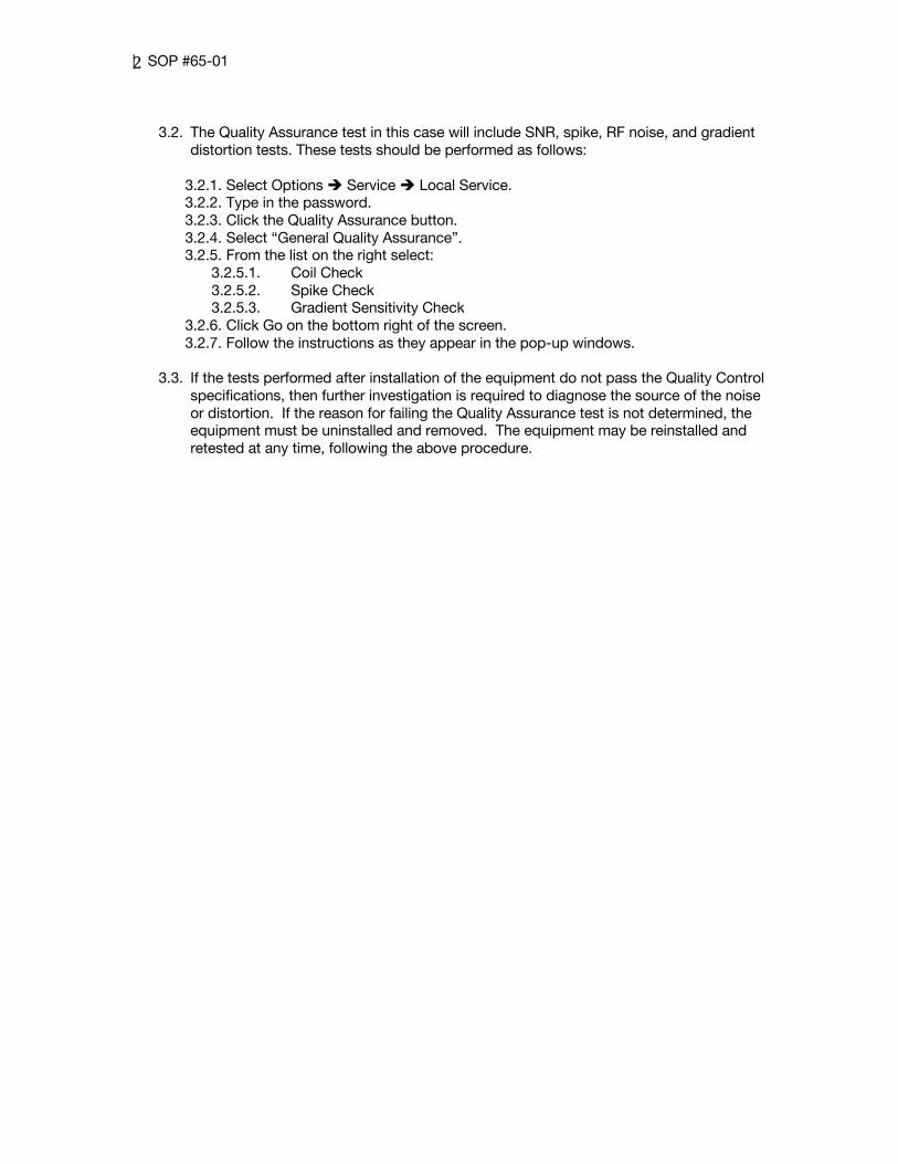
3.2. The Quality Assurance test in this case will include SNR, spike, RF noise, and gradient
distortion tests. These tests should be performed as follows:
3.2.1. Select Options è Service è Local Service. 3.2.2. Type in the password. 3.2.3. Click the Quality Assurance button. 3.2.4. Select “General Quality Assurance”. 3.2.5. From the list on the right select:
3.2.5.1. Coil Check 3.2.5.2. Spike Check 3.2.5.3. Gradient Sensitivity Check
3.2.6. Click Go on the bottom right of the screen. 3.2.7. Follow the instructions as they appear in the pop-up windows.
3.3. If the tests performed after installation of the equipment do not pass the Quality Control
specifications, then further investigation is required to diagnose the source of the noise or distortion. If the reason for failing the Quality Assurance test is not determined, the equipment must be uninstalled and removed. The equipment may be reinstalled and retested at any time, following the above procedure.
York MRI Facility
Standard Operating Procedure #70-01
System Billing Guide and Standard Rates
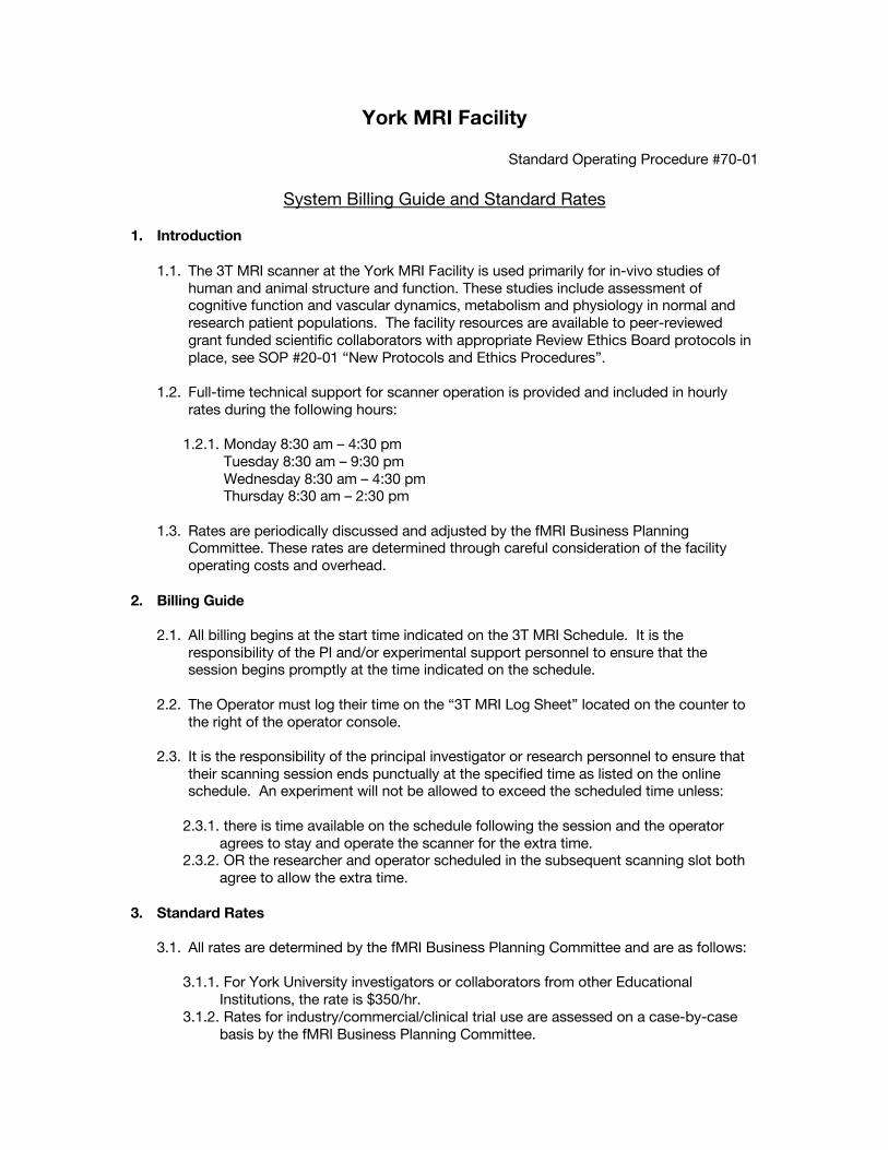
1. Introduction 1.1. The 3T MRI scanner at the York MRI Facility is used primarily for in-vivo studies of
human and animal structure and function. These studies include assessment of cognitive function and vascular dynamics, metabolism and physiology in normal and research patient populations. The facility resources are available to peer-reviewed grant funded scientific collaborators with appropriate Review Ethics Board protocols in place, see SOP #20-01 “New Protocols and Ethics Procedures”.
1.2. Full-time technical support for scanner operation is provided and included in hourly rates during the following hours:
1.2.1. Monday 8:30 am – 4:30 pm
Tuesday 8:30 am – 9:30 pm Wednesday 8:30 am – 4:30 pm Thursday 8:30 am – 2:30 pm
1.3. Rates are periodically discussed and adjusted by the fMRI Business Planning
Committee. These rates are determined through careful consideration of the facility operating costs and overhead.
2. Billing Guide
2.1. All billing begins at the start time indicated on the 3T MRI Schedule. It is the
responsibility of the PI and/or experimental support personnel to ensure that the session begins promptly at the time indicated on the schedule.
2.2. The Operator must log their time on the “3T MRI Log Sheet” located on the counter to the right of the operator console.
2.3. It is the responsibility of the principal investigator or research personnel to ensure that
their scanning session ends punctually at the specified time as listed on the online schedule. An experiment will not be allowed to exceed the scheduled time unless:
2.3.1. there is time available on the schedule following the session and the operator
agrees to stay and operate the scanner for the extra time. 2.3.2. OR the researcher and operator scheduled in the subsequent scanning slot both
agree to allow the extra time.
3. Standard Rates 3.1. All rates are determined by the fMRI Business Planning Committee and are as follows:
3.1.1. For York University investigators or collaborators from other Educational
Institutions, the rate is $350/hr. 3.1.2. Rates for industry/commercial/clinical trial use are assessed on a case-by-case
basis by the fMRI Business Planning Committee.

|SOP#70-01 2
4. Scheduling 4.1. Access for editing the 3T MRI schedule is restricted to operators. Investigators and/or
experimental support personnel are required to contact their operator to book time on 3T MRI schedule.
4.2. The principal investigator or research personnel must inform their operator of a cancelation of scan time as soon as possible. The operator will then immediately remove the entry from the online schedule so that another user may book the time.
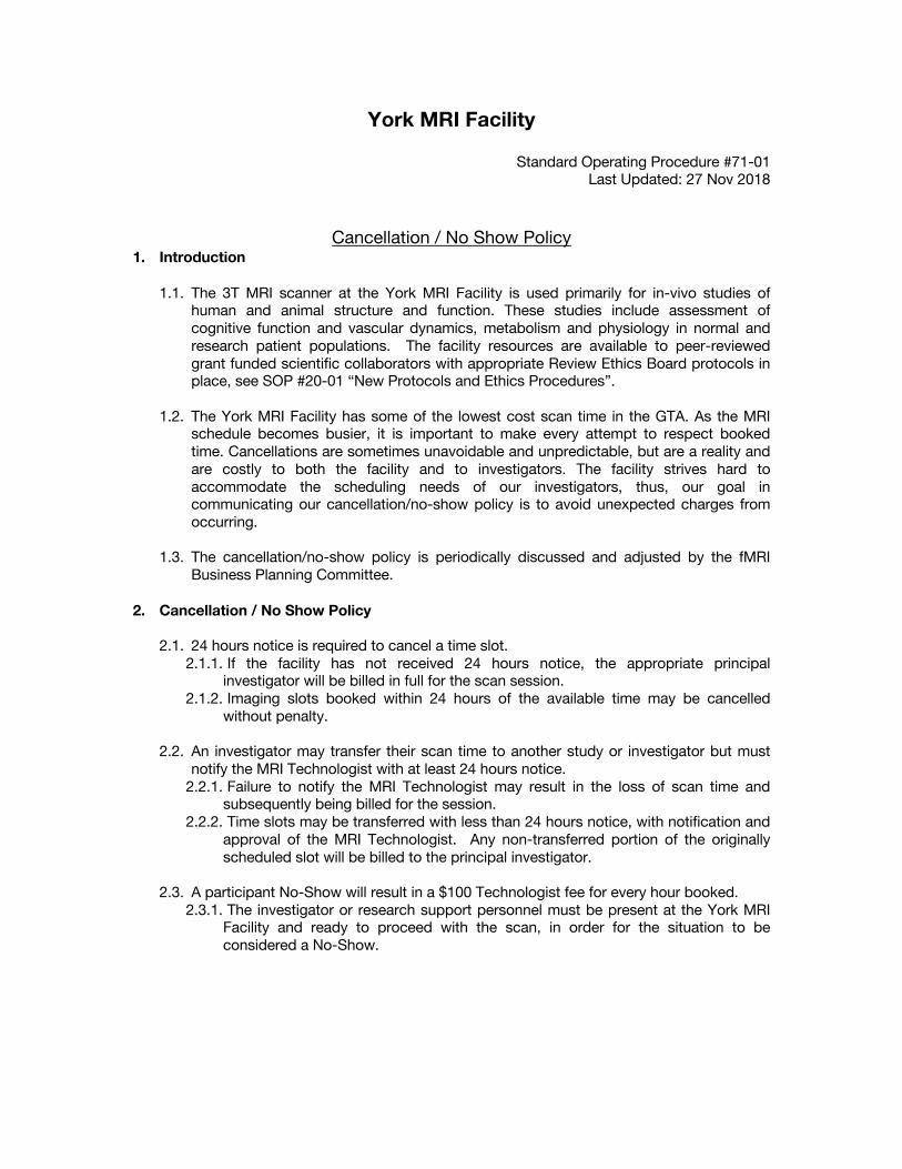
York MRI Facility
Standard Operating Procedure #71-01 Last Updated: 27 Nov 2018
Cancellation / No Show Policy 1. Introduction
1.1. The 3T MRI scanner at the York MRI Facility is used primarily for in-vivo studies of human and animal structure and function. These studies include assessment of cognitive function and vascular dynamics, metabolism and physiology in normal and research patient populations. The facility resources are available to peer-reviewed grant funded scientific collaborators with appropriate Review Ethics Board protocols in place, see SOP #20-01 “New Protocols and Ethics Procedures”.
1.2. The York MRI Facility has some of the lowest cost scan time in the GTA. As the MRI
schedule becomes busier, it is important to make every attempt to respect booked time. Cancellations are sometimes unavoidable and unpredictable, but are a reality and are costly to both the facility and to investigators. The facility strives hard to accommodate the scheduling needs of our investigators, thus, our goal in communicating our cancellation/no-show policy is to avoid unexpected charges from occurring.
1.3. The cancellation/no-show policy is periodically discussed and adjusted by the fMRI
Business Planning Committee. 2. Cancellation / No Show Policy
2.1. 24 hours notice is required to cancel a time slot. 2.1.1. If the facility has not received 24 hours notice, the appropriate principal
investigator will be billed in full for the scan session. 2.1.2. Imaging slots booked within 24 hours of the available time may be cancelled
without penalty.
2.2. An investigator may transfer their scan time to another study or investigator but must notify the MRI Technologist with at least 24 hours notice.
2.2.1. Failure to notify the MRI Technologist may result in the loss of scan time and subsequently being billed for the session.
2.2.2. Time slots may be transferred with less than 24 hours notice, with notification and approval of the MRI Technologist. Any non-transferred portion of the originally scheduled slot will be billed to the principal investigator.
2.3. A participant No-Show will result in a $100 Technologist fee for every hour booked.
2.3.1. The investigator or research support personnel must be present at the York MRI Facility and ready to proceed with the scan, in order for the situation to be considered a No-Show.
York MRI Facility
Standard Operating Procedure #80-01
Decontamination Procedure
1. Introduction 1.1. The 3T MRI scanner at the York MRI Facility is used primarily for in-vivo studies of
human and animal structure and function. These studies include assessment of cognitive function and vascular dynamics, metabolism and physiology in normal and research patient populations. The facility resources are available to peer-reviewed grant funded scientific collaborators with appropriate Review Ethics Board protocols in place, see SOP #20-01 “New Protocols and Ethics Procedures”.
1.2. Research involving animals presents unique hazards related to naturally occurring pathogenic organisms which may be transmitted from the animals and infect the people working with and caring for these animals. Similarly, humans may transmit diseases to animals.
1.3. The static magnetic field in the York MRI Facility is always present. It is important that
all those entering the facility be aware of the presence of the field, as it cannot be detected in any way, i.e. magnetic fields cannot be felt, seen or smelled. Ferromagnetic objects brought into the magnet room could quickly become dangerous projectiles, and the magnetic field can also interfere with the operation of certain medical implants.
2. Preparation for Animal Scans
2.1. The York MRI Facility should be prepared as follows, before an animal scan begins.
2.2. Cover the windows.
2.3. Remove all foam, padding, coils and human use devices/equipment from the MRI bed/
bore and any surfaces that will be needed during the scan.
2.4. Disinfect the MRI bed and bore using Percept:
2.4.1. Spray all surfaces. 2.4.2. Allow a contact time of 5 minutes. 2.4.3. Wipe all surfaces with a clean cloth or paper towel.
3. Procedures during the Scan Session
3.1. All appropriate PPE must be worn according to Universal Precautions and Vivaria SOP
I-02.
3.2. The MRI operator station must remain a “clean” area. Remove PPE and wash hands thoroughly before using the operator console.
3.3. Vivaria SOP’s must be followed for cleanup of all spills.
|SOP#80-01 2
4. Decontamination 4.1. Immediately following the MRI session, the York MRI Facility must be decontaminated
following the steps below.
4.2. Remove all “dirty” items from the magnet room.
4.3. Disinfect the MRI bed and bore using Percept:
4.3.1. Spray all surfaces. 4.3.2. Allow a contact time of 5 minutes. 4.3.3. Wipe all surfaces with a clean cloth or paper towel. 4.3.4. Cloth or paper towel must be discarded as Biohazardous Waste.
4.4. Disinfect the floor.
4.4.1. Fill the MR compatible mop bucket with the appropriate water and Percept solution.
4.4.2. Mop the magnet room, hallway and prep room floors. 4.4.3. Allow to air dry.
4.5. All dirty PPE is returned to BSB for cleaning, along with any Biohazardous waste.
York MRI Facility
Standard Operating Procedure #90-01 Updated: 05 Dec 2019
MRI Simulator Usage and Booking Policy
1. Introduction 1.1. The MRI simulator is a valuable shared resource at the York MRI Facility. It is located in
Sherman Health Science Research Centre Room 1036.
1.2. The MRI simulator was purchased from Psychology Software Tools. More information can be found on their website https://pstnet.com/products/mri-simulator/.
1.3. The MRI simulator allows the researcher to simulate the MR environment to prepare
participants in advance of MRI data acquisition, without incurring safety risks and expenses. The bore diameter of the MRI simulator matches that of the MRI scanner. The button boxes available in the simulator also match those used in the MRI suite.
2. Scheduling the MRI Simulator
2.1. Use of the MRI simulator must be scheduled on the online google calendar. Please contact the MRI Technologist at the York MRI Facility for access to the google calendar.
2.2. If a simulator session is no longer needed, the researcher must remove the booking from the google calendar.
2.3. Simulator sessions are generally scheduled on a first come first served basis.
2.4. If there are multiple groups interested in booking the same time on the MRI Simulator,
the group with the most imminent MRI session should have priority. 3. Access to the MRI Simulator
3.1. Use of the MRI simulator is limited to researchers conducting MRI studies.
3.2. Access to the MRI simulator is at the discretion of the Director or MRI Technologist of the York MRI Facility.
3.3. The key to the MRI simulator room, Sherman 1036, is contained in the lockbox
mounted beside the door.
3.4. The code to the lockbox can be obtained by contacting either the Director or the MRI Technologist of the York MRI Facility.
3.5. Training sessions for proper use of the MRI simulator are available upon request.
Please contact the MRI Technologist to book a training session.
|SOP#90-01
2
4. Startup procedure for the MRI Simulator and Accessory Components
4.1. Please display the sign stating “In Use Please Do NOT Disturb” on the door to avoid interruptions during your simulator session.
4.2. Turn on the computer and allow it to fully boot.
4.3. Turn on the projector.
4.4. Turn on the simulator.
4.5. Turn on the MoTrak console.
4.6. Open the MoTrak software.
4.7. Allow 5 minutes for the system to warm up.
4.8. Open SimFX software and select the appropriate sound files.
5. Preparing the MRI Simulator and Participant
5.1. The researcher is responsible for ensuring the health and safety of the participant
during the simulator session.
5.2. Place a clean sheet on the MRI simulator bed.
5.3. Fasten the MoTrak headband on the participant, ensuring it is secure and comfortable.
5.4. Position the participant on the MRI simulator bed with their head in the head coil.
5.5. Place the anterior portion of the head coil over the participant with the mirror in place, and confirm that the participant can see behind them, into the bore.
5.6. When the participant is comfortable and ready, press the “Table” button to advance the
bed into the bore of the simulator.
5.7. Adjust the simulator lights and fan as needed.
5.8. Confirm that the participant is comfortable in the bore and is not experiencing claustrophobia.
6. Motion Tracking
6.1. With the computer fully booted and MoTrak software started
6.1.1. Press “Run” to start the MoTrak session 6.1.2. Press “Centre” to move the crosshairs to the centre of the bullseye. Re-centre
crosshairs as needed throughout the session. 6.1.3. The MoTrak sensor on the headband must be in close proximity to the transmitter
to ensure reliable tracking (i.e. inside the bore).
6.2. Adjust the size of the bullseye on the screen, so it is clearly visible by the participant.
|SOP#90-01
3
6.3. Inform the participant that the crosshairs indicate their head position.
6.4. Ask the participant to perform any of the following while watching the crosshairs for motion.
6.4.1. Wiggle toes. 6.4.2. Take a deep breath. 6.4.3. Touch their nose. 6.4.4. Move their ankles. 6.4.5. Any other movement deemed appropriate.
6.5. Re-centre the crosshairs and ask the participant to practice laying still.
6.6. Play selected scanner sounds for desired length of time.
6.7. The researcher may also allow the participant to practice the task in the MRI simulator.
7. Completing the MRI Simulator Session
7.1. When the MRI Simulator Session is complete, and the participant is ready to come out,
press the “Table” button to move the bed out of the bore.
7.2. Remove the anterior portion of the head coil and the MoTrak headband.
7.3. The participant may now step down off the bed. Please use the step stool or step stool with handle as necessary, based on the participant’s needs and ability.
7.4. The researcher should now take some time to discuss the session with the participant
as any feedback will assist with the setup in the MRI scanner.
7.5. Place dirty linens in the laundry hamper.
7.6. Return all other equipment to its respective place for the next user.
7.7. Remove the sign from the door of the MRI Simulator room. 8. Shutdown of the MRI Simulator
8.1. On the computer, save any necessary session information.
8.2. Close the MoTrak software.
8.3. Close the SimFX software.
8.4. Switch off the MoTrak console.
8.5. Switch off the Simulator.
8.6. Turn off the projector (Press power button twice).
8.7. Shutdown the computer.