yag capsulotomy - colorado home capsulotomy powerpoint... · -pco must interfere with activities of...
TRANSCRIPT

YAG CapsulotomyAaron McNulty, OD, FAAO
Louisville Eye CenterLouisville, KY
Disclosure statement
Nothing to disclose
Course Outline
- Brief overview of laser tissue interactions- Posterior capsular opacification (PCO)- Overview of YAG laser- Capsulotomy indications/contraindications- Preoperative preparation- Capsulotomy techniques- Postoperative management- Risks and complications
Mrs. B
- Chief complaint “blurry vision”
- 20/50 BCVA OD, OS- Posterior segment
unremarkable
Mrs. B Mrs. B
- “Why did I have this complication? Did the surgeon mess up?”
- “Why did you send me to a quack surgeon?”
Mrs. B
- “Is this procedure safe? What's the risk of complication? Can I go blind?”
Mrs. B
- “Is my insurance going to cover this?”
- “I have a high deductible. What's this going to cost me?”
Mrs. B
- “What if I decide to wait? Is there harm in waiting? Does the procedure become more difficult or risky?”
● Certain laser characteristics make them useful medically○ Single Wavelength○ Low divergence
■ Energized■ Focused■ Controlled
Laser Characteristics
● Wavelength● Spot size● Pulse duration
● Laser variables interact to determine characteristics of energy delivered to the eye
Laser Variables that Influence Interactions
● Wavelength○ Determines which pigment/tissue will absorb
energy○ In general, longer wavelengths penetrate deeper
■ Ultraviolet: cornea (excimer 193um)■ Green/yellow/red lasers: retina
Laser Variables that Influence Interactions

● Wavelength○ Infrared (longer wavelengths)
■ Photodisruption■ Nd:YAG
Laser Variables that Influence Interactions
� Spot size◦ Smaller spot size has a greater energy density
Laser Variables that Influence Interactions
● Spot size○ YAG and SLT: fixed spot sizes○ Laser lens: optional in YAG procedures
● Tightens the spot size ● Effectively increases energy density
Laser Variables that Influence Interactions
� Pulse duration◦ A shorter pulse duration delivers a very
concentrated burst of power in a brief time◦ A longer pulse duration delivers less concentrated
power (slow burn)
Laser Variables That Influence Interactions
� Pulse duration◦ Short pulses (.02 - .05sec): photovaporization or
photodisruption◦ Longer pulses (.1 - .2 sec): photocoagulation
Laser Variables That Influence Interactions
� Transparency� Pigment� Water Content
Tissue Variables That Influence Interactions
� Transparency◦ Tissue transparency depends on wavelength◦ Healthy ocular media is transparent to 400nm
(blue) to 700nm (red)
Tissue Variables That Influence Interactions
� Transparency and pathology◦ Corneal pathology can affect transparency◦ Scars, edema, infiltrates make cornea opaque◦ Cornea absorbs more laser energy and may cause
corneal burn
Tissue Variables That Influence Interactions
● Transparency and pathology○ Pathology in the aqueous can also affect
transparency○ Cell/flare, hyphema○ Increased absorption of laser energy
■ Increased complications
Tissue Variables That Influence Interactions
● Pigmentation ○ Some lasers are dependent upon pigment
for their effect (Argon)○ Some lasers are pigment independent
(YAG)
Tissue Variables That Influence Interactions Specific Laser-Tissue Interactions
● Pigment independent● High energy, small spot size, brief pulse duration
○ Extremely high energy density● 15,000ºC increase● Optical breakdown: laser energy reduces tissue to
plasma● Molecules are stripped of electrons
Photodisruption
● Produces small explosion○ Hydrodynamic waves and acoustic pulses travel
back toward the surgeon■ These shockwaves disrupt tissue
● Therefore, the focal point must be posterior to the target tissue
Photodisruption
Posterior Capsular Opacification
- Pathophysiology- Anterior capsulorrhexis in cataract surgery- Residual lens epithelial cells proliferate
- Prevalence- 3-50% 5 years postoperatively- Higher in young patients- Arises months-years postoperatively
Posterior Capsular Opacification
- Signs- Translucent or opacified film behind IOL- Elschnig’s Pearls
- Vesicles with/without turbid fluid inside- Contracted collagen
- Fibrosis and wrinkles- Symptoms
- Similar to cataract- Blur, glare, decreased contrast sensitivity
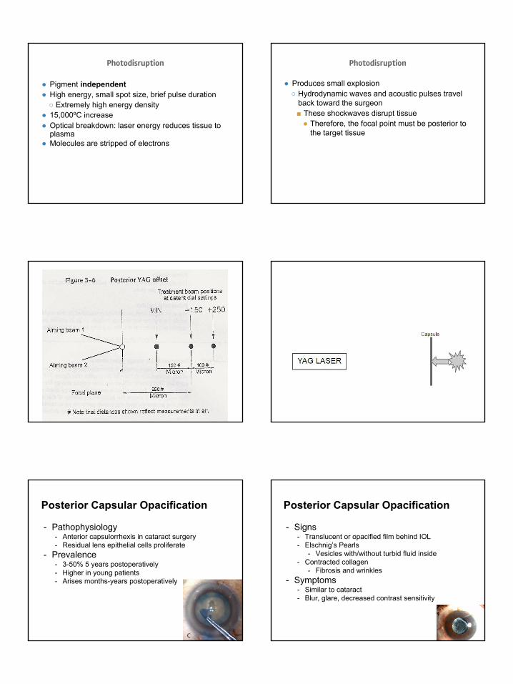
Nd:YAG Laser
- Developed in early 1980’s- Photodisruptive laser
- Pigment independent- 1064nm near infrared
- High energy, small spot, brief duration (very concentrated)- Localized temperature increase of 15,000℃
- Produces explosion and acoustic shockwaves- Shockwaves break through tissue- Shockwaves are directed back toward the doctor- Therefore focus of laser (explosion) must be just
posterior to the capsule
YAG LASER
Nd:YAG Laser
Capsule
Nd:YAG Laser
- Laser Offset- Red light is aiming beam (helium neon)- Indicates focal plane- Explosion is just posterior to the aiming beam- Acoustic shockwave travels anteriorly and disrupts
capsule at plane of aiming beams- “Offset” is adjustable on lasers. Recommend
250-350um posterior offset for capsulotomy
Capsulotomy Indications
- PCO must interfere with activities of daily living (ADL) and quality of life
- May be considered if PCO critically interferes with visualization of retina
- Needed earlier in eyes with multifocal IOL- Multifocal IOLs cause decreased contrast sensitivity- Exacerbated by mild PCO
Capsulotomy Contraindications
- Active uveitis- Corneal pathology/opacity- Macular edema- Retinal disease
- Consider retinal consult/clearance

Preoperative Exam
- BCVA, glare test, pinhole- IOP- Slit lamp- Dilated fundus exam- Rule out other causes for decreased vision
Preoperative Preparation
- Note size/shape of undilated pupil- Just large enough to avoid glare
- Dilate pupil- Topical anesthetic- Brimonidine or apraclonidine
- Reduces IOP spike risk
Capsulotomy Techniques
- Laser settings- Initial energy ~1mJ- Offset: 250-350um posterior- Fixed spot size, duration
- Focus carefully on posterior capsule- Avoid IOL and anterior hyaloid face
Capsulotomy Techniques
- Laser lens- Magnifies target tissue- Stabilizes eye and lids- Decreases spot size (concentrates energy)- Decreases acoustic propagation into eye- Downside: uncomfortable for patient
Capsulotomy Techniques
- Fire initial shot- If no tissue response, refocus and repeat- If still no response, increase energy by 0.3-0.5mJ
- Each shot creates small localized break- Aim shots such that each break is continuous with
the previous one
Capsulotomy Techniques
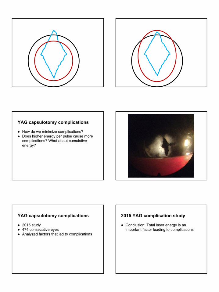
- Various shape approaches

Capsulotomy Techniques Capsulotomy and Crystalens
Capsulotomy and Crystalens
● Capsulotomy may spontaneously enlarge with lens translation○ Increases risk of IOL dislocation
● Recommendations○ Maximum 4mm diameter○ Avoid acute edges
■ Circular or octagonal approach
- Z Syndrome- Rarely occurs after Crystalens implantation- Capsular contraction causes “Z” configuration- Capsulotomy may improve or complicate the
situation
Capsulotomy and Crystalens
Postoperative Management
- Immediate postop brimonidine or apraclonidine
- Check IOP in 1 hour- Prednisolone acetate QID x 1 week
- Recent literature questions this- Postop visits in 1 week and 1 month
Postoperative Management
- Postop visits- Acuity- IOP - Slit lamp- Dilate at one month

Capsulotomy techniques
● Techniques are largely based on practitioner preference
● How to determine best practices?
Capsulotomy techniques
● 2011 survey of British ophthalmologists○ 300 surveyed, 158 replied
● Use of dilating drops, capsulotomy shape/size, use of contact lens, steroid use, follow-up schedule
Capsulotomy techniques
● Dilation○ 98.5% dilate before capsulotomy
● Size○ 64% aim for size larger than undilated pupil
● Shape○ 47% cruciate, 27% circular, 24% combination
● Use of contact lens○ 88% use one
Capsulotomy techniques
● Topical steroid use○ 42% use postoperative prophylactic steroids
● Postoperative follow-up○ 39% see patients for routine postoperative visits
■ Mostly within one month
Risks & Complications
- IOP Spike- Most common complication that requires treatment- FDA cohort of 213 patients
- 39% had IOP spike (>5mmHg) 1-6 hrs postop- None treated with prophylactic hypotensives
- Prospective randomized trial (1988)- Pre- and postop apraclonidine vs placebo- Placebo tended to spike 3 hrs postop- Apraclonidine group had lower postop IOP
Risks & Complications
- Iritis- Incidence 1-2%- Despite low rate, postop steroids are
commonly used- Prednisolone acetate QID x 1 wk

Risks & Complications
- Cystoid macular edema- Retinal detachment
- May occur weeks to years after capsulotomy- Risk factors: axial myopia, pre-existing vitreoretinal
disease, male gender, young age, vitreous prolapse, spontaneous extension of capsulotomy
- IOL displacement- IOL damage (lens pits)
- Most common complication in FDA cohort of 2110 patients
- Visually inconsequential (usually) YAG LASER
IOL Pits
CapsuleIOL
YAG LASER
IOL Pits
CapsuleIOL
A note on vitreous prolapse
● Prolapse of vitreous humor into anterior chamber is possible if capsulotomy done improperly
IOL optic
Capsulotomy
Capsulorrhexis
YAG capsulotomy complications
● How do we minimize complications?● Does higher energy per pulse cause more
complications? What about cumulative energy?
YAG capsulotomy complications
● 2015 study● 474 consecutive eyes● Analyzed factors that led to complications
2015 YAG complication study
● Conclusion: Total laser energy is an important factor leading to complications

2015 YAG complication study
Complication Incidence
Uveitis 9.9%
IOP spike 12.6%
IOL pitting 7.8%
Cystoid macular edema 2.9%
Retinal detachment 2.3%
2015 YAG complication studyComplication Mean total
energy with complication
(mJ)
Mean total energy without complication
(mJ)
Uveitis 65 42
IOP spike 76 42
IOL pitting 62 43
Cystoid macular edema
71 42
Retinal detachment 78 43
Overall average 66 37
2015 YAG complication study
● Retinal detachment: a closer look● 11 RDs in 474 eyes (2.3%)● Mean onset 11.7 months post YAG
○ Range 4-15 months● Risk factors
○ Higher total laser energy○ Higher axial length
● Recommendation: avoid large capsulotomy in patients with high axial length
2015 YAG complication study
● Mean energy level by PCO subtype
Pearl: 1.8mJ starting energy; 22mJ total
Fibrous: 2.8mJ starting energy; 65mJ total
Capsulotomy pearls
- Patient education- Expect bright flashes of light and “pops” of sound
- Lens glare and obtaining clear image- Practice with gonioscopy
- Localized fibrosis- “Curve” your treatment to avoid localized dense
fibrosis

Maximizing shot efficiency Billing & Coding
- CPT 66821- 90-day global period- $332 per eye ($314 if done in ASC)
- Be aware of ASC fees- Generally separate eyes by 1 week
Video examples
Mrs. B
- “Why did I have this complication? Did the surgeon mess up?”
- “Why did you send me to a quack surgeon?”
Mrs. B
- “Is this procedure safe? What's the risk of complication? Can I go blind?”

Mrs. B
- “Is my insurance going to cover this?”
- “I have a high deductible. What's this going to cost me?”
Mrs. B
- “What if I decide to wait? Is there harm in waiting? Does the procedure become more difficult or risky?”
Questions?
Thank you!