xyloglucan xylosyltransferases xxt1, xxt2, and xxt5 and the glucan synthase cslc4 form
TRANSCRIPT

Xyloglucan Xylosyltransferases XXT1, XXT2, and XXT5and the Glucan Synthase CSLC4 Form Golgi-LocalizedMultiprotein Complexes1[W][OA]
Yi-Hsiang Chou, Gennady Pogorelko, and Olga A. Zabotina*
Department of Biochemistry, Biophysics, and Molecular Biology (Y.-H.C., G.P., O.A.Z.) and InterdepartmentalPlant Biology Program (Y.-H.C.), Iowa State University, Ames, Iowa 50011
Xyloglucan is the major hemicellulosic polysaccharide in the primary cell walls of most vascular dicotyledonous plants and hasimportant structural and physiological functions in plant growth and development. In Arabidopsis (Arabidopsis thaliana), the 1,4-b-glucan synthase, Cellulose Synthase-Like C4 (CSLC4), and three xylosyltransferases, XXT1, XXT2, and XXT5, act in the Golgito form the xylosylated glucan backbone during xyloglucan biosynthesis. However, the functional organization of theseenzymes in the Golgi membrane is currently unknown. In this study, we used bimolecular fluorescence complementationand in vitro pull-down assays to investigate the supramolecular organization of the CSLC4, XXT1, XXT2, and XXT5 proteinsin Arabidopsis protoplasts. Quantification of bimolecular fluorescence complementation fluorescence by flow cytometry allowedus to perform competition assays that demonstrated the high probability of protein-protein complex formation in vivo andrevealed differences in the abilities of these proteins to form multiprotein complexes. Results of in vitro pull-down assays usingrecombinant proteins confirmed that the physical interactions among XXTs occur through their catalytic domains. Additionally,coimmunoprecipitation of XXT2YFP and XXT5HA proteins from Arabidopsis protoplasts indicated that while the formation ofthe XXT2-XXT2 homocomplex involves disulfide bonds, the formation of the XXT2-XXT5 heterocomplex does not involvecovalent interactions. The combined data allow us to propose that the proteins involved in xyloglucan biosynthesis functionin a multiprotein complex composed of at least two homocomplexes, CSLC4-CSLC4 and XXT2-XXT2, and three heterocomplexes,XXT2-XXT5, XXT1-XXT2, and XXT5-CSLC4.
The major structural components of plant cell walls arepolysaccharide networks composed of pectins, hemi-celluloses, and cellulose. The precise arrangement andcomposition of these components exert important in-fluences on plant growth and development. Moreover,understanding (and possibly manipulating) cell wallformation and structure is key for industrial applicationssuch as the production of biofuels and biomaterials. Indicotyledons and nongraminaceous monocotyledons,xyloglucan (XyG) is the major hemicellulosic polysac-charide (Scheller and Ulvskov, 2010). XyG has a back-bone made of b-1,4-linked glucosyl residues and isbranched, with short side chains made of Xyl, Gal, andFuc. For example, in Arabidopsis (Arabidopsis thaliana),the basic repeating XyG subunit is XXXG, which iscomposed of a b-1,4-glucan, where three of four glu-cosyl residues are linked to a-D-xylosyl residues at the
O-6 position. These basic XXXG repeating subunits canbe further substituted at the O-2 position of the xylosylresidues by either a b-D-galactosyl residue (L) or adimer, a-L-fucosyl-(1,2)-b-D-galactosyl (F), formingXXLG, XLLG, and XLFG subunits, respectively (Fryet al., 1993).
All polysaccharides in plants, except cellulose andcallose, are assembled by Golgi membrane-boundglycan synthases and glycosyltransferases (Keegstraand Raikhel, 2001) from various nucleotide diphos-phate sugars synthesized mainly in the cytosol (Reyesand Orellana, 2008). Glycosyltransferases transfersugar residues from a nucleotide diphosphate sugaronto the polysaccharide backbone, such as glucan inXyG. Most glycosyltransferases are localized in theGolgi membrane and have a type II membrane proteintopology (Perrin et al., 2001), with a short N-terminalfragment most likely protruding into the cytosol, onehelical transmembrane domain, and a catalytic domaincontaining a DXD motif, which is attached to a flexiblestem region; both the catalytic domain and the stemregion are localized in the Golgi lumen (Albersheimet al., 2010). These glycosyltransferases are highlyspecific; it is postulated that a distinct enzyme is re-quired to create each type of linkage (Keegstra andRaikhel, 2001). The other Golgi-resident enzymes in-volved in polysaccharide biosynthesis are glycan syn-thases belonging to the Cellulose Synthase-Like (CSL)superfamily. Like cellulose synthases, CSLs have sev-eral transmembrane domains and the catalytic domain
1 This work was supported by the National Science Foundation(grant no. 1121163) and the Roy J. Carver Charitable Trust (grant no.09–3384).
* Corresponding author; e-mail [email protected] author responsible for distribution of materials integral to the
findings presented in this article in accordance with the policy de-scribed in the Instructions for Authors (www.plantphysiol.org) is:Olga A. Zabotina ([email protected]).
[W] The online version of this article contains Web-only data.[OA] Open Access articles can be viewed online without a subscrip-
tion.www.plantphysiol.org/cgi/doi/10.1104/pp.112.199356
Plant Physiology�, August 2012, Vol. 159, pp. 1355–1366, www.plantphysiol.org � 2012 American Society of Plant Biologists. All Rights Reserved. 1355
Dow
nloaded from https://academ
ic.oup.com/plphys/article/159/4/1355/6109427 by guest on 18 D
ecember 2021

synthesizing glycan polymeric chains, which is pre-dicted to have the D,D,D,Q/RXXRW motif (Saxenaet al., 1995; Charnock et al., 2001; Doblin et al., 2002).
It has been proposed that glycosyltransferases formmultienzyme complexes to synthesize complex poly-saccharide structures (Keegstra, 2010). Recently, severalpieces of evidence supporting this hypothesis haveemerged. First, several putative protein complexes in-volved in polysaccharide biosynthesis have been iden-tified. Using a wheat (Triticum aestivum) microsomalfraction and a coimmunoprecipitation approach, Zenget al. (2010) demonstrated that three Golgi-residentglycosyltransferases, members of the GT43, GT47, andGT75 families, are involved in arabinoxylan biosyn-thesis and can be coimmunoprecipitated with anti-bodies specific to GT43. Atmodjo et al. (2011) reportedthat GAUT1, a galacturonosyltransferase involved inpectin biosynthesis, interacts with another homolo-gous protein, GAUT7, forming an active multiproteincomplex localized in the Golgi membrane. Two otherputative glycosyltransferases involved in pectin bio-synthesis, ARABINAN DEFICIENT1 (ARAD1) andARAD2 were shown to form homocomplexes andheterocomplexes in the Golgi when transiently coex-pressed in Nicotiana benthamiana (Sakuragi et al., 2011;Harholt et al., 2012). In yeast, Stolz and Munro (2002)demonstrated that several mannosyltransferases in-volved in cell wall mannan synthesis form two types ofprotein complexes, M-Pol I and II.
The second line of evidence supporting the in-volvement of multiprotein complexes in glycan bio-synthesis is the finding that glycosyltransferases eithertend to form or are even required to form homo-oligomers and heterooligomers or complexes in orderto be targeted to and retained in the Golgi. Moreover,some glycosyltransferases function as homooligomersor heterooligomers rather than in monomeric form(McCormick et al., 2000; van Meer, 2001; Hassinenet al., 2010). For example, for cellulose biosynthesis,cellulose synthases must form homodimers and het-erodimers organized into a rosette-like multisubunitcomplex (Kurek et al., 2002; Taylor et al., 2003;Desprez et al., 2007; Carpita, 2011). Studying Cellu-lose Synthase-Like C4 (CSLC4; Cocuron et al., 2007)demonstrated that coexpression of XXT1 and CSLC4 inPichia cells increases the length of the synthesizedglucan polymer more than the expression of CSLC4alone, despite the fact that the glucan chain cannot bexylosylated because yeast do not produce UDP-Xyl. Itwas proposed that XXT1 may assist CSLC4 functionthrough protein-protein interactions.
In Arabidopsis, at least seven enzymes are involvedin XyG biosynthesis: a b-glucan synthase, CSLC4(Cocuron et al., 2007); three a-1,6-xylosyltransferases,XXT1, XXT2, and XXT5 (Cavalier and Keegstra, 2006;Cavalier et al., 2008; Zabotina et al., 2008); two b-1,2-galactosyltransferases, MUR3 (Madson et al., 2003) andXTLT2 (Jensen et al., 2012); and an a-fucosyltransferase,FUT1 (Perrin et al., 1999; Vanzin et al., 2002). Althoughthe structure of XyG is well characterized and most of
the enzymes involved in XyG biosynthesis have beenidentified, knowledge about the mechanism of XyGbiosynthesis is still limited.
In order to understand the function of ArabidopsisXyG-synthesizing enzymes, we first investigated com-plex formation in vivo and potential physical interactionsamong three xylosyltransferases, XXT1, XXT2, and XXT5,and a glucan synthase, CSLC4. We used two indepen-dent approaches: (1) bimolecular fluorescence comple-mentation (BiFC) assays in Arabidopsis protoplasts,visualizing the signal by fluorescence microscopy andquantifying its intensity by flow cytometry (Walteret al., 2004; Li et al., 2010); and (2) in vitro pull-downassays using recombinant proteins expressed in Esch-erichia coli. Here, we report results that, to our knowledgefor the first time, directly demonstrate protein-proteincomplex formation among the xylosyltransferases andglucan synthase in the Golgi membrane in Arabidopsiscells.
RESULTS
To study the in vivo colocalization of glycosyltransfer-ases involved in XyG biosynthesis into putative protein-protein complexes, we conducted BiFC assays usingproteins transiently expressed in Arabidopsis protoplasts(Hu et al., 2002). For the assay, we made two constructsfor each enzyme (three xyloglucan xylosyltransferases,XXT1, XXT2, and XXT5, and the glucan synthase CSLC4);each enzyme was fused to either the N- or C-terminalfragment of yellow fluorescent protein (YFP; N-YFP andC-YFP) in the pSAT vectors (Citovsky et al., 2008) to formNC and CC forms, respectively (Fig. 1A). We fused theYFP fragment to the N terminus of the enzyme for allproteins, presuming that the short cytosolic N terminus isless likely to affect YFP reconstitution than the big bulkytransferase catalytic domain at the C terminus.
BiFC Assays in Arabidopsis Protoplasts
BiFC constructs for each pair of xylosyltransferases,XXT1-XXT2, XXT1-XXT5, and XXT2-XXT5, were tran-siently coexpressed in Arabidopsis protoplasts usingthe polyethylene glycol (PEG) method (Jin et al., 2001),and the signal from reconstituted YFP was examinedby fluorescence microscopy. All three pairs of XXTsshow detectable fluorescence, which forms punctatestructures (Fig. 2, A, E, and F). Xylosyltransferases arepredicted to localize to the Golgi; to test the localiza-tion of the expressed fusion proteins, Golgi were vi-sualized using the marker G-CK (Nelson et al., 2007),which is composed of cyan fluorescent protein (CFP)fused with a Golgi membrane signal peptide. Theprotoplasts were cotransfected with three constructssimultaneously, XXT2NC, XXT5CC, and G-CK, andexamined by fluorescence microscopy (Fig. 2B). Themerged image (Fig. 2C) confirms colocalization of thepunctate YFP signal from the BiFC pair and the CFPsignal marking the Golgi.
1356 Plant Physiol. Vol. 159, 2012
Chou et al.
Dow
nloaded from https://academ
ic.oup.com/plphys/article/159/4/1355/6109427 by guest on 18 D
ecember 2021

Three different negative controls confirmed that theobserved BiFC signals result from specific protein-protein colocalization of glycosyltransferases and notfrom incidental nonspecific YFP reconstitution. Thefirst negative control was coexpression of two BiFCconstructs, where the two YFP complementary frag-ments were fused to the opposite termini of the twoglycosyltransferases: one YFP fragment was fused tothe N terminus of XXT2 and the other YFP fragmentwas fused to the C terminus of XXT5. Another negative
control was a transfection of protoplasts with two BiFCconstructs, one of which carried an XXT protein fusedwith one YFP fragment and another construct thatcarried only the complementary YFP fragment withouta fused protein. As a third negative control, we chosean Arabidopsis class I a-mannosidase (MNS1), whichis a type II membrane protein with hydrolytic activityand is involved in protein glycosylation in the Golgi(Liebminger et al., 2009). MNS1 fused with the YFPfragment was coexpressed with each of the XXT and
Figure 1. Plasmid constructs used for BiFC. A,Each gene was cloned into the pSAT vector withthe N- or C-terminal fragment of YFP (N-YFP andC-YFP), HA-tagged vector, and full-length YFP-tagged vector. Genes were fused to the C terminusof each YFP fragment (GTNC and GTCC), full-length YFP tag (GTYFP), and HA tag (GTHA). Oneadditional construct was made that fused C-terminal fragments of YFP to the C terminus ofCSLC4 (GTCN). B, The truncated versions of thexylosyltransferases with His, HA, or Myc tagscloned into the pET-15b backbone vector. C, Twoexpression cassettes, GTNC and GTCC, werecloned into the coexpression vector (pPZP2N5C).
Figure 2. Fluorescence images of BiFCsignal for homodimers and heterodimersin Arabidopsis protoplasts. A and B, Arab-idopsis root protoplasts coexpressingXXT2NC and XXT5CC (A) and Golgimarker G-CK (B). C, Merged image of Aand B to confirm BiFC signal localizationin the Golgi. D, Bright-field image of thesame protoplast. E to P, Arabidopsis leafprotoplasts coexpressing XXT1CC andXXT2NC (E), XXT1NC and XXT5CC (F),CSLC4NC and XXT1CC (G), CSLC4NCand XXT5CC (H), XXT2NC and XXT2CC(I), XXT5NC and XXT5CC (J), CSLC4NCand CSLC4CC (K), CSLC4NC and CSLC4CN(L), XXT2NN and XXT5CC (M), XXT2NCand MNS1CC (N), XXT5CC and MNS1NC(O), and CSLC4CC and MNS1NC (P). Rep-resentative images from three independentexperiments are shown. Bar = 10 mm for allimages.
Plant Physiol. Vol. 159, 2012 1357
Multiprotein Complexes Involved in Xyloglucan Biosynthesis
Dow
nloaded from https://academ
ic.oup.com/plphys/article/159/4/1355/6109427 by guest on 18 D
ecember 2021

CSLC4 proteins fused with the complementary YFPfragment. For all negative controls, we did not ob-serve any BiFC signal by fluorescence microscopy(Fig. 2, M–P). The BiFC results confirm that XXT1,XXT2, and XXT5 have the same topology, with theirN termini on the same side of the Golgi membraneforming heterocomplexes, and that the YFP comple-mentary fragments fused to the N termini of XXTs donot affect their localization.
To confirm whether the fusion proteins used in BiFCassays preserved their functions in vivo, XXT5NCand XXT2CC were constitutively coexpressed in Arabi-dopsis xxt2 xxt5 double mutant plants. The observedcomplementation of the mutant root hair phenotype(Supplemental Fig. S4) suggests that XXT5NC andXXT2CC are functional proteins localized in the Golgi.
Fluorescence Quantification by Flow Cytometry
To evaluate the probability of complex formation,BiFC fluorescence intensities of XXT pairs coexpressedin Arabidopsis protoplasts were quantified by flow
cytometry (Fig. 3, A and B). The combination of BiFCwith flow cytometry provides a straightforward andsensitive estimation of the number of positive events(Morell et al., 2008). Fluorescence intensity wasexpressed as total fluorescence, which was calculatedas the percentage of fluorescent cells (events) multi-plied by the mean fluorescence of each fluorescent cell(Li et al., 2010). Both values were measured by flowcytometry, where the number of events is equal to thenumber of protoplasts in the P3 area (Fig. 3A) and themean fluorescence is the average fluorescence intensityof these protoplasts. The results presented here areexpressed as the fluorescence index, the ratio betweenthe total fluorescence of transfected protoplasts andthe total fluorescence of nontransfected protoplasts.Nontransfected protoplasts treated with PEG show alow level of fluorescence that was not visible by fluo-rescence microscopy but was detectable by flow cytom-etry (Fig. 3A). Therefore, to account for this backgroundfluorescence, the fluorescence index of nontransfectedprotoplasts was set to 1.0. Two control experiments wereperformed to investigate the dependence of the BiFC
Figure 3. Heterodimerization of XXTs.A, Flow cytometry quantification ofBiFC signal. The plots show the resultsobtained after protoplasts were ana-lyzed by flow cytometry. A Negativecontrol (nontransfected protoplasts)and a positive control (XXT5YFP) wereused to determine the gated area (P3)by the brightness of each protoplast(FL1-A). Each protoplast (a dot) local-ized in the P3 area represents a fluo-rescent event (an example is shown forXXT2NC and XXT5CC coexpression).B, BiFC signal intensities determinedby flow cytometry for different XXTpairs and all negative controls. Thecalculation of fluorescence index isdescribed in “Materials and Methods.”Values shown are means6 SE. Asterisksindicate significant differences (t test,P , 0.05; n = 5) among experiments,negative controls, and nontransfectedprotoplasts. C, Estimation of proteinexpression in Arabidopsis protoplasts.The protein expression of GTNC con-structs (all three XXTNCs, approximately75 kD; CSLC4NC, approximately 99kD; MNS1NC, approximately 80 kD)was detected using monoclonal GFPantibody. Asterisks indicate nonspecificbinding.
1358 Plant Physiol. Vol. 159, 2012
Chou et al.
Dow
nloaded from https://academ
ic.oup.com/plphys/article/159/4/1355/6109427 by guest on 18 D
ecember 2021
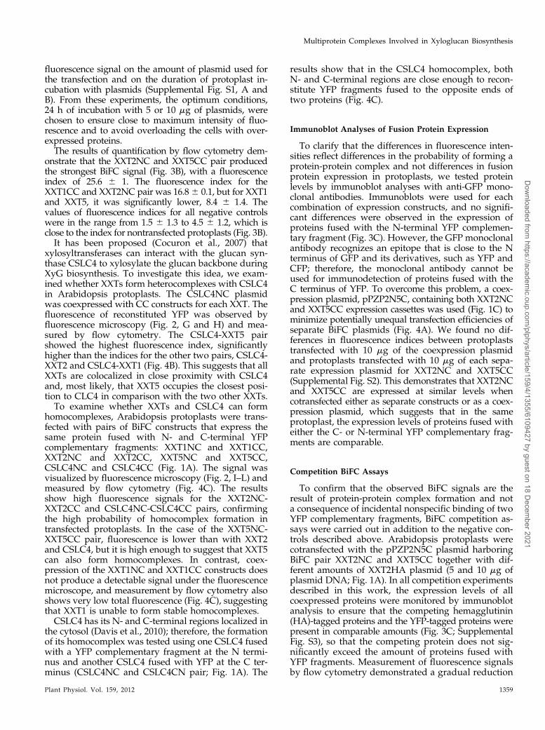
fluorescence signal on the amount of plasmid used forthe transfection and on the duration of protoplast in-cubation with plasmids (Supplemental Fig. S1, A andB). From these experiments, the optimum conditions,24 h of incubation with 5 or 10 mg of plasmids, werechosen to ensure close to maximum intensity of fluo-rescence and to avoid overloading the cells with over-expressed proteins.The results of quantification by flow cytometry dem-
onstrate that the XXT2NC and XXT5CC pair producedthe strongest BiFC signal (Fig. 3B), with a fluorescenceindex of 25.6 6 1. The fluorescence index for theXXT1CC and XXT2NC pair was 16.86 0.1, but for XXT1and XXT5, it was significantly lower, 8.4 6 1.4. Thevalues of fluorescence indices for all negative controlswere in the range from 1.5 6 1.3 to 4.5 6 1.2, which isclose to the index for nontransfected protoplasts (Fig. 3B).It has been proposed (Cocuron et al., 2007) that
xylosyltransferases can interact with the glucan syn-thase CSLC4 to xylosylate the glucan backbone duringXyG biosynthesis. To investigate this idea, we exam-ined whether XXTs form heterocomplexes with CSLC4in Arabidopsis protoplasts. The CSLC4NC plasmidwas coexpressed with CC constructs for each XXT. Thefluorescence of reconstituted YFP was observed byfluorescence microscopy (Fig. 2, G and H) and mea-sured by flow cytometry. The CSLC4-XXT5 pairshowed the highest fluorescence index, significantlyhigher than the indices for the other two pairs, CSLC4-XXT2 and CSLC4-XXT1 (Fig. 4B). This suggests that allXXTs are colocalized in close proximity with CSLC4and, most likely, that XXT5 occupies the closest posi-tion to CLC4 in comparison with the two other XXTs.To examine whether XXTs and CSLC4 can form
homocomplexes, Arabidopsis protoplasts were trans-fected with pairs of BiFC constructs that express thesame protein fused with N- and C-terminal YFPcomplementary fragments: XXT1NC and XXT1CC,XXT2NC and XXT2CC, XXT5NC and XXT5CC,CSLC4NC and CSLC4CC (Fig. 1A). The signal wasvisualized by fluorescence microscopy (Fig. 2, I–L) andmeasured by flow cytometry (Fig. 4C). The resultsshow high fluorescence signals for the XXT2NC-XXT2CC and CSLC4NC-CSLC4CC pairs, confirmingthe high probability of homocomplex formation intransfected protoplasts. In the case of the XXT5NC-XXT5CC pair, fluorescence is lower than with XXT2and CSLC4, but it is high enough to suggest that XXT5can also form homocomplexes. In contrast, coex-pression of the XXT1NC and XXT1CC constructs doesnot produce a detectable signal under the fluorescencemicroscope, and measurement by flow cytometry alsoshows very low total fluorescence (Fig. 4C), suggestingthat XXT1 is unable to form stable homocomplexes.CSLC4 has its N- and C-terminal regions localized in
the cytosol (Davis et al., 2010); therefore, the formationof its homocomplex was tested using one CSLC4 fusedwith a YFP complementary fragment at the N termi-nus and another CSLC4 fused with YFP at the C ter-minus (CSLC4NC and CSLC4CN pair; Fig. 1A). The
results show that in the CSLC4 homocomplex, bothN- and C-terminal regions are close enough to recon-stitute YFP fragments fused to the opposite ends oftwo proteins (Fig. 4C).
Immunoblot Analyses of Fusion Protein Expression
To clarify that the differences in fluorescence inten-sities reflect differences in the probability of forming aprotein-protein complex and not differences in fusionprotein expression in protoplasts, we tested proteinlevels by immunoblot analyses with anti-GFP mono-clonal antibodies. Immunoblots were used for eachcombination of expression constructs, and no signifi-cant differences were observed in the expression ofproteins fused with the N-terminal YFP complemen-tary fragment (Fig. 3C). However, the GFP monoclonalantibody recognizes an epitope that is close to the Nterminus of GFP and its derivatives, such as YFP andCFP; therefore, the monoclonal antibody cannot beused for immunodetection of proteins fused with theC terminus of YFP. To overcome this problem, a coex-pression plasmid, pPZP2N5C, containing both XXT2NCand XXT5CC expression cassettes was used (Fig. 1C) tominimize potentially unequal transfection efficiencies ofseparate BiFC plasmids (Fig. 4A). We found no dif-ferences in fluorescence indices between protoplaststransfected with 10 mg of the coexpression plasmidand protoplasts transfected with 10 mg of each sepa-rate expression plasmid for XXT2NC and XXT5CC(Supplemental Fig. S2). This demonstrates that XXT2NCand XXT5CC are expressed at similar levels whencotransfected either as separate constructs or as a coex-pression plasmid, which suggests that in the sameprotoplast, the expression levels of proteins fused witheither the C- or N-terminal YFP complementary frag-ments are comparable.
Competition BiFC Assays
To confirm that the observed BiFC signals are theresult of protein-protein complex formation and nota consequence of incidental nonspecific binding of twoYFP complementary fragments, BiFC competition as-says were carried out in addition to the negative con-trols described above. Arabidopsis protoplasts werecotransfected with the pPZP2N5C plasmid harboringBiFC pair XXT2NC and XXT5CC together with dif-ferent amounts of XXT2HA plasmid (5 and 10 mg ofplasmid DNA; Fig. 1A). In all competition experimentsdescribed in this work, the expression levels of allcoexpressed proteins were monitored by immunoblotanalysis to ensure that the competing hemagglutinin(HA)-tagged proteins and the YFP-tagged proteins werepresent in comparable amounts (Fig. 3C; SupplementalFig. S3), so that the competing protein does not sig-nificantly exceed the amount of proteins fused withYFP fragments. Measurement of fluorescence signalsby flow cytometry demonstrated a gradual reduction
Plant Physiol. Vol. 159, 2012 1359
Multiprotein Complexes Involved in Xyloglucan Biosynthesis
Dow
nloaded from https://academ
ic.oup.com/plphys/article/159/4/1355/6109427 by guest on 18 D
ecember 2021
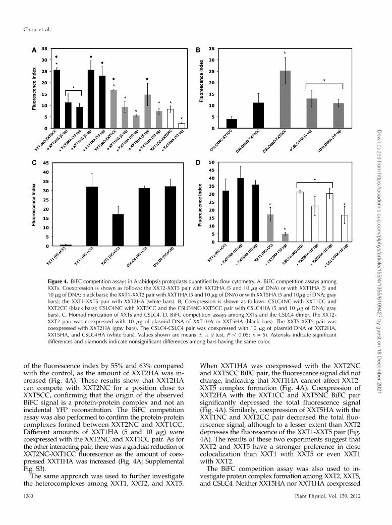
of the fluorescence index by 55% and 63% comparedwith the control, as the amount of XXT2HA was in-creased (Fig. 4A). These results show that XXT2HAcan compete with XXT2NC for a position close toXXT5CC, confirming that the origin of the observedBiFC signal is a protein-protein complex and not anincidental YFP reconstitution. The BiFC competitionassay was also performed to confirm the protein-proteincomplexes formed between XXT2NC and XXT1CC.Different amounts of XXT1HA (5 and 10 mg) werecoexpressed with the XXT2NC and XXT1CC pair. As forthe other interacting pair, there was a gradual reduction ofXXT2NC-XXT1CC fluorescence as the amount of coex-pressed XXT1HA was increased (Fig. 4A; SupplementalFig. S3).
The same approach was used to further investigatethe heterocomplexes among XXT1, XXT2, and XXT5.
When XXT1HA was coexpressed with the XXT2NCand XXT5CC BiFC pair, the fluorescence signal did notchange, indicating that XXT1HA cannot affect XXT2-XXT5 complex formation (Fig. 4A). Coexpression ofXXT2HA with the XXT1CC and XXT5NC BiFC pairsignificantly depressed the total fluorescence signal(Fig. 4A). Similarly, coexpression of XXT5HA with theXXT1NC and XXT2CC pair decreased the total fluo-rescence signal, although to a lesser extent than XXT2depresses the fluorescence of the XXT1-XXT5 pair (Fig.4A). The results of these two experiments suggest thatXXT2 and XXT5 have a stronger preference in closecolocalization than XXT1 with XXT5 or even XXT1with XXT2.
The BiFC competition assay was also used to in-vestigate protein complex formation among XXT2, XXT5,and CSLC4. Neither XXT5HA nor XXT1HA coexpressed
Figure 4. BiFC competition assays in Arabidopsis protoplasts quantified by flow cytometry. A, BiFC competition assays amongXXTs. Coexpression is shown as follows: the XXT2-XXT5 pair with XXT2HA (5 and 10 mg of DNA) or with XXT1HA (5 and10 mg of DNA; black bars); the XXT1-XXT2 pair with XXT1HA (5 and 10 mg of DNA) or with XXT5HA (5 and 10mg of DNA; graybars); the XXT1-XXT5 pair with XXT2HA (white bars). B, Coexpression is shown as follows: CSLC4NC with XXT1CC andXXT2CC (black bars); CSLC4NC with XXT5CC and the CSLC4NC-XXT5CC pair with CSLC4HA (5 and 10 mg of DNA; graybars). C, Homodimerization of XXTs and CSLC4. D, BiFC competition assays among XXTs and the CSLC4 dimer. The XXT2-XXT2 pair was coexpressed with 10 mg of plasmid DNA of XXT1HA or XXT5HA (black bars). The XXT5-XXT5 pair wascoexpressed with XXT2HA (gray bars). The CSLC4-CSLC4 pair was coexpressed with 10 mg of plasmid DNA of XXT2HA,XXT5HA, and CSLC4HA (white bars). Values shown are means 6 SE (t test, P , 0.05; n = 5). Asterisks indicate significantdifferences and diamonds indicate nonsignificant differences among bars having the same color.
1360 Plant Physiol. Vol. 159, 2012
Chou et al.
Dow
nloaded from https://academ
ic.oup.com/plphys/article/159/4/1355/6109427 by guest on 18 D
ecember 2021

with the XXT2NC-XXT2CC pair affected its fluorescence,but XXT2HA coexpressed with the XXT5NC-XXT5CCpair decreased its fluorescence intensity (Fig. 4D). Also,the fluorescence of the CSLC4NC-CSLC4CC pair wasnot affected by coexpression of XXT5HA or by coex-pression of XXT2HA (Fig. 4D), but it was depressed bycoexpression of CSLC4HA. The latter confirms that theBiFC signal observed for the CSLC4-CSLC4 pair wasdue to protein-protein homocomplex formation andnot due to accidental YFP reconstitution. Similarly,coexpression of CSLC4HA with the CSLC4NC andXXT5CC BiFC pair resulted in decreased fluorescencesignal, supporting CSLC4-XXT5 heterocomplex for-mation (Fig. 4B).
Coimmunoprecipitation of XXT2 and XXT5 fromArabidopsis Protoplasts
To further our investigation of XXT2-XXT5 hetero-complex formation in vivo, the coimmunoprecipitationassay was performed using protoplasts prepared fromArabidopsis plants expressing XXT5HA (Zabotinaet al., 2008) and transfected with the XXT2YFP con-struct to introduce tagged XXT2 protein. A total pro-tein extract (prepared as described in “Materials andMethods”) was applied to the anti-HA agarose col-umn, and the collected fractions were analyzed bySDS-PAGE under reducing and nonreducing condi-tions. The XXT2YFP and XXT5HA proteins weredetected by western blot. The results show that bothproteins can be detected in the elution fraction (Fig.5A) and not in the flow through or washes. Undernonreducing conditions, XXT2YFP was detected in theelution fraction as a monomer (approximately 83 kD)and also in a set of larger bands (approximately 166,250, and 300 kD); by contrast, XXT5HA was detectedonly in monomeric form (approximately 53 kD) underboth reducing and nonreducing conditions. To rule outthe possibility of XXT2YFP nonspecifically interactingwith anti-HA agarose, XXT2YFP was expressed inprotoplasts prepared from wild-type Arabidopsisplants, and extracted proteins were applied to anti-HAagarose. XXT2YFP was detected only in the flow-through and first wash fractions but not in the elutionfraction (Fig. 5B), confirming that XXT2YFP was re-tained on the column because of its interaction withXXT5HA and not because of a nonspecific interactionwith anti-HA agarose. The results obtained in reducingand nonreducing conditions suggest that formation ofthe XXT2-XXT2 homocomplex most likely involvesdisulfide bonds, but the XXT2-XXT5 heterocomplex isformed through noncovalent interactions.
In Vitro Pull-Down Assays
To confirm the formation of protein-protein com-plexes and investigate possible physical interactionsamong XXT proteins, in vitro pull-down assays wereperformed using recombinant proteins expressed in E.
coli. To obtain soluble proteins, truncated XXT1, XXT2,and XXT5 mutant proteins, lacking their N termini andtransmembrane domains, were fused with HA, His,and Myc tags, respectively (tGTtag; Fig. 1B) andexpressed in E. coli (BL21) under the control of aninducible promoter in the pET-15b vector (www.novagen.com). The transmembrane domains of theXXTs were predicted using the program HMMTOP(http://www.enzim.hu/hmmtop), and 45-, 41, and71-amino acid coding sequences were truncated fromthe 59 ends of XXT1, XXT2, and XXT5, respectively.The proteins were designated with a “t” for truncated.The in vitro pull-down assays were performed twodifferent ways: (1) two lysates prepared from cellsexpressing two different truncated proteins weremixed and then applied to an affinity column; (2) oneof the lysates was first applied to the affinity column,
Figure 5. Coimmunoprecipitation of XXT5HA and XXT2YFP fromArabidopsis protoplasts. A, XXT2YFP was transiently expressed inprotoplasts prepared from XXT5HA-expressing plants. Total proteinextracts from protoplasts treated with Triton X-100 were applied to ananti-HA agarose column. The elution fractions were treated with orwithout b-mercaptoethanol (2-ME) SDS-PAGE under reducing andnonreducing conditions, respectively. Proteins were detected by eitherpolyclonal anti-HA or monoclonal anti-GFPantibody (Ab). B, Negativecontrol for immunoprecipitation of XXT5HA and XXT2YFP. Proteinextract from wild-type protoplast transiently expressed XXT2YFP wasapplied to an anti-HA agarose column. The flow-through, wash (W1and W3), and elution fractions were separated by SDS-PAGE anddetected by monoclonal anti-GFP antibodies.
Plant Physiol. Vol. 159, 2012 1361
Multiprotein Complexes Involved in Xyloglucan Biosynthesis
Dow
nloaded from https://academ
ic.oup.com/plphys/article/159/4/1355/6109427 by guest on 18 D
ecember 2021

and after washing, the second lysate was applied to thesame column. After washing, the proteins bound to thecolumn were eluted and examined by immunoblottingwith antibodies specific for the tag on each protein. Bothassays showed very similar results; therefore, only re-sults from variant 1 are presented here.
After lysates from tXXT2His- and tXXT5Myc-expressing cells were applied to a nickel-nitrilotriaceticacid agarose (Ni-NTA) affinity column, both tXXT2His(approximately 50 kD) and tXXT5Myc (approximately46 kD) were detected in the elution fraction (Fig. 6A).When either tXXT2His or tXXT5Myc alone was ap-plied to the Ni-NTA affinity column, only tXXT2Hiswas detected in the elution fraction (Fig. 6A). Similarly,both tXXT1HA and tXXT2His could be pulled downand detected by both anti-HA and anti-His antibodieswhen lysates from tXXT1HA- (approximately 48 kD)and tXXT2His-expressing cells were applied to the Ni-NTA column (Fig. 6B). When tXXT1HA was applied tothe column alone, it was not detected in the elutionfraction. We were unable to pull down tXXT1HA andtXXT5His together; tXXT1HA was detected in theflow-through fraction and not in the elution fraction(Fig. 6C). These results are in agreement with the BiFCresults, where very weak fluorescence was observed
for the XXT1-XXT5 pair. Next, the ability of XXT2 andXXT5 to form homodimers was confirmed using amixture of tXXT2His and tXXT2HA and a mixture oftXXT5Myc and tXXT5His, respectively (Fig. 6, D andE). By contrast, a pull-down assay using tXXT1HA andtXXT1His did not show an interaction (Fig. 6F), con-firming the BiFC results and demonstrating that XXT1does not homodimerize.
To verify that the observed in vitro interactionsamong truncated XXTs were specific and not dueto nonspecific attraction between soluble truncatedproteins, we performed pull-down assays using thenative full-length XXT5HA (approximately 53 kD)and tXXT2His proteins. Total protein extract fromprotoplasts prepared from XXT5HA-expressing plants(Zabotina et al., 2008) treated with detergent (as de-scribed in “Materials and Methods”) was mixed withlysate from tXXT2His-expressing E. coli. The mixturewas applied either to a Ni-NTA affinity column or ananti-HA agarose column. Both XXT5HA and tXXT2Hiswere detected in elution fractions obtained from eitherNi-NTA or HA columns (Fig. 6G).
Since CSLC4 has six predicted transmembrane do-mains distributed through its protein sequence, it isnot feasible to obtain this protein in soluble form.
Figure 6. Interactions between XXTs confirmed by in vitro pull-down assay. A, In vitro Ni-NTA pull-down assay for interactionbetween tXXT2His and tXXT5Myc. tXXT2His and tXXT5Myc lysates and a mixture of the two lysates were applied to Ni-NTAaffinity columns. The elution fractions were detected by either anti-His or anti-Myc antibody (Ab). Asterisks indicate nonspecificbound signal. B, In vitro Ni-NTA pull-down assay for the interaction of tXXT1HA and tXXT2His. tXXT1HA and tXXT2His lysatemixture was applied onto a Ni-NTA affinity column. The elution fractions were detected by either anti-His or anti-HA antibody.Asterisks indicate nonspecific bound signal. C, In vitro Ni-NTA pull-down assay for the interaction of tXXT1HA and tXXT5His.tXXT1HA and tXXT5His lysate mixture was applied onto a Ni-NTA affinity column. The elution fractions were detected by eitheranti-His or anti-HA antibody. The flow-through fraction was detected by anti-HA antibodies to confirm the presence of nonboundtXXT1HA. D, In vitro Ni-NTA pull-down assay for interaction between tXXT2His and tXXT2HA. tXXT2His and tXXT2HA lysatemixture was applied onto a Ni-NTA affinity column. The elution fractions were detected by either anti-His or anti-HA antibody. E,In vitro Ni-NTA pull-down assay for interactions between tXXT5His and tXXT5Myc. tXXT5His and tXXT5Myc lysate mixture wasapplied onto a Ni-NTA affinity column. The elution fractions were detected by either anti-His or anti-Myc antibody. F, In vitro Ni-NTA pull-down assay for interactions between tXXT1His and tXXT1HA. tXXT1His and tXXT1HA lysate mixture was applied onto aNi-NTA affinity column. The elution fractions were detected by either anti-His or anti-HA antibody. The flow through fraction wasdetected by anti-HA antibodies to confirm the presence of nonbound tXXT1HA. G, In vitro Ni-NTA and anti-HA agarose pull-downassay for interactions between tXXT2His and HA tag-fused full-length XXT5. tXXT2His lysate was mixed with protein extract fromXXT5HA-expressing transgenic plants. The mixture was pulled down by either a Ni-NTA or HA agarose column. Both elutionfractions were detected by anti-HA and anti-His antibodies.
1362 Plant Physiol. Vol. 159, 2012
Chou et al.
Dow
nloaded from https://academ
ic.oup.com/plphys/article/159/4/1355/6109427 by guest on 18 D
ecember 2021

Therefore, in this study, we were unable to performpull-down experiments using CSLC4.
DISCUSSION
Previous studies revealed that three xylosyltransferases,XXT1, XXT2, and XXT5, and a glucan synthase, CLSC4,are involved in the synthesis of the xylosylated glucanbackbone of XyG in Arabidopsis (Cavalier and Keegstra,2006; Cocuron et al., 2007; Cavalier et al., 2008; Zabotinaet al., 2008). Here, we investigated the protein-proteininteractions among these enzymes to shed light on theirfunctional organization and to explore the putative mul-tiprotein complex involved in XyG formation in theGolgi. The idea that XyG assembly involves a multi-enzyme complex localized in the Golgi membrane firstemerged from biochemical studies demonstrating thecooperativity of glucan synthase and xylosyltransfer-ase activities in vitro (Zhang and Staehelin, 1992;Cocuron et al., 2007). In addition, recent reverse-geneticstudies indicated that three xylosyltransferases, XXT1,XXT2, and XXT5, play different roles in XyG biosyn-thesis (Cavalier et al., 2008; Zabotina et al., 2008), andthe presence of all three proteins is essential for theformation of the wild-type XyG (Zabotina et al., 2012).In this study, we investigated only one glucan syn-thase, CSLC4, as a potential member of the XyG syn-thetic complex, although other CSLC proteins, such asCSLC5 and CSLC6, have been implicated in XyG for-mation also (Cavalier and Keegstra, 2010). Our selec-tion is based on the facts that CSLC4 was shown tohave b-glucan synthase activity when expressed het-erologously and that this activity was enhanced bycoexpression of CSLC4 and XXT1 (Cocuron et al.,2007). CSLC4 is also expressed in all Arabidopsis tis-sues and has the highest expression level of allAtCSLCs (http://wardlab.cbs.umn.edu/arabidopsis/).This does not exclude the participation of other CSLCsin the multienzyme complex involved in XyG biosyn-thesis, and this needs to be investigated in the future.Available transcription data demonstrate that all threeXXTs and CSLC4 are coexpressed in all Arabidopsistissues studied, and XXT2 and CSLC4 have levels ofexpression about two times higher than XXT1 andXXT5 (Supplemental Fig. S5; Schmid et al., 2005).Higher expression levels of XXT2 and CSLC4 fit withour observation of two homocomplexes, XXT2-XXT2and CSLC4-CSLC4, while XXT1 and XXT5 are mostlikely present only in heterocomplexes.The absence of a fluorescence signal from coex-
pression of XXT proteins with YFP complementaryfragments fused to their opposite termini confirms thatall three xylosyltransferases are localized in the Golgimembrane in the same orientation, which brings theirN termini into close proximity. It was suggested thatXXT1, XXT2, and XXT5 proteins are positioned in theGolgi membrane with their N termini on the cytosolicside and their C termini inside the Golgi lumen(Søgaard et al., 2012). Unlike XXTs, which have only
one predicted transmembrane domain and span theGolgi membrane only once, CSLC4 has six predictedtransmembrane domains spanning the membrane sixtimes, with the catalytic site and both termini locatedon the cytosolic side of the Golgi membrane (Daviset al., 2010). This information about the topologies ofXXTs and CSLC4 prompted us to fuse the YFP frag-ments to the N terminus of each protein in the BiFCexperiments, because the C termini of XXT and CSLC4proteins are localized on opposite sides of the Golgimembrane, which would prevent YFP reconstitutionin XXT-CSLC4 pairs. The results obtained in this studysupport the earlier assumptions about the XXTs andCSLC4 topologies.
The competition BiFC assays, together with all thenegative controls used in our experiments, confirmthat the observed fluorescence signals are the result ofspecific protein-protein complex formation. Addition-ally, the competition assay allowed some insights intothe putative composition of the protein-protein com-plexes. For example, it was demonstrated that XXT2and XXT5 prefer to form a heterocomplex and that thepotential interaction between XXT2 and XXT1 occursmost likely via different residues in XXT2. In addition,XXT2 can simultaneously form homocomplexes andheterocomplexes, most likely involving different in-teracting surfaces. The formation of the XXT2-XXT2homocomplex and the XXT5-XXT2 heterocomplex isalso supported by coimmunoprecipitation of the XXT2YFPhomocomplex and the XXT2YFP-XXT5HA heterocomplexfrom Arabidopsis protoplasts (Fig. 5). Additionally, coim-munoprecipitation demonstrated that the XXT2-XXT2homocomplex is linked by disulfide bonds, but for-mation of the XXT2-XXT5 heterocomplex does notinvolve covalent interactions. Currently, we cannotexplain the presence of two bigger bands (approxi-mately 250 and 300 kD) detected with GFP antibodiesunder nonreducing conditions (Fig. 5). It is possiblethat XXT2 can form larger homocomplexes or canpull down other components of the multiproteincomplex, interacting with them through disulfidebonds.
The weaker ability of XXT5 to compete with theXXT1-XXT2 complex suggests that the XXT1 and XXT2interaction is stronger than the interaction betweenXXT1 and XXT5 and, most likely, that XXT2 can form acomplex with XXT1 and XXT5 simultaneously. Thisconclusion is supported by in vitro pull-down assays.XXT2 can be pulled down with XXT5 and XXT1, butXXT1 and XXT5 were not pulled down together, whichdemonstrates weak or no interaction between XXT1and XXT5. Perhaps XXT1 and XXT5 do not directlyinteract in vivo but colocalize in the Golgi closeenough to reconstitute YFP in the BiFC assay.
The BiFC assay using complementary YFP frag-ments fused to the N termini of two CSLC4 proteins orto the C terminus of one protein and the N terminus ofanother showed comparable fluorescence indices, whichis in accordance with the hexagonal rosette-like struc-ture of glucan synthase, similar to cellulose synthases.
Plant Physiol. Vol. 159, 2012 1363
Multiprotein Complexes Involved in Xyloglucan Biosynthesis
Dow
nloaded from https://academ
ic.oup.com/plphys/article/159/4/1355/6109427 by guest on 18 D
ecember 2021

In addition, this suggests that the termini of two CSLC4molecules are in closest proximity while their catalyticloops are positioned on opposite sides. The presence ofat least two CSLC4s in the complex is also supportedby an earlier suggestion that the synthesis of a glucanchain with alternating Glc residues in opposite orien-tations requires two glucan synthases working alter-nately (Sandhu et al., 2009; Carpita, 2011). Davis et al.(2010) proposed that CSLC4 has a topology similar tothat of cellulose synthases and therefore operates in asimilar manner (i.e. by coupling the addition of Glc tothe elongating glucan chain with translocation of thechain across the Golgi membrane). BiFC assays dem-onstrate that expression of XXT5 or XXT2 with CSLC4gives a strong fluorescence signal, but coexpression ofXXT5HA or XXT2HA with the CSLC4-CSLC4 BiFCpair does not depress the fluorescence signal of thelatter. This implies that XXT proteins colocalize withtwo CSLC4 proteins, most likely in the same complex,to xylosylate the elongating glucan chain. Interactionsof XXTs, most likely through their catalytic domains,may support complex integrity, holding the xylosyl-transferases around the synthesized glucan backbone.Localization of xylosyltransferases close to the glucansynthases is the most plausible complex organization,assuming that xylosylation of the glucan backboneoccurs upon its elongation in a processive manner.There could be a few reasons for the lower fluores-cence signal observed for interactions between CSLC4and XXT1 and XXT2, in comparison with the signalobserved for the interaction between CSLC4 andXXT5. First, the distance between the interaction sideof CSLC4 and its N terminus fused with the YFP com-plementary fragment is longer than the distance be-tween each XXT’s interaction side and N terminus. Thisdifferent spatial proximity likely affects YFP reconstitu-tion. XXT5 has a longer N-terminal cytosolic tail thanthe other two XXTs, which makes it easier for the YFPfragment fused to the XXT5 N terminus to reach the YFPcomplementary fragment fused to the CSLC4 N termi-nus. Second, it is possible that in the CSLC4-XXT het-erocomplex, the N termini are separated by CSLC4’sactive site loop, which can disturb YFP reconstitution.Finally, other complex components may be localizedclose to the interaction side between CSLC4 and XXTs,increasing the distance between two YFP complemen-tary fragments; these components may include, for ex-ample, other glycosyltransferases or nucleotide sugartransporters (Zhang et al., 2011).
In conclusion, we propose that the putative XyGsynthase complex contains at least two glucan syn-thases colocalized with their N and C termini in closeproximity to each other, two XXT2 proteins interactingwith each other through disulfide bonds, and XXT5 andXXT1, which interact with XXT2-XXT2 and CSLC4-CSLC4 homocomplexes. XXT2, XXT5, and XXT1 phys-ically interact with each other through their catalyticdomains localized in the Golgi lumen, and most likely,these interactions do not involve covalent bonds. Bycontrast, since the catalytic domains of CSLC4 and the
XXTs are localized on opposite sides of the Golgi mem-brane, most likely CSLC4 and the XXTs interact throughtheir transmembrane domains, or the catalytic domainsor stem regions of XXTs interact with CSLC4’s loops thatprotrude into the Golgi lumen. Further investigation willbe required to address this question.
Future research will also include other glycosyl-transferases known to be involved in XyG decoration,to define the full composition and structure of theXyG synthase complex in Arabidopsis. Demonstra-tion of these complexes in vivo further extends ourunderstanding of XyG biosynthesis; this knowledgewill shed light on polysaccharide formation and offeropportunities for the direct manipulation of polysac-charide formation to modify plant cell walls for var-ious industrial applications.
MATERIALS AND METHODS
Plant Material and Growth Conditions
Seeds of Arabidopsis (Arabidopsis thaliana) ecotype Columbia were steril-ized in 70% (v/v) NaOCl solution with 0.1% (v/v) Triton X-100. Sterilizedseeds were germinated, and seedlings were grown for 10 d on plates with one-half-strength Murashige and Skoog medium under 16-h-light/8-h-dark pho-toperiod conditions in a growth incubator at 22°C.
DNA Constructs
Constructs for BiFC Assays
All genes, XXT1, XXT2, XXT5, and CSLC4, were amplified using gene-specific primers from full-length complementary (cDNA) clones (XXT1 andXXT2 cDNA constructs were obtained from Prof. K. Keegstra). CSLC4 wasamplified from Arabidopsis cDNA directly. After PCR amplification, PCRproducts were inserted into the Gateway entry vector pCR8⁄GW⁄TOPO TACloning vector (Invitrogen). Gene-TOPO DNAs were recombined into bothdestination Gateway pSAT-BiFC vectors, pSAT4-DEST-nEYFP-C1 (N-terminalYFP fragment) and pSAT5-DEST-cEYFP-C1(B) (C-terminal YFP fragment;Arabidopsis Biological Resource Center [ABRC]) and into pEarleyGate104(ABRC). For cloning coexpression plasmids, the expression cassette with theN- or C-terminal YFP fragment was digested with a homing endonuclease,I-SceI or I-CeuI, respectively, and cloned into the pPZP-RCS2-ocs-bar-R1(ABRC) coexpression binary vector.
Constructs for Pull-Down Assays
The N-terminal His-tagged truncated tXXT1 and tXXT2 were prepared byamplification of XXT1 and XXT2 from the corresponding cDNAs and cloninginto the pET-15b vector (Novagen) containing an N-terminal 6xHis tag usinggene-specific forward and reverse primers containing NdeI and BamHI sites,respectively. The N-terminal HA-tagged truncated tXXT1 was made by re-moving the N-terminal 6xHis tag sequence from the pET-15b vector andreplacing it with the HA-tXXT1 sequence, which was made by amplificationfrom XXT1 cDNA using a gene-specific forward primer containing both theHA sequence (59-ACTCATGAATGTACGACGTACCAGATTACGCTACG-CCGGAGAAAGATATCGAGG-39) and a BspHI site as well as a gene-specificreverse primer with a BamHI site (59-ACGGATCCTCACGTCGTCGTCG-TACTAAGCT-39). The N-terminal Myc-tagged truncated tXXT5 was made byamplification from the cDNA using a gene-specific forward primer containingboth the Myc tag and an NcoI site (59-AGCCATGGATGGAACAAAA-ACTCATCTCAGAAGAGGATCTGAACCTAGGAAGCTCAAGCGCCG-39)as well as a gene-specific reverse primer containing an NdeI site (59-ACCA-TATGCTAGTTCTGTGGTTTGGTTTCCAC-39). The amplified Myc-tXXT5PCR fragment was then cloned into pET-15b digested with NcoI and NdeI togenerate the plasmid with the Myc-tXXT5 sequence.
1364 Plant Physiol. Vol. 159, 2012
Chou et al.
Dow
nloaded from https://academ
ic.oup.com/plphys/article/159/4/1355/6109427 by guest on 18 D
ecember 2021

Preparation of Protoplasts
Forty Arabidopsis seedlings grown on plates for 10 d were harvested andincubated in 5mL of enzyme solution (0.25% [w/v]Macerozyme, 1.0%Cellulase,0.4 mM mannitol, 8 mM CaCl2, 5 mM MES-KOH, pH 5.6, and 0.1% bovine serumalbumin) for 10 to 12 h in the dark with gentle agitation at 50 rpm. After in-cubation, suspended protoplasts were filtered through a 100-mm cell strainer,laid onto 10 mL of 21% (w/v) Suc solution, and centrifuged at 300g for 5 min.The supernatant, which contained the protoplasts, was collected and dilutedwith 10 mL of W5 solution (154 mM NaCl, 125 mM CaCl2, 5 mM KCl, 5 mM Glc,and 1.5 mM MES-KOH, pH 5.6). Protoplasts were collected by centrifugation at300g for 5 min and resuspended in 1 mL of W5 solution. The amount of pro-toplasts was measured using a hemocytometer (0.1 mm deep).
Transient Expression in Protoplasts
The protocol for protoplast transfection was adapted from Jin et al. (2001).After protoplast suspensions were counted, protoplasts were pelleted again bycentrifugation at 300g for 5 min and resuspended to a density of 23 105 mL21 ina solution of 400 mM mannitol, 15 mM MgCl2, and 5 mM MES-KOH, pH 5.6.Each plasmid (5 or 10 mg) was added into 100 mL of protoplast suspensionfollowed by the addition of 120 mL of PEG solution [30% (w/v) PEG-4000,400 mM mannitol, and 15 mM Ca(NO3)2]. The transfection mixture was incu-bated at room temperature for 30 min. After incubation, the transfection mix-ture was diluted with 4 mL of W5 solution to terminate the transfection process.The transfected protoplasts were collected by centrifugation at 300g for 5 minand resuspended in 1 mL of W5 solution. The transfected protoplast suspensionwas incubated at room temperature for 8 h in the dark and then moved to 4°Cfor 10 h. The BiFC fluorescence signal was visualized using a fluorescencemicroscope (DMIRE; Leica) with distinct filter cubes for YFP (filter set: excita-tion, 485/20; emission, 460/20) and for CFP (filter set: excitation, 436/20;emission, 480/40), and examined with the attached digital camera.
Western Blot of Expressed Fusion Proteins
Transfected protoplasts expressing fusion proteins (approximately 60,000 persample) were pelleted by centrifugation at 300g for 5 min and resuspended in300 mL of protein extraction buffer (40 mM HEPES, 0.45 M Suc, 1 mM EDTA, 1 mM
MgCl2, 1 mM KCl, 1 mM dithiothreitol, and protease inhibitor cocktail [Roche],pH 8.0) by vortexing three times for 10 s each. The protein extract was treatedwith 1% Triton X-100 for 30 min at 4°C to solubilize membrane-bound proteins.After solubilization, proteins were precipitated with 10% (v/v) TCA. Precipi-tated proteins were resuspended in loading buffer (30 mgmL21) with or withoutb-mercaptoethanol for reducing or nonreducing SDS-PAGE, respectively. AfterSDS-PAGE separation, the proteins were electrophoretically transferred to anitrocellulose membrane (0.2 mm; Bio-Rad) for immunodetection. Monoclonalanti-GFP antibodies (MMS-118P; Covance) were used (1:5,000 dilution) for thedetection of YFP. Monoclonal anti-HA antibodies (LT0422; LifeTein) were used(1:500 dilution) to detect HA-fused proteins. Polyclonal His antibodies (sc-803;Santa Cruz Biotechnology) were used (1:10,000 dilution) to detect His-fusedproteins, and monoclonal Myc antibodies (MA1-21316; Thermo) were used(1:2,000 dilution) to detect Myc-fused proteins. Membranes were treated with thereagents to detect peroxidase activity and immediately visualized by ChemiDocXRS+ (Bio-Rad). Prestained size markers were visualized on the samemembraneusing visible light. Protein concentration was measured using the Bio-Rad kit(Quick Start Bradford Dye reagent 1X; catalog no. 500-0205) following themanufacturer’s instructions.
Flow Cytometry
Fluorescence intensities of the BiFC signal in transfected protoplasts werequantified by flow cytometry (FACSCanto; BD Biosciences). Approximately20,000 to 25,000 protoplasts (counted by hemocytometer) were suspended in500 mL of W5 solution. The YFP was excited with a laser at 488 nm andcaptured with a FL1-A sensor (the emission wavelength was 505–554 nm). Thefluorescence intensity was calculated as described by Li et al. (2010) with thefollowing equation: total fluorescence = mean fluorescence level 3 the per-centage of fluorescent events. The fluorescence intensity index was determinedas total fluorescence of transfected protoplasts divided by total fluorescence ofnontransfected but PEG-treated protoplasts. For each BiFC pair describedhere, five independent protoplast transfections were performed, and fluores-cence was measured two times for each of five transfections.
Coimmunoprecipitation Assay
Protoplasts from XXT5HA-expressing or wild-type plants (around 80,000protoplasts) were transfected with the XXT2YFP construct (10 mg), and proteinextraction and solubilization with Triton X-100 were performed as describedabove. Total protein extract was diluted with extraction buffer to reduce the finalconcentration of detergent to 0.2%, and the extract was applied to an affinitycolumn with anti-HA-conjugated agarose. After 1.5 h of incubation at 4°C, theanti-HA agarose column was washed with 500 mL of wash buffer (25 mM Trisand 150 mM sodium chloride, pH 7.2) three times and eluted with 200 mL ofelution buffer (200 mM Gly, pH 2.8). Collected flow-through, wash, and elutionfractions were mixed with loading buffer with and without b-mercaptoethanolfor reducing and nonreducing conditions, respectively. Proteins were separatedby SDS-PAGE and detected by western blotting as described above.
In Vitro Pull-Down Assay
All prepared plasmids with truncated XXT-tagged proteins were transformedinto Escherichia coli (BL21) using the heat shock method. Transformed E. coli cellswere incubated in 4 mL of lysogeny broth medium at 37°C for 2.5 h at 150 rpm.When the cells reached an optical density at 600 nm of 0.5 to 0.6, the culture wasmoved to 16°C for 1 h with continuous shaking. After the 16°C treatment, cellswere induced with 0.5 mM isopropyl-b-D-thiogalactopyranoside and incubatedat 37°C for an additional 3 h. Isopropyl-b-D-thiogalactopyranoside-induced cellswere pelleted and lysed by incubation in lysis buffer (1 mg mL21 lysozyme,20 mM HEPES, 100 mM NaCl, 10 mg mL21 DNaseI, and 5 mM MgCl2, pH 7.0) for1 h at room temperature followed by five freeze/thaw cycles in liquid nitrogen.Lysate containing soluble proteins including tXXT was separated from the in-soluble pellet by centrifugation at 13,000 rpm for 10 min.
In vitro pull-down assays were performed using Ni-NTA affinity resin(Thermo Scientific; 88221) or HA agarose (Pierce) following each manufacturer’sinstructions. For Ni-NTA, two truncated protein lysates (1 mg of total crudeprotein) were mixed with 300 mL of equilibration buffer (20 mM sodium phos-phate, 300 mM sodium chloride, and 10 mM imidazole, pH 7.4). The mixture wasthen added to the affinity resin and incubated at 4°C overnight with end-to-endshaking. The column was washed with 600 mL of wash buffer (20 mM sodiumphosphate, 300 mM sodium chloride, and 25 mM imidazole, pH 7.4) two times andeluted with 300 mL of elution buffer (20 mM sodium phosphate, 300 mM sodiumchloride, and 250 mM imidazole, pH 7.4). All fractions were analyzed by westernblotting with tag-specific antibodies. For anti-HA agarose, two truncated proteinlysates (1 mg of total crude protein) were mixed with 300 mL of equilibrationbuffer, applied to the column with anti-HA-conjugated agarose, and then washedand eluted as described above for the coimmunoprecipitation assay.
Sequence data from this article can be found in the GenBank/EMBL datalibraries under accession numbers 2081625 (XXT1), 2132293 (XXT2), 2019090(XXT5), and 2089730 (CSLC4).
Supplemental Data
The following materials are available in the online version of this article.
Supplemental Figure S1. BiFC signal dependence on the amount of plas-mid DNA and time of incubation.
Supplemental Figure S2. BiFC signal intensity for XXT2NC and XXT5CCcoexpressed using two approaches.
Supplemental Figure S3. Expression level of HA-tagged proteins used incompetition BiFC assays.
Supplemental Figure S4. Expression of XXT2NC and XXT5CC in the Ara-bidopsis xxt2 xxt5 double mutant.
Supplemental Figure S5. Expression of XXT1, XXT2, XXT5, and CSLC4 inArabidopsis organs.
ACKNOWLEDGMENTS
We thank Prof. Kenneth Keegstra from Plant Research Laboratory,Michigan State University for the cDNA clones of XXT1 and XXT2 he kindlyprovided for this work.
Received April 27, 2012; accepted June 1, 2012; published June 4, 2012.
Plant Physiol. Vol. 159, 2012 1365
Multiprotein Complexes Involved in Xyloglucan Biosynthesis
Dow
nloaded from https://academ
ic.oup.com/plphys/article/159/4/1355/6109427 by guest on 18 D
ecember 2021

LITERATURE CITED
Albersheim P, Darvill A, Roberts K, Sederoff R, Staehelin A (2010) PlantCell Walls. Garland Science, New York
Atmodjo MA, Sakuragi Y, Zhu X, Burrell AJ, Mohanty SS, Atwood JAIII, Orlando R, Scheller HV, Mohnen D (2011) Galacturonosyl-transferase (GAUT)1 and GAUT7 are the core of a plant cell wall pectinbiosynthetic homogalacturonan:galacturonosyltransferase complex.Proc Natl Acad Sci USA 108: 20225–20230
Carpita NC (2011) Update on mechanisms of plant cell wall biosynthesis: howplants make cellulose and other (1→4)-b-D-glycans. Plant Physiol 155: 171–184
Cavalier DM, Keegstra K (2006) Two xyloglucan xylosyltransferases cat-alyze the addition of multiple xylosyl residues to cellohexaose. J BiolChem 281: 34197–34207
Cavalier DM, Keegstra K (2010) Members of Arabidopsis cellulose synthase-like C(CSLC) family are involved in xyloglucan biosynthesis. In S Coimbra, LG Per-eira, eds, Abstract Book: XII Cell Wall Meeting, Porto-Portugal, July 25-30. In-vulgar, Penafiel, Portugal, p 84
Cavalier DM, Lerouxel O, Neumetzler L, Yamauchi K, Reinecke A, FreshourG, Zabotina OA, Hahn MG, Burgert I, Pauly M, et al (2008) Disrupting twoArabidopsis thaliana xylosyltransferase genes results in plants deficient in xy-loglucan, a major primary cell wall component. Plant Cell 20: 1519–1537
Charnock SJ, Henrissat B, Davies GJ (2001) Three-dimensional structuresof UDP-sugar glycosyltransferases illuminate the biosynthesis of plantpolysaccharides. Plant Physiol 125: 527–531
Citovsky V, Gafni Y, Tzfira T (2008) Localizing protein-protein interactions bybimolecular fluorescence complementation in planta. Methods 45: 196–206
Cocuron J-C, Lerouxel O, Drakakaki G, Alonso AP, Liepman AH, Keegstra K,Raikhel NV, Wilkerson CG (2007) A gene from the cellulose synthase-like Cfamily encodes a b-1,4 glucan synthase. Proc Natl Acad Sci USA 104: 8550–8555
Davis J, Brandizzi F, Liepman AH, Keegstra K (2010) Arabidopsis mannansynthase CSLA9 and glucan synthase CSLC4 have opposite orientationsin the Golgi membrane. Plant J 64: 1028–1037
Desprez T, Juraniec M, Crowell EF, Jouy H, Pochylova Z, Parcy F, HöfteH, Gonneau M, Vernhettes S (2007) Organization of cellulose synthasecomplexes involved in primary cell wall synthesis in Arabidopsisthaliana. Proc Natl Acad Sci USA 104: 15572–15577
Doblin MS, Kurek I, Jacob-Wilk D, Delmer DP (2002) Cellulose biosyn-thesis in plants: from genes to rosettes. Plant Cell Physiol 43: 1407–1420
Fry SC, York WS, Albersheim P, Darvill A, Hayashi T, Joseleau J-P, Kato Y,Lorences EP, Maclachlan GA, McNeil M, et al (1993) An unambiguousnomenclature for xyloglucan derived oligosaccharides. Physiol Plant 89: 1–3
Harholt J, Jensen JK, Verhertbruggen Y, Søgaard C, Bernard S, Nafisi M,Poulsen CP, Geshi N, Sakuragi Y, Driouich A, et al (2012) ARADproteins associated with pectic arabinan biosynthesis form complexeswhen transiently overexpressed in planta. Planta 236: 115–128
Hassinen A, Rivinoja A, Kauppila A, Kellokumpu S (2010) Golgi N-glycosyltransferases form both homo- and heterodimeric enzyme com-plexes in live cells. J Biol Chem 285: 17771–17777
Hu C-D, Chinenov Y, Kerppola TK (2002) Visualization of interactionsamong bZIP and Rel family proteins in living cells using bimolecularfluorescence complementation. Mol Cell 9: 789–798
Jensen JK, Schultink A, Keegstra K,Wilkerson CG, PaulyM (April 2 2012) RNA-Seq analysis of developing nasturtium seeds (Tropaeolum majus): identificationand characterization of an additional galactosyltransferase involved in xylo-glucan biosynthesis. Mol Plant http://dx.doi.org/10.1093/mp/sss032
Jin JB, Kim YA, Kim SJ, Lee SH, Kim DH, Cheong G-W, Hwang I (2001) newdynamin-like protein, ADL6, is involved in trafficking from the trans-Golginetwork to the central vacuole in Arabidopsis. Plant Cell 13: 1511–1526
Keegstra K (2010) Plant cell walls. Plant Physiol 154: 483–486Keegstra K, Raikhel NV (2001) Plant glycosyltransferases. Curr Opin Plant
Biol 4: 219–224Kurek I, Kawagoe Y, Jacob-Wilk D, Doblin M, Delmer D (2002) Dimerization
of cotton fiber cellulose synthase catalytic subunits occurs via oxidation of thezinc-binding domains. Proc Natl Acad Sci USA 99: 11109–11114
Li M, Doll J, Weckermann K, Oecking C, Berendzen KW, Schöffl F (2010)Detection of in vivo interactions between Arabidopsis class A-HSFs,using a novel BiFC fragment, and identification of novel class B-HSFinteracting proteins. Eur J Cell Biol 89: 126–132
Liebminger E, Hüttner S, Vavra U, Fischl R, Schoberer J, Grass J,Blaukopf C, Seifert GJ, Altmann F, Mach L, et al (2009) Class Ia-mannosidases are required for N-glycan processing and root devel-opment in Arabidopsis thaliana. Plant Cell 21: 3850–3867
Madson M, Dunand C, Li X, Verma R, Vanzin GF, Caplan J, Shoue DA,Carpita NC, Reiter W-D (2003) The MUR3 gene of Arabidopsis encodes axyloglucan galactosyltransferase that is evolutionarily related to animalexostosins. Plant Cell 15: 1662–1670
McCormick C, Duncan G, Goutsos KT, Tufaro F (2000) The putative tu-mor suppressors EXT1 and EXT2 form a stable complex that accumu-lates in the Golgi apparatus and catalyzes the synthesis of heparansulfate. Proc Natl Acad Sci USA 97: 668–673
Morell M, Espargaro A, Aviles FX, Ventura S (2008) Study and selection ofin vivo protein interactions by coupling bimolecular fluorescence com-plementation and flow cytometry. Nat Protoc 3: 22–33
Nelson BK, Cai X, Nebenführ A (2007) A multicolored set of in vivo or-ganelle markers for co-localization studies in Arabidopsis and otherplants. Plant J 51: 1126–1136
Perrin RM, DeRocher AE, Bar-Peled M, Zeng W, Norambuena L, Orellana A,Raikhel NV, Keegstra K (1999) Xyloglucan fucosyltransferase, an enzymeinvolved in plant cell wall biosynthesis. Science 284: 1976–1979
Perrin RM, Wilkerson C, Keegstra K (2001) Golgi enzymes that synthesizeplant cell wall polysaccharides: finding and evaluating candidates in thegenomic era. Plant Mol Biol 47: 115–130
Reyes F, Orellana A (2008) Golgi transporters: opening the gate to cell wallpolysaccharide biosynthesis. Curr Opin Plant Biol 11: 244–251
Sakuragi Y, Norholm MHH, Scheller HV (2011) Visual mapping of cell wallbiosynthesis. In ZA Popper, ed, The Plant Cell Wall: Methods and Protocols.Springer Science-Business Media, Humana Press, New York pp 153–167
Sandhu APS, Randhawa GS, Dhugga KS (2009) Plant cell wall matrixpolysaccharide biosynthesis. Mol Plant 2: 840–850
Saxena IM, Brown RM Jr, Fevre M, Geremia RA, Henrissat B (1995)Multidomain architecture of b-glycosyl transferases: implications formechanism of action. J Bacteriol 177: 1419–1424
Scheller HV, Ulvskov P (2010) Hemicelluloses. Annu Rev Plant Biol 61: 263–289Schmid M, Davison TS, Henz SR, Pape UJ, Demar M, Vingron M,
Schölkopf B, Weigel D, Lohmann JU (2005) A gene expression map ofArabidopsis thaliana development. Nat Genet 37: 501–506
Søgaard C, Stenbæk A, Bernard S, Hadi M, Driouich A, Scheller HV,Sakuragi Y (2012) GO-PROMTO illuminates protein membrane topol-ogies of glycan biosynthetic enzymes in the Golgi apparatus of livingtissues. PLoS ONE 7: e31324
Stolz J, Munro S (2002) The components of the Saccharomyces cerevisiaemannosyltransferase complex M-Pol I have distinct functions in mannansynthesis. J Biol Chem 277: 44801–44808
Taylor NG, Howells RM, Huttly AK, Vickers K, Turner SR (2003) Inter-actions among three distinct CesA proteins essential for cellulose syn-thesis. Proc Natl Acad Sci USA 100: 1450–1455
van Meer G (2001) What sugar next? Dimerization of sphingolipid glyco-syltransferases. Proc Natl Acad Sci USA 98: 1321–1323
Vanzin GF, Madson M, Carpita NC, Raikhel NV, Keegstra K, Reiter W-D(2002) The mur2 mutant of Arabidopsis thaliana lacks fucosylated xylo-glucan because of a lesion in fucosyltransferase AtFUT1. Proc Natl AcadSci USA 99: 3340–3345
Walter M, Chaban C, Schütze K, Batistic O, Weckermann K, Näke C,Blazevic D, Grefen C, Schumacher K, Oecking C, et al (2004) Visual-ization of protein interactions in living plant cells using bimolecularfluorescence complementation. Plant J 40: 428–438
Zabotina OA, Avci U, Cavalier D, Pattathil S, Chou YH, Eberhard S,Danhof L, Keegstra K, Hahn MG (2012) Mutations in multiple XXTgenes of Arabidopsis reveal the complexity of xyloglucan biosynthesis.Plant Physiol 159: 1367–1384
Zabotina OA, van de Ven WTG, Freshour G, Drakakaki G, Cavalier D,Mouille G, Hahn MG, Keegstra K, Raikhel NV (2008) ArabidopsisXXT5 gene encodes a putative a-1,6-xylosyltransferase that is involvedin xyloglucan biosynthesis. Plant J 56: 101–115
Zeng W, Jiang N, Nadella R, Killen TL, Nadella V, Faik A (2010) A glucurono(arabino)xylan synthase complex from wheat contains members of the GT43,GT47, and GT75 families and functions cooperatively. Plant Physiol 154: 78–97
Zhang B, Liu X, Qian Q, Liu L, Dong G, Xiong G, Zeng D, Zhou Y (2011)Golgi nucleotide sugar transporter modulates cell wall biosynthesis andplant growth in rice. Proc Natl Acad Sci USA 108: 5110–5115
Zhang GF, Staehelin LA (1992) Functional compartmentation of the Golgiapparatus of plant cells: immunocytochemical analysis of high-pressurefrozen- and freeze-substituted sycamore maple suspension culture cells.Plant Physiol 99: 1070–1083
1366 Plant Physiol. Vol. 159, 2012
Chou et al.
Dow
nloaded from https://academ
ic.oup.com/plphys/article/159/4/1355/6109427 by guest on 18 D
ecember 2021