xrd/hsa-interactions, hirshfeld analysis, homo/lumo and
TRANSCRIPT
Moroccan Journal of Chemistry
ISSN: 2351-812X
http://revues.imist.ma/?journal=morjchem&page=login
Ismail Warad / Mor. J. Chem. 9 N°1 (2021) 168-176
Mor. J. Chem. 9 N°1 (2021) 168-176
168
XRD/HSA-interactions, Hirshfeld analysis, HOMO/LUMO and MEP of
new N'-(di(pyridin-2-yl)methylene DFT)benzohydrazide
Ismail Warad
Department of Chemistry and Earth Sciences, College of Arts and Sciences, Qatar University P. O. Box 2713, Doha,
Qatar.
* Corresponding author:
Received 03 Nov 2020,
Revised 23 Dec 2020,
Accepted 04 Jan 2021
Abstract
A new functionalized Schiff base N'-(di(pyridin-2-yl)methylene)benzohydrazide
Schiff base (HZSB) was prepared in a high yield via condensation of benzohydrazide
with di(pyridin-2-yl)methanone in ethanol solution under reflux condition. The
prepared HZSB was characterized by XRD, DFT and computed via Hirshfeld surface
analysis (HSA). The measured XRD structural parameters like angles lengths and
bonds were matching with their DFT computed relatives.
Keywords: Crystal structures; MEP; DFT, Hirshfeld surface.

Mor. J. Chem. 9 N°1 (2021) 168-176
169
1. Introduction
Hydrazide Schiff base (HZSB) forms a wide variety of N,O-polychelate organic ligands, it can be prepared by
condensation of hydrazide with aldehyde or ketone under reflux condition with and without using acid or base
catalytic medium [1-6]. The imine –N=C together with carbonyl functional groups can promote such material to have
pharmaceutical role in addition to being good bi-chelate N,O-ligands. HZSB are just motivating as they existing
incorporation of donor centers, like a protonated/deprotonated C=N and O=C functional groups [1]. Therefore, such
chelate with its bi-delocalization groups created several applications in clinical and biological fields like anti- anti-
fungal, bacterial, anti-inflammatory, anti-convulsing, analgesic, anti-malarial, anti-tuberculosis, anti-cancer anti-
platelets, and insecticidal [7-13]. These activities are assigned to the coordination ability of HZSB to form stable metal
ions complexes that enhanced physiological procedures [14-16]. The coordination of HZSB with transition metal ions
to form various complexes played a critical role in inorganic and organic applications [1-3]. Recently, several HZSB
complexes with structural, biological chemical and industrial applications have been prepared [17-24]. Biological
studies have reflected HZSB/complexes as significant DNA binding potential, as this interconnectedness may be an
ambitious introduction to the development of therapeutic drugs for many diseases, especially cancer [17-19]. The
preparation of HZSB has been performed via the condensation of benzohydrazide with di(pyridin-2-yl)methanone.
The HZSB molecular structure was supported by the XRD-crystal and computed by DFT and HSA. The MEP, MAC,
and NPA were served to support the XRD/HAS- interactions, moreover, DFT- bond lengths and angle values
resembled to the XRD-measured parameters.
2. Materials and methods
2.1. Computational
Gaussian 09W 32 bit software was served for all DFT operations in gaseous state at DFT/B3LYP method and 6-
311G(d) as basis set [25], the HSA and 2D-FP were carried out via Crystal Explorer 3.1 [26].
2.2. Chemicals
All chemicals and the solvents were purchased from Sigma-Aldrich and used without purification.
2.3. Synthesis of HZSB
Benzohydrazide (0.01 mol) was dissolved in 10 mL of ethanol, then added to stirring di(pyridin-2-yl)methanone (0.01
mol) dissolved 5 mL of ethanol. The reaction mixture was refluxed for 4 h, the precipitate product was filtrated and
washed with n-hexane (81%, yield).
2.4. XRD-analysis
The structure was solved [24], with the SHELXT; the model was refined with version 2018/3 of SHELXL [27]. The
crystal structure refinement parameters of the HZSB are illustrated in Table 1.
3. Results and Discussions
3.1. Synthesis, XRD and DFT
The N'-(di(pyridin-2-yl)methylene)benzohydrazide Schiff base (HZSB) product was prepared via one to one
condensation of benzohydrazide with di(pyridin-2-yl)methanone in ethanol under reflux conditions for 4
hours, as seen in Scheme 1. Fig. 1 illustrates the XRD and DFT-structure analysis of the desired HZSB

Mor. J. Chem. 9 N°1 (2021) 168-176
170
ligand, which was crystallized in Monoclinic/P21/c, with the following lattice parameters: a = 8.2741(5), b
=22.1436(14), and c = 8.8006(5) Å, meanwhile, β =108.974 (2)° [24]. The selected experimental XRD bond
angle and bond lengths and the DFT outcome are illustrated in Table 2.
Scheme 1. Synthesis of desired HZSB ligand.
Table 1. Refined data of the HZSB
Fig.1. (a) Molecular structure and (b) DFT optimized of HZSB.
Mor. J. Chem. 9 N°1 (2021) 168-176
171
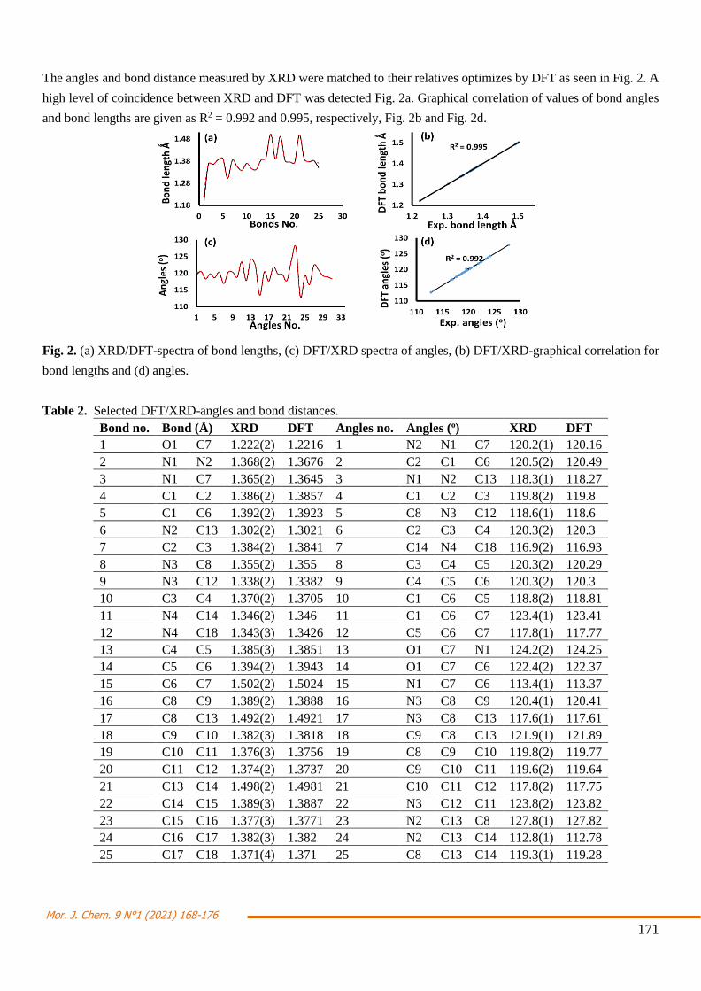
The angles and bond distance measured by XRD were matched to their relatives optimizes by DFT as seen in Fig. 2. A
high level of coincidence between XRD and DFT was detected Fig. 2a. Graphical correlation of values of bond angles
and bond lengths are given as R2 = 0.992 and 0.995, respectively, Fig. 2b and Fig. 2d.
Fig. 2. (a) XRD/DFT-spectra of bond lengths, (c) DFT/XRD spectra of angles, (b) DFT/XRD-graphical correlation for
bond lengths and (d) angles.
Table 2. Selected DFT/XRD-angles and bond distances.
Bond no. Bond (Å) XRD DFT Angles no. Angles (o) XRD DFT
1 O1 C7 1.222(2) 1.2216 1 N2 N1 C7 120.2(1) 120.16
2 N1 N2 1.368(2) 1.3676 2 C2 C1 C6 120.5(2) 120.49
3 N1 C7 1.365(2) 1.3645 3 N1 N2 C13 118.3(1) 118.27
4 C1 C2 1.386(2) 1.3857 4 C1 C2 C3 119.8(2) 119.8
5 C1 C6 1.392(2) 1.3923 5 C8 N3 C12 118.6(1) 118.6
6 N2 C13 1.302(2) 1.3021 6 C2 C3 C4 120.3(2) 120.3
7 C2 C3 1.384(2) 1.3841 7 C14 N4 C18 116.9(2) 116.93
8 N3 C8 1.355(2) 1.355 8 C3 C4 C5 120.3(2) 120.29
9 N3 C12 1.338(2) 1.3382 9 C4 C5 C6 120.3(2) 120.3
10 C3 C4 1.370(2) 1.3705 10 C1 C6 C5 118.8(2) 118.81
11 N4 C14 1.346(2) 1.346 11 C1 C6 C7 123.4(1) 123.41
12 N4 C18 1.343(3) 1.3426 12 C5 C6 C7 117.8(1) 117.77
13 C4 C5 1.385(3) 1.3851 13 O1 C7 N1 124.2(2) 124.25
14 C5 C6 1.394(2) 1.3943 14 O1 C7 C6 122.4(2) 122.37
15 C6 C7 1.502(2) 1.5024 15 N1 C7 C6 113.4(1) 113.37
16 C8 C9 1.389(2) 1.3888 16 N3 C8 C9 120.4(1) 120.41
17 C8 C13 1.492(2) 1.4921 17 N3 C8 C13 117.6(1) 117.61
18 C9 C10 1.382(3) 1.3818 18 C9 C8 C13 121.9(1) 121.89
19 C10 C11 1.376(3) 1.3756 19 C8 C9 C10 119.8(2) 119.77
20 C11 C12 1.374(2) 1.3737 20 C9 C10 C11 119.6(2) 119.64
21 C13 C14 1.498(2) 1.4981 21 C10 C11 C12 117.8(2) 117.75
22 C14 C15 1.389(3) 1.3887 22 N3 C12 C11 123.8(2) 123.82
23 C15 C16 1.377(3) 1.3771 23 N2 C13 C8 127.8(1) 127.82
24 C16 C17 1.382(3) 1.382 24 N2 C13 C14 112.8(1) 112.78
25 C17 C18 1.371(4) 1.371 25 C8 C13 C14 119.3(1) 119.28
Mor. J. Chem. 9 N°1 (2021) 168-176
172
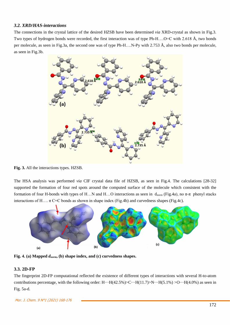
3.2. XRD/HAS-interactions
The connections in the crystal lattice of the desired HZSB have been determined via XRD-crystal as shown in Fig.3.
Two types of hydrogen bonds were recorded, the first interaction was of type Ph-H….O=C with 2.618 Å, two bonds
per molecule, as seen in Fig.3a, the second one was of type Ph-H….N-Py with 2.753 Å, also two bonds per molecule,
as seen in Fig.3b.
Fig. 3. All the interactions types. HZSB.
The HSA analysis was performed via CIF crystal data file of HZSB, as seen in Fig.4. The calculations [28-32]
supported the formation of four red spots around the computed surface of the molecule which consistent with the
formation of four H-bonds with types of H…N and H…O interactions as seen in dnorm (Fig.4a), no π-π phenyl stacks
interactions of H…. π C=C bonds as shown in shape index (Fig.4b) and curvedness shapes (Fig.4c).
Fig. 4. (a) Mapped dnorm, (b) shape index, and (c) curvedness shapes.
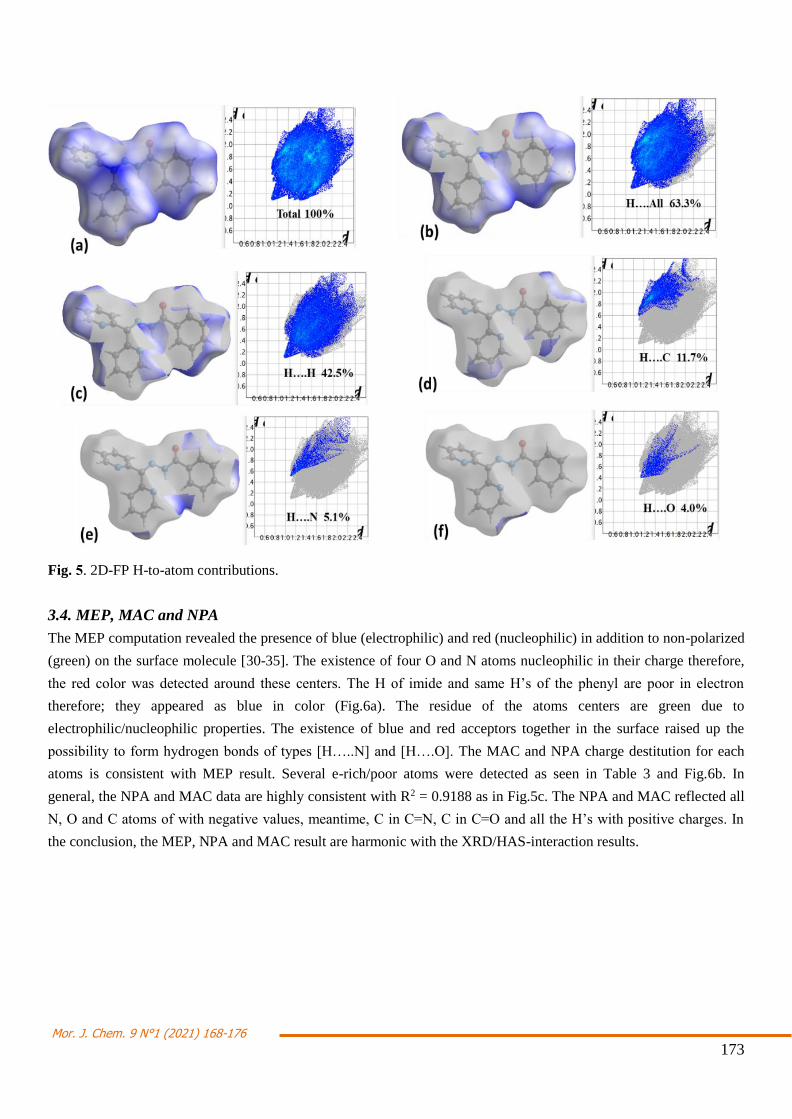
3.3. 2D-FP
The fingerprint 2D-FP computational reflected the existence of different types of interactions with several H-to-atom
contributions percentage, with the following order: H···H(42.5%)>C···H(11.7)>N···H(5.1%) >O···H(4.0%) as seen in
Fig. 5a-d.
Mor. J. Chem. 9 N°1 (2021) 168-176
173
Fig. 5. 2D-FP H-to-atom contributions.
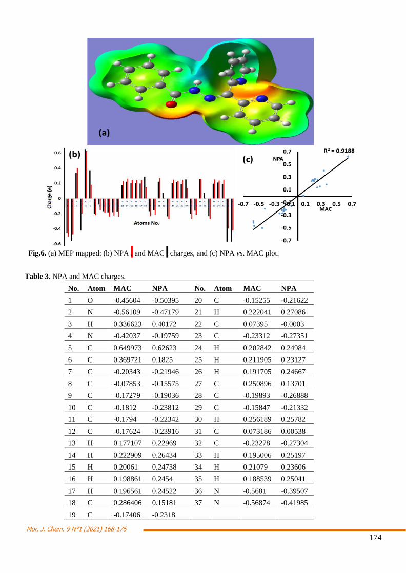
3.4. MEP, MAC and NPA
The MEP computation revealed the presence of blue (electrophilic) and red (nucleophilic) in addition to non-polarized
(green) on the surface molecule [30-35]. The existence of four O and N atoms nucleophilic in their charge therefore,
the red color was detected around these centers. The H of imide and same H’s of the phenyl are poor in electron
therefore; they appeared as blue in color (Fig.6a). The residue of the atoms centers are green due to
electrophilic/nucleophilic properties. The existence of blue and red acceptors together in the surface raised up the
possibility to form hydrogen bonds of types [H…..N] and [H….O]. The MAC and NPA charge destitution for each
atoms is consistent with MEP result. Several e-rich/poor atoms were detected as seen in Table 3 and Fig.6b. In
general, the NPA and MAC data are highly consistent with R2 = 0.9188 as in Fig.5c. The NPA and MAC reflected all
N, O and C atoms of with negative values, meantime, C in C=N, C in C=O and all the H’s with positive charges. In
the conclusion, the MEP, NPA and MAC result are harmonic with the XRD/HAS-interaction results.
Mor. J. Chem. 9 N°1 (2021) 168-176
174
Fig.6. (a) MEP mapped: (b) NPA . and MAC . charges, and (c) NPA vs. MAC plot.
Table 3. NPA and MAC charges.
No. Atom MAC NPA No. Atom MAC NPA
1 O -0.45604 -0.50395 20 C -0.15255 -0.21622
2 N -0.56109 -0.47179 21 H 0.222041 0.27086
3 H 0.336623 0.40172 22 C 0.07395 -0.0003
4 N -0.42037 -0.19759 23 C -0.23312 -0.27351
5 C 0.649973 0.62623 24 H 0.202842 0.24984
6 C 0.369721 0.1825 25 H 0.211905 0.23127
7 C -0.20343 -0.21946 26 H 0.191705 0.24667
8 C -0.07853 -0.15575 27 C 0.250896 0.13701
9 C -0.17279 -0.19036 28 C -0.19893 -0.26888
10 C -0.1812 -0.23812 29 C -0.15847 -0.21332
11 C -0.1794 -0.22342 30 H 0.256189 0.25782
12 C -0.17624 -0.23916 31 C 0.073186 0.00538
13 H 0.177107 0.22969 32 C -0.23278 -0.27304
14 H 0.222909 0.26434 33 H 0.195006 0.25197
15 H 0.20061 0.24738 34 H 0.21079 0.23606
16 H 0.198861 0.2454 35 H 0.188539 0.25041
17 H 0.196561 0.24522 36 N -0.5681 -0.39507
18 C 0.286406 0.15181 37 N -0.56874 -0.41985
19 C -0.17406 -0.2318

Mor. J. Chem. 9 N°1 (2021) 168-176
175
3.5. Molecular orbitals, DOS and Absorption/TD-DFT
The energy of electron transfer was determined by HOMOLUMO and the density of state (DOS) methods as in
Fig.7. The LUMO/HOMO FMO energy levels are both in negative area that increased the stability and softness of the
HZSB. The ΔEHOMO/LUMO was found to be with 4.490 eV (Fig.7a) whereas; the ΔEDOS was with 4.510 eV (Fig.7b).
Both methods used to calculate ΔE are very close in their values with 0.02 eV small deviation.
Fig.7. (a) LUMO/HOMO, and (b) DOS method for ΔE calculation.
4. Conclusions
The N'-(di(pyridin-2-yl)methylene)benzohydrazide Schiff base (HZSB) was made available in good yield via
dehydration of di(pyridin-2-yl)methanone and benzohydrazide in ethanol under reflux condition. The XRD/HSA-
interactions and DFT-optimization structure parameters matched well and confirmed the real 3D-structure of
HZSB. Moreover, the MAC, NPA charge populations matched well with the MEP result.
References
[1] S. Mondal, B. Pakhira, A. J. Blake, M. Drew and S. K. Chattopadhyay. Polyhedron 117 (2016): 327–337
[2] I. Warad, A. A. Khan, M. Azam, S. I. Al-Resayes, and S. F. Haddad. J. Mol. Struct. 1062 (2014): 167-173.
[3] M. Azam, I. Warad, S. I. Al-Resayes, N. Alzaqri, M. R. Khan, R. Pallepogu, S. Dwivedi, J. Musarrat, and M.
Shakir. J. Mol. Struct., 1047 (2013): 48-54.
[4] V. Nagalakshmi, M. Sathya, M. Premkumar, D. Kaleeswaran, G. Venkatachalam, and K. Balasubramani, J.
Organomet. Chem. 914 (2020): 121220-121230.
[5] I. Warad, M. Azam, U. Karama, S. Al-Resayes, A. Aouissi, and B. Hammouti. J. Mol. Struct., 1002, (2011): 107-
112.
[6] I. Warad, M. Abdoh, N. Shivalingegowda, N. K. Lokanath, R. Salghi, M. Al-Nuri, S. Radi, and B. Hammouti, J.
Mol. Struct.,1099 (2015): 323-329.
[7] K.K. Bedia, O. Elçin, U. Seda, K. Fatma, S. Nathaly, R.A. Sevim, and A. Dimoglo, Eur. J. Med. Chem. 41 (2006) :
1253-1260.
[8] P. Melnyk, V. Leroux, C. Sergheraert, P. Grellier, and C. Sergheraert, Bioorg. Med. Chem. Lett. 16 (2006) 31-40.

Mor. J. Chem. 9 N°1 (2021) 168-176
176
[9] C. Cunha, J.M. Figueiredo, J.L.M. Tributino, A.L.P. Miranda, H.C. Castro, R.B. Zingali, C.A.M. Fraga, M.C.B. de
Souza, V.F. Ferreira, and E. Barreiro, J. Bioorg. Med. Chem. 11 (2003): 2051-2061.
[10] L. Savanini, L. Chiasserini, A. Gaeta, and C. Pellerano, Bioorg. Med. Chem. 10 (2002): 2193-2199.
[11] R. Albertini, S. Pinelli, and P. Lunghi, Inorg. Chim. Acta 286 (1999) : 134-130.
[12] H. Elo, I. Sunila, and P. Lumme, Inorg. Chim. Acta 136 (1987) : 61-70.
[13] T. Todorovic, U. Rychlewska, B. Warzajtis, D. Radanovic, N. Filipovic, I. Pajic, D. Sladic, and K. Andelkovic,
Polyhedron 28 (2009): 2397-2407.
[14] M. Katyal, and Y. Dutt, Talanta 22 (1975): 151-160.
[15] M. Mohan, M.P. Gupta, L. Chandra, and N.K. Jha, Inorg. Chim. Acta 151 (1988) : 61-70.
[16] S. A. Beyramabadi, M. Saadat-Far, A. Faraji-Shovey, M. Javan-Khoshkholgh, and A. Morsali, J. Molec. Struc.,
1208 (2020): 127898-127905.
[17] Y. Liu, Na Wang, W. Mei, F. Chen, H. Li-Xin, L. Jian, and R. Wang, Transition Met. Chem. 32 (2007) : 332-340.
[18] I. Warad, A. A. F. Eftaiha, M. A. Al-Nuri, A. I. Husein, M. Assal, A. Abu-Obaid, N, Al-Zaqri, T. B. Hadda, and
B. Hammouti. J. Mater. Environ. Sci., 4 (2013): 542-557.
[19] A. Mansri, B. Bouras, B. Hammouti, I. Warad, and A. Chetouani. Res. Chem. Intermed., 39 (2013): 1753- 1765.
[20] M. Suleiman, M. Al-Masri, A. A. Ali, D. Aref, A. Hussein, I. Saadeddin, and I. Warad, J Mater Environ Sci. 6,
(2015): 513-518.
[21] I. Warad, F. Eftaiha, M. Al-Nuri, I. Husein, M. Assal, A. Abu-Obaid, N. Al-Zaqri, B. Hadda and B. Hammouti, J
Mater Environ Sci., 4, (2013): 542-557.
[22] M. Rbaa, F. Benhiba, B. Obot, H. Oudda, I. Warad, B. Lakhrissi, and A. Zarrouk, J. Molec. Liq. 276 (2019):
120-133.
[23] S. Tighadouini, S. Radi, M. Bacquet, J.P. Dacquin, Y.N. Mabkhot, I.Warad, and M. Zaghrioui. Sep. Sci. Technol.
50 (2015): 710-717.
[24] I. Warad, M. Al-Nuri, S. Al-Resayes, K. Al-Farhana and M. Ghazzalia, Acta Cryst. E65 (2009) : 1597-1599.
[25] S. K. Wolff, D. J. Grimwood, J. J. McKinnon, D. Jayatilaka, and M. A. Spackman, Crystal explorer 2.1.
University of Western Australia, Perth (2007).
[26] M.J. Frisch, G.W. Trucks, et. al. Gaussian 09, Gaussian Inc., Wallingford CT, 2009.
[27] G. M. Sheldrick, Acta Cryst., A64 (2008): 112-114.
[28] I. Warad, M. R. H. Siddiqui, S. Al-Resayes, A. Al-Warthan, and R. Mahfouz, Transit. Metal Chem, 34, (2009):
347-352.
[29] I. Warad, Z. Al-Othman, S. Al-Resayes, S. S. Al-Deyab, and E. Kenawy. Molecules 15 (2010): 1028-1040. [30]
M. E. Belghiti, Y. Karzazi, S. Tighadouini, A. Dafali, C. Jama, I. Warad, B. Hammouti, and S. Radi. J Mater Environ
Sci., 7 (2016): 956-967.
[31] A. Chetouani, K. Medjahed, S. Al-Deyab, I. Warad, and A. Mansri, J Mater Environ Sci., 7 (2012): 6025-6043.
[32] M. Belghiti, Y. Karzazi, S. Tighadouini, A. Dafal, C. Jama, I. Warad and B. Hammouti, and S. Radi, J Mater
Environ Sci., 7 (2016): 956-967.
[33] I. Warad, O. Bsharat, S. Tabti, A .Djedouani, M.Al-Nuri, N. Al-Zaqri, K. Kumara, N.K. Lokanath, S. Amereih,
and Ib. M. Abu-Reidah, J. Mol. Struct.,1185 (2019): 290-299.
[34] M. R. Aouad, M. Messali, N. Rezki, N. Al-Zaqri, I. Warad, J. Mol. Liq., 264 (2018): 621-630
[35] M. R. Aouad, M. Messali, N. Rezki, M. A. Said, D. Lentz, L. Zubaydi, I. Warad, J. Mol. Struct.,1180 (2019):
455-461.