xmdff: molecular dynamics flexible fitting of low...
TRANSCRIPT

research papers
2344 doi:10.1107/S1399004714013856 Acta Cryst. (2014). D70, 2344–2355
Acta Crystallographica Section D
BiologicalCrystallography
ISSN 1399-0047
xMDFF: molecular dynamics flexible fitting oflow-resolution X-ray structures
Ryan McGreevy,a‡ Abhishek
Singharoy,a‡ Qufei Li,b Jingfen
Zhang,c Dong Xu,c Eduardo
Perozob and Klaus Schultena,d*
aBeckman Institute for Advanced Science and
Technology, University of Illinois at
Urbana-Champaign, Urbana, IL 61801, USA,bDepartment of Biochemistry and Molecular
Biology, The University of Chicago, Chicago,
IL 60637, USA, cDepartment of Computer
Science, University of Missouri, Columbia,
MO 65211, USA, and dDepartment of Physics,
University of Illinois at Urbana-Champaign,
Urbana, IL 61801, USA
‡ These authors contributed equally to this
work.
Correspondence e-mail: [email protected]
X-ray crystallography remains the most dominant method
for solving atomic structures. However, for relatively large
systems, the availability of only medium-to-low-resolution
diffraction data often limits the determination of all-atom
details. A new molecular dynamics flexible fitting (MDFF)-
based approach, xMDFF, for determining structures from
such low-resolution crystallographic data is reported. xMDFF
employs a real-space refinement scheme that flexibly fits
atomic models into an iteratively updating electron-density
map. It addresses significant large-scale deformations of the
initial model to fit the low-resolution density, as tested with
synthetic low-resolution maps of d-ribose-binding protein.
xMDFF has been successfully applied to re-refine six low-
resolution protein structures of varying sizes that had already
been submitted to the Protein Data Bank. Finally, via
systematic refinement of a series of data from 3.6 to 7 A
resolution, xMDFF refinements together with electro-
physiology experiments were used to validate the first all-
atom structure of the voltage-sensing protein Ci-VSP.
Received 14 February 2014
Accepted 13 June 2014
1. Introduction
X-ray crystallography is arguably the most versatile and
dominant technique for delivering atomic structures of
biomolecules. An increasing number of structures are
submitted each year to the Protein Data Bank, with over 90%
of the current entries coming from X-ray crystal structures.
Traditional methods for determining X-ray structures include
least-squares with gradient descent (Hendrickson, 1985),
maximum likelihood (Pannu & Read, 1996; Bricogne & Irwin,
1996; Murshudov et al., 1997), simulated annealing (Brunger,
1988) and knowledge-based conformational sampling
(Depristo et al., 2005). However, investigating the structure of
large biomolecular complexes has posed a serious challenge to
traditional crystallographic techniques. The inherent flexibility
of such large systems and the presence of disordered solvent
and lipids or ligands often cause the crystals to diffract at low
resolutions. Furthermore, in the low-resolution limit the
number of atomic coordinates to be determined often exceeds
the number of observed diffraction intensities. At moderate to
low resolutions, knowledge of the stereochemistry of the
system must be incorporated to achieve accurate atomic
positions. Lower resolutions, >5 A, pose a greater challenge to
refinement; however, even at �7 A resolution there are in
principle enough independent Bragg reflections to determine

the backbone torsion angles of protein crystal structures
(Brunger et al., 2012).
Solving structures from low-resolution diffraction data
is a difficult, time-consuming process. Consequently, low-
resolution data sets are usually chosen to be discarded
(Karmali et al., 2009).
However, new methods are being developed to better
handle low-resolution data. For example, DEN refinement
incorporates deformable elastic network models with generic
stereochemistry and homology information (Schroder et al.,
2010) to address the issue. Other notable recent developments
applicable to refining low-resolution structures include
normal-mode refinement (Delarue, 2008), the Rosetta imple-
mentation of physical energy functions (DiMaio et al., 2011)
and its combination with reciprocal-space X-ray refinement in
PHENIX (DiMaio et al., 2013), torsional optimization proto-
cols (Haddadian et al., 2011) and external structure restraints
or jelly-body refinement in REFMAC (Murshudov et al.,
2011). Indeed, the number of low-resolution X-ray structures
has grown rapidly in recent years (Karmali et al., 2009).
Here, we present a new method, xMDFF (molecular
dynamics flexible fitting for X-ray crystallography), for real-
space (Diamond, 1971; Chapman, 1995; Chapman & Blanc,
1997) X-ray refinement. Our method specifically targets the
handling of low-resolution data and
the large-scale deformations that often
separate the target and reference
models. To create this method, we
extended a previous hybrid method,
molecular dynamics flexible fitting
(MDFF), developed to solve atomic
models from cryo-EM densities. In
MDFF, an initial atomic model is
subject to a molecular dynamics (MD)
simulation with a modified potential
energy function that includes a term
derived from the cryo-EM density map
(Trabuco et al., 2008, 2009). The accu-
racy and robustness of MDFF has been
widely demonstrated in many applica-
tions solving structural models for the
ribosome (Villa et al., 2009; Gumbart et
al., 2009, 2011; Becker et al., 2009;
Seidelt et al., 2009; Trabuco et al., 2010;
Frauenfeld et al., 2011; Agirrezabala et
al., 2011; Li et al., 2011), photosynthetic
proteins (Hsin et al., 2009; Sener et al.,
2009) and the first all-atom structure of
the HIV capsid (Zhao et al., 2013). In
xMDFF, the MDFF protocol is modified
to use iteratively updated model-phased
maps to fit and subsequently refine
densities derived from low-resolution
X-ray diffraction data.
xMDFF was tested via the refinement
of a model with a known final structure
at resolutions of 3.5–5 A. Next, xMDFF
successfully further refined six low-
resolution (4–4.5 A) protein structures
of varying sizes that had already been
submitted to the PDB (Fig. 1). Finally,
xMDFF was applied in parallel with an
independent experimental investigation
to resolve crystallographic uncertainty
in the three-dimensional structure of
Ciona intestinalis voltage-sensing
protein (Ci-VSP). xMDFF refinements
were evaluated by (i) Rfree, (ii) r.m.s.d.
to known targets (test case and voltage-
research papers
Acta Cryst. (2014). D70, 2344–2355 Li et al. � xMDFF 2345
Figure 1xMDFF re-refinement characteristics for six structures. The structures with diffraction data between4 and 4.5 A resolution had been deposited earlier in the Protein Data Bank and were used as initialmodels for xMDFF refinement. Improvements were evaluated using the Rfree values of the xMDFF-refined structures (shown in red) against their respective starting values taken from the depositedstructure (blue). In addition, the increase in the percentage of favored Ramachandran angles owingto xMDFF refinement and other structural statistics, as summarized by the overall MolProbityscore, were used to measure the improvement in structural geometries. Further details of the refinedstructures are provided in Table 1.

sensing protein) and (iii) improvements in structural geometry
of the model. In all cases xMDFF successfully refined the
structures and demonstrated an ability to work at very low
resolution (7 A) and with starting models that are very
divergent from the target (>5 A).
2. Methods
2.1. Concept
xMDFF is derived from the MDFF method which solves
atomic models of biomolecules imaged by cryo-electron
microscopy. In MDFF, an initial atomic model is subjected to
an MD simulation with a modified potential energy function
that includes a term derived from the cryo-EM density map
(Trabuco et al., 2008, 2009). Through the density-dependent
term, atoms experience steering forces, f fit, that locally drive
them towards high-density regions, thereby fitting the atoms
to the map. For use in low-resolution X-ray crystallography,
the MDFF protocol was modified to work with model-phased
densities, using the phases ’calc calculated from a tentative
model and the amplitudes Fobs from the X-ray diffraction data
to produce a 2mFobs � DFcalc density map. The density is
biased by the model, but contains sufficient information from
the Fobs to determine the experimental structure. Next, the
tentative model is flexibly fitted into the electron-density map
using MDFF. In addition to the steering forces derived from
the density data, structural restraints are applied to preserve
the secondary structure of proteins and nucleic acids (Trabuco
et al., 2008, 2009), as well as to ensure stereochemical
correctness (Schreiner et al., 2011), thus avoiding overfitting
the model into the map. The xMDFF-fitted structure provides
new ’calc that, together with Fobs, are used to regenerate the
electron-density map. The fitted structure is then employed as
an updated search model to be driven into the new model-
phased density map, and this process continues iteratively. In
effect, f fit drives the structure in a direction biased by the Fobs
contribution to the density. Consequently, ’calc improvement
is indicated by a decrease in R factors with each subsequent
iteration. The iterations continue until the Rfree and Rwork
values reach a minimum or become lower than a predefined
tolerance. The quality of the xMDFF-refined all-atom struc-
tures is further analyzed via computing correlation coefficients
(CCs) between the electron-density map generated from the
refined ’calc with Fobs and a simulated map at the target
resolution.
Next, the MDFF method is briefly discussed in addition
to the algorithmic and computational extensions required to
address the crystallographic aspects of xMDFF. All of the
software required to use xMDFF is currently distributed in
released versions of NAMD (Phillips et al., 2005), VMD
(Humphrey et al., 1996) and PHENIX (Adams et al., 2010). A
script for running xMDFF can be found as part of the
Supporting Information1 for this article.
2.2. MDFF
xMDFF is a real-space refinement technique and relies on
a previously developed method, molecular dynamics flexible
fitting (MDFF), which uses a modified potential energy
function, Utotal, during MD to fit a structure into a low-
resolution cryo-EM density (Trabuco et al., 2008, 2009). This
function has three terms, namely
Utotal ¼ UMD þ UEM þ USS: ð1Þ
The first term, UMD, is the conventional MD potential energy
function. The second term, UEM, is a potential energy function
derived from the electron density that is used to drive the
structure from areas of low density to areas of high density.
The third term, USS, is a potential which helps to preserve the
secondary structure of proteins and nucleic acids through
restraints (Trabuco et al., 2008, 2009) in addition to ensuring
stereochemical correctness in chirality and peptide-bond
conformations (Schreiner et al., 2011). Symmetry restraints
can also be introduced if the system exhibits noncrystallo-
graphic symmetry (Chan et al., 2011).
The VMD (Humphrey et al., 1996) plugin mdff can be
employed to generate a potential energy function UEM defined
on a three-dimensional grid based on the cryo-EM density
map,
UEMðRÞ ¼P
j
wjVEMðrjÞ; ð2Þ
where
VEMðrÞ ¼� 1�
�ðrÞ ��thr
�max ��thr
� �if �ðrÞ � �thr,
� if �ðrÞ<�thr.
8<: ð3Þ
Here �(r) is the density at position r; it and its maximum value
�max are obtained from the cryo-EM data. In equation (3), a
threshold �thr is introduced to clamp the �(r) values that are
lower than �thr to the �thr value, effectively removing the
solvent contribution from the map and creating a flat potential
for those regions. The global scaling factor � uniformly adjusts
the strength of the influence of the cryo-EM map on the
molecular system. In addition, VEM(r) has a weight wj for each
atom j present at position rj. Generally, wj is set to the atomic
mass; this weighting avoids strong differences in the accel-
eration of atoms owing to mass disparities, ensuring stability of
the simulation. This choice of weighting factor is also in line
with the rough correspondence between the mass of atoms
and their density in a cryo-EM map.
The potential energy function UEM defined by (2) and (3) is
incorporated into an MD simulation using the gridForces
feature (Wells et al., 2007) of NAMD (Phillips et al., 2005). The
gridForces feature allows an arbitrary potential defined on a
three-dimensional grid to be added to an MD simulation. The
gradient of the potential is calculated by finite-difference
methods and forces are applied to each atom depending on its
position on the grid using an interpolation scheme. In the case
of MDFF, for each atom i in the system, the resulting force is
given by
research papers
2346 Li et al. � xMDFF Acta Cryst. (2014). D70, 2344–2355
1 Supporting information has been deposited in the IUCr electronic archive(Reference: RR5069).

fEMi ¼ �
@
@ri
UEMðRÞ ¼ wi
@
@ri
VEMðriÞ: ð4Þ
The force fiEM can be tuned via the scaling factor � (3), which is
the same for all atoms, and the weight wi, which can be defined
on a per-atom basis.
2.3. xMDFF
To extend MDFF to low-resolution X-ray crystallography
and create xMDFF, the MDFF protocol was adjusted to work
with electron densities produced from X-ray diffraction
instead of a cryo-EM source (Fig. 2). To this end, xMDFF uses
model-phased maps which incorporate the phases ’calc from
a tentative model and the amplitudes Fobs from the X-ray
diffraction data. Ideally, this approach produces a density,
which although biased by the phasing model, is expected
to contain a sufficient contribution from the diffraction data
such that the density can be used as a target for refinement.
However, this may not be the case if the experimental data are
too low resolution (>�7 A as seen in the Ci-VSP case below)
or excessively noisy, or if the phasing model greatly differs
from the experimental structure. The model-phased density
can be used as a potential (2) to steer the structure into the
appropriate locations using MDFF forces (4), now termed ffit.
Once the structure is fitted into the density, it provides new
phases ’calc to be used with the Fobs to generate an updated
density. This structure is then fitted into the new map using
MDFF, and the process proceeds iteratively until a sufficiently
low Rfree is obtained.
To create the densities, xMDFF utilizes tools in the
PHENIX software suite (Adams et al., 2010) to generate
2mFobs � DFcalc maps. These maps highlight the areas of the
density where the difference between Fobs and Fcalc is greatest,
suggesting that these parts of the structure require refinement.
xMDFF employs additional features of PHENIX to improve
the densities, such as bulk-solvent correction and �-factor
sharpening, which improves the maps, particularly at low
resolutions (DeLaBarre & Brunger, 2003). All density maps
generated for use in xMDFF exclude the Rfree reflections. This
results in poorer quality maps, but allows proper use of the
Rfree metric for an unbiased evaluation, which is especially
useful for low-resolution data. To help correct for model bias
caused by using a homology model to supply phase informa-
tion, xMDFF employs kicked maps (Praznikar et al., 2009),
which are produced by randomly perturbing the structure
multiple times, calculating densities with the new ’calc and
averaging the results. Additionally, xMDFF can make use of
inherent sampling owing to the MD-based nature of the
research papers
Acta Cryst. (2014). D70, 2344–2355 Li et al. � xMDFF 2347
Figure 2Overview of the xMDFF workflow. Amplitudes Fobs from the X-ray diffraction data are combined with computed phases ’calc from a tentative model toproduce an electron-density map �(r). Biasing forces f fit derived from a �(r)-dependent potential are used to flexibly fit the phasing model into the map,yielding structures most consistent with the electron density. With the most recently fitted structure as a new phasing model, density maps are synthesizedand flexibly fitted until a structure is found with sufficiently low Rfree, providing the best possible interpretation of the diffraction data.
method and can increase or lower the temperature to control
the thermal fluctuations of the system. Conventional MD
simulations generate ensembles of atomic structures under
constraints such as constant pressure (P), volume (V),
temperature (T) and number of particles (N). Generally,
xMDFF is compatible with any such ensemble-generation
scheme, e.g. constant NPT or NVT, and microenvironmental
conditions, such as vacuum, explicit/implicit solvent or
membrane, achievable within typical NAMD simulations. For
the examples in the subsequent sections, constant volume and
temperature, i.e. NVT, ensembles were chosen in vacuum.
Details of MD conditions are provided in the xMDFF scripts
in the Supporting Material. Much of the analysis of the
xMDFF-refined structures was performed with the phenix.
model_vs_data package in PHENIX, including the computa-
tion of Rwork, Rfree and MolProbity (Chen et al., 2010) statistics.
All analysis was performed on structures with B factors
obtained through individual ADP refinement in phenix.refine.
2.3.1. Refinement protocol. xMDFF refinement is
performed in multiple stages, tweaking the parameters as
outlined here, which follows the general protocol employed
for the cases presented in this paper. If the initial phasing
model is thought to differ from the reference model by large-
scale conformational changes (>�2 A r.m.s.d.), it is best to
first only couple the backbone atoms to the density-derived
potential. Furthermore, the global scaling factor � (3) is set to
a low value, �0.1, which helps to reduce the overall force felt
by each of the selected atoms owing to the density. Both of
these settings allow the system to remain more flexible and not
be heavily constrained to the map, which is likely to be quite
noisy at the early stage of the refinement. The flexibility is also
required for adequate sampling of the density map. As the
r.m.s.d. of the system stabilizes with time, the side-chain atoms
can be coupled to the density-derived potential and � can be
increased to �0.3–0.5. Once the r.m.s.d. stabilizes again, it can
be beneficial to further increase � and begin reducing the
temperature of the system to 0 K. The refinement protocol is
illustrated using the change of the r.m.s.d. of the phasing
model relative to a known reference structure as shown in
Supplementary Fig. S1 for a simple test system as described in
x3.1.
As in other refinement techniques, xMDFF can also
perform simulated annealing by increasing and subsequently
decreasing the temperature of the system for multiple
iterations to help avoid the structure becoming trapped in any
local energy minimum. At this stage of the refinement it is
important to frequently analyze the geometry, Rwork and Rfree
of the structure. Poor geometries such as bad dihedral angles
and a large difference between Rwork and Rfree can be indi-
cative of overfitting a structure to the density and should be
avoided.
Real-space refinement methods are expected to have a wide
convergence radius, as has been formally shown, provided
that the initial phases are of good quality (Diamond, 1971).
molecular dynamics and simulated-annealing protocols
further improved the convergence radius of real-space
refinements (Brunger et al., 1987). However, MD sampling of
the side chains at low resolutions becomes computationally
expensive. Historically, such issues have been addressed with
dihedral sampling techniques (Rice & Brunger, 1994). As
noted here, xMDFF uses simulated annealing to address the
sampling of side chains. To increase the speed of fitting side
chains and to improve their placement, future versions of
xMDFF will incorporate data from rotamer libraries (Subra-
maniam & Senes, 2012). Improved fitting of side chains can
also be achieved by using more realistic simulation environ-
ments. All of the refinements discussed here were performed
in vacuum; however, it has been shown that MDFF-derived
structures can be improved by the inclusion of explicit water
molecules during the simulation, or with the use of a gener-
alized Born implicit solvent for better computational perfor-
mance with similar results (Tanner et al., 2011). Additionally,
membrane proteins can be simulated in a membrane, as in the
previous case of MDFF studies of the ribosome (Frauenfeld et
al., 2011).
The computational cost of a typical xMDFF refinement will
vary based on the system size and the number of iterations
required, which is very system-dependent. Generally, we find
research papers
2348 Li et al. � xMDFF Acta Cryst. (2014). D70, 2344–2355
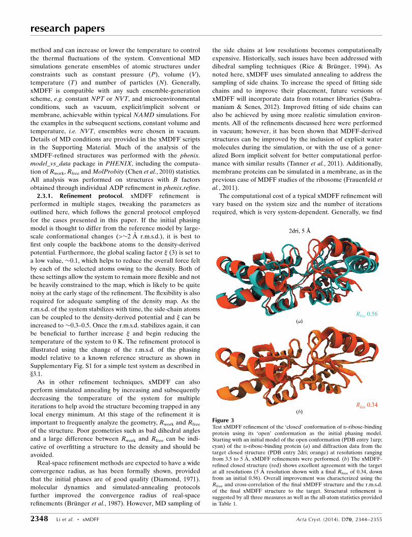
Figure 3Test xMDFF refinement of the ‘closed’ conformation of d-ribose-bindingprotein using its ‘open’ conformation as the initial phasing model.Starting with an initial model of the open conformation (PDB entry 1urp;cyan) of the d-ribose-binding protein (a) and diffraction data from thetarget closed structure (PDB entry 2dri; orange) at resolutions rangingfrom 3.5 to 5 A, xMDFF refinements were performed. (b) The xMDFF-refined closed structure (red) shows excellent agreement with the targetat all resolutions (5 A resolution shown with a final Rfree of 0.34, downfrom an initial 0.56). Overall improvement was characterized using theRfree and cross-correlation of the final xMDFF structure and the r.m.s.d.of the final xMDFF structure to the target. Structural refinement issuggested by all three measures as well as the all-atom statistics providedin Table 1.
that xMDFF can be computationally more demanding than
other refinement software owing to the full molecular
dynamics nature of the method. However, because xMDFF
is an extension of MDFF, which was developed as part of
NAMD, it is able to scale up from a single CPU core to
thousands, potentially allowing the protocol to be applied to
large systems such as the multi-million-atom HIV capsid
(Zhao et al., 2013). Furthermore, NAMD, and thus xMDFF,
can utilize GPU acceleration to improve simulation perfor-
mance (Stone et al., 2007, 2010).
3. Results
3.1. Proof of principle
The performance of xMDFF was evaluated on a test
structure, ribose-binding protein, with two known conforma-
tions at high resolution. The open conformation (PDB entry
1urp; 2.3 A resolution) was used as an initial phasing model,
with the closed conformation (PDB entry 2dri; 1.6 A resolu-
tion) as a target model (Fig. 3). The refinement was performed
using the diffraction data for the target model at four reso-
lutions (3.5, 4, 4.5 and 5 A), created by truncating the original
intensities at each resolution limit. The four refinements began
with the same initial phasing model and were evaluated
against the same target; the final refined structures were
evaluated using the overall improvement in Rfree as well as the
root-mean-squared deviation (r.m.s.d.) from the target model.
xMDFF refinements improved the Rfree value dramatically at
every resolution, with an initial value of 0.57 and a final value
of 0.23 at a resolution of 3.5 A (Table 1). The all-heavy-atom
r.m.s.d. for the refined structure at every resolution was 3.0 A
from the high-resolution 2dri target, down from the initial
5.46 A. However, the final r.m.s.d. of the backbone alone at
3.5 A resolution was 0.53 A (down from an initial 4.46 A),
demonstrating proper backbone placement relative to the
target model. In this case, the all-atom r.m.s.d.s are much
higher relative to the backbone r.m.s.d.s because the target
model was originally refined against 1.6 A resolution data,
where the side chains are much better resolved. Using the low-
resolution synthetic data, the backbone can still be fitted quite
well, but it is much harder to refine the side chains to the same
extent.
To verify that xMDFF was performing as well as it could,
a standard MDFF simulation was performed using density
created directly from the 2dri target model and Fobs reduced to
3.5 A resolution, which underwent non-iterative refinement.
The final structure obtained from this fitting had an all-atom
r.m.s.d. of 3.01 A and a backbone r.m.s.d. of 0.56 A, very close
to those of the xMDFF refinements and demonstrating that
xMDFF performs well against a more appropriate benchmark.
Although the synthesized 3.5 A resolution map manifests the
best possible density at this resolution, it has much less side-
chain information than the original 1.6 A resolution map. This
lack of side-chain density negatively affects side-chain fitting,
and thus increased the side-chain r.m.s.d. relative to the known
high-resolution target and accounts for the poor side-chain
refinements in xMDFF. Since the initial model was originally
obtained through refinement against high-resolution data,
the overall structural geometry including the percentage of
favored Ramachandran angles started very high at 98.5%.
However, xMDFF did manage to improve the overall
research papers
Acta Cryst. (2014). D70, 2344–2355 Li et al. � xMDFF 2349
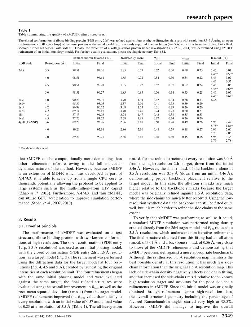
Table 1Table summarizing the quality of xMDFF-refined structures.
The closed conformation of ribose-binding protein (PDB entry 2dri) was refined against four synthetic diffraction data sets with resolution 3.5–5 A using an openconformation (PDB entry 1urp) of the same protein as the initial model. Six previously reported low-resolution (4–4.5 A) structures from the Protein Data Bankshowed further refinement with xMDFF. Finally, the structure of a voltage-sensor protein under investigation (Li et al., 2014) was determined using xMDFFrefinement of an initial homology model. For further quality evaluations, please see Supplementary Table S1.
Ramachandran favored (%) MolProbity score Rfree Rwork R.m.s.d. (A)
PDB code Resolution (A) Initial Final Initial Final Initial Final Initial Final Initial Final
2dri 3.5 98.51 97.01 1.85 0.77 0.62 0.30 0.58 0.23 5.46 3.014.46† 0.53†
4.0 98.51 96.64 1.85 0.72 0.54 0.30 0.54 0.22 5.46 3.024.46† 0.55†
4.5 98.51 95.90 1.85 0.92 0.57 0.37 0.52 0.24 5.46 3.064.46† 0.68†
5.0 98.51 96.27 1.85 0.85 0.56 0.34 0.53 0.23 5.46 3.054.46† 0.67†
1av1 4.0 90.20 95.01 3.70 1.94 0.42 0.34 0.38 0.33 N/A1xdv 4.1 95.30 95.05 2.87 2.01 0.41 0.33 0.39 0.291yi5 4.2 86.99 90.72 3.08 1.73 0.31 0.29 0.26 0.261aos 4.2 89.14 92.17 3.40 2.45 0.24 0.23 0.20 0.211jl4 4.3 87.15 91.03 3.24 1.47 0.42 0.38 0.35 0.331ye1 4.5 77.25 84.72 2.68 1.89 0.27 0.24 0.26 0.264g80 (Ci-VSP) 3.6 89.20 92.96 2.86 2.10 0.50 0.28 0.49 0.26 5.96 2.47
5.75† 1.84†4.0 89.20 92.14 2.86 2.10 0.48 0.29 0.48 0.27 5.96 2.60
5.75† 2.06†7.0 89.20 90.73 2.86 2.18 0.46 0.40 0.45 0.38 5.96 3.36
5.75† 2.78†
† Backbone-only r.m.s.d.

MolProbity (Chen et al., 2010) scores, primarily through a
decrease in rotamer outliers as well as in steric clashes. In
practice, xMDFF improves the MolProbity statistics of almost
all models discussed here owing to the MD-based nature of
the method, which provides excellent structural restraints on
the system.
We further tested the refinement capabilities of xMDFF by
employing more realistic low-resolution synthetic data. After
truncating the high-resolution data to 5 A, the data were
smoothed using a B factor of 35.00 A2, in a fashion similar to
the technique used in Schroder et al. (2010). Such a smoothing
procedure further reduces the signal-to-noise ratio of the 5 A
resolution diffraction data, posing perhaps a more realistic
refinement scenario. The refinement resulted in very similar
but slightly worse results, with a backbone r.m.s.d. of 0.93 A,
an Rfree of 0.34 and an Rwork of 0.28. To test the robustness of
xMDFF refinements to the choice of initial structure, the 5 A
resolution diffraction data were refined using another search
model that has the same overall r.m.s.d. from the target but
with a shift in the positioning of the helices. The new search
model, shown in Supplementary Fig. S1, has a backbone
r.m.s.d. of 4.45 A from 2dri, which is comparable to that of
1urp, but it also has an r.m.s.d. of 1.5 A relative to 1urp. The
refinement results are slightly worse but very similar, with an
r.m.s.d. of 0.8 A, a final Rfree of 0.34 (down from an initial 0.62)
and a final Rwork of 0.26 (down from 0.56).
Additionally, during refinements of the test model the
scaling factor which determines the overall strength of the
steering forces being applied was varied in order to determine
the optimal parameterization for future work (see x2 for
additional information). A lower scaling factor was deter-
mined to be useful during early stages of backbone refinement
to keep the system flexible, but increasing the scaling factor is
considered useful as refinement progresses in order to couple
the structure more strongly to the density and improve the fit;
however overfitting needs to be avoided at this stage.
3.2. Improving refinements of reported PDB structures
To test whether xMDFF is capable of improving previously
refined structures, it was applied to six structures at 4–4.5 A
resolution deposited previously into the PDB (Fig. 1). The
structures served as an initial phasing model and xMDFF was
able, without any further knowledge of the reference model,
to improve the Rfree by at least 0.01 (PDB entry 1aos) and up
to a maximum of 0.08 (PDB entries 1av1 and 1xdv) in the case
of all six structures (Table 1). Furthermore, the Rfree and Rwork
values of each system were relatively close, indicating that
xMDFF is not overfitting the structures. Additionally, every
xMDFF-refined structure exhibits an improved structural
geometry, as shown by a higher percentage of Ramachandran
favored angles and a lower overall MolProbity score over
those of the initial structures (Table 1). In one of the most
improved cases (PDB entry 1xdv), the main cause for the
lowered Rfree was improvement in a highly flexible region with
relatively large root-mean-square fluctuations (Supplemen-
tary Fig. S2).
This region shifted the most during xMDFF refinement,
with an r.m.s.d. of 4.3 A from the initial to the final model (part
of which is shown in Fig. 4). Flexible regions often diffract
poorly and can be difficult to properly place using traditional
means of X-ray refinement, especially at low resolution.
Densities were generated using Fobs and also ’calc from the
initial and final models, respectively (Fig. 4). A significant
improvement in the quality of the densities can be seen in
terms of their respective completeness and how well the
structure fits inside the density, with the latter captured by an
increase in the local CC from 0.47 (initial, blue) to 0.63 (final,
red).
The application of force-field-based MD simulations for
model refinement inherently provides geometries consistent
with some energy minima. Not only does the procedure
improve protein conformations, but it also conserves the
structures of cofactors, keeping them compatible with the
surrounding protein. For example, xMDFF refinement of
human hemoglobin (PDB entry 1ye1) preserves the planar
research papers
2350 Li et al. � xMDFF Acta Cryst. (2014). D70, 2344–2355
Figure 4Refinement of the highly flexible region in the case of PDB entry 1xdv.Substantial density improvements are observed in a flexible region of1xdv, illustrated by the difference in the density map between the initial(blue) (a) and xMDFF-refined final (red) (b) structures; local cross-correlations increase from 0.47 to 0.63, implying a more unambiguousplacement of the atoms.

coformation of the heme group. Consequently, distortions in
cofactors accompanying multiple conformational states of
protein crystals can be accounted for by low-resolution
diffraction data (Supplementary Fig. S3a).
xMDFF provides the atomic positions as well as the
chemical bonding in the low-resolution X-ray map. In many
cases, the assumed connectivity between atoms is reflected in
the degree of structural refinement, which in turn can affect
predictions of the associated function. For example, in the case
of the pentameric cobratoxin (Cbtx)-bound acetylcholine-
binding protein (AChBP) complex (PDB entry 1yi5), the
complex has a dense core composed primarily of the AChBPs.
The cobratoxins are composed of two antiparallel �-sheets
forming slightly concave discs, five of which emerge from the
dense AChBP core, with each disc sporting ten cysteine resi-
dues. However, given only the positions of atoms it is unclear
whether the cysteines are in oxidized or reduced states, i.e.
whether disulfide bridges are present or not. Using the 4.2 A
resolution diffraction data, xMDFF refinements were
performed assuming either the presence or the absence of
the disulfide bridges (Supplementary Fig. S3b). A more
pronounced refinement was achieved with the assumption of
oxidized cysteines (a final Rwork of 0.26 and an Rfree of 0.29
in contrast to the final Rwork of 0.27 and Rfree of 0.31 for the
reduced state), implying the existence of disulfide bridges. The
presence of the five xMDFF-predicted disulfide bridges per
Cbtx disc has been validated in biochemical studies, whereby
four bridges are key to the stability and the concave shape of
the disc and the fifth is resposible for optimal Cbtx–AChBP
binding (Bourne et al., 2005). Thus, xMDFF provided
biomolecular structures with energetically favorable geome-
tries that interpret low-resolution density maps as well as help
to clarify structures’ relevant biological functions.
The xMDFF-based structures [PDB entries 1av1 (Borhani
et al., 1997), 1xdv (Sondermann et al., 2004), 1yi5 (Bourne et
al., 2005), 1aos (Turner et al., 1997), 1jl4 (Wang et al., 2001) and
1ye1 (Kavanaugh et al., 2005)] in Fig. 1 showed improvement
with regard to R factor and geometry over structures that
had resulted from conventional approaches, e.g. REFMAC or
CNS/X-PLOR (PDB entries 1yi5, 1jl4, 1ye1, 1av1, 1xdv and
1aos), applied to the same diffraction data, most likely owing
to the combination of starting xMDFF with good initial phases
from an already refined search model and having molecular
dynamics incorporated in xMDFF. An identical conclusion
on the use of good initial phases was drawn from X-PLOR
refinements with MD simulations. However, models 1jl4 and
1av1, which were published in 2001 and 1997, respectively,
resulted from early-generation refinement methods and
accordingly had relatively high R-factor values, posing an easy
challenge for our present xMDFF treatment. In other cases
(1yi5 and 1ye1) the original refinements involved initial
REFMAC or X-PLOR rigid-body fitting of the search model
used and the authors had to resort to manual fitting employing
FRODO to account for the needed structural flexibility. The
automated, force-field-based flexible fitting algorithm in
xMDFF should avoid errors arising in manual fitting. Alto-
gether, for the discussed examples, the xMDFF-refined models
are found to be better or at least as good as the published
models.
3.3. xMDFF refinement and experimental validation of theCi-VSP crystal structure
Finally, xMDFF was applied to solve the structure of a
voltage-sensing protein, Ci-VSP, using 3.6, 4 and 7 A resolu-
tion diffraction data (Fig. 5). To validate the reliability of the
xMDFF refinement, the work was carried out in parallel with
an independent experimental investigation of the structure
and function of Ci-VSP (Li et al., 2014).
Voltage-sensing protein is a common scaffold present in
voltage-gated ion channels, voltage-sensitive enzymes and
voltage-gated proton channels, which are related to diverse
important physiological functions. As illustrated in Fig. 5, the
protein under current investigation, Ci-VSP, is arranged as
an antiparallel four-transmembrane-helix bundle S1–S4;
the overall structure is in agreement with the basic three-
dimensional architecture of all known voltage-sensor proteins
(Jiang et al., 2003; Long et al., 2007; Payandeh et al., 2011;
Zhang et al., 2012). The positively charged S4 helix within Ci-
VSP reorients upon stimulus from a transmembrane electric
field, leading to downstream responses. Despite a wealth of
structural and functional data, the details of this conforma-
tional change remain controversial, in particular the move-
ment of the S4 helix.
According to electrophysiological results, Ci-VSP at 0 mV
assumes the resting (Down) state in the wild type (WT) but
the activated (Up) state in the R217E mutant. Crystal struc-
tures of both states have been determined experimentally:
R217E at 2.5 A resolution and WT at 3.6 A resolution (Li et
al., 2014). Unfortunately, there was crystallographic uncer-
tainty in the S4 position in the Ci-VSP WT 3.6 A resolution
electron-density map. Spectroscopic data had limited the S4
position of Ci-VSP WT to three options in reference to the
R217E structure: no conformational change, one click down
and two clicks down, where a click refers to the offset of a
helix by one turn. Obviously, the confirmation of the S4
position in Ci-VSP WT became the key to the puzzle. xMDFF
was applied to predict the WT Ci-VSP structure and, thereby,
to resolve the uncertainty in the low-resolution data.
Refinement started from a MUFOLD-predicted (Zhang
et al., 2010) medium-confidence homology model developed
using information from 13 proteins (Supporting Information).
During refinement, the tentative model underwent a
remarkable large-scale deformation with an r.m.s.d. of 5.96 A.
Unlike many traditional refinement techniques, xMDFF is
able to handle such large-scale structural deformations
between the initial and final structures, producing in the
present case final Rfree values of 0.28 and 0.29, starting from
initial Rfree values of 0.50 and 0.48, at 3.6 and 4 A resolution,
respectively (Table 1). Positioning of the functionally relevant
S4 helices is in excellent agreement with the one-click-down
model of WT Ci-VSP, with an r.m.s.d. ranging from 0.4 to 1 A.
To further confirm the S4 position, potential structural
models were generated by gradual shift and rotation of the S4
research papers
Acta Cryst. (2014). D70, 2344–2355 Li et al. � xMDFF 2351

helix from the Up-conformation model to the two-click-down
model in 2000 even steps (in all a �10 A vertical displacement
and �110� rotation). Two independent parameters for model
evaluation were calculated from each of these 2000 structures:
(i) the crystallographic Rfree value and (ii) the correlation
coefficient (CC) between the experimental density map of the
S4 region and the calculated electron density from the refined
model (Supplementary Fig. S4). Fig. 5(b) shows that the
structure corresponding to the Rfree minimum and CC
maximum from Supplementary Fig. S4 resides in a region that
unambiguously places the position of the S4 helix in the one-
click-down position. Assuming that movement of the S4 helix
in Ci-VSP follows the classic helix-screw or sliding-helix mode
(Li et al., 2014), other degrees of freedom that are not involved
in any helix-screw type of motion are considered to be
redundant. Subsequently, the Rfree minimum identified by
xMDFF along the chosen line of helix-screw offsets is likely to
be global for the resting state of WT Ci-VSP. xMDFF clearly
differentiates the pattern of side chains associated with a
specific S4 helix position even though the individual side
chains are not fully visible at 3.6 A resolution. The resulting S4
position is three residues lower than that of the Up structure,
positioning Arg residues into the protein interior and away
from lipids, and is in excellent agreement with the one-click-
down model.
The refinement with the 7 A resolution data was not as
pronounced as those with the 3.6 and 4 A resolution data.
However, improvements were still observed in the r.m.s.d. and
R factors. The R factors from the resulting structure were
considerably higher than those obtained from higher resolu-
tions. Unlike the higher resolution maps, the 7 A resolution
data failed to distinguish between the Up and one-click-down
research papers
2352 Li et al. � xMDFF Acta Cryst. (2014). D70, 2344–2355
Figure 5xMDFF refinement of the voltage-sensing protein Ci-VSP. (a) A MUFOLD-predicted homology model (cyan) was used as an initial phasing model inxMDFF; this model has an r.m.s.d. of 6 A from an independently refined Ci-VSP structure (orange; Li et al., 2014). Ci-VSP includes a transmembrane(TM) domain and was crystallized with an antibody (Ab). Inset: the placement of the S4 helix within the TM region which determines the voltage-gatingcapabilities of the protein is of particular interest. (b) xMDFF refinement with 4 A resolution diffraction data produced a final structure (red) 2.6 A awayfrom the independently refined Ci-VSP structure (orange). Inset: the placement of the S4 helix in the Down position and the alignment of the regularlyplaced voltage-sensing Arg residues is in agreement with the independently refined model (Li et al., 2014).
models of Ci-VSP. Furthermore, using the 7 A resolution data
the R factors derived from the high-resolution Up or one-
click-down structures were comparable to that of the xMDFF
output. Thus, we conclude that the 7 A resolution map is too
coarse to resolve any information relevant to S4 placement.
The present example reaffirms the capability of xMDFF to
address large-scale structural deformations, as has been shown
for the test case with synthetic data sets, to produce significant
refinements yielding realistic structures, but now from more
noisy low-resolution experimental data.
4. Discussion
The introduction of MD and simulated-annealing algorithms
have facilitated structure determination from low-resolution
diffraction data sets (Brunger et al., 1987). xMDFF provides
a significant step forward among the MD-based algorithms
as a crystallographic refinement tool. A brief comparison of
xMDFF predictions with those from other available refine-
ment methods is provided in Supplementary Table S2. Broadly
speaking, xMDFF refinements provide the lowest R factors,
minimal overfitting and improved structural statistics among
the methods compared [DEN in both CNS and PHENIX
(Brunger et al., 1998; Schroder et al., 2010) in addition to
default PHENIX (Adams et al., 2010) refinement]. However,
as further discussed in the Supporting Information, although
we try to achieve a fair comparison, the results might depend
on the user’s knowledge of the system, the application of
the software and the usage of manual fitting. For a more
controlled comparison, xMDFF was used to refine two struc-
tures using the same initial models (PDB entries 3kso and
3k0i) and reflection data (PDB entry 3k07) from a previous
comparison of Rosetta–PHENIX (DiMaio et al., 2013), CNS
DEN (Brunger et al., 1998; Schroder et al., 2010), PHENIX
(Adams et al., 2010) and REFMAC5 (Murshudov et al., 2011).
The two structures are sufficiently low resolution, >�4 A, and
the search models are displaced by >�5 A relative to the
target. In the case of 3kso, xMDFF achieved a better refined
structure than the best one from DiMaio and coworkers, which
was obtained using CNS DEN (using Rwork and Rfree as the
determinants). This improvement is evident in the lower Rwork
(0.2518), Rfree (0.3509) and MolProbity score (4.03) than those
obtained for the CNS DEN-refined structure (Rwork 0.319,
Rfree 0.387, MolProbity score 4.15). When starting with the
3k0i search model, the xMDFF-refined structure has a higher
Rwork (0.3226) and Rfree (0.3944) but a lower MolProbity score
(3.00) than those obtained for the best refined structure, again
using CNS DEN (Rwork 0.307, Rfree 0.368, MolProbity score
3.94). However, xMDFF still produces better results than the
rest of the methods in the study. It should also be noted that
while the Rfree and Rwork of Rosetta–PHENIX-refined struc-
tures in these two cases are worse than those with CNS DEN,
they have much better MolProbity scores of 2.00 for 3ks0 and
1.91 for 3k0i, a trend that is observed in all but two of the test
cases.
Additional considerations must be made to satisfactorily,
if not conclusively, compare the multiple refinement protocols.
Restricting discussions to only the real-space refinement
protocols compared here brings forth several differences.
For example, elastic networks, as implemented in DEN-based
protocols, robustly predict collective global motions but do not
work as well for describing local changes. Therefore, addi-
tional care should be taken when dealing with side chains
through the use of library-based refinement protocols.
Furthermore, elastic network or heurestic force-field-based
algorithms often require optimization of the interatomic
interactions with each new class of applications. Universal
force-field-based protocols such as ours are expected to be
more physically correct as the atomic interactions are cali-
brated against very accurate quantum calculations. However,
sometimes the application of different force fields leads to
different results owing to differences in the calibration
protocol used for force-field development. Thus, any further
comparison of the various real-space refinement protocols is
beyond the scope of this paper.
xMDFF guides the dynamics of a search model to a refined
structure through use of first principles via universal force
fields together with restraints from the X-ray maps. Conse-
quently, manual real-space fitting, as is commonly used with
reciprocal-space refinement protocols, is often avoided. This
benefit is of relevance to protocols in drug discovery, which
often require determination of multiple crystal structures
distributed over a broad range of conformations or ligand-
bound states. If performed manually, refinement in drug
discovery requires extensive repetition of the same task for
each structure. xMDFF naturally provides a semi-automated
computational platform to systematically perform several
model-building and repetitive refinement steps amenable to
high-throughput crystallography.
The initial xMDFF implementation presented here has
several limitations that we hope to address through future
development. Firstly, the quality of the refinement results
might depend on the nature of the force field used, the effects
of which require further investigation. Additionally, the
present implementation is unable to handle changes in
secondary structure during the simulation, and thus the
refinement is strongly biased by the folds present in the search
model. Any change in the secondary structure can only be
invoked at the homology-modelling stage and not during the
refinement. Finally, the current xMDFF implementation is
limited to refinement of biomolecules for which force fields
are already available. Refinement of nonbiological systems is
subject to force-field availibility.
In summary, the application of xMDFF to synthetic as well
as experimental low-resolution X-ray data demonstrated the
capability of the software to refine phasing models 6 A away
from target data even with maps as coarse as 7 A resolution,
a feat thus far achieved, to the best of our knowledge, very
rarely in low-resolution X-ray crystallography. MD naturally
provides the necessary sampling required to flexibly fit a
model into an electron-density map. Through flexible fitting
into iteratively updated model-phased maps, xMDFF can
bring about a series of large-scale deformations of the
initial structure relevant to its refinement. Detailed features
research papers
Acta Cryst. (2014). D70, 2344–2355 Li et al. � xMDFF 2353

characterizing macromolecular function, such as the location
of helices in voltage-sensor proteins or disulfide-bridge
networks in cobratoxin, have been accurately determined. The
application of force fields within xMDFF achieves realistic
atomic structures with sterically and conformationally accep-
table geometries. Low MolProbity scores together with a
consistently small difference between Rwork and Rfree values
imply negligible overfitting in xMDFF refinements. The
quality of the overall refinement is confirmed via improve-
ments in cross-correlations with simulated density. Finally,
xMDFF output structures require little post-processing to
initiate MD simulations for subsequent analysis of their
dynamics in a host medium. In summary, xMDFF together
with sequence information and homology modelling provides
a general approach to determining all-atom structures from
low-resolution X-ray data.
This work was supported by grants NIH 9P41GM104601,
NIH 5R01GM098243-02 and NIH U54GM087519 from the
National Institutes of Health. The authors also acknowledge
the Beckman Postdoctoral Fellowship program for supporting
A. Singharoy.
References
Adams, P. D. et al. (2010). Acta Cryst. D66, 213–221.Agirrezabala, X., Schreiner, E., Trabuco, L. G., Lei, J., Ortiz-Meoz,
R. F., Schulten, K., Green, R. & Frank, J. (2011). EMBO J. 30,1497–1507.
Becker, T., Bhushan, S., Jarasch, A., Armache, J.-P., Funes, S., Jossinet,F., Gumbart, J., Mielke, T., Berninghausen, O., Schulten, K.,Westhof, E., Gilmore, R., Mandon, E. C. & Beckmann, R. (2009).Science, 326, 1369–1373.
Borhani, D. W., Rogers, D. P., Engler, J. A. & Brouillette, C. G. (1997).Proc. Natl Acad. Sci. USA, 94, 12291–12296.
Bourne, Y., Talley, T., Hansen, S., Taylor, P. & Marchot, P. (2005).EMBO J. 24, 1512–1522.
Bricogne, G. & Irwin, J. (1996). Proceedings of the CCP4 StudyWeekend. Macromolecular Refinement, edited by E. Dodson, M.Moore, A. Ralph & S. Bailey, pp. 85–92. Warrington: DaresburyLaboratory.
Brunger, A. T., Adams, P. D., Fromme, P., Fromme, R., Levitt, M. &Schroder, G. F. (2012). Structure, 20, 957–966.
Brunger, A. T., Kuriyan, J. & Karplus, M. (1987). Science, 235,458–460.
Brunger, A. T. (1988). Crystallographic Computing 4: Techniques andNew Technologies, edited by N. W. Isaacs & M. R. Taylor. Oxford:Clarendon Press.
Brunger, A. T., Adams, P. D., Clore, G. M., DeLano, W. L., Gros, P.,Grosse-Kunstleve, R. W., Jiang, J.-S., Kuszewski, J., Nilges, M.,Pannu, N. S., Read, R. J., Rice, L. M., Simonson, T. & Warren, G. L.(1998). Acta Cryst. D54, 905–921.
Chan, K.-Y., Gumbart, J., McGreevy, R., Watermeyer, J. M., Sewell,B. T. & Schulten, K. (2011). Structure, 19, 1211–1218.
Chapman, M. S. (1995). Acta Cryst. A51, 69–80.Chapman, M. S. & Blanc, E. (1997). Acta Cryst. D53, 203–206.Chen, V. B., Arendall, W. B., Headd, J. J., Keedy, D. A., Immormino,
R. M., Kapral, G. J., Murray, L. W., Richardson, J. S. & Richardson,D. C. (2010). Acta Cryst. D66, 12–21.
DeLaBarre, B. & Brunger, A. T. (2003). Nature Struct. Biol. 10,856–863.
Delarue, M. (2008). Acta Cryst. D64, 40–48.Depristo, M. A., de Bakker, P. I. W., Johnson, R. J. K. & Blundell,
T. L. (2005). Structure, 13, 1311–1319.
Diamond, R. (1971). Acta Cryst. A27, 436–452.DiMaio, F., Echols, N., Headd, J. J., Terwilliger, T. C., Adams, P. D. &
Baker, D. (2013). Nature Methods, 10, 1102–1104.DiMaio, F., Terwilliger, T. C., Read, R. J., Wlodawer, A., Oberdorfer,
G., Wagner, U., Valkov, E., Alon, A., Fass, D., Axelrod, H. L., Das,D., Vorobiev, S. M., Iwai, H., Pokkuluri, P. R. & Baker, D. (2011).Nature (London), 473, 540–543.
Frauenfeld, J., Gumbart, J., van der Sluis, E. O., Funes, S., Gartmann,M., Beatrix, B., Mielke, T., Berninghausen, O., Becker, T., Schulten,K. & Beckmann, R. (2011). Nature Struct. Mol. Biol. 18, 614–621.
Gumbart, J., Schreiner, E., Trabuco, L. G., Chan, K.-Y. & Schulten, K.(2011). Molecular Machines in Biology, edited by J. Frank, pp. 142–157. Cambridge University Press.
Gumbart, J., Trabuco, L. G., Schreiner, E., Villa, E. & Schulten, K.(2009). Structure, 17, 1453–1464.
Haddadian, E. J., Gong, H., Jha, A. K., Yang, X., DeBartolo, J.,Hinshaw, J. R., Rice, P. A., Sosnick, T. R. & Freed, K. F. (2011).Biophys. J. 101, 899–909.
Hendrickson, W. A. (1985). Methods Enzymol. 115, 252–270.Hsin, J., Gumbart, J., Trabuco, L. G., Villa, E., Qian, P., Hunter, C. N.
& Schulten, K. (2009). Biophys. J. 97, 321–329.Humphrey, W., Dalke, A. & Schulten, K. (1996). J. Mol. Graph. 14,
33–38.Jiang, Y., Lee, A., Chen, J., Cadene, M., Chait, B. T. & MacKinnon, R.
(2003). Nature (London), 423, 33–41.Karmali, A. M., Blundell, T. L. & Furnham, N. (2009). Acta Cryst.
D65, 121–127.Kavanaugh, J. S., Rogers, P. H. & Arnone, A. (2005). Biochemistry, 44,
6101–6121.Li, Q., Wanderling, S., Paduch, M., Medovoy, D., Singharoy, A.,
McGreevy, R., Villalba-Galea, C., Hulse, R. E., Roux, B., Schulten,K., Kossiako, A. & Perozo, E. (2014). Nature Struct. Mol. Biol. 21,244–252.
Li, W., Trabuco, L. G., Schulten, K. & Frank, J. (2011). Proteins, 79,1478–1486.
Long, S., Tao, X., Campbell, E. & MacKinnon, R. (2007). Nature(London), 15, 376–382.
Murshudov, G. N., Skubak, P., Lebedev, A. A., Pannu, N. S., Steiner,R. A., Nicholls, R. A., Winn, M. D., Long, F. & Vagin, A. A. (2011).Acta Cryst. D67, 355–367.
Murshudov, G. N., Vagin, A. A. & Dodson, E. J. (1997). Acta Cryst.D53, 240–255.
Pannu, N. S. & Read, R. J. (1996). Acta Cryst. A52, 659–668.Payandeh, J., Scheuer, T., Zheng, N. & Catterall, W. (2011). Nature
(London), 475, 353–358.Phillips, J. C., Braun, R., Wang, W., Gumbart, J., Tajkhorshid, E., Villa,
E., Chipot, C., Skeel, R. D., Kale, L. & Schulten, K. (2005). J.Comput. Chem. 26, 1781–1802.
Praznikar, J., Afonine, P. V., Guncar, G., Adams, P. D. & Turk, D.(2009). Acta Cryst. D65, 921–931.
Rice, L. M. & Brunger, A. T. (1994). Proteins, 19, 277–290.Schreiner, E., Trabuco, L. G., Freddolino, P. L. & Schulten, K. (2011).
BMC Bioinformatics, 12, 190.Schroder, G. F., Levitt, M. & Brunger, A. (2010). Nature (London),
464, 1218–1222.Seidelt, B., Innis, C. A., Wilson, D. N., Gartmann, M., Armache, J.-P.,
Villa, E., Trabuco, L. G., Becker, T., Mielke, T., Schulten, K., Steitz,T. A. & Beckmann, R. (2009). Science, 326, 1412–1415.
Sener, M. K., Hsin, J., Trabuco, L. G., Villa, E., Qian, P., Hunter, C. N.& Schulten, K. (2009). Chem. Phys. 357, 188–197.
Sondermann, H., Soisson, S. M., Boykevisch, S., Yang, S.-S., Bar-Sagi,D. & Kuriyan, J. (2004). Cell, 119, 393–405.
Stone, J. E., Hardy, D. J., Umfitsev, I. S. & Schulten, K. (2010). J. Mol.Graph. Model. 29, 116–125.
Stone, J. E., Phillips, J. C., Freddolino, P. L., Hardy, D. J., Trabuco,L. G. & Schulten, K. (2007). J. Comput. Chem. 28, 2618–2640.
Subramaniam, S. & Senes, A. (2012). Proteins, 80, 2218–2234.
research papers
2354 Li et al. � xMDFF Acta Cryst. (2014). D70, 2344–2355

Tanner, D. E., Chan, K.-Y., Phillips, J. & Schulten, K. (2011). J. Chem.Theor. Comput. 7, 3635–3642.
Trabuco, L. G., Schreiner, E., Eargle, J., Cornish, P., Ha, T., Luthey-Schulten, Z. & Schulten, K. (2010). J. Mol. Biol. 402, 741–760.
Trabuco, L. G., Villa, E., Mitra, K., Frank, J. & Schulten, K. (2008).Structure, 16, 673–683.
Trabuco, L. G., Villa, E., Schreiner, E., Harrison, C. B. & Schulten, K.(2009). Methods, 49, 174–180.
Turner, M. A., Simpson, A., McInnes, R. R. & Howell, P. L. (1997).Proc. Natl Acad. Sci. USA, 94, 9063–9068.
Villa, E., Sengupta, J., Trabuco, L. G., LeBarron, J., Baxter, W. T.,Shaikh, T. R., Grassucci, R. A., Nissen, P., Ehrenberg, M., Schulten,K. & Frank, J. (2009). Proc. Natl Acad. Sci. USA, 106, 1063–1068.
Wang, J., Meijers, R., Xiong, Y., Liu, J., Sakihama, T., Zhang, R.,Joachimiak, A. & Reinherz, E. L. (2001). Proc. Natl Acad. Sci.USA, 98, 10799–10804.
Wells, D., Abramkina, V. & Aksimentiev, A. (2007). J. Chem. Phys.127, 125101.
Zhang, J., Wang, Q., Barz, B., He, Z., Kosztin, I., Shang, Y. & Xu, D.(2010). Proteins, 78, 1137–1152.
Zhang, X., Ren, W., DeCaen, P., Yan, C., Tao, X., Tang, L., Wang, J.,Hasegawa, K., Kumasaka, T., He, J., Wang, J., Clapham, D. E. &Yan, N. (2012). Nature (London), 486, 130–134.
Zhao, G., Perilla, J. R., Yufenyuy, E. L., Meng, X., Chen, B., Ning, J.,Ahn, J., Gronenborn, A. M., Schulten, K., Aiken, C. & Zhang, P.(2013). Nature (London), 497, 643–646.
research papers
Acta Cryst. (2014). D70, 2344–2355 Li et al. � xMDFF 2355