x-ray based craniofacial visualization and surgery simulation junjun pan 1, jian j zhang 1, yanning...
TRANSCRIPT

X-ray Based Craniofacial Visualization and SX-ray Based Craniofacial Visualization and S
urgery Simulationurgery Simulation
Junjun Pan1, Jian J Zhang1, Yanning Zhang2, Hong Zhou3 E-mail:
[email protected], [email protected]
[email protected], [email protected] 1. National Center for Computer Animation, Bournemouth University, UK
2. School of Computer Science, Northwestern Polytechnical University, China
3. Stomatology Hospital of Xi an Jiao tong University, China

OutlinesOutlines
• Research background• Objectives• Methodology• Algorithms• Experiment and results• Conclusion and future work

Research background
In orthodontics and plastic surgery, the analysis and measurement of cranium are often needed to diagnose whether there is structure malformation in the mandible or maxilla. At present there are two main kinds of approaches :
• One is the conventional measurement method, which is directly manipulated on the frontal or lateral cephalo X-rays. (2D method, widely used, the accuracy is limited)
• Another is the measurement on the 3D cranium model reconstructed by CT. ( 3D method, high accuracy)

Frontal and lateral cephalo X-rays 3D face and skull model from CT

Research objectives
• One disadvantage of CT is that large dose of X-ray radiation could have a serious adverse effect to the human body, especially for the children.
• In addition, compared with ordinary X-rays, CT scan is also more costly, which could be an issue for developing countries and underprivileged regions.
• In this research, instead of the CT based option, we want to design a low radiation, low cost way to reconstruct and visualize the 3D cranium with a small number of X-rays

Methodology
We use the method of computer vision to reconstruct the 3D face of patient from three cephalo X-rays (00, 600 , 900 projection with lead markers). Then with two of three X-rays and M-ultrasound scan, we can get the thickness of soft tissue and create the 3D skull model by subtracting soft tissue from facial surface. In this process, only X-ray machine, adhesive taps with lead markers and the M-ultrasonograph are needed.

X-ray photographing
X-ray photographing with cephalometer Cephalometer X-ray machine

60o Projection 90o Projection A testee with a tape of lead markers on lips in X-ray photographing

System structure

60o Projection 90o Projection Original X-ray image pair

60o Projection 90o Projection Binary images detected feature points

Correlated vision The calculation of 3D coordinates of markers by the correlated
vision.
x
y
z
=
l
l
l
x
y
z
+
',',
l i
l l l i
l
x
t y
F
R
=
r
r
r
x
y
z
+
',
',
r i
r r r i
r
x
t y
F
R
x
y
z
2' ', ,
2 ' ', ,
' ', ,
l l i r r i
l l l l i r r r r i
l l i r r i
x x x x
d y t y y t y
z z z z
R R
2
l l r rt t b r r

3D face and skull model of a subject in experiment
Visualization resultsVisualization results

Demo 1

Surgery simulation and soft tissue
deformation
LeFort I osteotomy Mandible vertical osteotomy
Two typical osteotomies and corresponding operation simulations

Demo 2

1. Corresponding points matching based on position similarity: When the difference of the projection angles is small for both
correlated images and the object. If place both correlated images in the same coordinate system, and let the barycenter of feature points in both images coincide, the summation of the Euclidean distance between pairs of corresponding points is minimum if the correct matching is found.
New algorithms

Corresponding matching by optimization
, 1
minn
ij iji j
d
s. t.1
1, 1,2,3....... ,n
ijj
i n
1
1, 1,2,3....... ,n
iji
j n
{0,1}, , 1, 2,3...... ,ij i j n
2 2( ) ( ) , , 1, 2,3...... ,ij li rj li rjd x x y y i j n

2. An supervised machine learning method to calculate the stiffness parameters in FEM:
After the computation of the soft tissue deformations using FEM,
the results are compared with the measured reference data (training data) taken from the previous operation. The errors are used to adjust the estimated parameters and this training process goes on until a predefined tolerance is satisfied. Then the trained parameters is more accurate and can be used in the following surgeries. The training formula is as follows:
New algorithms
1 ( )i i iT t k
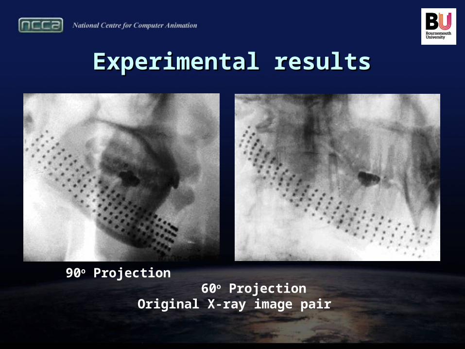
Experimental resultsExperimental results
90o Projection 60o Projection Original X-ray image pair

Experiment 1
(a) 3D points matched by epipolar lines searching (b) 3D points matched by sequence matching algorithm (c) 3D points matched by the algorithm in our paper
(a) (b) (c)
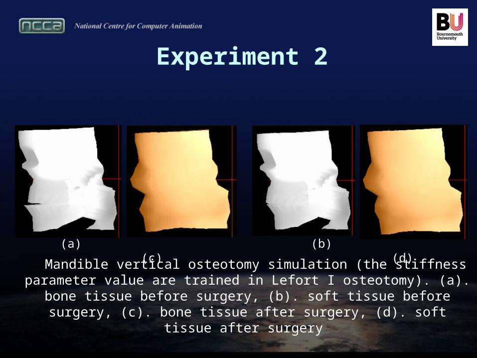
Experiment 2
Mandible vertical osteotomy simulation (the stiffness parameter value are trained in Lefort I osteotomy). (a). bone tissue before surgery, (b). soft tissue before
surgery, (c). bone tissue after surgery, (d). soft tissue after surgery
(a) (b) (c) (d)
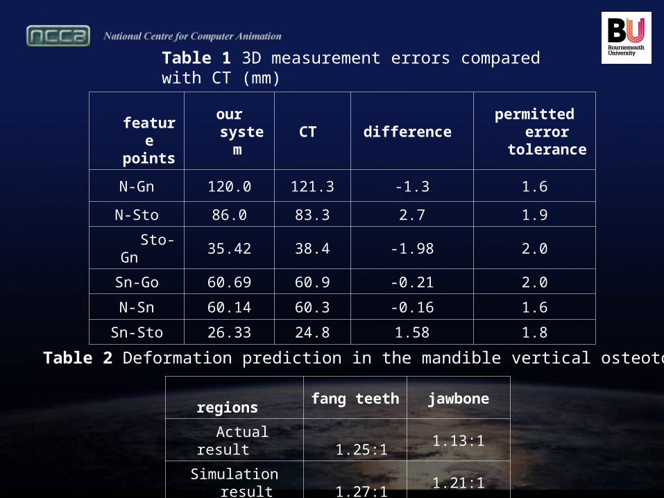
feature
pointsour system CT difference
permitted error tolerance
N-Gn 120.0 121.3 -1.3 1.6
N-Sto 86.0 83.3 2.7 1.9
Sto-Gn 35.42 38.4 -1.98 2.0
Sn-Go 60.69 60.9 -0.21 2.0
N-Sn 60.14 60.3 -0.16 1.6
Sn-Sto 26.33 24.8 1.58 1.8
Table 1 3D measurement errors compared with CT (mm)
regions fang teeth jawbone
Actual result 1.25:1 1.13:1
Simulation result 1.27:1 1.21:1
Table 2 Deformation prediction in the mandible vertical osteotomy

Conclusion• We present a low radiation, low cost alternative to CT-based
method for the visualization of 3D cranium using only three X-rays. We also present a new algorithm to solve the corresponding matching by evolutionary programming and designed a supervised learning method to estimate the soft tissue stiffness parameters.
Discussion• From the proof of groups medical experiment, our system is
easy-to-use and more accurate than the traditional 2D X-rays based measurement.
• Compared with CT, our technique remains deficiency of modeling accuracy in some area where the markers can not be placed.
Conclusion and discussion

• The current method for placing the lead markers is quite awkward and user-unfriendly. We are producing a special face mask to let the markers touch the face automatically, which can also guide the ultrasound scan with accurate placement of the probe.
• Using the developed prototype system, we also plan to investigate the relationship of the stiffness parameter values with human age, race, gender, ethnic origins and some other related factors.
Future work

Thank you!