x-ray acquisition software acquisition and diagnostic software · 2016-11-17 · dicom dx-r...
TRANSCRIPT

Acquisition anddiagnostic softwarefor X-ray images from DR flat panels or CR systems in
human and veterinary medicine
DX-RdicomPACS R
X-ray Acquisition Software
Acq
uis
itio
nand
dia
gnost
icso
ftw
are
for
X-r
ay
imag
es


Acq
uis
itio
n a
nd
dia
gnost
ic s
oft
ware
for
X-r
ay
imag
es
dicom DX-R
dicom DX-R
dicom DX-R
dicom
dicom DX-R
PACS
PACS
PACS
PACS
PACS
®
®
®
®
®
is a professional acquisition software for X-ray images
from flat panel systems (DR) and CR units (computed radiography with imaging
plates) by any manufacturer. In addition, the software controls X-ray generators
and X-ray units of various manufacturers, providing a smooth and systematic
workflow. A simple and user friendly GUI (graphical user interface) operated
by touchscreen or mouse completes the system.
The professional image processing can be adapted to
individual user needs and offers outstanding image quality in human and
veterinary medicine. It has been specially developed to enable organ specific
optimisation, guaranteeing the highest quality X-ray images.
Many helpful integrated functions such as the radiographic positioning guide
and intuitive operation simplify daily routine tasks greatly.
In addition, allows integration with existing patient
management systems. The integrated full viewer even allows the
user to diagnose X-ray images within the acquisition software. Therefore, the
system can also be applied as fully-fledged diagnostic workstation with the
option to upgrade to a PACS(Picture Archiving and Communication System).
forms the core of a direct digital X-ray unit, whether it
is a retrofit system to upgrade existing X-ray units, a complete new unit including
generator control, or a portable suitcase solution for mobile X-ray generators.
What is dicom DX-RPACS®
DX-RdicomPACS R
X-ray Acquisition Software
Professional
acquisition
software for
X-ray images

dicom DX-R function principlePACS®
- operation softwarefor generator and panel
- image processing- image management
DX-RdicomPACS R
X-ray Acquisition Software
HIS/RIS etc.(Patient management
system)
DICOM Worklist
Delivery of patient
data and examination
instructio
ns
Rawim
ages
Panel controlX-ray devices
(motorised)
Cont
rol o
f the
mot
orise
d sy
stem
,
collim
ator
etc
.Po
sitio
ning
prot
ocol
when instructio
ns have
been carried outConfirmation
Unlessprovided by
HIS/RISDICOMWorklist
PACS(e.g. )dicomPACS
®
DICOMstore
X-ray
generator
KVp, mAS,body part etc.
Exposureprotcol
Output of processedimages incl. all patientand exposure data
2
Flat panel(different manufacturers,
also dental panel)
dicom DX-RPACS®
software
Function principles

Benefits of dicom DX-RPACS®
Modern graphical user interface (GUI) adaptable to almost
operation – to ensure quick and efficient work and
a smooth workflow
Capture of patient data via
or other protocols – data may also be captured manually
Use of for the transfer of all relevant
examination data directly from the connected patient management
system (HIS/RIS)
body parts with more than
and numerous possible adjustments in
already included
Safe and fast
Allows the user to of a patient, for
instance to avoid having to re-position the patient frequently
Allows the user to to an examination, even
after that examination has already been completed
Special tools for veterinary medicine, such as an extra dialog
box for patient and owner data, integrated
for alternating
between mobile and stationary X-ray systems and much more…
Entry of recurring ,
e.g. thorax screenings or pre-purchase examination for horses
for each examination
in human and veterinary medicine incl. comprehensive notes, photos, videos
and correct X-ray images
Option to control a digital X-ray system via incl.
display of the worklist, preview of the image taken for checking
and much more
any language
Touchscreen
DICOM Worklist, BDT/GDT, HL7
DICOM Procedure Codes
Freely configurable 400 projections
human and veterinary medicine
registration of emergency patients
switch between examinations
subsequently add images
hip dysplasia measuring,
special image filters, multi generator operation
examination procedures as macros
Fully integrated radiographic positioning guide
wireless remote
Wireless remote
control for the
taking of images
3
BenefitsUser friendliness and smooth workflow

Benefits of dicom DX-RPACS®
4
Steve Miller
DX-RdicomPACS R
X-ray Acquisition Software
dicom DX-RPACS®
software
Screenshots
Job creation
Switch to
the planning
of X-ray jobs
for children
The correct settingsfor adults and children -
or for horses, dogs andcats – are available
at a mouse click
Chart for theplanning of anindividual
X-ray job
Radiographic positioning guide
Video with sound
for the step by
step positioning
of the patient
Shows anexample of a
correct X-ray
image
Opens examplesof inaccurateX-ray imageswith comments
Presentation of helpfulhints for the positioningof the patient, central beam,tips and tricks, frequent
errors etc.

Integration of various by different manufacturers
Option to (bucky, wall stand and mobile)
to one system
The enables the user to control
X-ray generators or X-ray systems by different manufacturers, delivering
the generator settings directly from the software
Option for the included
in the standard package. The user has the choice to take the next image with
either the flat panel or the integrated CR system. This flexibility also provides an
in case of a defect flat panel.
(Automatic Exposure Control) and (Anatomical Programmed
Radiography) allow the user to
for each projection with an option to subsequently edit the
image manually
Integration of (DAP) – the readings are
saved directly to the relevant image
Electronic X-ray log
flat panel and CR systems
connect up to 3 flat panels
configurable generator interface
parallel operation of a flat panel and a CR system
excellent emergency concept
AEC ARP
automatically adjust all X-ray options
dose area product meters
Benefits of dicom DX-RPACS®
5
BenefitsFlexible image acquisition
DR flat panelradiology
Network
The software allows the controlof one CR system and one or moreflat panels
DX-RdicomPACS R
X-ray Acquisition Software
inklus
iveDiv
arioCR 36O
vet
CR-Sys
teme mit
Zukunf
tDX-R Akquis
itions
-Softw
are
CR system
DR flat paneldental vet
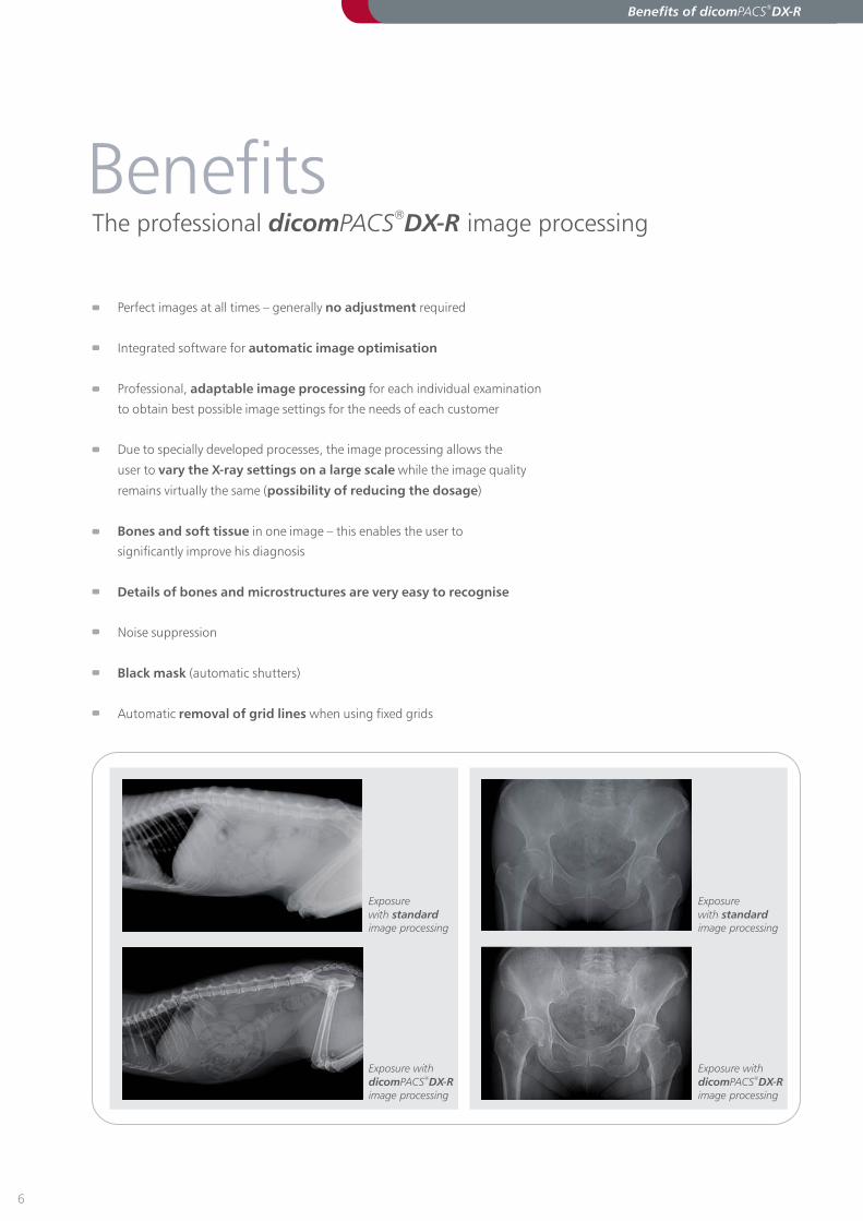
Perfect images at all times – generally required
Integrated software for
Professional, for each individual examination
to obtain best possible image settings for the needs of each customer
Due to specially developed processes, the image processing allows the
user to while the image quality
remains virtually the same ( )
in one image – this enables the user to
significantly improve his diagnosis
Noise suppression
(automatic shutters)
Automatic when using fixed grids
no adjustment
automatic image optimisation
adaptable image processing
vary the X-ray settings on a large scale
possibility of reducing the dosage
Bones and soft tissue
Details of bones and microstructures are very easy to recognise
Black mask
removal of grid lines
Exposure
withimage processing
standard
Exposure with
image processingdicom DX-RPACS
®
Benefits of dicom DX-RPACS®
6
Exposure
withimage processing
standard
Exposure with
image processingdicom DX-RPACS
®
The professional image processingdicom DX-RPACS®
Benefits

Varying X-ray doses
Example of a series of X-ray images taken with varying X-ray doses. The
image processing software allows the user to apply a strongly
reduced radiation dose while the image quality remains virtually the same. This
is of particular importance when taking X-rays of children. The foot images
depicted were taken with a Varian “PaxScan 4343R” (GadOx) flat panel.
dicom DX-RPACS®
54 KVp and 1.80 mAs 54 KVp and 1.05 mAs
54 KVp and 0.60 mAs 54 KVp and 0.15 mAs
Benefits of dicom DX-RPACS®
7

Viewer
Completely integrated ,
further processing and storage of images in an SQL database incl. image
manipulations, export options, layout adjustments, freely configurable
user interface and much more
Stepless etc.
Insertion of , e.g. free texts, arrows, ellipses etc.
of distances, angles, areas and density
(Specialised filters for the
optimised depiction of bones and soft tissue, measurements for and
, determination, etc.)
Adjustment of window/level options and ,
sharpening filters, noise suppression
Many additional functions such as calculation of
etc.
Printing of images both on Windows printers and laser imagers via
Creation of with free
to JPEG, TIFF, BMP and DICOM formats
Easily upgradable to the (PACS)
dicomPACS®
Viewer for image diagnosis
zoom, PAN, magnifyer, ROI, crop, rotate, mirror
image annotations
Measuring
Special purpose tools for the veterinarian
TPLO
TTA distraction index cardiac measurements
gamma correction
Cobb's angle,
HD measurements, pelvic obliquity measurements, integrated
capturing of diagnostic reports
DICOM Basic Print
DICOM patient CDs WEB viewer
Export of images
integrated image management system
Benefits of dicom DX-RPACS®
8
Outstandingly sophisticated image diagnosis
Benefits

Integrated image distribution
Worldwide image distribution
Web Server
direct auto routing
images can be archived externally via the web server
several
databases DICOM store
to colleagues or patients via the
(optional) – Images can be accessed from any
PC with internet access
Option of of images to external radiologists
On request,
Images can be sent to image management systems or
via
dicomPACS®
Web preview
Web viewer
Benefits of dicom DX-RPACS®
9
Benefits

dicom DX-RPACS®
is a generally open system. Its conception and
development was independent of hardware manufacturers.
Components from the following manufacturers have already been
integrated (We are continuously working on the integration of new
models and manufacturers):
Flat panel
Generator control
The generator screen displays all recommended values and
settings (kVp, mAs, focus etc.). These settings may be
adapted to the system used.
Supported modalities
CR systems
10
DX-RdicomPACS R
X-ray Acquisition Software
Which flat panels and CR systems doessupport?dicom DX-RPACS
®
Modalities
VAR ANmedical systems
DÜRRMEDICAL

dicom DX-R
dicom DX-R
PACS
PACS
®
®
may not only be used as a software for the acquisition
and processing of X-ray images, but can also be upgraded to a MiniPACS or
even to an Enterprise Multi Modality PACS. 5,000 installed workstations in
over 600 image management systems in over 40 countries (as of 10/2010)
prove that our customers are satisfied.
A single workstation system with installed software
can be upgraded with the following options (extract):
Further optional viewer functions:
Upgrading dicom DX-RPACS®
11
May be installed on systems
Generation of full leg/full spine images
Preparation of diagnostic reports with integrated images
in MS Word
Connection of of several diagnostic monitors
Capturing of additional patient and examination data with
their freely configurable
Working with
and documentation - Prosthesis templates can be
selected from a set and inserted into the image as
annotations
Additional radiological functions such as Maximum Intensity
Projection ( ), Multiplanar Reconstruction ( ), hanging
protocols and mammography tools
Fast and easy preparation of
with automatically inserted X-ray images
(only for Germany)
And much more…
Apple MAC and Linux
(Image stitching)
statistical analysis
digital prosthesis templates for surgery
planning
MIP MPR
equine pre-purchase
examinations
The stitching module merges a
number of separate digital X-ray
images into a single image. You can
load, correctly align and merge any
number of original images.
ExtensionOptions for upgrading dicom DX-RPACS
®

DICOM reception
DICOM distribution
DICOM DIR import
DICOM Query/Retrieve
Pre-fetching
DICOM Print Server
DICOM Compression
film and document scanners
endoscopy, angiography
synchronisation
Exchange of images and diagnostic
Web Server Intranet
Web Server Internet
from any DICOM sources, e.g. CT,
MRI, scintigraphy, ultrasound etc
with freely configurable rules
for archiving patient CDs by
other manufacturers
(SCP/ SCU)
DICOM Auto
to convert DICOM Basic Print into
Windows print jobs
according to freely
configurable rules
DICOM CD/DVD Backup Module, also via robot systems
Integration of
Digitalisation of standard and non-standard video signals,
e.g. etc.
Fully automatic of two image databases,
e.g. laptop and main archive
reports between
individual clinics by means of teleradiology
: distributes images within a hospital
and displays the images in a web browser
: enables worldwide image
distribution to referring doctors and patients via
the internet
Upgrading dicom DX-RPACS®
12
Upgrade to an integratedmulti-modality PACS
Extension

Upgrading dicom DX-RPACS®
Network overview
Image sources
Image viewing
Image processing
Picture archiving
Multimonitorworkstation
Homeworkstation
ISDNTelemedicine/
web server
Interface toHL7 / BDT
Archive server
CD backupsystem
Jukebox
Documentscanner
Mammography
MRI/CT/NM
Ultrasound/endoscopy
CR system
X-ray DR system
X-ray scanner
Mobile suitcase
Surgerydocumentation
Diagnosticworkstation
Patient CDwriter
Video projector
Laser printer
Laser imager
Viewing station
X-raygenerator
Imagedisplaying Network
dicomPACSDigital
Image Management
R

Ver
sion 0
04_10_2010
[Stamp distribution partner]
Info hotline: +49 (0)381 - 20 36 126
OR Technology
18057 Rostock, Germany, Waldemarstr. 20 g/h, Tel. +49 (0)381 - 20 36 126
Fax +49 (0)381 - 20 36 111, www.or-technology.com, [email protected]
An integrated prosthesis documentation module providespreoperative planning (optional).
The system enables fast and easy customisation of the operatinginterface for individual customer preferences.
The stitching module merges a number of separate digital X-rayimages into a single image.
Useful tools such as the configurable measuring magnifiermake diagnosis much easier.
A number of measuring tools, such us the HD measurement of dogsshown here, save the customer a lot of effort.
Comprehensive search tools enable the comparison of X-rayexaminations of one or more patients.
R TechnologyDigital X-ray and
Imaging Solutions
and
O