focalpoints
TRANSCRIPT

Cataract Surgery in the Developing WorldGeoffrey C. Tabin, MD
Michael R. Feilmeier, MD
VOLUME XXIX NUMBER 9
SEpTEMBER 2011 (MODULE 3 OF 3)
Reviewers and Contributing Editor
D. Michael Colvard, MD, FACS, Editor for Cataract Surgery
James C. Bobrow, MD, Basic and Clinical Science Course Faculty, Section 3
Edward K. Isbey III, MD, Practicing Ophthalmologists Advisory Committee for Education
FocalPoints
Consultants
Harry S. Brown, MD, FACS
Janak M. Shah, MB DO, DO MS, MMedSc
Clinical Modules for Ophthalmologists
Completecapsulectomy
by tearingcapsule
Straight needlecapsulotomy
2 o’clock 10 o’clock
1–1.5 mm
1.5–2 mm
Limbus
Scissorscapsulectomyincision
Entry intothe anteriorchamberthroughclear cornea
Horizontal extent of sclerocorneal
tunnel
6–7 mm scleral incision
FPv29n09_0911.indd 1 8/2/11 3:56 PM

Claiming CME CreditAcademy members: To claim Focal Points CME cred-
its, visit the Academy web site and access CME Central
(http://one.aao.org/CE/MyCMEPortfolio/default.aspx)
to view and print your Academy transcript and report
CME credit you have earned. You can claim up to two
AMA PRA Category 1 Credits™ per module. This will give
you a maximum of 24 credits for the 2011 subscrip-
tion year. CME credit may be claimed for up to three (3)
years from date of issue. Non- Academy members: For
assistance please send an e- mail to customer_service
@aao.org or a fax to (415) 561-8575.
Focal Points (ISSN 0891- 8260) is published quarterly by the American Acad-emy of Ophthalmology at 655 Beach St., San Francisco, CA 94109- 1336. For domestic subscribers, print with online 1- year subscription is $187 for Academy members (2 years, $337; 3 years, $477) and $252 for nonmembers (2 years, $455; 3 years, $642). International subscribers, please visit www.aao.org/focalpoints for more information. Online- only 1- year subscription is $155 for Academy members (2 years, $277; 3 years, $395) and $209 for nonmembers (2 years, $375; 3 years, $535). Periodicals postage paid at San Francisco, CA, and additional mailing offices. POSTMASTER: Send address changes to Focal
Points, P.O. Box 7424, San Francisco, CA 94120- 7424.The American Academy of Ophthalmology is accredited by the Accredita-
tion Council for Continuing Medical Education to provide continuing medical education for physicians.
The American Academy of Ophthalmology designates this educational activity for a maximum of two AMA PRA Category 1 Credits™. Physicians should only claim credit commensurate with the extent of their participation in the activity.
Reporting your CME online is one benefit of Academy membership. Non-members may request a Focal Points CME Claim Form by contacting Focal
Points, 655 Beach St., San Francisco, CA 94109- 1336.The Academy provides this material for educational purposes only. It is not
intended to represent the only or best method or procedure in every case, nor to replace a physician’s own judgment or give specific advice for case manage-ment. Including all indications, contraindications, side effects, and alternative agents for each drug or treatment is beyond the scope of this material. All information and recommendations should be verified, prior to use, with current information included in the manufacturers’ package inserts or other indepen-dent sources and considered in light of the patient’s condition and history. Reference to certain drugs, instruments, and other products in this publica-tion is made for illustrative purposes only and is not intended to constitute an endorsement of such. Some material may include information on applica-tions that are not considered community standard, that reflect indications not included in approved FDA labeling, or that are approved for use only in restricted research settings. The FDA has stated that it is the responsibility of the physi-cian to determine the FDA status of each drug or device he or she wishes to use, and to use them with appropriate informed patient consent in compliance with applicable law. The Academy specifically disclaims any and all liability for injury or other damages of any kind, from negligence or otherwise, for any and all claims that may arise out of the use of any recommendations or other information contained herein. The author(s) listed made a major contribution to this module. Substantive editorial revisions may have been made based on reviewer recommendations.
Subscribers requesting replacement copies 6 months and later from the cover date of the issue being requested will be charged the current module replacement rate.
©2011 American Academy of Ophthalmology®. All rights reserved.
ii F o C a l P o i n t s : M o d u l E 9 , 2 0 1 1
This icon in text denotes video clips in the online edition.
http://www.aao.org/focalpoints
Focal Points Editorial Review BoardGeorge A. Stern, MD, Missoula, MT
Editor in Chief; Cornea & External Disease
William S. Clifford, MD, Garden City, KS
Glaucoma Surgery; Liaison for Practicing Ophthalmologists Advisory
Committee for Education
D. Michael Colvard, MD, FACS, Encino, CA
Cataract Surgery
Bradley S. Foster, MD, Springfield, MA
Retina & Vitreous
Syndee J. Givre, MD, phD, Raleigh, NC
Neuro-Ophthalmology
Ramana S. Moorthy, MD, FACS, Indianapolis, IN
Ocular Inflammation & Tumors
Eric p. purdy, MD, Fort Wayne, IN
Oculoplastic, Lacrimal, & Orbital Surgery
Steven I. Rosenfeld, MD, FACS, Delray Beach, FL
Refractive Surgery, Optics & Refraction
C. Gail Summers, MD, Minneapolis, MN
Pediatric Ophthalmology & Strabismus
Focal Points StaffSusan R. Keller, Acquisitions Editor
Kim Torgerson, Publications Editor
Clinical Education Secretaries and StaffGregory L. Skuta, MD, Senior Secretary for Clinical Education,
Oklahoma City, OK
Louis B. Cantor, MD, Secretary for Ophthalmic Knowledge,
Indianapolis, IN
Richard A. Zorab, Vice President, Ophthalmic Knowledge
Hal Straus, Director of Print Publications
FPv29n09_0911.indd 2 8/2/11 3:56 PM

F o C a l P o i n t s : M o d u l E 9 , 2 0 1 1 1
IntroductionOver the past 2 decades, innovations and advances in phacoemulsification have helped to improve surgical outcomes for millions of patients, with fewer compli-cations and faster visual rehabilitation. However, the high cost associated with this technology limits its use throughout the developing world, where the majority of cataract- blind people live (Figure 1). The need to provide high- quality, low-cost cataract surgery to underserved patients in underdeveloped countries has fostered inno-vations in sutureless, non- phaco surgical techniques and novel refinements in the delivery of surgical services.
ContentsIntroduction 1
Scope of the problem 2
patient Selection and preoperative Management 2
Delivery of High‑Volume Surgery 3
Manual Small‑Incision Cataract Surgery 4
• Scleral Tunnel 4
• Side-Port Entry 5
• Capsulotomy 5
• Internal Corneal Incision 6
• Hydrodissection 6
• Prolapse of Nucleus Into the Anterior Chamber 6
• Nucleus Extraction 6
• Epinucleus Removal, Cortex Aspiration, and IOL Implantation 7
• Wound Closure 8
• Avoiding Potential Complications 8
Comparison of Outcomes 8
phacoemulsification in the Developing World 10
Conclusion 10
Clinicians’ Corner 11
Financial Disclosures
The authors, reviewers, and consultants disclose the following finan-
cial relationships. William S. Clifford, MD: (S) Transcend Medical.
D. Michael Colvard, MD, FACS: (C) Abbott Medical Optics, Bausch
& Lomb; (P) OASIS Medical. Steven I. Rosenfeld, MD, FACS:
(C) Inspire Pharmaceuticals; (L) Allergan. C. Gail Summers, MD:
(L) BioMarin Pharmaceutical.
The following contributors state that they have no significant financial
interest or other relationship with the manufacturer of any commer-
cial product discussed in their contributions to this module or with
the manufacturer of any competing commercial product:, Harry S.
Brown, MD, FACS; Michael R. Feilmeier, MD; Bradley S. Foster, MD;
Syndee J. Givre, MD; Edward K. Isbey III, MD; Daniel Mummert;
Ramana S. Moorthy, MD; Eric P. Purdy, MD; Janak M. Shah, MB,
DO, DO MS, MMedSc; George A. Stern, MD; Geoffrey C. Tabin, MD;
Kim Torgerson.
C = Consultant fee, paid advisory boards or fees for attending a
meeting
L = Lecture fees (honoraria), travel fees or reimbursements when
speaking at the invitation of a commercial sponsor
P = Patents and/or royalties that might be viewed as creating a
potential conflict of interest
S = Grant support for the past year (all sources) and all sources used
for this project if this form is an update for a specific talk or manu-
script with no time limitation
Learning ObjectivesUpon completion of this module, the reader should be able to:
• Describe the manual small- incision cataract surgery (MSICS) technique
• Discuss the potential complications and expected outcomes of MSICS compared to other surgical techniques
• Outline the keys to delivering high- volume surgery in the developing world
FPv29n09_0911.indd 1 8/2/11 3:56 PM

2 F o C a l P o i n t s : M o d u l E 9 , 2 0 1 1
In fact, ultra- efficient delivery systems, sustainable economic strategy, and indigenous production of con-sumables have made high- quality, 5-minute cataract surgeries available throughout the developing world for $20 USD. This sutureless non- phaco technique, termed manual small- incision cataract surgery (MSICS), provides excellent results for even the most advanced and chal-lenging cataracts. This module outlines the provision of high- volume cataract surgery and reviews the surgical techniques used throughout the developing world.
Scope of the ProblemCataract is the leading cause of blindness (defined as visual acuity less than 20/400) worldwide and is responsi-ble for approximately 50% of blindness in the developing world, affecting nearly 20 million people (Figure 2). In response to this crisis, in 1999 the International Agency for the Prevention of Blindness and the World Health Organization collaborated to launch the “Vision 2020: The Right to Sight” initiative. Its mission is to develop the infrastructure, personnel, and economic strategy necessary for sustainable provision of high- quality cat-aract surgical services throughout the underdeveloped world. At the onset of the initiative, the number of indi-viduals blinded by cataracts was projected to double by the year 2020 without significant improvements in global eye care delivery.
Over the past decade, significant progress has been made in both the quantity and quality of cataract sur-gery provided in the developing world. For example, the cataract surgery rate (operated cataracts per million pop-ulation per year) in India and Nepal has reached a stable prevalence level, and 98% of cataract surgeries are per-formed using high- quality IOLs (Figure 3).
Patient Selection and Preoperative ManagementPreoperative management begins with the surgeon examining patients who have been prescreened for vision and relative afferent pupillary defects by ophthal-mic assistants. Priority is given to patients who are bilat-erally blind from advanced cataracts. However, multiple
Cataract47.8%
Glaucoma12.3%
Diabetic retinopathy
4.8%
Cornealopacities
5.1%
Childhoodblindness
3.9%
Trachoma3.6%
Onchocerciasis0.8%
Others13%
Age-related macular
degeneration8.7%
Figure 2 Global causes of blindness due to eye disease. (Reprinted, with permission, from Resnikoff s, Pascolini d, Etya’ale d, et al. Policy and practice: global data on visual impairment in the year 2002. Bull World Health Organ. 2004;82:849.
Blindness Prevalence(%)
<0.3>0.3 <0.5>0.5 <1>1
Figure 1 Prevalence of blindness. (Re-printed with permission from the World Health organization, www.who.int/blindness/data_maps/blindness.jpg.)
FPv29n09_0911.indd 2 8/2/11 3:56 PM

F o C a l P o i n t s : M o d u l E 9 , 2 0 1 1 3
surgeries should be under 3 minutes. Table 1 summa-rizes instruments necessary for the procedure.
Different strategies are employed to maximize effi-ciency and patient f low. At Aravind Eye Hospital (India), for example, the operating room is set up with 2 par-allel tables, each supplied with 2 equipment trays and 1 microscope. Two scrub nurses are assigned to each table. In this setting, the surgeon takes a table-to-table approach: as surgery is proceeding on table 1, the assis-tant moves a patient to table 2 and prepares the patient and the instruments for surgery. As the surgeon finishes at table 1, surgery begins at table 2 on the pre-prepped patient while the patient from table 1 is removed. A new patient is then brought to table 1 and prepared for sur-gery. Using this technique, the typical turnover time between cases is less than 1 minute.
studies have demonstrated the additional benefits of cat-aract surgery on the fellow eye, and surgery on the sec-ond eye is also performed when possible. Many patients in the developing world have mature cataracts with no view to the posterior pole. Prior to surgery, these patients should undergo B-scan ultrasonography, when available, at the time of their biometry measurements.
Before the patient is brought into the operative room, the eye is prepped with 5% povidone- iodine (Betadine) solution and a peribulbar block is administered by an ophthalmic assistant or anesthetic technician, after which a Betadine- soaked gauze is placed over the eye. Once the patient is brought to the operating table, the surgeon or assistant performs a final prep with instilla-tion of a small amount of Betadine into the fornix.
Delivery of High‑Volume SurgeryThe importance of successfully delivering high- volume surgery in the setting of the developing world cannot be emphasized enough. High- volume surgery not only allows the provision of surgery to more individuals, it significantly lowers the unit cost of each procedure due to economy of scale. To maximize efficiency in the oper-ating room, turnover time should be minimized and multiple instrument sets should be available for each operating surgeon. With proper division of labor, suf-ficient support staff, and sufficient number of surgical instrument sets, the turnover time between cataract
No Data1–124125–248249–498499–1,9981,999–2,9982,999–3,9983,999–4,9984,999 and More
Figure 3 2006 global cataract surgical rates. the volume of surgery is per million in the population. (Reprinted with permis-sion from the World Health organization, www.who.int/blindness/CsR2006.jpg.)
Table 1. Instruments Necessary for Manual Small-Incision Cataract Surgery
1 ml syringe25–27 gauge needle27 gauge cannula3 ml syringe4-0 silk5% povidone- iodine (Betadine)Bevel-up crescent bladeCautery (low-temp or wet-field)Gauze padsdish for gauze padsEyelid speculumKeratome
needle driversimcoe irrigation/aspiration
cannulasinskey hooksuperior rectus forcepstoothed forceps
(0.12 or 0.3)tying forcepsVannas scissorsViscoelasticWestcott scissors
FPv29n09_0911.indd 3 8/2/11 3:56 PM

4 F o C a l P o i n t s : M o d u l E 9 , 2 0 1 1
extraction (ECCE) in that it involves removal of an intact crystalline lens from the eye while maintaining the integrity of the posterior capsule. However, in contrast to traditional ECCE, in MSICS the lens is delivered through a 6 to 7 mm wedge- shaped, multiplanar, self- sealing sclerocorneal tunnel large enough to allow removal of the nucleus and insertion of a rigid posterior chamber intraocular lens (Figure 4).
Scleral Tunnel
MSICS can be performed from a superior or temporal approach. When a superior tunnel is performed, a supe-rior rectus bridle suture may facilitate globe positioning intraoperatively.
Superior wound construction is less technically chal-lenging for the beginning surgeon; however, for more experienced surgeons, the temporal approach is preferred because it induces less astigmatism (0.5 to 1.0 diopters) compared to the superior approach and tends to coun-teract the pre- existing against-the-rule astigmatism that is commonly present in elderly patients with visually sig-nificant cataracts. The temporal approach also improves working space by eliminating the anatomic crowding associated with a prominent brow or deep sockets.
A fornix- based conjunctival f lap of 7 to 8 mm is cre-ated. Following dissection of Tenon’s capsule away from the scleral bed, light cautery is applied. A 33% to 50% depth external scleral groove of approximately 6 to 7 mm in width is made 1.5 to 2 mm posterior to the lim-bus using a crescent blade, side-port blade, or 20-gauge needle. The incision should be tangential to the limbus or frown-shaped to minimize surgically induced astig-matism and ensure sutureless wound closure (Figure 4).
At Tilganga Institute of Ophthalmology (Nepal), each surgeon uses a single table. A scrub nurse and scrub aide assist the surgeon and prepare the instruments and ster-ilized drapes to be used for the next case. As soon as the surgeon finishes with a case, a third operating assis-tant escorts the patient to the postoperative recovery room while another patient, who has already been given a peribulbar block, is brought to the table. Using this technique, surgeries take place in rapid and smooth suc-cession, with a typical turnover time of 1 to 2 minutes between surgical cases.
Manual Small‑Incision Cataract SurgeryIt is well established that the combination of continu-ous curvilinear capsulorrhexis, phacoemulsification, and in-the-bag placement of an intraocular lens (IOL) is the standard of care in industrialized nations. However, the high cost of purchasing and maintaining a phaco machine, the dependence on unreliable amenities, and the limited availability of appropriate training for technicians and surgeons are significant obstacles cur-rently limiting the widespread use of this technique in the developing world. Fortunately, the MSICS technique offers a high- quality, cost-effective surgical alternative to phacoemulsification.
First described by Blumenthal in 1994, MSICS has since received significant international attention, par-ticularly throughout Asia and Africa, as an inexpensive, low- technology, high-quality alternative to phacoemul-sification. MSICS is similar to extracapsular cataract
Completecapsulectomy
by tearingcapsule
Straight needlecapsulotomy
2 o’clock 10 o’clock
1–1.5 mm
1.5–2 mm
Limbus
Scissorscapsulectomyincision
Entry intothe anteriorchamberthroughclear cornea
Horizontal extent of sclerocorneal
tunnel
6–7 mm scleral incision
Figure 4 small incision cataract surgery diagram. (Reprinted from Ruit s, Paudyal G, Gurung R, et al. an innovation in developing world cataract surgery: sutureless extracapsular cataract extraction with intraocular lens implantation. Clin Experiment Oph-thalmol. 2000;28:274–279, © 2000, with permission from John Wiley and sons.)
FPv29n09_0911.indd 4 8/2/11 3:56 PM

F o c a l P o i n t s : M o d u l e 9 , 2 0 1 1 5
If the surgeon uses a triangular capsulotomy, this step should be performed prior to creation of the internal corneal incision and entry into the anterior chamber. A straight 25- to 27-gauge needle attached to a 1 mL syringe filled with balanced saline solution (BSS) is advanced in the scleral tunnel just anterior to the limbus, angled parallel to the iris plane, and advanced into the ante-rior chamber. The bevel tip of the needle is used to make a linear cut from 4 o’clock to 12 o’clock and then from 8 o’clock to 12 o’clock so that the incisions meet at 12 o’clock (Figure 4). Thus, a triangular or V-shaped f lap of anterior lens capsule is created with its base still attached. The apex of the V should be oriented toward the surgeon and the base of the capsulotomy away from the surgeon (Figures 4, 5). Each point of the triangle
A bevel-up crescent blade is used to advance the tun-nel anteriorly and parallel to the surface of the eye. The tunnel should be uniform in thickness, follow a single dissection plane, and extend approximately 1.5 mm into the clear cornea along the entire width of the incision. It is important that the dissection is of sufficient depth and carried anterior in a plane parallel to the ocular surface to avoid early entry into the anterior chamber, creation of buttonhole tears, and tearing of the wound edges. The inner aspect of the tunnel should extend peripheral to the limbus, creating a trapezoidal tunnel (Figure 4). At this point, the anterior chamber has not been entered.
ONLINEVIDEO:
Sclerocorneal Tunnel, 02 min 15 sec
Side-PortEntry
Creation of a side port at this point is optional, but it may be useful for injection of viscoelastic and capsular stain-ing agents, performing a capsulotomy, aspirating cortex and viscoelastic (especially in cases with iris prolapse), and adjusting the intraocular pressure at the end of surgery.
ONLINEVIDEO:
Side-Port Incision, 01 min 27 sec
Capsulotomy
Several different capsulotomy techniques are possible with the MSICS technique. Continuous curvilinear cap-sulorrhexis (CCC) may provide optimal IOL positioning but can be difficult in the setting of large mature, hyper-mature, or Morgagnian cataracts and in the setting or poor surgical visibility due to corneal scars, pterygium, and suboptimal operating microscopes. These are all common circumstances when operating in the develop-ing world. The triangular capsulotomy and can- opener capsulotomy can be particularly useful in these subopti-mal surgical settings, especially when capsular staining techniques are not available.
If the surgeon uses a CCC, the capsulotomy can be per-formed either before or after completion of the internal corneal incision. However, if the CCC is performed after creation of the internal incision, it is usually necessary to use viscoelastic to maintain the anterior chamber depth. The capsulotomy should have a minimum diameter of 5.5 to 6 mm, depending on the size and maturity of the nucleus. If the diameter of the CCC is less than 5.5 to 6 mm, it is prudent to make several relaxing incisions in the capsulotomy to facilitate prolapse of the nucleus into the anterior chamber. Capsular staining is helpful in the setting of white, dense brown, or black cataracts.
a
b c
d
Base
Apex
Figure 5 triangular capsulotomy. a. illustration of the cre‑ation of a triangular capsulotomy. left image: First, 2 capsu‑lar incisions are made with a 27‑gauge needle, demarcated by a dotted line, and joined to create a V‑shaped window in the anterior lens capsule. center image: the capsular win‑dow is then created by peeling the apex of the capsulotomy distally with the tip of the needle. Right image: the capsular bag is freed from the underlying lens cortex by injection of balanced saline solution. intraoperative photographs illustrating the first (b) and second (c) anterior lens capsular incisions created with a 27‑gauge needle. d. intraoperative photograph illustrating the creation of the capsular window by peeling the apex of the capsulotomy distally. (Reprinted, with permission, from tabin G. small incision cataract surgery in underdeveloped countries. in: steinert RF, ed. Cataract Surgery. 3rd ed. new York, nY: elsevier; 2010:128.)
FPv29n09_0911.indd 5 8/5/11 11:45 AM

6 F o C a l P o i n t s : M o d u l E 9 , 2 0 1 1
should be approximately 3 mm from the center of the pupil. Next, the apex is lifted with the tip of the needle and peeled away from the surgeon. This confirms the capsulotomy incisions are complete.
ONLINE VIDEO:
Capsulotomy, 01 min 58 sec
Internal Corneal Incision
Advancing a sharp angled keratome into the anterior chamber at the anterior edge of the corneal dissection completes the corneal tunnel. The tip of the keratome is advanced to the anterior edge of the internal wound, and the heel of the keratome is raised until the blade becomes parallel to the iris plane. The keratome is then advanced anteriorly and extended along the length of the incision, with care taken to maintain a single plane.
Hydrodissection
Hydrodissection can be performed using a 27-gauge bent-tip cannula attached to a syringe filled with BSS or by using an irrigation and aspiration cannula. In cases of can- opener or triangular capsulotomy, gentle hydrodis-section is performed to avoid capsular stress and cap-sular tears. The nucleus should be freely mobile within the capsular bag at the end of a successful hydrodissec-tion. In the setting of a white or hypermature cataract, hydrodissection may not be necessary as the lens is freely mobile within the capsule.
prolapse of Nucleus Into the Anterior Chamber
Hydroprolapse With CCC. Often, when the initial hydrodissection is performed, a pole of the nucleus will prolapse into the anterior chamber along with the f luid wave. At the site of this prolapse, injection of viscoelas-tic posterior to the nucleus will further prolapse the nucleus. A Sinskey hook can be used to rotate the lens and complete the prolapse.
prolapse With Triangular or Can‑ Opener Capsulot‑omy. Prolapse of the lens in this setting is achieved using hydrostatic and gentle mechanical pressure. Irrigating under the displaced triangular capsule f lap, as well as under the temporal and nasal edges of the f lap, with a f lowing Simcoe cannula will mobilize the lens nucleus and delaminate the lens components. The nucleus is then gently directed away from the tunnel incision within the capsular bag. Gentle downward pressure applied at the limbus within the tunnel incision using the tip of the Simcoe cannula will prolapse the edge of
the nucleus anterior to the iris. The irrigating cannula is then used to completely prolapse the nucleus into the anterior chamber, through a combination of hydrostatic pressure and mechanical rotation.
Nucleus Extraction
Once the nucleus is prolapsed into the anterior cham-ber, viscoelastic is injected anterior and posterior to the nucleus to protect the endothelium and posterior capsule. The nucleus can be extracted through the tun-nel using one of the following techniques. In each tech-nique, passing an instrument around the nucleus must be performed carefully to avoid damaging the corneal endothelium and posterior lens capsule.
Simcoe Extraction Technique. Following injection of viscoelastic anterior and then posterior to the nucleus, a vigorously f lowing Simcoe cannula is passed posterior to the nucleus until the tip is fully visible beyond the distal pole of the nucleus, anterior to the iris. A toothed forceps is used to gently rotate the eye away from the external wound. The accumulating irrigation f luid from the can-nula will engage the nucleus into the internal mouth of the sclerocorneal tunnel. Hydrostatic pressure and slow retraction of the cannula will promote migration of the nucleus through the sclerocorneal tunnel. Gentle down-ward pressure on the posterior lip of the wound using the heel of the cannula and gentle retraction of the can-nula will complete delivery of the nucleus (Figure 6). If the surgeon has difficulty in delivering the nucleus, the wound should be lengthened using a keratome blade to avoid damaging the endothelium.
phacosandwich Technique. In this technique, a Sin skey hook is used in addition to the irrigating vectis or Simcoe cannula. Once the nucleus is prolapsed into the anterior chamber, a protective layer viscoelastic should be placed anterior and posterior to the lens to protect the endothe-lium and posterior capsule. The vectis or cannula is then placed beneath the nucleus, and a Sinskey hook is care-fully introduced and placed on top of the nucleus, sand-wiching it between the vectis and the Sinskey hook. The tip of the Sinskey hook is placed beyond the central por-tion of the lens. With the Sinskey hook in the dominant hand and vectis in the other, the surgeon sandwiches and extracts the nucleus, again using gentle downward pres-sure on the posterior lip of the wound.
Irrigating Vectis Technique. This technique uses a com-bination of mechanical and hydrostatic forces to deliver the nucleus. A good superior rectus bridle suture is necessary for this technique. The bridle suture is held
FPv29n09_0911.indd 6 8/2/11 3:56 PM

F o C a l P o i n t s : M o d u l E 9 , 2 0 1 1 7
loosely in the left hand. After checking the patency of the ports, the surgeon inserts an irrigating vectis con-cave side up under the nucleus but anterior to the iris. As the superior rectus bridle suture is pulled tight, the irrigating vectis is slowly withdrawn without irrigating, until the superior pole of the nucleus is engaged in the tunnel. Gentle irrigation is then performed and the vec-tis is slowly withdrawn while placing gentle downward pressure on the posterior scleral lip. The force of irriga-tion should be carefully reduced when the maximum diameter of the nucleus just crosses the inner lip of the tunnel. Excessive hydrostatic pressure can result in forceful expulsion of the nucleus and other intraocular contents. If the wound is placed temporally, the lateral rectus bridal suture will not provide sufficient traction and traction on the nasal conjunctiva by the assistant may be necessary to aid in nucleus extraction.
phacofracture Technique. This technique of manual nuclear fragmentation is used for removing a large nucleus through a small (4 to 5 mm) incision. Following prolapse of the nucleus into the anterior chamber and
injection of viscoelastic above and below the nucleus, a bisector or trisector instrument is used to section the nucleus. The vectis is inserted posterior to the lens. Steady and constant downward pressure with the bisec-tor or the trisector, and gentle lifting pressure with the irrigating vectis, will split the nucleus. The split entities can be removed one by one using the irrigating vectis.
Fishhook Technique. In this technique, a 30-gauge dis-posable needle is bent in the form of a fishhook and used in the nucleus extraction. After a thorough hydrodissec-tion or hydrodelineation, the superior pole of the nucleus is brought into the anterior chamber, and viscoelastic is injected anterior and posterior to the nucleus. The modified 30-gauge needle is introduced into the ante-rior chamber sideways to prevent endothelial injury. It is then maneuvered behind the nucleus to hook the under-surface, distal pole of the nucleus. Viscoelastic should be reinjected posteriorly if there is difficulty in positioning the fishhook. Once the nucleus is hooked, it is delivered using gentle downward pressure on the posterior lip of the tunnel. The nucleus is thus delivered without per-forming extensive maneuvering in the anterior chamber.
ONLINE VIDEO:
Nucleus Delivery Into the Anterior Chamber, 01 min 59 sec
ONLINE VIDEO:
Nucleus Delivery From the Anterior Chamber, 02 min 00 sec
ONLINE VIDEO:
Small Pupil Techniques, 01 min 14 sec
Epinucleus Removal, Cortex Aspiration, and IOL Implantation
After the extraction of nucleus from the anterior cham-ber, a mixture of epinucleus, cortex and viscoelastic remains in the anterior chamber and capsular bag. The Simcoe cannula is used in standard fashion to remove all nuclear and cortical debris from the anterior chamber and capsular bag. If the surgeon experiences iris prolapse and difficulty in maintaining the anterior chamber, aspi-ration should be continued through the side-port inci-sion, or the wound may be temporarily shortened by placing single suture in the wound.
Next, viscoelastic is injected to inf late the capsular bag and a rigid, single- piece polymethylmethacrylate (PMMA) IOL is inserted into the capsular bag. Alterna-tively, the IOL can be inserted after filling the anterior chamber and capsular bag with air.
In the setting of a triangular capsulotomy, the apex of the capsulotomy tear should be folded away from the surgeon. The capsular f lap typically assumes this
a b
c
Figure 6 Extraction of the nucleus from the anterior chamber. intraoperative photographs of a flowing simcoe cannula aiding delivery of the lens nucleus from the anterior chamber. a. the tip of the cannula is passed posterior to the nucleus beyond the distal pole and the eye is gently rotated downward with toothed forceps. b. accumulating irrigation fluid from the cannula beyond the distal pole of the nucleus engages the nucleus into the internal mouth of the sclero-corneal tunnel. c. Hydrostatic pressure plus gentle lifting and retraction with the lip of the cannula aides the nucleus into the tunnel and the nucleus is delivered through the external foramen of the tunnel with gentle downward pressure from the heel of the simcoe cannula. (Reprinted, with permission, from tabin G. small incision cataract surgery in underdevel-oped countries. in: steinert RF, ed. Cataract Surgery. 3rd ed. new York, nY: Elsevier; 2010:129.)
FPv29n09_0911.indd 7 8/2/11 3:56 PM

8 F o C a l P o i n t s : M o d u l E 9 , 2 0 1 1
configuration upon injection of viscoelastic or air into the anterior chamber and capsular bag. The leading hap-tic of the IOL is passed into the capsular bag, behind the base of the triangular capsulotomy. The folded anterior capsule f lap at the base of the triangular capsulotomy serves as an easily identifiable landmark and facilitates in-the-bag IOL placement. The trailing haptic is then passed into the capsular bag.
If a triangular capsulotomy was performed, the ante-rior capsular f lap is removed to prevent obscuration of the visual axis. A small incision is made in the anterior capsule at the edge of the base of the triangular f lap with fine Vannas scissors. During this step, the anterior cham-ber should be maintained with viscoelastic or an irrigat-ing Simcoe cannula. The capsular f lap is then engaged with aspiration near the incision site, and the tear is propagated across the base of the f lap and removed from the anterior chamber. This step should always be per-formed after insertion of the IOL.
ONLINE VIDEO:
Cortical Cleanup and IOL Insertion, 3 min 38 sec
Wound Closure
At the end of the procedure, the wounds should be watertight. If the sclerocorneal tunnel is properly con-structed, the wound will be self- sealing, with no need for sutures. In cases of superior tunnel, a single interrupted 10-0 nylon suture can help minimize surgically induced astigmatism, although its effects are temporary. If the wound construction is suboptimal, the wound should be secured with one or more sutures. Cautery or a single conjunctival suture can be used to replace the conjunc-tiva over the incision site. Proper coverage of the incision site with conjunctiva may decrease the risk of postopera-tive infection. Subconjunctival antibiotics and steroids are routinely provided to decrease postoperative inflam-mation and infection.
Avoiding potential Complications
Each stage of the procedure is associated with a particu-lar set of potential complications.
Wound Construction. Common errors in wound con-struction include a tunnel that is too shallow, resulting in buttonhole tears, or too deep, resulting in premature entry into the anterior chamber. If the tunnel is too shal-low, or a buttonhole tear appears, we recommend either redirecting the blade angle to slightly deepen the tunnel or beginning a new, deeper tunnel away from the pri-mary tunnel.
Nucleus Delivery. To facilitate nucleus delivery, it is important to make sure the wound is sized according to the size of the nucleus. If the wound is too small, the surgeon will struggle to deliver the nucleus and risks significantly damaging the corneal endothelium. When the nucleus cannot be delivered out of the wound, sim-ply replace the nucleus and use the keratome blade and toothed forceps to enlarge the wound. Wounds of 7.5 mm are sometimes necessary to deliver large cata-racts. Despite their size, these larger wounds will reli-ably self-seal if constructed properly. Additionally, the tendency of beginning surgeons is to lift the nucleus when delivering the cataract out of the wound, unknow-ingly causing significant endothelial damage. This can be avoided with gentle downward pressure with the heel of the irrigating vectis or cannula at the external lip of the wound during nucleus delivery.
Cortical Cleanup. Particularly in the setting of a can- opener or a triangular capsulotomy, removal of corti-cal material needs to be performed gently and carefully. With manual aspiration, it is easy for the inexperi-enced surgeon to unknowingly aspirate a capsular tag and cause a posterior capsular tear. Therefore, excess care and attention should be given to this stage of the procedure.
IOL Implantation. Particularly in the setting of triangu-lar capsulotomy, it is imperative to ensure proper IOL placement. Improper sulcus placement of the IOL can result in undesired refractive results, and pigment dis-persion. Therefore, during IOL insertion, it is important to ensure that the leading haptic is placed under the anterior capsule. Equally important is the placement of the trailing haptic in the capsular bag.
Comparison of OutcomesTraditionally, removal of the lens nucleus using ECCE involves a large 8 to 12 mm limbal incision requiring several sutures for proper wound closure. The need for suture placement results in longer operative times, high levels of postoperative astigmatism, more frequent post-operative office visits for suture removal, and slower visual rehabilitation compared to MSICS. Many studies have shown that MSICS, compared to ECCE, results in higher surgical volume, faster visual rehabilitation, fewer postoperative visits, significantly less postoperative astig-matism, and improved uncorrected visual acuity.
FPv29n09_0911.indd 8 8/2/11 3:56 PM
F o C a l P o i n t s : M o d u l E 9 , 2 0 1 1 9
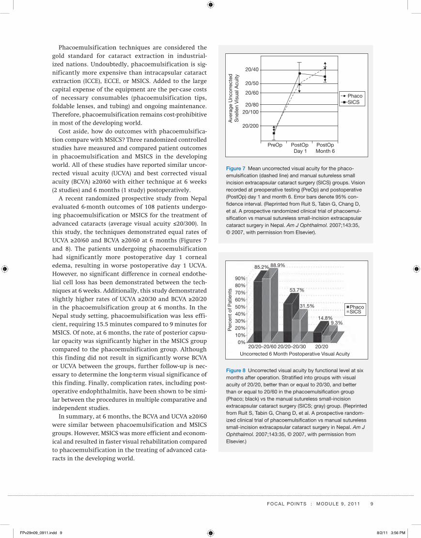
Phacoemulsification techniques are considered the gold standard for cataract extraction in industrial-ized nations. Undoubtedly, phacoemulsification is sig-nificantly more expensive than intracapsular cataract extraction (ICCE), ECCE, or MSICS. Added to the large capital expense of the equipment are the per-case costs of necessary consumables (phacoemulsification tips, foldable lenses, and tubing) and ongoing maintenance. Therefore, phacoemulsification remains cost- prohibitive in most of the developing world.
Cost aside, how do outcomes with phacoemulsifica-tion compare with MSICS? Three randomized controlled studies have measured and compared patient outcomes in phacoemulsification and MSICS in the developing world. All of these studies have reported similar uncor-rected visual acuity (UCVA) and best corrected visual acuity (BCVA) ≥20/60 with either technique at 6 weeks (2 studies) and 6 months (1 study) postoperatively.
A recent randomized prospective study from Nepal evaluated 6-month outcomes of 108 patients undergo-ing phacoemulsification or MSICS for the treatment of advanced cataracts (average visual acuity ≤20/300). In this study, the techniques demonstrated equal rates of UCVA ≥20/60 and BCVA ≥20/60 at 6 months (Figures 7 and 8). The patients undergoing phacoemulsification had significantly more postoperative day 1 corneal edema, resulting in worse postoperative day 1 UCVA. However, no significant difference in corneal endothe-lial cell loss has been demonstrated between the tech-niques at 6 weeks. Additionally, this study demonstrated slightly higher rates of UCVA ≥20/30 and BCVA ≥20/20 in the phacoemulsification group at 6 months. In the Nepal study setting, phacoemulsification was less effi-cient, requiring 15.5 minutes compared to 9 minutes for MSICS. Of note, at 6 months, the rate of posterior capsu-lar opacity was significantly higher in the MSICS group compared to the phacoemulsification group. Although this finding did not result in significantly worse BCVA or UCVA between the groups, further follow- up is nec-essary to determine the long-term visual significance of this finding. Finally, complication rates, including post-operative endophthalmitis, have been shown to be simi-lar between the procedures in multiple comparative and independent studies.
In summary, at 6 months, the BCVA and UCVA ≥20/60 were similar between phacoemulsification and MSICS groups. However, MSICS was more efficient and econom-ical and resulted in faster visual rehabilitation compared to phacoemulsification in the treating of advanced cata-racts in the developing world.
PhacoSICS
PostOpMonth 6
PostOp Day 1
PreOp
20/40
20/50
20/60
20/8020/100
20/200
Ave
rage
Unc
orre
cted
Sne
llen
Vis
ual A
cuity
Figure 7 Mean uncorrected visual acuity for the phaco-emulsification (dashed line) and manual sutureless small incision extracapsular cataract surgery (siCs) groups. Vision recorded at preoperative testing (Preop) and postoperative (Postop) day 1 and month 6. Error bars denote 95% con-fidence interval. (Reprinted from Ruit s, tabin G, Chang d, et al. a prospective randomized clinical trial of phacoemul-sification vs manual sutureless small- incision extracapsular cataract surgery in nepal. Am J Ophthalmol. 2007;143:35, © 2007, with permission from Elsevier).
Per
cent
of P
atie
nts
Uncorrected 6 Month Postoperative Visual Acuity20/20–20/60 20/20–20/30 20/20
0%10%20%30%40%50%60%70%80%90%
85.2% 88.9%
53.7%
31.5%
14.8%9.3%
PhacoSICS
Figure 8 uncorrected visual acuity by functional level at six months after operation. stratified into groups with visual acuity of 20/20, better than or equal to 20/30, and better than or equal to 20/60 in the phacoemulsification group (Phaco; black) vs the manual sutureless small- incision extracapsular cataract surgery (siCs; gray) group. (Reprinted from Ruit s, tabin G, Chang d, et al. a prospective random-ized clinical trial of phacoemulsification vs manual sutureless small- incision extracapsular cataract surgery in nepal. Am J Ophthalmol. 2007;143:35, © 2007, with permission from Elsevier.)
FPv29n09_0911.indd 9 8/2/11 3:56 PM

10 F o C a l P o i n t s : M o d u l E 9 , 2 0 1 1
Phacoemulsification in the Developing WorldPhacoemulsification is increasingly being used in the developing world (Table 2), especially at large tertiary care centers and teaching hospitals in metropolitan areas, where such a procedure is economically feasible.
Multiple explanations are available for this phenom-enon. Access to technology is improving, as phacoemul-sification equipment is increasingly being donated or available to physicians at a justifiable cost. Also, access to the necessary consumables, including viscoelastic and foldable hydrophilic lenses, has increased significantly due to local production. Such local production has also driven down the price of the procedure to an affordable level, making phacoemulsification economically feasible in many developing nations. As patients become educated regarding their treatment options, demand for state-of-the-art cataract surgery increases and patients are willing to pay a premium price for this technique. As a result, institutionalization of phacoemulsification becomes important from a cost- recovery perspective. Finally, phy-sicians in these institutions strive to acquire the neces-sary skills to provide state-of-the-art surgical options similar to those available in industrialized nations.
ConclusionTen years after the launch of the “Vision 2020: The Right to Sight” initiative, those working to eradicate prevent-able cataract blindness are gaining ground. (For more information, visit www.vision2020.org.) The devel-opment, implementation, and sustainability of high- volume, high- quality, low-cost cataract surgery have been demonstrated in some of the world’s poorest regions. These successful models can be replicated through coor-dinated efforts in countries where economic factors prevent implementation of more expensive and time- consuming cataract surgery delivery systems. Through the collaborative and continued efforts of government and nongovernment organizations, using the techniques discussed in this module and already proven in many regions of the world, the lofty goal of eliminating cata-ract blindness can be achieved in our lifetime.
Geoffrey C. Tabin, MD, is a corneal specialist and Direc-tor of the International Ophthalmology Division at John A. Moran Eye Center, Salt Lake City, Utah.
Michael R. Feilmeier, MD, is the Medical Director of the International Division of Ophthalmology, University of Nebraska Medical Center, and a cornea and external eye specialist at Midwest Eye Care, Omaha, Nebraska.
Table 2. Cataract Surgical Trends in Nepal, 1994–2008
Y E a R
t o ta l
C ata R a C t
o P E R at i o n s
i C C E / E C C E ,
n o i o l a
i C C E W i t H
a C i o l b
E C C E W i t H
P C i o l c
M s i C s W i t H
P C i o l d P H a C o E M u l s i F i C at i o n
1994 37,500 75% 7.5% 17.5% 0% 0%
1996 40,500 47% 4.5% 48.5% 0% 0%
1998 55,500 21% 2% 46% 31% 0%
2000 85,000 8% 1.50% 32.50% 57% 1.0%
2002 120,000 4% 0.75% 29% 65% 1.25%
2004 149,000 3% 0.9% 20% 72% 4%
2006 159,500 1.25% 0.5% 17% 69.25% 12%
2008 168,500 0.5% 0.5% 9% 67% 23%
aintracapsular cataract extraction/extracapsular cataract extraction, no intraocular lensbintracapsular cataract extraction with anterior chamber intraocular lenscExtracapsular cataract extraction with posterior chamber intraocular lensdManual small- incision cataract surgery with posterior chamber intraocular lens
FPv29n09_0911.indd 10 8/2/11 3:56 PM

F o C a l P o i n t s : M o d u l E 9 , 2 0 1 1 11
Clinicians’Corner
Clinicians’ Corner provides additional viewpoints on
the subject covered in this issue of Focal Points. Con-
sultants have been invited by the Editorial Review
Board to respond to questions posed by the Acade-
my’s Practicing Ophthalmologists Advisory Committee
for Education. While the advisory committee reviews
the modules, consultants respond without reading the
module or one another’s responses. – Ed.
1. Given the different techniques used predominantly in the developing world, such as manual small‑ incision cataract surgery (MSICS) rather than phacoemulsification, how can phaco‑ trained sur‑geons in industrialized nations interact and aid ophthalmologists in the developing world?
Dr. Brown: Certainly the phaco surgeon should be famil-iar with MSICS techniques before performing surgery. Courses are available through Surgical Eye Expeditions International (www.seeintl.org), the American Academy of Ophthalmology (www.aao.org), and the American Society of Cataract and Refractive Surgery (www.ascrs.org). Phaco- trained surgeons in industrialized nations can inform and educate colleagues in the developing world about phacoemulsification cataract surgery. Con-siderations include financial factors (cost of the machine and the availability and cost of tubing), importance of trained technical staff and phaco training courses, the long learning curve, availability of a retina surgeon, patient selection, and possible complications and their management. Patients with a rock hard nucleus, seen in much of the developing world, are not prime candidates for phaco.
Dr. Shah: In the developing world, one setting is urban and the second involves camps in rural areas. In urban areas phacoemulsification is the norm. In camp-based settings the volumes are huge, which justifies MSICS. Phaco- trained surgeons in an urban setting would likely do phacoemulsification rather than MSICS. The problem would arise when volumes are huge in a rural setting where either they can do conventional extracapsular cataract extraction or jump to MSICS (where I feel the learning curve is very short). So, in summary, the phaco- trained surgeon would benefit from learning MSICS for use in rural- based settings.
FPv29n09_0911.indd 11 8/2/11 3:56 PM

12 F o C a l P o i n t s : M o d u l E 9 , 2 0 1 1
Clinicians’Corner
2. How are intraoperative complications, such as dropped nuclear material and vitreous loss, addressed in the setting of cataract surgery in the developing world?
Dr. Brown: Intraoperative complications demand good surgical judgment. Fortunately, dropped nucleus in MSICS is not common. If it does occur it is frequently best left alone. Fishing for a lost nuclear fragment often leads to more serious complications. Vitreous loss is han-dled in the traditional way of carefully using sponges and scissors in removing vitreous from the wound, anterior chamber, and iris. Some clinics may have a vitreous cut-ter available. An anterior IOL is put in place.
Dr. Shah: In an urban setting, I would refer the patient to a vitreoretinal surgeon who would take the surgery forward. In a rural setting where a vitreoretinal surgeon is difficult to find, the patient can be referred to the nearest city for a nuclear drop. For vitreous loss without nuclear drop, it would be better to put in a sulcus- based IOL if your capsulorrhexis is intact and manually cut the vitreous using Weck ophthalmic sponges. I would also consider employing a scleral-fixated IOL as I feel it is better to put in the IOL than to leave the patient aphakic.
3. Describe your preoperative evaluation and postop‑erative management of MSICS. Who provides these services?
Dr. Brown: Preliminary preoperative screening of patients is frequently done by the local ophthalmolo-gist. This should include a general health assessment and an ocular evaluation. The visiting surgeon exam-ines each patient to determine the appropriate surgical management.
Postoperative examination by the operating surgeon the day following surgery is important to identify any problems that need to be addressed before the patient is discharged. Subsequent postoperative exams and fol-low- up are undertaken by the local ophthalmologist.
Dr. Shah: A complete eye examination would include slit-lamp evaluation for the grade of cataract, intraocular pressure, conjunctival sac patency, posterior segment
evaluation of the retinal status, and B-scan ultrasonog-raphy in patients with mature cataracts. I would also order investigations such as complete blood count, blood glucose, urinalysis, and an electrocardiogram. Previous history of any illness is documented. Finally, A-scan ultrasonography for measurement of axial length and IOL power calculation is done.
Postoperative management includes examination on day 1, day 3, and then after 15 days. The patient is started on systemic antibiotics and analgesics, steroid–antibiotic eyedrops, and nonsteroidal anti-inflammatory eyedrops, which are eventually taped off. In an urban setting, the operating surgeon or the resident provide these services. In a rural setting the operating surgeon and later on the local eye surgeon would follow up and provide the services.
4. How do you handle IOL calculations in MSICS in the developing world?
Dr. Brown: A-scan machines and keratometers for IOL calculation are sometimes available at clinics or brought by the visiting team. If these are not available, IOL cal-culation is approximated based on the patient’s visual history. Generally IOL powers between 18 and 22 diop-ters are used.
Dr. Shah: In an urban setting, the calculations would be done by using the water immersion A-scan technique. Holladay or SRK-II formula K readings would be auto-mated and applied. In a rural setting, calculations would be done by using an A-scan probe, applying the SRK-II formula, and using a manual keratometer to obtain K readings.
5. When should the phaco‑ trained surgeon consider MSICS surgery instead of extracapsular cataract extraction?
Dr. Brown: MSICS offers a number of advantages com-pared to extracapsular cataract extraction. MSICS is safer, results in less astigmatism and less bleeding, and does not require stitches. There are fewer complications. Phaco surgeons should be familiar with MSICS tech-niques before attempting the surgery.
FPv29n09_0911.indd 12 8/2/11 3:56 PM

F o C a l P o i n t s : M o d u l E 9 , 2 0 1 1 13
8. What are the primary sources of funding for cata‑ract surgical care in underdeveloped countries?
Dr. Brown: The cost of providing cataract surgical care is spread across a broad base. Sources of funding include nongovernment organizations that recruit, organize, and deploy surgical teams; volunteer doctors and nurses who pay their own travel expenses and donate their profes-sional services; the host country that provides in- country transportation, food, and lodging for the visiting team for the duration of the surgery clinic; and government social service agencies and local hospitals that cooper-ate in support of the clinic. Additional sources of fund-ing include donations from international service clubs (such as Lions and Rotary) that provide financial support; ophthalmic industries that supply in-kind donations of consumable supplies, IOLs, medications, and operative packs; and visiting teams that transport surgical equip-ment (microscopes, instruments sets, sterilizers, and lasers) needed to augment local circumstances to con-duct the cataract clinics.
Dr. Shah: In an urban setting, the patients pay or, if they are insured, then the insurance company covers the costs. In a rural setting, typically the funding is through charitable organizations and non- governmental organi-zations. The government helps in certain cases by provid-ing consumables for the surgery.
9. How do you handle patients with coexisting glau‑coma and cataracts in this setting?
Dr. Brown: Severity of glaucoma, visual field loss, size of cataract, and preoperative IOP will help determine surgical management. Since removing the cataract will lower the IOP in many cases, most surgeons do not do combined procedures. Postoperative IOP will determine if further surgery is an option for the patient.
Dr. Shah: In an urban setting, I would do phacoemulsi-fication with trabeculectomy or deep sclerectomy with mitomycin. In a rural setting, I would do superior MSICS along with trabeculectomy by raising the f lap at the end of the incision and cutting the meshwork from that end. I would also put a 10-0 suture at that end.
Dr. Shah: The phaco- trained surgeon should consider MSICS in the following conditions: pseudoexfoliation, calcified cataract, brown/black cataract, hypermature Morgagnian cataract, subluxated cataract, and in certain traumatic cases where lens is dislocated in the anterior chamber.
6. What are your considerations for a temporal approach versus a superior approach for MSICS?
Dr. Brown: The superior approach offers some protection from the upper eyelid covering the wound. Deep-set eyes with over- hanging brow may make this approach diffi-cult. Generally the surgeon should use the technique with which he or she is most familiar.
Dr. Shah: I always prefer a superotemporal approach for MSICS as the astigmatism is least in this approach and it is easier because you have the brow to support your hand, which is not so in the temporal approach. A tem-poral incision is exposed to the atmosphere as compared to superior incisions that are covered by the eyelid.
7. What is the rate of posterior capsular opacifica‑tion with MSICS and how is this addressed? Do you perform primary posterior capsulotomies in selected situations?
Dr. Brown: Accurate figures on the rate of posterior cap-sular opacification (PCO) after MSICS in the developing world are scanty at best. Careful cortical cleanup, pos-terior capsular polishing, and choice of IOLs known to reduce the incidence of PCO may help. YAG laser capsu-lotomy is ideal but not available in many clinics. Primary posterior capsulotomies are performed if there is calcifi-cation, clouding or opacity of the posterior capsule.
Dr. Shah: I believe the rate of posterior capsular opacifi-cation is equal between MSICS and phacoemulsification. Normally such patients undergo YAG capsulotomy, but if the capsule has become hard and you cannot use the YAG technique, then you need to do a surgical capsulec-tomy. I do perform primary capsulotomies but only in the pediatric age group.
FPv29n09_0911.indd 13 8/2/11 3:56 PM

14 F o C a l P o i n t s : M o d u l E 9 , 2 0 1 1
Clinicians’Corner
Harry S. Brown, MD, FACS, a retired ophthalmologist in Santa Barbara, California, founded Surgical Eye Expe-ditions (SEE) International in 1974. He obtained his resi-dency in ophthalmology at the Jules Stein Eye Institute at UCLA, Los Angeles, California. He notes that his com-ments are from his personal experience and interviews with John Crowder, MD, Medical Director of SEE Inter-national, and SEE affiliate surgeons Jack Aaron, MD, Jeff Rutgard, MD, and Doug Katsev, MD.
Janak M. Shah, MB, DO, DO MS, MMedSc, graduated from King Edward Memorial Hospital, Mumbai Univer-sity. He is the Director of Netrapuja Eye Care Pvt Ltd, Mumbai, India. His special interests include manual small- incision cataract surgery, paediatric ophthalmol-ogy, and oculoplastics.
Suggested Reading
Brian G, Taylor H. Cataract blindness—challenges for the 21st
century. Bull World Health Organ. 2001;79:249–256.
Chang MA, Congdon NG, Baker SK, Bloem MW, Savage H,
Sommer A. The surgical management of cataract: barriers,
best practices, and outcomes. Int Ophthalmol. 2008;28:247–260.
Foster A. Cataract and “Vision 2020—the right to sight”
initiative. Br J Ophthalmol. 2001;85:635–637.
Frick KD, Foster A. The magnitude and cost of global blind-
ness: an increasing problem that can be alleviated. Am J
Ophthalmol. 2003;135:471–476.
Gogate P, Deshpande M, Nirmalan PK. Why do phacoemulsifi-
cation? Manual small- incision cataract surgery is almost as
effective, but less expensive. Ophthalmology. 2007;114:965–968.
Gogate PM, Kulkarni SR, Krishnaiah S, et al. Safety and
efficacy of phacoemulsification compared with manual
small- incision cataract surgery by randomized controlled
clinical trial. Ophthalmology. 2005;112:869–874.
Lewallen S, Courtright P. Blindness in Africa: present situation
and future needs. Br J Ophthalmol. 2001;85:897–903.
Resnikoff S, Pascolini D, Etya’ale D, et al. Policy and practice:
global data on visual impairment in the year 2002. Bull World
Health Organ. 2004;82:849.
Riaz Y, Mehta JS, Wormald R, et al. Surgical interventions for
age- related cataract. Cochrane Database Syst Rev. 2006;Oct 18:
CD001323.
Ruit S, Tabin G, Chang D, et al. A prospective randomized
clinical trial of phacoemulsification vs manual sutureless
small- incision extracapsular cataract surgery in Nepal. Am J
Ophthalmol. 2007;143:32–38.
Ruit S, Tabin GC, Wykoff CC. Fighting Global Blindness: Improving
World Vision Through Cataract Elimination. Washington, DC:
American Public Health Association; 2006.
Ruit S, Tabin GC, Nissman SA, Paudyal G, Gurung R. Low-cost
high-volume extracapsular cataract extraction with posterior
chamber intraocular lens implantation in Nepal. Ophthalmol-
ogy. 1999;106:1887–1892.
Tabin G, Chen M, Espandar L. Cataract surgery for the
developing world. Curr Opin Ophthalmol. 2008;19:55–59.
Yorston D, Abiose A. Cataract blindness—the African perspec-
tive. 2001. Bull World Health Organ. 2001;79:249–256.
Related Academy Materials
Khanna RC. Blumenthal manual small- incision cataract
surgery in hypermature cataract: alternative technique for
the developing world. Video. http://aao.scientificposters.com/
vodONE.cfm?id=V08&yr=2006. Accessed June 21, 2011.
Raju LV, Ghanta M, Raju VK. Techniques for manual small-
incision extracapsular cataract extraction. Current Insight.
http://one.aao.org/ce/news/currentinsight/detail.
aspx?cid=ea87e277-d336-4a8b-a0cb-2a2a359c90a9. Accessed
June 21, 2011.
029033C
FPv29n09_0911.indd 14 8/2/11 3:56 PM