wt1 targets gas1 to maintain nephron progenitor cells by...
TRANSCRIPT

RESEARCH ARTICLE STEM CELLS AND REGENERATION
WT1 targets Gas1 to maintain nephron progenitor cells bymodulating FGF signalsMartin Kann1,2,3, Eunnyung Bae1,2,*, Maximilian O. Lenz3, Liangji Li4, BaoTran Trannguyen1,2,Valerie A. Schumacher1,2, Mary E. Taglienti1,2, Liliana Bordeianou1,2,‡, Sunny Hartwig5, Markus M. Rinschen3,Bernhard Schermer3,6, Thomas Benzing3,6, Chen-Ming Fan4 and Jordan A. Kreidberg1,2,7,§
ABSTRACTDevelopment of the metanephric kidney depends on tightly regulatedinterplay between self-renewal and differentiation of a nephronprogenitor cell (NPC) pool. Several key factors required for thesurvival of NPCs have been identified, including fibroblast growthfactor (FGF) signaling and the transcription factor Wilms’ tumorsuppressor 1 (WT1). Here, we present evidence that WT1 modulatesFGF signaling by activating the expression of growth arrest-specific1 (Gas1), a novel WT1 target gene and novel modulator of FGFsignaling. We show that WT1 directly binds to a conserved DNAbinding motif within the Gas1 promoter and activates Gas1 mRNAtranscription in NPCs. We confirm that WT1 is required for Gas1expression in kidneys in vivo. Loss of function of GAS1 in vivo resultsin hypoplastic kidneys with reduced nephron mass due to prematuredepletion of NPCs. Although kidney development in Gas1 knockoutmice progresses normally until E15.5, NPCs show decreased rates ofproliferation at this stage and are depleted as of E17.5. Lastly, weshow that Gas1 is selectively required for FGF-stimulated AKTsignaling in vitro. In summary, our data suggest amodel in whichWT1modulates receptor tyrosine kinase signaling in NPCs by directing theexpression of Gas1.
KEY WORDS: Kidney development, Fibroblast growth factorsignaling, Nephron progenitor cell, Mouse
INTRODUCTIONThe mammalian kidney is essential to the maintenance of bodyhomeostasis by controlling water and salt balance and excretingwaste products throughout life. This excretory function depends onits number of available kidney filtration units, nephrons, which isdetermined during kidney development. In mice, nephrogenesisceases shortly after birth when the nephron progenitor cell (NPC)pool is completely exhausted (Short et al., 2014), whereas inhumans the induction of new nephrons occurs entirely in the fetus.Crucially, in all mammals, it is not possible to induce new nephrons
after the disappearance of NPCs. As such, the number of nephronsendowed during fetal kidney development in humans has beenidentified as a key risk factor for various diseases occurring later inlife, including diabetes, hypertension and chronic kidney disease(Luyckx and Brenner, 2010). Therefore, pathways influencingnephron endowment during kidney development are nowconsidered to be crucial to the pathogenesis of many diseasesthroughout life.
The development of themetanephric kidney depends on a tightlyregulated and complex interplay of several cell lineages andstructures derived from the intermediate mesoderm (Dressler,2009).Metanephric development is initiated when themetanephricmesenchyme induces the outgrowth of the ureteric bud (UB) fromtheWolffian duct. At the same time, the metanephric mesenchymeestablishes a population of NPCs that give rise to nephrons.Signaling in both directions between the UB andNPCs results in aniterative process during which the UB undergoes branchingmorphogenesis to form ureteric tips and collecting ducts, whileNPCs maintain a balance between self-renewal and differentiation.Signals from the UB induce NPCs to condense into pretubularaggregates (PTA) before undergoing mesenchymal-to-epithelialtransition to form renal vesicles (RVs). RVs are further patternedand differentiated into primordial nephrons within S-shapedbodies, before a new round of branching and nephrogenesis istriggered (Dressler, 2009). Although the factors that affectbranching morphogenesis and nephron induction were previouslythought to be quantitatively constant throughout the period ofnephrogenesis, recent studies have shown evidence of temporal andfunctional discontinuity in both processes (Short et al., 2014). Assuch, the NPC pool as a SIX2-positive (SIX2+) cell population canbe divided into CITED1+, self-maintaining and CITED1-negative(CITED1−), induced compartments (Brown et al., 2013).Interestingly, proliferation rates in these compartments differbetween early and late stages of renal development (Brown et al.,2013; Short et al., 2014). To date, the mechanisms controllingproliferation in these compartments remain unclear.
Research into nephrogenesis and NPCs has led to the identificationof a large number of signaling pathways and molecules involved inregulating the balance between NPC self-renewal and differentiation,including the transcription factor Wilms’ tumor suppressor protein 1(WT1). In the absence of WT1, the metanephric mesenchymeundergoes apoptosis, resulting in renal agenesis (Kreidberg et al.,1993).WithinNPCs,WT1maintains the expressionof a large numberof genes required to promote the growth and differentiation of NPCs,including components and ligands of twomajor signaling pathways inNPCs – bone morphogenetic protein (BMP) and fibroblast growthfactor (FGF) signaling, respectively (Hartwig et al., 2010; Motamediet al., 2014). Both pathways are required for adequate proliferationrates within the NPC pool (Barak et al., 2012; Blank et al., 2009;Received 10 November 2014; Accepted 13 February 2015
1Division of Nephrology, Department of Medicine, Boston Children’s Hospital,Boston, MA 02115, USA. 2Department of Pediatrics, Harvard Medical School,Boston, MA 02115, USA. 3Department II of Medicine and Center for MolecularMedicine Cologne, University of Cologne, 50931 Cologne, Germany.4Department of Embryology, Carnegie Institution of Washington, Baltimore, MD21218, USA. 5Department of Biomedical Sciences, Atlantic Veterinary College,University of Prince Edward Island, Charlottetown, PE, Canada C1A 4P3. 6Clusterof Excellence on Cellular Stress Responses in Ageing-Associated Diseases(CECAD) and Systems Biology of Ageing Cologne, University of Cologne, 50931Cologne, Germany. 7Harvard Stem Cell Institute, Cambridge, MA 02138, USA.*Present address: Department of Cancer Biology, Lerner Research Institute,Cleveland Clinic, Cleveland, OH 44195, USA. ‡Present address: Department ofSurgery, Massachusetts General Hospital, Boston, MA 02114, USA.
§Author for correspondence ( [email protected])
1254
© 2015. Published by The Company of Biologists Ltd | Development (2015) 142, 1254-1266 doi:10.1242/dev.119735
DEVELO
PM
ENT

Brown et al., 2011) and for an adequate NPC response todifferentiation inductive signals (Blank et al., 2009; Brown et al.,2011; Motamedi et al., 2014). Interestingly, intracellular signalingcascades utilized by FGF and BMP signals overlap in variouscontexts. However, in NPCs, BMP has been shown to predominantlyactivate themitogen-activated protein kinases (MAPKs) JNKand p38(Blank et al., 2009; Di Giovanni et al., 2011; Motamedi et al., 2014),whereas FGF predominantly activates phosphoinositide 3-kinase(PI3K)-AKT pathways (Brown et al., 2011). Alteration of the activityofMAPKandPI3K signaling cascades has been linked to severeNPCdefects, underscoring a need for tight control of signaling cascadelevels and balances (Blank et al., 2009; Brown et al., 2011). To date, itis unclear how the balance between these intracellular cascades isregulated and how the correct levels of these signals are controlled inNPCs.Based on our previous analysis ofWT1 target genes in embryonic
kidneys (Hartwig et al., 2010), the aim of this study is to identify theglycosylphosphatidylinositol (GPI)-anchored membrane proteingrowth-arrest-specific 1 (GAS1) as a novel and direct target geneof WT1 and characterize its function in NPCs. GAS1 is structurallyrelated to glial-derived neurotrophic factor (GDNF) receptor alpha 1(GFRA1) and has been shown to modulate intracellular signalingcascades in response to GDNF-Ret receptor tyrosine kinase (RTK)signaling (Cabrera et al., 2006). Furthermore, GAS1 has beenidentified as a co-receptor that binds to sonic hedgehog (SHH),resulting in amplification of SHH signals (Allen et al., 2007;Martinelli and Fan, 2007). We show that a WT1-responsive elementin the Gas1 promoter is sufficient and required for Gas1 expressionin embryonic kidneys. Loss of function of GAS1 results inhypoplastic kidneys due to decreased proliferation rates andpremature depletion of NPCs. However, this phenotype does notmanifest until later stages of kidney development, supporting thehypothesis that NPCs at early and late stages in development aregoverned by distinct though overlapping regulatory mechanisms.Finally, we link GAS1 to the activation of AKT downstream of FGFsignals, providing a mechanistic basis for understanding the role ofGAS1 in the control of NPC proliferation and differentiation. Takentogether, these findings demonstrate how transcriptional regulationof gene expression can affect the level of RTK signaling inprogenitor cells.
RESULTSIdentification of Gas1 as a WT1 target geneWT1 is a transcription factor that is crucial to the maintenance anddifferentiation of NPCs in developing kidneys. Among the WT1target genes identified in our previous study (Hartwig et al., 2010),Gas1 was of interest as a potential modifier of signal transductionpathways, the study of which would offer insight into howtranscriptional regulation by WT1 would affect signaling inNPCs. Furthermore, the GUDMAP database had identified highlevel expression of Gas1 in NPCs.TheGas1 promoter was previously found to be bound byWT1 in
a chromatin immunoprecipitation followed by array hybridization(ChIP-on-chip) screen in embryonic kidneys (Hartwig et al.,2010) with a 4.5-fold enrichment over background (adjustedP-value<0.0001, Fig. 1A, brown bars; supplementary materialFig. S1A). In order to confirm this interaction, we extractedchromatin from stage postnatal day (P) 0 wild-type kidneys andperformed ChIP followed by qPCR using seven primer pairstiled along the Gas1 promoter, 5′UTR and open reading frame(Fig. 1A,C). These experiments replicated the finding of WT1binding to a conserved site, which was located close to the Gas1
transcription start site (TSS) in the same position as the two ChIP-on-chip probes with the highest enrichment. The specificity of theseexperiments was controlled by using an intronic site lacking a WT1binding motif within the Gapdhs gene as negative control and thepromoter of Kdm3a, which is tightly bound byWT1 (Hartwig et al.,2010), as a positive control (Fig. 1C, n=3). WT1 is thus bound to theGas1 promoter in vivo.
To then assess the functional significance of WT1 binding to theGas1 gene, we determined whether loss of WT1 affected levels ofGas1 mRNA. Initial evidence that WT1 indeed controls Gas1transcription was acquired through characterization of a novelWT1-expressing cell line, LB-22. LB-22 is an immortalizedmesenchymal cell line derived from the nephrogenic zone ofmouse embryonic kidneys that constitutively expresses abundantWT1. Characterization of the cell line by RNA expressionmicroarray experiments revealed expression of several genes thatare bona fide WT1 target genes in NPCs in vivo, such as Cxxc5,Pbx2, Sox4 and Rps6ka3 (Hartwig et al., 2010). However, the cellline lacks expression of most established marker genes of NPCs,such as Six2, Cited1 and Sall1. Hence, LB-22 does not accuratelymodel NPCs in cell culture but, based on its constitutive WT1expression, it is a useful tool to address WT1-mediated regulation ofgene expression. In order to show that the binding of WT1 to theGas1 promoter as described in embryonic kidneys is present in LB-22 cells, we repeated the ChIP-qPCR experiments, revealing thesame binding site of WT1 in LB-22 cells (Fig. 1D). LB-22 cells cantherefore serve as an accurate model of WT1-mediated regulation ofGas1 in cell culture.
Furthermore, we established siRNA-mediated knockdown ofWT1 in LB-22 cells (supplementary material Fig. S1B) and usedmRNA expression arrays (n=4) to analyze the effects of WT1depletion on mRNA levels (supplementary material Fig. S1C,D).Unbiased analysis of summarized array datasets identified Gas1 asone of the most significantly downregulated genes upon treatmentwith Wt1 siRNA (supplementary material Fig. S1D). These resultswere validated by RT-qPCR and western blotting (supplementarymaterial Fig. S1B). In conclusion, WT1 binds the Gas1 promoterand affects Gas1 mRNA and protein levels in LB-22 cells.
AWT1-responsive DNA element within the Gas1 promoter isrequired and sufficient for Gas1 expressionWe used promoter-reporter and mutagenesis studies to establishdirect regulation of Gas1 by WT1. Direct target genes containfunctional transcription factor binding DNA motifs within their cis-regulatory domain (CRD). We therefore screened the conservedCRD surrounding the Gas1 TSS within −1.5 kb to 0.5 kb forinstances of WT1 binding motifs using a published WT1 positionalweight matrix (Hartwig et al., 2010). Based on the location of WT1motifs, conservation, ChIP-qPCR and ChIP-on-chip results, wedivided the Gas1 CRD into five sections that were subsequentlycloned in several combinations (Fig. 1A, green and red bars) into aluciferase reporter vector containing a minimal promoter. Whenmeasuring relative luciferase signals generated by these fragmentsupon transfection into LB-22 cells, exclusively fragmentscontaining a 200 bp region close to the Gas1 TSS produced asignificant increase in reporter signals, indicating that a functionalcis-regulatory element was located in this region (Fig. 1E). Thispromoter element overlapped with the WT1-bound region asidentified by ChIP (Fig. 1A, red shaded box). To confirm that WT1was indeed responsible for eliciting reporter signals, we repeated thereporter assays in LB-22 cells treated with WT1 and controlsiRNAs. Reporter assays in the presence of WT1 siRNA showed
1255
RESEARCH ARTICLE Development (2015) 142, 1254-1266 doi:10.1242/dev.119735
DEVELO
PM
ENT
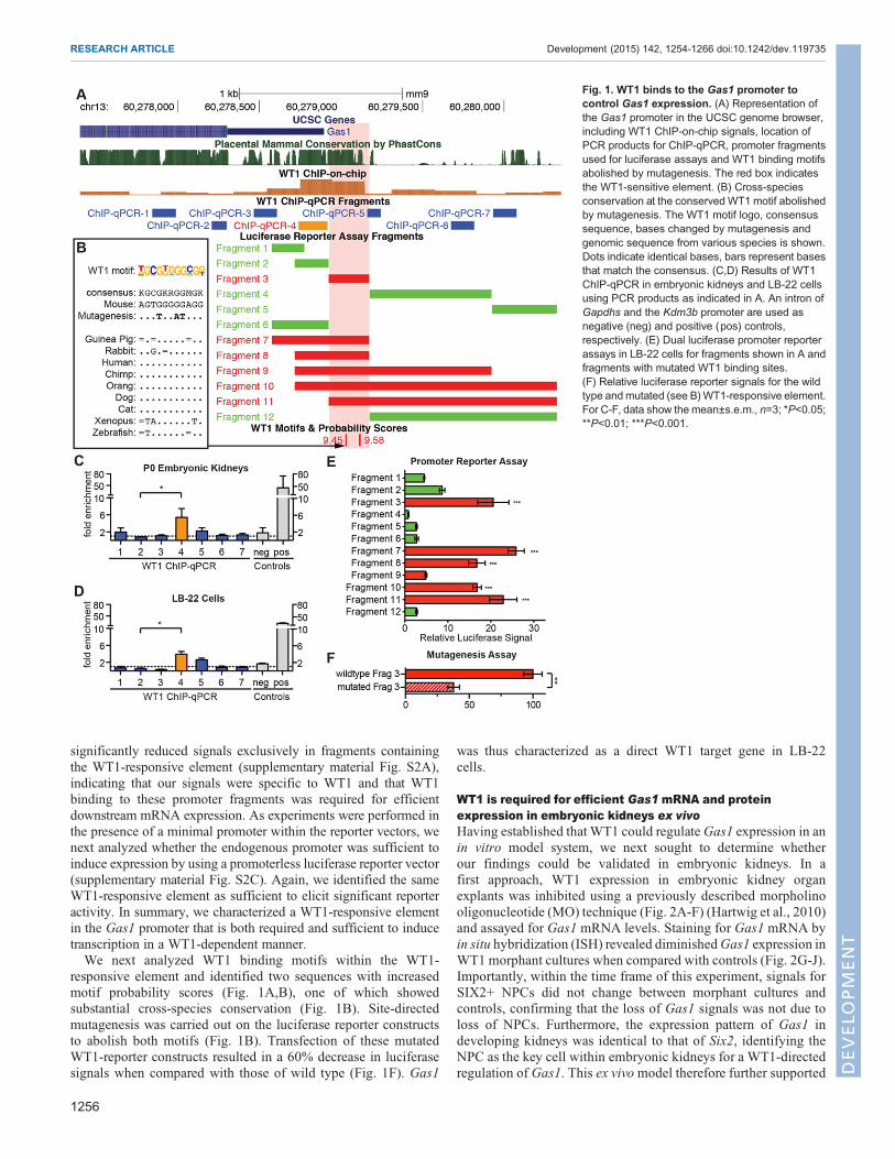
significantly reduced signals exclusively in fragments containingthe WT1-responsive element (supplementary material Fig. S2A),indicating that our signals were specific to WT1 and that WT1binding to these promoter fragments was required for efficientdownstream mRNA expression. As experiments were performed inthe presence of a minimal promoter within the reporter vectors, wenext analyzed whether the endogenous promoter was sufficient toinduce expression by using a promoterless luciferase reporter vector(supplementary material Fig. S2C). Again, we identified the sameWT1-responsive element as sufficient to elicit significant reporteractivity. In summary, we characterized a WT1-responsive elementin the Gas1 promoter that is both required and sufficient to inducetranscription in a WT1-dependent manner.We next analyzed WT1 binding motifs within the WT1-
responsive element and identified two sequences with increasedmotif probability scores (Fig. 1A,B), one of which showedsubstantial cross-species conservation (Fig. 1B). Site-directedmutagenesis was carried out on the luciferase reporter constructsto abolish both motifs (Fig. 1B). Transfection of these mutatedWT1-reporter constructs resulted in a 60% decrease in luciferasesignals when compared with those of wild type (Fig. 1F). Gas1
was thus characterized as a direct WT1 target gene in LB-22cells.
WT1 is required for efficient Gas1 mRNA and proteinexpression in embryonic kidneys ex vivoHaving established that WT1 could regulateGas1 expression in anin vitro model system, we next sought to determine whetherour findings could be validated in embryonic kidneys. In afirst approach, WT1 expression in embryonic kidney organexplants was inhibited using a previously described morpholinooligonucleotide (MO) technique (Fig. 2A-F) (Hartwig et al., 2010)and assayed for Gas1 mRNA levels. Staining for Gas1 mRNA byin situ hybridization (ISH) revealed diminishedGas1 expression inWT1 morphant cultures when compared with controls (Fig. 2G-J).Importantly, within the time frame of this experiment, signals forSIX2+ NPCs did not change between morphant cultures andcontrols, confirming that the loss of Gas1 signals was not due toloss of NPCs. Furthermore, the expression pattern of Gas1 indeveloping kidneys was identical to that of Six2, identifying theNPC as the key cell within embryonic kidneys for a WT1-directedregulation of Gas1. This ex vivomodel therefore further supported
Fig. 1. WT1 binds to the Gas1 promoter tocontrol Gas1 expression. (A) Representation ofthe Gas1 promoter in the UCSC genome browser,including WT1 ChIP-on-chip signals, location ofPCR products for ChIP-qPCR, promoter fragmentsused for luciferase assays and WT1 binding motifsabolished by mutagenesis. The red box indicatesthe WT1-sensitive element. (B) Cross-speciesconservation at the conserved WT1 motif abolishedby mutagenesis. The WT1 motif logo, consensussequence, bases changed by mutagenesis andgenomic sequence from various species is shown.Dots indicate identical bases, bars represent basesthat match the consensus. (C,D) Results of WT1ChIP-qPCR in embryonic kidneys and LB-22 cellsusing PCR products as indicated in A. An intron ofGapdhs and the Kdm3b promoter are used asnegative (neg) and positive (pos) controls,respectively. (E) Dual luciferase promoter reporterassays in LB-22 cells for fragments shown in A andfragments with mutated WT1 binding sites.(F) Relative luciferase reporter signals for the wildtype andmutated (see B)WT1-responsive element.For C-F, data show the mean±s.e.m., n=3; *P<0.05;**P<0.01; ***P<0.001.
1256
RESEARCH ARTICLE Development (2015) 142, 1254-1266 doi:10.1242/dev.119735
DEVELO
PM
ENT

our previous findings of the requirement of WT1 to activate Gas1transcription in NPCs.
A novel Wt1 hypomorphic mouse model confirms regulationof Gas1 by WT1As RNA interference (RNAi)-based and ex vivo studies can becompromised by off-target effects and confounders introducedwithin the organ culture system, we sought to extend our ex vivofindings in an independent in vivo model where expression of Wt1was reduced in embryonic kidneys. To this end, we generatedWt1ckd mice as a tool to investigate WT1 target genes. Wt1ckd miceconditionally express a short hairpin (sh)RNA (Coumoul et al.,2004; Shukla et al., 2007) directed against Wt1 in NPCs andderivatives by Six2:Cre-driven recombination. Wt1ckd mice wereborn in Mendelian ratios and were viable for up to 5 weeks.Although kidney development appeared to progress normally untilstage embryonic day (E)14.5 (data not shown), at stage E16.5 WT1hypomorphic phenotypes became evident, with premature loss ofcondensing mesenchyme surrounding the ureteric tips and lack ofcomma- and S-shaped bodies (Fig. 3A,B). Kidneys of newbornWt1ckd mice were hypoplastic (Fig. 3C,D) and had reduced nephronendowment. We subsequently assayed the efficacy of our RNAiapproach by confirmation of decreased Wt1 mRNA and proteinlevels in Wt1ckd kidneys using ISH at stage E14.5 (Fig. 3E,F) andimmunofluorescence staining at stage E15.5 (Fig. 3K,L),respectively. Importantly, the SIX2+ NPC pool was preserved atstage E14.5 (Fig. 3I,J) and only moderately depleted at stage E15.5(Fig. 3Q,R), rendering these stages suitable for WT1 target geneanalysis.The subsequent analysis of Gas1 expression in WT1ckd
kidneys identified decreased Gas1 mRNA in NPCs at stageE14.5 (Fig. 3G,H) and decreased Gas1 protein at stage E15.5(Fig. 3M,N) when compared with Cre-negative controls. Of note,although Wt1 mRNA and protein expression is increased inpretubular aggregates and renal vesicles (Fig. 3F,L), Gas1expression in these structures was similar to that of Six2. Gas1mRNA expression was abolished, whereas protein was retained at
low levels until the renal vesicle stage before vanishing completelyat the S-shaped body stage (Fig. 3H,N), suggesting that Gas1function and gene regulation are specific to the NPC pool inembryonic kidneys. In summary, we established Gas1 as a bonafide direct WT1 target gene in embryonic kidneys in vivo.
GAS1 loss of function results in hypoplastic kidneys withdecreased nephron endowmentAs high expression levels of Gas1 in NPCs and the sharp decline ofGas1 levels upon nephron induction had already suggested arelevant function ofGas1 in kidney development, we next sought todetermine the effects ofGas1 loss of function in embryonic kidneys.To this end, we made use of the previously published Gas1knockout mouse (Martinelli and Fan, 2007).
Initial histologic analysis of Gas1 null kidneys at stage E15.5did not show significant differences when compared withheterozygous null controls (Fig. 4A,B,I). The overall size ofGas1 null kidneys at E15.5 was similar to that of controlcounterparts; condensing mesenchyme was clearly visiblesurrounding the ureteric tips and nephron induction appeared tobe occurring normally, with evidence of one to two inducednephron structures (renal vesicles, comma- or S-shaped bodies)per ureteric branch (Fig. 4C,D). Similar numbers of fullydifferentiated glomeruli with positive nephrin expression werepresent in Gas1 null and wild-type kidneys (data not shown).However, analysis of kidney size at stage E17.5 and later showedsignificant hypoplasia of Gas1 null kidneys, with knockoutkidneys at stage P0 being about half the size of heterozygousknockout control kidneys (Fig. 4E,F,I). High-power histologicanalysis at stage P0 suggested perturbation of the NPC pool andnephrogenesis in Gas1 knockout kidneys with prematuredepletion of condensing mesenchyme surrounding the ureterictips and a reduced number of induced nephrons (Fig. 4G,H).Decreased nephron endowment as measured by countingglomeruli was also evident (Fig. 4J). Therefore, loss of functionof Gas1 did not appear to affect early renal development andestablishment of the nephrogenic zone; however, a quantitative
Fig. 2. Knockdown of WT1 in kidney organ cultures results in decreasedGas1mRNA. (A-F) Morpholino oligonucleotide (MO)-based knockdown of WT1 inkidney organ cultures results in decreased levels of WT1 protein. (G-J) In situ hybridization for Gas1mRNA shows decreased signals in WT1 morphant cultureswhile Six2+ nephron progenitor cells are maintained. Scale bars: 100 µm.
1257
RESEARCH ARTICLE Development (2015) 142, 1254-1266 doi:10.1242/dev.119735
DEVELO
PM
ENT

effect on nephrogenesis at stages in renal development where thebulk of nephrons is formed was clearly evident.
GAS1 controls nephron endowment by ensuring adequatenephron progenitor cell proliferationWe continued to investigate the SIX2+ NPC pool inGas1 knockoutkidneys in order to elucidate the role for GAS1 in NPC self-renewaland differentiation. The expression pattern of Gas1 and thedepletion of condensed mesenchyme in Gas1 knockout kidneyssuggested a role for GAS1 in the maintenance of NPCs. Similar tothe histological findings, at stage E15.5 no differences in numbersof SIX2+ NPCs or their distribution were evident upon examinationof SIX2 expression and quantitative analyses of SIX2-expressingcells (Fig. 5A,B,G). Beginning at stage E17.5, however, prematuredepletion of the NPC pool became visible, with fewer SIX2+ cellssurrounding ureteric tips and a significant reduction in NPCnumbers (Fig. 5C,D,G). NPC depletion became significantly morepronounced by P0 (Fig. 5E-G).NPCs can be depleted either by differentiation, cell death or
inadequate self-renewal. GAS1 had previously been implicated inapoptosis and cell-cycle control (Del Sal et al., 1992; Liu et al., 2001;Mellstrom et al., 2002). In order to identify the defect responsible forpremature depletion of the NPC pool, we first examined NPCproliferation and cell death in Gas1 knockout kidneys. A quantitative
analysis of TUNEL+ cells in the nephrogenic zone did not reveal adifference between Gas1 knockout and control kidneys at stagesE15.5 and E17.5, suggesting that NPC death was not a primary factorin their depletion (Fig. 5H). However, quantitative analysis of NPCproliferation demonstrated fewer BrdU+/SIX2+NPCs as a fraction ofthe overall SIX2+ NPCs at both E15.5 and E17.5 (Fig. 5I). Asnumbers of SIX2+ cells did not yet differ at E15.5 between Gas1knockout and control kidneys, themechanismbywhichGAS1 affectsproliferation of NPCs appears to be particularly relevant to themaintenance of NPCs at later stages in kidney development.
A role for GAS1 in the maintenance of NPCs was corroboratedin a second model using two overlapping MOs directed againstGas1 mRNA to block translation in E12.5 kidney organ cultures.In this model, an efficient knockdown of GAS1 was achievedwithin 18 h of cultivation in the presence of a lowMO concentration(supplementary material Fig. S4A-G). IncreasedMO concentrationsand longer cultivation periods resulted in rapid depletion andfinally abolishment of NPCs in Gas1 morphant cultures in a time-and dose-dependent manner (supplementary material Fig. S4H-M).As opposed to the NPC depletion phenotype occurring atlater stages in kidney development of Gas1 knockout kidneys,within the context of this ex vivo model, this phenotype wasexacerbated and already evident within 24 h of cultivation in thepresence of MOs.
Fig. 3. Conditional knockdown of WT1 in nephron progenitors in vivo results in decreased Gas1 expression. (A,B) High-power histology of thenephrogenic zone of WT1 knockdown (Wt1ckd) kidneys and controls at stage E16.5. Loss of condensed mesenchyme and pretubular aggregates/inducednephrons at a preserved ureteric bud branch. (C,D) Low-power H&E stain of Wt1ckd kidneys at stage P0 shows hypoplasia with cystic malformations andincreased stroma. (E-J) In situ hybridization for Wt1, Gas1 and Six2 mRNA at stage E14.5. Decreased signals for Wt1 and Gas1 are evident in Wt1ckd kidneys.NPCs are preserved at this stage. (K-R) Decreased WT1 and GAS1 in Wt1ckd are visualized by immunofluorescence at stage E15.5. NPCs are decreased innumber in Wt1ckd kidneys. Scale bars: 10 µm in A,E,K; 50 µm in C.
1258
RESEARCH ARTICLE Development (2015) 142, 1254-1266 doi:10.1242/dev.119735
DEVELO
PM
ENT

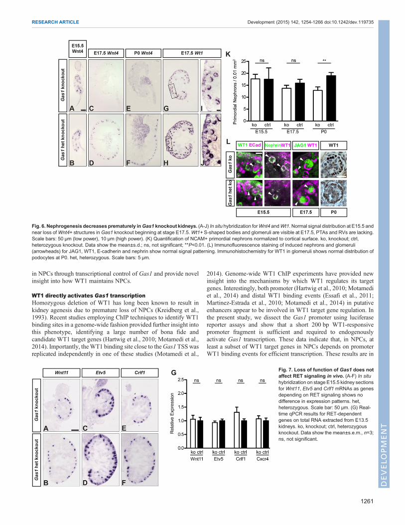
Gas1 knockout kidneys were also examined for abnormalitiesin nephron differentiation. Consistent with the histology, atstage E15.5, Gas1 knockout kidneys did not show any defects inthe induction of Wnt4+ PTAs or RVs (Fig. 6A,B). Withdeclining numbers of NPCs at E17.5, however, very few Wnt4+structures were present in Gas1 knockout kidneys, progressingto their complete absence at stage P0 (Fig. 6C-F). Similarfindings were obtained when analyzing induced nephrons usingincreased Wt1 levels in PTAs and RVs as a marker (Fig. 6G-J).Of note, S-shaped bodies that were induced in Gas1 knockoutkidneys at any stage, including E17.5 and later, showed normalpatterning in terms of glomerular WT1+, proximal tubularJAG1+ and distal tubular E-cadherin+ domains, and werecapable of forming morphologically normal glomeruli(Fig. 6L), consistent with a role for GAS1 in maintainingNPCs, rather than affecting their differentiation into nephrons.Indeed, the number of NCAM+ primordial nephrons normalizedto section area was significantly reduced in Gas1 knockoutkidneys at stage P0 (Fig. 6K; supplementary material Fig. S3).Furthermore, there was no indication of ectopic or precociousdifferentiation of nephrons in Gas1 knockout kidneys. Insummary, loss of GAS1 results in decreased proliferation ofNPCs and establishes GAS1 as a novel factor required for themaintenance of NPCs during mid and later stages of kidneydevelopment in vivo.
GAS1 directs the intracellular response to FGF9 and FGF20stimulation towards activation of AKTGAS1 has previously been shown to function in two majorsignaling pathways with relevance to kidney development; namely,SHH signaling and the GDNF-Ret RTK axis (Allen et al., 2007;Cabrera et al., 2006; Izzi et al., 2011; Martinelli and Fan, 2007).With regard to SHH signaling, GAS1 functions as a SHHco-receptor facilitating SHH signal transmission to GLI1 and full-length GLI3 (Allen et al., 2007; Martinelli and Fan, 2007).However, the kidney cortical mesenchyme and NPCs requiresignaling of repressive, cleaved GLI3-R isoforms that are generatedin the absence of SHH ligand (Cain et al., 2009), inconsistent with apossible role for GAS1 in potentiating SHH signaling in NPCs. Wetherefore investigated whether GAS1 plays a role in RTK signalingin NPCs.
As GAS1 has been shown to modulate signaling downstream ofRET (Cabrera et al., 2006), a key pathway in ureteric branching thatalso feeds back to NPCs through WNT11 (Costantini and Kopan,2010; Majumdar et al., 2003), we investigated whether expressionpatterns and levels of ureteric tip genes dependent onRETwere alteredin Gas1 knockout kidneys (Fig. 7A-G). No changes in expressionpatterns by ISH and mRNA levels by RT-qPCR were identified forWnt11, Crlf1, Etv4 and Cxcr4. These data indicated that GAS1 doesnot exert a non-cell-autonomous effect on RET signals in ureteric tipcells, and further suggest that decreased NPC proliferation in Gas1
Fig. 4. Gas1 knockout kidneys are hypoplastic and show disorganization of the nephrogenic zone. (A-D) Histology of Gas1 knockout kidneys at stageE15.5. Kidneys are normal and adequately sized with condensingmesenchyme (yellow line) and two renal vesicles (green line) surrounding a ureteric bud branch(black line). het, heterozygous. Scale bars: 50 µm (low power), 10 µm (high power). (E-H) Histology at stage P0. Gas1 knockout kidneys are hypoplastic.Condensed mesenchyme is reduced and only a single renal vesicle is present. (I) The longitudinal diameters of kidney sections including the papilla at variousstages. ko, knockout; ctrl, heterozygous knockout. Data show themean±s.d., n≥4; ns, not significant; **P<0.01; ***P<0.001. (J) The numbers of glomeruli inGas1knockout and control kidneys at P0. Data show the mean±s.d.; **P<0.01.
1259
RESEARCH ARTICLE Development (2015) 142, 1254-1266 doi:10.1242/dev.119735
DEVELO
PM
ENT

knockout kidneys is not a consequence of decreased RET-dependentpro-proliferative signals from ureteric tips.Signaling of FGF9 and FGF20 to NPCs is required for their
maintenance and proliferation (Barak et al., 2012). Interestingly, themodulating effect of Gas1 on RET signaling has been shown to bedependent on the interaction of RET with FRS2A (Cabrera et al.,2006), a phospho-tyrosine-binding RTK adaptor molecule that alsoplays key roles in mediating FGF signaling. Therefore, we nextinvestigated whether GAS1 modulates FGF signaling by knockingdown Gas1 in LB-22 cells. LB-22 cells endogenously express FGFreceptor 2 (FGFR2), the receptor for FGF8, FGF9 and FGF20, keyFGF ligands expressed in NPCs and RVs (Barak et al., 2012;Grieshammer et al., 2005; Perantoni et al., 2005). We stimulatedLB-22 cells with these FGF ligands after transfection with Gas1siRNA or scramble control siRNA and assayed for thephosphorylation of p42/p44 MAPK (ERK; MAPK3/MAPK1,respectively – Mouse Genome Informatics) and AKT (Fig. 8A,B).Interestingly, the phosphorylation of AKT upon FGF stimulationwas reduced to baseline levels in the Gas1 knockdown condition,whereas ERK phosphorylation was not affected. These findingsindicate a novel function for GAS1, in potentiating signaling fromFGF receptors that is selective to the AKT pathway as opposed toaffecting activation of ERK. Therefore, we assayed AKTphosphorylation in NPCs of E15.5 Gas1 knockout kidneys andcontrols by immunohistochemistry (Fig. 8C-G). Indeed, phospho-AKT signals in NPCs were considerably reduced upon loss ofGAS1, whereas signals in ureteric tips, the kidney capsule and
developing nephrons appeared to be unchanged. Furthermore, weaimed to rescue the FGF signaling defect by overstimulating Gas1morphant kidney explants with excess FGF9 (data not shown).However, in line with GAS1 modulating RTK signals on theintracellular level, neither the dynamics of NPC depletion nor theextent of the NPC pool appeared to be different betweenoverstimulation and control conditions. In summary, we identify amodulating effect of GAS1 on FGF signaling in vitro as well asin vivo and suggest that GAS1 drives the NPC response to FGFsignals towards intracellular activation of AKT to promote NPCproliferation.
DISCUSSIONThe data provided in this study establish the GPI-anchoredmembrane molecule GAS1 as a novel and direct WT1 target genein NPCs. Furthermore, we show in embryonic kidneys that GAS1controls nephron endowment by ensuring NPC proliferation issufficient to maintain the SIX2+ NPC population. In the absenceof GAS1, early metanephric development, establishment of anephrogenic niche and initial nephrogenesis proceed normally.However, loss of GAS1 results in premature depletion of the NPCpool at later stages in kidney development with subsequentdecreased nephrogenesis and kidney hypoplasia. Mechanistically,our data suggest that GAS1 affects signaling downstream of FGF9and FGF20 to phosphorylate AKT but not ERK, allowing for theactivation of pro-proliferative pathways downstream of AKT inNPCs. These data establish a link between WT1 and FGF signaling
Fig. 5. Progressive loss of SIX2+ nephron progenitor cells in Gas1 knockout kidneys due to decreased proliferation. (A-F) Immunofluorescencestaining for SIX2 onGas1 knockout and control kidneys at various stages. A decreased number of SIX2+ nuclei is evident with progression of renal development.het, heterozygous. Scale bar: 10 µm. (G) Quantification of SIX2+ nuclei normalized to cortex surface at various embryonic stages. Data show themean±s.d., n≥4;ns, not significant; *P<0.05; ***P<0.001. (H) Quantification of TUNEL+ signals normalized to cortical area. Increased cell death is excluded as a reason for NPCloss. Data show themean±s.d., n=3; ns, not significant. (I) Quantification of SIX2+/BrdU+ cells at E15.5 andE17.5. The proliferative fraction of SIX2+ cells inGas1knockout kidneys is decreased at E15.5 and E17.5. ko, knockout; ctrl, heterozygous knockout. Data show the mean±s.d., n=3; **P<0.01.
1260
RESEARCH ARTICLE Development (2015) 142, 1254-1266 doi:10.1242/dev.119735
DEVELO
PM
ENT
in NPCs through transcriptional control of Gas1 and provide novelinsight into how WT1 maintains NPCs.
WT1 directly activates Gas1 transcriptionHomozygous deletion of WT1 has long been known to result inkidney agenesis due to premature loss of NPCs (Kreidberg et al.,1993). Recent studies employing ChIP techniques to identify WT1binding sites in a genome-wide fashion provided further insight intothis phenotype, identifying a large number of bona fide andcandidate WT1 target genes (Hartwig et al., 2010; Motamedi et al.,2014). Importantly, theWT1 binding site close to theGas1 TSSwasreplicated independently in one of these studies (Motamedi et al.,
2014). Genome-wide WT1 ChIP experiments have provided newinsight into the mechanisms by which WT1 regulates its targetgenes. Interestingly, both promoter (Hartwig et al., 2010; Motamediet al., 2014) and distal WT1 binding events (Essafi et al., 2011;Martinez-Estrada et al., 2010; Motamedi et al., 2014) in putativeenhancers appear to be involved in WT1 target gene regulation. Inthe present study, we dissect the Gas1 promoter using luciferasereporter assays and show that a short 200 bp WT1-responsivepromoter fragment is sufficient and required to endogenouslyactivate Gas1 transcription. These data indicate that, in NPCs, atleast a subset of WT1 target genes in NPCs depends on promoterWT1 binding events for efficient transcription. These results are in
Fig. 6. Nephrogenesis decreases prematurely inGas1 knockout kidneys. (A-J) In situ hybridization forWnt4 andWt1. Normal signal distribution at E15.5 andnear loss ofWnt4+ structures in Gas1 knockout beginning at stage E17.5.Wt1+ S-shaped bodies and glomeruli are visible at E17.5, PTAs and RVs are lacking.Scale bars: 50 µm (low power), 10 µm (high power). (K) Quantification of NCAM+ primordial nephrons normalized to cortical surface. ko, knockout; ctrl,heterozygous knockout. Data show the mean±s.d.; ns, not significant; **P<0.01. (L) Immunofluorescence staining of induced nephrons and glomeruli(arrowheads) for JAG1, WT1, E-cadherin and nephrin show normal signal patterning. Immunohistochemistry for WT1 in glomeruli shows normal distribution ofpodocytes at P0. het, heterozygous. Scale bars: 5 µm.
Fig. 7. Loss of function of Gas1 does notaffect RET signaling in vivo. (A-F) In situhybridization on stage E15.5 kidney sectionsfor Wnt11, Etv5 and Crlf1 mRNAs as genesdepending on RET signaling shows nodifference in expression patterns. het,heterozygous. Scale bar: 50 µm. (G) Real-time qPCR results for RET-dependentgenes on total RNA extracted from E13.5kidneys. ko, knockout; ctrl, heterozygousknockout. Data show the mean±s.e.m., n=3;ns, not significant.
1261
RESEARCH ARTICLE Development (2015) 142, 1254-1266 doi:10.1242/dev.119735
DEVELO
PM
ENT

agreement with previous findings that WT1 binding at targetpromoters in NPCs activates transcription of a large number of keygenes in renal development (Hartwig et al., 2010; Motamedi et al.,2014). A puzzling aspect of the transcriptional regulation of Gas1and other WT1 target genes in NPCs is the fact that their expressionin NPCs depends onWT1, whereas they are silenced in RVs despiteincreased WT1 levels in these structures. Consistent with thesefindings, Gas1 mRNA has been shown to be downregulated uponinduction of canonical WNT signaling in FACS-sorted NPCs, aneffect that was attributed to a co-operative SIX2 and β-cateninbinding site downstream of the Gas1 locus (Park et al., 2012).Therefore, Gas1 transcription in NPCs might be dependent onco-operative SIX2 and WT1 effects, with SIX2 being bound at adistal regulatory element and WT1 at the promoter. Once thedifferentiation of NPCs is induced by canonical WNT signals(Carroll et al., 2005; Karner et al., 2011), β-catenin binding at theSIX2-bound regulatory element might disrupt this co-operation(Park et al., 2012) and result in Gas1 silencing in RVs. Anotherpossible explanation for the incongruence of Gas1 and Wt1expression patterns might be a differential utilization of the WT1-dependent co-activator CBP/p300 (Wang et al., 2001) and co-repressor BASP1 (Carpenter et al., 2004) between NPCs and RVs,resulting in different epigenetic regulation on the chromatin level atthe Gas1 locus as has been described for theWnt4, Snai1 and Cdh1loci in various tissues (Essafi et al., 2011; Martinez-Estrada et al.,2010). In the light of our data that implicate GAS1 in modulatingFGF signals during kidney development, differential transcriptionalregulation of Gas1 between NPCs and RVs might, in part, account
for differences in FGF activity within NPCs versus nephrons. Forexample, FGF8 signals derived from RVs are required forWnt4 andLhx1 expression within the vesicles (Grieshammer et al., 2005;Perantoni et al., 2005). However, in contrast to NPC-derivedFGF20, FGF8 signals are not sufficient to maintain the progenitorstatus in NPCs and might direct FGF signals towards responsesother than AKT phosphorylation (Barak et al., 2012). In summary,our results suggest that WT1 binding to promoters is a key event inWT1 target gene regulation in NPCs, which is in contrast to themore prevalent enhancer-based transcriptional regulation asobserved in SIX2 targets in NPCs (Park et al., 2012).
GAS1 maintains NPC expansion at late stages in kidneydevelopmentDecreased nephron endowment during renal development has beenshown to be a key risk factor for hypertension and chronic kidneydisease later in life (Luyckx et al., 2013), highlighting theimportance of identifying genetic modifiers of nephron numberduring kidney development. Reduced nephron endowment canresult from perturbations within several cell pools and in differentmorphogenetic processes during kidney development, including theprogenitor cell niche, the kidney stroma, branching morphogenesisand nephrogenesis (Kopan et al., 2014). The majority of loss-of-function phenotypes affecting genes expressed in NPCs result inrenal agenesis, failed establishment of the NPC niche or in severeearly impairment of nephrogenesis, featuring kidney rudiments withfew if any induced nephrons (Kopan et al., 2014). Loss-of-functionphenotypes associated with reduced NPC proliferation have been
Fig. 8. Depletion of Gas1 modulates FGF signaling through AKT in vitro and in vivo. (A) Western blots of whole-cell lysates from LB-22 cells treated withGas1 siRNA or scramble control and stimulated with 50 ng/ml FGF as indicated. (B) Densitometry of repeat western blots as in A. Results are normalized to thenon-stimulated, vehicle-treated control and to total AKT, total ERK or tubulin, respectively. Data show the mean±s.e.m., n≥3; ns, not significant.(C-G) Immunohistochemistry for phospho-Akt at E15.5 shows diminished phospho-Akt signal in condensed mesenchyme upon Gas1 knockout. Black boxes inC,D correspond to high-power panels. Ureteric tips are labeled with asterisks. Arrowheads indicate condensed mesenchyme. Scale bars: 200 µm (low power),25 µm (high power), 75 µm (mid power).
1262
RESEARCH ARTICLE Development (2015) 142, 1254-1266 doi:10.1242/dev.119735
DEVELO
PM
ENT

reported, including Bmp7 (Blank et al., 2009; Dudley et al., 1995),c-myc (Couillard and Trudel, 2009), Dlg1 and Cask (Ahn et al.,2013), Mi-2-NurD (Denner and Rauchman, 2013) and the miR-17-92 cluster (Marrone et al., 2014). However, with the exception of themiR-17-92 knockout, these phenotypes are characterized by earlygrowth deficits and reduced nephron numbers evident by E13.5(Ahn et al., 2013) or E14.5 (Denner and Rauchman, 2013; Dudleyet al., 1995). By contrast, establishment of the niche and thenephrogenic zone in early metanephric development appear to beunaffected in Gas1 knockout kidneys. A possible explanation forthese divergent phenotypes all associated with NPC proliferationdeficits is differential regulation of proliferation in early (E13.5) ascompared to late (E17.5) NPC pools. In fact, temporal discontinuitywith respect to NPC proliferation rates and cell cycle lengths in bothCITED1+ and CITED1− NPC compartments has recently beendescribed (Short et al., 2014), with the majority of proliferatingNPCs being contributed by a slow-cycling compartment at E17.5,whereas at E13.5 proliferation was predominantly in a fast-cyclingNPC compartment. Given that Gas1 knockout kidneys arephenotypically normal until E15.5, our data suggest that GAS1might primarily regulate the slow-cycling NPC population.
GAS1 selectively modulates the AKT branch of the FGFsignaling pathwayMultiple signalingpathways havebeen reported to regulate the balancebetween self-maintenance anddifferentiation inNPCs, includingFGF,BMP and canonical WNT signals (Barak et al., 2012; Blank et al.,2009; Brown et al., 2011, 2013; Carroll et al., 2005; Park et al., 2012),all of which interact with or are activated by WT1 (Akpa et al., 2014;Hartwig et al., 2010; Kim et al., 2010; Motamedi et al., 2014). TheFGF ligand FGF20 is transcriptionally activated by WT1 in NPCs(Motamedi et al., 2014) and is connected toNPCproliferation throughactivation of PI3K-AKT (Barak et al., 2012; Brown et al., 2011),whereas Bmp7 is a WT1 target gene (Hartwig et al., 2010) thatpromotesNPCproliferation byactivation of JNKMAPK (Blanket al.,2009; Motamedi et al., 2014). Both MAPK and PI3K-AKT cascadeshave well-established roles in cell cycle control; however, themechanisms by which these signaling cascades are balanced andregulated in response to BMP and FGF signals is not well understood.Our results suggest that WT1-dependent transcriptional control ofGas1might promote FGF-induced intracellular signals to activation ofPI3K-AKT. InterestinglymiR-17-92, whose loss of function results ina late NPC proliferation defect as well (Marrone et al., 2014), has alsobeen shown to modulate the PI3K-AKT pathway (Olive et al., 2009),underlining the importance of this signaling cascade in NPCs. Takentogether, we expand the findings fromWT1 ChIP-on-chip and ChIP-seq studies in NPCs (Hartwig et al., 2010; Motamedi et al., 2014) toshow that WT1 can affect signaling in NPCs by transcriptionalregulation on multiple levels, including ligands, receptors and RTK-interacting cofactors such as GAS1.GAS1has previously been shown to complexwithGFRA1,GDNF
and RET to modulate PI3K-AKT RTK signaling in the GDNF-RETpathway (Cabrera et al., 2006). Importantly, direct GDNF-GAS1interactions were not required for modulation of intracellular signals,suggesting a GAS1-dependent mechanism downstream of ligandbinding. Accordingly, our results that implicate a role for GAS1 in theFGF signaling pathway suggest that GAS1 modulates signalingdownstream of FGF ligands, as well. Altered FGF expression inresponse to GAS1 loss of function has been previously described inthe developing limb (Liu et al., 2002). Given the widespreadexpression of GAS1 in various anlagen (Lee and Fan, 2001) and thecritical role of FGF signaling in a large number of developmental
processes, GAS1 might play an important regulatory role in FGFsignaling in other developing organs.
In summary, we identify a novel mechanism by which WT1regulates the progenitor population by transcriptional activation ofGas1. GAS1 adjusts the gain on RTK signaling in NPCs to regulateNPC proliferation through the PI3K-AKT pathway in a manner thatresults in defects late in kidney development. Eventually, thesemechanisms governed by GAS1 in embryonic kidneys play animportant role in determining nephronmass and are therefore relevantto the risk of various diseases later in life.
MATERIALS AND METHODSLB-22 cellsLB-22 cells were derived from metanephric organ cultures (see below) ofE12.5 embryos obtained from ‘immortomice’ (Charles River Laboratories)(Jat et al., 1991). The nephrogenic zone was dissected from the organoid aftercultivation for 48 h and triturated into a single-cell suspension. Clones fromindividual cells were expanded in DMEM-F12/50:50 medium withL-glutamine (Cellgro) supplemented with 10% fetal bovine serum (FBS,Hyclone), 25 ng/ml prostaglandin E1 (Calbiochem), 2 ng/ml heparan-stabilized basic FGF, 100 nM hydrocortisone, 2 nM triiodothyronine, 5 µg/ml insulin (all fromSigma) and 5 µg/ml transferrin (Roche), and subsequentlyscreened for WT1 expression by western blotting. The WT1-positive cloneLB-22 wasmaintained at 33°C in the presence of 10 U/ml interferon γ (R&D)and transferred to interferon-free medium at 37°C upon transfection.
RNAi experimentsStealth siRNA duplexes (Invitrogen) were used to knock down Wt1 andGas1 in LB-22 cells (for oligo sequences see supplementary materialTable S1). Cells were transfected with 25 pmol gene-specific or controlsiRNA in 25 µl Lipofectamine 2000 (Invitrogen) per 5×10⁵ cells. Aftertransfection, cells were maintained at 70-80% confluency. RNA and proteinwere harvested at 48 and 72 h after transfection, respectively.
Stimulation of LB-22 cells with FGFs was carried out at 70% confluencyafter 6 h of serum starvation. FGF8, FGF9 or FGF20 (all fromPeprotech)wereadded at concentrations as indicated together with 5 µg/ml heparin (Sigma).
Western blottingWhole-cell lysates fromLB-22 cells and kidneyorgan cultureswere preparedeither in high-salt RIPA (500 mM NaCl, 50 mM Tris-HCl pH 7.4, 5 mMEDTA, 1 mMEGTA, 0.1% SDS, 1% Igepal, 0.5% sodium deoxycholate, 1×Roche protease inhibitor mix) for experiments involving WT1 or in RIPA(same as above, except 300 mMNaCl, 0.4 mMNa3VO4, 4 mMNaF) for allother experiments (all reagents from Sigma). Equal amounts of proteinlysates were resolved on 10% polyacrylamide gels and transferred to PVDFmembranes. Standard western blotting was performed with antibodiesagainst WT1 (C19, Santa Cruz sc-192, rabbit polyclonal, 1:500), β-tubulin(Santa Cruz sc-9104, rabbit polyclonal, 1:1000), GAS1 (R&DAF2644, goatpolyclonal, 1:500), SIX2 (Proteintech 11562-1-AP, rabbit polyclonal,1:500), phospho-AKT, AKT, phospho-ERK and ERK (4695S, 4370,4060, 4691, all from Cell Signaling, rabbit monoclonals, 1:1000).
RNA isolation, RT-qPCR and microarraysRNA was isolated from LB-22 cells and embryonic kidneys by means of theRNeasy Mini Kit (Qiagen) according to the manufacturer’s instructions. RNAwas quantified on a Nanodrop-2000 instrument and equal amounts per samplewere reverse transcribed into cDNAwith the SuperScript IIIRTKit (Invitrogen)with oligo-dT primers. Real-time quantitative PCRwas carried out in triplicateusing the QuantiTect SYBR Green HotStart Kit (Qiagen) on a CepheidSmartCycler. Primer sequences are provided in supplementary materialTable S1. Data analysis was carried out as described previously (Pfaffl, 2001).
For microarray studies, RNA samples were hybridized to Mouse 430.2expression microarrays (Affymetrix) at the Boston Children’s HospitalMicroarray Core Facility after processing according to the manufacturer’sinstructions. Microarray data have been deposited at Gene ExpressionOmnibus (GEO) under accession number GSE66356.
1263
RESEARCH ARTICLE Development (2015) 142, 1254-1266 doi:10.1242/dev.119735
DEVELO
PM
ENT

Chromatin immunoprecipitationChromatin immunoprecipitation on P0 kidneys was carried out as describedpreviously (Hartwig et al., 2010). LB-22 cells were crosslinked with 1%formaldehyde in PBS for 5 min, washed in PBS and sonicated in RIPAbuffer (300 mM NaCl, 1 mM EDTA, 10 mM Tris-HCl pH 7.4, 1% TritonX-100, 0.1% sodium dodecyl sulfate, 0.1% sodium deoxycholate, 0.1 MDTT, 0.25% N-lauroyl-sarcosine) using a Misonix S-4000 sonicator (allreagents from Sigma). ChIP was carried out using a WT1 antibody (C19,Santa Cruz sc-192) and normal rabbit serum (both from Santa Cruz) ascontrol. Real-time qPCR of ChIP and input DNA was carried out asdescribed above. For primers see supplementary material Table S1.
Dual luciferase reporter assays and mutagenesisFragments of theGas1 promoter were amplified frommouse genomic DNAand directionally cloned into the Gateway-compatible pENTR-dTOPOentry vectors (Invitrogen). The pGL4.14 and pGL4.26 firefly luciferasevectors (Promega) were rendered Gateway-compatible destination vectorsby means of the Gateway Vector Conversion System (Invitrogen). Entry anddestination vectors were then recombined using the LR Clonase II Kit(Invitrogen) to yield functional reporter plasmids. The pGL4.74 Renillaluciferase vector (Promega) was used as the transfection control.
LB-22 cells were transfected for 6 h with 1 µg reporter plasmid and 0.1 µgRenilla plasmid in Lipofectamine 2000 (Invitrogen) per well in 24-wellplates. Luciferase signals were assayed 48 h after transfection usingDualGlo chemistry (Promega) on a FLUOstar luminometer (Omega). Forreporter assays in WT1 knockdown cells, LB-22 cells were first transfectedwith siRNAs as described above and re-transfected with luciferase plasmidsat 24 h, and luciferase was assayed at 72 h.
For mutagenesis studies, the QuikChange2 Site DirectedMutagenesis Kit(Stratagene) was used according to the manufacturer’s instructions. Primersused for mutagenesis can be found in supplementary material Table S1.
Mice and metanephric organ culturesWT1 knockdown mice were established by cloning a WT1-specific shRNA(see supplementary material Table S1 for sequence) into the pBS/U6-loxPNeo vector (Shukla et al., 2007). Plasmids were microinjected into thepronuclei of single-cell mouse embryos, which were transferred intopseudopregnant foster mothers. Germline transmission was established andF1 offspring were genotyped for the presence of the transgene.
Six2:Cre mice and Gas1 knockout mice have been described previously(Kobayashi et al., 2008; Martinelli and Fan, 2007). Genotyping primers andconditions are given in supplementary material Table S1. Embryos formetanephric organ cultures were harvested from pregnant CD-1 wild-typemice at stage E12.5.
Metanephric organ cultures andMO knockdown ofWT1 were performedas described previously (Hartwig et al., 2010). ForGas1MO knockdown inorgan culture, kidneys of E12.5 CD-1 wild-type embryos were treated withMOs (Vivo-Morpholino, GeneTools) for 18 h or 24 h, with a morphantkidney being exposed to amixture of twoGas1-blocking oligonucleotides atconcentrations as indicated in the figure (supplementary material Fig. S4),while the opposite kidney of the same embryowas treated with either controlMO only or a mixture of sub-efficient Gas1 MO1 and control MO atconcentrations as indicated. All animal studies were carried out inaccordance with the guidelines of the Institutional Animal Care and UseCommittee at Boston Children’s Hospital.
Immunofluorescence, immunohistochemistry, in situhybridization and phenotypic quantificationImmunofluorescence was carried out using antibodies against WT1 (C19,Santa Cruz sc-192, 1:250), GAS1 (R&D AF2644, 1:250), SIX2(Proteintech 11562-1-AP, 1:200), E-cadherin (BD Biosciences 610404,mouse monoclonal, 1:200), NCAM (Sigma C6680, mouse monoclonal,1:200), JAG1 (Santa Cruz sc-6011, goat polyclonal, 1:200) and pan-cytokeratin (Sigma C5992, mouse monoclonal, 1:150). Whole-mountimages of organ cultures were edited using the background subtractionfunction in NIKON NIS Elements software to remove autofluorescence ofthe polyethylene membrane. The Apoptag Fluorescein Direct In Situ
Detection Kit (Millipore) was used for TUNEL staining as per themanufacturer’s instructions. BrdU labeling of proliferating cells wasachieved by injecting mice with 300 µg of BrdU (Roche) per gram ofbody weight at 1 h before sacrifice. BrdU was detected with the BrdULabeling and Detection Kit (Roche) according to the manufacturer’sinstructions.
For immunohistochemistry, paraffin-embedded formalin-fixed tissue wasde-waxed in xylenes and rehydrated. Epitope retrieval was carried out byboiling in TE (10 mM Tris pH 9, 1 mM EDTA) for 20 min. Slides wereblocked in 3%H2O2, 5% (w/v)BSA and using theAvidin/BiotinBlockingKit(Vector Labs) as per the manufacturer’s instructions. The phospho-Aktantibody (1:100, Cell Signaling) was applied at 4°C over night, slides werewashed and the signal was detected using a biotinylated anti-rabbit secondaryantibody (1:400, Jackson) with the Vectastain ABC Kit (Vector Labs) anddi-aminobenzidine, as per the manufacturer’s instructions. Slides werecounterstained in Hematoxylin, dehydrated and mounted with histomount.
Whole-mount and section in situ hybridization was carried out followingthe Genito-Urinary Molecular Anatomy Project protocols accessible athttp://www.gudmap.org/Research/Protocols/McMahon.html. Probes wereSix2, Wt1, Gas1 (a gift of Andrew P. McMahon, Keck School of Medicineof USC, Los Angeles, CA, USA), Wnt4, Wnt9b, Etv5, Crlf1 and Wnt11.
For phenotypic characterization, at least four kidneys per stage andgenotypewere evaluated. SIX2+ nuclei were counted on immunofluorescentsections and normalized to the product of section thickness and the length ofthe capsular circumference present on the section. For evaluation ofTUNEL signals, the area between capsule and corticomedullary border wascomputed, and TUNEL+ nuclei within this area were counted andnormalized to cortical area. The proliferative index was calculated bydividing the numbers of SIX2+/BrdU+ nuclei by numbers of all SIX2+nuclei. Only nuclei located on the capsular face of adjacent ureteric tips wereevaluated. Nephrogenesis was assessed by counting NCAM+ comma- andS-shaped bodies and normalizing this number to the product of capsularcircumference and section thickness.
StatisticsAll statistical analyses were carried out using GraphPad Prism 5. One-wayANOVA with Tukey’s post hoc test was used to analyze ChIP-qPCR andluciferase reporter assay data. Significance was assumed at P<0.05 inTukey’s test. Two-way ANOVAwith Bonferroni’s post hoc test was used toanalyze luciferase reporter assays in RNAi experiments, western blotdensitometry results for AKT and ERK, and RT-qPCR experiments withsignificance requiring P<0.05 in Bonferroni’s test. Student’s t-test was usedto test for differences between groups in BrdU incorporation assays, TUNELassays, densitometry of Gas1 knockdown and phenotypic quantifications.
Microarray results were normalized to the trimmed mean (top and bottom2% of values excluded for mean calculation) in each sample. Perseusframework software was used to compute q-values, and for principalcomponent analysis (Cox and Mann, 2012).
Screening for WT1 motifs in theGas1 promoter was done by adding log-odds scores for each base in the WT1 positional weight matrix (Hartwiget al., 2010) using the TRED algorithm. (Zhao et al., 2005).
AcknowledgementsWe thank Martyna Bruetting for excellent technical assistance, Martin Hoehne foradvice in image processing, Andrew P. McMahon for providing reagents, Shan Qinfor valuable discussions and the Harvard Rodent Histopathology Core Facility forhistology services.
Competing interestsThe authors declare no competing or financial interests.
Author contributionsM.K. and J.A.K. developed the concept of the study. M.K., E.B., M.O.L., L.L., B.T.,V.A.S., M.E.T., L.B. and C.-M.F. performed experiments. M.K., E.B., S.H. andM.M.R. analyzed data. M.K. and J.A.K. wrote the manuscript with input from S.H.,C.-M.F., T.B., B.S. and all other co-authors.
FundingM.K. received scholarships from the German Research Foundation [KA3217/2-1];the GermanHypertension Society; andCECADCologne.M.O.L. was a recipient of a
1264
RESEARCH ARTICLE Development (2015) 142, 1254-1266 doi:10.1242/dev.119735
DEVELO
PM
ENT

KoelnFortune scholarship. M.M.R. was supported by a Fritz-Scheler scholarship ofthe KfH Foundation for Preventive Medicine; and by a UoC postdoctoral grant in theframework of the German Research Foundation Excellence Initiative. T.B. issupported by the German Research Foundation [BE2212 and SFB329]. J.A.K.received funding from the National Institutes of Health/the National Institute ofDiabetes and Digestive and Kidney Diseases [NIDDKR01DK087794-A1]. E.B. wassupported by a fellowship grant from the National Kidney Foundation. B.S. wassupported by the German Research Foundation [SCHE 1562/2-1]. Deposited inPMC for release after 12 months.
Supplementary materialSupplementary material available online athttp://dev.biologists.org/lookup/suppl/doi:10.1242/dev.119735/-/DC1
ReferencesAhn, S.-Y., Kim, Y., Kim, S. T., Swat, W. and Miner, J. H. (2013). Scaffoldingproteins DLG1 and CASK cooperate to maintain the nephron progenitorpopulation during kidney development. J. Am. Soc. Nephrol. 24, 1127-1138.
Akpa, M. M., Iglesias, D. M., Chu, L. L., Cybulsky, M., Bravi, C. and Goodyer, P.(2014). Wilms tumour suppressor, WT1, suppresses epigenetic silencing of thebeta-catenin gene. J. Biol. Chem. 290, 2279-2288.
Allen, B. L., Tenzen, T. and McMahon, A. P. (2007). The Hedgehog-bindingproteins Gas1 and Cdo cooperate to positively regulate Shh signaling duringmouse development. Genes Dev. 21, 1244-1257.
Barak, H., Huh, S.-H., Chen, S., Jeanpierre, C., Martinovic, J., Parisot, M., Bole-Feysot, C., Nitschke, P., Salomon, R., Antignac, C. et al. (2012). FGF9 andFGF20 maintain the stemness of nephron progenitors in mice and man. Dev. Cell22, 1191-1207.
Blank, U., Brown, A., Adams, D. C., Karolak, M. J. andOxburgh, L. (2009). BMP7promotes proliferation of nephron progenitor cells via a JNK-dependentmechanism. Development 136, 3557-3566.
Brown, A. C., Adams, D., de Caestecker, M., Yang, X., Friesel, R. and Oxburgh,L. (2011). FGF/EGF signaling regulates the renewal of early nephron progenitorsduring embryonic development. Development 138, 5099-5112.
Brown, A. C., Muthukrishnan, S. D., Guay, J. A., Adams, D. C., Schafer, D. A.,Fetting, J. L. and Oxburgh, L. (2013). Role for compartmentalization in nephronprogenitor differentiation. Proc. Natl. Acad. Sci. USA 110, 4640-4645.
Cabrera, J. R., Sanchez-Pulido, L., Rojas, A. M., Valencia, A., Manes, S.,Naranjo, J. R. and Mellstrom, B. (2006). Gas1 is related to the glial cell-derivedneurotrophic factor family receptors alpha and regulates Ret signaling. J. Biol.Chem. 281, 14330-14339.
Cain, J. E., Islam, E., Haxho, F., Chen, L., Bridgewater, D., Nieuwenhuis, E., Hui,C.-C. and Rosenblum, N. D. (2009). GLI3 repressor controls nephron number viaregulation of Wnt11 and Ret in ureteric tip cells. PLoS ONE 4, e7313.
Carpenter, B., Hill, K. J., Charalambous, M., Wagner, K. J., Lahiri, D., James,D. I., Andersen, J. S., Schumacher, V., Royer-Pokora, B., Mann, M. et al.(2004). BASP1 is a transcriptional cosuppressor for the Wilms’ tumor suppressorprotein WT1. Mol. Cell. Biol. 24, 537-549.
Carroll, T. J., Park, J.-S., Hayashi, S., Majumdar, A. and McMahon, A. P. (2005).Wnt9b plays a central role in the regulation of mesenchymal to epithelialtransitions underlying organogenesis of the mammalian urogenital system. Dev.Cell 9, 283-292.
Costantini, F. and Kopan, R. (2010). Patterning a complex organ: branchingmorphogenesis and nephron segmentation in kidney development. Dev. Cell 18,698-712.
Couillard, M. and Trudel, M. (2009). C-myc as amodulator of renal stem/progenitorcell population. Dev. Dyn. 238, 405-414.
Coumoul, X., Li, W., Wang, R.-H. and Deng, C. (2004). Inducible suppression ofFgfr2 and Survivin in ES cells using a combination of the RNA interference (RNAi)and the Cre-LoxP system. Nucleic Acids Res. 32, e85.
Cox, J. and Mann, M. (2012). 1D and 2D annotation enrichment: a statisticalmethod integrating quantitative proteomics with complementary high-throughputdata. BMC Bioinformatics 13 Suppl. 16, S12.
Del Sal, G., Ruaro, M. E., Philipson, L. and Schneider, C. (1992). The growtharrest-specific gene, gas1, is involved in growth suppression. Cell 70, 595-607.
Denner, D. R. and Rauchman, M. (2013). Mi-2/NuRD is required in renal progenitorcells during embryonic kidney development. Dev. Biol. 375, 105-116.
Di Giovanni, V., Alday, A., Chi, L., Mishina, Y. and Rosenblum, N. D. (2011). Alk3controls nephron number and androgen production via lineage-specific effects inintermediate mesoderm. Development 138, 2717-2727.
Dressler, G. R. (2009). Advances in early kidney specification, development andpatterning. Development 136, 3863-3874.
Dudley, A. T., Lyons, K. M. and Robertson, E. J. (1995). A requirement for bonemorphogenetic protein-7 during development of the mammalian kidney and eye.Genes Dev. 9, 2795-2807.
Essafi, A., Webb, A., Berry, R. L., Slight, J., Burn, S. F., Spraggon, L., Velecela,V., Martinez-Estrada, O. M., Wiltshire, J. H., Roberts, S. G. E. et al. (2011).
A wt1-controlled chromatin switching mechanism underpins tissue-specific wnt4activation and repression. Dev. Cell 21, 559-574.
Grieshammer, U., Cebrian, C., Ilagan, R., Meyers, E., Herzlinger, D. and Martin,G. R. (2005). FGF8 is required for cell survival at distinct stages of nephrogenesisand for regulation of gene expression in nascent nephrons. Development 132,3847-3857.
Hartwig, S., Ho, J., Pandey, P., MacIsaac, K., Taglienti, M., Xiang, M., Alterovitz,G., Ramoni, M., Fraenkel, E. and Kreidberg, J. A. (2010). Genomiccharacterization of Wilms’ tumor suppressor 1 targets in nephron progenitorcells during kidney development. Development 137, 1189-1203.
Izzi, L., Levesque, M., Morin, S., Laniel, D., Wilkes, B. C., Mille, F., Krauss, R. S.,McMahon, A. P., Allen, B. L. and Charron, F. (2011). Boc and Gas1 each formdistinct Shh receptor complexes with Ptch1 and are required for Shh-mediated cellproliferation. Dev. Cell 20, 788-801.
Jat, P. S., Noble, M. D., Ataliotis, P., Tanaka, Y., Yannoutsos, N., Larsen, L.and Kioussis, D. (1991). Direct derivation of conditionally immortal cell linesfrom an H-2Kb-tsA58 transgenic mouse. Proc. Natl. Acad. Sci. USA 88,5096-5100.
Karner, C. M., Das, A., Ma, Z., Self, M., Chen, C., Lum, L., Oliver, G. and Carroll,T. J. (2011). Canonical Wnt9b signaling balances progenitor cell expansion anddifferentiation during kidney development. Development 138, 1247-1257.
Kim, M. S., Yoon, S. K., Bollig, F., Kitagaki, J., Hur, W., Whye, N. J., Wu, Y. P.,Rivera, M. N., Park, J. Y., Kim, H. S. et al. (2010). A novel Wilms tumor 1 (WT1)target gene negatively regulates the WNT signaling pathway. J. Biol. Chem. 285,14585-14593.
Kobayashi, A., Valerius, M. T., Mugford, J. W., Carroll, T. J., Self, M., Oliver, G.and McMahon, A. P. (2008). Six2 defines and regulates a multipotent self-renewing nephron progenitor population throughout mammalian kidneydevelopment. Cell Stem Cell 3, 169-181.
Kopan, R., Chen, S. and Little, M. (2014). Nephron progenitor cells: shifting thebalance of self-renewal and differentiation. Curr. Top. Dev. Biol. 107, 293-331.
Kreidberg, J. A., Sariola, H., Loring, J. M., Maeda, M., Pelletier, J., Housman, D.and Jaenisch, R. (1993). WT-1 is required for early kidney development. Cell 74,679-691.
Lee, C. S. and Fan, C.-M. (2001). Embryonic expression patterns of the mouse andchick Gas1 genes. Mech. Dev. 101, 293-297.
Liu, Y., May, N. R. and Fan, C.-M. (2001). Growth arrest specific gene 1 is a positivegrowth regulator for the cerebellum. Dev. Biol. 236, 30-45.
Liu, Y., Liu, C., Yamada, Y. and Fan, C. M. (2002). Growth arrest specific gene 1acts as a region-specific mediator of the Fgf10/Fgf8 regulatory loop in the limb.Development 129, 5289-5300.
Luyckx, V. A. and Brenner, B. M. (2010). The clinical importance of nephron mass.J. Am. Soc. Nephrol. 21, 898-910.
Luyckx, V. A., Bertram, J. F., Brenner, B. M., Fall, C., Hoy, W. E., Ozanne,S. E. and Vikse, B. E. (2013). Effect of fetal and child health on kidneydevelopment and long-term risk of hypertension and kidney disease. Lancet382, 273-283.
Majumdar, A., Vainio, S., Kispert, A., McMahon, J. and McMahon, A. P. (2003).Wnt11 and Ret/Gdnf pathways cooperate in regulating ureteric branching duringmetanephric kidney development. Development 130, 3175-3185.
Marrone, A. K., Stolz, D. B., Bastacky, S. I., Kostka, D., Bodnar, A. J. and Ho, J.(2014). MicroRNA-17∼92 is required for nephrogenesis and renal function. J. Am.Soc. Nephrol. 25, 1440-1452.
Martinelli, D. C. and Fan, C.-M. (2007). Gas1 extends the range of Hedgehogaction by facilitating its signaling. Genes Dev. 21, 1231-1243.
Martinez-Estrada, O. M., Lettice, L. A., Essafi, A., Guadix, J. A., Slight, J.,Velecela, V., Hall, E., Reichmann, J., Devenney, P. S., Hohenstein, P. et al.(2010). Wt1 is required for cardiovascular progenitor cell formation throughtranscriptional control of Snail and E-cadherin. Nat. Genet. 42, 89-93.
Mellstrom, B., Cena, V., Lamas, M., Perales, C., Gonzalez, C. and Naranjo, J. R.(2002). Gas1 is induced during and participates in excitotoxic neuronal death.Mol.Cell. Neurosci. 19, 417-429.
Motamedi, F. J., Badro, D. A., Clarkson, M., Rita Lecca, M., Bradford, S. T.,Buske, F. A., Saar, K., Hubner, N., Brandli, A. W. and Schedl, A. (2014). WT1controls antagonistic FGF and BMP-pSMAD pathways in early renal progenitors.Nat. Commun. 5, 4444.
Olive, V., Bennett, M. J., Walker, J. C., Ma, C., Jiang, I., Cordon-Cardo, C., Li, Q.-J., Lowe, S. W., Hannon, G. J. and He, L. (2009). miR-19 is a key oncogeniccomponent of mir-17–92. Genes Dev. 23, 2839-2849.
Park, J.-S., Ma, W., O’Brien, L. L., Chung, E., Guo, J.-J., Cheng, J.-G., Valerius,M. T., McMahon, J. A., Wong, W. H. and McMahon, A. P. (2012). Six2 and Wntregulate self-renewal and commitment of nephron progenitors through sharedgene regulatory networks. Dev. Cell 23, 637-651.
Perantoni, A. O., Timofeeva, O., Naillat, F., Richman, C., Pajni-Underwood, S.,Wilson, C., Vainio, S., Dove, L. F. and Lewandoski, M. (2005). Inactivation ofFGF8 in early mesoderm reveals an essential role in kidney development.Development 132, 3859-3871.
Pfaffl, M.W. (2001). A newmathematical model for relative quantification in real-timeRT-PCR. Nucleic Acids Res. 29, e45.
1265
RESEARCH ARTICLE Development (2015) 142, 1254-1266 doi:10.1242/dev.119735
DEVELO
PM
ENT

Short, K. M., Combes, A. N., Lefevre, J., Ju, A. L., Georgas, K. M., Lamberton, T.,Cairncross, O., Rumballe, B. A., McMahon, A. P., Hamilton, N. A. et al. (2014).Global quantification of tissue dynamics in the developing mouse kidney. Dev.Cell 29, 188-202.
Shukla, V., Coumoul, X. and Deng, C.-X. (2007). RNAi-based conditional geneknockdown in mice using a U6 promoter driven vector. Int. J. Biol. Sci. 3, 91-99.
Wang, W., Lee, S. B., Palmer, R., Ellisen, L. W. and Haber, D. A. (2001). Afunctional interaction with CBP contributes to transcriptional activation by theWilms tumor suppressor WT1. J. Biol. Chem. 276, 16810-16816.
Zhao, F., Xuan, Z., Liu, L. and Zhang, M. Q. (2005). TRED: a transcriptionalregulatory element database and a platform for in silico gene regulation studies.Nucleic Acids Res. 33 Suppl. 1, D103-D107.
1266
RESEARCH ARTICLE Development (2015) 142, 1254-1266 doi:10.1242/dev.119735
DEVELO
PM
ENT