wouter willaert, paul sessink and wim ceelen* occupational ... · a standardized safety checklist...
TRANSCRIPT
Wouter Willaert, Paul Sessink and Wim Ceelen*
Occupational safety of pressurized intraperitonealaerosol chemotherapy (PIPAC)
https://doi.org/10.1515/pap-2017-0018Received June 17, 2017; accepted July 23, 2017;previously published online August 12, 2017
Abstract
Background: Pressurized intraperitoneal aerosol che-motherapy (PIPAC) has emerged as a novel method totreat extensive, small volume peritoneal metastases. Theclinical use of chemotherapy containing aerosols repre-sents a potential occupational health hazard. We reportthe results of toxicological analysis during the first twoclinical PIPAC procedures performed at Ghent UniversityHospital.Methods: After extensive preparation and in vitro testing,two patients were treated with PIPAC: the first usingdoxorubicin (2.86mg in 51.43 mL) and cisplatin(14.28mg in 164.3 mL), the second using oxaliplatin(182.10mg in 186.42 mL). A standardized safety checklistwas developed and used. Aerosol delivery was combinedwith electrostatic precipitation (ePIPAC). The followingsamples were obtained at several time points and loca-tions: environmental air, floor surface wipes, surgeon’sgloves, surgeon’s hand wipes, circuit filters, and fluidfrom the water seal collection chamber container placedalong the closed aerosol waste evacuating line. Platinumconcentration was measured in these samples using vol-tammetry. Sample collection and analysis were per-formed by an independent external laboratory.Results: Platinum was not detected on the four floorlocations after both procedures (detection limit 0.02 ng/cm2). Similarly, no platinum was detected in environmen-tal air during both PIPACs at the surgeon’s or anesthe-siologist’s position (detection limit 4.0–27 ng/m3). Noplatinum contamination was detected on the hands,outer pair of gloves, or inner pair of gloves of the surgeon(detection limit 70 and 50 ng respectively). Platinum was
not detected on the filters and in the air-seal containerliquid.Conclusions: With adequate preparation and precau-tions, a clinical PIPAC program can be established with-out measurable chemotherapy exposure to the operatingroom environment or healthcare workers.
Keywords: carcinomatosis, occupational, PIPAC
Introduction
Peritoneal metastasis is a defining feature of stage IIIovarian cancer and occurs in approximately 13% of gastriccancers, 9% of pancreas cancers, and 8% of colorectalcancers [1–4]. In selected patients, cytoreductive surgerycombined with hyperthermic intraperitoneal chemoperfu-sion (HIPEC) results in a significant survival advantagecompared to palliative treatment alone [5, 6]. The morbid-ity of the combined procedure is, however, considerable,and a substantial proportion of patients have locally irre-sectable disease [7].
In 2012, Marc Reymond and coworkers proposed, in ananimal model, a novel approach to intraperitoneal drugdelivery, during which chemotherapy is administered asan aerosol during CO2 pneumoperitoneum [8]. The aerosolis generated by a high pressure line connected to a nozzle,hence the term pressurized intraperitoneal aerosol che-motherapy or PIPAC. Advantages of this approach includeminimal morbidity, efficient drug distribution, and tissuepenetration, and the possibility to repeat the procedurewhich allows for visual and histological assessment oftreatment response. In patients with widespread, smallvolume but unresectable peritoneal metastasis, prelimin-ary clinical experience has demonstrated the safety andantitumor efficacy of PIPAC [9–11]. Recently, the samegroup proposed to combine nebulization of chemotherapywith electrostatic precipitation using the Ultravision™ sys-tem. This device, originally developed to clear smoke fromthe laparoscopic operating field using an electrostaticforce, uses a stainless steel microfilament brush(Ionwand™) which is inserted into the abdominal cavity.A high DC voltage (7.5–9.5 kV, ≤ 10 µA) is applied to thewand resulting in a corona discharge and a stream ofnegatively charged ions, which attach to suspended
*Corresponding author: Wim Ceelen, Department of GI Surgery,Ghent University Hospital, route 1275, De Pintelaan 185, 9000,Ghent, Belgium; Cancer research institute Ghent (CRIG), Ghent,Belgium, E-mail: [email protected] Willaert, Department of GI Surgery, Ghent UniversityHospital, route 1275, De Pintelaan 185, 9000, Ghent, Belgium,E-mail: [email protected] Sessink, Exposure Control Sweden AB, Bohus-Björkö, Sweden,E-mail: [email protected]
Pleura and Peritoneum 2017; 2(3): 121–128
Brought to you by | Ghent University LibraryAuthenticated
Download Date | 2/13/18 12:06 PM
particles. These now negatively charged smoke particlesare attracted to the positively charged tissue surfaces ofthe abdominal cavity, which is conferred a weak positivecharge by the patient return electrode. In theory, the com-bination of electrostatic precipitation with PIPAC, termedePIPAC, could result in better tissue penetration of theaerosol. In a recent porcine model, the addition of electro-static precipitation to PIPAC resulted in higher tissue con-centrations of a tracer substance [12]. The first clinicalapplication in three patients with peritoneal metastaseswas recently reported, and showed ePIPAC to be techni-cally feasible and well tolerated [13].
A possible drawback of (e)PIPAC is the challenge tosafely deliver a chemotherapy aerosol intraperitoneallyduring laparoscopy, while preventing exposure of theinvolved healthcare workers. In 2013, the group ofReymond in Bochum performed analytical measurementsof air samples during two PIPAC procedures [14]. Twopatients were treated with PIPAC using cisplatin (7.5mg/m2) and doxorubicin (1.5mg/m2); analysis of air samplestaken at the place of the surgeon as well as that of theanesthesiologist was unable to detect cisplatin air con-tamination (detection limit < 0.000009mg/m3). Graversenand coworkers from the Odense University Hospital inDenmark recently reported the results of air sample ana-lysis and biological monitoring in two surgeons duringand after PIPAC in two patients: one treated with cispla-tin and doxorubicin, and the second treated with oxali-platin [15]. No traces of platinum were found in the airsamples (detection limit 0.0001mg), and blood samplesof the surgeons showed no traces of platinum.
Here, we report an additional, comprehensive toxico-logical analysis including air samples, surface wipe sam-ples, and analysis of surgeon’s gloves and hands after
clinical PIPAC procedures using cisplatin/doxorubicinand oxaliplatin.
Patients and methods
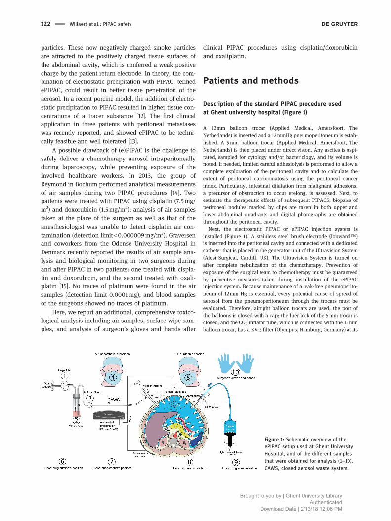
Description of the standard PIPAC procedure usedat Ghent university hospital (Figure 1)
A 12mm balloon trocar (Applied Medical, Amersfoort, TheNetherlands) is inserted and a 12mmHg pneumoperitoneum is estab-lished. A 5mm balloon trocar (Applied Medical, Amersfoort, TheNetherlands) is then placed under direct vision. Any ascites is aspi-rated, sampled for cytology and/or bacteriology, and its volume isnoted. If needed, limited careful adhesiolysis is performed to allow acomplete exploration of the peritoneal cavity and to calculate theextent of peritoneal carcinomatosis using the peritoneal cancerindex. Particularly, intestinal dilatation from malignant adhesions,a precursor of obstruction to occur erelong, is assessed. Next, toestimate the therapeutic effects of subsequent PIPACS, biopsies ofperitoneal nodules marked by clips are taken in both upper andlower abdominal quadrants and digital photographs are obtainedthroughout the peritoneal cavity.
Next, the electrostatic PIPAC or ePIPAC injection system isinstalled (Figure 1). A stainless steel brush electrode (Ionwand™)is inserted into the peritoneal cavity and connected with a dedicatedcatheter that is placed in the generator unit of the Ultravision System(Alesi Surgical, Cardiff, UK). The Ultravision System is turned onafter complete nebulization of the chemotherapy. Prevention ofexposure of the surgical team to chemotherapy must be guaranteedby preventive measures taken during installation of the ePIPACinjection system. Because maintenance of a leak-free pneumoperito-neum of 12mm Hg is essential, every potential cause of spread ofaerosol from the pneumoperitoneum through the trocars must beevaluated. Therefore, airtight balloon trocars are used; the port ofthe balloons is closed with a cap; the luer lock of the 5mm trocar isclosed; and the CO2 inflator tube, which is connected with the 12mmballoon trocar, has a KV-5 filter (Olympus, Hamburg, Germany) at its
Figure 1: Schematic overview of theePIPAC setup used at Ghent UniversityHospital, and of the different samplesthat were obtained for analysis (1–10).CAWS, closed aerosol waste system.
122 Willaert et al.: PIPAC safety
Brought to you by | Ghent University LibraryAuthenticated
Download Date | 2/13/18 12:06 PM
origin to prevent chemotherapy to enter the insufflator. A highpressure line sealed to the nebulizer (CapnoPen™, CapnomedGmbH, Villingendorf, Germany) and surrounded by a plastic cameracover is used. The nebulizer is then inserted in the 12mm balloontrocar and secured with the tip just inside the peritoneal cavity. Thetip is permanently visualized with a 5mm 30° camera (EndoEye™,Olympus, Hamburg, Germany) that is placed in the 5mm balloontrocar and secured with a laparoscopic scope holder (Integra,Zaventem, Belgium, and Cook Medical, Limerick, Ireland). Aftercompletion of the ePIPAC procedure, the abdomen is desufflatedusing a line attached to the 5mm trocar and equipped with a smokeevacuation filter (MTP Gmbh, Neuhausen Ob Eck, Germany). AfterCO2 has passed through this filter, it enters a water seal drainagesystem (Atrium, Mijdrecht, The Netherlands) that is attached to awall-mounted suction unit equipped with an infant-pediatric elec-trostatic filter HME (Medtronic, Brussels, Belgium). This closedwaste evacuation assembly is installed before the start of the proce-dure. After completing the ePIPAC installation, protective sheets arelaid out under the injector and next to the patient; team memberswear safety glasses and two pairs of gloves (outer pair: Gammex™;inner pair: Gammex Latex Chemo™, both Ansell Healthcare,Brussels, Belgium) and chemotherapy waste containers are providedin the operating room. Then, patient’s name and chemotherapy doseon the label of the chemotherapy infusion bag are verified andchemotherapy is completely aspirated through an infusion lineinto the syringe(s) of the Accutron™ CT-D injector (Euro Medical,Ham, Belgium). Afterward, the end of the syringe is firmly connectedto the high pressure line and this connection is surrounded with theplastic camera cover. Then, standard injector settings for ePIPAC areapplied (i. e., flow rate of 30 mL/min and maximal pressure of 20Bar). Before the team leaves the operating room, patients are curar-ized for 40 minutes; laminar air flow is activated, and an ePIPACdoor warning sign ensures that everyone is kept out the operatingroom during ePIPAC. The locally used ePIPAC safety checklist isprovided as Appendix.
Outside the operating room, the injector is activated through aremote control system that allows real-time assessment and controlof the established pressure in the nebulizer, the flow rate of theinjected chemotherapy and the administration time. A DVI cablethat passes through the operating room wall provides real-timelaparoscopy imaging and monitoring of the anesthesiology proce-dure. After complete administration of the chemotherapy (i. e., 5–6minutes, depending on the dose), the surgeon enters the operatingroom and activates the Ultravision™ System. A pneumoperito-neum of 12mmHg is maintained for 30 minutes and promotestumor penetration of chemotherapy. After ePIPAC, the surgeondesufflates the pneumoperitoneum and laparoscopic incisions areclosed.
Description of the clinical procedures performed for thebiohazard analysis
On September 23rd 2015, the first two ePIPACs were performed atGhent University Hospital, Belgium. The first procedure was done ina male 52 years old patient with a diffuse-type signet-ring cell gastricadenocarcinoma. After neoadjuvant treatment with docetaxel, cis-platin and fluorouracil, a total gastrectomy (ypT4aN2M0) was per-formed followed by radiotherapy (50.4 Gy) and fluorouracil.
Metachronous peritoneal carcinomatosis was diagnosed after 10months and treated with fluorouracil plus leucovorin and irinotecanin combination with ePIPAC using doxorubicin (2.86mg in 51.43 mL)and cisplatin (14.28mg in 164.3 mL). The second ePIPAC was per-formed in a 80 years old male patient with a history of a well-differentiated sigmoid adenocarcinoma (pT4bN2aM0). Adjuvantcapecitabine was administered after sigmoid resection. Seventeenmonths after diagnosis, metachronous peritoneal carcinomatosiswas observed and treated with cytoreductive surgery and intraper-itoneal chemotherapy. Six months later, ePIPAC with oxaliplatin(182.10mg in 186.42 mL) was initiated because of recurrent perito-neal disease.
Sample collection
Wipe samples were taken from potentially contaminated floor sur-faces in the operating room after the PIPACs. For wipe sampling,Cyto Wipe Kits were used (Exposure Control Sweden AB, Bohus-Björkö, Sweden). The wipe samples were taken with 2 tissues and17 mL of 0.05M HCl. The liquid was dripped on the defined surfaceand spread over the whole surface with one tissue. The secondtissue was used to remove the remaining liquid from the surface.Both tissues were collected. A blank sample (2 tissues and 17 mL0.05M HCl) was also analyzed. The air samples were collectedaccording to standard procedures. Institute of OccupationalMedicine (IOM)-samplers connected to VSS-5 Buck pumps (A.P.Buck Inc., Orlando, USA) were used. Total particulate matter wascollected on polytetrafluoroethylene (PTFE) filters (25mm diameterand 1.0 µm pore size, Whatman, GE Healthcare UK Limited, LittleChalfont, United Kingdom). The air flow was 2.0 L/min. A blanksample (filter) was also analyzed. Both pairs of surgeon gloveswere collected and analyzed for contamination. The hands of thesurgeon were checked for contamination to establish if the doublepair of gloves offered effective protection. The hands were wipedwith 3 moist tissues (verfrissingsdoekjes, Kruitvat, Renswoude, TheNetherlands). Blank samples (gloves and 3 moist tissues) were alsoanalyzed. To ascertain that the results of the monitoring study werenot influenced by previous working activities, a cleaning wasperformed before the first PIPAC, and wipe samples were collectedbefore and after cleaning. Stationary air samples were collectedduring the night before the PIPAC to measure background levels ofplatinum in the operation room.
Sample storage, preparation and analysis
After sampling and during transport to the lab, all samples werestored at room temperature followed by storage at -20 °C until sam-ple preparation and analysis.
A known volume of 0.5M HCl was added to the wipe samples,gloves, tissues of the hands, and the filters followed by extraction.Next, 0.5mL extract or water seal liquid (no extraction needed) wasdestructed with hydrogen peroxide and hydrogen acid using UVlight. During this process, platinum containing cytostatic drugssuch as cisplatin and oxaliplatin but also other platinum containingcompounds are converted into platinum (PT) ions [12]. Hence, it isvery important that no contamination is observed in the environ-ment from previous surgical activities before the PIPAC as this could
Willaert et al.: PIPAC safety 123
Brought to you by | Ghent University LibraryAuthenticated
Download Date | 2/13/18 12:06 PM
negatively influence the results. Platinum was finally analyzedwith voltammetry on a Computrace (Metrohm Ltd, Herisau,Switzerland) [16]. The results were corrected for potential back-ground values of platinum (compounds) being present in the envir-onment but who were not from platinum containing drugs. Thedetection limit for platinum was set at 0.5 ng/mL HCl extract.
Results
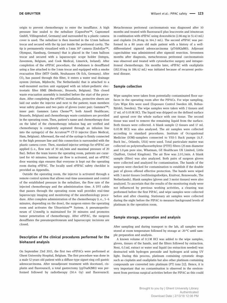
Platinum was not detected on the four floor positionsafter both PIPACs (Table 1). This was also the case forthe background testing before cleaning and before PIPAC1 indicating no contamination before the start of PIPAC 1.The limit of quantification was 0.02 ng/cm2. Platinumwas not detected in environmental air during bothPIPACs (Table 2). This was also the case for the back-ground testing after cleaning the day before indicating noplatinum in environmental air before the start of PIPAC 1.The limit of quantification depending on the air volumecollected was between 4.0 and 27 ng/m3.
Platinum was not detected on the hands, and theouter and inner pair of gloves of the surgeon (Table 3).The limit of quantification was 70 and 50 ng, respec-tively. Platinum was not detected on the filters and inthe liquid of the water locks (Table 4).
Discussion
The perioperative use of cytotoxic agents demands closeattention to the occupational health risks of the involvedstaff. Based on in vitro studies, animal experimentation,and epidemiological data, the International Agency ofResearch on Cancer (IARC) classifies some cytotoxic agentsin Group 1 (carcinogenic to humans; includes chlorambu-cil and cyclophosphamide), Group 2A (probably carcino-genic to humans; includes cisplatin and doxorubicin), and
Group 2B (possibly carcinogenic to humans; includes mito-mycin-C) [17]. There are no published epidemiological orexperimental data on the carcinogenicity of oxaliplatin,and it is not listed by the IARC. Nevertheless, given thesimilarity to cisplatin in structure and DNA interaction, asimilar degree of carcinogenicity is probable. The healthrisks of occupational exposure to cytotoxic drugs havebeen documented. A recent meta-analysis showed a 67%higher frequency of micronuclei in peripheral lymphocytes(a marker of genome toxicity) in exposed health care work-ers compared to controls [18].
The results from studies investigating the occupa-tional hazards for personnel involved in HIPEC proce-dures have been recently reviewed [19]. In summary,none of the included studies could detect platinum ormitomycin C in urine or plasma of health care workers,or in air samples. Villa and coworkers identified theoperating table, operating room floor, and surgeon’sovershoes as the most important sources of contamina-tion after open HIPEC with oxaliplatin [20].
Protection of the health care personnel and workingenvironment becomes even more critical when, duringPIPAC, chemotherapy is administered as an aerosol.Monitoring of surface contamination by wipe sampling,measuring glove and skin contamination is rather easyto perform and is a standard procedure in manyhospitals where cytostatic drugs are prepared and admi-nistered. Monitoring is a tool to evaluate routines,procedures and cleaning to prevent environmental con-tamination and potential exposure to hazardous drugsknown to cause adverse health effects [21].
The first safety analysis of the procedure wasreported by Solass and colleagues in 2013 [14]. Twopatients were treated with PIPAC using cisplatin(7.5mg/m2) and doxorubicin (1.5mg/m2); analysis of airsamples taken at the place of the surgeon as well as thatof the anesthesiologist was unable to detect cisplatin air
Table 1: Analysis of surfaces for presence of platinum (PT).
Description of the surfacea PT,ng/mL HClb
PT,ng/cm
PT,ng/mL HClb
PT,ng/cm
PT,ng/mL HClb
PT,ng/cm
PT,ng/mL HClb
PT,ng/cm
Background beforecleaning
Before PIPAC After PIPAC beforecleaning
After PIPAC beforecleaning
Floor surgeon ND <. ND <. ND <. ND <.Floor drug administration ND <. ND <. ND <. ND <.Floor drug suction equipment ND <. ND <. ND <. ND <.Floor anesthesiologist ND <. ND <. ND <. ND <.
aSurface area 4900 cm2. bTotal extraction volume 160 mL. ND, not detected ( <0.5 ng/mL HCl).
124 Willaert et al.: PIPAC safety
Brought to you by | Ghent University LibraryAuthenticated
Download Date | 2/13/18 12:06 PM
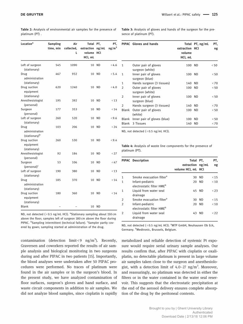
contamination (detection limit < 9 ng/m3). Recently,Graversen and coworkers reported the results of air sam-ple analysis and biological monitoring in two surgeonsduring and after PIPAC in two patients [15]. Importantly,the blood analyses were undertaken after 50 PIPAC pro-cedures were performed. No traces of platinum werefound in the air samples or in the surgeon’s blood. Inthe present study, we have analyzed contamination offloor surfaces, surgeon’s gloves and hand surface, andwaste circuit components in addition to air samples. Wedid not analyze blood samples, since cisplatin is rapidly
metabolized and reliable detection of systemic Pt expo-sure would require serial urinary sample analyses. Ourresults confirm that, after PIPAC with cisplatin or oxali-platin, no detectable platinum is present in large volumeair samples taken close to the surgeon and anesthesiolo-gist, with a detection limit of 4.0–27 ng/m3. Moreover,and reassuringly, no platinum was detected in either thefilters or in the water contained in the water seal reser-voir. This suggests that the electrostatic precipitation atthe end of the aerosol delivery ensures complete absorp-tion of the drug by the peritoneal contents.
Table 3: Analysis of gloves and hands of the surgeon for the pre-sence of platinum (PT).
PIPAC Gloves and hands Totalextraction
volumeHCl, mL
PT, ng/mLHCl
PT,ng
Outer pair of glovessurgeon (white)
ND <
Inner pair of glovessurgeon (blue)
ND <
Hands surgeon ( tissues) ND < Outer pair of gloves
surgeon (white) ND <
Inner pair of glovessurgeon (blue)
ND <
Hands surgeon ( tissues) ND <Blank Outer pair of gloves
(white) ND <
Blank Inner pair of gloves (blue) ND <Blank Tissues ND <
ND, not detected ( <0.5 ng/mL HCl).
Table 4: Analysis of waste line components for the presence ofplatinum (PT).
PIPAC Description Totalextraction
volume HCl, mL
PT,ng/mLHCl
PT,ng
Smoke evacuation filtera ND < Infant-pediatric
electrostatic filter HMEb ND <
Liquid from water sealdrainage
ND <
Smoke evacuation filtera ND < Infant-pediatric
electrostatic filter HMEb ND <
Liquid from water sealdrainage
ND <
ND, not detected ( <0.5 ng/mL HCl). aMTP GmbH, Neuhausen Ob Eck,Germany. bMedtronic, Brussels, Belgium.
Table 2: Analysis of environmental air samples for the presence ofplatinum (PT) .
Locationa Sampling
time, min
Air
collected,
L
Total
extraction
volume
HCl, mL
PT,
ng/mL
HCl
PT,
ng/m
Left of surgeon
(stationary)
ND < .
Drug
administration
(stationary)
ND < .
Drug suction
equipment
(stationary)
ND < .
Anesthesiologist
(personal)
ND <
Surgeon
(personal)
ND <
Left of surgeon
(stationary)
ND < .
Drug
administration
(stationary)b
ND <
Drug suction
equipment
(stationary)
ND < .
Anesthesiologist
(personal)
ND <
Surgeon
(personal)c ND <
Left of surgeon
(stationary)
ND <
Drug
administration
(stationary)
ND <
Drug suction
equipment
(stationary)
ND <
Blank – – ND
ND, not detected ( <0.5 ng/mL HCl). aStationary sampling about 150 cmabove the floor; samples left of surgeon 180 cm above the floor duringPIPAC. bSampling intermittent (technical failure). cSampler partly cov-ered by gown; sampling started at administration of the drug.
Willaert et al.: PIPAC safety 125
Brought to you by | Ghent University LibraryAuthenticated
Download Date | 2/13/18 12:06 PM
We were unable to identify platinum contamination ofeither the inner or the outer pair of surgeon’s gloves. Incontrast, during HIPEC with open abdomen perfusion andthe surgeon’s hand stirring the abdominal contents, glovecontamination is considerable. Regardless of the procedure,prevention of cutaneous exposure by wearing appropriategloves is essential. In Europe, there are no specific require-ments or test methodologies for medical gloves used forhandling cytotoxic agents. In contrast, in the US medicalgloves used for this purpose must fulfill the ASTMInternational (American Society of Testing and Materials)standard D 6978-05 requirements. Nitrile or natural rubberlatex are the preferred basic glovematerials. Importantly, allglove material displays a general trend towards greater per-meation over time (fivefold increase between 15 and 60min).[22] Therefore, a glove change is recommended every 15–20minutes, or more frequent with increasing temperature orcontinuous hand movement.
In conclusion, using the proposed technical setupand precautions, we were unable to detect any surface,air, or material contamination with platinum during orafter two clinical PIPAC procedures. These results con-firm that, with adequate preparation, a clinical PIPACprogram can be established without measurable che-motherapy exposure to health care workers. It is recom-mended that toxicological analyses are performed beforestarting a clinical PIPAC program in order to ensureadequacy of the protective measures that are put in place.
Author contributions: All the authors have acceptedresponsibility for the entire content of this submittedmanuscript and approved submission.Research funding: None declared.Employment or leadership: W. Ceelen is a senior clinicalresearcher from the Fund for Scientific Research –Flanders (FWO).Honorarium: None declared.Competing interests: The funding organization(s) playedno role in the study design; in the collection, analysis,and interpretation of data; in the writing of the report; orin the decision to submit the report for publication.
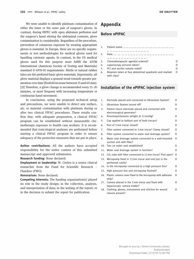
Appendix
Before ePIPAC
Installation of the ePIPAC injection system
. Patient name: … … … … … … … … … … … … … … … … … … …… … … … … … … … … … … … … … …
. Date: … . … . … … … … … … … … … … … … … … … … … … …… … … … … … … … … … … … … … .
. Chemotherapeutic agent(s) ordered? O. Laparoscopy pictures taken? O. PCI and ascites volume noted? O. Biopsies taken at four abdominal quadrants and marked
with clips?O
. Electrode placed and connected to Ultravision System? O
. Ultravision System turned off? O
. Patient return electrode placed and connected withelectrosurgical generator?
O
. Pneumoperitoneum airtight at mmHg? O
. Cap applied to balloon port of both trocars? O
. Port of mm trocar closed? O
. Filter system connected to mm trocar? Clamp closed? O
. Filter system connected to water seal drainage system? O
. Water seal drainage system connected to a wall-mountedsuction unit with filter?
O
. Two cm water seal established? O
. Water seal drainage system in function? O
. CO tube with filter connected to mm trocar? Port open? O
. Micropump fixed in mm trocar and end just in theperitoneal cavity?
O
. Is the micropump connected to a high pressure line? O
. High pressure line and micropump flushed? O
. Plastic camera cover fixed to the micropump with adhesivestrip?
O
. Camera placed in the mm trocar and fixed withlaparoscopic camera holder?
O
. Clothing, gloves, instruments and stitches for woundclosure present?
O
126 Willaert et al.: PIPAC safety
Brought to you by | Ghent University LibraryAuthenticated
Download Date | 2/13/18 12:06 PM
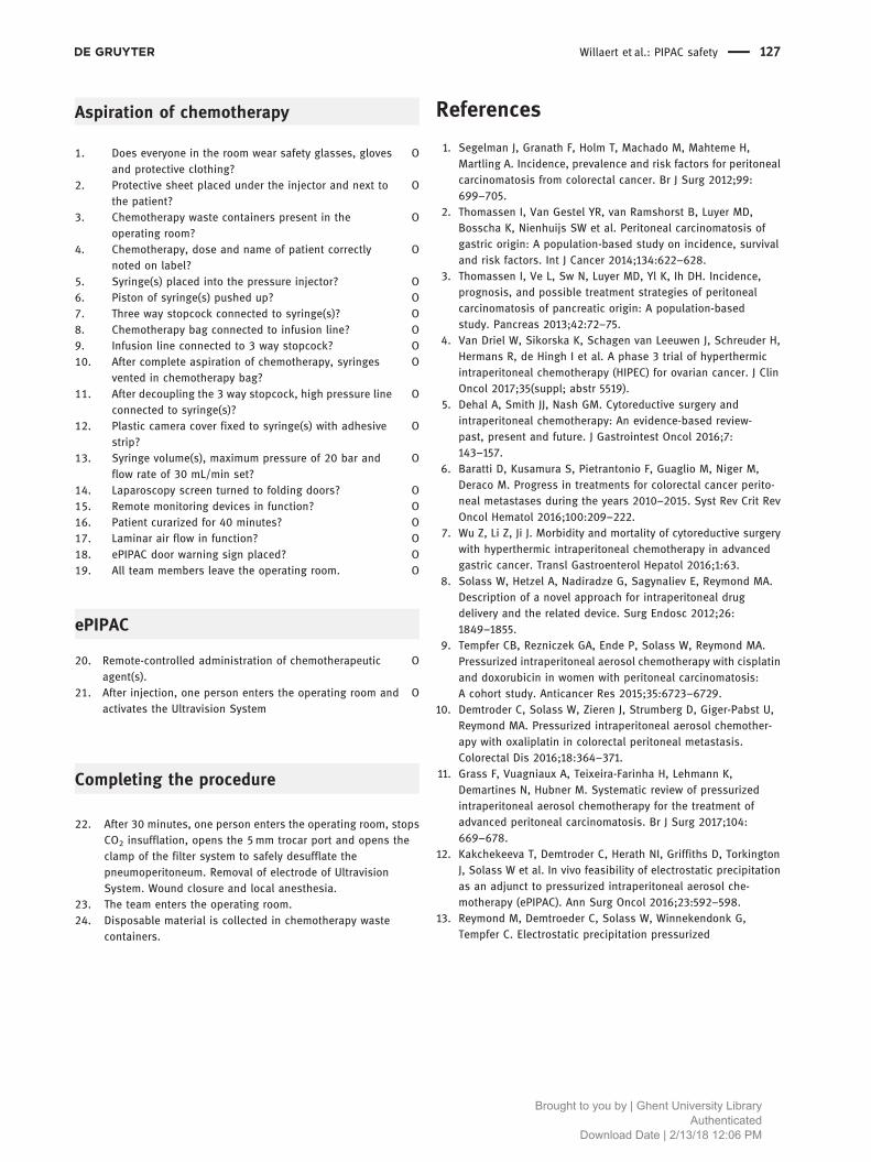
Aspiration of chemotherapy
ePIPAC
Completing the procedure
References
1. Segelman J, Granath F, Holm T, Machado M, Mahteme H,Martling A. Incidence, prevalence and risk factors for peritonealcarcinomatosis from colorectal cancer. Br J Surg 2012;99:699–705.
2. Thomassen I, Van Gestel YR, van Ramshorst B, Luyer MD,Bosscha K, Nienhuijs SW et al. Peritoneal carcinomatosis ofgastric origin: A population-based study on incidence, survivaland risk factors. Int J Cancer 2014;134:622–628.
3. Thomassen I, Ve L, Sw N, Luyer MD, Yl K, Ih DH. Incidence,prognosis, and possible treatment strategies of peritonealcarcinomatosis of pancreatic origin: A population-basedstudy. Pancreas 2013;42:72–75.
4. Van Driel W, Sikorska K, Schagen van Leeuwen J, Schreuder H,Hermans R, de Hingh I et al. A phase 3 trial of hyperthermicintraperitoneal chemotherapy (HIPEC) for ovarian cancer. J ClinOncol 2017;35(suppl; abstr 5519).
5. Dehal A, Smith JJ, Nash GM. Cytoreductive surgery andintraperitoneal chemotherapy: An evidence-based review-past, present and future. J Gastrointest Oncol 2016;7:143–157.
6. Baratti D, Kusamura S, Pietrantonio F, Guaglio M, Niger M,Deraco M. Progress in treatments for colorectal cancer perito-neal metastases during the years 2010–2015. Syst Rev Crit RevOncol Hematol 2016;100:209–222.
7. Wu Z, Li Z, Ji J. Morbidity and mortality of cytoreductive surgerywith hyperthermic intraperitoneal chemotherapy in advancedgastric cancer. Transl Gastroenterol Hepatol 2016;1:63.
8. Solass W, Hetzel A, Nadiradze G, Sagynaliev E, Reymond MA.Description of a novel approach for intraperitoneal drugdelivery and the related device. Surg Endosc 2012;26:1849–1855.
9. Tempfer CB, Rezniczek GA, Ende P, Solass W, Reymond MA.Pressurized intraperitoneal aerosol chemotherapy with cisplatinand doxorubicin in women with peritoneal carcinomatosis:A cohort study. Anticancer Res 2015;35:6723–6729.
10. Demtroder C, Solass W, Zieren J, Strumberg D, Giger-Pabst U,Reymond MA. Pressurized intraperitoneal aerosol chemother-apy with oxaliplatin in colorectal peritoneal metastasis.Colorectal Dis 2016;18:364–371.
11. Grass F, Vuagniaux A, Teixeira-Farinha H, Lehmann K,Demartines N, Hubner M. Systematic review of pressurizedintraperitoneal aerosol chemotherapy for the treatment ofadvanced peritoneal carcinomatosis. Br J Surg 2017;104:669–678.
12. Kakchekeeva T, Demtroder C, Herath NI, Griffiths D, TorkingtonJ, Solass W et al. In vivo feasibility of electrostatic precipitationas an adjunct to pressurized intraperitoneal aerosol che-motherapy (ePIPAC). Ann Surg Oncol 2016;23:592–598.
13. Reymond M, Demtroeder C, Solass W, Winnekendonk G,Tempfer C. Electrostatic precipitation pressurized
. After minutes, one person enters the operating room, stopsCO insufflation, opens the mm trocar port and opens theclamp of the filter system to safely desufflate thepneumoperitoneum. Removal of electrode of UltravisionSystem. Wound closure and local anesthesia.
. The team enters the operating room.. Disposable material is collected in chemotherapy waste
containers.
. Does everyone in the room wear safety glasses, glovesand protective clothing?
O
. Protective sheet placed under the injector and next tothe patient?
O
. Chemotherapy waste containers present in theoperating room?
O
. Chemotherapy, dose and name of patient correctlynoted on label?
O
. Syringe(s) placed into the pressure injector? O. Piston of syringe(s) pushed up? O. Three way stopcock connected to syringe(s)? O. Chemotherapy bag connected to infusion line? O. Infusion line connected to way stopcock? O. After complete aspiration of chemotherapy, syringes
vented in chemotherapy bag?O
. After decoupling the way stopcock, high pressure lineconnected to syringe(s)?
O
. Plastic camera cover fixed to syringe(s) with adhesivestrip?
O
. Syringe volume(s), maximum pressure of bar andflow rate of mL/min set?
O
. Laparoscopy screen turned to folding doors? O. Remote monitoring devices in function? O. Patient curarized for minutes? O. Laminar air flow in function? O. ePIPAC door warning sign placed? O. All team members leave the operating room. O
. Remote-controlled administration of chemotherapeuticagent(s).
O
. After injection, one person enters the operating room andactivates the Ultravision System
O
Willaert et al.: PIPAC safety 127
Brought to you by | Ghent University LibraryAuthenticated
Download Date | 2/13/18 12:06 PM

intraperitoneal aerosol chemotherapy (ePIPAC): First in-humanapplication. Pleura and Peritoneum 2016;1:109–116.
14. Solass W, Giger-Pabst U, Zieren J, Reymond MA. Pressurizedintraperitoneal aerosol chemotherapy (PIPAC): Occupationalhealth and safety aspects. Ann Surg Oncol 2013;20:3504–3511.
15. Graversen M, Pedersen PB, Mortensen MB. Environmentalsafety during the administration of pressurized intraperitonealaerosol chemotherapy (PIPAC). Pleura and Peritoneum2016;1:203–208.
16. Kyriazanos I, Kalles V, Stefanopoulos A, Spiliotis J, Mohamed F.Operating personnel safety during the administration ofhyperthermic intraperitoneal chemotherapy (HIPEC). Surg Oncol2016;25:308–314.
17. Grosse Y, Baan R, Straif K, Secretan B, El Ghissassi F, BouvardV, et al. A review of human carcinogens–Part A:Pharmaceuticals. Lancet Oncol 2009;10:13–14.
18. Villarini M, Gianfredi V, Levorato S, Vannini S, Salvatori T, MorettiM. Occupational exposure to cytostatic/antineoplastic drugs andcytogenetic damage measured using the lymphocyte cytokinesis-block micronucleus assay: A systematic review of the literatureand meta-analysis. Mutat Res 2016;770:35–45.
19. Villa AF, El Balkhi S, Aboura R, Sageot H, Hasni-Pichard H, PocardM, et al. Evaluation of oxaliplatin exposure of healthcare workersduring heated intraperitoneal perioperative chemotherapy(HIPEC). Ind Health 2015;53:28–37.
20. NIOSH list of antineoplastic and other hazardous drugs inhealthcare settings, 2016. https://www.cdc.gov/niosh/docs/2016-161/pdfs/2016-161.pdf [last accessed May 27,2017].
21. Landeck L, Gonzalez E, Koch OM. Handling chemotherapydrugs-Do medical gloves really protect? Int J Cancer2015;137:1800–1805.
128 Willaert et al.: PIPAC safety
Brought to you by | Ghent University LibraryAuthenticated
Download Date | 2/13/18 12:06 PM