wound healing: how we do it. - fort healthcare jc wound... · wound healing: how we do it. robert...
TRANSCRIPT

Wound Healing: How
we do it.
Robert Goldman MD, CWS, FAAPM&R
Medical Director
Fort Healthcare Wound and Edema Center


Background: RJ Goldman, MD
MD University of Texas, Galveston
Rehabilitation Medicine Residency (Albert
Einstein, Bronx, NY)
NIH Fellow (University of Pennsylvania)
Associate Professor, Rehabilitation Medicine
(U of PA)

Wound healing experience:
University of Pennsylvania Health System (1996-2005)
Rehabilitation Wound Clinic:
Developed Outpatient Wound Program
Treated all types of chronic wounds
Limb salvage
Electrotherapy (NIH Grant)


Electrotherapy Reverses
Inframalleolar Ischemia: A
Retrospective Observational
Study Robert Goldman1, MD; Barbara Brewley1, RN-C;
Michael Golden2, MD 1Departments of Rehabilitation Medicine,
and 2Surgery
University of Pennsylvania

Home based electrotherapy
improves healing of
ischemic wounds: A phase
I prospective study.
Robert Goldman1, MD; Barbara Brewley1, RN-C;
Linque Zhou1, MD
Michael Golden2, MD 1Departments of Rehabilitation Medicine,
and 2Surgery
University of Pennsylvania

Wound care experience (continued)
Visiting Faculty, University
of Texas, Houston (2006)
Wound care and Hyperbaric
Oxygen Center
Memorial Hermann Hospital
Caroline Fife, MD, Director
Supervised 1000 HBO
treatments, wound care.
Board certified, Undersea
and Hyperbaric Medicine,
2010

Wound Care Update:
Objectives
List wound types: Similarities and differences
Review wound healing principles
Understand wound-specific standards of care.

Mary Carvalho, RN, BSN, MBA
Clinical Coordinator, Fort HC Wound Edema
Center.
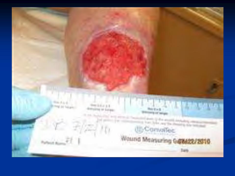
“Wound and skin assessments”
Assess size and appearance of wounds
“Interactive Wound Care Demonstrations”

Length
Width
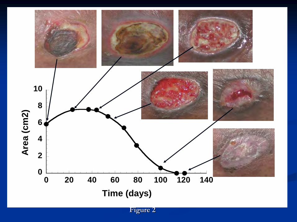
0
2
4
6
8
10
0 20 40 60 80 100 120 140
Are
a (
cm
2)
Time (days)
Figure 2

Reduced bio-burden leads to healing
Wound bed preparation
Invasive infection
Critical colonization (>100,000 CFU
per gram tissue)
Colonization
Debridement
Rapid closure

Moist Wound Healing
“not too wet, not too dry”
Wound bed preparation
Highly absorbent dressings
Silver dressings, cadexomer Iodine
Antibiotics
Foam, Hydrogels
“Frequent” dressing changes.
WET DRY
Sibbald, R., et al., Preparing the wound bed -- Debridement, Bacterial
Balance and Moisture Balance. Ostomy Wound Management., 2000. 46(11): p. 14-35

Moist wound healing
Healing phase
Less absorbant dressings, gels, much less antibiotics
used
Infrequent dressing changes, e.g., weekly
Lisa Reil, RN, speaker today on “Dressing types
and when to use them”

Why check arterial flow for wound
care?
Venous stasis wounds: Establish safety of
compression
Arterial wounds: Localize segment of arterial
disease
All wounds: Establish prognosis for healing
Goldman, R. et al. More than One Way to Measure a Wound: An
Overview of Tools and Techniques. Advances in Skin & Wound Care, 2002. 15(5): p. 236-243.

Tissue Hypoxia
Leads to the following alterations in normal
healing:
inflammation
wound repair
new blood vessels.

Vascular testing
Bedside Ankle brachial index (ABI)
Normal 0.8-1.3
Skin perfusion pressure (normal >35 mmHg)
Transcutaneous Oximetry (TCOM).
Segmental arterial studies (lab test).
Angiogram (referral).
Vikramjit Chhokar, MD (speaker today)

Definition: TCOM
Evaluates “dose” of O2 at tissue-at-risk.

Transcutaneous Oximetry (TcPO2)
adds to vascular workup
Room air = 21% (760 mm Hg) =157 mm O2
TcPO2>40 mm Hg normal: GOOD PX
TcPO2<20 mm Hg ischemic: BAD PX
Confirmed by 100% O2 challenge
Padberg, F., et al., Transcutaneous oxygen (TcPO2) estimates probability of
healing in the ischemic extremity. Journal of Surgical Research, 1996. 60: p. 365-369.

Understand wounds:
Types of Chronic Wounds
Post-surgical
Pressure
Venous
Neuropathic
Ischemic (Arterial) Diabetic Foot Ulcer

Post Surgical Wounds
Types
Dehiscence
I&D (e.g., abscess)
Post surgical debridement.
Treatment
Get to granulation.
Topical (bulky, moist dressings)
Negative pressure therapy.


Negative Pressure Therapy
Indications:
Early healing phase (80% red; minimal yellow)
Measurable depth
Benefits.
Rapid granulation and depth reduction of post surgical and pressure ulcers.
Reduced dressings costs, nursing visits
Joseph, E., et al., A prospective randomized trial of vacuum-assisted closure versus
standard therapy of chronic nonhealing wounds. Wounds, 2000. 12(3): p. 60-67.

60 year old with perforated
sigmoid diverticulum
Morbid Obesity, HTN
7/7/09: Left hemicolectomy with colostomy
7/31: Presented to Fort Healthcare Wound
Edema Center
8/6: Wound saucerized and debrided.

Post op week six: 14cm x 6.5cm x 9
cm: VAC therapy at Black Granufoam
@ 125 cont.

Post op week 8

Post op week 15: 99% closed.

Aug Oct

Negative Pressure Therapy
Contraindications (FDA warning):
Untreated osteomyelitis.
Untreated deep soft tissue infection.
Exposed artery
Neoplasm.
There have been 7 deaths and 50
hospitalizations using NPT in 2009.
With these caveats NPT is “de facto”
standard of care

Pressure Ulcers

Pressure ulcer risk: Braden scale
Moisture (Incontinence)
Activity
Mobility
Friction and shear (contractures)
Nutrition
Impaired sensation (lethargy)
Bergstrom, N., et al., The Braden Scale for Predicting Pressure Sore Risk. Nurs Res, 1987. 36: p. 205-210.

II
IV
III
IV

What stage????

“Deep Soft Tissue Injury Under
Intact Skin”
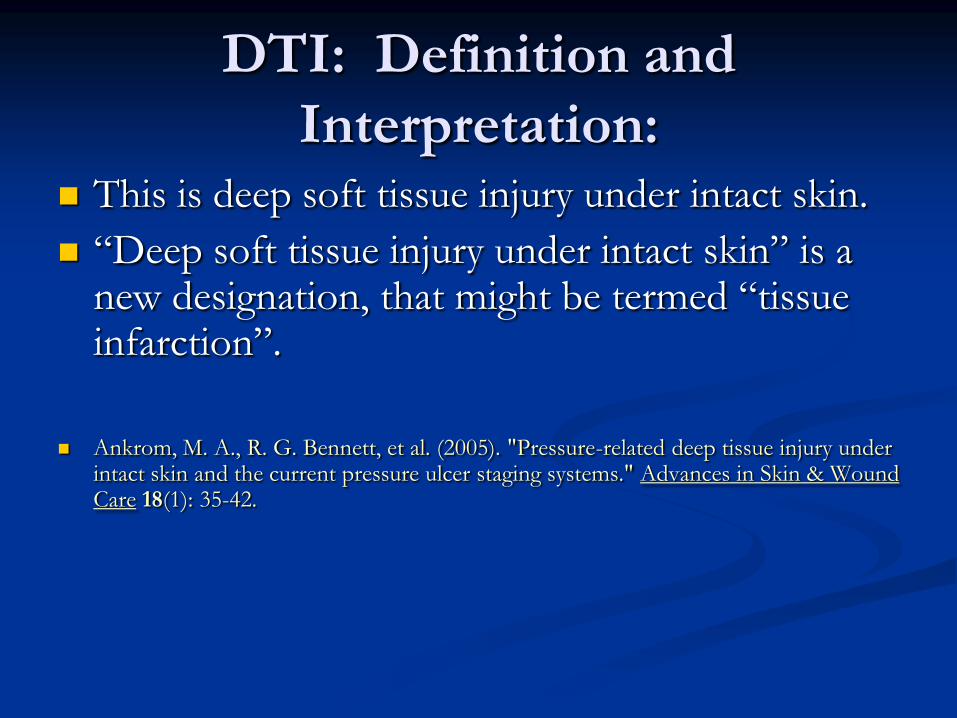
DTI: Definition and
Interpretation: This is deep soft tissue injury under intact skin.
“Deep soft tissue injury under intact skin” is a new designation, that might be termed “tissue infarction”.
Ankrom, M. A., R. G. Bennett, et al. (2005). "Pressure-related deep tissue injury under intact skin and the current pressure ulcer staging systems." Advances in Skin & Wound Care 18(1): 35-42.

Evolution of DTI:
Evolves according to forensic principles, consistent with post-mortem decomposition of the human body.
2 days: Livido (redness which could be confused with a stage I pressure ulcer).
1 week: Purple
2 weeks: Black or yellow eschar.
4 weeks: Necrosis
Farid, K. J. (2007). "Applying observations from forensic science to understanding the
development of pressure ulcers." Ostomy Wound Management 53(4): 26-8.

Evolution of DTI (continued)
Once DTI becomes visible, it is already too late!
Already “unstagable” which may evolve into
stage IV.
“Aggressive prevention”.

84 year old female with DTI
Fell at home right femur fracture.
Found on floor 2/6.
ORIF at Madison Hopsital
Evaluated at Johnson Creek 2/26: Copious foul
smelling drainage.


DTI c/b Osteomyelitis.
3/15: MRI: Positive for “superficial
osteomyelitis”.
Etrapenem IV via PICC for six weeks.
On NPT since D/C from Madison Hospital.
NPT 125 mm Hg “black foam”.


Lower Extremity
Edema

LE Edema: Lymphedema
Stage I: Pitting.
Stage II: Non-pitting, fibrosis.
Stage III: Elephantiasis.


Other end of the spectrum: Venous
Stasis Disease
Venous stasis disease.
Disease of veins.
Venule damage, inflammation, hypoxia,
sequestration of growth factors.
Treat edema = heal wound.
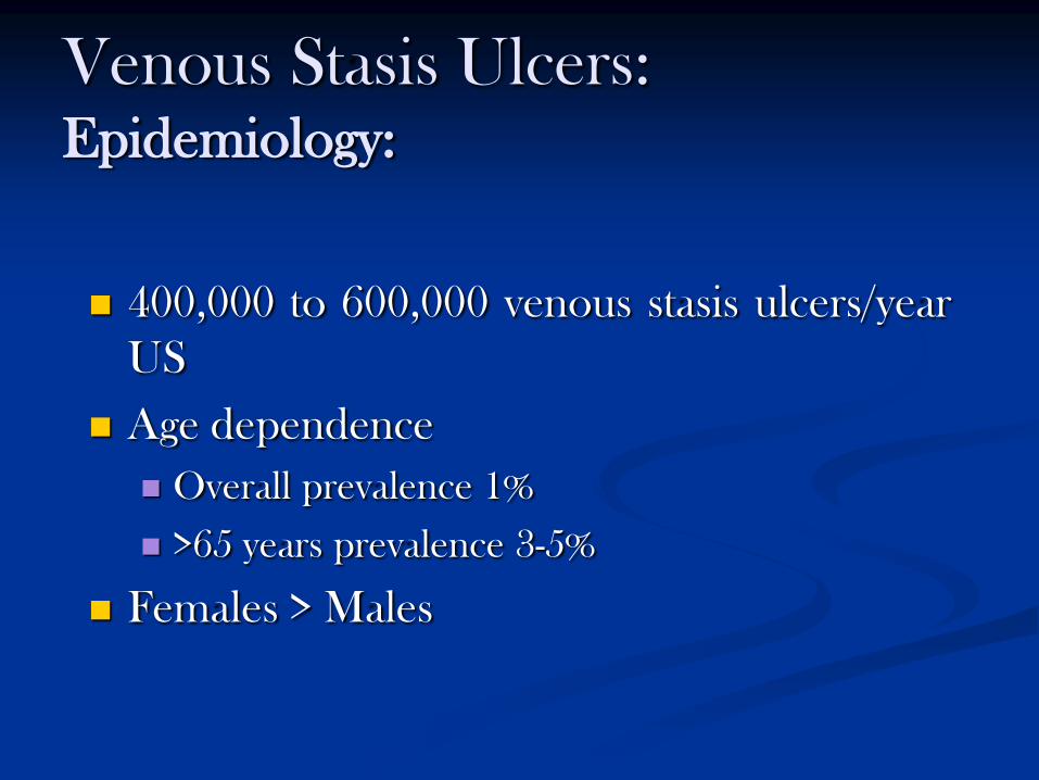
Venous Stasis Ulcers: Epidemiology:
400,000 to 600,000 venous stasis ulcers/year
US
Age dependence
Overall prevalence 1%
>65 years prevalence 3-5%
Females > Males

Presentation
Between foot and knee
Medial malleolus
Lateral malleolus
Hyperpigmentation
Red base
Irregular borders
Lipodermosclerosis
Pitting edema

Venous Ulcers: Pathophysiology
Venous stasis
Venous return
Incompetence of valves
Iatrogenic
Chronic deep venous thrombosis
Vein removal

Venous Ulcers: Treatment
Standard of care: Compression
Debridement
Drainage management
Vascular workup
Pain management
Education
Supply logistics
McGuckin M., Waterman R., Brooks J., et al.: Validation of venous leg ulcer guidelines in the United States and United Kingdom. American Journal of Surgery. 2002; 183: 132-7.

What can a leg ulcer be?
Venous stasis (85%)
Arterial disease
Edema (SCI, CVA)
Lymphedema
Sickle Cell Anemia
Neoplastic:
Basal cell CA
Cutaneous Lymphoma
Infectious:
Cutaneous TB (olden days)
Cutaneous Anthrax
Autoimmune:
Vasculitis,
Pyoderma gangrenosum
Phillips T, Dover J. Leg Ulcers. Journal of the American Academy of Dermatology 1991;25:965-985.

A
B

Methods to apply leg compression
All TOE TO KNEE
More aggressive compression necessary for
healing (>40 mm Hg)
Long stretch (continuous pressure)
Short stretch, no stretch (transient pressure).
Less aggressive to maintain healing (20-30 mm
Hg)
Medical grade stockings (e.g., Jobst)

Three layer elastic compression
Wound dressing
Under layer gauze toe to knee
Elastic (short stretch, long stretch, both)
compression toe to knee

Elastic Compression – 3 -layer


Multi-layer compression wrap

Multi-layer compression “Profore”

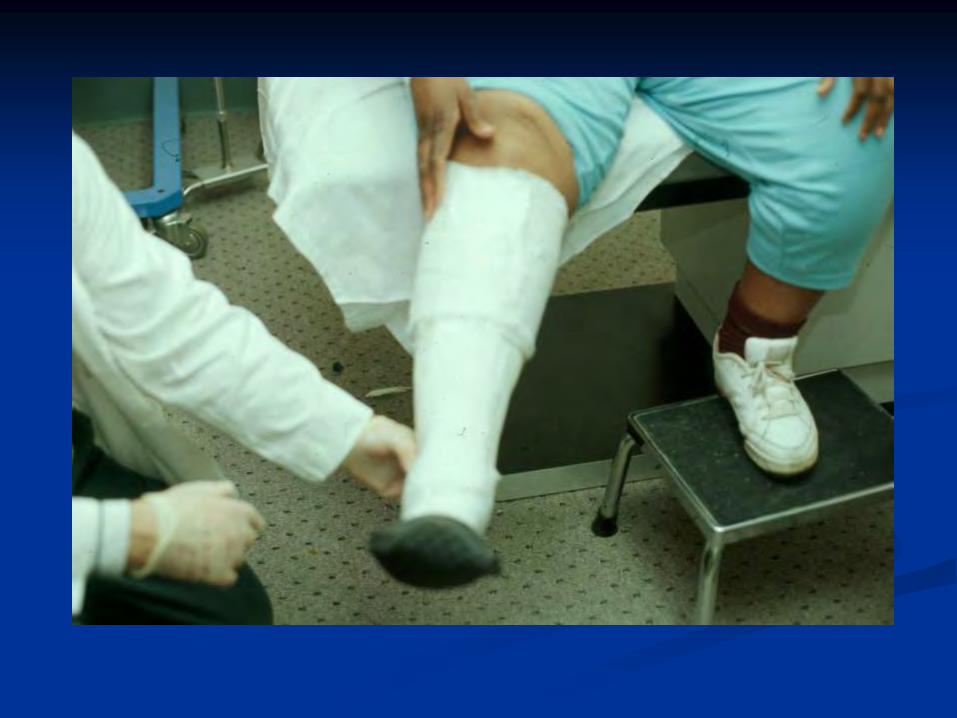
Unna “boot”
How applied (figure
of 8; strips)
Watch excessive
drainage!!


Long duration venous stasis
wounds: Patient DB DB, 66 year old female
Varicose veins, otherwise healthy (no diabetes)
1 year history of leg ulcers, left one year.
Works on feet many years.
Good arterial flow (ABI 1.06 bilaterally)
Frequency of dressing changes decreased, compression increased, drainage gradually decreased:
Returned to work after few weeks.
Local infection noted five times; Predominantly Staph Aureus.


DB: Healed!

DB: Wound healing course

Prevention; long term maintenance
20-30 mm Hg compression stockings.

Neuropathic Ulcer

Neuropathic ulcers
Polyneuropathy (usually from diabetes)
Etiology
Diabetes
EtOH
Congenital (e.g., HSMN type I)
Insensitivity to the 5.07 monofilament
Foot deformities
Develop ulcers at weight bearing bony prominences
Brent Yaeggi DPM (speaker today).

Figure 7:
A B
C D

Neuropathic ulcers
Presentation
Workup- MRI, TcPO2, PVR
Treatment
Debridement
Drainage management
Vascular workup
Standard of care: Off loading

Myth: All Off-loading is created
equal
Wound healing at 12 weeks:
Half shoe 58.3%
Removable “walker” 65%
Total contact cast 89.5% (p<.05)
Armstrong D. G., Nguyen H. C., Lavery L. A., et al.: Off-loading the diabetic foot wound: a randomized clinical trial. Diabetes Care. 2001; 24: 1019-22.

“Diabetic Healer Walker” AKA
DH Walker

Patient: JA
90 year old lady
Ulcer bottom of foot >4 years duration.
Diabetes, Stroke
Numb foot: “walks on wound”
Foot deformity due to diabetes
Serious infection 1/2009 septic shock.
On hospice because of wound.

Initial presentation: Celulitis


Prevention; long term maintenance
of healing: Orthopedic oxford shoes

Arterial Ulcers

Arterial or Ischemic ulcers
Cardiac risk factors
HTN
DM
Chloresterol
Family Hx
Smoking
Bony prominences, lateral foot margins

Figure 8:
C B A

Ischemic ulcers
Presentation – wounds expand and deepen!
Workup- MRI, TcPO2, PVR, MRA, Arteriogram
Treatment
Standard of care: Revascularization Off loading (similar to neuropathic)
Drainage management
Debridement – be careful!
Adjunctive therapies
Hyperbaric Oxygen Grolman R. E., Wilkerson D. K., Taylor J., et al.: Transcutaneous oxygen
measurements predict a beneficial response to hyperbaric oxygen therapy in patients with nonhealing wounds and critical limb ischemia. American Surgeon 2001; 67: 1072-9.
Electrotherapy Goldman R, Brewley B, Zhou L, Golden M. Electrotherapy reverses
inframalleolar ischemia: A retrospective, observational study. Advances in Skin & Wound Care 2003;16(3):79-89.
Timed compression boot (Dillon boot).

Healing of Arterial Ulcers (yellow
bars)
0
100
200
300
400
500
600
700
800
-4 0 4 8 12 16 20 24 28 32 36 40 44 48 52
HVPC+Std-of-care
Standard-of-care
Wo
un
d A
rea (
%)
Time (Weeks)
(no
rmalized
to
100%
at
week 0
)
* * * * * * *
*
*

Micro-circulation Improved in
HVPC Group
0
10
20
30
40
50
HVPC+std-of-care Standard-of-care
TcP
O2 (
mm
Hg
)
Condition
*
(ma
xim
um
)


Complex Diabetic Ulcer


Hyperbaric Oxygen
Monoplace Chamber


Odds ratio: Amputation reduction DFU
Favors HBOT

Improved healing with HBOT:
Randomized controlled trial

Case Study: RW
66 year old male with Rheumatoid Arthritis
No diabetes
PAD right leg and foot
OM of the big toe by plain film
Hallux amp 9/21/10.
Large plantar flap down to the proximal phalanx of
the toe.

Patient RW
Seen by a general surgeon in past six weeks (Sept
2010): “no reconstructable arterial disease”.
No diabetes.
ABI right = 0.94
TCOM: forefoot 2, 23 and 29 mm Hg, increased by
20-40 mm Hg with 100% O2 by FM.

RW: Photos

RW Healing Rate
HBOT

Conclusion: Chronic wound care
in five easy steps:
Ulcer Diagnosis Standard of care
Post-surgical Negative pressure
Pressure Negative pressure, nutrition,
offloading
Venous Compression
Neuropathic Off-loading
Arterial Revascularization

Pressure ulcers: Standard of Care
Measurement
Off-loading Support surface
Turn and Position
Optimal nutrition
Treatment Debridement
Moisture balance.
Surgical referral (Stage III/IV)
Bergstrom, N., M. Bennett, and C. Carlson, Treatment of
Pressure Ulcers. Treatment of Pressure Ulcers. Clinical Practice Guideline Number 14. AHCPR Publication No. 95-0642. 1994, Rockville, MD:

Overall approach (continued)
Venous stasis ulcers:
Compression
TOE TO KNEE DRESSINGS
Staged debridements
Moist wound environment

Overall approach (continued)
Neuropathic
Wound bed preparation
Off loading
Sharp debridement
Total contact casting

Overall approach
Arterial/Ischemic:
Standard of care: Revascularization
Non-invasive arterial testing.
Also use compression or off-loading (standard of care for
neuropathic or venous ulcers).
Hyperbaric Oxygen (especially for diabetic foot ulcers).


THANK YOU FOR YOUR
ATTENTION!