work report - iisc · work report understanding cellular variability using droplet microfluidics...
TRANSCRIPT

Work Report
Understanding cellular variability using droplet microfluidics
Sidhant Swarup Rout
Department of Biotechnology and Medical
Engineering
NIT Rourkela
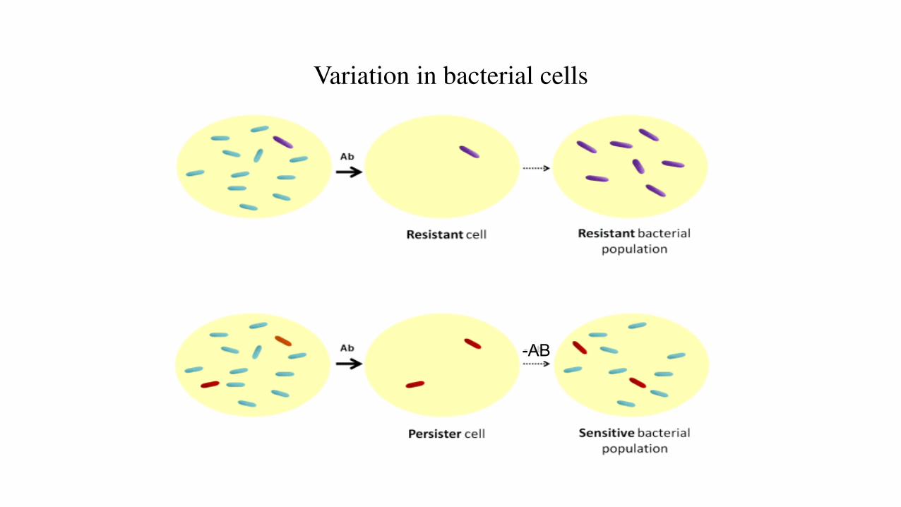
Variation in bacterial cells
-AB
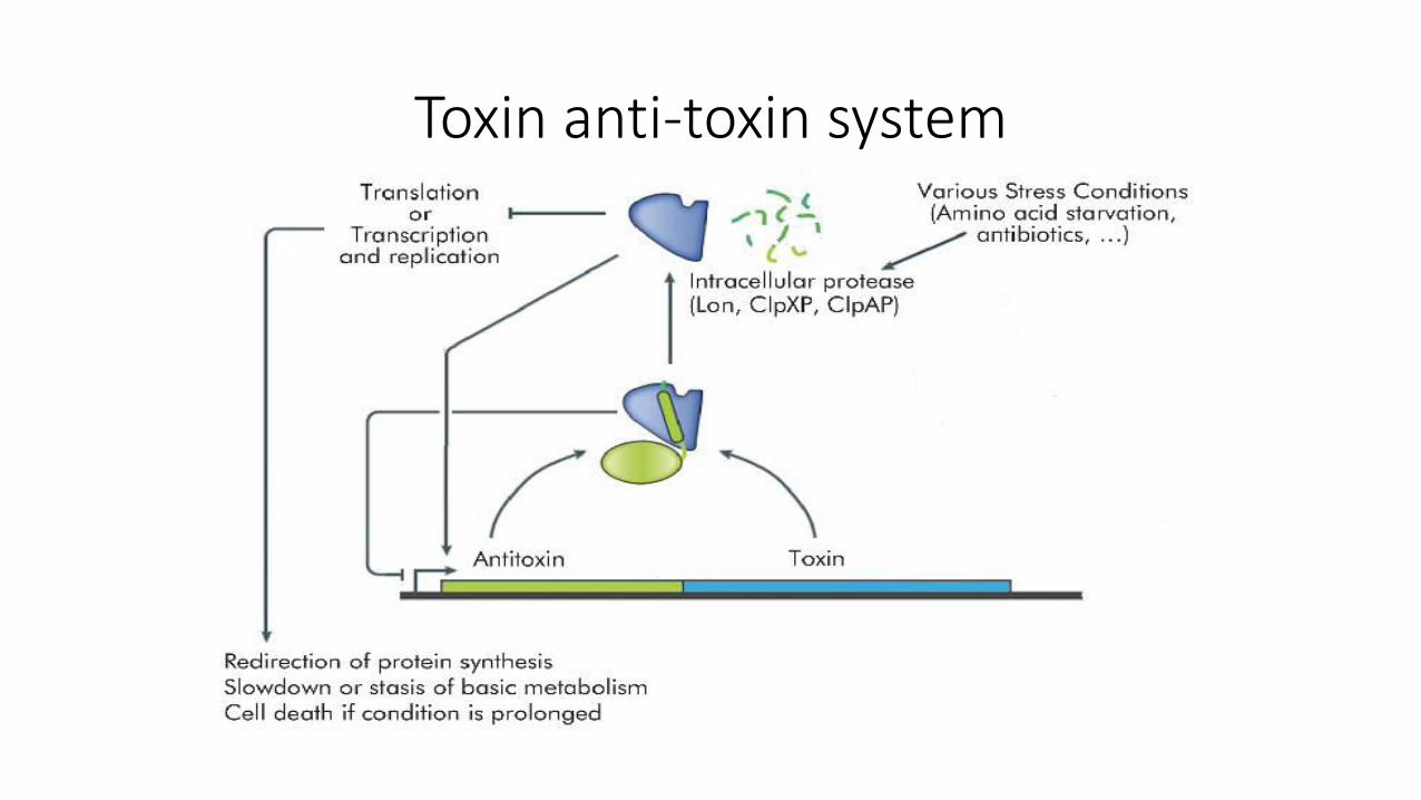
Toxin anti-toxin system
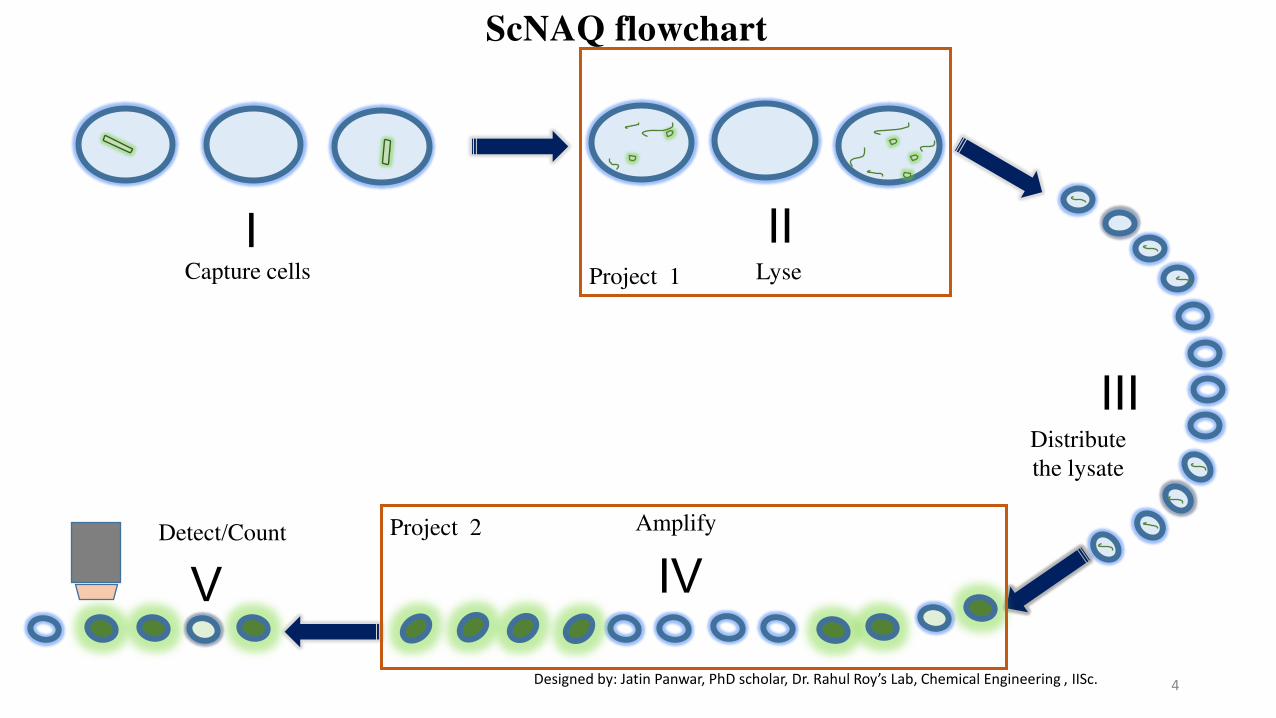
Capture cells Lyse
II I
Distribute
the lysate
III
Detect/Count
V
Amplify
IV
4
ScNAQ flowchart
Designed by: Jatin Panwar, PhD s holar, Dr. Rahul Roy’s La , Che i al E gi eeri g , IISc.
Project 1
Project 2
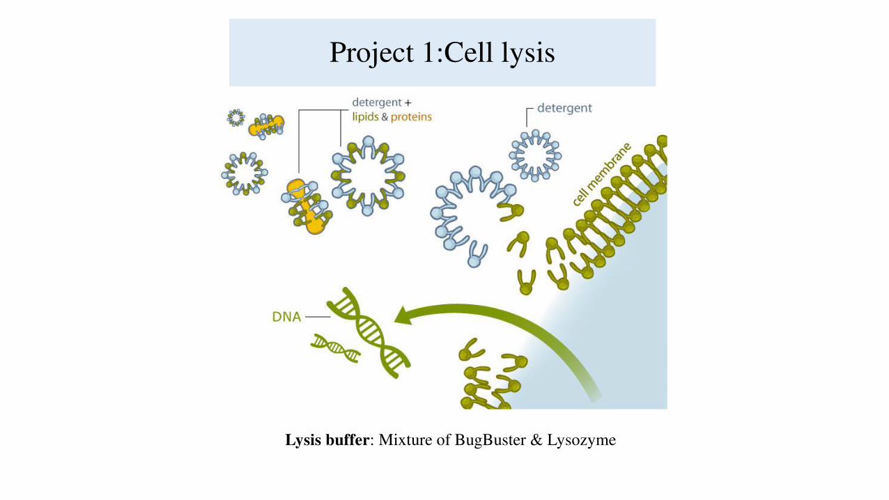
Project 1:Cell lysis
Lysis buffer: Mixture of BugBuster & Lysozyme
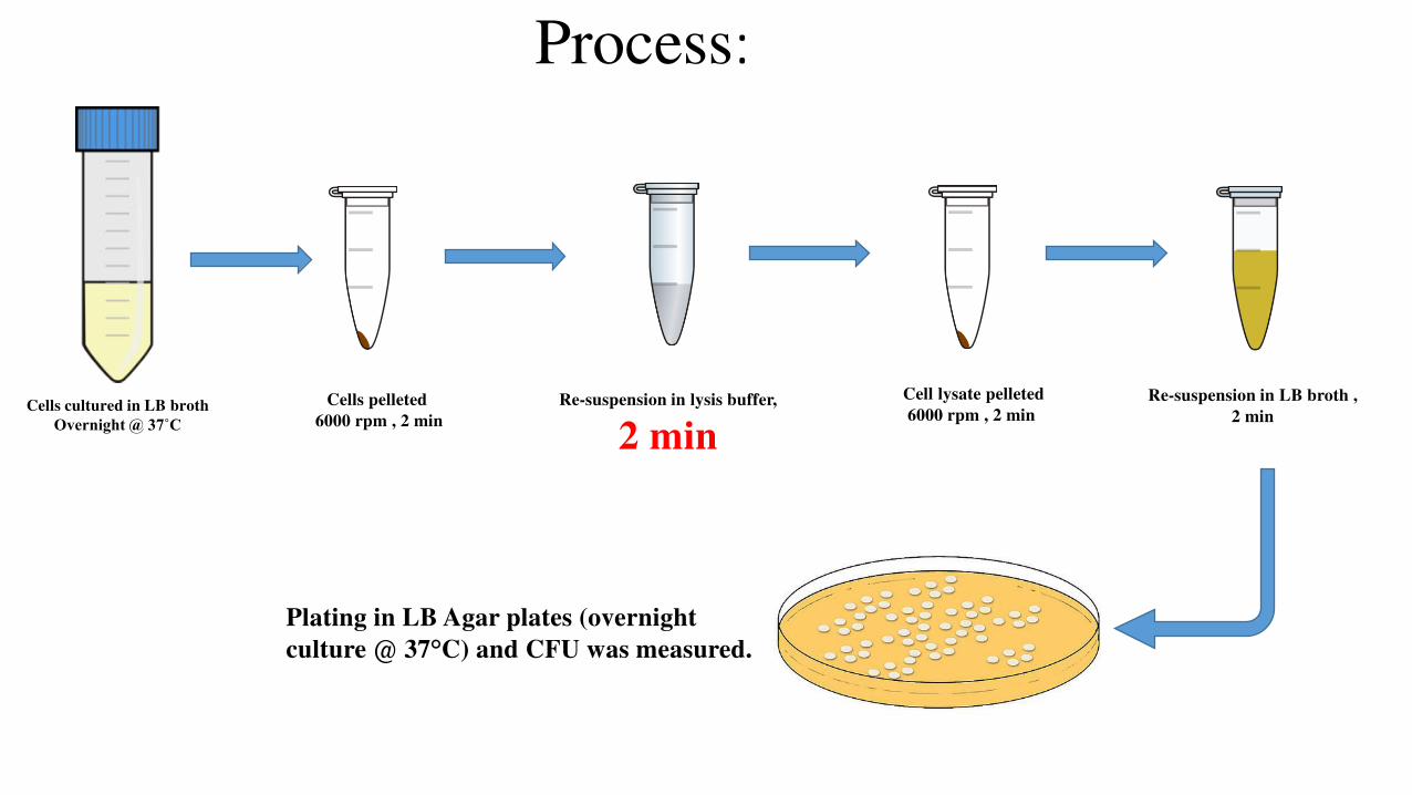
Process:
Cells cultured in LB broth
Overnight @ 37˚C
Cells pelleted
6000 rpm , 2 min
Cell lysate pelleted
6000 rpm , 2 min
Re-suspension in lysis buffer,
2 min
Re-suspension in LB broth ,
2 min
Plating in LB Agar plates (overnight
culture @ 37°C) and CFU was measured.

Optimization of Lysis buffer Concentration
3 16 5 17
500
0
100
200
300
400
500
600
0.6x B +
20KU
lysozyme
0.6x B +
40KU
lysozyme
0.6x B +
60KU
lysozyme
0.6x B +
80KU
lysozyme
0.6x B +
100KU
lysozyme
1
45
10
38
110 120
0
20
40
60
80
100
120
140
0.6x BB +
40 KU
Lysozyme
0.6x BB +
60 KU
Lysozyme
0.4x BB +
40 KU
Lysozyme
0.4x BB +
60 KU
Lysozyme
0.2x BB +
40 KU
Lysozyme
0.2x BB +
60 KU
Lysozyme
No. of colonies
Varying lysozyme concentration, Keeping
BugBuster concentration as 0.6x
Varying BugBuster concentration, Keeping
lysozyme concentration as 40 KU and 60 KU
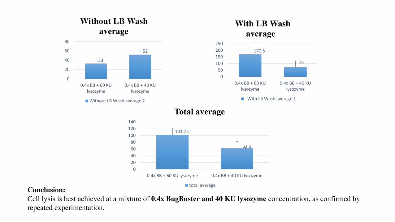
170,5
73
0
50
100
150
200
250
0.4x BB + 60 KU
lysozyme
0.4x BB + 40 KU
lysozyme
With LB Wash
average
With LB Wash average 1
33
52
0
20
40
60
80
0.4x BB + 60 KU
lysozyme
0.4x BB + 40 KU
lysozyme
Without LB Wash
average
Without LB Wash average 2
101,75
62,5
0
20
40
60
80
100
120
140
0.4x BB + 60 KU lysozyme 0.4x BB + 40 KU lysozyme
Total average
total average
Conclusion:
Cell lysis is best achieved at a mixture of 0.4x BugBuster and 40 KU lysozyme concentration, as confirmed by
repeated experimentation.
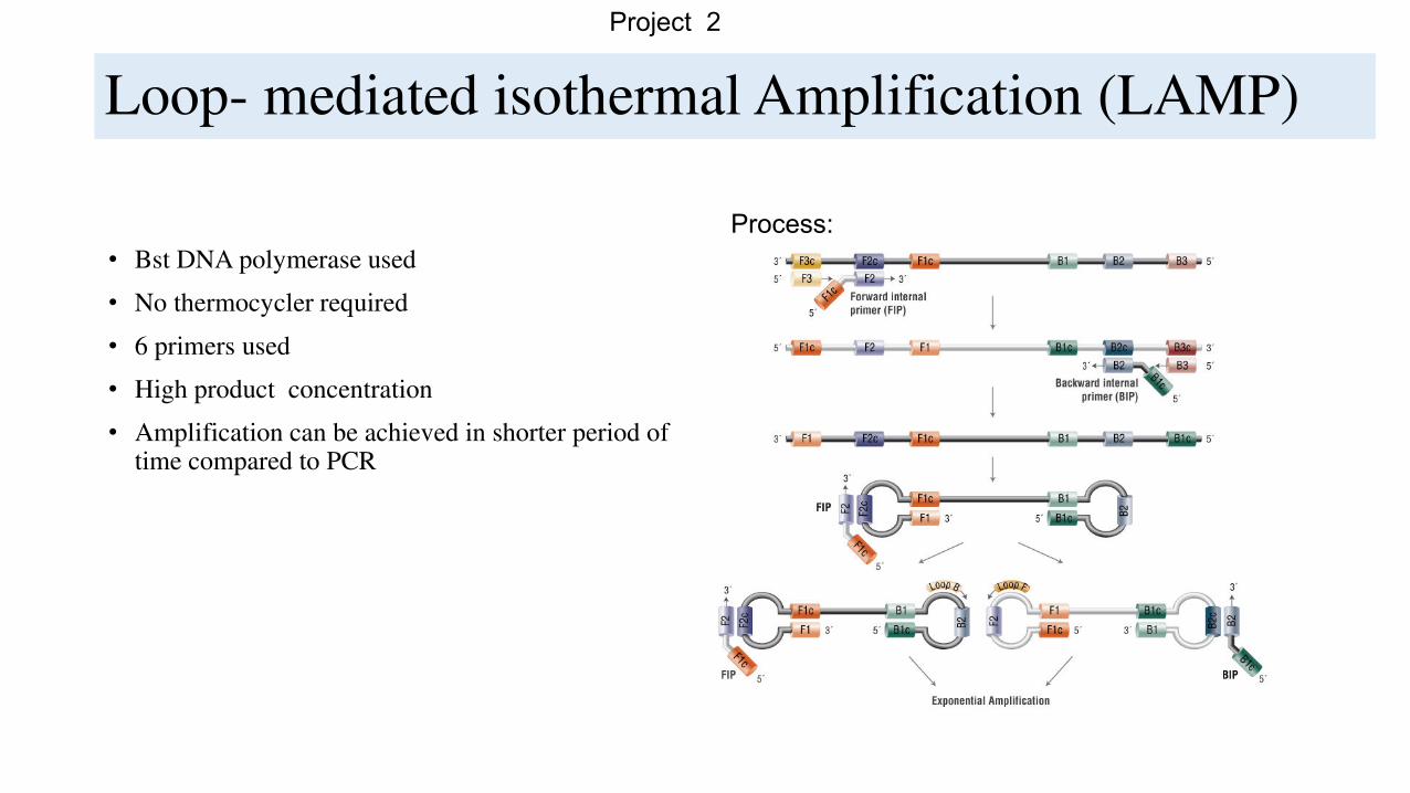
Loop- mediated isothermal Amplification (LAMP)
• Bst DNA polymerase used
• No thermocycler required
• 6 primers used
• High product concentration
• Amplification can be achieved in shorter period of time compared to PCR
Process:
Project 2

LAMP
Ct values average obtained after optimization
of Bst DNA polymerase concentration by
evaluating LAMP reaction at to 1x, 2x and 5x
concentration..
0
10
20
30
40
50
60
JEV 10^4 copies JEV 10^3 copies No template DEN II 10^4 copies
Bst DNA polymerase Optimization
Bst Pol 1X Ct Avg Bst Pol 2X Ct Avg Bst Pol 5X Ct Avg

Gel Image:
Gel image showing negative control (no template)
amplification in case of no heat denaturation (left) and
also negative control (no template and Dengue II)
amplification in case of additional heat denaturation
(right).
Gel image showing duplicates of a LAMP reaction ,
decreasing the JEV DNA copy number from 10^5 to 1
Project 3: Nucleic acid quantification using magnetic beads

A)
B)
C)
Streptavidin magnetic beads
Biotinylated capture probe
Target DNA
Concept:
Annealing
Probe attachment

Streptavidin magnetic beads
• Magnetic beads captured inside the droplets (100 µm) (60X magnification)
Control: 1X kapa HiFi
buffer
• Magnetic beads captured inside the droplets (100 µm) (60X magnification)
Magnetic beads(1 µm)
7.3 X 10^2 beads/ µL

Microscopic detection using channels
50bp
DNA
ladder
1Kb DNA
ladder
Sample
1
Sample
2 control Gel image:
Sample 1: probe attached Template (10^6 copies) + magnetic beads (10^8 beads) – 1000 fold
dilution
Sample 2: probe attached Template (10^6 copies) + magnetic beads (10^8 beads) – 10000 fold
dilution
Control : Droplets without annealing step with biotinylated probe.
Merged droplets
*The droplets were imaged using
488 nm (blue) illumination
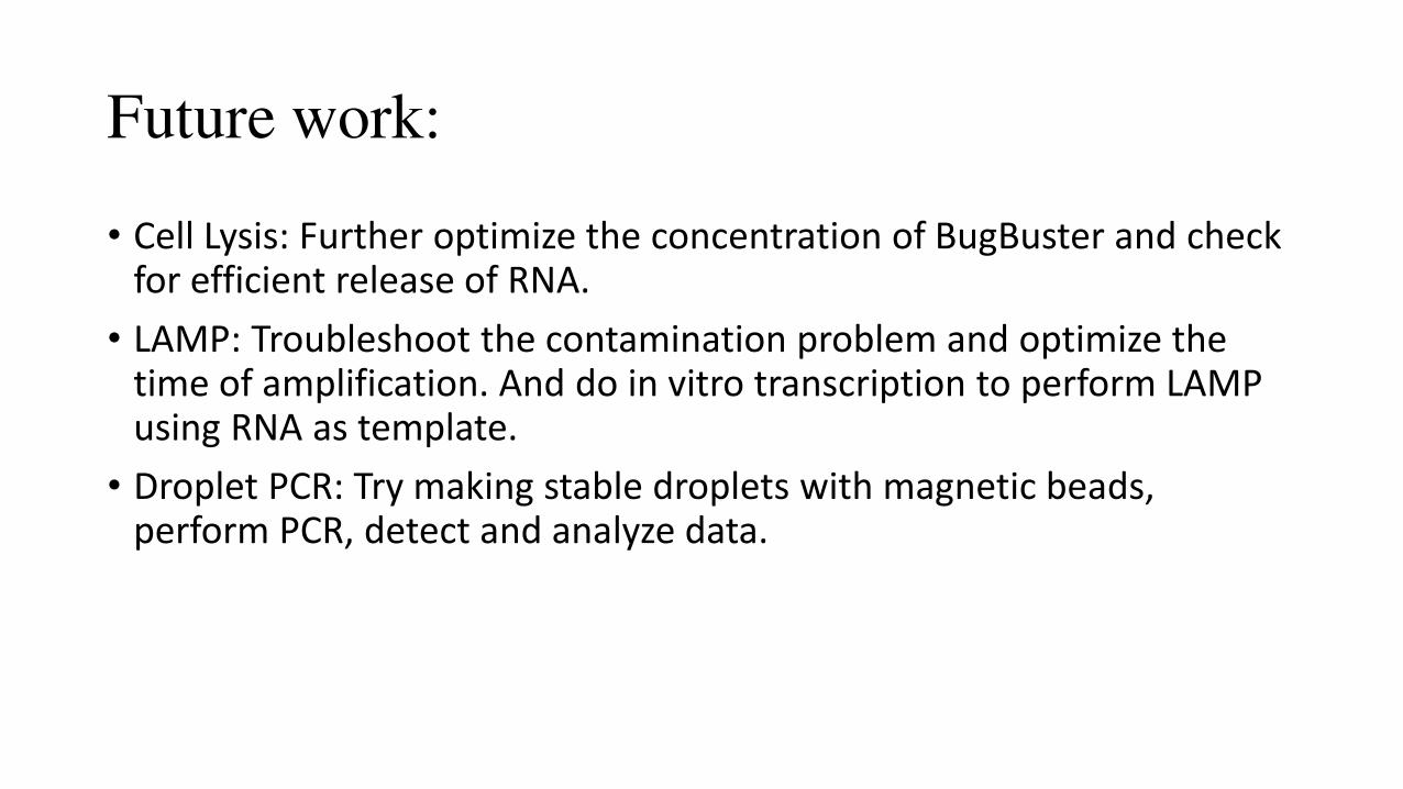
Future work:
• Cell Lysis: Further optimize the concentration of BugBuster and check
for efficient release of RNA.
• LAMP: Troubleshoot the contamination problem and optimize the
time of amplification. And do in vitro transcription to perform LAMP
using RNA as template.
• Droplet PCR: Try making stable droplets with magnetic beads,
perform PCR, detect and analyze data.
ACKNOWLEDGMENT
I want to thank centre of BioSystem Science and Engineering for giving this opportunity to pursue my
research interest through BioEngineering Summer Training (BEST) program.
I want to express my gratitude to Dr. Rahul Roy for his able guidance and valuable suggestions. I want
to thank Ms. Sunanda and Prof. Siddharth and Prof. Ananthasuresh for there able guidance and
support throughout the program.
I also want to thank all the research scholars in Dr. Rahul’s lab especially Jatin, Monisha, Saranya and
Priyanka for their assistance in designing and analyzing the experimental data.

Thank you!