work-related carpal tunnel syndrome diagnosis …related carpal tunnel syndrome. median nerve...
TRANSCRIPT
Effective April 1, 2009; reviewed Jan. 23, 2014; Hyperlink and formatting update 2016
Work-Related Carpal Tunnel Syndrome
Diagnosis and Treatment Guideline
Table of Contents
I. Guideline Summary
II. Introduction
III. Establishing Work-Relatedness
IV. Making the Diagnosis
A. Symptoms and Signs
B. Electrodiagnostic Testing
i. Nerve Conduction Velocity
ii. Needle Electromyography
iii. Quantitative Sensory Testing
C. Other Diagnostic Tests
V. Treatment for Carpal Tunnel Syndrome
A. Conservative Treatment
B. Surgical Carpal Tunnel Release
VI. Return to Work (RTW)
A. Early Assessment
B. Returning to Work following Surgery
VII. Hand Diagram
VIII. Electrodiagnostic Worksheet
Updated January 2014; Hyperlink Update 2016 Page 2
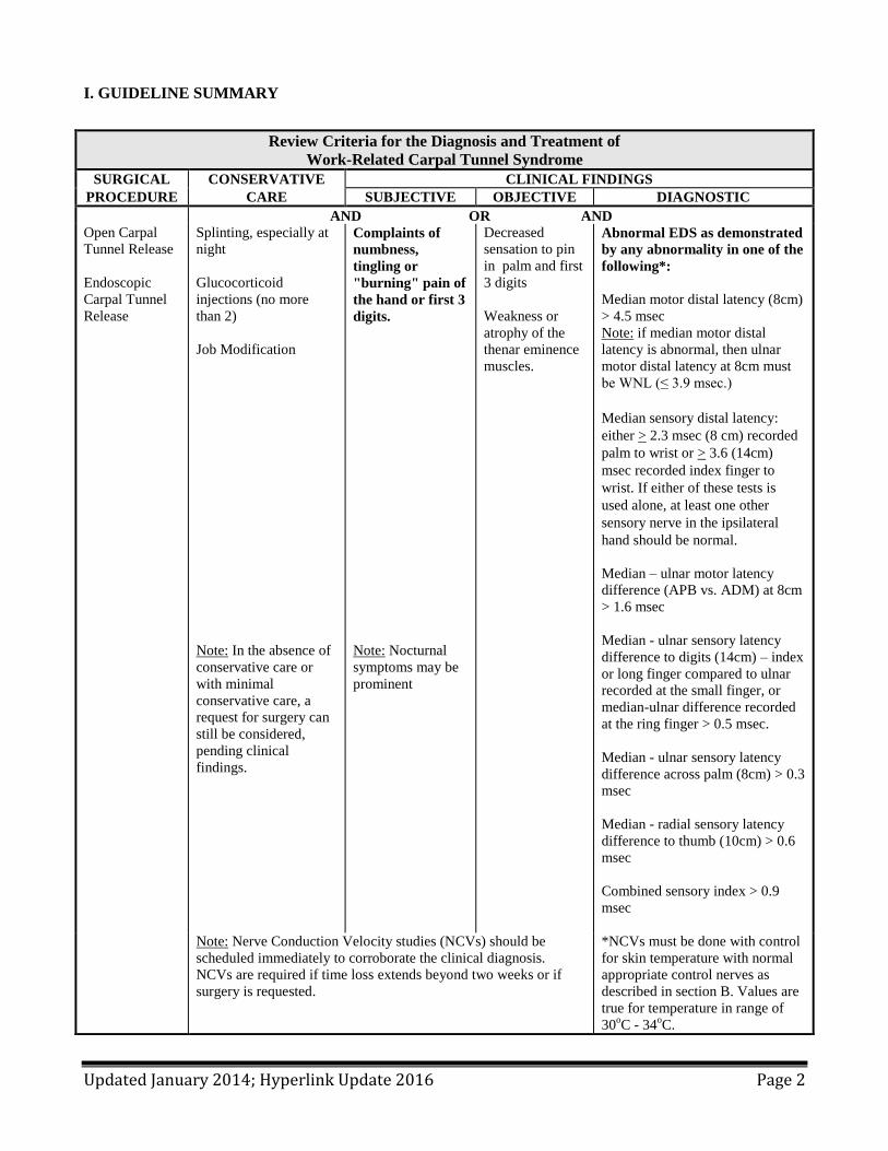
I. GUIDELINE SUMMARY
Review Criteria for the Diagnosis and Treatment of
Work-Related Carpal Tunnel Syndrome
SURGICAL CONSERVATIVE CLINICAL FINDINGS
PROCEDURE CARE SUBJECTIVE OBJECTIVE DIAGNOSTIC
AND OR AND
Open Carpal
Tunnel Release
Endoscopic
Carpal Tunnel
Release
Splinting, especially at
night
Glucocorticoid
injections (no more
than 2)
Job Modification
Note: In the absence of
conservative care or
with minimal
conservative care, a
request for surgery can
still be considered,
pending clinical
findings.
Complaints of
numbness,
tingling or
"burning" pain of
the hand or first 3
digits.
Note: Nocturnal
symptoms may be
prominent
Decreased
sensation to pin
in palm and first
3 digits
Weakness or
atrophy of the
thenar eminence
muscles.
Abnormal EDS as demonstrated
by any abnormality in one of the
following*:
Median motor distal latency (8cm)
> 4.5 msec
Note: if median motor distal
latency is abnormal, then ulnar
motor distal latency at 8cm must
be WNL (≤ 3.9 msec.)
Median sensory distal latency:
either > 2.3 msec (8 cm) recorded
palm to wrist or > 3.6 (14cm)
msec recorded index finger to
wrist. If either of these tests is
used alone, at least one other
sensory nerve in the ipsilateral
hand should be normal.
Median – ulnar motor latency
difference (APB vs. ADM) at 8cm
> 1.6 msec
Median - ulnar sensory latency
difference to digits (14cm) – index
or long finger compared to ulnar
recorded at the small finger, or
median-ulnar difference recorded
at the ring finger > 0.5 msec.
Median - ulnar sensory latency
difference across palm (8cm) > 0.3
msec
Median - radial sensory latency
difference to thumb (10cm) > 0.6
msec
Combined sensory index > 0.9
msec
Note: Nerve Conduction Velocity studies (NCVs) should be
scheduled immediately to corroborate the clinical diagnosis.
NCVs are required if time loss extends beyond two weeks or if
surgery is requested.
*NCVs must be done with control
for skin temperature with normal
appropriate control nerves as
described in section B. Values are
true for temperature in range of
30oC - 34
oC.
Updated January 2014; Hyperlink Update 2016 Page 3
II. INTRODUCTION This guideline is intended as an educational resource for physicians who treat injured workers in the Washington
workers’ compensation system under Title 51 RCW and as review criteria for the department’s utilization
review team to help ensure that diagnosis and treatment of carpal tunnel syndrome is of the highest quality. This
guideline was developed in 2008 using published medical evidence and expert consensus. The medical
literature search focused on specific topics and areas of interest to the department and Washington’s injured
worker population. A list of references used in this guideline can be found at the end of the document.
A hand diagram, diagnostic worksheet and guideline summary are appended to the end of this document.
Providers are encouraged to use these tools as references in the diagnosis, evaluation, and treatment of work-
related carpal tunnel syndrome. Median nerve compression at the wrist is the most common peripheral nerve entrapment disorder. It produces a constellation of specific symptoms and signs, described as carpal tunnel syndrome (CTS). The annual incidence in the general population has been reported to be approximately 1/1000.
1 The incidence in Washington’s
workers’ compensation population peaked at approximately 2.73/1000 in the mid-1990s.2
Both documentation of appropriate symptoms and signs and a statement attesting to probable work-relatedness must be present for Labor and Industries to accept a CTS claim. Nerve conduction velocity studies (NCVs) should be scheduled immediately to corroborate the clinical diagnosis. Completion of a nerve conduction study for a presumptive case of CTS is required if time loss extends beyond two weeks or if surgery is requested. III. ESTABLISHING WORK-RELATEDNESS
CTS may result from numerous conditions, including inflammatory or non-inflammatory arthropathies, recent or
remote wrist trauma or fractures, diabetes mellitus, obesity, hypothyroidism, pregnancy, and genetic factors.3 4
Risk for CTS strongly increases with age and among peri-menopausal females for unclear reasons. In the
unusual instance that CTS is acutely, traumatically induced, e.g. a patient has both CTS and concomitant trauma
(fracture or dislocation), the patient may require prompt carpal tunnel release. Work-related activities may also
cause or contribute to the development of CTS. To establish a diagnosis of work-related carpal tunnel
syndrome, all of the following are required:
1. Exposure: Workplace activities that contribute to or cause CTS, and 2. Outcome: A diagnosis of CTS that meets the diagnostic criteria under Section IV, and 3. Relationship: Generally accepted scientific evidence, which establishes on a more probable than not
basis (greater than 50%) that the workplace activities (exposure) in an individual case contributed to the development or worsening of the condition (outcome).
When the department receives notification of an occupational disease, the Occupational Disease & Employment History form is mailed to the worker, employer or attending provider. The form should be completed and returned to the Department as soon as possible. If the worker’s attending provider completes the form, provides a detailed history in the chart note, and gives an opinion on causality, he or she may be paid for this (use billing code 1055M). Additional billing information is available in the Attending Doctor’s Handbook. Work-related CTS is most often associated with activities requiring extensive, forceful, repeated, or prolonged use of the hands and wrists, particularly if these potential risk factors are present in combination (e.g., force and repetition or force and posture). Usually, one or more of the following work conditions occurs on a regular basis to support work-relatedness:
1. Forceful use, particularly if repeated
Updated January 2014; Hyperlink Update 2016 Page 4
2. Repetitive hand use combined with some element of force, especially for prolonged periods 3. Constant firm gripping of objects 4. Moving or using the hand and wrist against resistance or with force 5. Exposing the hand and wrist to strong regular vibrations 6. Regular or intermittent pressure on the wrist
The types of jobs most mentioned in the literature or reported in L&I’s data as being associated with CTS are listed in Table 1. This is not an exhaustive list and is meant only as a guide in the consideration of work-relatedness.
Table 1. Work Exposures and the Probability of Work-Relatedness
Exposure
Examples of types of jobs
Probability of
work-relatedness
Combinations of high force with high
repetition and awkward posture; regular
strong vibrations
Seafood, fruit, or meat processing or canning,
carpentry, roofing, dry-wall installation, boat
building, book binding
High,
Relative risk > 4
Medium-high force, high repetition or
awkward posture alone, on a nearly
continuous basis
Dental hygienists, wood products production
Medium,
Relative risk 2-4
Low force or medium-low repetition
alone, on an intermittent basis
Computer or keyboard use
Low,
Relative risk < 2
IV. MAKING THE DIAGNOSIS A. SYMPTOMS AND SIGNS A case definition for the presence or absence of CTS requires both appropriate symptoms and abnormal NCVs for the diagnosis.
5 Appropriate symptoms include numbness, tingling, or burning pain in the volar aspects of one
or both hands, especially noted after work or at night. Nocturnal symptoms are prominent in 50-70% of patients. Patients frequently awaken at night or early morning and shake their hands to relieve these symptoms. The location of these symptoms may be reported as involving the entire hand or localized to the thumb and first two or three fingers. A hand pain diagram has been validated for use in localizing sensory symptoms of CTS (appended to end of guideline).
6
If the nerve symptoms are prominent only in the fourth and fifth fingers, a different diagnosis (e.g. ulnar neuropathy or C-8 radiculopathy) should be considered. Although burning pain is often prominent in the hands and palm side of the wrists, an aching pain may radiate to the medial elbow region or more proximally to the shoulder. Proximal symptoms, especially tingling in the radial hand combined with lateral elbow pain should raise questions about a possible C-6 radiculopathy. Findings on physical examination, signs, are frequently absent or non-specific. Hoffmann-Tinel’s sign (paresthesias radiating in a median nerve distribution with tapping on the wrist or over the median nerve) and Phalen’s sign (paresthesias radiating in a median nerve distribution within 60 seconds of sustained flexion of the wrist) are frequently described, but by themselves are not sensitive or specific for the diagnosis of CTS. Their presence may corroborate the presence of other clear neurologic symptoms. Likewise, non-specific symptoms, (e.g., pain without numbness, tingling or burning; “dropping things”) by themselves are not diagnostic of CTS.
Updated January 2014; Hyperlink Update 2016 Page 5
Signs that occur as CTS becomes more severe include decreased sensation to pin or light touch in the first three digits or weakness or atrophy of the muscles of the thenar eminence (especially the abductor pollicis brevis). Unlike Tinel’s or Phalen’s, the presence of thenar atrophy or weakness may suggest more acute or advanced nerve injury and perhaps the need for more aggressive treatment.
Every effort should be made to objectively verify the diagnosis of CTS before considering surgery. Although
some evidence is conflicted, it has been suggested that patients who have undergone carpal tunnel surgery with
normal or near normal pre-surgical nerve conduction test results have poorer outcomes than those with
electrodiagnostic evidence of median nerve entrapment across the carpal tunnel.7 In rare cases, a steroid
injection can be performed into the carpal canal as a therapeutic and diagnostic challenge test. Patients noting a
dramatic improvement in symptoms for weeks or months following the injection, but then having recurrence of
symptoms, may be considered candidates for surgical carpal tunnel release (CTR). Patients with a negative
response may be referred to an appropriate specialist (e.g., neurologist, orthopedist or physiatrist) for further
diagnostic evaluation if warranted, or be followed for a 12-month period to monitor for neurologic findings that
may develop. If CTS is not documented by clinical criteria and NCV testing, other clinical problems potentially related to work exposures (e.g. tendonitis) should be investigated and treated appropriately. It would also be important to rule out other neurologic causes of tingling in the hands. Referral to an appropriate specialist (neurologist, physiatrist) would be prudent in such cases.
CTS is a common physiologic condition in pregnancy. This is theorized to be due to increased plasma volume
and fluid retention that raise the pressure within the carpal tunnel. The symptoms of CTS often improve after
childbirth. If they do not, other etiologies should be pursued.
B. ELECTRODIAGNOSTIC TESTING (EDS) i. Nerve Conduction Velocity An easy-to-use worksheet for interpreting electrodiagnostic tests is available at the end of this guideline. The worksheet should be used only when the main purpose of the study is to evaluate a patient for CTS. It is critical to conduct NCV testing in the following situation:
1. The diagnosis of CTS is being considered, or 2. Patient is on time-loss for more than two weeks, or 3. Carpal tunnel decompression surgery is requested
Conceptually, validation of the clinical diagnosis of CTS depends on the finding of slowing of sensory and/or motor fibers of the median nerve across the carpal tunnel. The nerve conduction study methods used to test for slowing should not be affected by temperature (either the temperature should be maintained over 32
o C, or tests
should be used that are not influenced by temperature). They should have a high specificity, good sensitivity, and high degree of reliability. Such tests should also minimize the possibility of age or polyneuropathy creating a misleading or false-positive result. This can often be accomplished by comparing the median nerve to another nerve across the same distance across the wrist. NCVs are highly sensitive and specific for CTS. If the patient has a positive clinical picture of CTS but the NCV results are negative, the physician should investigate other competing clinical diagnoses such as pronator syndrome, cervical radiculopathy or tendonitis. Less than 10% of patients with clinical CTS have normal NCV results.
8 In these cases, the treating physician should be sure the most sensitive and specific NCVs are done. If
not, a request for these tests should be made. In some cases of suspected CTS, the NCVs can be repeated. However, unless there is a significant intervening event or a substantial change in the clinical assessment, there should be a delay of at least one year before repeating the NCV test, as it is unlikely that a difference will be seen at a shorter time interval.
Updated January 2014; Hyperlink Update 2016 Page 6
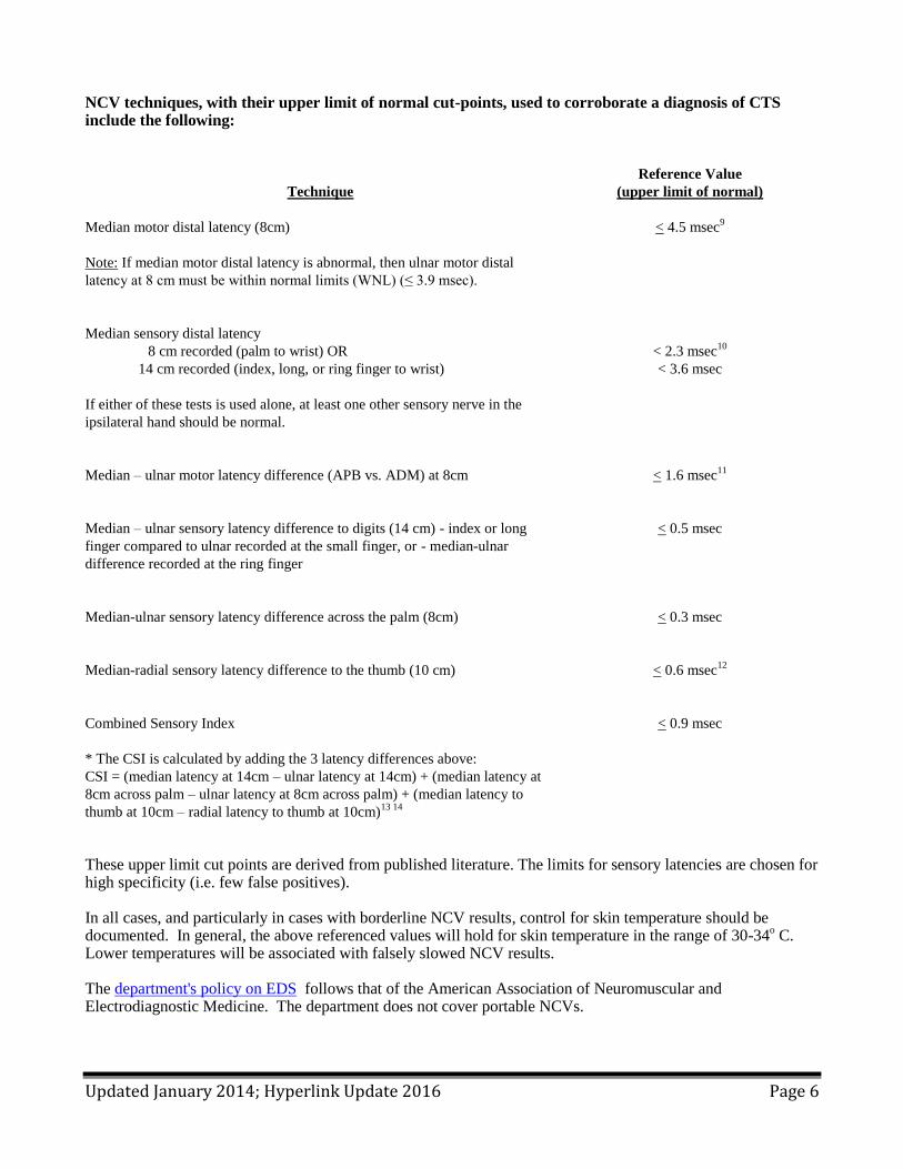
NCV techniques, with their upper limit of normal cut-points, used to corroborate a diagnosis of CTS include the following:
Technique
Reference Value
(upper limit of normal)
Median motor distal latency (8cm)
Note: If median motor distal latency is abnormal, then ulnar motor distal
latency at 8 cm must be within normal limits (WNL) (≤ 3.9 msec).
< 4.5 msec9
Median sensory distal latency
8 cm recorded (palm to wrist) OR
14 cm recorded (index, long, or ring finger to wrist)
If either of these tests is used alone, at least one other sensory nerve in the
ipsilateral hand should be normal.
< 2.3 msec10
< 3.6 msec
Median – ulnar motor latency difference (APB vs. ADM) at 8cm
< 1.6 msec11
Median – ulnar sensory latency difference to digits (14 cm) - index or long
finger compared to ulnar recorded at the small finger, or - median-ulnar
difference recorded at the ring finger
< 0.5 msec
Median-ulnar sensory latency difference across the palm (8cm)
< 0.3 msec
Median-radial sensory latency difference to the thumb (10 cm)
< 0.6 msec12
Combined Sensory Index
* The CSI is calculated by adding the 3 latency differences above:
CSI = (median latency at 14cm – ulnar latency at 14cm) + (median latency at
8cm across palm – ulnar latency at 8cm across palm) + (median latency to
thumb at 10cm – radial latency to thumb at 10cm)13 14
< 0.9 msec
These upper limit cut points are derived from published literature. The limits for sensory latencies are chosen for high specificity (i.e. few false positives). In all cases, and particularly in cases with borderline NCV results, control for skin temperature should be documented. In general, the above referenced values will hold for skin temperature in the range of 30-34
o C.
Lower temperatures will be associated with falsely slowed NCV results. The department's policy on EDS follows that of the American Association of Neuromuscular and Electrodiagnostic Medicine. The department does not cover portable NCVs.
Updated January 2014; Hyperlink Update 2016 Page 7
ii. Needle Electromyography Needle electromyography sometimes has a role in the electrodiagnostic evaluation of CTS. If the clinical presentation is classic for CTS symptoms and no other signs and/or symptoms, and the nerve conduction study is entirely normal, no needle EMG or only limited EMG studies are acceptable. However, there are circumstances in which it would be reasonable to do needle EMG during an evaluation of CTS:
a. Nerve conduction studies are abnormal in a manner indicating CTS, and the patient demonstrates wasting or clinical weakness of the thenar muscles, or the median motor nerve conduction study is significantly abnormal
b. The electromyographer suspects another possible diagnosis or a neuropathic process other than, or in addition to, CTS (e.g., diabetes)
c. There is a history of an acute crush injury or other major trauma to the distal upper extremity d. There are proximal symptoms (e.g., neck stiffness, radiating pain) that suggest cervical
radiculopathy may be present. iii. Quantitative Sensory Testing The department does not cover quantitative sensory tests (QST). Several tests of sensory function (vibration, temperature, pressure) have been reported in the scientific literature to be useful in investigational settings to differentiate between patients with and without neuropathy. However, because these techniques cannot localize peripheral nerve lesions, they are not useful for diagnosing specific entrapment neuropathies.
15
C. OTHER DIAGNOSTIC TESTS Some studies have suggested that Magnetic Resonance Imaging (MRI) neurography
16 and ultrasound
17 may
have utility in the diagnosis of CTS. However, these tests have not been shown to be more accurate than EDX in high quality studies
18 19. The department does not cover these services.
V. TREATMENT FOR CARPAL TUNNEL SYNDROME A. CONSERVATIVE TREATMENT A critical element for any conservative CTS intervention is to document improved function and ability to return to work. Because findings of median nerve involvement on NCV strongly predict a good outcome with CTS surgery, any worker suspected of median nerve involvement or with documented increased median nerve latencies who does not gain meaningful and sustainable functional improvement within 6-8 weeks of any conservative intervention approach should be referred to a specialist or surgeon. To date, although most studies have demonstrated meaningful and significant short term benefit, better-designed longer term follow-up studies are needed to clarify the sustainability of relief. Several conservative interventions have demonstrated utility in reducing symptoms and improving function:
1. Neutral position wrist splits used nocturnally and intermittently during work exposures have been
shown to be effective in reducing symptoms, increasing grip strength and in improving NCV 20 21 22
.
Studies report that 30-70% of patients respond favorably within several months of initiating this
intervention.
2. Glucocorticoids - Local steroid injections into the carpal tunnel have been demonstrated to provide good
short term relief of CTS.23
About half of all patients receiving this treatment require surgery within one
year. No more than two injections should be done. Oral steroids are not recommended. Although there
can be a short term benefit from oral steroids, the risk of serious adverse effects (e.g. avascular necrosis)
outweighs the benefits24 25 26
.
Updated January 2014; Hyperlink Update 2016 Page 8
3. Forearm/wrist stretching home exercise regimens may be of benefit and can be demonstrated to the
patient when the diagnosis is considered.
Occupational-centered interventions to reduce exposure are believed to be of value, based primarily on epidemiological studies and consensus opinion.
27 28
Job modification - Reducing the intensity of manual tasks when feasible may prevent progression and promote recovery from CTS. In most cases, the patient can continue working during conservative treatment. If job modification is not possible or if the patient cannot continue working despite conservative treatment, then surgical CTR should be considered as a treatment option.
The following treatments are not recommended for Carpal Tunnel Syndrome because there is inadequate or conflicting evidence concerning their effectiveness:
22 27
1. Vitamin B6 (pyridoxine) 2. Oral diuretics 3. Magnets* 4. Lasers (Not covered, see coverage decision) 5. Botulinum toxin injections* (Not FDA-approved for carpal tunnel syndrome, see coverage decision) 6. Iontophoresis*
*Not covered per WAC 296-20-03002 B. SURGICAL CARPAL TUNNEL RELEASE For patients with CTS confirmed by electrodiagnostic studies (EDS), carpal tunnel surgery is more effective in relieving symptoms than conservative treatment such as splinting.
20 Decompression of the median nerve at the
wrist with release of the transverse carpal ligament is the surgical procedure of choice and can be effectively performed by either open or endoscopic approaches.
29 30 31 Both are covered by the department. There is no
quality evidence that tenosynovectomy, internal neurolysis and several other adjunct procedures improves the clinical outcome of carpal tunnel release, and these procedures increase the risk of additional neurological trauma to the median nerve.
32 33 34 35
All of the following criteria must be met for surgery to be authorized:
1. The clinical presentation is consistent with CTS, and 2. The EDS criteria for CTS have been met, and 3. The patient has failed to respond to conservative treatment that included wrist splinting and/or injection
If symptoms return after surgery
Recurring carpal tunnel syndrome is uncommon. The results of revision surgery are unpredictable. In order to
determine whether or not a patient who has had prior CTS surgery is appropriate for revision surgery, at least
one of the following criteria should be met:
1. The symptoms should be at least as severe as pre-operatively, or
2. The EDS should be at least as severe as pre-operatively, or
3. There are new signs of median nerve dysfunction.
In general, it is helpful to wait at least 6 months from the time of initial surgery before considering revision
surgery, unless there are signs of significant surgical complication. This waiting period allows an adequate time
for healing, scar maturation, rehabilitation, and clinical improvement.
Updated January 2014; Hyperlink Update 2016 Page 9
VI. RETURN TO WORK (RTW)
A. EARLY ASSESSMENT
In the United States, approximately 7% of workers with upper extremity musculoskeletal disorders account for
75% of the disability in this population.36
A large prospective study of work-related carpal tunnel syndrome in
the Washington workers’ compensation system identified several important predictors of long-term disability:
low expectations of return to work, no offer of a job accommodation, and high physical demands on the job.37
Identifying and attending to these risk factors when patients have not returned to work within 2-3 weeks of the
initial clinical presentation may improve their chances of returning to work.
Timeliness of the CTS diagnosis can be a critical factor influencing RTW. Washington workers diagnosed
accurately and early were far more likely to RTW than workers whose CTS was diagnosed weeks or months
later.38
Early coordination of care with improved timeliness and effective communication with the workplace is
also likely to help prevent long-term disability in CTS. A recent quality improvement project in Washington
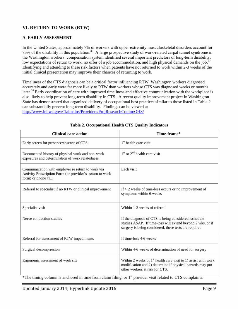
State has demonstrated that organized delivery of occupational best practices similar to those listed in Table 2
can substantially prevent long-term disability. Findings can be viewed at
http://www.lni.wa.gov/ClaimsIns/Providers/ProjResearchComm/OHS/
Table 2. Occupational Health CTS Quality Indicators
Clinical care action Time-frame*
Early screen for presence/absence of CTS 1st health care visit
Documented history of physical work and non-work
exposures and determination of work relatedness
1st or 2
nd health care visit
Communication with employer re return to work via
Activity Prescription Form (or provider’s return to work
form) or phone call
Each visit
Referral to specialist if no RTW or clinical improvement
If > 2 weeks of time-loss occurs or no improvement of
symptoms within 6 weeks
Specialist visit Within 1-3 weeks of referral
Nerve conduction studies If the diagnosis of CTS is being considered, schedule
studies ASAP. If time-loss will extend beyond 2 wks, or if
surgery is being considered, these tests are required
Referral for assessment of RTW impediments If time-loss 4-6 weeks
Surgical decompression Within 4-6 weeks of determination of need for surgery
Ergonomic assessment of work site Within 2 weeks of 1st health care visit to 1) assist with work
modification and 2) determine if physical hazards may put
other workers at risk for CTS.
*The timing column is anchored in time from claim filing, or 1st provider visit related to CTS complaints.
Updated January 2014; Hyperlink Update 2016 Page 10
B. RETURNING TO WORK FOLLOWING SURGERY
RTW after surgery should be possible for many patients regardless of whether open or endoscopic release was
performed. Average times for returning to work (panel consensus) are within 2-4 weeks for clerical and light
duty workers and within 5-6 weeks for heavy labor workers. These time frames tend to be shorter for
endoscopic surgery; time from surgery to return to work or to activities of daily living is approximately 6 days
less with endoscopic than with open surgery.39
In a number of well-designed studies, the majority of patients recovered function and did not have a permanent impairment that would result in disability.
29 31 40 The panel’s experience is that many patients can successfully
return to the job of injury. If neurologic symptoms reappear after RTW, repeat EDS and referral to a specialist may be indicated.
Continue on to next page for hand diagram.
Updated January 2014; Hyperlink Update 2016 Page 11
VII. HAND DIAGRAM*
This diagram can be printed and completed by the patient.
Pain
Patient Name: ______________________Claim#:_______________Date:____________
Comments: Tingling
Numbness
Decreased
Sensation
* Permission to use this hand diagram was obtained from Dr. Jeffrey N. Katz. The legend was modified for
better readability.
Right Hand Left Hand
Updated January 2014; Hyperlink Update 2016 Page 12
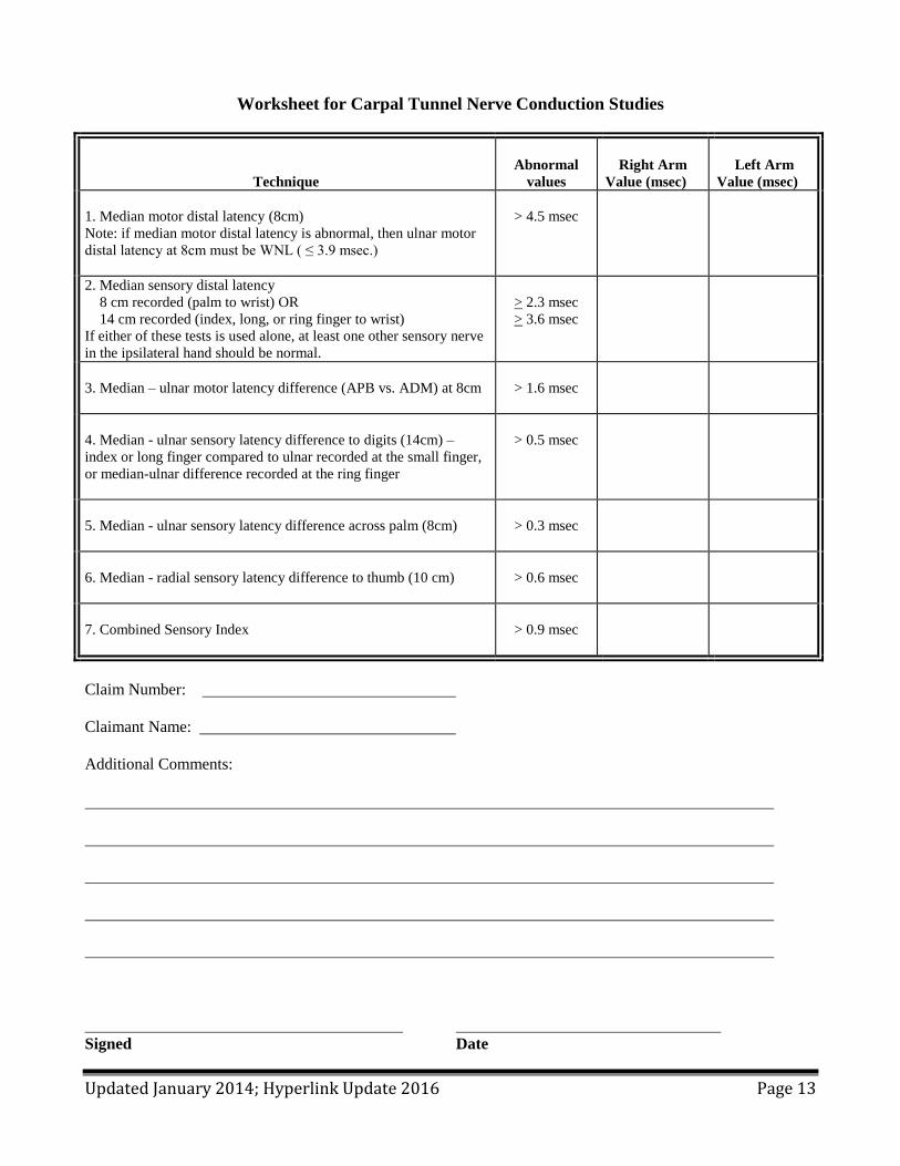
VIII. ELECTRODIAGNOSTIC WORKSHEET PURPOSE AND INSTRUCTIONS 1. The purpose of this worksheet is to help the department’s medical and nursing staff interpret
electrodiagnostic tests (EDS) that you do for L&I patients. The worksheet should be used only when the main purpose of your study is to evaluate a patient for CTS. It is for this reason that the worksheet focuses on distal latency from NCV. It should accompany but not replace the detailed report normally submitted to the department.
2. We encourage you to use the Electrodiagnostic Worksheet that is appended to this guideline to report EDS
results, but the department will accept the results on a report generated by your office system. 3. On the worksheet, sensory distal latency should be measured to response peak and motor distal latency
should be measured to response onset. 4. It is not necessary to do all the NCVs listed on the worksheet. You should do only the studies needed to
rule CTS in or out. 5. It is sometimes necessary to do EDS other than ones listed on the worksheet. If you do any additional
studies bearing on the diagnosis of CTS, please write them in the blank area below the listed studies. 6. The value of other studies of median nerve function has not been proven. Those tests are NOT
recommended for the diagnosis of CTS. The following quotation is taken from a literature review published in Muscle & Nerve, 1993, Vol. 16, p. 1392-1414:
“Several other variations on median sensory and motor NCV have been reported to be useful for the evaluation of patients with OCTS [occupational carpal tunnel syndrome]. The committee’s review of the literature indicated that the value of these tests for the clinical electrodiagnostic evaluation of patients with OCTS remains to be established. These electrodiagnostic studies include the following: (1) studies of the median motor distal latency recorded from the lumbrical muscles,.. (2) measurement of the refractory period of the median nerve,.. (3) median motor residual latency measurements,.. (4) terminal latency ratio,.. (5) median F-wave abnormalities,.. (6) median motor nerve conduction amplitude comparisons with stimulation above and below the carpal ligament,.. (7) anterior interosseous/median nerve latency ratio,.. (8) change in median motor response configuration with median nerve stimulation at the wrist and elbow in the presence of Martin-Gruber anastomosis,.. (9) sensory amplitude measurements,.. and (10) measurement of median sensory and motor nerve conduction across the wrist before and after prolonged wrist flexion.”
Continue on to next page for worksheet.
Updated January 2014; Hyperlink Update 2016 Page 13
Worksheet for Carpal Tunnel Nerve Conduction Studies
Technique
Abnormal
values
Right Arm
Value (msec)
Left Arm
Value (msec)
1. Median motor distal latency (8cm)
Note: if median motor distal latency is abnormal, then ulnar motor
distal latency at 8cm must be WNL ( ≤ 3.9 msec.)
> 4.5 msec
2. Median sensory distal latency
8 cm recorded (palm to wrist) OR
14 cm recorded (index, long, or ring finger to wrist)
If either of these tests is used alone, at least one other sensory nerve
in the ipsilateral hand should be normal.
> 2.3 msec
> 3.6 msec
3. Median – ulnar motor latency difference (APB vs. ADM) at 8cm
> 1.6 msec
4. Median - ulnar sensory latency difference to digits (14cm) –
index or long finger compared to ulnar recorded at the small finger,
or median-ulnar difference recorded at the ring finger
> 0.5 msec
5. Median - ulnar sensory latency difference across palm (8cm)
> 0.3 msec
6. Median - radial sensory latency difference to thumb (10 cm)
> 0.6 msec
7. Combined Sensory Index
> 0.9 msec
Claim Number:
Claimant Name:
Additional Comments:
Signed Date
Updated January 2014 Page 14
References
1. Stevens JC, Sun S, Beard CM. Carpal tunnel syndrome in Rochester, Minnesota, 1961-1980. Neurology
1988; 38: 134-138.
2. Silverstein B, Welp E, Nelson N, Kalat J. Claims incidence of work-related disorders of the upper
extremities. Am J Public Health 1998 Dec; 88(12): 1827-1833.
3. Stevens J, Beard CM, O’Failon WM, Kurland LT. Conditions associated with carpal tunnel syndrome.
Mayo Clin Proc 1992; 67: 541-548.
4. Hakim AJ, Cherkas L, El Zayat S, MacGregor AJ, Spector TD. The genetic contribution to carpal tunnel
syndrome in women: a twin study. Arthritis and Rheumatism 2002; 47: 275–279.
5. Rempel D, Evanoff B, Amadio PC, et al. Consensus criteria for the classification of carpal tunnel
syndrome in epidemiologic studies. Am J of Pub Health 1998; 88(10): 1447-1451.
6. Katz JN, Stirrat CR. A self-administered hand diagram for the diagnosis of carpal tunnel syndrome. J
Hand Surg 1990; 15A: 360-363.
7. Higgs PE, Edwards DF, Martin DS, Weeks PM. Relation of preoperative nerve-conduction values to
outcome in workers with surgically treated carpal tunnel syndrome. J Hand Surg 1997; 22A: 216-221.
8. Prakash KM, Fook-Chong S, Leoh TH, Dan YF, Nurjannah S, Tan YE, Lo YL. Sensitivities of sensory
nerve conduction study parameters in carpal tunnel syndrome. J Clin Neurophysiol 2006 Dec; 23(6):
565-567.
9. Buschbacher RM. Median nerve motor conduction to the abductor pollicis brevis. Am J Phys Med
Rehabil 1999 Nov-Dec; 78(6 Suppl): S1-8.
10. Sander HW, Quinto C, Saadeh PB, Chokroverty S. Median and ulnar palm-wrist studies. Clin
Neurophysiol 1999 Aug; 110(8): 1462-1465.
11. Grossar EA, Prahlow ND, Buschbacher RM. Acceptable difference in sensory and motor latencies
between the median and ulnar nerves. J Long Term Eff Med Implants 2006; 16(5): 395-400.
12. Berkson A, Lohman J, Buschbacher RM. Comparison of median and radial sensory studies to the
thumb. J Long Term Eff Med Implants 2006; 16(5): 387-394.
13. Robinson LR, Micklesen PJ, Wang L. Strategies for analyzing nerve conduction data: superiority of a
summary index over single tests. Muscle & Nerve 1998; 21: 1166-1171.
14. Robinson LR. Electrodiagnosis of carpal tunnel syndrome. Phys Med Rehabil Clin N Am 2007; 18: 733-
746.
15. Shy ME, Frohman EM, So YT, Arezzo JC, Cornblath DR, Giuliani MJ, Kincaid JC, Ochoa JL, Parry
GJ, Weimer LH. Quantitative sensory testing: report on the therapeutics and technology assessment
subcommittee of the American Academy of Neurology. Neurology 2003; 60: 898-904.
16. Jarvik JG, Yuen E, Haynor DR, Bradley CM, Fulton-Kehoe D, Weller-Smith T, Wu R, Kliot M, Kraft
G, Wang L, Erlich V, Heagerty PJ, Franklin GM. MR Nerve imaging in a prospective cohort of patients
with suspected carpal tunnel syndrome. Neurology 2002; 58: 1597-1602.
Updated January 2014 Page 15
17. Wong SM, Griffith JF, Hui ACF, Lo SK, Fu M, Wong KS. Carpal tunnel syndrome: diagnostic
usefulness of sonography. Radiology 2004; 232: 93-99.
18. Descatha, A., Huard, L., Aubert, F., Barbato, B., Gorand, O., and Chastang, J.F., Meta-analysis on the
performance of sonography for the diagnosis of carpal tunnel syndrome. Semin Arthritis Rheum, 2012.
41(6): p. 914-22.
19. Fowler, J.R., Gaughan, J.P., and Ilyas, A.M., The sensitivity and specificity of ultrasound for the
diagnosis of carpal tunnel syndrome: a meta-analysis. Clin Orthop Relat Res, 2011. 469(4): p. 1089-94.
20. Gerritsen AAM, de Vet HCW, Scholten RJPM, et al. Splinting vs surgery in the treatment of carpal
tunnel syndrome: a randomized controlled trial. JAMA 2002; 288 (10): 1245-1251.
21. Nobuta S, Sato K, Nakagawa T, Hatori M, Itoi E. Effects of wrist splinting for carpal tunnel syndrome
and motor nerve conduction measurements. Upsala J Med Sci 2008; 113 (2): 181-192.
22. O’Connor D, Marshall S, Massy-Westropp N. Non-surgical treatment (other than steroid injection) for
carpal tunnel syndrome. Cochrane Database of Systematic Reviews 2003, Issue 1. Art. No.: CD003219.
DOI: 10.1002/14651858.CD003219.
23. American Academy of Orthopedic Surgeons (AAOS, 2007). Carpal Tunnel Syndrome Guideline.
Retrieved September 24, 2008, from AAOS. Web site:
http://www.aaos.org/research/guidelines/guide.asp
24. Ly-Pen D. Andreu JL, de Blas G. Sanchez-Olaso A. Millan I. Surgical decompression versus local
steroid injection in carpal tunnel syndrome: a one-year, prospective, randomized, open, controlled
clinical trial. Arthritis & Rheumatism 2005 Feb. 52(2): 612-619.
25. Marshall S, Tardif G, Ashworth N. Local corticosteroid injection for carpal tunnel syndrome. Cochrane
Database of Systematic Reviews 2007. Issue 2. Art. No.: CD001554. DOI:
10.1002/14651858.CD001554.pub2.
26. Hui ACF, Wong S, Leung CH, Tong P, Mok V, Poon D, et al. A randomized controlled trial of surgery
vs steroid injection for carpal tunnel syndrome. Neurology 2005; 64(12): 2074-2078.
27. Verhagen AP, Karels C, Bierma-Zeinstra SM, Feleus A, Dahaghin S, Burdorf A, De Vet HC, Koes BW.
Ergonomic and physiotherapeutic interventions for treating work-related complaints of the arm, neck or
shoulder in adults. A Cochrane systematic review. Eura Medicophys. 2007 Sep; 43(3):391-405.
28. De Kesel R, Donceel P, De Smet L. Factors influencing return to work after surgical treatment for
carpal tunnel syndrome. Occup Med (London) 2008 May; 58 (3): 187-190.
29. Trumble TE, Diao E, Abrams RA, Glibert-Anderson MM. Single-portal endoscopic carpal tunnel
release compared with open release: a prospective, randomized trial. J Bone Joint Surg 2002; 84: 1107-
1115.
30. Palmer DH, Paulson JC, Lane-Larsen CL, Peulen VK, Olsen JD. Endoscopic carpal tunnel release: a
comparison of two techniques with open release. Arthroscopy 1993; 9(5): 498-508.
Updated January 2014 Page 16
31. Brown RA, Gelberman RH, Seiler JG, Abrahamsson S, Weiland AJ, Urbaniak JR, Schoenfeld DA,
Furcolo D. Carpal tunnel release: a prospective, randomized assessment of open and endoscopic
methods. J Bone Joint Surg 1993; 9: 1265-1275.
32. Gelberman RH, Pfeffer GB, Galbraith RT, Szabo RM, Rydevik B, Dimick M. Results of treatment of
severe carpal-tunnel syndrome without internal neurolysis of the median nerve. J Bone Joint Surg 1987
Jul; 69(6): 896-903.
33. Mackinnon SE, Dellon AL. Anatomic investigations of nerves at the wrist: I. Orientation of the motor
fascicle of the median nerve in the carpal tunnel. Ann Plast Surg 1988 Jul; 21(1): 32-5.
34. Mackinnon SE. Secondary carpal tunnel surgery. Nerurosurg Clin N Am 1991; 2: 75-91.
35. Kerr CD, Sybert DR, Albarracin NS. An analysis of the flexor synovium in idiopathic carpal tunnel
syndrome: report of 625 cases. J Hand Surg 1992 Nov; 17(6): 1028-1030.
36. Hashemi L, Webster BS, Clance EA, Courtney TK. Length of disability and cost of work-related
musculoskeletal disorders of the upper extremity. J Occup Environ Med 1998; 40: 261-269.
37. Turner JA, Franklin G, Fulton-Kehoe D. Early predictors of chronic work disability associated with
carpal tunnel syndrome: a longitudinal workers’ compensation cohort study. Am J Ind Med 2007; 50:
489-500.
38. Daniell WE, Fulton-Kehoe D, Chiou LA, Franklin GM. Work-related carpal tunnel syndrome in
Washington State worker’s compensation: temporal trends, clinical practices, and disability. Am J Ind
Med 2005; 48: 259-269.
39. Scholten RJPM, Mink van der Molen A, Uitdehaag BMJ, Bouter LM, de Vet HCW. Surgical treatment
options for carpal tunnel syndrome. Cochrane Database of Systematic Reviews 2007. Issue 4. Art. No.:
CD003905. DOI: 10.1002/14651858.CD003905.pub3.
40. Agee JM, McCarroll HR, Tortosa RD, Berry DA, Szabo RM, Peimer CA. Endoscopic release of the
carpal tunnel: a randomized prospective multicenter study. J Hand Surg 1992; 17A: 987-995.
41. ACOEM Evidence-based Chronic Pain Panel. Chronic Pain. In: Hegmann KT, ed. Occupational
Medicine Practice Guidelines. 2nd ed. Rev. Elk Grove Village, Ill: American College of Occupational
and Environmental Medicine 2008; 6:395.
Updated January 2014 Page 17
Acknowledgements
This guideline was developed in 2008 by Labor and Industries’ Industrial Insurance Medical Advisory
Committee (IIMAC) and its subcommittee on Upper Extremity Entrapment Neuropathies. Acknowledgement
and gratitude go to all subcommittee members, clinical experts, and consultants who contributed to this
important guideline:
IIMAC Committee Members
Gregory T. Carter MD MS
Dianna Chamblin MD – Chair
G.A. DeAndrea MD MBA
Jordan Firestone, MD PhD MPH
Andrew Friedman MD
Subcommittee Clinical Experts
Christopher H. Allan MD
Douglas P. Hanel MD
Michel Kliot MD
Lawrence R. Robinson MD
Thomas E. Trumble MD
Nicholas B. Vedder MD
Michael D. Weiss MD
Consultation Provided by:
Terrell Kjerulf MD, Qualis Health
Ken O’Bara MD, Qualis Health
Scott Carlson MD
Jeffrey (Jerry) G. Jarvik MD
Department staff who helped develop and prepare this guideline include:
Gary M. Franklin MD MPH, Medical Director
Simone P. Javaher BSN, MPA, Occupational Nurse Consultant
Reshma N. Kearney MPH, Epidemiologist
Bintu Marong BS, MS, Epidemiologist