wood microstructure of ligneous species of rhamnaceae from ... · pearson and brown (1932)...
TRANSCRIPT

Journal of Tropical Forest Science 23(3): 239–251 (2011) Gupta S & Saxena V
239© Forest Research Institute Malaysia
Wood Microstructure of Ligneous species of rhaMnaceae froM india
s gupta* & V saxena
Forest Research Institute, Dehradun 248006, India
Received May 2010
gupta s & saxena V. 2011. Wood microstructure of ligneous species of rhamnaceae from india. This study dealt with the wood microstructure of the family Rhamnaceae from India. The wood microstructure and salient diagnostic features of 22 species belonging to 8 genera were described. An identification key was developed for the 22 species. Perforated ray cells were reported for the first time in Sageretia brandrethiana, Rhamnus purpurea, Rhamnus wightii, Zizyphus mauritiana, Z. oenoplia, Z. oxyphylla and Z. xylopyrus. The study revealed a need for reclassification of the family as anatomically the family is quite heterogeneous at the generic level.
Keywords: Wood anatomy, perforated ray cell, identification key, systematic position, crystals
gupta s & saxena V. 2011. Mikrostruktur kayu spesies rhamnaceae berlignin dari india. Kajian ini adalah tentang mikrostruktur kayu famili Rhamnaceae dari India. Mikrostruktur kayu dan ciri pengecaman penting bagi 22 spesies daripada 8 genus dihuraikan. Sel jejari berliang dilaporkan pertama kalinya dalam Sageretia brandrethiana, Rhamnus purpurea, Rhamnus wightii, Zizyphus mauritiana, Z. oenoplia, Z. oxyphylla and Z. xylopyrus. Kajian ini menunjukkan bahawa famili ini perlu dikelaskan semula memandangkan anatomi famili ini agak heterogen pada peringkat genus.
* E-mail: [email protected]
introduction The family Rhamnaceae comprises about 70 genera and 1500 species of erect or scandent shrubs and small- to medium-sized trees, distributed throughout the tropical and temperate regions of the world. A total of 12 genera and about 57 species occur in India (Bhandari & Bhansali 2000). The family is of little significance from the point of view of timber but many of its members have medicinal value and some yield valuable dyes. Yellow and green dyes are obtained from several species of Rhamnus, which are used in the textile industry and for calico printing. Gouania leptostachya, Ventilago madraspatana and Zizyphus jujuba are used in native medicine (Anonymous 1963). The wood microscopic features were studied but at the generic level (Metcalfe & Chalk 1950, Carlquist 1988). Schirarend (1991) gave a systematic wood anatomy of the Rhamnaceae. Inside Wood database (http://insidewood.lib.ncsu.edu/) dealt only with Hovenia dulcis, Z. jujuba and Z. xylopyrus.
On the Indian front, Gamble (1922) briefly described general features of the woods. Pearson and Brown (1932) described the wood microstructure of Z. jujuba and Z. xylopyrus Anonymous (1963) reported physical properties and gross structure of the wood. A comprehensive study on the wood microstructure of the species of this family occurring in India is lacking. Thus, the present study was undertaken. The study was also aimed at developing a key for the identification of the species. The study also compared findings of earlier studies.
MateriaLs and Methods The study examined 41 wood samples from 22 species belonging to 8 genera of the family Rhamnaceae, housed at the Forest Research Institute, Dehradun. Details of the specimens are given in Table 1 along with their accession number, specific gravity and locality. Data on specific gravity are as per Anonymous (1963).
Journal of Tropical Forest Science 23(3): 239–251 (2011) Gupta S & Saxena V
240© Forest Research Institute Malaysia
For microscopic examination, 15–20 µm thick transverse, radial and tangential sections were obtained using a microtome. The sections were stained in Heidenhain’s haematoxylin and safranin, and mounted on slides. For determination of fibre and vessel characteristics, small radial chips were macerated using 30% nitric acid and a pinch of potassium chlorate. The
data on fibre and vessel characteristics as well as ray frequency (rays/mm) were means of 25, 10 and 10 counts respectively. Photomicrographs of diagnostic features were taken using microscope and image analyser. For microstructure, the terminology published by the International Association of Wood Anatomists (IAWA 1989) was used. The ratio between fibre length and vessel
Species Accession number Locality Specific gravity
Berchemia floribunda (Wall.) Brongn. DDw 2864 Darjeeling, West Bengal 0.52Gouania leptostachya DC. DDw 5369 Pilibhit, Uttar Pradesh 0.54Hovenia dulcis Thunb DDw 8398 Arunachal Pradesh 0.62
DDw 3808 United States of America 0.58Rhamnus nepalensis (Wall.) M. Lawson DDw 3364 Darjeeling, West Bengal 0.62
DDw 3346 West Bengal 0.65R. persica Boiss. DDw 5004 Dehradun,Uttaranchal 1.00R. purpurea Edgew. DDw 70 Himachal Pradesh 0.66
DDw 4418 Jaunsar, Uttaranchal 0.63DDw 4707 Jaunsar, Uttaranchal 0.76
R. triquetra (Wall.) Brandis DDw 4808 Jaunsar, Uttaranchal 0.93R. virgata Roxb. DDw 79 Simla, Himachal Pradesh 0.90
DDw 2877 Simla, Himachal Pradesh 0.84R. wightii W.&A. DDw 3745 Nilgiri, Madras, Tamilnadu 0.89Sageretia brandrethiana Aitch. DDw 914 Simla, Himachal Pradesh 0.97S. thea (Osbeck) Johnst. DDw 2946 Simla, Himachal Pradesh 0.89
DDw 2951 Simla, Himachal Pradesh 0.96Scutia myrtina (Burm. F.) Kurz DDw 5725 Tamil Nadu 0.89Ventilago maderaspatana Gaertn. DDw 3843 Madhya Pradesh 0.52
DDw 6448 Myanmar 0.61Ziziphus glabrata Heyne ex Roth. DDw 5634 Madras, Tamilnadu 1.15Z. incurva Roxb. DDw 8067 Dehradun, Uttaranchal 0.68Z. jujuba Mill. DDw 885 Pakistan 0.69Z. mauritiana Lam. DDw 1128 Bombay, Maharashtra 0.85
DDw 6273 Dehradun, Uttaranchal 0.62DDw 5314 Dehradun, Uttaranchal 0.56DDw 6066 Mysore, Karnataka 0.68DDw 4736 Uttar Pradesh 0.69
Z. nummularia W. & A. DDw 3077 Punjab 0.61DDw 442 Ajmer, Rajasthan 0.70DDw 2931 Simla, Himachal Pradesh 0.63
Z. oenoplia (L.) Mill. DDw2753 Maharashtra 0.56Z. oxyphylla Edgew. DDw4818 Jaunsar, Uttaranchal 0.64
DDw2949 Punjab 0.58Z. rugosa Lam. DDw 2336 Darjeeling, West Bengal 0.69
DDw 8343 Tamilnadu 0.78Z. xylopyrus Willd. DDw 4735 Uttar Pradesh 0.76
DDw 6274 Dehradun, Uttaranchal 0.66DDw 8336 Tamil Nadu 0.77DDw 3559 Orissa 0.71DDw 6067 Mysore, Karnataka 0.78DDw 8469 Karnataka 0.75
table 1 Species of Rhamnaceae studied, their accession number, locality and specific gravity

Journal of Tropical Forest Science 23(3): 239–251 (2011) Gupta S & Saxena V
241© Forest Research Institute Malaysia
element length (F/V ratio) was calculated for establishment of evolutionary trends (based on degree of intrusiveness) in the family (Carlquist 1977). The vulnerability and mesomorphy figures were calculated as per formula given by Carlquist (1977).
resuLts The quantitative and qualitative data collected are given in Tables 2 and 3.
Berchemia neck. ex dc.
Species studied: Berchemia floribunda (Wall.) Brongn. Microscopic features: Growth rings indistinct, wood diffuse porous, vessels exclusively solitary, rarely in radial multiples of 2–5 cells, round to oval shaped, perforation plate simple. Intervessel pits alternate, minute, rarely coalescent aperture present; vessel ray pits are similar to intervessel pits in shape and size. Fibres septate with simple pits. Axial parenchyma scanty paratracheal, in strands of 6–11 cells. Rays 1–8 seriate, rays of two distinct sizes, uniseriate ray height 5–20 celled, mean multiseriate ray height more than 1 mm. Body ray cells mixed throughout. Prismatic crystals present in ray cell, non-chambered, rarely chambered.
Gouania Jacq.
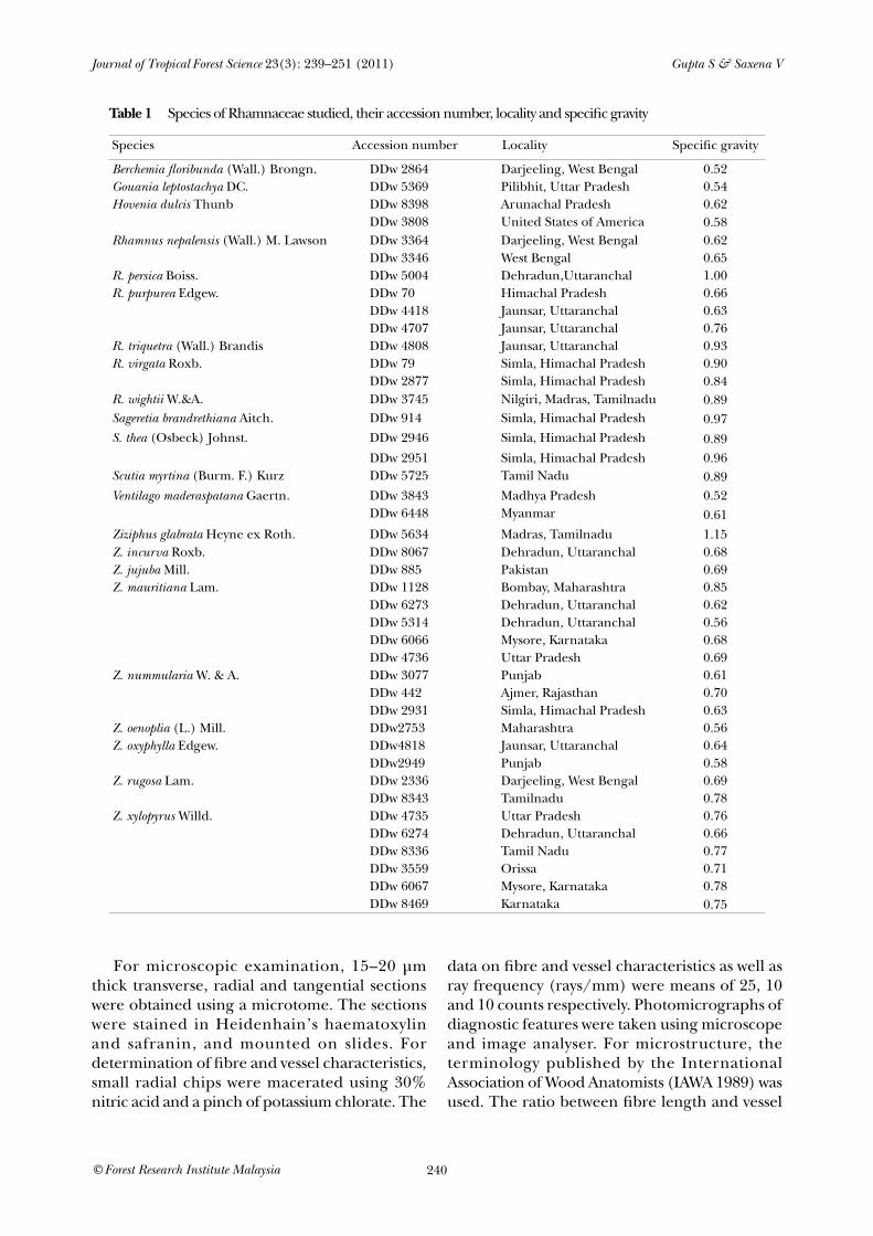
Species studied: Gouania leptostachya DC.Microscopic features: Growth rings distinct due to fibrous tissue, wood diffuse porous, vessels solitary or in radial multiples of 2–6 cells, very rarely clustered, round to oval shaped, perforation plate simple. Intervessel pits alternate, large, vessel ray pits are smaller than intervessel pits with 8–11 µm diameter. Fibres non-septate with simple pits. Axial parenchyma in marginal or in seemingly marginal bands 4–7 cells wide, scanty paratracheal, in strands of 3–7 cells. Rays 1–7 seriate, ray of two distinct sizes, uniseriate ray height 13–41 celled, mean multiseriate ray height more than 1 mm. Body ray cells homogeneous consisting of procumbent cells. Prismatic crystals non-chambered, rarely chambered, present in ray and parenchyma cells. Druses present in ray cells (Figure 1).
Hovenia thunb.
Species studied: Hovenia dulcis Thunb.Microscopic features: Growth rings indistinct. Wood semi-ring to diffuse porous. Vessels solitary or in radial multiples of 2–3 cells, round to oval shaped, perforation plate simple. Intervessel pits alternate, small to medium, vessel ray pits smaller than intervessel pits with 4–6 µm diameter. Fibres non-septate with minutely bordered pits. Axial parenchyma scanty paratracheal rarely diffuse, in strands of 5–8 cells. Rays 1–5 seriate, uniseriate ray height 3–8 celled, multiseriate ray height 6–25 celled. Body ray cells procumbent with 1–2 rows of upright and square cells. Rays sometimes irregularly storied. Prismatic non-chambered crystals present in ray cells. In the present study, two samples of H. dulcis were studied, of which one sample (DDw 8398) was from India and the other, (DDw 3808) from USA. The samples showed differences in ray seriation and parenchyma type (Table 4).
Rhamnus L.
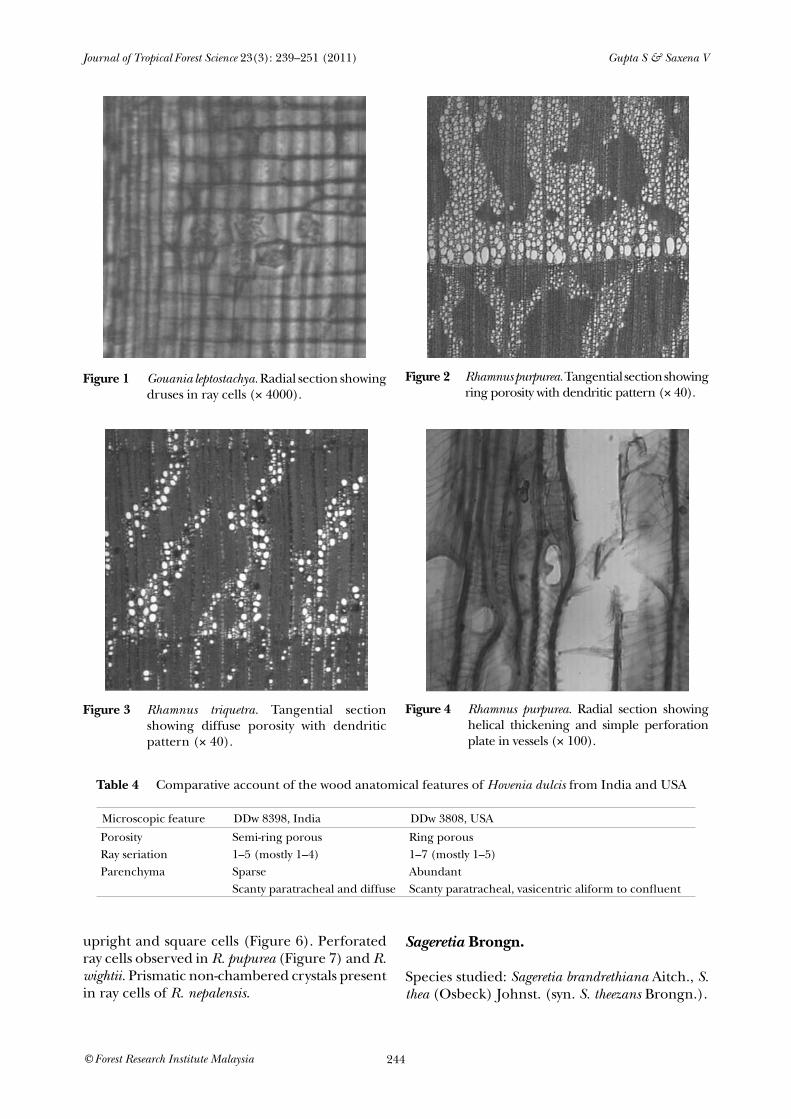
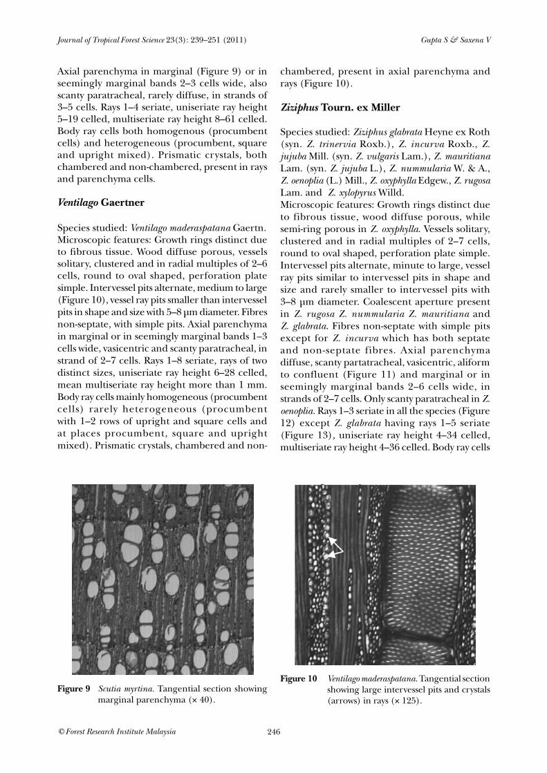
Species studied: Rhamnus nepalensis (Wall.) M. Lawson, R persica Boiss., R purpurea Edgew., R triquetra (Wall.) Brandis, R virgata Roxb. & R wightii W. & A.Microscopic features: Growth rings distinct due to fibrous tissue. Wood ring porous to semi-ring porous in R. nepalensis, R. purpurea (Figure 2), R. virgata and R. wightii while diffuse porous in R. persica and R. triquetra (Figure 3). Vessels arranged in dendritic pattern, round to oval shaped, perforation plate simple. Intervessel pits alternate, minute to large, coalescent aperture rarely present in R. virgata. Vessel ray pits are similar to intervessel pits in size and shape throughout the ray cells while rarely smaller to intervessel pits with 3–8 µm diameter in R. pupurea and R. virgata. Helical thickening present throughout body of vessel element (Figure 4). Fibres non-septate in R. persica, R. purpurea, R. triquetra and R. virgata while both septate and non-septate in R. wightii and R. nepalensis (Figure 5) with distinctly bordered pits and slit-like aperture. Axial parenchyma diffuse to scanty paratracheal in strands of 4–10 cells. Rays 1–6 seriate, rays of two types, uniseriate ray height 3–15 celled, multiseriate ray height 4–51 celled. Body ray cells procumbent with 1–3 rows of

Journal of Tropical Forest Science 23(3): 239–251 (2011) Gupta S & Saxena V
242© Forest Research Institute Malaysia
table 2 Wood anatomical characters of vessels and fibres in species of Rhamnaceae
Typ
e of
pla
nt
Vess
el in
den
driti
c pa
tter
n
Gro
wth
rin
g
Poro
sity
Mea
n ve
ssel
dia
met
er (
µm)
Vess
el fr
eque
ncy
per
mm
²
Mea
n ve
ssel
leng
th (
µm)
Inte
rves
sel p
it (µ
m)
Hel
ical
thic
keni
ng in
ves
sel
Mea
n fib
re le
ngth
(µm
)
Mea
n fib
re d
iam
eter
(µm
)
Sept
atio
n in
fibr
e
Vul
nera
bilit
y
Mes
omor
phy
F/V
rat
io
Berchemia floribunda
C/SS – – D 185(± 51)
10–28 437(± 72)
3–4 – 732(± 70)
17(± 1)
+ 9.7 4239 1.67
Gouania leptostachya
C/SS – + D 228(± 84)
7–21 430(± 105)
11–16 – 1106(± 293)
17(± 3)
– 16.2 6966 2.5
Hovenia dulcis T – – SR, D 106(± 20)
3–7 399(± 87)
5–9 – 1152(± 208)
22(± 6)
– 21.2 8459 2.8
Rhamnus nepalensis
S + + Dd-SR – – 409(± 83)
5–8 +, – 834(± 143)
17/21(± 3)
+, – – – 2.0
R. persica S/ST + + Dd-D 28(± 6)
118–127 286(± 47)
6–8 + 752(± 116)
17(± 5)
– – – 2.6
R. purpurea S/ST + + Dd-SR – – 299(± 74)
5–10 + 815(± 128)
16(± 3)
– – – 2.7
R. triquetra S/ST + + Dd-D 55(± 27)
98–121 339(± 59)
5–8 + 1349(± 219)
14(± 3)
– – – 2.2
R. virgata S/ST + + Dd-SR – – 252(± 25)
5–10 + 754(± 279)
13(± 6)
– – – 2.7
R. wightii S + + Dd-SR – – 360(± 100)
3–8 + 834(± 168)
14(± 3)
+, – – – 2.3
Sageretia brandrethiana
S/ST – + D 44(± 11)
98–126 249(± 44)
5–8 – 683(± 165)
14(± 23)
– 0.39 98 2.7
Sageretia thea S/ST – + D 43(± 14)
37–83 243(± 44)
2–3 – 578(± 165)
11(± 3)
– 0.75 132 2.4
Scutia myrtina S – + D 89(± 30)
34–63 363(± 76)
3–6 – 792(± 95)
13(± 2)
– 1.81 657 2.18
Ventilago maderaspatana
C/SS – + D 213(± 83)
5–15 344(± 207)
8–12 – 918(± 180)
14(± 3)
– 22 17220 1.3
Ziziphus glabrata
ST – + D 109(± 17)
36–67 403(± 90)
6–10 – 907(±43)
14(± 3)
– 2.2 756 2.67
Z. incurva S – + D 86(± 24)
14–33 383(± 23)
6–9 – 883 (±170)
15(± 2)
+, – 3.58 1372 2.3
Z. jujuba T – + D 109(± 26)
5–9 314(± 82)
4–8 – 1018(± 169)
17(± 4)
– 15.6 4889 3.2
Z. mauritiana T – + D 142(± 43)
4–28 352(± 58)
5–10 – 1002(± 171)
14(± 18)
– 13 4551 2.84
Z. nummularia S – + D 114(± 30)
3–22 404(± 18)
5–10 – 917(± 140)
15(± 13)
– 8.5 3444 2.26
Z. oenoplia C/SS – + D 111(± 22)
33–51 553(± 92)
5–8 – 903(± 155)
14(± 34)
– 2.64 1462 1.6
Z. oxyphylla S – + SR 68(± 21)
44–79 368(± 62)
3–5 – 604(± 76)
14(± 3)
– 1.09 401 1.6
Z. rugosa ST – + D 145(± 38)
9–15 577(± 99)
5–11 – 941(± 231)
17(± 4)
– 12.08 6970 1.63
Z. xylopyrus ST – + D 122(± 43)
5–20 411(± 111)
5–13 – 969(± 208)
16(± 4)
– 8.76 3781 2.35
– = absent, + = present, ± = rarely present; D = diffuse porous, SR = semi-ring porous, R = ring porous, Dd = dendritic pattern; C/SS = large climber or scrambling shrub, S/ST = straggling shrub or small tree, T = tree; F/V = fibre length/vessel length
Species

Journal of Tropical Forest Science 23(3): 239–251 (2011) Gupta S & Saxena V
243© Forest Research Institute Malaysia
table 3 Wood anatomical characters of axial parenchyma and rays in the studied species of Rhamnaceae
Parenchyma Ray
Dif
fuse
Scan
ty
para
trac
heal
Alif
orm
to c
onfl
uent
Vasi
cent
ric
Mar
gina
l
Seri
atio
n
Freq
uenc
y/m
m
Avg
uni
seri
ate
ray
wid
th
Avg
uni
seri
ate
ray
heig
ht
Avg
mul
tiser
iate
ray
w
idth
Avg
mul
tiser
iate
ray
he
ight
Stor
ied
Cry
stal
Dru
se
Pith
flec
k
Perf
orat
ed r
ay c
ell
Berchemia floribunda
– + – – – 1–8 4–8 15(± 3)
285(± 108)
56(± 26)
1092 ± 1011
– + R – – –
Gouania leptostachya
– ± – – + 1–7 5–8 11(± 2)
347(± 161)
57(± 23)
1695(± 835)
– + R, P R – –
Hovenia dulcis + + – – – 1–5 4–9 18(± 3)
213(± 61)
38(± 8)
363(± 164)
± + R – – –
Rhamnus nepalensis
– + – – – 1–6 4–9 13(± 2)
284(± 112)
46(± 11)
575(± 186)
– + R – + –
R. persica + – – – – 2–4 5–8 – – 19(± 6)
202(± 75)
– – – – –
R. purpurea – + – – – 1–6 4–10 13(± 3)
148(± 14)
35(± 11)
307(± 112)
– – – – +
R. triquetra – + – – – 1–6 4–7 13(± 2)
178(± 67)
51(± 11)
337(± 101)
– – – – –
R. virgata – + – – – 1–4 4–9 11(± 3)
140(± 42)
23(± 6)
260(± 93)
– – – – –
R. wightii + + – – – 1–5 4–9 14(± 2)
246(± 94)
27(± 7)
395(± 130)
– – – + +
Sageretia brandrethiana
+ ± – – + 1–4 13 13(± 2)
248(± 95)
29(± 3)
367(± 121)
± + R, P – – +
Sageretia thea + ± – ± + 1–4 6–12 12(± 3)
223(± 103)
22(± 4)
364(± 143)
– + R, P – – +
Scutia myrtina ± + – – + 1–4 6–12 12(± 3)
347(± 120)
28(± 5)
551(± 208)
– + R, P – – –
Ventilago maderaspatana
– + – + + 1–8 3–15 14(± 3)
244(± 104)
62(± 23)
1075(± 482)
– + R, P – – –
Ziziphus glabrata + + – + + 1–5 4–10 15(± 2)
144(± 39)
30(± 11)
395(± 114)
– + R, P – – –
Z. incurva – + ± + + 1 6–12 13(± 2)
318(± 126)
– – – + R, P R – –
Z. jujuba ± – + + + 1–3 6–10 23(± 4)
289(± 79)
43(± 8)
385(± 210)
– + R, ± P – – +
Z. mauritiana ± ± + + + 1–3 5–11 21(± 6)
363(± 202)
35(± 13)
498(± 239)
– + R – – +
Z. nummularia – – + + + 1–3 5–12 22(± 6)
399(± 205)
36(± 8)
401(± 163)
– + R – – –
Z. oenoplia – + – – – 1–2 7–12 15(± 2)
635(± 337)
20(± 4)
841(± 82)
– + R, P – – +
Z. oxyphylla ± + – ± ± 1–2 5–10 14(± 3)
544(± 304)
19(± 4)
590(± 304)
– + R, ± P – – +
Z. rugosa ± – ± + + 1–2 5–16 19(± 3)
678(± 424)
27(± 5)
529(± 149)
– + R – – +
Z. xylopyrus + – + + + 1–3 4–15 15(± 3)
342(± 120)
29(± 8)
391(± 121)
– + R – + +
Species
– = absent, + = present, ± = rarely present; R = ray, P = parenchyma; Avg = average
Journal of Tropical Forest Science 23(3): 239–251 (2011) Gupta S & Saxena V
244© Forest Research Institute Malaysia
upright and square cells (Figure 6). Perforated ray cells observed in R. pupurea (Figure 7) and R. wightii. Prismatic non-chambered crystals present in ray cells of R. nepalensis.
Sageretia Brongn.
Species studied: Sageretia brandrethiana Aitch., S. thea (Osbeck) Johnst. (syn. S. theezans Brongn.).
figure 2 Rhamnus purpurea. Tangential section showing ring porosity with dendritic pattern (× 40).
figure 3 Rhamnus triquetra. Tangential section showing diffuse porosity with dendritic pattern (× 40).
figure 4 Rhamnus purpurea. Radial section showing helical thickening and simple perforation plate in vessels (× 100).
figure 1 Gouania leptostachya. Radial section showing druses in ray cells (× 4000).
table 4 Comparative account of the wood anatomical features of Hovenia dulcis from India and USA
Microscopic feature DDw 8398, India DDw 3808, USA
Porosity Semi-ring porous Ring porousRay seriation 1–5 (mostly 1–4) 1–7 (mostly 1–5)Parenchyma Sparse Abundant
Scanty paratracheal and diffuse Scanty paratracheal, vasicentric aliform to confluent

Journal of Tropical Forest Science 23(3): 239–251 (2011) Gupta S & Saxena V
245© Forest Research Institute Malaysia
Microscopic features: Growth rings distinct due to parenchyma bands. Wood diffuse porous. Vessels solitary or in radial multiples of 2–6, round to oval shaped, perforation plate simple. Intervessel pits alternate, minute to medium, coalescent aperture present, vessel ray pits similar to intervessel pits in shape and size. Fibres non-septate with simple pits. Axial parenchyma marginal or in seemingly marginal bands 2–7 cells wide, diffuse, rarely vasicentric and scanty paratracheal, in strands of 2–4 cells. Rays 1–4 seriate, uniseriate ray height 4–19 celled, multiseriate ray height 8–37 celled. Body ray cells heterogeneous (procumbent, square and upright mixed in S. thea while procumbent and upright mixed in S. brandrethiana). Rays show storied tendency at few places in S. brandrethiana. Perforated ray cells
observed in S. brandrethiana (Figure 8). Prismatic crystals, both chambered and non-chambered, present in ray and parenchyma cells. Two distinct sizes of crystals in same ray cells present in S. thea.
Scutia (dc.) comm. ex Brongn.
Species studied: Scutia myrtina (Burm. F.) Kurz. (syn. S. indica Brongn.).Microscopic features: Growth rings distinct due to fibrous tissue, wood diffuse porous, vessels solitary, clustered and in radial multiples of 2–8 cells, round to oval shaped, perforation plate simple. Intervessel pits alternate, minute to small, vessel ray pits similar to intervessel pits in shape and size. Fibres non-septate with simple pits.
figure 5 Rhamnus nepalensis. Tangential section showing septate fibres (× 125).
figure 6 Rhamnus triquetra. Radial section showing heterogeneous rays (× 100).
figure 7 Rhamnus purpurea. Tangential section showing perforated ray cell (arrow) (× 400).
figure 8 Sageretia brandrethiana. Tangential section showing perforated ray cell (arrow) (× 400).
Journal of Tropical Forest Science 23(3): 239–251 (2011) Gupta S & Saxena V
246© Forest Research Institute Malaysia
Axial parenchyma in marginal (Figure 9) or in seemingly marginal bands 2–3 cells wide, also scanty paratracheal, rarely diffuse, in strands of 3–5 cells. Rays 1–4 seriate, uniseriate ray height 5–19 celled, multiseriate ray height 8–61 celled. Body ray cells both homogenous (procumbent cells) and heterogeneous (procumbent, square and upright mixed). Prismatic crystals, both chambered and non-chambered, present in rays and parenchyma cells.
Ventilago gaertner
Species studied: Ventilago maderaspatana Gaertn.Microscopic features: Growth rings distinct due to fibrous tissue. Wood diffuse porous, vessels solitary, clustered and in radial multiples of 2–6 cells, round to oval shaped, perforation plate simple. Intervessel pits alternate, medium to large (Figure 10), vessel ray pits smaller than intervessel pits in shape and size with 5–8 µm diameter. Fibres non-septate, with simple pits. Axial parenchyma in marginal or in seemingly marginal bands 1–3 cells wide, vasicentric and scanty paratracheal, in strand of 2–7 cells. Rays 1–8 seriate, rays of two distinct sizes, uniseriate ray height 6–28 celled, mean multiseriate ray height more than 1 mm. Body ray cells mainly homogeneous (procumbent cells) rarely heterogeneous (procumbent with 1–2 rows of upright and square cells and at places procumbent, square and upright mixed). Prismatic crystals, chambered and non-
chambered, present in axial parenchyma and rays (Figure 10).
Ziziphus tourn. ex Miller
Species studied: Ziziphus glabrata Heyne ex Roth (syn. Z. trinervia Roxb.), Z. incurva Roxb., Z. jujuba Mill. (syn. Z. vulgaris Lam.), Z. mauritiana Lam. (syn. Z. jujuba L.), Z. nummularia W. & A., Z. oenoplia (L.) Mill., Z. oxyphylla Edgew., Z. rugosa Lam. and Z. xylopyrus Willd. Microscopic features: Growth rings distinct due to fibrous tissue, wood diffuse porous, while semi-ring porous in Z. oxyphylla. Vessels solitary, clustered and in radial multiples of 2–7 cells, round to oval shaped, perforation plate simple. Intervessel pits alternate, minute to large, vessel ray pits similar to intervessel pits in shape and size and rarely smaller to intervessel pits with 3–8 µm diameter. Coalescent aperture present in Z. rugosa Z. nummularia Z. mauritiana and Z. glabrata. Fibres non-septate with simple pits except for Z. incurva which has both septate and non-septate fibres. Axial parenchyma diffuse, scanty partatracheal, vasicentric, aliform to confluent (Figure 11) and marginal or in seemingly marginal bands 2–6 cells wide, in strands of 2–7 cells. Only scanty paratracheal in Z. oenoplia. Rays 1–3 seriate in all the species (Figure 12) except Z. glabrata having rays 1–5 seriate (Figure 13), uniseriate ray height 4–34 celled, multiseriate ray height 4–36 celled. Body ray cells
figure 9 Scutia myrtina. Tangential section showing marginal parenchyma (× 40).
figure 10 Ventilago maderaspatana. Tangential section showing large intervessel pits and crystals (arrows) in rays (× 125).

Journal of Tropical Forest Science 23(3): 239–251 (2011) Gupta S & Saxena V
247© Forest Research Institute Malaysia
both homogeneous (procumbent cells) (Figure 14) and heterogeneous (procumbent with 1–2 rows of upright and square cells and procumbent, square and upright mixed). Perforated ray cells present in Z. jujuba (Figure 15), Z. mauritiana, Z. oenoplia, Z. oxyphylla and Z. xylopyrus (Figure 16). Prismatic crystals, both chambered and non-chambered, present in ray and parenchyma cells. Druses present in ray cells of Z. incurva.
Schiararend (1991) studied seven species (studied by us too) of Ziziphus viz. Z. mauritiana Lam. (syn. Z. jujuba Lam.), Z. jujuba Mill. (Z. vulgaris Lam.), Z. oenoplia (L.) Mill, Z. rugosa Lam., Z. xylopyrus Willd., Z. nummularia (Burm.) DC. and Z. glabrata Heyne ex Roth. A comparative account with the above data revealed differences in quantitative features, size of intervessel pits and mineral inclusions (Table 5).
figure 11 Zizyphus vulgaris. Tangential section showing diffuse, vasicentric, aliform to confluent parenchyma (× 40).
figure 12 Zizypus xylopyrus. Tangential showing rays exclusively 1–2 seriate (× 100).
figure 13 Zizypus glabrata. Tangential section showing rays 1–5 seriate (× 100).
figure 14 Zizypus xylopyrus. Radial section showing homogeneous rays with crystals (× 100).

Journal of Tropical Forest Science 23(3): 239–251 (2011) Gupta S & Saxena V
248© Forest Research Institute Malaysia
figure 15 Zizypus jujuba. Tangential section showing perforated ray cell (arrows) (× 100).
figure 16 Zizypus xylopyrus. Tangential section showing perforated ray cell (arrow) (× 100).
species identification key of the family 1. Vessels in dendritic pattern, helical
thickening present ...................................... 21*. Vessels not arranged in dendritic pattern,
helical thickening absent.............................. 72. Wood ring to semi-ring porous .................. 32*. Wood diffuse to semi-ring porous .............. 53. Vessels arranged in prominent dendritic
pattern, fibre non-septate... Rhamnus purpurea3*. Vessels show only a tendency of dendritic
pattern, fibre both septate to non-septate... 44. Rays mostly 1–5 seriate, rarely 1–6 seriate and
crystals in rays present .. Rhamnus nepalensis4*. Rays mostly 1–3 seriate, rarely 1–5 seriate and
crystals in rays absent ......... Rhamnus wightii
5. Rays 1–6 seriate ................. Rhamnus triquetra5*. Rays 1–4 seriate ........................................... 66. Intervessel pits 6–8 µm in diameter, ray vessel
pits similar to intervessel pits ... Rhamnus persica
6*. Intervessel pits 5–10 µm in diameter, ray vessel pits smaller to intervessel pits (4–8 µm) ...................................................... Rhamnus virgata
7. Rays 1–8 seriate, mean vessel diameter > 150 µm ............................................................ 8 7*. Rays < 5 seriate, mean vessel diameter < 150 µm ............................................................10 8. Intervessel pits minute with 3–4 µm diameter, fibre septate to non-septate and crystals in rays only ................................ Berchemia floribunda
table 5 Comparative account of the wood anatomical features of Ziziphus
Species Study Mean vessel
frequency
Mean vessel
diameter(µm)
Mean vessel length (µm)
Mean fibre
length(µm)
Ray seriation
Average ray
height(µm)
Inter- vessel
pit(µm)
Crystal in ray
Z. mauritiana Schiararend (1991) 10/mm² 125 410 780 1–2 370 8–12 ++
NA+
Present 4–28/mm² 142 352 1002 1–3 498 5–10 Z. jujuba Schiararend (1991) 33/mm² 90 360 750 1–2 480 8–12
Present 5–9/mm² 109 314 1018 1–3 385 4–8 Z. rugosa Schiararend (1991) 14/mm² 145 590 935 1–3 440 8–14 –
Present 9–15/mm² 145 577 941 1–3 529 5–11 +Z. xylopyrus Schiararend (1991) 10/mm² 110 405 860 1–2 500 8–14 +
+
NA+
NA+
Present 5–20/mm² 122 411 969 1–3 391 5–13Z. glabrata Schiararend (1991) 37/mm² 95 425 870 1–5
1–5NA
1–3
340 4–8 Present 36–67/mm² 109 403 907 395 6–10
Z. oenoplia Schiararend (1991) 33/mm² 110 500 750 315 8–12Present 33–51/mm² 111 553 903 841 5–8
NA = not available, + = present, – = absent

Journal of Tropical Forest Science 23(3): 239–251 (2011) Gupta S & Saxena V
249© Forest Research Institute Malaysia
8*. Intervessel pits medium to large with 8– 16 µm diameter, fibre non-septate and crystals in rays and axial parenchyma ..... 9
9. Druses in rays present, rays homogeneous, intervessel pits large (11–16µm) and axial parenchyma scanty paratracheal and marginal (4–7 cells wide)............................................................... Gouania leptostachya
9*. Druses in rays absent, rays heterogeneous, intervessel pits medium to large (8–12 µm) and parenchyma scanty paratracheal, vasicentric and marginal (2–7 cells).............................................Ventilago maderaspatana
10. Marginal parenchyma absent. Wood semi-ring to diffuse porous .............................. 11
10*. Marginal parenchyma present. Wood diffuse porous ...................................................... 12
11. Rays 1–5 seriate, intervessel pits small to medium (5–9 µm diameter)... Hovenia dulcis
11*. Rays 1–2 seriate, intervessel pits small (3– 5 µm diameter) ............... Ziziphus oxyphylla
12. Marginal parenchyma band 2–6 cells wide, mean vessel diameter < 50 µm ............... 13
12*. Marginal parenchyma band 1–3 cells wide, mean vessel diameter > 50 µm ................14
13. Intervessel pits minute with 2–3 µm diameter, rays not storied ....................... Sageretia thea
13*. Intervessel pits small to medium with 5– 8 µm diameter, rays storied at places ................................................ Sageretia brandrethiana
14. Rays 1–5 seriate, wood very hard and very heavy ........................................................ 15
14*. Rays 1–3 seriate, wood hard and moderately heavy to heavy .......................................... 16
15. Rays 1–4 seriate, intervessel pits < 6 µm, mean uniseriate ray height > 200 µm, mean multiseriate ray height > 400 µm .................................................................... Scutia myrtina
15*. Rays 1–5 seriate, intervessel pits > 6 µm, mean uniseriate ray height < 200 µm, mean multiseriate ray height < 400 µm ................................................................. Ziziphus glabrata
16. Vessel diameter < 70 µm, vessel frequency > 50 mm2 , wood semi-ring porous, parenchyma scanty paratracheal, rarely marginal, exclusively uniseriate rays ...................................................................... Ziziphus oxyphylla
16*. Vessel diameter > 70 µm, vessel frequency < 50 mm2, diffuse porous, parenchyma aliform and marginal, rays uni to biseriate, rarely triseriate .......................................... 17
17. Druses present in rays, fibres both septate and non-septate ......................... Ziziphus incurva
17*. Druses absent, fibres non-septate ...........1818. Axial parenchyma scanty paratracheal,
multiseriate ray height > 700 µm .............................................................. Ziziphus oenoplia
18*. Axial parenchyma scanty paratracheal, aliform, vasicentric and marginal (1–2 cells wide) multiseriate ray height < 700 µm ............................................................................ 19
19. Intervessel pits 4–8µm diameter, vessel frequency < 8 mm2, uniseriate ray height < 300 µm ................................. Ziziphus jujuba
19*. Intervessel pits 5–10 µm (rarely 12 µm), vessel frequency > 8 mm2, uniseriate ray height > 300 µm ......................................... 20 20. Mean uniseriate ray height > 400 µm, axial parenchyma scanty paratracheal and narrow bands ................................... Ziziphus rugosa20*. Mean uniseriate ray height 300–400 µm, axial
parenchyma vasicentric, aliform with short lateral extensions and narrow bands ....... 21
21. Mean multiseriate ray height > 450 µm ................................................. Ziziphus mauritiana
21*. Mean multiseriate ray height < 450 µm .... 2222. Mean uniseriate ray height > 350 µm .............
..................................... Ziziphus nummularia 22*.Mean uniseriate ray height < 350 µm ..............
.......................................... Ziziphus xylopyrus
discussion The study indicated that there was very little difference in anatomy within a genus and most of the species of a genus mainly differed from one another in a few quantitative features, some of which might be site dependent. Gouania leptostachya and V. maderaspatana are large climbers or scrambling shrubs showing quite similar anatomy with soft, usually light (specific gravity 0.44–0.66 air dry), coarse-textured wood containing large pores, large intervessel pits, marginal parenchyma, non-septate fibres and high rays. Although B. floribunda and Z. oenoplia are also climbers/scrambling shrubs, their anatomies differ from those of Gouania and Ventilago species. The F/V ratio (Table 2) suggested that G. leptostachya, H. dulcis, R. persica, R. virgata, S. brandrethiana, Z. glabrata, Z. jujuba and Z. mauritiana had a highly evolved mechanical system with a high degree of intrusiveness (F/V = ≥ 2.5), while V. maderaspatana showed a lower ratio

Journal of Tropical Forest Science 23(3): 239–251 (2011) Gupta S & Saxena V
250© Forest Research Institute Malaysia
(F/V = 1.3) indicating primitive features and a low degree of intrusiveness. The vulnerability and mesomorphy figures calculated as per formula given by Carlquist (1977) indicated that all the species were mesic except for S. brandrethiana and S. thea which showed xeric values (Table 2). The characters mentioned in the Inside Wood database for fossil wood of Hovenia dulcis were quite different while those of Z. jujuba and Z. xylopyrus matched the present study. The study of two samples of H. dulcis from India and USA showed differences in ray seriation and parenchyma type reflecting that within a species these features vary. Similarly, a comparative account of the present study with Schiararend’s (1991) seven species of Ziziphus revealed differences in quantitative features, size of intervessel pits and mineral inclusions indicating that even feature like ‘size of intervessel pits’, which usually was considered to be a fixed character, varied greatly. Thus, it can be concluded that feature variation within a species is of much importance especially for the preparation of species identification key. Rhamnaceae has been studied by a number of researchers but the presence of perforated ray cells has never been reported. In the present study this feature was observed in S. brandrethiana, S. thea, R. purpurea, R. wightii, Z. jujuba, Z. mauritiana, Z. oenoplia, Z. oxyphylla, Z. rugosa and Z. xylopyrus. Of the 41 wood samples studied, perforated ray cells were observed in 13 wood samples of the said species. These perforated ray cells occurred as uniseriate extensions of multiseriate rays. The perforation plates in the perforated ray cells were all simple. The ray cells having perforation plates were generally much larger than the surrounding ray cells and were of the same size as the vessel.
systematic position of the family
Schiararend (1991) investigated the systematic wood anatomy of the Rhamnaceae (Rhamnales), tribe Zizipheae. He divided the genus Ziziphus into three group, namely, Ziziphus-A, Ziziphus-B and Ziziphus-C on the basis of ray width, parenchyma distribution and type of vessel perforation. The present study revealed that parenchyma type varied with site. All our samples were found to have simple perforation plate. Our study supports formation of two groups on the basis of ray seriation only: one with 1–3 seriate rays and the other with 1–5 seriate.
Suessenguth (1953) divided the family Rhamnaceae into five tribes, of which four, viz. Gouanieae, Rhamneae, Ventilagineae and Zizipheae, occurred in India. The present study showed that the tribes Rhamneae and Zizipheae were quite heterogeneous. Gouanieae and Ventilagineae had single genus and hence no comparison of these tribes could be done. Richardson et al. (2000a) revised this classification and divided Rhamnaceae into 12 tribes, of which four tribes (Rhamneae, Ventilagineae, Paliureae and Gouanieae) were represented in India. Suessenguth (1953) placed Hovenia, Rhamnus, Sageretia and Scutia together in the tribe Rhamneae, and Berchemia and Ziziphus together in the tribe Ziziphae. Richardson et al. (2000a) placed Berchemia with Rhamnus, Sageretia and Scutia in the tribe Rhamneae and Hovenia and Ziziphus in different tribe Paliureae. This classification of Rhamnaceae was again revised by Richardson et al. (2000b) on the basis of DNA sequences-rbcL analysis. Our studied genera were placed in the same tribes as in his previous classification. The present study revealed that the genera placed in the tribe Rhamneae and Ziziphae by Suessenguth (1953) and the tribes Rhamneae and Paliureae by Richardson et al. (2000a, b) are dissimilar (Tables 2 and 3) in their wood anatomy and hence need to be revised.
acKnoWLedgeMents
The authors are thankful to SS Negi, Director of the Forest Research Institute and S Nautiyal, Head of the Botany Division for administrative support. The financial support from the Indian Council of Forestry Research and Education, Dehradun is duly acknowledged.
references
Anonymous 1963. Indian Woods, Their Identification, Properties and Uses.Volume 2. Forest Research Institute, Dehradun.
BhAndAri mm & AK BhAnsAli. 2000. Rhamnaceae. Pp 161–245 in Singh NP et al. (Eds) Flora of India. Botanical Survey of India, Calcutta.
CArlquist s. 1977. Ecological factors in wood evolution: a floristic approach. American Journal of Botany 64: 704–713.
CArlquist s. 1988. Comparative Wood Anatomy: Systematic, Ecological, and Evolutionary Aspects of Dicotyledon Wood. Springer-Verlag, Berlin.

Journal of Tropical Forest Science 23(3): 239–251 (2011) Gupta S & Saxena V
251© Forest Research Institute Malaysia
GAmBle Js.1922. A Manual of Indian Timbers. Sampson Low, Marston and Co. Ltd, London.
iAWA. 1989. IAWA list of microscopic features by IAWA committee for hardwoods identification. IAWA Bulletin n.s. 10: 219–332.
metCAlfe Cr & ChAlK l. 1950. Anatomy of the Dicotyledons. Clarendon Press, Oxford.
PeArson rs & BroWn hP. 1932. The Commercial Timbers of India. Volume 1. Central Publication Branch, Calcutta.
riChArdson Je, fAy mf, CronK qCB & ChAse mW. 2000a. A revision of the tribal classification of Rhamnaceae. Kew Bulletin 55: 311–340.
riChArdson Je, fAy mf, CronK qCB, BoWmAn d & ChAse mW . 2000b. A phylogenetic analysis of Rhamnaceae using rbcL and trnL-F plastid DNA sequences. American Journal of Botany 87: 1309–1324.
sChirArend C. 1991. The systematic wood anatomy of the Rhamnaceae Juss. (Rhamnales). I. Tribe Zizipheae. IAWA Bulletin n.s. 12: 359–388.
suessenGuth K. 1953. Rhamnaceae, Vitaceae, Leeaceae. In Engler A & Prantl K (Eds) Die Naturlichen Pflanzenfamilien 2. Duncker and Humbolt, Berlin.