wing myology of caracaras (aves, falconiformes): muscular ... · p, m.b.j. & m, m.c.: ing...
TRANSCRIPT
177ISSN 1864-5755
68 (2): 177 –190
15.8.2018© Senckenberg Gesellschaft für Naturforschung, 2018.
Wing myology of Caracaras (Aves, Falconiformes): muscular features associated with flight behavior
Mariana Beatriz Julieta Picasso 1 * & María Clelia Mosto 1
1 Museo de La Plata-Facultad de Ciencias Naturales y Museo, Universidad Nacional de La Plata – CONICET. Paseo del Bosque s/n, La Plata (1900), Buenos Aires, Argentina — *corresponding author: [email protected]
Accepted July 12, 2018. Published online at www.senckenberg.de/vertebrate-zoology on July 27, 2018.
Editor in charge: Martin Päckert
AbstractCaracaras (Aves, Falconiformes, Falconidae) are Neotropical diurnal raptors that belong to the subfamily Polyborinae. The forelimb myology of this group has not been comprehensively studied or compared with that of other Falconidae. Thus, the aims of this study were i) to describe the forelimb muscles of two species of Polyborinae (Caracara plancus and Milvago chimango), ii) to explore the possible relationship between muscular features and their function in flight behavior and iii) to compare the muscular features of these species with those of species of the subfamily Falconinae. To this end, the forelimb muscles of C. plancus (n = 4) and M. chimango (n = 4) were dissected. Additonally, to complement this data set, two specimens of M. chimachima were dissected. The mass of each muscle of one wing and its percentage with respect to the body mass were obtained. A total of 45 muscles were identified, and differences with respect to Falconinae were related to the presence of single or additional bellies. The total forelimb muscle mass represented between 7.68 and 10.26 % of the body mass. The muscle pectoralis represented ̴ 5% of the body mass, followed by the muscles scapulohumeralis caudalis (0.64 – 0.79%), deltoideus major (0.43 – 0.53%), supracoracoideus (0.34 – 0.38%) and biceps brachii (0.26 – 0.39%). The high values of these muscles are in agreement with their important function: they are involved in the downstroke and upstroke phases of the flapping flight. On the other hand, the muscles that seemed to contribute little to the mechanical power for flight presented low values that ranged between 0.01 and 0.25%. Comparison of the forelimb muscles of caracaras with published data on Falconinae species suggests that their muscular features might be associated with their type of flight, which is more erratic and less powerful than that of Falconinae.
Key wordsForelimb morphology, Falconidae, Polyborinae.
Introduction
Since flight is a demanding mode of locomotion, the wings of flying birds have numerous anatomical features to meet this demand (Heers et al., 2016). These include a complex and varied morphology comprising many muscles (between 45 and 50) (GeorGe & BerGer, 1966; rai-kow, 1985), which generate and control their movements (Videler, 2006) and the different flight behaviors (e.g. flapping, gliding, hovering, etc.). Although the features of these muscles, i.e. number of bellies, relative development and mass, vary between taxa (Hartman, 1961; GeorGe & BerGer, 1966), this variability and its possible association with flight behavior have been scarcely investigated both in birds in general and particularly in
Falconiformes (see Hartman, 1961; meyers, 1992a, b; CorVidae et al., 2006; Hertel, maldonado & sustaita, 2014; CanoVa et al., 2015a, b, c). Although the Falconidae have been classically grouped together with the Accipitridae (hawks, eagles, kites) due to their morphology, there is a general consensus that the Falconidae are phylogenetically distant from these other diurnal birds of prey (HaCkett, et al., 2008; JarVis et al., 2014; Prum et al., 2015). The family Falconidae consists of three subfamilies, Polyborinae Falconinae and Herpetotherinae, which show diversity in their locomotor behavior and diet. The Polyborinae (caracaras) are endemic to the New World and distinguished

Picasso, M.B.J. & Mosto, M.C.: Wing myology of Caracaras (Aves, Falconiformes)
178
as ambulatory birds that mostly forage by walking on the ground and in which flight is not as important as in the other two families (Cade & diGBy, 1982; wHite, et al., 1994; FuCHs, et al. 2012). Moreover, the caracaras have an erratic and slow flight in which they alternate flapping and gliding (CaneVari et al., 1991; wHite et al., 1994). The caracaras also have a diverse diet: some genera like Daptrius and Ibicter are omnivorous and arboreal, whereas other genera, such as Milvago, Caracara and Phalcoboenus, are opportunistic, feeding on invertebrates, vertebrates, garbage and carrion (CaneVari et al., 1991; Grin, 2018). In contrast, the Falconinae (cosmopolitan falcons) have a more diverse and complex flight behavior: they hunt in flight at high speeds to strike their prey (mainly birds and insects) and some species can also hover or soar (wHite et al., 1994; sustaita, 2008). Finally, the Herpetotherinae (forest and laughing falcons) are secretive birds that inhabit the humid forests of the Neotropics and are poorly known (wHite et al., 1994). Despite these interesting flight variations, little is known about the forelimb myology of the Polyborinae, and previous research consists mainly on descriptive studies based on the Falconinae. BerGer (1956), studied the appendicular myology of Polihierax semitorquatus (Pygmy falcon), and described only 18 muscles of the wing. Jollie (1977), in his extensive study on birds of prey, briefly mentioned some muscles for the forelimbs of Falco sparverius (American kestrel) and Polyborus cheriway, with scarce information and few illustrations. meyers (1992a, 1996) studied the brachial and antebrachial muscles of Falco sparverius, being, to date, the most complete descriptive works on the forelimb myology of Falconidae. More recently, in a comparative framework with other nonFalconiformes bird species, CanoVa et al. (2015 a, b) studied some muscles of Falco tinnunculus. The only quantitative information regarding the mass of wing muscles was published by Hartman in 1961. However, these data do not refer to individual muscles, but to the entire wing, with the exception of the muscle pectoralis and the muscle supracoracoideus. Thus, to expand our knowledge on the forelimb myology of the Polyborinae, the aims of the present study were i) to describe, photograph and illustrate the forelimb muscles of three species of Polyborinae (Caracara plan-cus, Milvago chimachima and Milvago chimango), ii) to explore the possible relationship between the muscular features of these species and their function in flight behavior, and iii) to compare the muscular features of these species with those of species of the subfamily Falconinae.
Materials and methods
Specimens: Healthy and unsexed adults of Caracara plancus (crested caracara, n=4), Milvago chimango (chimango caracara, n=4) and Milvago chimachima (yel
low headed caracara, n=2) were used. The specimens of C. plancus and M. chimachima were obtained from La Marcela farm (26°17035″S; 59°06067″W), Pirané, Formosa province, Argentina, with authorization of Ministerio de la Producción y Ambiente, Dirección de Fauna y Parques of Formosa Province (guía de tránsito nº 003384) during 2014. The specimens of M. chimango were obtained from Programa de Control de Aves en Rellenos Sanitarios y Areas Aledañas (ProCoA) in the province of Buenos Aires, Argentina, during 2013. This research complied with protocols approved by the animal care committee and adhered to the legal requirements of Argentina.
Data collection: The body mass of each specimen was weighed with a digital scale, except for that of the two specimens of M. chimachima, which was taken from dunninG (2008). Then, the anterior corporal region was carefully separated from the rest of the body, without damaging the muscles. The hind limbs were studied in mosto et al. (2013; 2016). Each individual region was properly identified and stored in individual bags and frozen until it was studied. The left wing muscles were unilaterally dissected during the six months following storage. The wing muscles were defrosted, identified, photographed, carefully removed and weighed (both the fleshy and tendinous components) with a digital scale (with 0.01g accuracy). The percentage of each muscle with respect to its body mass was calculated considering one wing. Also, the forelimb muscle mass was calculated as the sum of the individual muscles except the small muscles of the manus (mm. abductor digiti majoris, ex-tensor brevis alulae, abductor alulae, flexor digiti mino-ris, flexor alulae, and adductor alulae), which give little mechanical power for flight (Biewener, 2011). Muscular mass data were explored considering the movements of the main bones and joints, including those of the muscles associated with the thoracic shoulder (ossa cinguli), the arm (humerus), the forearm (ulna and radius), the elbow (the juncture cubiti) and the wrist joint (juncture carpi). The main roles of the muscles during flight were determined following that described in the works of raikow (1985), dial (1992) and Vazquez (1995, Table 1). The muscular description is the same in the three species and only the differences among them are mentioned. The interpretations about muscle mass were based only on the data obtained for C. plancus and M. chimango, given that the data available are more complete. Similarly, the interpretation about the small muscles of the manus and digits was based on the complete data obtained for C. plancus. Because of the low number of specimens of M. chimachima, the muscle mass data are provided only as complementary data. The mm. subcoracoideus and sub-scapularis were weighed together because of the common tendon of insertion, and the small m. propatagialis p. caudalis was not weighed. The anatomical nomenclature follows Baumel et al. (1993), and the abbreviations used in text are as follows: m. (muscle), mm. (muscles), and p. (pars).

179
VERTEBRATE ZOOLOGY — 68 (2) 2018
Tabl
e 1.
Win
g m
uscl
es, t
he m
ean
mus
cula
r mas
s (g)
, sta
ndar
d de
viat
ion
(SD
) and
per
cent
age
of b
ody
mas
s (%
BM
), in
bra
cket
s, th
e av
erag
e bo
dy m
ass.
DW
/UP:
hum
eral
mus
cles
that
act
s dur
ing
the
dow
nst
roke
and
ups
troke
resp
ectiv
ely
(fol
low
ing
Dia
l, 19
92).
Mus
cle
func
tion
acco
rdin
g to
: 1 R
aiko
w (1
985)
, 2 D
ial (
1992
), 3 V
azqu
ez (1
995)
.
Mus
cle
Mai
n fu
nctio
nC
arac
ara
plan
cus
(135
8.00
g)
Milv
ago
chim
ango
(302
.50g
)M
ilvag
o ch
imac
him
a( 3
15.5
0g)
Mea
nSD
%B
MM
ean
SD%
BM
Mea
nSD
%B
MM
. lat
issi
mus
dor
si c
rani
alis
retra
cts t
he h
umer
us 1
0.83
0.27
0.06
0.10
0.04
0.03
0.22
0.13
0.07
M. l
atis
sim
us d
orsi
cau
dalis
1.68
0.33
0.12
0.32
0.03
0.11
0.21
0.13
0.06
M. r
hom
boid
eus (
supe
rfici
alis
+ p
rofu
ndus
)st
abili
zatio
n of
the
thor
acic
gird
le 1
2.38
0.71
0.17
0.41
0.02
0.14
0.41
0.10
0.13
M. s
erra
tus s
uper
ficia
lis c
rani
alis
st
abili
zatio
n of
the
thor
acic
gird
le 1
0.37
0.09
0.03
0.09
0.04
0.03
0.05
0.01
0.02
M. s
erra
tus s
uper
ficia
lis c
auda
lis1.
100.
380.
080.
220.
070.
070.
210.
060.
06M
. ser
ratu
s pro
fund
us
0.62
0.12
0.05
0.10
0.06
0.03
0.06
0.03
0.02
M. p
ecto
ralis
p. t
hora
cica
retra
cts a
nd d
epre
ss th
e hu
mer
us/D
W 2
71.9
710
.73
5.27
15.9
92.
005.
2815
.26
0.79
4.84
M. d
elto
ideu
s pro
pata
gial
is p
. cra
nial
is
tens
e th
e pr
opat
agiu
m 2
2.59
1.41
0.19
0.38
0.10
0.12
0.36
0.01
0.11
M. d
elto
ideu
s pro
pata
gial
is p
. cau
dalis
te
nse
the
prop
atag
ium
2—
——
——
——
——
M. s
upra
cora
coid
eus
elev
ates
hum
erus
/UP 2
5.26
0.96
0.38
1.04
0.14
0.34
0.68
0.01
0.21
M. d
elto
ideu
s p. m
ajor
el
evat
ion
of h
umer
us a
nd w
ings
/UP 2
7.25
2.30
0.53
1.29
0.19
0.43
1.26
0.16
0.40
M. d
elto
ideu
s p. m
inor
Pr
otra
cts a
nd e
leva
tes t
he h
umer
us 1
0.29
0.23
0.02
0.01
—0.
000.
12—
0.04
M. c
orac
obra
chia
lis c
rani
alis
pr
otra
cts a
nd d
epre
ss th
e hu
mer
us 1
0.63
0.17
0.05
0.18
0.11
0.06
0.11
0.01
0.03
M. c
orac
obra
chia
lis c
auda
lis
depr
ess a
nd ro
tate
s the
hum
erus
/DW
12.
591.
370.
190.
380.
090.
130.
290.
060.
09M
. sca
pulo
hum
eral
is c
auda
lisre
tract
s the
hum
erus
, rot
atio
n of
the
win
g/D
W 2
1.08
0.33
0.79
0.11
0.26
0.64
0.04
0.20
—M
. sca
pulo
hum
eral
is c
rani
alis
retra
cts t
he h
umer
us 1
0.01
0.00
0.02
0.01
0.01
——
——
M. s
ubsc
apul
aris
re
tract
s the
hum
erus
/DW
2
2.31
0.75
0.17
0.53
0.03
0.18
0.41
0.04
0.13
M. s
ubco
raco
ideu
sde
pres
s and
rota
tes t
he h
umer
us d
orsa
lly/D
W 2
M. s
capu
lotr
icep
s st
abili
zes t
he e
lbow
/UP 2
4.16
1.20
0.30
0.63
0.15
0.21
0.64
0.11
0.20
M. h
umer
otri
ceps
ex
tend
s the
elb
ow/D
W 2
4.47
1.20
0.33
0.74
0.05
0.25
0.64
0.05
0.20
M. b
icep
s bra
chii
stab
ilize
s the
elb
ow, fl
ex th
e fo
rear
m/D
W 2
5.34
1.11
0.39
0.80
0.08
0.26
0.94
0.21
0.30
M. b
rach
ialis
fle
x th
e el
bow
10.
490.
150.
040.
080.
020.
030.
08—
0.03
M. p
rona
tor s
uper
ficia
lis
vent
ral r
otat
ion
of th
e w
ing 2
0.97
0.27
0.07
0.15
0.03
0.05
0.12
0.00
0.04
M. p
rona
tor p
rofu
ndus
ve
ntra
l rot
atio
n of
fore
arm
11.
940.
390.
140.
360.
040.
120.
310.
060.
10M
. flex
or d
igito
rum
supe
rfici
alis
fle
x th
e w
rist a
nd e
xten
d di
giti
maj
oris
30.
660.
150.
050.
140.
040.
050.
110.
040.
03M
. flex
or c
arpi
uln
aris
fle
x th
e w
rist 2
2.25
0.72
0.17
0.49
0.07
0.16
0.43
0.09
0.13
M. fl
exor
dig
itoru
m p
rofu
ndus
fle
x th
e w
rist 3
0.72
0.20
0.05
0.16
0.03
0.05
0.12
0.00
0.04
M. u
lno-
met
acar
palis
ven
tral
is
flex
and
pron
ates
the
wris
t 30.
580.
120.
040.
120.
010.
040.
120.
040.
04M
. sup
inat
orR
otat
es fo
rear
m 1
0.31
0.08
0.02
0.06
0.02
0.02
0.02
—0.
01M
. ext
enso
r car
pi ra
dial
is
exte
nds t
he w
rist 2
2.43
0.41
0.18
0.42
0.05
0.14
0.43
0.03
0.14
M. e
xten
sor d
igito
rum
com
mun
is
wris
t flex
ion
and
stab
iliza
tion 3
0.69
0.13
0.05
0.14
0.02
0.04
0.11
0.01
0.03
M. e
xten
sor c
arpi
uln
aris
fle
x th
e w
rist 3
0.77
0.23
0.06
0.14
0.02
0.05
0.11
0.02
0.03
M. e
ctep
icon
dilo
uln
aris
el
evat
es fo
rear
m 1
1.02
0.21
0.07
0.19
0.02
0.06
0.13
0.01
0.04
M. e
xten
sor l
ongu
s alu
lae
exte
nds t
he w
rist 3
0.83
0.31
0.06
0.15
0.02
0.05
0.13
0.01
0.04
M. e
xten
sor l
ongu
s dig
iti m
ajor
is
exte
ds d
igiti
maj
oris
30.
530.
110.
040.
120.
020.
040.
080.
030.
03

Picasso, M.B.J. & Mosto, M.C.: Wing myology of Caracaras (Aves, Falconiformes)
180
Results
Muscular description
The m. latissimus dorsi (Fig. 1A – D) has two independent portions: the p. cranialis and the p. caudalis. The p. cranialis (Fig. 1A – C) is a fleshy band-shaped muscle that originates on the spinous process of the cervical vertebrae and inserts into the medial facet of the corpus humeri, covering the second proximal quarter (Fig. 2A).
The p. caudalis (Fig. 1A – D) is also a fleshy band-shaped muscle, although wider and thicker than the p. cranialis, which is situated on the m. rhomboideus superficialis. It inserts into the humerus via a tendon (Fig. 2A), which is common to that of the origin of the m. scapulotriceps.
The m. rhomboideus superficialis (Fig. 1A – D) is a fleshy muscle with two distinct and contiguous fleshy portions (cranial and caudal) that originate on the last cervical ver-tebrae. The cranial portion is squareshaped and inserts into the first quarter of the scapula (Fig. 2C), whereas the caudal portion is wider than the cranial one and inserts into the scapula, reaching the region of the angulus sub-terminalis (sensu liVezey & suzy, 2006; Fig. 2C).
The m. rhomboideus profundus (Fig. 2D) is a fleshy muscle that originates on the cervical vertebrae and inserts into the medial surface of the scapula. It is located right beneath the m. rhomboideus superficialis, from which it is difficult to separate.
The m. serratus superficialis (Fig. 1A) consists of two independent bellies: the p. cranialis and the p. caudalis. The p. cranialis is a fleshy band-shaped muscle that originates on the ribs near the processus uncinatus, and attaches on the cranial half of the scapula between the two bellies of the m. subescapularis (Fig. 2D). The p. cauda-lis is a fleshy, broad and flat muscle that originates on the caudal ribs, covering the processus uncinatus, and inserts into the medial surface and ventral end of the scapula, on the last caudal third (Fig. 2C).
The m. serratus profundus is a fleshy and flat muscle that originates on the first ribs and inserts into the medial surface at the third caudal quarter of the scapula (Fig. 2D).
The m. pectoralis p. thoracica (Fig. 3A, C) is a fleshy welldeveloped muscle that originates on various structures: the carina sterni, the ventral aspect of the clavicula (Fig. 2B) and the membranes sternocoracoclavicularis and cristoclavicularis. The muscle inserts into the proximal region of the corpus humeri on the crista deltopec-toralis (Fig. 2B).
The m. pectoralis propatagialis p. longus (Fig. 4A, B) is a small, fleshy and elongated portion of the muscle pec-toralis p. thoracica, with an elastic tendon that penetrates the propatagium and gives support to its leading edge. Ta
ble
1 co
ntin
ued.
Mus
cle
Mai
n fu
nctio
nC
arac
ara
plan
cus
(135
8.00
g)
Milv
ago
chim
ango
(302
.50g
)M
ilvag
o ch
imac
him
a( 3
15.5
0g)
Mea
nSD
%B
MM
ean
SD%
BM
Mea
nSD
%B
MM
. uln
omet
acar
palis
dor
salis
fle
x th
e w
rist 3
0.57
0.07
0.04
0.06
0.00
0.02
0.05
0.03
0.02
M. i
nter
osse
ous d
orsa
lis
elev
ates
dig
iti m
ajor
is 3
0.19
0.03
0.01
0.04
—0.
010.
04—
0.01
M. i
nter
osse
us v
entr
alis
fle
x di
giti
maj
oris
b 30.
170.
030.
010.
050.
020.
020.
03—
0.01
M. e
xten
sor b
revi
s alu
lae
exte
nds a
lula
r dig
it 3
0.06
0.05
0.01
——
—
—
—M
. abd
ucto
r alu
lae
exte
nds a
nd d
epre
ss a
lula
r dig
it 3
0.09
0.02
0.01
——
—
——
—M
. flex
or a
lula
e de
pres
s alu
lar d
igit
30.
070.
060.
01
——
——
—M
. add
ucto
r alu
lae
flex
alul
ar d
igit
30.
220.
080.
02—
——
——
—M
. abd
ucto
r dig
iti m
ajor
is
exte
nds d
igiti
maj
oris
30.
140.
110.
01—
——
——
—M
. flex
or d
igiti
min
oris
fle
x d
igiti
maj
oris
and
men
oris
30.
080.
020.
01—
——
——
—Fo
relim
b m
ass
130.
62—
10.2
624
.27
—-
7.68
——
—U
pstr
oke
mus
cles
16.6
7
—1.
222.
96
—
-0.
98—
——
Dow
nstr
oke
mus
cles
87.7
6
—7.
1418
.56
—-
6.73
——
—

181
VERTEBRATE ZOOLOGY — 68 (2) 2018
The tendon inserts into the thick aponeurosis that covers the carpometacarpus, on the region of the processus extensorius.
The m. deltoideus propatagialis p.cranialis (Fig. 4A – B) is a well-developed muscle that originates fleshy on the dorsal aspect of the furcula (Fig. 3B). The belly gives rise to a tendon that penetrates the patagium and bifurcates into two tendons, both of which insert into the tendon of origin of the m. extensor metacarpi radialis.
The m. deltoideus propatagialis p. caudalis (Fig. 4B) is a small fleshy cranial portion of the m. deltoideus propa-tagialis p. cranialis, from which it is separated by a thin tendinous raphe. The muscle has a short tendon that fuses to the elastic tendon of the m. pectoralis propatagialis p. longus.
The m. supracoracoideus (Fig. 5A – B) is a welldeveloped muscle that lies deep to the m. pectoralis, with a fleshy origin on the cranial half of the sternum (Fig. 3A), along the coracoideum (Fig. 3E) and on the membrane sternoclavicularis. The muscle passes through the cana-lis triosseus and inserts into the proximal end of the hu-merus, on the tuberculum dorsale (Fig. 2A).
The m. deltoideus p. major (Figs. 1A – B and 4A) has two bellies that join together and insert via a tendon along
the crista deltopectoralis of the humerus (Fig. 2A). The p. cranialis originates fleshy on the fibrocartilage hum-eroscapsularis and adjacent ligaments. The p. caudalis is fleshy and originates on the scapula (Fig. 2D).
The m. deltoideus p. minor (Fig. 1A, B) is a poorly developed muscle located on the tendon of insertion of the m. supracoracoideus.
The m. coracobrachialis cranialis (Fig. 5B) is a short fleshy muscle that originates on the coracoideum, on the extremitas omalis coracoidei (Fig. 3E). The muscle crosses the joint and inserts into the extremitas proxima-lis humeri (Fig. 2B).
The m. coracobrachialis caudalis (Fig. 5A – B) originates fleshy on the sternal region, proximal to the cora-coideum and the adjacent sternum (Fig. 3A, E). This muscle inserts by a tendon on the tuberculum ventrale of the humerus (Fig. 2A).
The m. scapulohumeralis cranialis (Fig. 1C – D) is a small fleshy muscle that originates on the dorsal surface of the scapula and inserts into the humerus near the fo-ramen pneumaticum, dividing the m. humerotriceps into internal and external bellies.
The m. scapulohumeralis caudalis (Figs. 1A and 5 A – B)
Fig. 1. Image and schematic drawings of the superficial muscles of shoulder and proximal humerus of Caracara plancus (A) and (D), Milvago chimachima (B) and Milvago chimango (C). Abbreviations: (dpmj) M. deltoideus p. major, (dpmn) M. deltoideus p. minor, (ht) M. humerotriceps, (ldca) M. latissimus dorsi caudalis, (ldcr) M. latissimus dorsi cranialis, (rs) M. rhomboideus (superficialis + profundus), (st) M. scapulotriceps, (se) M. subscapularis, (shca) M. scapulohumeralis caudalis, (shcr) M. scapulohumeralis cranialis, (spc) M. supra-coracoideus, (ssca) M. serratus superficialis caudalis. Scale bars: 1 cm.
A
C
B
D

Picasso, M.B.J. & Mosto, M.C.: Wing myology of Caracaras (Aves, Falconiformes)
182
has a fleshy origin along the caudal half of the scapula. The insertion is via a tendon on the caudal aspect of the foramen pneumaticum of the humerus.
The m. subescapularis (Figs. 1D and 5C) has two fleshy portions, the caput laterale and the caput mediale, with the m. serratus superficialis between them. The caput laterale originates on the dorsal surface of the scapula (Fig. 2C), whereas the caput mediale originates on the ventral surface (Fig. 2D). Both portions join and give rise to a common tendon that inserts into the tuberculum ven-trale of the humerus along with the tendon of the m. sub-coracoideus (Fig. 2A).
The m. subcoracoideus (Fig. 5A – C) is a fleshy muscle located on the medial aspect of the coracoideum, originated on the medial surface of the corpus coracoidei (Fig. 3D). It inserts by a common tendon into with the m. subescapularis on the tuberculum ventrale of the hu-merus to (Fig. 2A).
The m. scapulotriceps (Figs.1 A – B and 5A) originates from an aponeurosis on the scapula, immediately posterior to the joint with the coracoideum (Fig. 2C). The fleshy portion shares the aponeurosis with the m. latissi-mus dorsi p. caudalis. The muscle inserts into the ulna on the impressio m. scapulotricipitis via a tendon (Fig. 6A).
The m. humerotriceps (Fig. 1D) originates fleshy on the fossa pneumotricipitalis and the corpus humeri (Fig. 2A). It inserts both fleshy and tendinous into the olecranon (Fig. 6A).
The m. biceps brachii (Figs. 5B and 7A) has a strong and wide tendon originated on the extremitas omalis of the coracoideum (Fig. 3E) and on the crista bicipitalis of the humerus (Fig. 2B). The muscle continues fleshy along the corpus humeri until the tendon of insertion approaches the ulna and radius, and then splits into two tendons, one of which inserts into the proximal end of the ulna and the other into the radius (Fig. 6B).
Fig. 2. Muscular maps showing the sites of origin (red) and insertion (blue) of humerus in (A) dorsal and (B) ventral aspect; scapula in dorsal (C) and ventral (D) aspect. Scale bars: 1cm.
A
C
B
D

183
VERTEBRATE ZOOLOGY — 68 (2) 2018
Fig. 3. Muscular maps showing the sites of origin in (A) the ventral aspect of the sternum, (B) and (C) cranial and caudal aspect respectively of the clavicula, (D) and (E) dorsal and ventral aspect respectively of the coracoideum. Scale bars: 1cm.
Fig. 4. Image and schematic drawings of Caracara plancus showing (A) the superficial dorsal muscles of the arm and forearm, (B) detailed view showing the small M. deltoideus p. propatagialis p.cranialis. Abbreviations: (dpmj) M. deltoideus p. major, (ecr) M. extensor carpi radialis, (st) M. scapulotriceps, (dpca) M. deltoideus p. propatagialis p. caudalis, (dpcr) M. deltoideus propatagialis p. cranialis, (pppl) M. pectoralis propatagialis p. longus Scale bars: 1cm.
A
A
B
B C
D E

Picasso, M.B.J. & Mosto, M.C.: Wing myology of Caracaras (Aves, Falconiformes)
184
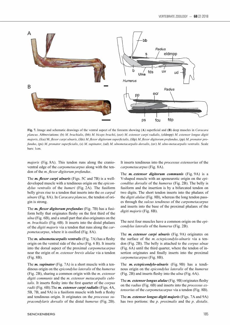
The m. brachialis (Fig. 7A) is a short and fleshy muscle that originates on the fossa brachialis of the distal humer-us, (Fig. 2B) and inserts into the depressio m. brachialis of the ulna (Fig. 6B).
The m. pronator superficialis (Figs. 5C and 7B) originates from a short and strong tendon on the distal hu-merus (Fig. 2B). It inserts both fleshy and tendinous into the proximal radius (Fig. 6B). In Caracara plancus, the muscle originates both aponeurotic and fleshy.
The m. pronator profundus (Fig. 7B) has a tendinous origin on the epicondilus ventralis of the humerus (Fig. 2B). The insertion is fleshy along the proximal third of the corpus radii (Fig. 6B). In Caracara plancus, the insertion reaches half way on the corpus radii.
The m. flexor digitorum superficialis (Fig. 7A) is a thin muscle that originates tendinous on the epicondylus ven-tralis of the humerus (Fig. 2B). The belly gives rise to a tendon that inserts into the proximal phalanx of the digiti
Fig. 5. Image and schematic drawings showing the ventral and deep muscles of the (A) sternum, (B) proximal humerus, (C) scapula and coracoideu, (A, C) Caracara plancus; (B) Milvago chimango. Abbreviations: (bb) M. biceps brachii, (ccr) M. coracobrachialis cranialis, (cca) M. coracobrachialis caudalis, (ecr) M. extensor carpi radialis, (fcu) M. flexor carpi ulnaris, (ps) M. pronator superficialis, (sbc) M. subcoracoideus, (se) M. subscapularis, (shca) M. scapulohumeralis caudalis, (spc) M. supracoracoideus, (st) M. scapulotriceps. Scale bars: 1cm.
Fig. 6. Muscular maps showing the sites of origin (red) and insertion (blue) of the forearm, (A) dorsal aspect, (B) ventral aspect. Scale bars: 1cm.
A
A
B
B
C
185
VERTEBRATE ZOOLOGY — 68 (2) 2018
majoris (Fig. 8A). This tendon runs along the cranioventral edge of the carpometacarpus along with the tendon of the m. flexor digitorum profundus.
The m. flexor carpi ulnaris (Figs. 5C and 7B) is a welldeveloped muscle with a tendinous origin on the epicon-dylus ventralis of the humeri (Fig. 2A). The fusiform belly gives rise to a tendon that inserts into the os carpal ulnare (Fig. 8A). In Caracara plancus, the tendon of origin is strong.
The m. flexor digitorum profundus (Fig. 7B) has a fusiform belly that originates fleshy on the first third of the ulna (Fig. 6B), and a small part that also originates on the m. brachialis (Fig. 6B). It inserts into the distal phalanx of the digiti majoris via a tendon that runs along the car-pometacarpus, where it is ossified (Fig. 8A).
The m. ulnometacarpalis ventralis (Fig. 7A) has a fleshy origin on the ventral side of the ulna (Fig. 6 B). It inserts into the dorsal aspect of the proximal carpometacarpus near the origin of m. extensor brevis alulae via a tendon (Fig. 8B).
The m. supinator (Fig. 7A) is a short muscle with a tendinous origin on the epicondylus lateralis of the humerus (Fig. 2B), sharing a common origin with the m. extensor digiti communis and the m. extensor metacarpalis cubi-talis. It inserts fleshy into the first quarter of the corpus radii (Fig. 6B).The m. extensor carpi radialis (Figs. 4A, 5B, 7B, and 9A) is a fusiform muscle with both a fleshy and tendinous origin. It originates on the processus su-pracondylaris dorsalis of the distal humerus (Fig. 2B).
It inserts tendinous into the processus extensorius of the carpometacarpus (Fig. 8A).
The m. extensor digitorum communis (Fig. 9A) is a Vshaped muscle with an aponeurotic origin on the epi-condilus dorsalis of the humerus (Fig. 2B). The belly is fusiform and the insertion is by a bifurcated tendon on two digits. The short tendon inserts into the phalanx of the digiti alulae (Fig. 8B), whereas the long tendon passes through the sulcus tendineus of the carpometacarpus and inserts into the base of the proximal phalanx of the digiti majoris (Fig. 8B).
The next four muscles have a common origin on the epi-condylus lateralis of the humerus (Fig. 2B).
The m. extensor carpi ulnaris (Fig. 9A) originates on the surface of the m. ectepicondylo-ulnaris via a tendon (Fig. 2B). The belly is attached to the corpus ulnae (Fig. 6A) until the third quarter, where the tendon of insertion originates and finally inserts into the proximal carpometacarpus (Fig. 8B).
The m. ectepicondylo-ulnaris (Fig. 9B) has a tendinous origin on the epicondylus lateralis of the humerus (Fig. 2B) and inserts fleshy into the ulna (Fig. 6A).
The m. extensor longus alulae (Fig. 9B) originates fleshy on the radius (Fig. 6B) and inserts into the processus ex-tensorius of the carpometacarpus via a tendon (Fig. 8B).
The m. extensor longus digiti majoris (Figs. 7A and 9A) has two portions: the p. proximalis and the p. distalis.
Fig. 7. Image and schematic drawings of the ventral aspect of the forearm showing (A) superficial and (B) deep muscles in Caracara plancus. Abbreviations: (b) M. brachialis, (bb) M. biceps brachii, (ecr) M. extensor carpi radialis, (eldmpp) M. extensor longus digiti majoris, (fcu) M. flexor carpi ulnaris, (fds) M. flexor digitorum superficialis, (fdp) M. flexor digitorum profundus, (pp) M. pronator pro-fundus, (ps) M. pronator superficialis, (s) M. supinator, (ud) M. ulnometacarpalis dorsalis, (uv) M. ulno-metacarpalis ventralis. Scale bars: 1cm.
A
B

Picasso, M.B.J. & Mosto, M.C.: Wing myology of Caracaras (Aves, Falconiformes)
186
The p. proximalis originates fleshy on the distal region of the medial surface of the radius (Fig. 6B). The muscle becomes tendinous near the wrist joint and inserts into a tubercle of the terminal phalanx of the digiti majoris (Fig. 8B). The p. distalis (Fig. 10B) is small and fleshy and originates on the proximal region of the carpometa-carpus. The muscle joins the tendon of the p. proximalis by a short tendon.
The m. ulnometacarpalis dorsalis (Figs. 7A and 10A – B) has a superficial belly with a triangular shape that originates on the distal ulna via a tendon (Fig. 6A). The muscle inserts fleshy into the surface of the proximal carpo-metacarpus near the os metacarpale minus (Fig. 8A – B).
The m. interosseus dorsalis (Fig. 10B) is a small muscle with a fleshy origin on the edges of the spatium inter-metacarpale (Fig. 8B). It inserts into the proximal end of the distal phalanx of the digiti majoris by a long tendon (Fig. 8B). In Caracara plancus, some fibers insert into the proximal angle of the spatium intermetacarpale.
The m. interosseus ventralis (Fig. 10A) originates fleshy on the edges of the spatium intermetacarpale (Fig. 8A) and inserts into the end of the distal phalanx of the digiti majoris via a tendon (Fig. 8B).
The m. extensor brevis alulae (Fig. 10B) is a small muscle with a fleshy origin on the dorsal surface of the car-pometacarpus (Fig. 8B), near the processus extensorius. It inserts into the proximal region of the alular phalanx via a tendon (Fig. 8B).
The m. abductor alulae (Fig. 10A) is an entirely fleshy muscle located on the ventral surface of the manus, with a superficial and a deep belly. The superficial belly originates fleshy on the tendon of insertion of the m. extensor
carpi radialis, whereas the deep belly is located beneath the superficial belly. Both parts are closely related and go through the processus extensorius of the carpometa-carpus and ligaments of the wrist joint and insert into the ventral aspect of the digiti alulae (Fig. 8A).
The m. flexor alulae (Fig. 10A) is a small and fleshy muscle located on the ventral surface of the manus. It originates on the proximal region of the carpometacar-pus (Fig. 8A) and inserts into the proximal region of the phalanx of the digiti alulae (Fig. 8A).
The m. adductor alulae (Fig. 10A) is a fleshy muscle that originates on the craniodorsal aspect of the carpometa-carpus (Fig. 8A) and inserts into a wide area of the medial aspect of the phalanx of the digiti alulae (Fig. 8A).
The m. abductor digiti majoris (Fig. 10A) has a fleshy origin on the processus pisiformis of the ventral surface of the carpometacarpus(Fig. 8A) and inserts into the proximal phalanx of the digiti majoris via a tendon (Fig. 8A).
The m. flexor digiti minoris (Fig. 10A) is a long and thin muscle that originates fleshy along the os metacarpale minus (Fig. 8A) and inserts into the proximal region of the phalanx of the digiti minoris via a tendon (Fig. 8A).
Muscle mass
The total forelimb muscle mass represented between 7.68 and 10.26% of the body mass (Table 1). The m. pec-toralis had the highest value of mass ( ̴ 5 % of the body mass), whereas the remaining muscles had lower values, ranging between 0.01 and 0.79% (Table 1).
Fig. 8. Muscular maps showing the sites of origin (red) and insertion (blue) of the carpometacarpus and digits in (A) ventral and (B) dorsal aspect. Scale bars: 1cm.
A
B

187
VERTEBRATE ZOOLOGY — 68 (2) 2018
The muscles that move the humerus (mm. scapulo-humeralis caudalis, deltoids major and supracoracoide-us) and those that move the forearm and elbow (mm. bi-ceps brachii, humerotriceps and scapulotriceps) had the highest values of muscle mass after the m. pectoralis, representing between 0.21 and 0.79% of the body mass (Table 1). Other humeral muscles, such as the latissimus dorsii caudalis, coracobrachialis cranialis, subscapula-ris and subcoracoideus showed values that ranged between 0.11 and 0.19 % of the body mass. The mass of the muscles involved in the downstroke represented between 6.73 and 7.56 % of the body mass, whereas the muscles involved in the upstroke represented between 0.21 and 1.22 % of the body mass (Table 1). Other muscles that had a considerable mass were those that act in the movements of the wrist and rotation of the forearm: the flexor carpi ulnaris, extensor metacarpalis radialis and prona-tor profundus, representing between 0.12 and 0.18% of the body mass (Table 1). The muscles that stabilize the thoracic shoulder (mm. serratus and rhomboideus) represented between 0.13 and 0.17 % (Table 1). The remaining muscles, i.e. those associated with the movements of the wrist and digits, represented a small portion of the body mass, ranging between 0.01 and 0.07%.
Discussion
Comparison with other Falconidae species
The forelimb myology of Caracara plancus, Milvago chimango and Milvago chimachima was similar to that described for Falco sparverius (meyers, 1992 a, 1996), Polihierax semitorquatus (BerGer, 1956), Falco tinnun-culus (CanoVa et al., 2015 a, b), and birds in general (e.g. GeorGe and BerGer, 1966; raikow, 1985). However, some specific differences can be identified with respect to Falco or Polihierax. The general morphology of the m. pectoralis was different from that of Falco sparverius. In caracaras, the m. pectoralis did not show the typical subdivisions as in F. sparverius, where this muscle has distinct deep muscular fascicle groups (meyers, 1992a; 1993). These deep muscular fascicles are involved in the gliding flight (meyers, 1993), commonly used by F. sparverius but not by caracaras. In the polyborines studied, single bellies were present in muscles like the humer-otriceps and subcoracoideus, whereas in Falco sparve-rius these muscles have two bellies (meyers, 1992a). BerGer (1956) described the m. subcoracoideus only in P. semitorquatus, being this similar to that here described for polyborines. The mm. flexor carpi ulnaris and exten-sor longus alulae of the polyborines studied presented a single belly in their origin, whereas those of Polihierax semitorquatus and Falco sparverius present two bellies (BerGer, 1956; meyers, 1996). In Falco tinnunculus, the m. flexor carpi ulnaris possesses three bellies (CanoVa et al., 2015b). The m. abductor alulae of the polyborines
here studied presented two bellies, like in Polihierax (BerGer 1956), whereas that of Falco sparverius presents a single belly (meyers, 1996). The presence of accessory bellies in certain muscles is a modification that increases the number of fibers and, consequently, the physiological crosssection is also increased, producing a greater force (BoCk, 1974). Therefore, the occurrence of one muscular belly in muscles that move the humerus (m. subcoracoi-deus), extend the elbow (m. humerotriceps) and flex and extend the wrist (m. flexor carpi ulnaris and m. extensor longus alulae) in Caracara and Milvago could be associated with a more generalized mode of flight. Finally, the polyborines studied here presented no sesamoids in the tendons of the insertions of the mm. flexor carpi ulnaris and extensor longus digiti majoris, unlike that observed in Falco sparverius, which presents these sesamoids (meyers, 1996). The absence of sesamoids could be associated with less mechanical advantage with respect to the muscles with sesamoids (BerGer & storer, 1995; sarin et al., 1999).
Muscle mass
In the Polyborinae here studied, the wing muscle mass represented an important portion of the body mass (7.68 to 10.26 %), whereas in the polyborine species studied
Fig. 9. Image and schematic drawings of the dorsal aspect of the forearm showing (A) superficial and (B) deep muscles in Cara-cara plancus. Abbreviations: (ec) M. ectepicondylo-ulnaris (ecr) M. extensor carpi radialis, (ecu) M. extensor carpi ulnaris, (edc) M. extensor digitorum communis, (ela) M. extensor longus alulae, (eldmpp) M. extensor longus digiti majoris p. proximalis. Scale bars: 1cm.
A
B

Picasso, M.B.J. & Mosto, M.C.: Wing myology of Caracaras (Aves, Falconiformes)
188
by Hartman (1961) (Caracara cheriway) the values are slightly greater (11.61%). In Falco sparverius, the only Falconinae studied by Hartman (1961), the wing muscles represent a higher proportion of the body mass (29.92%), and the mass of the individual muscles (pecto-ralis and supracoracoideus) greatly exceeds that of those of the polyborines here studied. The relatively low values found in the muscle mass of caracaras could be associated with their less powerful flight, where flapping and gliding are alternated. Instead, the high values of Falco sparverius could be associated with the opposite situation: a flight characterized by fast flapping, with an ability to hover and stoop when a prey is caught (CaneVari et al., 1991; Grin, 2018). Most of the muscle mass of the wing is involved in the movements of the articulatio omalis (see Table 1); this joint has the greatest degree of mobility in the wing and its movements affect the entire limb (raikow, 1985). The main humerus movements that produce the downstroke and upstroke, i.e. the two main phases of the flapping flight, are depression, elevation, protaction, retraction and rotation (dial et al. 1988). The downstroke provides the propulsive force required to generate both the lift and thrust (dial et al., 1988; Goslow et al., 1990), and, in caracaras, this is in accordance with the great muscle mass
dedicated to this movement. Instead, the upstroke prepares the wing for the next downstroke (dial et al., 1988) and the relevant muscle mass is lower (see Table 1). In the polyborines studied, the m. pectoralis was the largest muscle of the wing, a feature in accordance with its important functions during flight. This muscle acts during the downstroke, generating the lift and thrust required for flight and counteracting the inertia of the wing (dial, 1992; dial & Biewener, 1993; toBalske, 2007). The second largest muscle of the polyborines studied was the m. scapulohumeralis caudalis, which is also a downstroke muscle. This muscle produces the retraction of the humerus and the ventral rotation of the wing, showing great activity (dial, 1992). Investigations in pigeons have established that the m. supracoracoideus (the primary upstroke muscle) is the second largest muscle in the wing (toBalske, 2007; toBalske & Biewener, 2008), but this does not hold true for caracaras. The significance of this difference remains to be studied in other birds with different styles of flight and/or phylogenetic relationship. Besides the mm. pectoralis and scapulohumeralis caudalis, the muscles with the largest masses were the upstroke muscles mm. deltoideus major and supracora-coideus. Electromyographic data have indicated that the m. deltoideus major helps the m. supracoracoideus in the
Fig. 10. Image and schematic drawings showing the muscles of manus and digits in (A) ventral and (B) dorsal aspect in Caracara plancus. Abbreviations: (aba) M. abductor alulae, (ada) M. ad-ductor alulae, (abdm) M. abductor digiti majoris, (eba) M. extensor brevis alulae, (eldmpd) M. extensor longus digiti majo-ris p. distalis, (fa) M. flexor alulae, (fdm) M. flexor digiti minoris, (id) M. interos-seus dorsalis, (iv) M. interosseus ventra-lis, (ud) M. ulnometacarpalis dorsalis. Scale bars: 1cm.
A
B

189
VERTEBRATE ZOOLOGY — 68 (2) 2018
elevation of the wing and that the m. supracoracoideus also elevates the humerus and contributes to decelerating the wing during the upstroke (dial, 1992). Poore et al. (1997) proposed that this muscle also produces the rotation of the humerus. It is difficult to assess the contribution of the remaining muscles to the humerus movements because they have not been studied with electromyographic techniques and, consequently, their roles in wing movements have only been inferred by their anatomical position (see raikow, 1985; meyers, 1992a; 1998). The other muscles with considerable mass were those involved in the movements of the forearm. The m. biceps brachii, in addition to flexing the forearm, is also a stabilizer of the elbow joint (together with the m. humerotri-ceps) during the final one-half of the downstroke (dial, 1992). The m. scapulotriceps also contributes to the stabilization of the elbow during flapping flight and the m. humerotriceps extends the elbow during descending flight (dial, 1992). The m. pronator profundus allows the rotation of the wing and is intensely active during takeoff and ascending flight (dial, 1992). The movements of the wrist are performed by several muscles but only two of them showed relatively higher mass: the mm. extensor metacarpi radialis and the flexor carpi ulnaris. This is concordant with their important function, since these muscles are responsible for synchronized extension and flexion of the elbow and wrist joints (Vazquez, 1994). Besides, the action of these muscles is intimately integrated with the “drawingparalells” mechanism, i.e. the coordinated movements of the forearm and manus during the wing beat (for details see Vazquez, 1994). Instead, the lowest mass values of the mm. extensor digitorum communis, flexor digitorum superficialis and extensor metacarpi ulnaris are in accordance with their poor contribution to the movements of the elbow and wrist (Vazquez, 1994). The small (or intrinsic) muscles that move the digits represented only a low percentage of the body mass. This could be related to the restriction in the movements of the manus during flight, which are subject to strong stresses (Vazquez, 1995), and to the fact that it is generally proposed that these muscles contribute little to the mechanics of the flight (Biewener, 2011). Other small muscles of caracaras like those located near the humeral joint (mm. latissimus dorsi cranialis, deltoideus minor, scapulo humeralis cranialis and bra-chialis) are involved in the postural control of the folded wing (meyers, 1992b).
Conclusions and future directions
Although the wing muscles have a multifunctional nature that complicates the interpretation of their role during flight (Videler, 2006), this study highlights the utility of muscular mass data to understand and explore the possible importance of individual muscles or muscular groups
during this mode of locomotion. This is possible because the muscle mass is proportional to the maximum muscle power output (Biewener & roBerts, 2000; roBerts, 2001). Moreover, this kind of information, combined with quantitative information, like electromyographic, histochemical or muscular architectural data, may allow us to achieve an integrative assessment of the muscle function. The lack of similar works in birds prevents comparison and detailed analysis about how muscle mass can vary in birds with different flying styles. However, this analysis is a first step towards the exploration of this area of study.
Acknowledgements
Thanks to Y. Davies (Museo Argentino de Ciencias Naturales Bernadino Rivadavia, MACN)
References
Baumel, J.J., kinG, s.a., Breazile, J.e., eVans, H.e. & BerGe, J.C. (1993): Handbook of Avian Anatomy, Nomina Anatomica Avium. Publication 23 of Nuttall Ornithological Club. Cambridge.
BerGer, a.J. (1956): The appendicular myology of the Pygmy Falcon (Polihierax semitorquatus). – American Midland Naturalist, 55: 326 – 333.
BerGe, J.C. & storer, r.w. (1995): Intratendinous ossification in birds: a review. – Journal of Morphology, 226: 47 – 77.
Biewener, a.a. (2011): Muscle function in avian flight: achieving power and control. – Philosophical Transactions of the Royal Society of London B, 366, 1496 – 1506.
Biewener, a.a. & roBert, t.J. (2000): Muscle and tendon contributions to force, work and elastic energy savings: a comparative perspective. – Exercise and Sport Sciences Reviews, 2803: 99 – 107.
BoCk, w.J. (1974): The avian skeletomuscular system. In: Farmer D. S., King J. R. (Eds.), Avian biology, vol. 4. Academic Press, London, 119 – 257 pp.
Cade, t.J. & diGBy, r.d. (1982): The falcons of the world. Cornell University Press, New York.
CaneVari, m., Carrizo, G.r., CaneVari, m. & CaneVari, P.C. (1991): Nueva guía de las aves argentinas. Fundación Acindar, Buenos Aires.
CanoVa, m., Bedoni, C., HarPer, V., ramBaldi, a.m., BomBardi, C. & Grandis, a. (2015a): Anatomical study of the musculus deltoideus and musculus flexor carpi ulnaris in 3 species of wild birds. – Veterinaria Italiana, 52: 37 – 44.
CanoVa, m., Bedoni, C., HarPer, V., Barazzoni, a.m., de FaVeri, a. & Grandis, a. (2015b): Anatomical differences in three wing muscles of the Grey heron (Ardea cinerea), the Common buzzard (Buteo buteo) and the Common kestrel (Falco tinnun-culus): a possible functional interpretation. – Rivista Italiana di Ornitologia, 85: 15 – 22.
CanoVa, m., ClaVenzani, P., BomBardi, C., mazzoni, m., Bedoni, C. & Grandis, a. (2015c): Anatomy of the shoulder and arm mus
Picasso, M.B.J. & Mosto, M.C.: Wing myology of Caracaras (Aves, Falconiformes)
190
culature of the common buzzard (Buteo buteo Linnaeus, 1758) and the European honey buzzard (Pernis apivorus Linnaeus, 1758). – Zoomorphology, 134: 291 – 308.
CorVidae, e.l., BierreGaard, r.o. & Peters, s.e. (2006): Comparison of wing morphology in three birds of prey: Correlations with differences in flight behavior. – Journal of Morpho-logy, 267: 612–622.
dial, k.P. (1992): Activity patterns of the wing muscles of the pigeon (Columba livia) during different modes of flight. – Journal of Experimental Zoology, Part A, Ecological Genetics and Physiology, 262: 357 – 373.
dial, k.P. & Biewener, a.a. (1993): Pectoralis muscle force and power output during different modes of flight in pigeons (Co-lumba livia). – Journal of Experimental Zoology, 176: 31 – 54.
dial, k.P., kaPlan, s.r., Goslow, G.e. & Jenkins, F.a. (1988): A functional analysis of the primary upstroke and downstroke muscles in the domestic pigeon (Columba livia) during flight. – Journal of Experimental Zoology, 134: 1 – 16.
dunninG, J.B. (2008): Handbook of avian body masses. Boca Raton, CRC Press.
FuCHs, J., JoHnson, J.a. & mindell, d.P. (2012): Molecular systematics of the caracaras and allies (Falconidae: Polyborinae) inferred from mitochondrial and nuclear sequence data. – Ibis, 154: 520 – 532.
GeorGe, J.C. & BerGer, a.J. (1966): Avian Myology. Academic Press, New York.
Goslow, G.e., dial, k.P. & Jenkins, F.a. (1990): Bird flight: insights and complications. – Bioscience, 40: 108 – 115.
Grin Global Raptor Information Network (2018): Species ac count. Available at: http://www.globalraptors.org/grin/index Alt. asp.
HaCkett, s.J., kimBall, r.t., reddy, s., Bowie, r.C., Braun, e.l., Braun, m.J. et al . (2008): A phylogenomic study of birds reveals their evolutionary history. – Science, 320: 1763 – 1768.
Hartman, F.a. (1961): Locomotor mechanisms of birds. – Smithsonian Miscellaneous Collections, 143: 1 – 91.
Heers, a.m., Baier, d.B., JaCkson, B.e., & dial, k.P. (2016): Flapping before flight: high resolution, three-dimensional ske - letal kinematics of wings and legs during avian development. – PloS one, 11: e0153446.
Hertel, F., maldonado, J.e. & sustaita, d. (2015): Wing and hindlimb myology of vultures and raptors (Accipitriformes) in relation to locomotion and foraging. – Acta Zoologica, 96: 283 – 295.
JarVis, e.d., miraraB, s., aBerer, a.J., li, B., Houde, P., li, C. et al. (2014): Wholegenome analyses resolve early branches in the tree of life of modern birds. – Science 346: 1320 – 1331.
Jollie, m.t. (1977): A contribution to the morphology and phylogeny of the Falconiformes. – Evolutionary theory, 3: 1 – 141.
liVezey, B.C. & zusi, r.l. (2006): Higherorder phylogeny of modern birds (Theropoda, Aves: Neornithes) based on comparative anatomy: 1. Methods and characters. – Bulletin of the Carnegie Museum of Natural History, 37: 1 – 544.
meyers, r.a. (1992a): Morphology of the shoulder musculature of the American kestrel, Falco sparverius (Aves), with implications for gliding flight. – Zoomorphology, 112: 91 – 103.
meyers, r.a. (1992b): The morphological basis of folded wing posture in the American kestrel, Falco sparverius. – Anatomical Record, 232: 493 – 498.
meyers, r.a. (1993): Gliding flight in the American kestrel (Falco sparverius): an electromyographic study. – Journal of Morphology, 215: 213 – 224.
meyers, r.a. (1996): Morphology of the antebrachial musculature of the American kestrel, Falco sparverius (Aves). – Annales of Anatomy, 178: 49 – 60.
mosto, m.C., Carril, J. & PiCasso, m.B.J. (2013): The hindlimb myology of Milvago chimango (Polyborinae, Falconidae). – Journal of Morphology, 274: 1191 – 1201.
mosto, m.C., PiCasso, m.B.J. & Biondi, l.m. (2016): Longlegged caracaras: terrestrial habitat and hindlimb morphology. – Journal of Zoology, 298: 274 – 284.
Poore, s.o., sanCHez-Haiman, a., & Goslow Jr, G.e. (1997): Wing upstroke and the evolution of flapping flight. – Nature, 387: 799 – 802.
Prum, r.o., BerV, J.s., dornBurG, a., Field, d.J., townsend, J. P., lemmon, e.m. & lemmon, a.r. (2015): A comprehensive phylogeny of birds (Aves) using targeted nextgeneration DNA sequencing. – Nature, 526: 569 – 577.
raikow, r.J. (1985): Locomotor system. In: King, A.S., McLelland, J. (Eds), Form and Function in Birds, vol. 3. Academic Press, London, 57 – 147pp.
roBerts, t.J. (2001): Muscle force and stress during running in dogs and wild turkeys. – Bulletin of the Museum of Comparative Zoology, 156: 283 – 295.
sarin, V.k., eriCkson, G.m., Giori, n.J., BerGman, a.G. & Carter, d.r. (1999): Coincident development of sesamoid bones and clues to their evolution. – Anatomical Record, 257: 174 – 180.
sustaita, d. (2008): Musculoskeletal underpinnings to differencesin killing behavior between North American accipiters (Falco
niformes: Accipitridae) and falcons (Falconidae). – Journal of Morphology, 269: 283 – 301.
toBalske, B.w. (2007): Biomechanics of bird flight. – Journal of Experimental Biology, 210: 3135 – 3146.
toBalske, B.w. & Biewener, a.a. (2008): Contractile properties of the pigeon supracoracoideus during different modes of flight. – Journal of Experimental Biology, 211: 170 – 179.
Vazquez, r.J. (1994): The automating skeletal and muscular mechanisms of the avian wing (Aves). – Zoomorphology, 114: 59 – 71.
Vazquez, r.J. (1995): Functional anatomy of the pigeon hand (Columba livia): a muscle stimulation study. – Journal of Morphology, 226: 33 – 45.
Videler, J.J. (2006): Avian flight. Oxford University Press, New York.
wHite, C.m., olsen, P.d. & kiFF, l.e. (1994): Family Falconidae (falcons and caracaras). In: Del Hoyo, J., Elliott, A., Sargatal, J. (Eds.), Handbook of the birds of the World, vol. XX Lynx Editions Barcelona, 216 – 277pp.