when to refer and why behind the md curtain: an internist’s perspective john winton, md john neal,...
TRANSCRIPT

When to Refer and Why
Behind the MD Curtain:An Internist’s Perspective
John Winton, MDJohn Neal, OD

Objectives
• Provide information on referring to an MD– When to refer– What information to convey
• Provide insight into what goes on after a referral is made– Diagnostic workup– Acute and long-term management
• Demonstrate the importance of MD / OD collaboration– Ocular implications– Systemic implications

Disclosures
•Drs. Neal and Winton put together a rock band in 2012 with a couple other guys.• It lasted about 6 weeks.• No. of practices: 8• No. of shows: 0
•No other disclosures, financial or otherwise

Diabetes
• 65 year old white male
• CC: Cloudy vision right eye x 1 month. “I think I need some stronger glasses.”
• PMHx: Unremarkable; “doesn’t like to go to the doctor”
• POHx: Spectacles since age 12

Diabetes
• BCVA: OD: 20/30 OS: 20/25
• CVF: FTFC OU
• IOP OD: 18 OS 19
• Anterior Segment: mild nuclear cataracts OD/OS
• Posterior Segment: see photo

Diabetes
“I can’t see right.”

Diabetes
“I can’t see right.”

Assessment/Plan
• Presumed proliferative diabetic retinopathy OD/OS
• Plan• Refer to retinal specialist for evaluation• Systemic health?• How worried should I be here?• What should I do in the office today?

Diabetes
• “Now what?”

Diabetes
• Fingerstick glucose & vital signs
• 200-400, vitals OK, not acutely ill: don’t panic. Outpatient MD followup within
one week.
• 200-400, tachycardic OR hypotensive OR ill-appearing: need ER
• >400: need ER

Diabetes
• Why ER? What’s the emergency?
• What can kill your patient today?• Electrolyte abnormalities (pH, potassium, etc.)• Dehydration• Silent myocardial infarction

Diabetes

Diabetes

Diabetes
• NOTICE: no mention of A1c to determine urgency• BUT...A1c is still a GREAT test• Average blood glucose level over the past 3 months • A1c ≥ 6.5 = diabetes• A1c > 7.0 = uncontrolled diabetes• Accurate within 0.5 percentage points

Diabetes

Diabetes
A1c%
6 7 8 9 10 11 12
eAGmg/dL
126 154 183 212 240 269 298
*eAG = estimated Average GlucoseTable taken from Diabetes Care. 2011; 34(Supp 1):S11–S61, table 9

Diabetes
Red blood cellglucose
low A1c high A1c

Diabetes
• A1c is subject to misinterpretation!• Newly diagnosed diabetes (average glucose)• “Brittle” diabetes (glucose roller coaster)• Hemoglobinopathies (sickle cell, thalassemia, etc.)• Iron deficiency or recent blood transfusion• Liver or kidney failure

Diabetes
• What does the internist do after acute management?
1.Decide why diabetic (type I vs. type II vs. pancreatic insufficiency)
2.Further testing (lipids, lytes, TSH, urine protein)3.Therapy (pills only / pills + insulin / insulin only)4.EDUCATE / follow up5.Arrange annual eye screening

Hypertension
• 50 year old white female
• CC: I have been getting this headache for a while. I think I need stronger glasses.
• PMHx: unremarkable
• POHx: occasional contact lens wear, spectacles for many years

Hypertension
• BCVA: OD: 20/20 OS: 20/20
• CVF: FTFC OU BP: 170/95
• Binocular testing yields no significant findings
• IOP: OD: 14 OS: 14
• Anterior segment: mild corneal neovascularization
• Posterior segment: see photo

Hypertension

Assessment/Plan
• Hypertensive retinopathy OD/OS
• Plan• Check Blood Pressure• Urgent/Emergent referral?• My vision is fine, why are you talking about my blood pressure?

Hypertension
“I can’t see right.”

Hypertension
• What Systolic BP is “too high”?• What Diastolic BP is “too high”?• Answer: whatever number scares you• Better question: What is the context of the high blood pressure?
• Symptoms?• Situation?• How was it measured?

Hypertension
• For high BP, consider:• Did a machine take the BP?• Was the cuff size appropriate?• Is the patient symptomatic?
• Blood in urine, confusion, shortness of breath, chest pain, abdominal pain, etc.

Hypertension
• Flame hemorrhages OR papilledema in a patient with SBP > 160 and / or DBP > 110 needs emergent treatment for malignant hypertension• **this is where your input is helpful!!• Electrolytes (looking for kidney disease)• CBC (looking for low platelets)• CT head (looking for intracranial hemorrhage)

Hypertension
What does the internist do?• Consider secondary causes of hypertension– Hyperaldosteronism, endocrine disorders, kidney disease, etc.
• Initiate treatment• DON’T LOWER TOO RAPIDLY!!!– (unless bleeding into brain)

Embolic Disease
• 72 yo AAM
• CC: A large part of my vision is missing. I just noticed it this morning.
• PMHx: HTN x 20 years, 2 heart stents 5 years ago
• POHx: Glasses x 30 years

Embolic Disease
• BCVA OD: 20/40 OS 20/25
• IOP OD: 18 OS: 17
• CVF: Extiniction superior quadrants OD FTFC OS
• Anterior seg: unremarkable
• Posterior seg: see photo
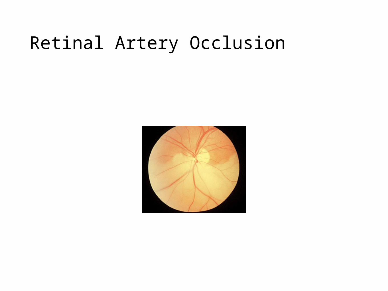
Retinal Artery Occlusion

Retinal Artery Occlusion

Assessment/Plan
• Branch retinal artery occlusion with visible embolus
• Plan• Breathe into a bag?• Referral for laser embolectomy?• Systemic workup?

Retinal Artery Occlusion
• CRAO / BRAO need workup to determine source– Imaging: carotid U/S or angiography, echo– Labs: CBC, Electrolytes, Lipids, RPR, HIV, ESR– Notice what I didn’t auto-order:
• CRP, ANA, ACE, Lyme titers, coagulopathy panel, homocysteine
• Optocase RAO

Retinal Artery Occlusion
Workup• Homocysteine - can reduce level, but not risk
• ANA - nonspecific. maybe later.
• CRP - will be elevated. Uninterpretable. Waste of $$.
• ACE - elevated in ALL granulomatous disease.
• Lyme - only if traveled to or live in endemic area.
• Coagulopathy - never during acute embolic event.

Retinal Artery Occlusion
Other Vasculitidies:
•Behçet's disease
•Systemic lupus erythematosus
•Giant cell arteritis
•Wegener granulomatosis
•Polyarteritis nodosa
•Multiple sclerosis
•Sarcoidosis
•HLA-B27 associated conditions–(BUT DON’T CHECK HLA-B27!!)
•Relapsing polychondritis
•Inflammatory bowel disease–Crohn disease
–Ulcerative colitis
•Toxoplasmosis
•Tuberculosis
•Syphilis
•Whipple’s disease

Nystagmus
• 55 yo AAM
• CC: My eyes started jumping around
• PMHx: DM x 12 years, HTN x 10 yrs
• POHx: unremarkable

Nystagmus
• BCVA: OD 20/40 OS 20/40
• CVF: FTFC OU but poor fixation• EOM: FROM OU with upbeat nystagmus
• IOP OD: 12 OS: 12
• Anterior segment: unremarkable• Posterior segment: unremarkable

Nystagmus
•Don’t laugh! You might get this referral!
•Always refer nystagmus to an internist or neurologist (preferably not the one who sent it to you)
• Information for referral:• Direction (up / down / left / right / rotational)• Timing (acute / chronic)• Associated symptoms (vertigo, tinnitus)

Nystagmus
• Differential Diagnosis:
– Vestibular (Labyrinthitis, Otolith, Benign Positional Peripheral Vertigo)
– Central (Stroke, tumor, demyelinating disease)
– Metabolic (Medications, drugs, nutritional deficiency)
– Thiamine deficiency (Wernicke encephalopathy)
• Workup: Imaging (unless obvious etiology, such as medication)

Sarcoidosis
•40 yo AAF
•CC: My eyes are always red and watery. I have to wear shades at all times
•PMHx: unremarkable
•POHx: “pink eye” 4 times in the past 5 years

Sarcoidosis
•BCVA: OD 20/25 OS 20/25
•CVF FTFC OU, FROM OU
• IOP OD: 9 OS: 8
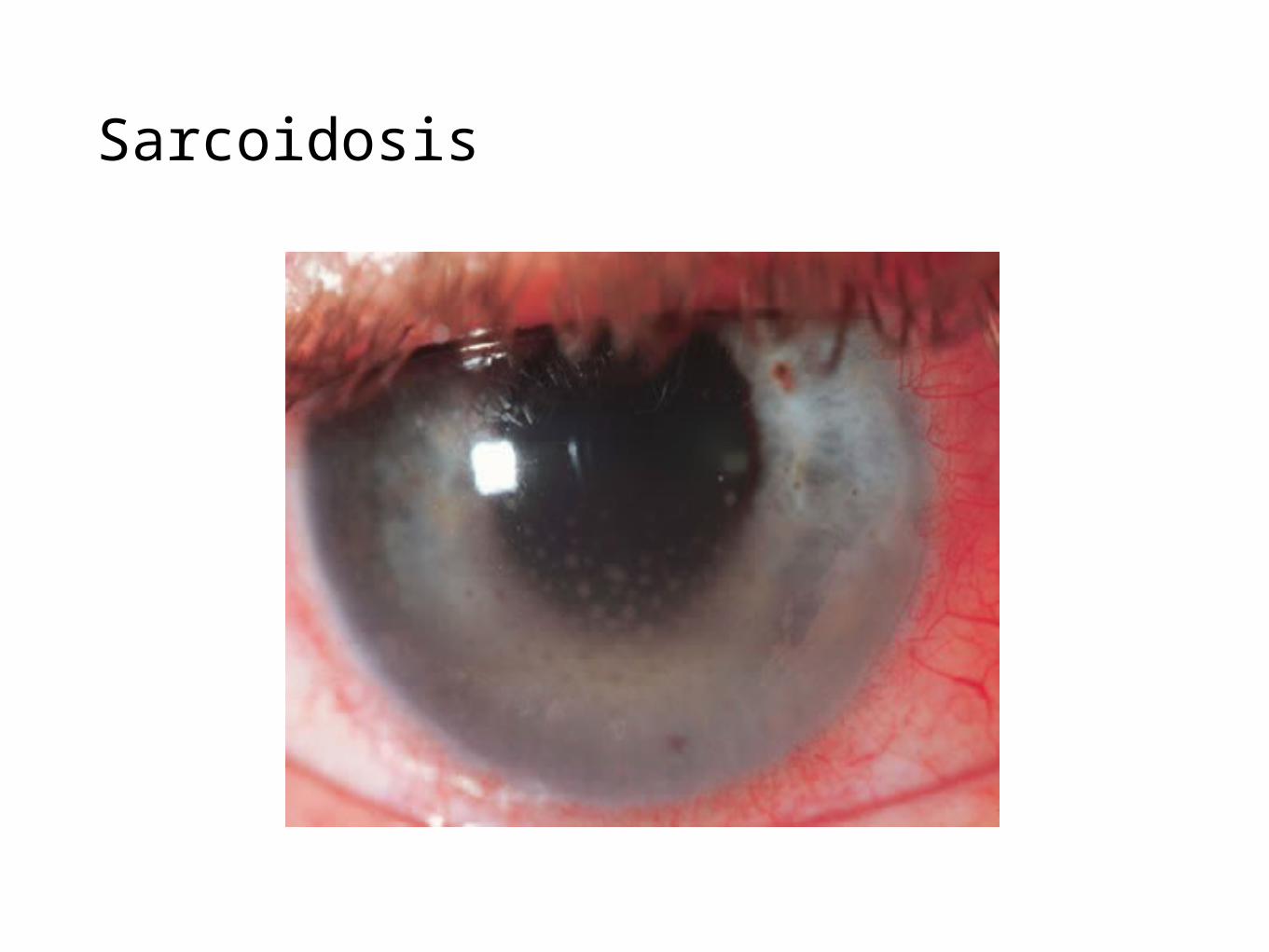
•Anterior seg: 3+ cells 2+ flare in the AC OD/OS•Keratic precipitates OD/OS•Post seg: unremarkable
Sarcoidosis

Assessment/Plan
•Acute bilateral granulomatous anterior uveitis
•Plan:• Begin Pred Forte q2h OD/OS, begin homatropine 5.0% BID OD/OS, monitor 4-
7 days
• Bloodwork? Imaging?

Sarcoidosis
• Systemic granulomatous disease of unknown etiology– Primarily manifests in lungs– Non-pulmonary sarcoid makes up 10% of disease
• Labs: Calcium, vitamin D, renal function, liver enzymes, PPD, sputum samples
– *ACE level is neither sensitive nor specific• Differential diagnosis– Multiple sclerosis, inflammatory bowel disease– Amyloidosis, lymphoma– Infection (TB, histoplasmosis, coccidioidiomycosis)

Sarcoidosis
Radiography: Chest XR
• Bilateral hilar lymphadenopathy
• Air trapping
• *CT of chest rarely contributes to diagnosis
• Other tests:– EKG
– PFT (Pulmonary function testing)

Sarcoidosis
Histology
• Obtained via fiberoptic bronchoscopy with transbronchial biopsy
• Hallmark: non-caseating (i.e., non-necrotizing) granulomas
• Diagnosis is made by histologic findings in the appropriate setting

Endophthalmitis
•44 yo WM
•CC: My vision in my right eye has been blurry and I have been feeling pretty sick since I was released from the hospital 2 weeks ago.
•PMHx: (+)HIV
•POHx: spectacle use since childhood

Endophthalmitis
• BCVA: OD: 20/400 OS: 20/20
• CVF: FTFC OU, FROM OU
• IOP OD: 8 OS: 16
• Anterior segment: 3+ injection along with 3+ cells and flare OD• Posterior segment: see photo

Endophthalmitis
“I can’t see right.”

Assessment/Plan
• Endophthalmitis OD
• Refer to retinal specialist?• ER?• Emergent?

Endophthalmitis
• Setting: immunocompromised patients and / or recently hospitalized patients
• Always an emergency (i.e., requires inpatient management)!
• What you can do:– Describe lesion as retinal vs. vitreal – both will require systemic antifungals
but vitreal disease requires vitrectomy + intra-ocular antifungals

Syphilis
• 38 yo WM
• CC: I would just like to get my eyes checked.
• PMHx: Unremarkable
• POHx: Unremarkable

Syphilis
• BCVA: OD 20/20 OS 20/20
• CVF FTFC OU
• IOP: OD: 14 OS: 13
• Anterior segment: old keratic precipitates OD/OS & old synechiae and pigment on anterior lens, no active inflammation• Posterior segment: see photos

Posterior Segment

Ocular Manifestations
• Ocular manifestations of syphilis:• Uveitis (anterior/intermediate/posterior)• Keratic Precipitates• Vitritis
• Optic atrophy• Retinal pigment changes• Interstitial Keratitis• Episcleritis/Conjunctivitis• Retinitis• Vasculitis (ground glass)

Assessment/Plan
• Inactive bilateral anterior uveitis• Chorioretinal scarring OD/OS
• Systemic inflammatory/infective workup• What lab tests?• Imaging?• Followup?

Syphilis• The great imitator• Can cause multiple systemic
ailments• Diagnosis, classification, and
lab interpretation is confusing – but important because of treatment implications
• Don’t ever type “syphilis” into google images
Syphilis
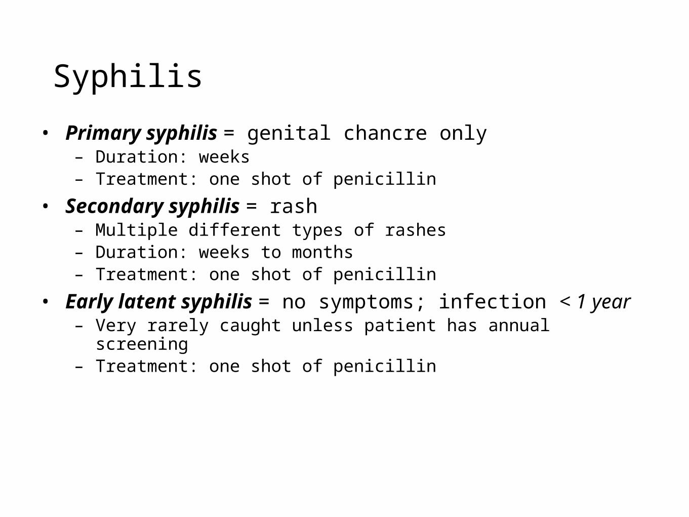
• Primary syphilis = genital chancre only– Duration: weeks– Treatment: one shot of penicillin
• Secondary syphilis = rash– Multiple different types of rashes– Duration: weeks to months– Treatment: one shot of penicillin
• Early latent syphilis = no symptoms; infection < 1 year – Very rarely caught unless patient has annual screening– Treatment: one shot of penicillin

Syphilis
• Late latent syphilis = no symptoms; infection > 1 year or unknown duration– Much more commonly diagnosed than early latent– Treatment: 1 shot of penicillin weekly x3 total
• Tertiary syphilis = late manifestations– Occurs years to decades after initial infection– Neurosyphilis, cardiac syphilis, gummatous syphilis– Treatment: IV penicillin 6 times / day for 2 weeks

Syphilis
• Laboratory interpretation: RPR– If non-reactive, no syphilis, you’re done– If weakly positive (1:8 or lower)
• Check confirmatory FTA-ABS or treponemal antibody – if that is nonreactive, your RPR was a false positive
• If confirmatory test reactive, check to see if RPR ever done in past (4-fold decrease from prior RPR = treated syphilis). Otherwise, it’s real, so determine which stage and treat accordingly.
– If strongly positive (1:16 or higher)• It’s probably syphilis. You can check confirmatory test but it will be reactive.
Determine stage and treat accordingly.

Syphilis
• Other considerations– RPR 1:32 or greater should probably be checked for neurosyphilis (i.e., CSF
studies) even if asymptomatic• Exception: obvious primary or secondary syphilis.
– Any reactive RPR in an HIV patient should be presumed real with strong consideration for ruling out neurosyphilis
– CSF diagnosis of neurosyphilis is inexact• Multiple labs must be considered (CSF protein, CSF lymphocytes, CSF FTA-ABS, CSF
VDRL) • When in doubt, treat as neurosyphilis.
– OCULAR SYPHILIS CAN OCCUR AT ANY STAGE

Optometric Medications with
Systemic Implications(or, When the MD Calls the OD)

Topical beta blockers
• Can be absorbed systemically and are generally nonselective (block beta-1 and beta-2 receptors)– Can cause bradycardia leading to dizziness or syncope in the elderly and in
patients with underlying heart disease• Use with care if already on systemic beta blocker
– Can cause bronchospasm and wheezing in patients with chronic lung disease (asthma, sarcoidosis, COPD)
– We may call you to let you know we’re changing your patient’s dorzolamide / timolol combo to single agent dorzolamide

Antibiotics
• Always check allergies– *Penicillin allergy does NOT preclude cephalosporin use as long as the
allergy is not anaphylactic shock
• Macrolides (erythromycin, azithromycin) should be given careful consideration!!– Can cause fatal arrhythmias, especially in combination with certain
medications or medical conditions
• Clindamycin is losing ground and is hard to take (3-4 time / day dosing)

Antibiotics
• Sulfamethoxazole / trimethoprim works, but only for staphylococcal disease– Poor coverage for streptococcal disease– Must be weight-based– Can raise potassium and creatinine in patients with underlying kidney
disease– Can cause bone marrow suppression
• Doxycyline and minocycline are great– Just remember to tell the patient to drink a full glass of water with it, don’t
take it less than 1 hour prior to bedtime, and stay out of the sun