when infecting western ower thrips, frankniella similar
TRANSCRIPT
Page 1/22
Beauveria bassiana ERL836 and JEF-007 withsimilar virulence show different gene expressionwhen infecting western �ower thrips, FrankniellaoccidentalisSihyeon Kim
Chonbuk National UniversityJong Cheol Kim
Chonbuk National UniversitySe Jin Lee
University of FloridaMi rong Lee
Chonbuk National UniversitySo Eun Park
Chonbuk National UniversityDongwei Li
Chonbuk National UniversitySehyeon Baek
Chonbuk National UniversityTae Young Shin
Chonbuk National UniversityJae Su Kim ( [email protected] )
Chonbuk National University https://orcid.org/0000-0003-0128-0684
Research article
Keywords: Beauveria bassiana, western �ower thrips, genome, transcription, cytochrome P450
Posted Date: July 22nd, 2020
DOI: https://doi.org/10.21203/rs.3.rs-17095/v2
License: This work is licensed under a Creative Commons Attribution 4.0 International License. Read Full License

Page 2/22
Version of Record: A version of this preprint was published at BMC Genomics on November 27th, 2020.See the published version at https://doi.org/10.1186/s12864-020-07253-y.

Page 3/22
AbstractBackground: Insect-killing fungal species, Beauveria bassiana, is as an environment-friendly pestmanagement tool, and many isolates are on the track of industrialization. However, some of B. bassianaisolates show similar morphology and virulence against insect pests, and so it is hard to differentiatethem. Herein we used two patented isolates, ERL836 and JEF-007, and investigated their virulenceagainst western �ower thrips, Frankliniella occidentalis, and further analyzed genome structures andtranscriptional responses when infecting the thrips to see possible differences. Results: The two isolatesshowed no signi�cant differences in fungal growth, conidial production, and virulence against thrips, andthey were structurally similar in genome. But, in transcription level, ERL836 appeared to infect thripseasily, while JEF-007 appeared to have more di�culty. In the GO analysis of ERL836 DEGs (differentiallyexpressed genes), the number of up-regulated genes was much larger than that of down-regulated genes,when compared to JEF-007 DEGs (more genes down-regulated). Interestingly, in the enrichment analysisusing shared DEGs between two infecting isolates, plasma membrane-mediated transporter activity andfatty acid degradation pathway including cytochrome P450 were more active in infecting ERL836.Conclusion: The two B. bassiana isolates had similar morphology and virulence as well as genomestructure, but in transcription level they differently infected western �ower thrips. This comparativeapproach using shared DEG analysis could be easily applied to characterize the difference of the two B.bassiana isolates, JEF-007 and ERL836.
BackgroundBeauveria bassiana has received great interest from both academia and industry because of its greatpotential as an alternative environment-friendly pest management tool to substitute synthetic pesticidesthat can damage the environment and cause insect resistance. Popularly available B. bassiana productsare BotaniGard® (isolate name: GHA), Naturalis-L® (ATCC74040), Chongchaesak® (ERL836), Broadband®
(PPRI 5339), and BioCeres® (ANT-03) and they are used to control agricultural insect pests such asmoths, beetles, white�y, aphid, mite, and thrips among others [1]. Particularly B. bassiana ERL836 GR hasbeen developed by LG-Chemical-a�liated FarmHannong and successfully launched and sold out in alocal market. Insect resistance to pesticides may not be an issue when using these fungal pathogens forpest control because this fungal group uses complicated mechanical hyphal penetration and enzymaticdegradation to kill the host. This mode of fungal action is completely different from that of chemicalstargeting synapses or energy metabolism. Although other entomopathogenic fungi such as Metarhizium,Cordyceps (previously Isaria or Paecilomyces), Akanthomyces (previously Lecanicillium and Verticillium)are also being investigated as pest management tools, B. bassiana has several advantages over theseother fungi in terms of �eld application, mass production, and long-term storage.
Numerous B. bassiana isolates have been collected form natural environments and have been transferredto fungal libraries around the world. USDA-ARS manages a huge entomopathogenic fungal library andsupport academic research worldwide. Japan and China have big fungal libraries and Brazil andEuropean countries also have entomopathogenic fungal libraries. Industrially important isolates from

Page 4/22
these libraries are placed in the research and development track for development as commercial orgovernment-supported products. Very few isolates successfully pass all steps of industrialization to belaunched in the biopesticide market. Nevertheless, the number of B. bassiana isolates being commerciallydeveloped is increasing. An important intellectual property (IP) issue is how to scienti�cally determinewhether a particular B. bassiana isolate is different from another isolate. Attempts have been made touse phylogenetic analysis based on one or a couple of housekeeping genes, but this approach is notalways adequate. As a prerequisite for intellectual property in South Korea, such as a patent for aparticular isolate, clear experimental data that describes the uniqueness of an isolate needs to besubmitted; this type of data could be used to resolve possible isolate-mediated con�icts in thebiopesticide industry.
As introduced above, phylogenetic approaches have been mainly used to determine the evolutionaryorigins of isolates and their genetic relationships. Rehner and Buckley investigated the correspondencebetween Beauveria and Cordyceps using EF1-α and ITS phylogenies [2]. Glare et al. analyzed rDNA todetermine phylogenetic relationships among 26 Beauveria isolates; more speci�cally, they used sequenceinformation from the 3' end of 16s rDNA across ITS 1, 5.8s rDNA, and ITS 2 to the 5' end of 28s rDNA [3].In phylogenies, three nuclear genes encoding elongation factor 1-α (TEF1), RNA polymerase II largestsubunit (RPB1), and RNA polymerase II second largest subunit (RPB2) were used by Rehner andcolleagues [4]. Furthermore, a new species, B. lii, was found by four-locus based phylogenetic analysis [5].However, these previous phylogenetic analyses were based on one or a couple of house-keeping genes,and could not be easily used to suggest that one isolate is different from another isolate in B. bassianaunder IP issues.
Although fungal phylogenetic analysis can be used to suggest the uniqueness of one isolate in patentsubmission and acquisition, but the question is how many genes should be analyzed for this purpose.Whole genome sequencing (WGS) can provide the sequences of the entire genome of an organism and tocompare the roles of genes or the diversity of genes in the isolates of same species of fungi. WGS of 10B. bassiana isolates have been performed to date. ARSEF2860 (33.70 Mb, ADAH00000000.1) was the�rst WGS of B. bassiana and was obtained using Roche 454 system and Illumina paired-end sequencingin 2012 [6]. Genomes of other B. bassiana isolates, such as D1-5 (36.69 Mb, ANFO00000000) in 2014,ARSEF1520 (36.97 Mb, JTCW01000000), ARSEF 2597 (38.83 Mb, JTCX00000000), ARSEF8028 (35.02Mb, JRHA00000000), ARSEF5078 (34.45 Mb, JTCZ00000000), ARSEF4305 (34.77 Mb, JTCY00000000)in 2016, BCC2660 (34.56 Mb, MWYT00000000) in 2017, and JEF-007 (36.54 Mb, MRVG00000000 fromour previous work) and Bv062 (34.84 Mb, MAQY00000000) in 2018 have been generated. Whole genomesequences of D1-05, ARSEF1520, ARSEF 2597, ARSEF5078, and ARSEF4305 were obtained by Illuminasequencing.
To more deeply understand fungal mode of action, transcriptome analyses have been conducted, andthis approach could be used to detect differences in patterns of gene expression among isolates of B.bassiana. De novo sequencing of non-model organisms is the most widely used strategy fortranscriptomic pro�ling [7, 8, 9]. Some studies have investigated the interaction between B. bassiana and

Page 5/22
host insects at the molecular level. Transcriptome analysis of the initial phases of B. bassiana infectionof the coffee berry borer, Hypothenemus hampei, was conducted [10]. Gene expression in silverleafwhite�y, Bemisia tabaci, in response to the infection by B. bassiana has also been analyzed [11].Immunity-related gene expression in the Asian corn borer, Ostrinia furnacalis, when infected by B.bassiana was studied using an RNA-seq approach [12, 13]. Infection of B. bassiana to bean bug,Riptortus pedestris has also been investigated [14], as has the resistance and susceptibility of twosilkworm species to B. bassiana infection [15]. Many virulence-related genes were found to be up-regulated in B. bassiana when infecting Anopheles stephensi mosquito [16]. Chen et al. studied themolecular mechanisms of B. bassiana infection to wax moth, Galleria mellonella [17]. Recently, in ourlaboratory Lee et al. performed transcriptome analysis of the bean bug in response to infection by B.bassiana [18]. In 2017, Wang et al. conducted a study to evaluate transcriptomic differences in two B.bassiana isolates when infecting silkworms [19].
Of the insects to be managed, thrips receives many attentions because of its high resistance to chemicalsand cryptic behaviors. Transcriptome analyses of thrips infected with various entomopathogens havebeen conducted, but most studies have focused on gene expression of thrips, rather than fungal genes.Differentially expressed genes in western �ower thrips, Frankliniella occidentalis, in response to tomatospotted wilt virus infection were analyzed by RNA-seq [20-23]. Zhang and colleagues identi�ed a total of36,339 thrips unigenes, and among them, 278 genes were involved in insecticide resistance [20]. Similarly,molecular responses of melon thrips, Thrips palmi, to capsicum chlorosis virus infection were analyzedby RNA-seq [24].
In this study, two patented B. bassiana isolates, JEF-007 (Patent No: 10-1666968, South Korea) andERL836 (Patent No:10-1974265, South Korea; commercialized in a local market) were used and thefollowing biological features were compared: hyphal growth, conidial production and virulence againstthrips. Secondly, the genomes of the two isolates were compared. The whole genome of ERL836(GenBank accession: PPTI00000000) was sequenced using PacBio technology and the genomesequence data for JEF-007 (GenBank accession: MRVG00000000) was obtained from our previous work[1]. Lastly the gene expression patterns of ERL836 and JEF-007 when infecting western �ower thrips, F.occidentalis, were analyzed using an RNA-seq technology.
ResultsFungal morphological growth
The two isolates showed similar hyphal growth and conidial production (Figure 1). The two isolates grewsimilarly on the 1/4SDA medium with white hyphal mass and conidia (Figure 1A). On the millet-basedsolid culture, JEF-007 and ERL836 produced 3.98 ± 0.71 × 10⁹ conidia g-1 and 5.12 ± 1.06 × 10⁹ conidiag-1, respectively and no signi�cant difference of conidial production was observed (F1,12 = 1.7, p = 0.783)(Figure 1B).
Page 6/22
Virulence against thrips
In the bioassay against western �ower thrips, JEF-007 and ERL836 showed similarly high virulenceagainst the adults under laboratory conditions (Figure 2). When the adults were sprayed with the twoisolates, the treated thrips showed fungus dose-dependent response and no signi�cant mortality betweenthe two isolate treatments. In 3 days after treatment, LC50 of JEF-007 was 7.9×10⁶ (1.8×10⁵ ~ 3.4×10⁸)
conidia ml-1 (R2 = 0.881) and LC50 of ERL836 was 2.2×10⁷ (3.6×10⁶ ~ 1.4×10⁸) conidia ml-1 (R2 = 0.916)(Figure 2A). The lethal time 50 (LT50) of the two isolates were similar in each conidial concentration. At
1×10⁷ conidia ml-1, the LT50 of JEF-007 was 3.04 (2.41~3.91) days and that of ERL836 was 2.87(1.92~4.30). Similar hyphal growth and sporulation on the insect cadavers (mycosis) were observed inthe two fungal isolates-treated adults in 5 days after the treatment (Figure 2B).
Genome features of two B. bassiana isolates
From the de novo sequencing of the two isolates (Supplementary Table S1), the genome sizes of B.bassiana JEF-007 and ERL836 were similar to each other; 36.5 Mb of JEF-007 and 35.5 Mb of ERL836(Table 1). These genome sizes were similar to the previously analyzed B. bassiana ARSEF2860 (33.7Mb). Most genomic features of the three B. bassiana isolates were not signi�cantly different, except fordifferent scaffold numbers due to different sequencing platforms (ARSEF2860: 242, ERL836 and JEF-007: 15 and 39, respectively). The G+C content of the three isolates was approximately 48~52%, and thenumbers of protein-coding genes were 10,631 (ERL836), 10,857 (JEF-007) and 10,366 (ARSEF2860).
Comparison of genome structure of two B. bassiana isolates
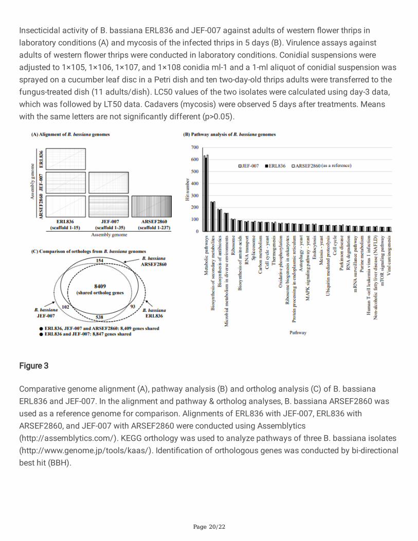
No big signi�cant differences in genome structure were observed between the two B. bassiana genomes(Figure 3A). When ERL836 genome was aligned with JEF-007 genome, although there were some minordifferences, but mostly genome structures (synteny) were similar to each other. When the genome ofERL836 was aligned to that of ARSEF2860 as a reference genome, two genome structures were similar,although some minor parts of the middle of the ARSEF2860 genome did not match the ERL836 genome.Additionally, when the three B. bassiana isolates were subjected to KEEG pathway analysis, no signi�cantdifferences were detected (Figure 3B). Metabolic pathway (9.7%), biosynthesis of secondary metabolites(3.9%) and biosynthesis of antibiotics (2.8%) were commonly major pathways in the three isolates. Whenorthologs of the two B. bassiana isolates were analyzed, ERL836 shared 8,847 genes with JEF-007 andthe two isolates shared 8,409 genes with ARSEF2860 as a reference (Figure 3C). In the three B. bassianaisolates, the percentages of the unique gene of each isolate were 0.19~0.35% of the total shared genesand the numbers are quite small.

Page 7/22
Comparative DEGs of two infecting B. bassiana isolates
B. bassiana ERL836 or JEF-007 was cultured on the 1/4SDA medium and adults of western �ower thripswere exposed to the fungal mass for three days. Collected infecting fungus was subjected to RNAextraction and differentially expressed genes were compared. In the DEG analysis using the JEF-007sequencing reads (Supplementary Table S2), a total of 643 genes were up-regulated, and 982 genes weredown-regulated when infecting western �ower thrips (Figure 4A). Approximately 1.5 times more contigsbelonged to down-regulated genes than up-regulated genes. Most fold changes were in the range of -2 to2 and accounted for 80.9% of total DEGs (1,625 DEGs). Among up-regulated genes (643 genes), 51%were assigned to the biological process category, 32% to the molecular function category, and 17% to thecellular component category. Among down-regulated genes (982 genes), 44% were assigned to themolecular function category, 41% to the biological process category, and 15% to the cellular componentcategory. Both up- and down-regulated genes in the biological process category were involved inmetabolic processes. Most up-regulated genes in the molecular function category had catalytic activity,whereas most down-regulated genes in this category were involved in binding. A similar number ofcontigs in most GO categories of the cellular component category were found among up-regulated genes,while genes with a membrane function were some of the most down-regulated genes.
In the DEG analysis using the ERL836 sequencing reads (Supplementary Table S2), a total of 1,197 geneswere up-regulated, and 360 genes were down-regulated (Figure 4B). ERL836 treatment resulted inapproximately twice more up-regulated contigs than down-regulated contigs. Most fold changes were inthe range of–2 to 2 and accounted for 71.96% of total DEGs (2,471 DEGs). Among up-regulated genes(1,197 genes), 38.4% were in the biological process category, 43.1% in the molecular function category,and 18.5% in the cellular component category. Similarly, among down-regulated genes, 39.8% were in thebiological process category, 40.0% in the molecular function category, and 20.1% in the cellularcomponent category. Metabolic process-related genes were the most abundant up-regulated genes.Among genes with a molecular function, most contigs were annotated as encoding proteins with catalyticactivity followed by binding and heterocyclic compound binding. Among cellular component genes,similar numbers of contigs were found in most of the GO categories.
Randomly selected up-regulated genes from the ERL836 and JEF-007 contigs were subjected to qRT-PCRfor validation. The fold change levels of the selected genes as analyzed by qRT-PCR were consistent withthe Illumina sequencing analysis results (Supplementary Figure S1).
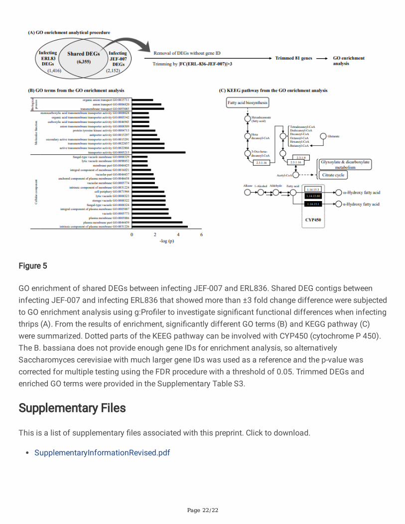
GO enrichment of shared DEGs between two isolates
A list of shared DEGs between infecting B. bassiana ERL836 and JEF-007 was derived from RNA-seqdata for enrichment analysis. A total 6,355 DEGs were shared by the two infecting B. bassiana isolates,
Page 8/22
but 3,576 genes without gene IDs were excluded from the analysis (Figure 5A). To investigate differences,81 genes were trimmed based on the fold change value between infecting ERL836 and JEF-007(|FC(ERL836-JEF-007)|>3). From the GO enrichment analysis (Supplementary Table S3), 30 GO termswere signi�cantly affected by shared DEG groups (FDR<0.05) (Figure 5B). Transporter activity(GO:0005215), plasma membrane part (GO: 0044459), and intrinsic component of plasma membrane(GO:0031226) functions were profoundly affected. According to pathway (KEGG) from the GOenrichment, the signi�cantly affected pathway was the fatty acid degradation pathway (KEGG:00071) (-log(p)>1.4), which is related to cytochrome P450 (Figure 5C). Some cytochrome P450 genes (CYP gene)in the shared gene groups showed different gene expression levels; highly up-regulated in ERL836 butdown-regulated in JEF-007. The fold change value of CYP539B1 in ERL836 was 2.34, while in JEF-007 itwas -0.73. The CYP655C1 had fold change value of 3.87 in ERL836 but -0.1 in JEF-007. Additionally,CYP5099A1 was upregulated at 2.66-fold in ERL836, but downregulated at -3.21-fold in JEF-007.
DiscussionIn this work, the two B. bassiana isolates, ERL836 and JEF-007 showed high similarities in biologicalfeatures, such as morphology, conidial productivity and virulence against western �ower thrips. Not onlythese two isolates, but also many other B. bassiana isolates look similar and are regarded to be similaractive ingredient when being developed as biopesticides. The highly similar morphological characteristicsof isolates can lead to potential con�icts when they are submitted to patent application and productdevelopment. So, in this work to further characterize two patented B. bassiana isolate ERL836 and JEF-007, their genome structures and gene expression patterns when infecting western �ower thrips wereanalyzed and compared. The genome structures of the two isolates were very similar, but geneexpression patterns were quite different. Different transcriptional responses could therefore be used as acharacteristic to differentiate the two B. bassiana isolates.
The two B. bassiana isolates, JEF-007 & ERL836 had similar genome structures, but their scaffoldnumbers were different from B. bassiana ARSEF2860, which might be due to the different sequencingtechnologies used to obtain the genomes of the isolates. JEF-007 and ERL836 isolates were sequencedusing the recent PacBio sequencing technology which reads ca. 10 Kb at a time (maximum 20 Kb)followed by error correction, whereas the genome of ARSEF2860 was generated using Illumina pair-endsequencing technology, which reads short fragments of 100 bp. Ten and nine contigs of more than 1 Mb(considered the minimum size of chromosome) were generated for JEF-007 and ERL836, respectively. Itis therefore possible that the two isolates have ca. 10 chromosomes, whereas ARSEF2860 has beenreported to have six chromosomes [25]. The ERL836, JEF-007 and ARSEF2860 as a reference shared8,409 genes, but each of isolate had less than 30 unique ortholog genes, very small. Unique genes ofeach isolate were predicted to be involved in fungal life cycles including growth (FluG domain-containingprotein, �occulin, methyltransferase, and protein kinase domain-containing protein), stress response(indoleamine-dioxygenase subfamily, tlh3, metallophosphoesterase, and transposase-like protein) andnutrient uptake (alpha/beta hydrolase fold-3 and ribose-phosphate pyrophosphokinase). The rest half ofthem is hypothetical protein gene.

Page 9/22
In GO analysis of ERL-836 DEGs, there were signi�cantly more up-regulated genes than down-regulatedgenes in various GO categories, when compared with JEF-007. Particularly highly up-regulated ERL836GO terms were detected in the biological process and molecular function of ERL836. This result indicatesthat when infecting thrips, possibly ERL836 much easily overcomes the defense mechanisms of thripscompared to JEF-007, but JEF-007 looks having di�culty in infecting the thrips. In ERL836, many fungalgenes were highly up-regulated in the infection process. For example, the eukaryotic aspartyl proteaseshowed an 11.2-fold increase in ERL836 when infecting western �ower thrips. This aspartyl protease hasa potential to degrade host insect tissues and disable anti-microbial peptides [26].
In the GO enrichment analysis, S. cerevisiae was used as a reference rather than B. bassiana becauseSaccharomyces GO IDs have been established much better than B. bassiana GO IDs and provide a goodeukaryotic model system for genetic and biochemical mechanisms. However, no gene IDs were found for3,576 genes which were shared by both of ERL836 and JEF-007. This is a limitation of this GOenrichment analysis. In spite of this limitation, GO categories of membrane transporters were signi�cantlydifferent between infecting JEF-007 and ERL836. ABC transporters are membrane transporters that playan essential role in B. bassiana infection as well as in infection by the phytopathogenic fungi ABC1 andBcatrB [27]. Transporter capability depends on activation of antioxidative enzymes [28]. B. bassianaharbors 21 ABC transporter genes, but not all ABC transporters contribute to fungal pathogenicity [29].
In the enrichment analysis, fatty acid degradation pathway was signi�cantly different between infectingJEF-007 and ERL836. The Cytochrome P450 genes have been found to be involved in fatty aciddegradation. Interestingly, most cytochrome P450 genes in infecting ERL836 were up-regulated, but inJEF-007 they were down-regulated. This suggests that these cytochrome P450 genes of ERL836 couldplay an important role in thrips infection in contrast to in JEF-007. In eukaryotic microorganisms,cytochrome P450 is involved in alkane oxidation. At least 83 genes encoding cytochrome p450s havebeen reported in B. bassiana, and a study of B. bassiana infection of silkworms revealed that cytochromeP450s are involved in infection and proliferation of B. bassiana [30]. In the previous study, CYP5099A1,CYP617A1, CYP617A2, CYP52G8, CYP539B1, and CYP655C1 were found to be involved in fatty aciddegradation (KO00071), which is related to insect hydrocarbon degradation [31]. Cytochrome P450enzymes can catalyze cascade formation of mono-oxidation products followed by diterminal oxidationand �nally produce α-ω acids [32]. CYP505D4 is involved in sulfur metabolism (KO00920). CYP540B16,CYP61A1, CYP586B1, and CYP682H1 are involved in steroid biosynthesis (KO00100). The two B.bassiana isolates, JEF-007 and ERL836, shared the expression of CYP.
ConclusionsIn summary, the two B. bassiana isolates ERL836 and JEF-007 showed similar biological characteristics,such as morphology and virulence against western �ower thrips as well as similar genome structure, butgene expression patterns were quite different when infecting western �ower thrips. B. bassiana ERL836appeared to infect thrips easily, while JEF-007 appeared to have more di�culty based on transcriptionalanalyses. Interestingly, in the enrichment analysis using shared DEGs between two infecting isolates,

Page 10/22
plasma membrane-mediated transporter activity and fatty acid degradation pathway includingcytochrome P450 were more active in infecting ERL836. This comparative approach using shared DEGanalysis could be applied to easily characterize the difference of the two patented B. bassiana isolates,ERL836 and JEF-007.
MethodsFungal isolates
B. bassiana ERL836 was obtained from the Entomology Research Laboratory (ERL) Worldwide Collectionof Entomopathogenic Fungi of the University of Vermont, USA, and was originally collected from soil inCalifornia, USA. B. bassiana JEF-007 was obtained from the Insect Microbiology and BiotechnologyLaboratory (IMBL), Chonbuk National University, Republic of Korea. Fungal isolates were grown onquarter strength Sabouraud dextrose agar (1/4SDA; Difco, USA) in the dark at 25 ± 1°C and stored in 20%(v/v) glycerol at -80°C.
Hyphal growth and conidial production
Fungal mycelia masses from 7-day-old B. bassiana isolates were collected into 1.5 ml Eppendorf tubescontaining 0.03% (v/v) siloxane solution. After 30 seconds of shaking on a vortex mixer (Vortex-Genie2TM; VWR Scienti�c, NY, USA), 2 ml of conidial suspension (1×10⁷ conidia ml-1) was dropped on themiddle of a ¼ SDA plate (90 mm diameter). Plates were kept in a 25°C incubator and observed for 9 daysunder a stereoscopic microscope. Fungal spore productivity was evaluated using millet-based solidcultured granules. Fungal granules were produced by modi�cation of the protocol of Kim et al [33]. Brie�y,200 g of millet was mixed with 100 ml of distilled water with 50% citric acid (160 ml) in a polyvinyl bag.Then the millet was autoclaved at 121°C for 15 minutes and cooled down at room temperature. Onemilliliter of fungal conidial suspension (1×10⁷ conidia ml-1) was inoculated and incubated at 25 ± 2°C for10 days. After drying the fungal granules till they had a moisture content of less than 10%, the 0.1 gmillet-based medium was collected into 1.5 ml microtubes with 0.03% (v/v) siloxane solution (Silwet,FarmHannong, Seoul, Republic of Korea). The fungal suspension was then vortexed vigorously for 3 minand the number of conidia was counted using a hemocytometer (Cat No. 2960408, Marienfeld, BadMergentheim, Germany) under a microscope (400×).
Bioassay against western �ower thrips
To compare the virulence of the two B. bassiana JEF-007 and ERL836, a colony of western �ower thripswas received from the National Institute of Horticultural and Herbal Science, Republic of Korea. Insectswere placed on �lter paper moistened with 2 ml of distilled water in a breeding dish (90 mm diameter, 50
Page 11/22
mm height). Each stage of thrips was reared in a different breeding dish and provided with fresh beansprouts every day. Old sprouts with eggs were transferred to new breeding dish every second day. Thripswere kept at 25 ± 2°C, 40 ± 10% relative humidity, and a 14:10 (L:D) photoperiod. For fungal bioassayagainst the thrips, conidial suspensions were prepared and adjusted to 1×10⁵, 1×10⁶, 1×10⁷, and 1×10⁸
conidia ml-1 using 0.03% (v/v) siloxane solution. A cucumber disc (diameter 60 mm) was placed on a�lter paper in a Petri-dish (diameter 60 mm) and 1 ml of conidial suspension was sprayed on thecucumber disc and dried for 10 min at room temperature. Distilled water containing 0.03% Silwet (v/v)served as a non-treated control. To maintain high humidity, 100 µl distilled water was added to the dishes.Eleven two-day-old thrips adults were transferred to the fungus-treated dish (11 adults dish-1) and covedwith lids. Treated dishes were kept at 24 ± 2°C and living adults were counted every day. Each treatmentwas replicated three times (3 plates per treatment). Lethal concentration 50 (LC50) values of the twoisolates were calculated using a Probit analysis (SPSS Inc., 2018).
Whole genome sequencing
For whole genome sequencing of B. bassiana ERL836 at Macrogen (www.macrogen.com; Macrogen Inc.,Seoul, Korea), genomic DNA and RNA were extracted from 7-day-old fungal mycelia. DNA quantity wasassessed using Pico-green staining (Invitrogen, Cat No. P7589) and Victor 3 �uorometry. To assess DNAquality, gel electrophoresis was performed. The concentration of genomic DNA (81.79 ng/µl) wasmeasured using a Nano Drop spectrophotometer (Thermo Scienti�c) and a Qubit �uorometer (LifeTechnology). For PacBio RS sequencing, 8 g of input genomic DNA was used for 20 kb librarypreparation. For gDNA with a size range less than 17 Kb, a Bioanalyzer 2100 (Agilent) was used todetermine the actual size distribution. Genomic DNA was sheared with g-TUBE (Covaris Inc., Woburn, MA,USA) and puri�ed using AMPure PB magnetic beads (Beckman Coulter Inc., Brea, CA, USA) if the apparentsize was greater than 40 kb. The gDNA concentration was measured using both a Nano Dropspectrophotometer and a Qubit �uorometer, and approximately 200 ng ml-1 of gDNA was run on a �eld-inversion gel. Total of 10 ml of library was prepared using the PacBio DNA Template Prep Kit 1.0 (for3~10 Kb). SMRT bell templates were annealed using PacBio DNA/Polymerase Binding Kit P6. PacBioDNA Sequencing Kit 4.0 and eight SMRT cells were used for sequencing. Subsequent steps were basedon the PacBio Sample Net-Shared Protocol, which is available at http://paci�cbiosciences.com/.
Genome analysis
All the genome-level comparisons were outsourced to Macrogen except genome alignment and KEEGanalysis. When B. bassiana JEF-007 and ERL836 genomes were compared using a Repeat Maskerprogram (v4.0.5), another B. bassiana ARSEF2860 was used as a reference. To obtain fungal secretedprotein proportions, TMHMM (v2.0) was used and the protein sequences of the B. bassiana isolates were

Page 12/22
subjected to ortholog analysis using OrthoMCL (v.2.0.3). Sequences encoding peptides shorter than 10amino acids and those with more than 20% stop codons were removed prior to blastp analysis (v2.2.25+;E-value 1E-5). To compare the sequences of two B. bassiana isolates, the Nucmer (NUCleotide MUMmer)module of the MUMmer package was used and data were analyzed using the following three steps:maximal extract matching, match clustering, and alignment extension. Alignments of ERL836 with JEF-007, ERL836 with ARSEF2860, and JEF-007 with ARSEF2860 were analyzed by Assemblytics(http://assemblytics.com/) and dot blot �gures were drawn. Lastly KEGG orthology (KO) was analyzed bythe web-based server KAAS (http://www.genome.jp/tools/kaas/). For gene ortholog analysis andpathway mapping, query sequences of JEF-007, ERL836 and ARSEF2860 were inputted, respectively.Identi�cation of orthologous genes was conducted by bi-directional best hit (BBH). A total of 348,165ascomycete sequences (taken from the KEGG databases) were used as reference sequences.
RNA extraction from thrips-infecting B. bassiana isolates
B. bassiana ERL836 or JEF-007 which was infecting adults of western �ower thrips was subjected to RNAextraction. Conidial suspension (70 ml, 1×10⁷ conidia ml-1) of each isolate was spread on ¼SDA plateand cultured at 27°C for 3 days. Then ca. 600 adults of western �ower thrips were transferred to thecultured plate and the plate was incubated at 27°C for 3 days. Infected thrips were collected into 1.5 mlmicrotubes with 0.03% (v/v) siloxane solution, and vortexed for 3 minutes. To exclude thrips bodies, thesuspensions were �ltered by sterilized iron mesh (ca. 80 mm2 pore size) and a fungal suspension wascollected. As a control, fungal mycelia of each isolate were harvested from 6-day-old cultures on ¼SDA.The two different treatments were replicated three times (3 samples from the thrips-treated B. bassianaand another 3 samples from the B. bassiana only). Total RNAs were extracted by TRIzol reagent(Invitrogen Life Technologies, CA, USA) following the manufacturer’s instructions. RNA purity and integritywere quanti�ed by ASP-2680 spectrophotometer (ACTGene, Piscataway, NJ, USA) and an AgilentTechnologies 2100 Bioanalyzer (Agilent Technologies, Palo Alto, Cambridge, USA).
RNA sequencing and analysis
Libraries of non-infecting and infecting ERL836 or JEF007 were made using the Truseq RNA kit (Illumina,San Diego, USA) following the manufacturer’s protocol at Macrogen. Multiple indexing adapters wereligated to the ends of the double-stranded cDNA and then enriched by PCR to create DNA librarytemplates. In each isolate, infecting and non-infecting samples were sequenced in parallel using anIllumina HiSeq 2000 sequencer. Before analyzing the sequences, quality scores were checked by Fast QC(ver 0.11.7). After receiving sequencing raw data from Macrogen, the following analyses were conductedin our laboratory. For e�cient and robust de novo reconstruction of transcriptomes, Trinity (ver 2.8.3) wasused (https://github.com/trinityrnaseq/). To identify candidate coding regions within transcriptsequences, Trans-Decoder (ver 5.5.0) was used. In the �rst step, long ORFs were extracted, and then likely

Page 13/22
coding regions were predicted. To cluster similar nucleotide sequences into clusters meeting a user-de�ned similarity threshold (0.9 in this analysis), CD-HIT-EST (version 4.7) was used. To quantifytranscript abundances, Kallisto (ver 0.45.0) was used to build an index form from the fasta form of targetsequences and non-infecting and infecting libraries were compared. Transcripts per million (TPM) of non-infecting and infecting samples was calculated. Raw signals were normalized using a log2-basedtransformation. Fold-change statistical tests were performed and log2|FC|≧2 was de�ned as statisticallysigni�cant differential expression. Genes were blasted using the Blast2Go program. Analysis wasconducted by local blast with B. bassiana ARSEF2860 as a reference sequence. Statistical signi�cancethreshold was 1.0E-10 and the number of blast hits was set to one. Gene ontology (GO) analysis of up-and down-regulated genes was performed using InterPro (online) in the Blast2Go program. The publicEMBL-EBI database was used to scan sequences against InterPro's signatures. Up- and down-regulatedgenes were annotated at GO level 2.
Validation of RNA-sequencing
Non-infecting and infecting RNA samples were subjected to reverse transcription (RT) using AccuPower®RT PreMix (Bioneer, Daejeon, Republic of Korea) with the oligo (dT) 15 primer (Promega, MI, USA). A setof primers for RT-PCR (Supplementary Table S4) were designed at Snap Dragon(http://www.�yrnai.org/snapdragon). RT-PCR was performed as follows: an initial denaturation at 94°C of5 minutes followed by 34 cycles of 30 seconds at 94°C, 30 seconds at (Tm)°C, and 30 seconds at 74°C,followed by a �nal extension for 10 minutes at 74°C (C-1000, Bio Rad, Hercules, CA, USA). To validate up-regulated genes, �ve genes of JEF-007 and three genes of ERL836 were randomly selected and subjectedto quantitative RT-PCR (qRT-PCR). To remove gDNA contamination, RNA extracted by the Trizol method(described above) was treated with 1 mg of DNaseI (Invitrogen Life Technologies, CA, USA). cDNAs weregenerated using the AccuPowder® Rocketscript RT PreMix kit (BioNeer) and oligo(dT) primer (Promega),and they were used as the template for PCR ampli�cation. Then qRT-PCR was performed usingThunderbird® Syber® qPCR mix (QPS-201, TOYOBO, Japan) on a 96-well Bio-Rad CFX96 Real-Time PCRSystem (Bio-Rad, USA). Cycling parameters for qRT-PCR were as follows: denaturation for 1 minute at95°C, and then 40 cycles of 15 seconds at 95°C, 1 minute at 60°C followed by melting with an increase intemperature of 0.5°C per 5 seconds starting from 65°C to 95°C. Primers for B. bassiana actin (=γ-actin,GenBank Accession No: HQ232398) were used as an internal control to obtain relative expression levels[34]. △Ct (threshold cycle) was calculated as (Ct value of up-regulated genes) - (Ct value of Bb-actin) andsubjected to the calculation of fold change value (2-△△Ct).
GO enrichment analysis
Page 14/22
Shared DEG contigs between infecting JEF-007 and infecting ERL836 which had more than 3-fold changedifference were subjected to GO enrichment analysis to investigate the different mechanisms of the twofungal isolates in pathogenesis. In this analysis, blastn was performed with an E-value of 1.0E-10. Non-blasted and non-GO ID contigs were removed in this analysis. Functional enrichment was performedusing the g:Pro�ler web server (http://bitt.cs.ut.ee/gpro�ler/) [35]. Input query data list was matched withSaccharomyces cerevisiae (https://�ybase.org/). The B. bassiana does not provide enough gene IDs forenrichment analysis, so alternatively S. cerevisiae with much larger analyzed gene IDs was used as areference. The related p-value was corrected for multiple testing using the Benjamini-Hochberg FalseDiscovery Rate (FDR) procedure with a threshold of 0.05.
Data analysis
Data on the numbers of conidia, percentage of live thrips and expression level in qRT-PCR were arc-sinetransformed and analyzed using an ANOVA or generalized linear model (GLM) followed by Tukey'shonestly signi�cant difference (HSD) for multiple comparisons. All the analyses were conducted usingSPSS (SPSS Inc., 2018) at the 0.05 (α) level of signi�cance.
Abbreviations1/4SDA: quarter strength Sabouraud dextrose agar; ANOVA: analysis of variance; DEG: differentiallyexpressed gene; FC: fold change; FDR: false discovery rate; GLM: generalized linear model; GO: geneontology; HSD: honestly signi�cant difference; KEEG: Kyoto encyclopedia of genes and genomes; LC50:lethal concentration 50%; and LT50: lethal time 50%
DeclarationsAcknowledgements
We appreciate Dr. Woo Jin Kim for comments and advice for the analyses of genome and RNAsequencing data, and National Institute of Horticultural and Herbal Science, Republic of Korea forproviding western �ower thrips colonies.
Authors’ contributions
JSK designed the whole experiments and wrote the manuscript. SK conducted bioassay against thripsand analyzed the NGS data and wrote manuscript. JCK conducted analyzed the WGS and RNA-seq datausing raw sequencing materials. SJL designed the analyses of WGS and RNA-seq data. MRL, SEP, DL andSB reared the thrips and supported the bioassays and statistical data analysis. TYS supported the designof experiments and helped troubleshooting. All authors reviewed this manuscript.

Page 15/22
Funding
This work was supported by Korea Institute of Planning and Evaluation for Technology in Food,Agriculture and Forestry (IPET) through Agriculture and Livestock Machinery / Equipment industryTechnology Development Program, funded by Ministry of Agriculture, Food and Rural Affairs (MAFRA)(Grant No: 118103-03).
Availability of data and materials
The genome of B. bassiana ERL836 has been deposited in the GenBank database under the projectaccession no. PPTI00000000.
Ethics approval and consent to participate
Not applicable.
Consent for publication
Not applicable.
Competing interests
The authors declare that they have no competing interests.
Author details
1Department of Agricultural Biology, Chonbuk National University, Jeonju 54596, Korea. 2 Department ofMicrobiology and Cell Science, University of Florida, Gainesville FL 32611-0700, USA.
References1. Lee SJ, Lee MR, Kim S, Kim JC, Park SE, Li D, Shin TY, Nai YS, Kim JS, Genomic Analysis of the
Insect-Killing Fungus Beauveria bassiana JEF-007 as a Biopesticide. Sci Rep. 2018;8:12388.

Page 16/22
2. Rehner SA, Buckley EA, Beauveria phylogeny inferred from nuclear ITS and EF1-α sequences:evidence for cryptic diversi�cation and links to Cordyceps teleomorphs. Mycologia 2005;97:84-98.
3. Glare TR, Inwood AJ, Morphological and genetic characterisation of Beauveria spp. from NewZealand. Mycol Res. 1998;102:250-256.
4. Rehner SA, Minnis AM, Sung GH, Luangsa-ard JJ, Devotto L, Humber RA, Phylogeny and systematicsof the anamorphic, entomopathogenic genus Beauveria. Mycologia 2011;103:1055-1073.
5. Zhang S, Xia Y, Keyhani NO, Contribution of the gas1 gene of the entomopathogenic fungusBeauveria bassiana, encoding a putative glycosylphosphatidylinositol-anchored β-1, 3-glucanosyltransferase, to conidial thermotolerance and virulence. Appl Environ Microbiol.2011;77:2676-2684.
�. Xiao G, Ying SH, Zheng P, Wang ZL, Zhang S, Xie XQ, Shang Y, St Leger RJ, Zhao GP, Wang C,Genomic perspectives on the evolution of fungal entomopathogenicity in Beauveria bassiana. SciRep. 2012;2:483.
7. Meyer E, Aglyamova GV, Wang S, Buchanan-Carter J, Abrego D, Colbourne JK, Willis BL, Matz MV,Sequencing and de novo analysis of a coral larval transcriptome using 454 GSFlx. BMC Genomics200;10:219.
�. Shu S, Chen B, Zhou M, Zhao X, Xia H, Wang M, De novo sequencing and transcriptome analysis ofWol�poria cocos to reveal genes related to biosynthesis of triterpenoids. PLoS One 2013;8:e71350.
9. Yang P, Zhu JY, Gong ZJ, Xu DL, Chen XM, Liu WW, Lin XD, Li YF, Transcriptome analysis of theChinese white wax scale Ericerus pela with focus on genes involved in wax biosynthesis. PLoS One2012;7: e35719.
10. Mantilla JG, Galeano NF, Gaitan AL, Cristancho MA, Keyhani NO, Gongora CE, Transcriptome analysisof the entomopathogenic fungus Beauveria bassiana grown on cuticular extracts of the coffee berryborer (Hypothenemus hampei). Microbiol. 2012;158:1826-1842.
11. Xia J, Zhang CR, Zhang S, Li FF, Feng MG, Wang XW, Liu SS, Analysis of white�y transcriptionalresponses to Beauveria bassiana infection reveals new insights into insect-fungus interactions. PLoSOne 2013;8:e68185.
12. Liu Y, Shen D, Zhou F, Wang G, An C, Identi�cation of immunity-related genes in Ostrinia furnacalisagainst entomopathogenic fungi by RNA-seq analysis. PLoS One 2014;9:e86436.
13. Chu Y, Liu Y, Shen D, Hong F, Wang G, An C, Serine proteases SP1 and SP13 mediate the melanizationresponse of Asian corn borer, Ostrinia furnacalis, against entomopathogenic fungus Beauveriabassiana. J Invertebr Pathol. 2015;128:64-72.
14. Yang YT, Lee SJ, Nai YS, Kim S, Kim JS, Up-regulation of carbon metabolism-related glyoxylate cycleand toxin production in Beauveria bassiana JEF-007 during infection of bean bug, Riptortuspedestris (Hemiptera: Alydidae). Fungal Biol. 2016;120:1236-1248.
15. Xing D, Yang Q, Jiang L, Li Q, Xiao Y, Ye M, Xia Q, RNA-seq analyses for two silkworm strains revealsinsight into their susceptibility and resistance to Beauveria bassiana infection. Int J Mol Sci.2017;18:234.

Page 17/22
1�. Lai Y, Chen H, Wei G, Wang G, Li F, Wang S, In vivo gene expression pro�ling of theentomopathogenic fungus Beauveria bassiana elucidates its infection stratagems in Anophelesmosquito. China Life Sci. 2017;60:839-851.
17. Chen A, Wnag Y, Shao Y, Zhou Q, Chen S, Wu Y, Chen H, Liu E, Genes involved in Beauveria bassianainfection to Galleria mellonella. Arch Microbiol. 2018;200:541-552.
1�. Lee SJ, Yang YT, Kim S, Lee MR, Kim JC, Park SE, Hossain MS, Shin TY, Nai YS, Kim JS,Transcriptional response of bean bug (Riptortus pedestris) upon infection with entomopathogenicfungus, Beauveria bassiana JEF‐007. Pest Manage Sci. 2019;75:333-345.
19 Wang JJ, Bai WW, Zhou W, Liu J, Chen J, Liu XY, Xiang TT, Liu RH, Wang WH, Zhang BL,Transcriptomic analysis of two Beauveria bassiana strains grown on cuticle extracts of the silkwormuncovers their different metabolic response at early infection stage. J Invertebr Pathol. 2017;145:45-54.
20 Zhang Z, Zhang P, Li W, Zhang J, Huang F, Yang J, Bei Y, Lu Y, De novo transcriptome sequencing inFrankliniella occidentalis to identify genes involved in plant virus transmission and insecticide resistance.Genomics 2013;101:296-305.
21 Schneweis DJ, Whit�eld AE, Rotenberg D, Thrips developmental stage-speci�c transcriptomeresponse to tomato spotted wilt virus during the virus infection cycle in Frankliniella occidentalis, theprimary vector. Virol. 2017;500:226-237.
22 Shrestha A. Champagne DE, Culbreath AK, Rotenberg D, Whit�eld AE, Srinivasan R, Transcriptomechanges associated with Tomato spotted wilt virus infection in various life stages of its thrips vector,Frankliniella fusca (Hinds). J Gen Virol. 2017;98:2156-2170.
23 Gupta R, Kwon SY, Kim ST, An insight into the tomato spotted wilt virus (TSWV), tomato and thripsinteraction. Plant Biotechnol Rep. 2018;12:157-163.
24 Gamage SMW, Rotenberg D, Schneweis DJ, Tsai CW, Dietzgen RG, Transcriptome-wide responses ofadult melon thrips (Thrips palmi) associated with capsicum chlorosis virus infection. PLoS One2018;13:e0208538.
25 Padmavathi J, Devi KU, Rao CUM, Reddy NNR, Telomere �ngerprinting for assessing chromosomenumber, isolate typing and recombination in the entomopathogen Beauveria bassiana. Mycol Res.2003;107:572-580.
26 Freimoser FM, Screen S, Hu G, Leger RS, EST analysis of genes expressed by the zygomycetepathogen Conidiobolus coronatus during growth on insect cuticle. Microbiol. 2003;149:1893-1900.
27 Stefanato FL, Abou-Mansour E, Buchala A, Kretschmer M, Mosbach A, Hahn M, Bochet CG, MétrauxJP, Schoonbeek HJ, The ABC transporter BcatrB from Botrytis cinerea exports camalexin and is avirulence factor on Arabidopsis thaliana. Plant J. 2009;58:499-510.

Page 18/22
28 Wang ZL, Zhang LB, Ying SH, Feng MG, Catalases play differentiated roles in the adaptation of afungal entomopathogen to environmental stresses. Environ Microbiol. 2013;15:409-418.
29 Song TT, Zhao J, Ying SH, Feng MG, Differential contributions of �ve ABC transporters to mutidrugresistance, antioxidion and virulence of Beauveria bassiana, an entomopathogenic fungus. PLoS One2013;8:e62179.
30 Hou C, Qin G, Liu T, Geng T, Gao K, Pan Z, Qian H, Guo X, Transcriptome analysis of silkworm,Bombyx mori, during early response to Beauveria bassiana challenges. PLoS One 2014;9:e91189.
31 Pedrini N, Ortiz-Urquiza A, Zhang S, Keyhani NO, Targeting of insect epicuticular lipids by theentomopathogenic fungus Beauveria bassiana: hydrocarbon oxidation within the context of a host-pathogen interaction. Front Microbiol. 2013;4:24.
32 Scheller U, Zimmer T, Becher D, Schauer F, Schunck WH, Oxygenation cascade in conversion of n-alkanes to α, ω-dioic acids catalyzed by cytochrome P450 52A3. J Biol Chem. 1998;273:32528-32534.
33 Kim JS, Kassa A, Skinner M, Hata T, Parker BL, Production of thermotolerant entomopathogenicfungal conidia on millet grain. J Ind Microbiol Biotechnol. 2011;38:697-704.
34 Zhou YH, Zhang YJ, Luo ZB, Fan YH, Tang GR, Liu LJ, Pei Y, Selection of optimal reference genes forexpression analysis in the entomopathogenic fungus Beauveria bassiana during development, underchanging nutrient conditions, and after exposure to abiotic stresses. Appl Microbiol Biotechnol.2012;93:679-685.
35 Reimand J, Arak T, Adler P, Kolberg L, Reisberg S, Peterson H, Vilo J, g:Pro�ler - a web server forfunctional interpretation of gene lists (2016 update). Nucleic Acids Res. 2016;44:W83-W89.
Supplementary InformationTable S1. de novo assembly of B. bassiana JEF-007 (A) and ERL846 (B) after the sequencing of wholegenomes using Pac Bio RSII technology with error correction.
Table S2. de novo assembly of B. bassiana RNA-sequencing raw data.
Table S3. GO enrichment analysis of differentially expressed and shared genes between infecting B.bassiana JEF-007 and ERL836.
Table S4. Primers used in qRT-PCR for validation of B. bassiana RNA-sequencing.
Figure S1. Validation of RNA-sequencing of B. bassiana ERL836 and JEF-007 using qRT-PCR.
Figures

Page 19/22
Figure 1
Hyphal growth (A) and conidial production (B) of B. bassiana ERL836 and JEF-007. A 2 l of conidialsuspension (1×10⁷ conidia/ml) was dropped on the middle of a ¼ SDA plate and kept in a 25°Cincubator for 9 days. Conidial productivity was evaluated using millet-based solid culture in polyethylenebags. A 1 ml of fungal conidial suspension (1×10⁷ conidia/mL) was inoculated to the bag including 200g millet and incubated at 25±2°C for 10 days. Means followed by the same letter are not signi�cantlydifferent (p>0.05).
Figure 2
Page 20/22
Insecticidal activity of B. bassiana ERL836 and JEF-007 against adults of western �ower thrips inlaboratory conditions (A) and mycosis of the infected thrips in 5 days (B). Virulence assays againstadults of western �ower thrips were conducted in laboratory conditions. Conidial suspensions wereadjusted to 1×10⁵, 1×10⁶, 1×10⁷, and 1×10⁸ conidia ml-1 and a 1-ml aliquot of conidial suspension wassprayed on a cucumber leaf disc in a Petri dish and ten two-day-old thrips adults were transferred to thefungus-treated dish (11 adults/dish). LC50 values of the two isolates were calculated using day-3 data,which was followed by LT50 data. Cadavers (mycosis) were observed 5 days after treatments. Meanswith the same letters are not signi�cantly different (p>0.05).
Figure 3
Comparative genome alignment (A), pathway analysis (B) and ortholog analysis (C) of B. bassianaERL836 and JEF-007. In the alignment and pathway & ortholog analyses, B. bassiana ARSEF2860 wasused as a reference genome for comparison. Alignments of ERL836 with JEF-007, ERL836 withARSEF2860, and JEF-007 with ARSEF2860 were conducted using Assemblytics(http://assemblytics.com/). KEGG orthology was used to analyze pathways of three B. bassiana isolates(http://www.genome.jp/tools/kaas/). Identi�cation of orthologous genes was conducted by bi-directionalbest hit (BBH).

Page 21/22
Figure 4
Differentially expressed genes (DEG) of B. bassiana ERL836 (A) and JEF-007 (B) when infecting western�ower thrips. Thrips were exposed to 3-day old fungus-cultured plates for 3 days. Infected thrips wereharvested using a surfactant solution and thrips bodies were excluded by �ltering and subjected to RNAextraction. As a non-infecting control, 6-day cultured fungus was used for RNA extraction. In eachinfecting isolate, the numbers of up- and down-regulated contigs (|fold change|>2) were analyzed and GOanalyses of DEGs were conducted using Blast2Go program.
Page 22/22
Figure 5
GO enrichment of shared DEGs between infecting JEF-007 and ERL836. Shared DEG contigs betweeninfecting JEF-007 and infecting ERL836 that showed more than ±3 fold change difference were subjectedto GO enrichment analysis using g:Pro�ler to investigate signi�cant functional differences when infectingthrips (A). From the results of enrichment, signi�cantly different GO terms (B) and KEGG pathway (C)were summarized. Dotted parts of the KEEG pathway can be involved with CYP450 (cytochrome P 450).The B. bassiana does not provide enough gene IDs for enrichment analysis, so alternativelySaccharomyces cerevisiae with much larger gene IDs was used as a reference and the p-value wascorrected for multiple testing using the FDR procedure with a threshold of 0.05. Trimmed DEGs andenriched GO terms were provided in the Supplementary Table S3.
Supplementary Files
This is a list of supplementary �les associated with this preprint. Click to download.
SupplementaryInformationRevised.pdf