what is a biochemical test? · what is a biochemical test? a biochemical test refers to the...
TRANSCRIPT

What is a biochemical test
A biochemical test refers to the chemical identification of unknown substances within a living thing The test quantitatively and qualitatively determines a particular substance like an enzyme within the bloodAbiochemical test can be used to diagnose a given disease
Most bacteria of medical importance can be grown on culture
Culture media can be selective or non selective Non selective media allow a wide variety of bacteria to grow (eg nutrient agar or blood agar)
Selective media allow only certain organisms to grow because they have specific inhibitors added to the media(eg the bile salts in MacConkey agar)The differential ability of these media is based on lactose fermentation which allows groups of biochemically related bacteria to be distinguished from other of bacteria
Gram Negative Rods
IMViC test is an acronym that stands for four different tests to distinguished between enteric microorganisms(identificationdifferentiation of members of family enterobacteriaceae
1- Indol test
2- Methyl red test
3- Voges-proskauer test
4- Citrate utilization test
To obtain the results of these four tests three tubes are inoculated
Tryptone broth(indole test) methyl red-Voges proskauer broth(MR-VP)broth and citrate agar
Indole Test
PurposeThe Indole Test identifies bacteria capable of producing
indole using the enzyme tryptophanase The Indole Test
is one component of the IMViC battery of tests (Indole
Methyl Red Voges-Proskauer and Citrate) used to differentiate
the Enterobacteriaceae
PrincipleThe Indole Test as it appears in this manual is performed
using SIM medium SIM medium also tests for motility and
sulfur reduction (SIM is an acronym for Sugar-Indole-
Motility) It is a semi-solid medium that is formulated with
casein and animal tissue as sources of amino acids an iron
containing
compound and sulfur in the form of sodium
thiosulfate
Indole production in the medium is made possible by
the presence of tryptophan (contained in casein and animal
protein) Bacteria possessing the enzyme tryptophanase can
hydrolyze tryptophan to pyruvate ammonia (by deamination)
and indole
The hydrolysis of tryptophan in SIM medium can be
detected by the addition of Kovacsrsquo reagent after a period
of incubation
Kovacsrsquo reagent contains dimethylamino -
benz aldehyde (DMABA) and HCl dissolved in amyl alcohol
When a few drops of Kovacsrsquo reagent are added to the tube
it forms a liquid layer over the solid medium
7-48)
The formation of red color in the reagent layer indicates
a positive reaction and the presence of tryptophanase
No red color is indole- negative
INDOLE TEST
RESULTS
INDOLE TEST RESULTS
This is SIM medium inoculated
with Morganella morganii on the right(+) and Enterobacter
aerogenes (_) on the left
Methyl Red Test
PurposeThe Methyl Red Test is a component of the IMViC battery
of tests (Indole Methyl Red Voges-Proskauer and Citrate)
used to differentiate the Entero bacteriaceae It identifies
bacterial ability to produce stable acid end products by
means of a mixed-acid fermentation of glucose
PrincipleMR-VP Broth is a combination medium used for both
Methyl Red (MR) and Voges-Proskauer (VP) tests (Refer
to page 98 for the VP test) It is a simple solution containing
only peptone glucose and a phosphate buffer The peptone
and glucose provide protein and a fermentable carbohydrate
respectively and the potassium phosphate resists pH
changes in the medium
The MR test is designed to detect organisms capable of
performing a mixed acid fermentation which overcomes the
phosphate buffer in the medium and lowers the pH (Figure
7-62) The acids produced by these organisms tend to be
stable whereas acids produced by other organisms tend to
be unstable and subsequently are converted to more
neutral
products
Mixed acid fermentation is verified by the addition of
methyl red indicator dye following incubation Methyl red is
red at pH 44 and yellow at pH 62 Between these two pH
values it is various shades of orange Red color is the
only
true indication of a positive result Orange is negative or
inconclusive Yellow is negative (Figure 7-63)
7-63 THE METHYL RED TEST
Escherichia coli (MR-positive) on the
left and Enterobacter aerogenes (MR-negative) on the right
Voges-Proskauer Test
PurposeThe Voges-Proskauer Test (VP) is a component of the IMViC
battery of tests (Indole Methyl Red Voges-Proskauer and
Citrate) used to distinguish between members of the Family
Entero bacteriaceae and differentiate them from other
Gram-negative rods It identifies organisms able to produce
acetoin from the degra dation of glucose during a 23-butanediol
fermentation
Principle
MR-VP Broth is the combination medium used for both
Methyl Red (MR) and Voges-Proskauer (VP) tests (Refer to
page 82 for the MR test) It is a simple solution containing
only peptone glucose and a phosphate buffer The peptone
and glucose provide protein and a fermentable carbohydrate
and the potassium phosphate resists pH changes in the
medium-98
The Voges-Proskauer test was designed for organisms
that are able to ferment glucose but quickly convert their
acid products to acetoin and 23-butanediol
Adding VP reagents to the medium after
incubation oxidizes the acetoin (if present) to diacetyl
which in turn reacts with guanidine nuclei from peptone
to produce a red color (Figure 7-98) A positive VP result
therefore is red No color change (or development of copper
color) after the addition of reagents is negative The copper
color is a result of interactions between the reagents and
should not be confused with the true red color of a positive
result (Figure 7-98) Use of positive and negative controls
for comparison is usually recommended
7-98 THE VOGES-PROSKAUER TEST
Escherichia coli (VP- negative)is on the left and Enterobacter aerogenes (VP-positive) is on the rightThe copper color at the top of the VP- negative tube is due to thereaction of KOH and 1048576 -naphthol and should not be confused witha positive result Layering of the red color in positive tubes may ormay not occur and is irrelevant to interpretation
Citrate Utilization Test
PurposeThe Citrate Utilization Test is used to determine the ability
of an organism to use citrate as its sole source of carbon
Citrate utilization is one part of a test series referred to as
the IMViC (Indole Methyl Red Voges-Proskauer and
Citrate tests) that distinguishes between members of the
family Enterobacteriaceae and differentiates them from
other Gram-negative rods
Principle Simmons Citrate Agar is a defined medium that contains
sodium citrate as the sole carbon source and ammonium
phosphate as the sole nitrogen source Bromthymol blue
dye which is green at pH 69 and blue at pH 76 is added
as an indicator Bacteria that survive in the medium and
utilize the citrate also convert the ammonium phosphate
to ammonia (NH3) and ammonium hydroxide (NH4OH)
both of which tend to alkalinize the agar As the pH goes
up the medium changes from green to blue (Figure 7-23)
Thus conversion of the medium to blue is a positive citrate
test result
Occasionally a citrate-positive organism will grow on a
Simmons Citrate slant without producing a change in color
7-23 CITRATE TEST
RESULTS
Simmons citrated
agar inoculate Citrobacter
diversus (
+) on the left
Bacillus cereus (_) in the
center and an uninoculated
control on the right
Triple Sugar Iron Agar
PurposeTriple Sugar Iron Agar (TSIA) is primarily used to differen -
tiate members of Enterobacteriaceae and to differentiate
them from other Gram-negative rods such as Pseudomonas
PrincipleTSIA is a rich medium designed to differentiate bacteria on
the basis of glucose fermentation lactose fermentation sucrose
fermentation and sulfur reduction In addition to the
three carbohydrates it includes beef extract yeast extract
and peptone as carbon and nitrogen sources and sodium
thiosulfate as a source of reducible sulfur Phenol red is the
pH indicator and the iron in ferrous sulfate is the hydrogen
sulfide indicator
The medium is prepared as a shallow agar slant with
a deep butt thereby providing both aerobic and anaerobic
growth environments It is inoculated by a stab in the agar
butt followed by a fishtail streak of the slant The incubation
period is 18 to 24 hours for carbohydrate fermentation
and up to 48 hours for hydrogen sulfide reactions Many
reactions in various combinations are possible (Figure 7-92
and Table 7-6)
When TSIA is inoculated with a glucose-only fermenter
acid products lower the pH and turn the entire medium
yellow within a few hours Because glucose is in short
supply (01) it will be exhausted within about 12 hours
As the glucose is used up the organisms located in the aerobic
region (slant) will begin to break down available amino
acids producing NH3 and raising the pH This process
which takes 18 to 24 hours to complete is called a reversion
and only occurs in the slant because of the anaerobic conditions
in the butt Thus a TSIA with a red slant and yellow
butt after a 24-hour incubation period indicates that the
organism ferments glucose but not lactose
Organisms that are able to ferment glucose and lactose
andor sucrose also turn the medium yellow throughout
However because the lactose and sucrose concentrations
are ten times higher than that of glucose greater acid production
results and both slant and butt will remain yellow
after 24 hours Therefore a TSIA with a yellow slant and
butt at 24 hours indicates that the organism ferments glucose and one or both of the other sugars Gas
produced by carbohydrate
fermentation will appear as fissures in the medium
or will lift the agar off the bottom of the tube
Hydrogen sulfide (H2S) may be produced by the reduction
of thiosulfate in the medium or by the breakdown of
cysteine in the peptone Ferrous sulfate in the medium reacts
with the H2S to form a black precipitate usually seen in the
butt Acid conditions must exist for thiosulfate reduction
therefore black precipitate in the medium is an indication
of sulfur reduction and fermentation If the black precipitate
obscures the color of the butt the color of the slant determines
which carbohydrates have been fermented (ie red
slant = glucose fermentation yellow slant = glucose and
lactose andor sucrose fermentation)
An organism that does not ferment any of the carbo hy -
drates but utilizes peptone and amino acids will alkalinize the
medium and turn it red If the organism can use the peptone
aerobically and anaerobically both the slant and butt will
appear red An obligate aerobe will turn only the slant red
Timing is critical in reading TSIA results An early reading
could reveal yellow throughout the medium leading
one to conclude that the organism is a lactose or sucrose
fermenter when it simply may not yet have exhausted the
glucose A reading after the lactose and sucrose have been
depleted could reveal a yellow butt and red slant leading
one to falsely conclude the organism is a glucose-only
fermenter
Tubes that have been interpreted for carbohydrate
fermentation and are negative for sulfur reduction can be
re-incubated for 24 hours before H2S determination Refer
to Table 7-6 for information on the correct symbols and
method of reporting the various reactions
7-92 TSI AGAR SLANTS
From left to right Pseudomonas aeruginosa
(KNC) uninoculated control Morganella morganii (KA atypically not
producing gas) Escherichia coli (AA G) and Proteus mirabilis (KA H2S)
Urease Tests
PurposeThe Urease Test is used to differentiate organisms based on
their ability to hydrolyze urea with the enzyme urease Urinary
tract pathogens from the genus Proteus may be distinguished
from other enteric bacteria by their rapid urease activity
Principle
Urea is a product of decarboxylation of certain amino acids
It can be hydrolyzed to ammonia and carbon dioxide by
bacteria containing the enzyme urease Many enteric bacteria
(and a few others) possess the ability to metabolize urea but
only members of Proteus Morgan ella and Providencia are
considered rapid urease-positive organisms
Urea Agar was formulated to differentiate rapid ureasepositive
organisms from slower urease-positive and ureasenegative
bacteria It contains urea peptone potassium
phosphate glucose phenol red and agar Peptone and
glucose provide essential nutrients for a broad range of
bacteria Potassium phosphate is a mild buffer used to resist
alkalini zation of the medium from peptone metabolism
Phenol red which is yellow or orange below pH 84 and
red or pink above is included as an indicator
Urea hydrolysis to ammonia by ureasepositive
organisms will overcome the buffer in the medium
and change it from orange to pink The agar must be
examined daily during incubation Rapid urease-positive
organisms will turn the entire slant pink within 24 hours
Weak positives may take several days) Ureasenegative
organisms either produce no color change in the
medium or turn it yellow from acid products (Figure 7-94)
7-94 UREASE
AGAR TEST RESULTS
Urease agar tubes
after a 24 hour incubation
Morganella
morganii (ureasepositive)
a rapid
urea splitter is on the
left and Hafnia alvei
(urease-negative)
is on the right An
uninoculated control
is in the center
Gram positive cocci
Hemolysis test
Blood agar is used for isolation and cultivation of many
types of fastidious bacteria It is also used to dif ferentiate
bacteria based on their hemolytic characteristics especially
within the genera Streptococcus Enterococcus and
Aerococcus
PrincipleSeveral species of Gram-positive cocci produce exotoxins
called hemolysins which are able to destroy red blood cells
(RBCs) and hemoglobin Blood Agar sometimes called
Sheep Blood Agar because it includes 5 sheep blood in
a Tryptic Soy Agar base allows differentiation of bacteria
based on their ability to hemolyze RBCs
The three major types of hemolysis are -hemolysis
1048576 -hemolysis and -hemolysis -hemolysis the complete
destruction of RBCs and hemoglobin results in a clearing of
the medium around the colonies (Figure 7-10) 1048576 -
hemolysis
is the partial destruction of RBCs and produces a
greenish
discoloration of the agar around the colonies (Figure 7-
11)
-hemolysis is actually nonhemolysis and appears as
simple
growth with no change to the medium (Figure 7-12)
7-10 -HEMOLYSISStreptococcus pyogenes demonstrates -hemolysis
The clearing around the growth is a result of complete lysis of red blood
cells This photograph was taken with transmitted light
7-11 -HEMOLYSISThis is a streak plate of Streptococcus pneumoniae
demonstrating 1048576 -hemolysis The greenish zone around the colonies
results from incomplete lysis of red blood cells-
Catalase Test
PurposeThe Catalase Test is used to identify organisms that produce
the enzyme catalase It is most commonly used to differentiate
members of the catalase-positive Staphylococcus from the
catalase-negative Streptococcaceae Variations on this test
may also be used in identification of Mycobacterium species
Principle
Bacteria that produce catalase can be detected easily
using typical store-grade hydrogen peroxide When hydrogen
peroxide is added to a catalase-positive culture oxygen
gas bubbles form immediately (Figures 7-20 and 7-21) If no
bubbles appear the organism is catalase-negative This test
can be performed on a microscope slide or by adding hydrogen
peroxide directly to the bacterial growth
7-20 CATALASE SLIDE TEST
Shown is the catalase slide test in which
visible bubble production indicates a positive result Staphylococcus
aureus (+) is on the left Streptococcus(-) is on the right of peroxide to contain aerosols produced in positive reactions
7-21 CATALASE TUBE
TEST
The catalase test
may also be performed on
an agar slant Staphylo -
coccus aureus (1048576 ) is on
the left Enterococcus
faecium () is on the right
Coagulase Tests
PurposeThe Coagulase Test is typically used to differentiate
Staphylococcus aureus from other species
Principle
Staphylococcus aureus is an opportunistic pathogen that
can be highly resistant to both the normal immune response
and antimicrobial agents Its resistance is due in part to
the production of a coagulase enzyme Coagulase works
in conjunction with normal plasma com ponents to form
protective fibrin barriers around individual bacterial cells
or groups of cells shielding them from phagocytosis and
other types of attack
Two forms of the Coagulase Test have been devised
to detect the enzymes the Tube Test and the Slide
Test
The Tube Test detects the presence of either bound
or free
coagulase while the Slide Test detects only bound
coagulase
Both tests utilize rabbit plasma treated with
anticoagulant
to interrupt normal clotting mechanisms
The Tube Test is performed by adding the test organism
to rabbit plasma in a test tube Coagulation of the plasma
(including any thickening or formation of fibrin threads)
within 24 hours indicates a positive reaction (Figure 7-24)
The plasma is typically examined for clotting (without
shaking) after about 4 hours because it is possible for
coagulation to take place early and revert to liquid within
24 hours
In the slide test bacteria are transferred to a slide containing
a small amount of plasma Agglutination of the
cells on the slide within one to two minutes indicates the
presence of bound coagulase (Figure 7-25) Equivocal or
negative Slide Test results are typically confirmed using the
7-25 COAGULASE SLIDE TEST
EmStaphylococcus epidermidis (_) on the left and
S aureus (+) on the right were prepared in sterile
saline Agglutination of the coagulase plasma is
indicative of a positive result for bound coagulase
7-24 COAGULASE TUBE TEST
Coagulase-negative Staphylococcus
epidermidis below and the more pathogenic coagulase- positive S aureus
above Coagulase increases bacterial resistance to phagocytosis and
antibodies by surrounding infecting organisms with a clot
Oxidase Test
PurposeThe Oxidase Test is used to identify bacteria containing the
respiratory enzyme cytochrome c oxidase
It can be useful in differentiating the oxidase negative
Enterobacteriaceae from the oxidase-positive
PseudomonadaceaeIt is also identification of the oxidase-positive
Neisseria
7-78 OXIDASE TEST ON BACTERIAL GROWTHA few drops of
reagent on oxidase-positive bacteria will produce a purple-blue color
immediately Oxidase-negative organisms will not turn purple The
bacterium on the left is its natural color not the color of an oxidase negative
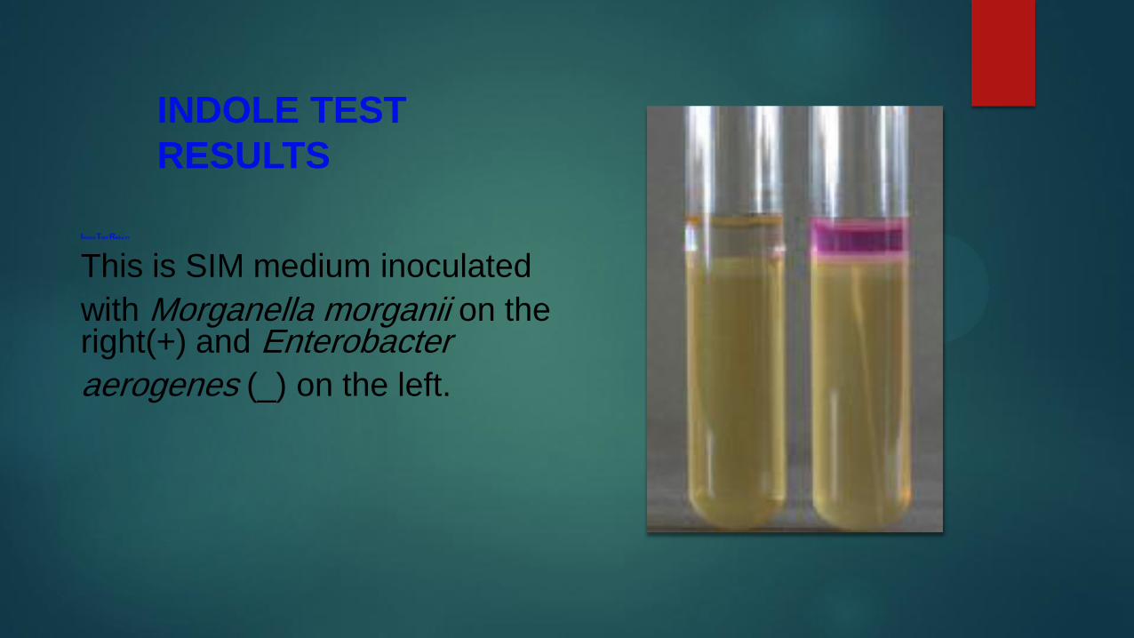
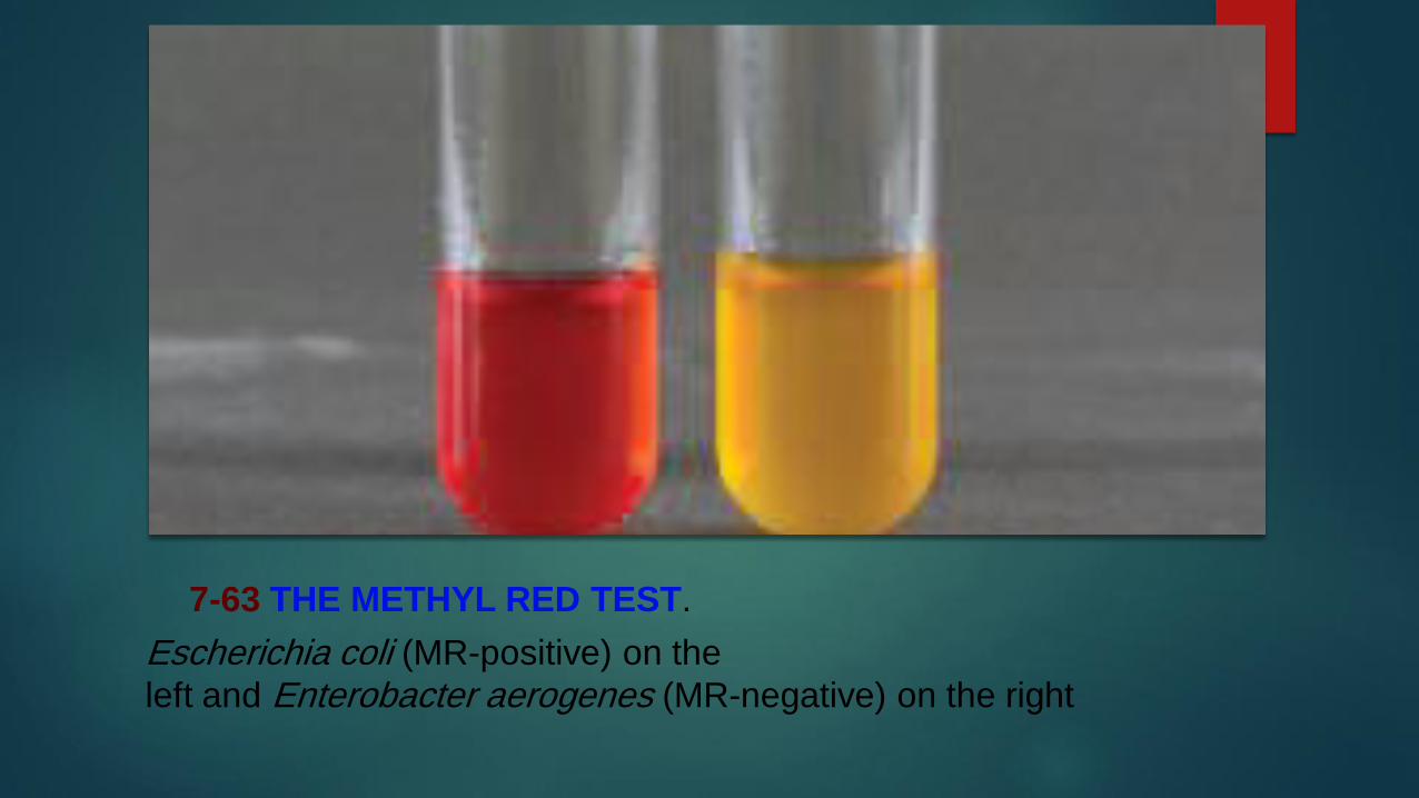
Principle
MR-VP Broth is the combination medium used for both
Methyl Red (MR) and Voges-Proskauer (VP) tests (Refer to
page 82 for the MR test) It is a simple solution containing
only peptone glucose and a phosphate buffer The peptone
and glucose provide protein and a fermentable carbohydrate
and the potassium phosphate resists pH changes in the
medium-98
The Voges-Proskauer test was designed for organisms
that are able to ferment glucose but quickly convert their
acid products to acetoin and 23-butanediol
Adding VP reagents to the medium after
incubation oxidizes the acetoin (if present) to diacetyl
which in turn reacts with guanidine nuclei from peptone
to produce a red color (Figure 7-98) A positive VP result
therefore is red No color change (or development of copper
color) after the addition of reagents is negative The copper
color is a result of interactions between the reagents and
should not be confused with the true red color of a positive
result (Figure 7-98) Use of positive and negative controls
for comparison is usually recommended
7-98 THE VOGES-PROSKAUER TEST
Escherichia coli (VP- negative)is on the left and Enterobacter aerogenes (VP-positive) is on the rightThe copper color at the top of the VP- negative tube is due to thereaction of KOH and 1048576 -naphthol and should not be confused witha positive result Layering of the red color in positive tubes may ormay not occur and is irrelevant to interpretation
Citrate Utilization Test
PurposeThe Citrate Utilization Test is used to determine the ability
of an organism to use citrate as its sole source of carbon
Citrate utilization is one part of a test series referred to as
the IMViC (Indole Methyl Red Voges-Proskauer and
Citrate tests) that distinguishes between members of the
family Enterobacteriaceae and differentiates them from
other Gram-negative rods
Principle Simmons Citrate Agar is a defined medium that contains
sodium citrate as the sole carbon source and ammonium
phosphate as the sole nitrogen source Bromthymol blue
dye which is green at pH 69 and blue at pH 76 is added
as an indicator Bacteria that survive in the medium and
utilize the citrate also convert the ammonium phosphate
to ammonia (NH3) and ammonium hydroxide (NH4OH)
both of which tend to alkalinize the agar As the pH goes
up the medium changes from green to blue (Figure 7-23)
Thus conversion of the medium to blue is a positive citrate
test result
Occasionally a citrate-positive organism will grow on a
Simmons Citrate slant without producing a change in color
7-23 CITRATE TEST
RESULTS
Simmons citrated
agar inoculate Citrobacter
diversus (
+) on the left
Bacillus cereus (_) in the
center and an uninoculated
control on the right
Triple Sugar Iron Agar
PurposeTriple Sugar Iron Agar (TSIA) is primarily used to differen -
tiate members of Enterobacteriaceae and to differentiate
them from other Gram-negative rods such as Pseudomonas
PrincipleTSIA is a rich medium designed to differentiate bacteria on
the basis of glucose fermentation lactose fermentation sucrose
fermentation and sulfur reduction In addition to the
three carbohydrates it includes beef extract yeast extract
and peptone as carbon and nitrogen sources and sodium
thiosulfate as a source of reducible sulfur Phenol red is the
pH indicator and the iron in ferrous sulfate is the hydrogen
sulfide indicator
The medium is prepared as a shallow agar slant with
a deep butt thereby providing both aerobic and anaerobic
growth environments It is inoculated by a stab in the agar
butt followed by a fishtail streak of the slant The incubation
period is 18 to 24 hours for carbohydrate fermentation
and up to 48 hours for hydrogen sulfide reactions Many
reactions in various combinations are possible (Figure 7-92
and Table 7-6)
When TSIA is inoculated with a glucose-only fermenter
acid products lower the pH and turn the entire medium
yellow within a few hours Because glucose is in short
supply (01) it will be exhausted within about 12 hours
As the glucose is used up the organisms located in the aerobic
region (slant) will begin to break down available amino
acids producing NH3 and raising the pH This process
which takes 18 to 24 hours to complete is called a reversion
and only occurs in the slant because of the anaerobic conditions
in the butt Thus a TSIA with a red slant and yellow
butt after a 24-hour incubation period indicates that the
organism ferments glucose but not lactose
Organisms that are able to ferment glucose and lactose
andor sucrose also turn the medium yellow throughout
However because the lactose and sucrose concentrations
are ten times higher than that of glucose greater acid production
results and both slant and butt will remain yellow
after 24 hours Therefore a TSIA with a yellow slant and
butt at 24 hours indicates that the organism ferments glucose and one or both of the other sugars Gas
produced by carbohydrate
fermentation will appear as fissures in the medium
or will lift the agar off the bottom of the tube
Hydrogen sulfide (H2S) may be produced by the reduction
of thiosulfate in the medium or by the breakdown of
cysteine in the peptone Ferrous sulfate in the medium reacts
with the H2S to form a black precipitate usually seen in the
butt Acid conditions must exist for thiosulfate reduction
therefore black precipitate in the medium is an indication
of sulfur reduction and fermentation If the black precipitate
obscures the color of the butt the color of the slant determines
which carbohydrates have been fermented (ie red
slant = glucose fermentation yellow slant = glucose and
lactose andor sucrose fermentation)
An organism that does not ferment any of the carbo hy -
drates but utilizes peptone and amino acids will alkalinize the
medium and turn it red If the organism can use the peptone
aerobically and anaerobically both the slant and butt will
appear red An obligate aerobe will turn only the slant red
Timing is critical in reading TSIA results An early reading
could reveal yellow throughout the medium leading
one to conclude that the organism is a lactose or sucrose
fermenter when it simply may not yet have exhausted the
glucose A reading after the lactose and sucrose have been
depleted could reveal a yellow butt and red slant leading
one to falsely conclude the organism is a glucose-only
fermenter
Tubes that have been interpreted for carbohydrate
fermentation and are negative for sulfur reduction can be
re-incubated for 24 hours before H2S determination Refer
to Table 7-6 for information on the correct symbols and
method of reporting the various reactions
7-92 TSI AGAR SLANTS
From left to right Pseudomonas aeruginosa
(KNC) uninoculated control Morganella morganii (KA atypically not
producing gas) Escherichia coli (AA G) and Proteus mirabilis (KA H2S)
Urease Tests
PurposeThe Urease Test is used to differentiate organisms based on
their ability to hydrolyze urea with the enzyme urease Urinary
tract pathogens from the genus Proteus may be distinguished
from other enteric bacteria by their rapid urease activity
Principle
Urea is a product of decarboxylation of certain amino acids
It can be hydrolyzed to ammonia and carbon dioxide by
bacteria containing the enzyme urease Many enteric bacteria
(and a few others) possess the ability to metabolize urea but
only members of Proteus Morgan ella and Providencia are
considered rapid urease-positive organisms
Urea Agar was formulated to differentiate rapid ureasepositive
organisms from slower urease-positive and ureasenegative
bacteria It contains urea peptone potassium
phosphate glucose phenol red and agar Peptone and
glucose provide essential nutrients for a broad range of
bacteria Potassium phosphate is a mild buffer used to resist
alkalini zation of the medium from peptone metabolism
Phenol red which is yellow or orange below pH 84 and
red or pink above is included as an indicator
Urea hydrolysis to ammonia by ureasepositive
organisms will overcome the buffer in the medium
and change it from orange to pink The agar must be
examined daily during incubation Rapid urease-positive
organisms will turn the entire slant pink within 24 hours
Weak positives may take several days) Ureasenegative
organisms either produce no color change in the
medium or turn it yellow from acid products (Figure 7-94)
7-94 UREASE
AGAR TEST RESULTS
Urease agar tubes
after a 24 hour incubation
Morganella
morganii (ureasepositive)
a rapid
urea splitter is on the
left and Hafnia alvei
(urease-negative)
is on the right An
uninoculated control
is in the center
Gram positive cocci
Hemolysis test
Blood agar is used for isolation and cultivation of many
types of fastidious bacteria It is also used to dif ferentiate
bacteria based on their hemolytic characteristics especially
within the genera Streptococcus Enterococcus and
Aerococcus
PrincipleSeveral species of Gram-positive cocci produce exotoxins
called hemolysins which are able to destroy red blood cells
(RBCs) and hemoglobin Blood Agar sometimes called
Sheep Blood Agar because it includes 5 sheep blood in
a Tryptic Soy Agar base allows differentiation of bacteria
based on their ability to hemolyze RBCs
The three major types of hemolysis are -hemolysis
1048576 -hemolysis and -hemolysis -hemolysis the complete
destruction of RBCs and hemoglobin results in a clearing of
the medium around the colonies (Figure 7-10) 1048576 -
hemolysis
is the partial destruction of RBCs and produces a
greenish
discoloration of the agar around the colonies (Figure 7-
11)
-hemolysis is actually nonhemolysis and appears as
simple
growth with no change to the medium (Figure 7-12)
7-10 -HEMOLYSISStreptococcus pyogenes demonstrates -hemolysis
The clearing around the growth is a result of complete lysis of red blood
cells This photograph was taken with transmitted light
7-11 -HEMOLYSISThis is a streak plate of Streptococcus pneumoniae
demonstrating 1048576 -hemolysis The greenish zone around the colonies
results from incomplete lysis of red blood cells-
Catalase Test
PurposeThe Catalase Test is used to identify organisms that produce
the enzyme catalase It is most commonly used to differentiate
members of the catalase-positive Staphylococcus from the
catalase-negative Streptococcaceae Variations on this test
may also be used in identification of Mycobacterium species
Principle
Bacteria that produce catalase can be detected easily
using typical store-grade hydrogen peroxide When hydrogen
peroxide is added to a catalase-positive culture oxygen
gas bubbles form immediately (Figures 7-20 and 7-21) If no
bubbles appear the organism is catalase-negative This test
can be performed on a microscope slide or by adding hydrogen
peroxide directly to the bacterial growth
7-20 CATALASE SLIDE TEST
Shown is the catalase slide test in which
visible bubble production indicates a positive result Staphylococcus
aureus (+) is on the left Streptococcus(-) is on the right of peroxide to contain aerosols produced in positive reactions
7-21 CATALASE TUBE
TEST
The catalase test
may also be performed on
an agar slant Staphylo -
coccus aureus (1048576 ) is on
the left Enterococcus
faecium () is on the right
Coagulase Tests
PurposeThe Coagulase Test is typically used to differentiate
Staphylococcus aureus from other species
Principle
Staphylococcus aureus is an opportunistic pathogen that
can be highly resistant to both the normal immune response
and antimicrobial agents Its resistance is due in part to
the production of a coagulase enzyme Coagulase works
in conjunction with normal plasma com ponents to form
protective fibrin barriers around individual bacterial cells
or groups of cells shielding them from phagocytosis and
other types of attack
Two forms of the Coagulase Test have been devised
to detect the enzymes the Tube Test and the Slide
Test
The Tube Test detects the presence of either bound
or free
coagulase while the Slide Test detects only bound
coagulase
Both tests utilize rabbit plasma treated with
anticoagulant
to interrupt normal clotting mechanisms
The Tube Test is performed by adding the test organism
to rabbit plasma in a test tube Coagulation of the plasma
(including any thickening or formation of fibrin threads)
within 24 hours indicates a positive reaction (Figure 7-24)
The plasma is typically examined for clotting (without
shaking) after about 4 hours because it is possible for
coagulation to take place early and revert to liquid within
24 hours
In the slide test bacteria are transferred to a slide containing
a small amount of plasma Agglutination of the
cells on the slide within one to two minutes indicates the
presence of bound coagulase (Figure 7-25) Equivocal or
negative Slide Test results are typically confirmed using the
7-25 COAGULASE SLIDE TEST
EmStaphylococcus epidermidis (_) on the left and
S aureus (+) on the right were prepared in sterile
saline Agglutination of the coagulase plasma is
indicative of a positive result for bound coagulase
7-24 COAGULASE TUBE TEST
Coagulase-negative Staphylococcus
epidermidis below and the more pathogenic coagulase- positive S aureus
above Coagulase increases bacterial resistance to phagocytosis and
antibodies by surrounding infecting organisms with a clot
Oxidase Test
PurposeThe Oxidase Test is used to identify bacteria containing the
respiratory enzyme cytochrome c oxidase
It can be useful in differentiating the oxidase negative
Enterobacteriaceae from the oxidase-positive
PseudomonadaceaeIt is also identification of the oxidase-positive
Neisseria
7-78 OXIDASE TEST ON BACTERIAL GROWTHA few drops of
reagent on oxidase-positive bacteria will produce a purple-blue color
immediately Oxidase-negative organisms will not turn purple The
bacterium on the left is its natural color not the color of an oxidase negative
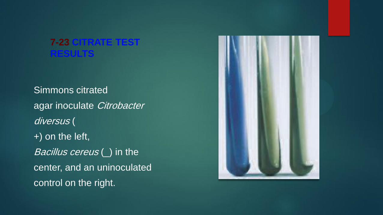
Principle
Urea is a product of decarboxylation of certain amino acids
It can be hydrolyzed to ammonia and carbon dioxide by
bacteria containing the enzyme urease Many enteric bacteria
(and a few others) possess the ability to metabolize urea but
only members of Proteus Morgan ella and Providencia are
considered rapid urease-positive organisms
Urea Agar was formulated to differentiate rapid ureasepositive
organisms from slower urease-positive and ureasenegative
bacteria It contains urea peptone potassium
phosphate glucose phenol red and agar Peptone and
glucose provide essential nutrients for a broad range of
bacteria Potassium phosphate is a mild buffer used to resist
alkalini zation of the medium from peptone metabolism
Phenol red which is yellow or orange below pH 84 and
red or pink above is included as an indicator
Urea hydrolysis to ammonia by ureasepositive
organisms will overcome the buffer in the medium
and change it from orange to pink The agar must be
examined daily during incubation Rapid urease-positive
organisms will turn the entire slant pink within 24 hours
Weak positives may take several days) Ureasenegative
organisms either produce no color change in the
medium or turn it yellow from acid products (Figure 7-94)
7-94 UREASE
AGAR TEST RESULTS
Urease agar tubes
after a 24 hour incubation
Morganella
morganii (ureasepositive)
a rapid
urea splitter is on the
left and Hafnia alvei
(urease-negative)
is on the right An
uninoculated control
is in the center
Gram positive cocci
Hemolysis test
Blood agar is used for isolation and cultivation of many
types of fastidious bacteria It is also used to dif ferentiate
bacteria based on their hemolytic characteristics especially
within the genera Streptococcus Enterococcus and
Aerococcus
PrincipleSeveral species of Gram-positive cocci produce exotoxins
called hemolysins which are able to destroy red blood cells
(RBCs) and hemoglobin Blood Agar sometimes called
Sheep Blood Agar because it includes 5 sheep blood in
a Tryptic Soy Agar base allows differentiation of bacteria
based on their ability to hemolyze RBCs
The three major types of hemolysis are -hemolysis
1048576 -hemolysis and -hemolysis -hemolysis the complete
destruction of RBCs and hemoglobin results in a clearing of
the medium around the colonies (Figure 7-10) 1048576 -
hemolysis
is the partial destruction of RBCs and produces a
greenish
discoloration of the agar around the colonies (Figure 7-
11)
-hemolysis is actually nonhemolysis and appears as
simple
growth with no change to the medium (Figure 7-12)
7-10 -HEMOLYSISStreptococcus pyogenes demonstrates -hemolysis
The clearing around the growth is a result of complete lysis of red blood
cells This photograph was taken with transmitted light
7-11 -HEMOLYSISThis is a streak plate of Streptococcus pneumoniae
demonstrating 1048576 -hemolysis The greenish zone around the colonies
results from incomplete lysis of red blood cells-
Catalase Test
PurposeThe Catalase Test is used to identify organisms that produce
the enzyme catalase It is most commonly used to differentiate
members of the catalase-positive Staphylococcus from the
catalase-negative Streptococcaceae Variations on this test
may also be used in identification of Mycobacterium species
Principle
Bacteria that produce catalase can be detected easily
using typical store-grade hydrogen peroxide When hydrogen
peroxide is added to a catalase-positive culture oxygen
gas bubbles form immediately (Figures 7-20 and 7-21) If no
bubbles appear the organism is catalase-negative This test
can be performed on a microscope slide or by adding hydrogen
peroxide directly to the bacterial growth
7-20 CATALASE SLIDE TEST
Shown is the catalase slide test in which
visible bubble production indicates a positive result Staphylococcus
aureus (+) is on the left Streptococcus(-) is on the right of peroxide to contain aerosols produced in positive reactions
7-21 CATALASE TUBE
TEST
The catalase test
may also be performed on
an agar slant Staphylo -
coccus aureus (1048576 ) is on
the left Enterococcus
faecium () is on the right
Coagulase Tests
PurposeThe Coagulase Test is typically used to differentiate
Staphylococcus aureus from other species
Principle
Staphylococcus aureus is an opportunistic pathogen that
can be highly resistant to both the normal immune response
and antimicrobial agents Its resistance is due in part to
the production of a coagulase enzyme Coagulase works
in conjunction with normal plasma com ponents to form
protective fibrin barriers around individual bacterial cells
or groups of cells shielding them from phagocytosis and
other types of attack
Two forms of the Coagulase Test have been devised
to detect the enzymes the Tube Test and the Slide
Test
The Tube Test detects the presence of either bound
or free
coagulase while the Slide Test detects only bound
coagulase
Both tests utilize rabbit plasma treated with
anticoagulant
to interrupt normal clotting mechanisms
The Tube Test is performed by adding the test organism
to rabbit plasma in a test tube Coagulation of the plasma
(including any thickening or formation of fibrin threads)
within 24 hours indicates a positive reaction (Figure 7-24)
The plasma is typically examined for clotting (without
shaking) after about 4 hours because it is possible for
coagulation to take place early and revert to liquid within
24 hours
In the slide test bacteria are transferred to a slide containing
a small amount of plasma Agglutination of the
cells on the slide within one to two minutes indicates the
presence of bound coagulase (Figure 7-25) Equivocal or
negative Slide Test results are typically confirmed using the
7-25 COAGULASE SLIDE TEST
EmStaphylococcus epidermidis (_) on the left and
S aureus (+) on the right were prepared in sterile
saline Agglutination of the coagulase plasma is
indicative of a positive result for bound coagulase
7-24 COAGULASE TUBE TEST
Coagulase-negative Staphylococcus
epidermidis below and the more pathogenic coagulase- positive S aureus
above Coagulase increases bacterial resistance to phagocytosis and
antibodies by surrounding infecting organisms with a clot
Oxidase Test
PurposeThe Oxidase Test is used to identify bacteria containing the
respiratory enzyme cytochrome c oxidase
It can be useful in differentiating the oxidase negative
Enterobacteriaceae from the oxidase-positive
PseudomonadaceaeIt is also identification of the oxidase-positive
Neisseria
7-78 OXIDASE TEST ON BACTERIAL GROWTHA few drops of
reagent on oxidase-positive bacteria will produce a purple-blue color
immediately Oxidase-negative organisms will not turn purple The
bacterium on the left is its natural color not the color of an oxidase negative
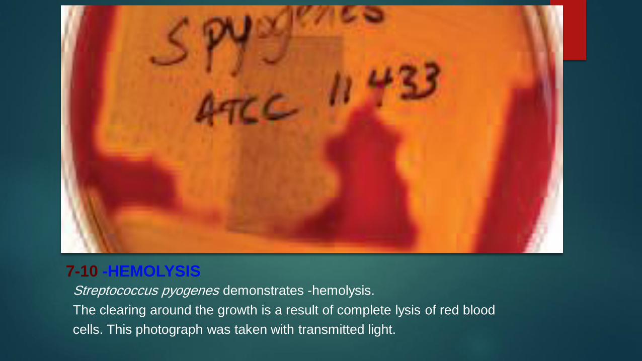

Principle
Bacteria that produce catalase can be detected easily
using typical store-grade hydrogen peroxide When hydrogen
peroxide is added to a catalase-positive culture oxygen
gas bubbles form immediately (Figures 7-20 and 7-21) If no
bubbles appear the organism is catalase-negative This test
can be performed on a microscope slide or by adding hydrogen
peroxide directly to the bacterial growth
7-20 CATALASE SLIDE TEST
Shown is the catalase slide test in which
visible bubble production indicates a positive result Staphylococcus
aureus (+) is on the left Streptococcus(-) is on the right of peroxide to contain aerosols produced in positive reactions
7-21 CATALASE TUBE
TEST
The catalase test
may also be performed on
an agar slant Staphylo -
coccus aureus (1048576 ) is on
the left Enterococcus
faecium () is on the right
Coagulase Tests
PurposeThe Coagulase Test is typically used to differentiate
Staphylococcus aureus from other species
Principle
Staphylococcus aureus is an opportunistic pathogen that
can be highly resistant to both the normal immune response
and antimicrobial agents Its resistance is due in part to
the production of a coagulase enzyme Coagulase works
in conjunction with normal plasma com ponents to form
protective fibrin barriers around individual bacterial cells
or groups of cells shielding them from phagocytosis and
other types of attack
Two forms of the Coagulase Test have been devised
to detect the enzymes the Tube Test and the Slide
Test
The Tube Test detects the presence of either bound
or free
coagulase while the Slide Test detects only bound
coagulase
Both tests utilize rabbit plasma treated with
anticoagulant
to interrupt normal clotting mechanisms
The Tube Test is performed by adding the test organism
to rabbit plasma in a test tube Coagulation of the plasma
(including any thickening or formation of fibrin threads)
within 24 hours indicates a positive reaction (Figure 7-24)
The plasma is typically examined for clotting (without
shaking) after about 4 hours because it is possible for
coagulation to take place early and revert to liquid within
24 hours
In the slide test bacteria are transferred to a slide containing
a small amount of plasma Agglutination of the
cells on the slide within one to two minutes indicates the
presence of bound coagulase (Figure 7-25) Equivocal or
negative Slide Test results are typically confirmed using the
7-25 COAGULASE SLIDE TEST
EmStaphylococcus epidermidis (_) on the left and
S aureus (+) on the right were prepared in sterile
saline Agglutination of the coagulase plasma is
indicative of a positive result for bound coagulase
7-24 COAGULASE TUBE TEST
Coagulase-negative Staphylococcus
epidermidis below and the more pathogenic coagulase- positive S aureus
above Coagulase increases bacterial resistance to phagocytosis and
antibodies by surrounding infecting organisms with a clot
Oxidase Test
PurposeThe Oxidase Test is used to identify bacteria containing the
respiratory enzyme cytochrome c oxidase
It can be useful in differentiating the oxidase negative
Enterobacteriaceae from the oxidase-positive
PseudomonadaceaeIt is also identification of the oxidase-positive
Neisseria
7-78 OXIDASE TEST ON BACTERIAL GROWTHA few drops of
reagent on oxidase-positive bacteria will produce a purple-blue color
immediately Oxidase-negative organisms will not turn purple The
bacterium on the left is its natural color not the color of an oxidase negative
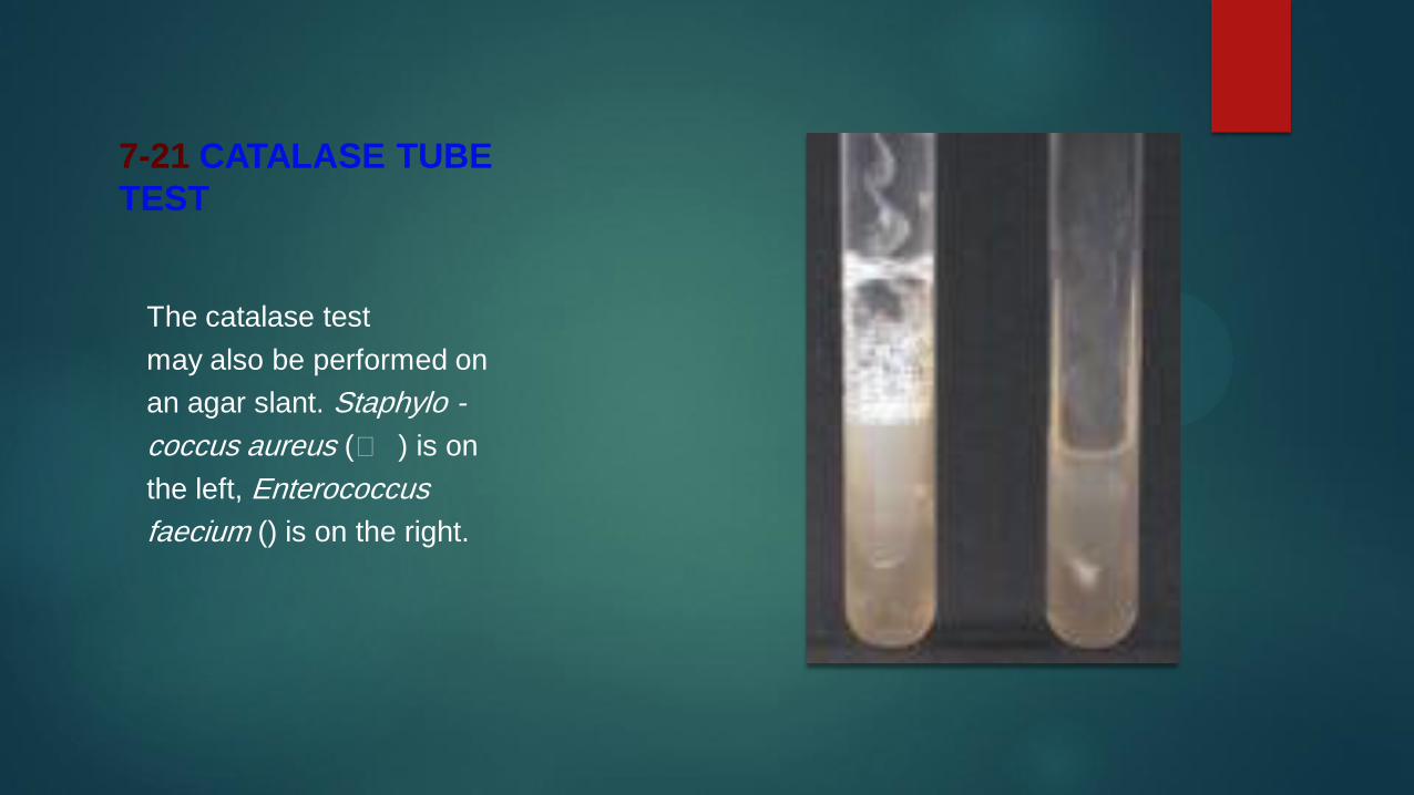

7-78 OXIDASE TEST ON BACTERIAL GROWTHA few drops of
reagent on oxidase-positive bacteria will produce a purple-blue color
immediately Oxidase-negative organisms will not turn purple The
bacterium on the left is its natural color not the color of an oxidase negative