wfsbp abstract book final
DESCRIPTION
asdaaaTRANSCRIPT

The World Journal of Biological Psychiatry
Volume 8Supplement 1, 2007
Guest EditorsFrederick Goodwin, Jose Luis Ayuso-Gutierrez, Veronica Larach-Walters, Siegfried Kasper
2nd International Congress of Biological PsychiatryApril 17th - April 21st, 2007
Santiago, Chile
The Official Journal of the World Federation of Societies of Biological Psychiatry
ABSTRACTS
The World Journal of Biological Psychiatry, Supplement 1, Vol 8, 2007
2
Founding EditorsHans-Jürgen Möller (Germany)Carlos Roberto Hojaij (Australia)Joseph Zohar (Israel)
Chief EditorHans-Jürgen MöllerDepartment of PsychiatryLudwig-Maximilians-UniversityNussbaumstrasse 780336 MunichGermanyTel: + 49 89 5160 5501Fax: + 49 89 5160 5522E-mail:[email protected]
Assistant Chief EditorRainer RupprechtDepartment of PsychiatryLudwig-Maximilians-UniversityNussbaumstrasse 780336 MunichGermanyTel: + 49 89 5160 2770Fax: + 49 89 5160 5524E-mail: [email protected]
Associate Editors Carlos Roberto HojaijThe Melbourne Institute of BiologicalPsychiatry511 Whitehorse RoadSurrey Hills 3127MelbourneAustraliaTel: + 61 3 9836 0088Fax: + 61 3 9836 0644
Joseph ZoharChaim Sheba Medical CenterDivision of PsychiatryTel-Hashomer, 52621IsraelTel: + 972 3 530 3300Fax: + 972 3 535 2788
Editorial BoardHagop Akiskal (USA)Michael Bauer (Germany)Robert H. Belmaker (Israel)Graham Burrows (Australia)Arvid Carlsson (Sweden)Giovanni B Cassano (Italy)Marcelo Cetkovich-Bakmas (Argentina)Delcir da Costa (Brazil)Frederick Goodwin (USA)Jose Luis Ayuso Gutierrez (Spain)Ralf P Hemmingsen (Denmark)Eric Hollander (USA)Florian Holsboer (Germany)Lewis L Judd (USA)Nobumasa Kato (Japan)Martin B Keller (USA)Yves Lecrubier (France)Brian Leonard (Ireland)Odd Lingjaerde (Norway)Henri Loo (France)Juan J Lopez-Ibor (Spain)Mario Maj (Italy)Herbert Y Meltzer (USA)Julien Mendlewicz (Belgium)Stuart Montgomery (UK)David Nutt (UK)Tatsuro Ohta (Japan)Ahmed Okasha (Egypt)Antonio Pacheco Palha (Portugal)Stanislaw Puzynski (Poland)Giorgio Racagni (Italy)Americo Reyes-Tucas (Honduras)Philippe H Robert (France)Bernd Saletu (Austria)Norman Sartorius (Switzerland)Jan Sikora (Czech Republic)Hernan Silva-Ibarra (Chile)Constantin Soldatos (Greece)Costas Stefanis (Greece)Dan J Stein (South Africa)Saburo Takahashi (Japan)Marcio Versiani (Brazil)Jerzy Vetulani (Poland)Daniel Weinberger (USA)
Regional EditorsAfricaDriss Moussaoui (Morocco)AsiaTakuya Kojima (Japan)EuropeBirte Glenthøj (Denmark)Siegfried Kasper (Austria)Latin-AmericaWagner Gattaz (Brazil)North AmericaCharles Nemeroff (USA)Owen M. Wolkowitz (USA)
Editorial Assistant Jacqueline KlesingDepartment of PsychiatryLudwig-Maximilians-UniversityNussbaumstrasse 780336 MunichGermanyTel: + 49 89 5160 5531Fax: + 49 89 5160 5530E-mail: [email protected]
PublisherTaylor & FrancisBox 12 PosthusetBiskop Gunnerusgt. 14ANO-0051 OsloNorwayTel: +47 23 10 34 60Fax: +47 23 10 34 61E-mail: [email protected]
PrintersTypeset by Datapage International Ltd., Dublin,IrelandPrinted by Hobbs the Printers, Hampshire, UK
The World Journal of Biological Psychiatry is one of the numerousbenefits available to World Federation of Societies of Biological Psychiatryregistered members, to whom the journal is sent free of charge and isaccessible on-line in the members only section of the Federation’s website www.wfsbp.org. Please contact the WFSBP Global Headquartersat [email protected] for more information.
Subscription InformationThe World Journal of Biological Psychiatry, ISSN print edition 1562-2975, online edition 1814-1412, is published four times a year.Visit our webpage for more information: www.tandf.no/wjbp
Annual subscription rates for volume 8, 2007: Institutions (print &online): $360, institutions (online only): $342, individuals (print &online) $150. Air speed postage included. Supplements are suppliedfree of charge to all subscribers.
Orders for subscription, back issues and change of address should be sent to: Taylor & Francis Customer Services, T&F InformaUK Ltd, Sheepen Place, Colchester, Essex CO3 3LP, UnitedKingdom. Tel: +44 (0) 20 7017 5544. Fax: +44 (0) 20 7017 5198. E-mail:[email protected]
Taylor & Francis retains a three year back issue stock of journals. Older volumes are held by our official stockists: Periodicals ServiceCompany (http://www.periodicals.com/tandf.html) 11 MainStreet, Germantown, NY 12526, USA, to whom all orders andenquiries should be addressed. Tel: 11 518 537 4700. Fax: 11 518 5375899. E-mail: [email protected]
Correspondence concerning publishing matters, copyright, permissions and reprints should be addressed to: Taylor & Francis,PO Box 12 Posthuset, NO-0051 Oslo, Norway. Tel: 147 23 10 34 60.Fax: 147 23 10 34 61. E-mail: [email protected]
The World Journal of Biological Psychiatry (USPS permit pending) is published in March, May, August and November. The 2007 institutional subscription price is $360. Application to mail atperiodicals postage rate is pending at Jamaica, NY by US MailingAgent Air Business Ltd. c/o Priority Airfreight NY Ltd. 147-29182nd Street, Jamaica, NY 11413, USA. US Postmaster: Please send address changes to sWBP, Air BusinessLtd. c/o Priority Airfreight NY Ltd. 147-29 182nd Street, Jamaica,NY 11413, USA.
Copyright © 2007 Taylor & FrancisAll articles are protected by copyright, which covers exclusiverights to reproduce and distribute the article. No material in thisjournal may be reproduced photographically or stored on micro-film, in electronic databases, video or compact disks etc. withoutprior written permission from Taylor & Francis. Special regula-tions for photocopies in the USA: Authorization to photocopyitems for internal or personal use, or the internal or personal useof specific clients, is granted by Taylor & Francis for libraries andother users registered with the Copyright Clearance Center(CCC), Transactional Reporting Service, provided the fee of $32per article is article paid to CCC, 222 Rosewood Drive, Danvers,MA 01923, USA. This authorization does not include copying forgeneral distribution, promotion, new works, or resale. In thesecases, specific written permission must be obtained from Taylor &Francis.
Indexed in:Index Medicus/MEDLINE, Science Citation Index Expanded, ISIAlerting Services, Current Contents/Clinical Medicine, CurrentContents/Life Sciences, NeuroScience Citation Index
Impact Factor: 2.800 (Source: 2005 JCR Science Edition)
The World Journal of Biological PsychiatryISSN print edition 1562 - 2975

Table of Contents
The World Journal of Biological Psychiatry, Supplement 1, Vol 8, 2007
3
FOREWORD ..................................................................4
OPENING LECTURE ......................................................5
PLENARY LECTURES ....................................................5
SYMPOSIAAddictive Disorders ......................................................7Affective Disorders (Bipolar)........................................10Affective Disorders (Unipolar) ....................................11Anxiety ......................................................................14Childhood & Adolescent Disorders ............................17Neurodegenerative Disorders......................................18Psychotic Disorders ....................................................21Brain Function ............................................................34Genetics ....................................................................37Neuroimaging ............................................................41Psychopharmacology ..................................................45Other ........................................................................52
FREE COMMUNICATIONSAddictive Disorders ....................................................60Affective Disorders (Bipolar)........................................62Affective Disorders (Unipolar) ....................................64Anxiety ......................................................................67Childhood & Adolescent Disorders ............................69Neurodegenerative Disorders......................................72Psychotic Disorders ....................................................75Brain Function ............................................................78Genetics ....................................................................80Neuroimaging ............................................................82Psychopharmacology ..................................................84Other ........................................................................86
POSTERSAddictive Disorders ....................................................92Affective Disorders (Bipolar)......................................100Affective Disorders (Unipolar) ..................................104Anxiety ....................................................................113Childhood & Adolescent Disorders ..........................121Neurodegenerative Disorders....................................126Psychotic Disorders ..................................................135Brain Function ..........................................................161Genetics ..................................................................166Neuroimaging ..........................................................171Psychopharmacology ................................................181Other ......................................................................205
SATELLITE SYMPOSIA ..............................................221
AUTHOR INDEX........................................................224

Foreword
The World Journal of Biological Psychiatry, Supplement 1, Vol 8, 2007
4
Dear Colleagues,
The Scientific Program is considered the core of the 2nd International Congress of Biological Psychiatry. Thus theInternational Scientific Program Committee is proud to have compiled a well balanced blend of Plenary Lectures,Symposia, WFSBP Treatment Guidelines Workshops, Debates, Free Communications and Guided Poster Tours coveringall aspects of psychiatric disorders from basic to clinical reasearch including the newest concepts for improving thepatients’ lives.
As with all congresses of the World Federation of Societies of Biological Psychiatry (WFSBP), the Scientific Program greatlydepends on the submitted proposals and abstracts. In this sense we first would like to thank all Chairs, Co-Chairs andSpeakers who accepted with enthusiasm to participate and who will present high quality work. A WFSBP congress is notpossible without the continuing support from its National Societies of Biological Psychiatry. We cordially thank the WFSBPNational Societies for their engagement and for spreading the ideas that we are committed to.
In the last couple of years there has been radical progress in neuroscience that is changing our concepts and attitudeson mental diseases. The advances in biology and molecular genetics, the availability of modern technologies such asbrain imaging, the development of a new generation of treatments based upon neuroplasticity concepts are openingour field to new and exciting horizons. With this Scientific Program we will have the opportunity to discuss these the-mes as well as the integration between the rapid development of scientifc knowledge and its impact on diagnoses, treat-ment, and rehabilition of patients with mental disorders.
It is most special to us that the 2nd International Congress of Biological Psychiatry is held in Santiago de Chile. TheWFSBP has traditionally strong Latin American roots, especially through the first WFSBP President Edmund Fisher. Today,exciting developments are taking place in Latin America, such as institutional reforms, increasing foreign direct invest-ment and the recent surge in exports. In the field of psychiatry an increasing number of world renown Latin Americanscientists are formative and influential. Therefore this congress is the ideal setting to share recent evolvements, exchangethoughts with decision makers from all around the globe, and discuss the newest innovations in our field.
We believe that you will find interesting updates for new and better treamtent of your patients and fruitful thoughts foryour research work in this Scientific Program.
Yours sincerely
Dr. Frederick Goodwin Prof. Jose Luis Ayuso-GuitierrezCo-Chair of the WFSBP International Scientific Co-Chair of the WFSBP International ScientificProgram Committee Program Committee
Prof. Siegfried KasperCongress PresidentPresident World Federation of Societiesof Biological Psychiatry

O-01Opening Lecture: Lost and found in translation-al research: The challenge and promise
T12 Other
O-01-01Lost and found in translational research: The challenge andthe promise
Raquel GurUniversity of Pennsylvania, Psychiatry, Philadelphia, USA
There has been increased focus on developing paradigms that enhancetranslational research. In complex disorders these efforts mandate inter-disciplinary collaborations. Two links are essential for such a bridge, onebetween basic and clinical neuroscience research and the other betweenclinical research in an academic setting to clinical practice in the field.Such translational paradigms will be exemplified with schizophrenia. Thechallenge is to “zoom in” on a theme and develop a research plan thatcan test complementary hypotheses in humans and animal models. Whatcan be lost in the first link is the limitation of comparing human behaviorto that modeled in basic studies. The pitfalls of linking academic researchto clinical settings are in loosing sight of the diversity of individuals affec-ted and the complexity of their environment. These dangers are especiallynoteworthy because the range of available tools and the required expert-ise lead to narrowing the scope of the research.
PL-01Is cyclicity or polarity more fundamental to theclassification of mood disorders?
T2 Affective Disorders (Bipolar)
PL-01-01Is cyclicy or polarity more fundamental to the classifica-tion of major mood disorders?
Frederick GoodwinGeorge Washington Univ., Medical Center, Bethesda, MD, USA
Kraepelin’s description of manic-depressive illness encompassed all majorrecurrent mood disorders. Subsequent investigators, principally Leonhard,Perris, Angst and Winokur subdivided manic-depressive illness into unipo-lar and bipolar based on the presence or absence of a prior history ofmania. The NIMH group (Dunner, Gershon and Goodwin) subsequentlysubdivided the bipolar group into BP I and BP II based, respectively, on ahistory of mania vs hypomania only. Like Kraepelin, the original UP-BPdistinction was based on studies of patients with recurrent affective dis-orders. Notwithstanding this history, the architects of DSM III and IV sepa-rated out bipolar disorder “from the top,” explicitly making polarity theprimary basis for organizing the mood disorders. This presentation willexamine both the reasons for this fundamental shift in the evolution ofthe DSM system and the evidence supporting Kraepelin’s original empha-sis on the importance of recurrence across the affective spectrum.
PL-02Ethics and neuropsychiatry
T12 Other
PL-02-01Ethics and neuropsychiatry
Fernando Lolas StepkeUniversity of Chile, Pan American Health Org., Santiago, Chile
This lecture addresses current issues in neuropsychiatric research in thecontext of Western ethical traditions, teleology and deontology. It isargued that contemporary bioethical thinking, by creating the social insti-tution of the committee, uses dialogue and deliberation for the formula-tion, justification, and resolution of dilemmas arising from scientificprogress and its applications to human affairs. Research is examined fromthe standpoint of its technical or instrumental value, its scientific or theo-retical importance, and its social or hermeneutic relevance. In each realm,ethical considerations are essential to achieve integration between valuesrelated to the search for truth, to the enhancement of knowledge and tothe improvement of the human condition. Several areas are identified: theimpact of neuroscientific information on conceptions about the humanbrain and its performance, the biological foundations of moral/socialbehaviour, the “conceptual nervous systems” underlying different philo-sophical theories about human life, among others. As the application ofneuroethics extends beyond laboratory and experiment, other aspectsacquire relevance, such as the ethics of mind-enhancing drugs or inter-ventions, the links between psychological and biological methods onbehaviour and mentation, and the accessibility of knowledge to people indifferent parts of the world or diverse social conditions. As a third area ofinquiry, the ethics of neuroscientific research is singled out in some of itscharacteristic features and its importance in the training of researchersand practitioners.
PL-03Neurobiology and treatment of cannabisdependence
T9 Genetics
PL-03-01Neurobiology and treatment of cannabis dependence
David A. GorelickNational Institute on Drug Abuse, Baltimore, USA
Introduction: Cannabis is the most widely used illegal drug in the world,with an estimated 160 million current users. Community-based epide-miological studies suggest that about 10% of users develop dependence.The psychoactive effects of cannabis are primarily due to activation ofcannabinoid CB1 receptors by THC. The endogenous ligands for thisreceptor include fatty acid derivatives such as anandamide (arachi-donylethanolamide) and 2-arachidonoylglycerol (2-AG), which appear tofunction as retrograde modulators of synaptic neurotransmission.
Opening/Plenary Lectures
The World Journal of Biological Psychiatry, Supplement 1, Vol 8, 2007
5
Opening Lectures
Plenary Lectures

Method: There are no proven, broadly effective medications for the treat-ment of cannabis dependence, and no medication is approved by anyregulatory authority for this indication. Treatment generally involves psy-chosocial methods, such as cognitive behavioral therapy and motivationalenhancement. Many cannabis users are adolescents, in which case familyinvolvement in treatment is essential. Many cannabis users have psychi-atric comorbidity, the treatment of which may also be helpful in reducingcannabis use. Both self-report studies and human experimental studiesindicate that chronic cannabis use can result in physical dependence anda withdrawal syndrome.Results: A retrospective survey of 104 adult cannabis smokers found that70% experienced at least 3 withdrawal symptoms during their quittingattempt (Copersino et al., 2006). A majority (56%) reported taking actionto relieve withdrawal symptoms, including some who resumed cannabisuse, indicating that withdrawal can be a negative reinforcer for relapse.One treatment approach under study is blockade of CB1 receptors byrimonabant. This medication blocks the acute psychological and cardio-vascular effects of a smoked cannabis cigarette, without altering THCpharmacokinetics (Huestis et al., 2001). Other medications under studyinclude buspirone, baclofen, quetiapine, and substitution treatment withoral THC.Conclusion: Cannabis use produces physical dependence in humans;treatment of withdrawal symptoms may reduce relapse. Several medica-tions, including the CB1 antagonist rimonabant, are under study as pos-sible treatments for cannabis dependence.References: Copersino et al., Am J Addict 15:8-14, 2006. Huestis et al.,Arch Gen Psychiat 58:322-328, 2001 Acknowledgment: Research sup-ported by the Intramural Research Program, NIH, NIDA and Sanofi-Aventis(studies with rimonabant).
PL-04Biological psychiatry integrated in clinical science
T12 Other
PL-04-01Biological psychiatry integrated in clinical science
Siegfried KasperMedical University, General Psychiatry, Vienna, Austria
Based on the sound knowledge of clinical psychiatry neuroscientistsembark since over 20 years to discover the biological bases of mental dis-orders. Whereas the „window to the brain“ was firstly dominated by neuro-endocrine and biochemical models, we now have the possibility to usesophisticated brain imaging techniques which span from electrophysiologyto techniques derived from nuclear medicine. Furthermore, the combina-tion of brain imaging techniques with genetic variables promises to havea close insight into brain-environment interactions. For treatment of psy-chiatric diseases, multidimensional criteria for selection of a psychotropicmedication is necessary to not only judge the clinical phenotype but alsothe functional phenotype and finally the genotype. Whereas the clinicalphenotype is characterized by psychiatric syndromes including psychiatricand somatic comorbidity, the functional phenotype can be uncoveredwith neuroendocrine measurements, sleep physiology, imaging tech-niques as well as proteomics. Finally, the genotype can be characterizedby single nucleotid polymorphisms (SNP), coupling as well as the determi-nation of candidate genes. Imaging genetics is a new way to achieveinsight in the genetic variables of changes on a cellular level which there-after influence information processing and finally are represented in com-plex functional interaction of human behavior. Based on this knowledgeit is evident that psychiatrists not only should be determined to the clini-cal symptomatology of their patients but also on the underlying biologi-cal processes which can in turn be influenced to various degress withpharmacological as well as non-pharmacological treatment modalities.
Plenary Lectures
The World Journal of Biological Psychiatry, Supplement 1, Vol 8, 2007
6
ADDICTIVE DISORDERS - Symposia
The World Journal of Biological Psychiatry, Supplement 1, Vol 8, 2007
7
S-04Comorbidity between substance use and mental health disorders
T1 Addictive Disorders
S-04-01Genetics of comorbid substance use and psychiatric disorders
James L. KennedyUniversity of Toronto, Ctr. Addiction & mental Health, CanadaJulien Renou, Bernard LeFoll
Introduction: Smoking behavior is inflenced by both genetic and envi-ronmental factors. Genetic and pharmacologic studies have suggestedthe alpha-7 nicotinic receptor gene (Deluca et al, 2004) and theDopamine Receptor D3 (DRD3) involvement in smoking behavior, whichare expressed in brain structures (eg nucleus accumbens) implicated indrug dependence.Method: We investigated the association between 10 DRD3 polymor-phisms and smoking behavior in a cohort of 236 Caucasian schizophrenicpatients (155 smokers and 81 non-smokers). Our sample included 172 males and 64 females. The comparisons of the frequencies of geno-types and alleles in smokers and non-smokers for each polymorphismwere performed with contingency tables. Homozygosity versus heterozy-gosity was also tested using chi-square. Bonferoni correction for multipletesting was used when applicable. Results: The distribution of genotypes were in accordance with HardyWeinberg equilibrium. We did not find significant differences betweenschizophrenia smokers and non-smokers in allele or genotype frequenciesfor each polymorphism (p > 0.05). Smoking frequencies for males (72%)and females (50%) populations differed significantly (¯2=11.58,p=0.0007). Consequently, we also analysed DRD3 polymorphisms strati-fying by gender. For male and female populations, no statistical diffe-rences were found in the genotype distributions or allele frequenciesbetween smokers and non-smokers (p > 0.05). Conclusion: We conclude from our studies thus far, that the DRD3 geneis not associated with smoking in our population of schizophrenicpatients. We have not yet performed interaction studies between DRD3and the alpha-7 nicotinic subunit gene or the brain-derived neurotrophicfactor (BDNF) gene, each of which has rationale for involvement incomorbid smoking behavior.
S-04-02Comorbidity between bipolar and alcohol use disorder:Genetic and environmental factors
Usoa BustoCAMH, Neuroscience, Toronto, CanadaBaseer Yasseen, James Kennedy, Laurie Zawertailo
Introduction: Bipolar Disorder (BP) has high rates of comorbidity withsubstance use disorders. Frequency of alcohol use disorders has beenfound to be 9 times more prevalent in BP than in the general population(1).Method: In a secondary analysis of data from the Toronto BipolarDisorders database (N=295) the demographic characteristics, environ-mental factors and the genetics of the comorbid disorder were determined.We hypothesize that alleles for the candidate genes (5HT2A, 5HT1Db,5HTT and DRD3) will be more prevalent in comorbid BP patients comparedto non-comorbid BP.
Results: Results show that patients had a mean age of 42.8 with a rangefrom 22 to 87 years, 63% females; and 80% high school graduates.There were gender differences between males and females: females weresignificantly more likely to be married (p< 0.008) and have family conflictsthan males (p< 0.006) but males had a higher frequency of alcohol usedisorders (36.3%) than females (19.1%; p< 0.001). Comparison betweencomorbid and non-comorbid groups of allele frequencies of the 4 candi-date genes showed that there was a significantly higher frequency(p<0.04) of the “C” allele in 5HT2A in BP and alcohol use disorderpatients compared to non-comorbid patients. The allele containing “12”repeats in the VNTR region of the 5HTT was significantly more prevalentin comorbid than non-comorbid patients (p<0.0007). There was no asso-ciation between DRD3 alleles and comorbidity.Conclusion: Patients with comorbid BP and alcohol use disorders mayshare a genetic predisposition to develop these disorders possibly due toalterations in the neurotransmitters and receptors associated with BP andalcohol use disorders.References: 1. Kessler R, McGonagle K, Zhao S et al: Lifetime and 12-month prevalence of DSM-III-R psychiatric disorders in the United States:Results from the National Comorbidity Survey. Archives of GeneralPsychiatry 1994;51:8-19
S-04-03A dimensional view of monoamines and the regulation ofmood and motivational states: Implications for co-morbidpsychiatric disorders
Marco LeytonMcGill University, Psychiatry, Montreal, Canada
Introduction: Recent studies suggest that individual differences in sero-tonin and dopamine neurotransmission correspond to continuums of per-sonality traits and vulnerability to pathological behaviors and psychiatricdisorders. High mesolimbic dopamine function might enhance tendenciestoward focused approach, novelty seeking, persistence, and optimisticcognito-affective states. Low dopamine, in comparison, might disrupt theability to sustain focus, increase susceptibility to impulsivity and lack ofpersistence, and affect susceptibility to pessimistic states. Low serotonin,in turn, might increase susceptibility to impulsive-aggression, labile moodregulation, and low social rank. Method: To investigate these associations further, we have been usingfunctional neuroimaging and biogenic amine precursor depletion methodsin a range of populations. Results: Preliminary studies suggest that subjects who are at risk forimpulsive, aggressive behaviors exhibit evidence of reduced serotonin syn-thesis (low alpha-[11C]methyl-tryptophan trapping) in the orbitofrontalcortex. Individual differences in a[11C]MTrp trapping throughout limbiccortico-striatal pathways correlate with impulsive responding on a Go/No-Go task. Experimentally reducing serotonin synthesis, using acute trypto-phan depletion, increases impulsive responding and lowers mood, andthese effects are greater in vulnerable populations. Recent studies suggestthat dopamine may also contribute to some of these same behaviors.Individual differences in the personality trait of novelty seeking predict dif-ferences in amphetamine-induced striatal dopamine release and suscepti-bility to drug-induced sensitization. In comparison, acute depletion of thedopamine precursors, phenylalanine and tyrosine, increases impulsivebehavior, particularly in response to cues that predict reward, decreasesfocused craving and the ability to sustain motivation during a progressiveratio alcohol self-administration task, and aggravates the mood-loweringresponse to a social stressor. Finally, a very recent study suggests thatacute tryptophan depletion increases the striatal dopamine response tointra-nasal cocaine. Conclusion: Together, these results raise the possibility that pre-existingtraits of low serotonin and dopamine tone are associated with impulsive,aggressive, emotionally volatile behaviors as well as disinhibited dopamineresponses to psychostimulant drugs. Collectively, these features mightaccount, in part, for an overlapping vulnerability to a range of co-morbidpsychiatric disorders.
Symposia

S-04-04Comorbid major depression and tobacco dependence:Neurobiology and behaviour in humans
Laurie ZawertailoCAMH, Clinical Neuroscience, Toronto, CanadaVlad Kushnir, Usoa Busto
Introduction: Chronic use of tobacco may induce neuroadaptations.Preclincal studies suggest that neuroadaptations induced by repeateddrug use involve altered neurotransmission and brain metabolism as wellas changes in behavioural and attentional processes triggered by specificdrug-related stimuli. It is not known whether these changes are differentin individuals with comorbid tobacco dependence and major depression.We will present two recent neuroimaging studies that measured neuralchanges in smokers compared to control subjects, with and without acurrent diagnosis of major depressive disorder (MDD). Method: In Study 1 we measured [11C]-raclopride (RAC) binding todopamine D2 receptors using PET, both prior to and 2-hours followingsingle-blind oral administration of 30mg d-amphetamine, in four differentsubject groups: non-smokers (n=11); smokers (n=10); MDD non-smokers(n=11) and MDD smokers (n=9). Subjective effects of amphetamine weremeasured during the PET scan using visual analog scales (VAS). In Study 2,we investigated the neural correlates of craving in never-smokers (i.e. lessthan 100 cigarettes/lifetime) (N=4), current smokers in withdrawal (N=4)and past smokers in early remission (i.e. <6 months remission) (N=4) usingauditory and visual cues during functional magnetic resonance imaging(fMRI). Results: In Study 1, there was a main effect of smoking on ampheta-mine-induced RAC displacement where smokers had significantly less dis-placement compared to non-smokers (p<0.002). Comorbod subjects hadsignficantly less binding comared to control non-smokers (p<0.03) andMDD non-smokers (p<0.01). Subjectively, smokers reported positive sub-jective effects similar to non-smokers but had significantly higher scoreson negative subjective effects (p<0.01). These results suggest that chronicsmokers have an altered dopaminergic system compared to non-smokers.In Study 2, nicotine deprived current smokers and former smokers in earlyremission exhibited similar neural responses to visual smoking cues inwell-defined regions (ventrolateral and dorsolateral prefrontal cortex,amygdala) relative to never smokers. Subjective measures of craving,however, showed that only current smokers deprived of nicotine reportedsignificant craving using the Questionnaire of Smoking Urges (QSU) thanboth past smokers in early remission and never smokers. Conclusion: These results suggest that neurochemical differences exist incomorbid individuals compared to individuals with a single disorder. Also,nicotine-induced neuroadaptations persist following cessation and neuralreactivity to smoking cues may occur without conscious awareness. Thesepreliminary findings may have implications for tobacco craving andincreased vulnerability to relapse in comorbid individuals.
S-14Treatment options in drug and behavioraladdictive disorders
T1 Addictive Disorders
S-14-01Treatments for different forms of cocaine dependence
Nils Noya-TapiaPostgraduate School in Psychiatry, Santa Cruz, Bolivia
The consumption of cocaine in the world has increased in the last years.The diverse routes of administration by which cocaine is consumed, likethe basic cocaine paste and “crack” cocaine, and their association withother abused drugs or alcohol has also increased. There is also the possi-bility of incremental cerebral damage from cocaine use that might be irre-versible. These facts gave a very poor prognosis for many patients. Untilnow, there is not a pharmacological treatment that has been completelysuccessful. New medications under study include modafinil, anti-convul-sants such as topiramate, and an anti-cocaine vaccine. During the last
years we have seen the development of dependence on coca leaf chew-ing that requires a deeper study to determine its health and political con-sequences.
S-14-02Developing treatments for cannabis dependence
David A. GorelickNational Institute on Drug Abuse, Baltimore, USA
Cannabis is the most widely used illegal drug in the world, with an esti-mated 160 million current users. Treatment generally involves psychoso-cial methods, such as cognitive behavioral therapy and motivationalenhancement. Many cannabis users are adolescents, in which case familyinvolvement in treatment is essential. No medication is approved for treat-ment of cannabis addiction. Medications under study include rimona-bant, a cannabinoid CB1 receptor antagonist, and substitution treatmentwith oral THC, the primary psychoactive chemical in cannabis. Manycannabis users have psychiatric comorbidity, the treatment of which mayalso be helpful in reducing cannabis use.
S-14-03How genetics can guide the development of treatments foralcoholism
Peter DoddUniversity of Queensland, Molecular Biosciences, Brisbane, Australia
There are substantial individual differences in alcohol craving, severity ofalcohol withdrawal, propensity for alcohol dependence and alcohol-induced brain damage. Some of this variability is due to genetics: mostcurrent estimates of the heritability of dependence are in the range 50-60%. Genetic control of neurotransmitter receptor subunit compositionin the brain, and of programmed cell death (apoptosis), may contributeto an individual’s liability to or protection from alcohol dependence andits consequences. Identification of specific relevant alleles will help guidedevelopment of new, more precisely targeted prevention and treatmentmedications. We have investigated genotype-phenotype interactions bystudying GABA-A and glutamate-NMDA receptor subunit expression inautopsy tissue extracts from well-characterized alcoholics with and with-out comorbid disease, and matched controls. Tissue samples were takenfrom superior frontal cortex, which is highly vulnerable to neuronal celldeath in alcoholics, and primary motor cortex, which is relatively sparedin this regard. Subjects were genotyped on a range of markers that havebeen associated with alcohol misuse and dependence in literaturereports. Expression was measured with a range of techniques: levels ofsubunit mRNA transcripts were quantified by competitive reverse-trans-cription PCR or by Real Time reverse-transcription PCR; amounts of sub-unit proteins were assayed by immunoblotting; and receptor densitieswere quantified by pharmacological binding assays. A limited number ofgenotypes modulated receptor subunit expression and density. For example,SLC1A2G603A, GABRB2C1412T, and 5HTTLPR genotypes mediated glutamate-NMDA receptor expression, most notably in female alcoholicswith cirrhosis. In contrast, there were significant, regionally selective,interactions of DRD2B1, SLC1A2G603A and APOE3 genotypes withGABA-A receptor beta-subunit protein expression when alcoholics with-out comorbid disease were compared with controls. In each of the latterinstances, possession of the alcoholism-associated allele increased thebeta-2 to beta-3 ratio in the pathologically vulnerable region, a responsethat would be likely to reduce inhibitory tone and hence promote localexcitotoxicity. Since subunit composition controls receptor pharmacology,these and similar data will underpin the development of novel therapeu-tics that can be exquisitely targeted to the most vulnerable cells. As well,the data will guide the counselling of individuals who may be at greaterrisk of alcohol-induced brain damage as a consequence of their geneticmake-up. Supported by the National Institutes of Alcoholism and AlcoholAbuse (USA) under grant NIH AA12404 and the National Health andMedical Research Council (Australia) under grant #981723.
ADDICTIVE DISORDERS - Symposia
The World Journal of Biological Psychiatry, Supplement 1, Vol 8, 2007
8

ADDICTIVE DISORDERS - Symposia
The World Journal of Biological Psychiatry, Supplement 1, Vol 8, 2007
9
S-14-04Genetics and treatment of pathological gambling: Whowill benefit from pharmacological treatment?
James L. KennedyUniversity of Toronto, Ctr. Addiction & Mental Health, CanadaDaniela S.S. Lobo, Sahar Ethesdam, Nicole King, Joanne Brathwaite,Nigel Turner
Pathological gambling (PG) is considered to be a behavioural addiction,sharing clinical similarities with substance addiction. Addictive beha-viours, including PG, are characterized by the engagement in a repetitivebehavioural pattern (repetitive drug-seeking/ drug taking, repetitive gam-bling) despite its evident negative consequences. The incentive-sensitiza-tion theory hypothesizes that the dysfunctional behavioural pattern inaddictions is the result of neuroadaptations that sensitize the brain’sreward system to drugs and to drug-associated cues. There has beenmounting evidence that gambling can promote priming-like effects inproblem gamblers. For instance, it has been found that exposure to abrief episode of gambling is associated with increased desire to gambleand decreased control over gambling in problem gamblers compared tocontrol subjects. These studies provide experimental evidence that gam-bling can promote sensitization-like effects and that psychostimulant-likeneurochemical activation underlies these effects in problem gamblers.According to the most recent findings on the neurobiology of psychostimu-lant action sites and decision-making processes, polymorphisms on theCOMT, BDNF and dopamine system genes have been found to have animpact on frontal lobe executive function and amphetamine response,presumably trough promoting a change on dopamine availability. Betterunderstanding of the processes underlying sensitization could have para-mount implications on treatment and relapse prevention. Clinical trialshave found that PrG present some response to SSRIs, opioid antagonistsand mood stabilizers. However, findings regarding decision making andamphetamine effects on PrG raise the possibility that PrG could respondbetter to drugs that increase synaptic dopamine, such as dopamine trans-port inhibitors, as opposed to SSRIs. Our group has found an associationof PG with the blunted response variants (D4.2 and D4.7) of DRD4. Thedopamine D3 receptor gene is a promising drug target due to its expres-sion in areas of the brain that are closely related to behavioural and sub-stance addictions. We will present evidence of the association of PrG infemales with polymorphisms in this gene. Being able to predict drugresponse through the genotypes of PrG could result in a better time andcost-effective treatment.

S-49Advances in the management and treatment ofthe bipolar disorder
T2 Affective Disorders (Bipolar)
S-49-01General health status in bipolar disorder patients undertreatment
Julio BobesOviedo, Spain
Introduction: Bipolar disorder is a highly prevalent and disabling conditionwith greater medical comorbidity and mortality rates than in the generalpopulation. This study evaluates the prevalence of metabolic syndrome(MetS) and ten-year fatal cardiovascular (CVM) risk in a group of 142 Spanish patients with bipolar disorder.Method: Bipolar patients (ICD-10 criteria) from 11 centres in Spain wereassessed cross-sectionally for MetS according to the NCEP ATP III criteriaand for ten-year CVM risk using the SCORE function.Results: The mean age was 47.3 (SD 14.5); 51.1% were male. On ave-rage, patients were receiving 2.8 (SD 1.3) drugs for treatment of theirbipolar disorder. Ninety-one percent were receiving mood stabilizers,63.4% antipsychotics, and 29.6 antidepressants. The overall prevalenceof MetS was 24.6% Fifty-seven percent of the sample met the criterionfor abdominal obesity, 37.4% for met the criterion for hypertriglyc-eridemia, 36.4% for low HDL-cholesterol, 25.2% for high blood pressureand 12.5% for high fasting glucose. No statistically significant differencewas found between with and without MetS for gender, illness status(acute versus in remission), CGI-S-BP scores and number of medicationsused. Ten-year CVM risk was 1.62% (5.6) in women and 2.51% (4.3) inmen. This risk significantly increased with age in both sexes.Conclusion: The prevalence of MetS in bipolar patients is high as well asit is ten-year CVM risk. Clinicians should be aware of these issues andmonitor patients with bipolar disorder for these conditions as part of thestandard care for these patients.
S-49-02Silvia GaviriaColumbia
S-49-03Valproic acid-induced hyperammonemia: Data, neuro-cognition and treatment
Eduardo CorreaUniversidad de Chile, Clinica Psiquiatrica Univ., Oviedo, ChileF. Herrera, A. Ortega, J.C. Martinez, F. Ivanovic-Zuvic, L. Risco
Hyperammonemia, one of the clinical expressions of hepatological disease,can be observed as a secondary effect from the use of valproic acid. Itusually appears without presenting any hepatic function alteration, and itis observed in almost half of the patients who receive this drug. In spiteof this, most patients remain apparently asymptomatic, without eviden-cing encephalopathy or they can present a slight compromise in cognitivefunctions such as attention and memory, leading to a negative influenceon treatment adhesion. First we review published data in neurology (1),then we have done several trials where we found a negative cognitiveimpact of frequent high ammonia levels in bipolar patients, never studiedbefore (2,3,4). This secondary hyperammonemia has a very well docu-mented management, where most of the patients can improve theammonium plasmatic values, without the necessity to suspend the use.We had made, until we know, the first standardized trial with adult psy-chiatric patients, using oral L-carnitine. We suggests that there is a fre-quent, poor known problem with valproic acid use, we show our neuro-cognition data and the effectiveness of L-carnitine in reducing hyperam-monemiaReferences: 1) Martinez JC, Correa Eduardo. Hiperamonemia secundariaa Acido Valproico. Trast Animo 2006; 2 (1):34-43 2) Herrera F, Correa E,Vel·squez V. Neurocognicion en hiperamonemia inducida por acido val-proico: efecto del tratamiento con L- carnitina. Trast Animo 2006; 2(2):113-120 3) Correa E, Ortega A, Risco L, Ivanovic-Zuvic F. Prueba deltrazo en bipolares con hiperamonemia secundaria a acido valproico. 4)Ortega A, Correa E, Risco L, Ivanovic-Zuvic F. Estudio de una paciente conhiperamonemia secundaria a ·cido valproico utilizando bateria neuropsi-coligica flexible.
S-49-04Anthony LevittCanada
AFFECTIVE DISORDERS (BIPOLAR) - Symposia
The World Journal of Biological Psychiatry, Supplement 1, Vol 8, 2007
10
AFFECTIVE DISORDERS (UNIPOLAR) - Symposia
The World Journal of Biological Psychiatry, Supplement 1, Vol 8, 2007
11
S-10Muscarinic receptors in depression- a promis-ing old hypothesis
T3 Affective Disorders (Unipolar)
S-10-01Antidepressant effects of scopolamine in major depressivedisorder patients
Maura FureyNIMH/NIH, Bethesda, Maryland, USA
Introduction: The cholinergic neurotransmitter system is implicated inthe pathophysiology of mood disorders. Increasing cholinergic activityprovides a challenge uniquely capable both of exacerbating symptoms incurrently depressed MDD patients, and inducing depressive symptoms inmanic bipolar (BD) patients (1,2). Evidence further suggests that thecholinergic muscarinic receptors in particular are hyper-responsive indepression (3,4). We evaluated the contribution of the cholinergic systemto depressive symptoms in MDD and BD patients by administering theantimuscarinic, scopolamine.Method: Eighteen depressed MDD and BD subjects participated in a dou-ble-blind, placebo-controlled, cross-over study. In each of 7 sessions, psy-chiatric evaluations were obtained using the Montgomery & AsbergDepression Rating Scale (MADRS) and Hamilton Anxiety Rating Scale(HARS) prior to a 15-min i.v. infusion of either placebo, or 4.0 µg/kg ofscopolamine. After a single-blind, placebo lead-in session (providing 2 baseline measures) subjects were randomized into either a P/S or S/Psequence where P constitutes a block of 3 placebo sessions and S a blockof 3 scopolamine sessions. Telephone follow-up evaluations wereacquired after session 7. Repeated measures ANOVA was used to assessgroup X study phase X repeated assessment effects; between and withingroup t-tests were used in planned comparisons.Results: Repeated measures ANOVA showed a significant group X assessment interaction (p< 0.0001). The 3-way ANOVA (group X studyphase X assessment) was significant (p= 0.005). The P/S group showed nochange from baseline during the placebo block, and reductions in MADRS(p< 0.0001) (fig1A) and HARS (p< 0.0001) (fig1B) scores during the sub-sequent scopolamine block. The S/P group also showed reductions frombaseline in MADRS (p<0.0001) and HARS (p<0.0001) during scopolamine,effects that persisted during the subsequent placebo block. Improvementwas significant at the first evaluation following scopolamine (p< 0.002).No difference in the magnitude of change in MADRS (p= 0.98) or HARS (p= 0.36) scores occurred based on diagnosis. Conclusion: These results demonstrate rapid, robust antidepressantresponses to the antimuscarinic, scopolamine. Patients with MDD and BDresponded similarly. These results provide a strong link between thecholinergic muscarinic receptors and mood disorders, and offer a new direc-tion for effective treatment.References: 1. Janowsky et al. Psychosomatic Medicine 1974. 2. Janowsky et al. Lancet 1972. 3. Gillin et al. Psychiatry Research 1979.4. Kasper et al. Pharmacopsychiatria 1981.
S-10-02Muscarinic 2 receptor abnormalities in bipolar disorder
Dara M. CannonNational Institute of Mental Health, Neurobiology of Mood Disorders, USA
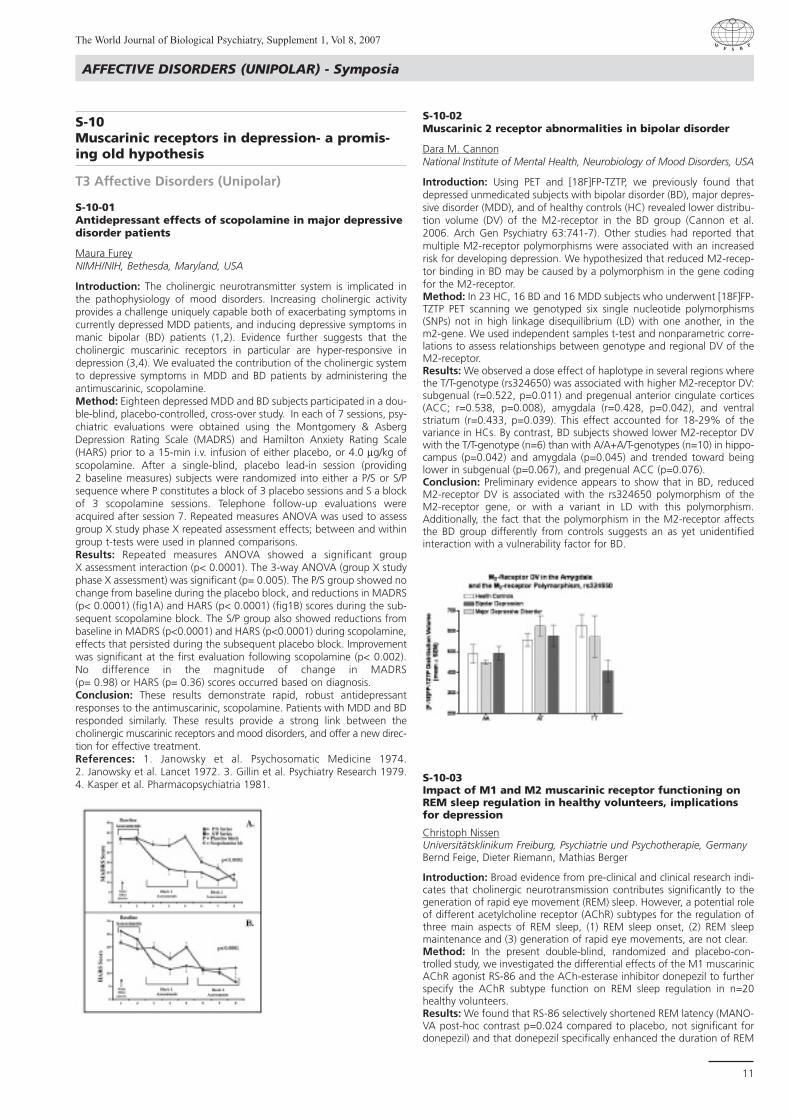
Introduction: Using PET and [18F]FP-TZTP, we previously found thatdepressed unmedicated subjects with bipolar disorder (BD), major depres-sive disorder (MDD), and of healthy controls (HC) revealed lower distribu-tion volume (DV) of the M2-receptor in the BD group (Cannon et al.2006. Arch Gen Psychiatry 63:741-7). Other studies had reported thatmultiple M2-receptor polymorphisms were associated with an increasedrisk for developing depression. We hypothesized that reduced M2-recep-tor binding in BD may be caused by a polymorphism in the gene codingfor the M2-receptor. Method: In 23 HC, 16 BD and 16 MDD subjects who underwent [18F]FP-TZTP PET scanning we genotyped six single nucleotide polymorphisms(SNPs) not in high linkage disequilibrium (LD) with one another, in the m2-gene. We used independent samples t-test and nonparametric corre-lations to assess relationships between genotype and regional DV of theM2-receptor.Results: We observed a dose effect of haplotype in several regions wherethe T/T-genotype (rs324650) was associated with higher M2-receptor DV:subgenual (r=0.522, p=0.011) and pregenual anterior cingulate cortices(ACC; r=0.538, p=0.008), amygdala (r=0.428, p=0.042), and ventralstriatum (r=0.433, p=0.039). This effect accounted for 18-29% of thevariance in HCs. By contrast, BD subjects showed lower M2-receptor DVwith the T/T-genotype (n=6) than with A/A+A/T-genotypes (n=10) in hippo-campus (p=0.042) and amygdala (p=0.045) and trended toward beinglower in subgenual (p=0.067), and pregenual ACC (p=0.076).Conclusion: Preliminary evidence appears to show that in BD, reducedM2-receptor DV is associated with the rs324650 polymorphism of theM2-receptor gene, or with a variant in LD with this polymorphism.Additionally, the fact that the polymorphism in the M2-receptor affectsthe BD group differently from controls suggests an as yet unidentifiedinteraction with a vulnerability factor for BD.
S-10-03Impact of M1 and M2 muscarinic receptor functioning onREM sleep regulation in healthy volunteers, implicationsfor depression
Christoph NissenUniversitätsklinikum Freiburg, Psychiatrie und Psychotherapie, GermanyBernd Feige, Dieter Riemann, Mathias Berger
Introduction: Broad evidence from pre-clinical and clinical research indi-cates that cholinergic neurotransmission contributes significantly to thegeneration of rapid eye movement (REM) sleep. However, a potential roleof different acetylcholine receptor (AChR) subtypes for the regulation ofthree main aspects of REM sleep, (1) REM sleep onset, (2) REM sleepmaintenance and (3) generation of rapid eye movements, are not clear.Method: In the present double-blind, randomized and placebo-con-trolled study, we investigated the differential effects of the M1 muscarinicAChR agonist RS-86 and the ACh-esterase inhibitor donepezil to furtherspecify the AChR subtype function on REM sleep regulation in n=20healthy volunteers.Results: We found that RS-86 selectively shortened REM latency (MANO-VA post-hoc contrast p=0.024 compared to placebo, not significant fordonepezil) and that donepezil specifically enhanced the duration of REM

sleep (% sleep period time, p=0.000 compared to placebo; p=0.003 com-pared to RS-86) and the number of REMs (p=0.000 compared to place-bo; p=0.000 compared to RS-86). Conclusion: These results provide evidence that the onset of REM sleepis, in part, mediated by M1 mAChR activity whereas the maintenance ofREM sleep and the number of REMs are mediated by non-M1, but presu-mably M2 mAChR activity. The findings are discussed in the context ofREM sleep abnormalities in depression.
S-10-04M1 muscarinic acetylcholine receptor agonism alters sleepwithout affecting memory consolidation
Mathias BergerGermanyChristoph Nissen, Ann E. Power, Bernd Feige, Ulrich Voderholzer, DieterRiemann
Preclinical studies have implicated cholinergic neurotransmission, andspecifically M1 muscarinic acetylcholine receptor (mAChR) activation, insleep-associated memory consolidation. In the present study, we investi-gated the effects of administering the direct M1 mAChR agonist RS-86 onpre-post sleep memory consolidation. Twenty healthy human participantswere tested in a declarative word-list task and a procedural mirror-tracingtask. RS-86 significantly reduced rapid eye movement (REM) sleep latencyand slow wave sleep (SWS) duration in comparison with placebo. Pre-sleepacquisition and post-sleep recall rates were within the expected ranges.However, recall rates in both tasks were almost identical for the RS-86and placebo conditions. These results indicate that selective M1 mAChRactivation in healthy humans has no clinically relevant effect on pre-postsleep consolidation of declarative or procedural memories at a dose thatreduces REM sleep latency and SWS duration.
S-21Genetic polymorphisms in anxiety disordersand depression; Recent findings
T3 Affective Disorders (Unipolar)
S-21-01Are genetic polymorphisms involved in antidepressanttreatment response?
Gianpaolo PernaVia Stamira d’Ancona, ItalyDaniela Caldirola, Massimiliano Grassi, Laura Bellodi
While pharmacotherapy with antidepressants is an effective treatment ofdepression and anxiety disorders, the outcome of drug therapy it still ishampered by a delayed time of onset of clinical improvement and is oftenunpredictable, ranging from beneficial effects to lack of efficacy to seriousadverse effects and a several side effects. Among the factors influencingthe interindividual variability in response to treatment with antidepres-sants, differences in genetic features play a significant role. Variations insingle genes are one well-recognized cause of such unpredictability,defining the field of pharmacogenetics. Such variations may involve genescontrolling drug metabolism, drug transport, disease susceptibility, ordrug targets. Several genetic polymorphisms have been associated withtherapeutic response to antidepressants in mood and anxiety disorders,including genetic variants of the 5-HT transporter, tryptophan hydroxylase,catechol-O-methyltransferase, brain-derived neurotrophic factor, inter-leukin-1beta although with conflicting results; also cytochrome P450drug-metabolising enzymes may be of a particular importance. The hopeis that the identification of these genetic components will eventually facili-tate the development of a customised treatment with antidepressants.The promise is to improve new drugs and ultimately individualizing theselection of appropriate drugs and dosages for each individual patient.
S-21-02Influence of the serotonin transporter gene on bipolar dis-order and suicidal behaviour
Vincenco DelucaCentre for Addiction and Mental Health, Toronto, CanadaJames L. Kennedy
Introduction: A serotonin transporter gene linked polymorphic regionpolymorphism (5-HTTLPR) has been investigated in several genetic associa-tion studies, including studies of bipolar disorder (BD) and suicidality. Method: This study was designed to examine whether the new A/G vari-ant polymorphism of the 5-HTTLPR region may be associated with the sui-cide attempts in 305 families with at least one member having BD.Results: No association with history of suicide attempt was found eitherin the multiallelic HTTLPR (LRS = 0.15659 DF = 2 p = 0.924692), or withthe intron 2 VNTR polymorphism (LRS = 0.8795 DF = 2 p = 0.6442).When we performed a haplotype analysis, we found no associationbetween suicide attempt and haplotype distribution (LRS = 1.8426 DF =4 p = 0.764681). Conclusion: These findings suggest that this new polymorphism in 5-HTTgene may not influence suicidal behaviour in bipolar disorder.
S-21-03Moderating effects of 5-HTT, MAO-A and COMT polymor-phisms on early life stress: Evidence from primate studies
Christina BarrPoolesville, MD, USA
Introduction: A loss-of-function polymorphism in the human serotonintransporter gene promoter (5-HTTLPR) increases risk for developingdepression in the face of adversity. In macaques, there exists an ortholo-gous polymorphism (rh5-HTTLPR), which has also been shown to diminishtranscriptional efficiency.Method: Rhesus macaques (M. mulatta) were raised either by theirmothers (MR) or in peer- only groups (PR), a model for early adversity.Animals were genotyped for the rh5-HTTLPR polymorphism, and theeffects of peer rearing and rh5-HTTLPR on responsivity to stress wasassessed using a social separation paradigm. Results: The rh5-HTTLPR short (s) allele interacts with exposures to adver-sity to influence behavioral and endocrine stress responding in infancy.Infants reared with their mothers that were carriers of the s allele weremore likely to sensitize to repeated mother-infant separation, where asthose homozygous for the l allele were able to adapt. When we examinedinteractive effects with early rearing history, we found that PR infants thatwere carriers of the s allele were more likely to become agitated duringstress exposure and were also more likely to develop behavioral patholo-gy during social separation. This was particularly evident among femaleinfants. Conclusion: These studies demonstrate an association of the rh-HTTLPRs allele with reactivity to stress in macaques exposed to early stress in theforms of peer rearing or repeated mother-infant separation. The identifi-cation of functionally-equivalent 5HTTLPR variants that are observed atrelatively high frequencies in both rhesus and humans may suggest thatthese loci are maintained by balancing selection. Our studies in macaquesdemonstrate that the rh5HTTLPR polymorphism influences behavioraladaptation to stress, despite the fact that it also confers increased risk forbehavioral pathology. It may be that similar selective pressures have main-tained the 5HTTLPR, and other functional 5HTT polymorphisms, in humanpopulations.
AFFECTIVE DISORDERS (UNIPOLAR) - Symposia
The World Journal of Biological Psychiatry, Supplement 1, Vol 8, 2007
12

AFFECTIVE DISORDERS (UNIPOLAR) - Symposia
The World Journal of Biological Psychiatry, Supplement 1, Vol 8, 2007
13
S-21-04The hunt for polymorphisms: Expectations and disappoint-ments
Johan A. Den BoerUniversity Medical Center, Department of Psychiatry, Groningen, The NetherlandsM. Jabbi, I. Kema, C. Hartman, J. Ormel
Introduction: Depression is a complex disease in which several differentcauses, including life events and genetic risk factors may interact in com-plex ways. We investigated the influence of genetic polymorphisms (cat-echol- O-methyltransferase polymorphism (COMT) and variants ofMonoAmino-Oxidase-A (MAO-A) genes) on endocrine and behaviouralresponses to a psychological and neuroendocrine stressor. Method: We included three groups of individuals with different degreesof susceptibility to major depression (MD) (i.e., healthy controls, first-degree relatives and patients suffering from MD). They underwent a psy-chological stressor, a simulated public speaking task (Groningen AcuteStress Test: GAST) and a neuroendocrine stressor, the DEX/CRH challenge. Results: Allelic variations of COMT polymorphism were found to influencethe degree of plasma epinephrine (EPI) response and subjective experienceof stress. Interactions between COMT and diagnostic groups in measuresof plasma EPI, cortisol and subjective responses to psychological stresswere also found. We found that COMT and MAOA functionally interacton the HPA-axis response to psychological stress, whereas during theendocrine challenge, only significant main effects of these genotypes wereobserved. Interestingly we found an interaction between COMT andMAOA with respect to the ACTH response to the GAST (see Fig.1).Conclusion: These observations support a possible role for COMT in theendocrine and subjective response to psychological stress and thus mayqualify as a possible candidate gene involved in the pathogenesis of MD.In addition, our findings show that interactions between MAOA andCOMT genes may influence the degree to which subjects respond to apsychological stressor, thus determining the vulnerability for the develop-ment of major depression. Figure 1 depicts low activity MAOA interac-tions with COMT allelic variations in mean (SEM) percentage change inplasma ACTH during all stressors except SP (speech preparation). Legend:PS= public speaking; MA= mental arithmetic; FG=rank ordering task withFinancial Gain; FL= Financial Loss.
S-07New perspectives in anxiety disorders
T4 Anxiety
S-07-01Needs and trends in the treatment of anxiety disorders
Jakov ShlikUniversity of Ottawa, Department of Psychiatry, Ottawa, Ontario,Canada
Anxiety disorders (AD) are the most common psychiatric illnesses world-wide. Lifetime prevalence for any AD nears 30% with a median age ofonset at 11 years. The prevalence in a given year reaches 18% with 30-50% of cases in the serious range of clinical severity and impairment.Many patients with AD develop unremitting course of illness complicatedby psychiatric and medical morbidiy, social maladjustment and disability.There is an increasing recognition of unmet needs in the management ofAD. Treatment directions in the field have come a long way from concep-tual arguments to evidence-based consensus. Recent authoritative guide-lines for the most part agree that medications and cognitive-behaviouraltherapies (CBT) are equally effective first-line treatments for all categoriesof AD. Among medications, the preference is given to selective serotoninreuptake inhibitors (SSRI) and serotonin and norepinephrine reuptakeinhibitors (SNRI) with a more limited role for benzodiazepine anxiolytics.Use of CBT should optimally follow empirically based protocols in a disor-der-specific format. However, numerous challenges exist in implementingthe guidelines in clinical reality. Many patients with AD fail to improve orcan not tolerate the indicated treatments. Miscellaneous algorithms forswitching, augmentation, combining and sequencing treatments are stillmore art than science. The prospects of biological and genetic markersand novel pharmacological approaches remain elusive. On the systemslevel there is a need for optimization of services across primary and spe-cialized care to ensure appropriate resources, accessibility, continuity andimproved outcomes in the management of patients with AD.
S-07-02Genetic and environmental risk factors for anxiety disorders
John M. HettemaInstitute for Psychiatric Genetics, Dept. of Psychiatry, Richmond, VA, USA
The anxiety disorders represent a heterogeneous group of syndromeswhich are common, chronic, and possess extensive comorbidity. They arecomplex genetic disorders that exhibit moderate familial aggregation andheritability. While the findings of family studies are mixed, twin studiessuggest that anxiety disorders share genetic risk with each other, majordepression, and stable personality traits such as neuroticism. This hasimportant implications for strategies targeted at identifying susceptibilitygenes underlying these conditions. Likewise, common patterns of envi-ronmental risk have been identified. In this presentation, we will reviewthese findings and their impact on recent gene finding efforts for the anxiety disorders.
S-07-03Neurobiology of anxiety disorders: Beyond serotonin
Eduard MaronUniversity of Tartu, Psychiatry, Estonia
Anxiety disorders are serious and most prevalent psychiatric diseases, theneurochemical and neurobiological origins of which are not fully under-stood. In the past decade, the specific involvement of serotonin in thepathogenesis and neurobiology of anxiety has been extensively tested ina broad scope of investigations including experimental studies, brainimaging, and genetics. Two direct serotoninergic interventions, includingtryptophan depletion and acute administration of serotonin precursors,appear to be particularly relevant in challenge studies. The results of chal-lenge studies confirm the dual role of serotonin in the modulation of va-rious forms of pathological anxiety. The brain-imaging research has pro-vided more evidence of neurobiological substrates in anxiety disorders.
The recent findings demonstrate that alterations in the brain serotoninsystem may vary among different anxiety disorders and probably dependon their clinical status. Furthermore there are increasing efforts to deter-mine anxiety vulnerability genes in the serotonin system. Overall, theassociation studies of gene polymorphisms in the serotonin system in an-xiety disorders have produced results that are inconsistent, negative, ornot clearly replicable. Therefore it would be an oversimplification to con-sider serotonin dysfunction as the single or primary pathogenetic factor inanxiety. However there is growing evidence suggesting that interactionsbetween serotonin and other systems are of particular interest for furtherunderstanding the neurobiology of anxiety disorders.
S-07-04Future classification of anxiety disorders
Joseph ZoharChaim Sheba Medical Center, Psychiatry A, Ramat Gan, Israel
Currently, although there is quite good agreement between the ICD-10and the DSM IV in regard to diagnosis of anxiety disorders, there is amajor difference between them, which is that, according to the DSM IV,OCD is part of the anxiety disorders, but according to the ICD-10, it is astand-alone disorder. This kind of different approach points out one ofthe major issues in diagnosis in general and diagnosis of anxiety disordersin particular, which is the low validity of the diagnosis. The current diag-nostic system, although it has improved the reliability of the diagnosis ofdifferent anxiety disorders, it did not take us too far in regard to impro-ving the validity. The way to increase the validity is by finding biologicalmarkers, including endophenotype markers that would give an externalvalidation of the current diagnostic system. Although we are far frombeing there, at least the intention of the different committees who areworking on the DSM V is to strive to find such biological markers, and toimplement them in future diagnostic systems.
S-11Impulsivity across the psychiatric disorders
T4 Anxiety
S-11-01Endophenotype of impulse control disorder: From conceptto treatment
Joseph ZoharChaim Sheba Medical Center, Psychiatry A, Ramat Gan, Israel
Impulsivity is a symptom dimension that goes behind specific diagnosis.In addition to the impulse control disorders listed in the DSM IV, there areother disorders with elements of dysregulated impulsivity. Those disordersinclude bipolar disorder, substance use, PTSD, trichotillomania, gambling,some of the paraphilia, etc.In each of these disorders, the presence anddegree of impulsivity should be taken into account in the treatment plan.In the session we will explore the different presentations of dysregulatedimpulsivity, not only in different psychiatric disorders, but across the life-span as well. However, manifestation of the trait of impulsivity changesthroughout development and therefore, the dysregulated impulsivity inadults is different as compared to children.The vast majority of psychiatricdisorders are often accompanied by comorbid disorders or associatedsymptom domains that shape their expression and the course of thosedisorders. For example, the presence of affective instability in patientswith impulsivity may suggest that a trial of mood stabilizers is appropri-ate. Although the exact mechanism by which the comorbidity of affectand impulse dysregulation occurs has not been identified as of yet, thepresence of more than one psychiatric disorder is the rule rather than theexception. The presence of a comorbid condition or associated symptomcomplicated both the diagnosis and treatment of impulse control disor-der.In the session, we will explore the implications for diagnosis of disor-ders with impulsivity throughout different diagnostic schema, and exa-mine their clinical implications along those lines. Focusing more attentionon this dimension will improve recognition of it, and encourage explo-ration of new treatment avenues for this important dimension.
ANXIETY - Symposia
The World Journal of Biological Psychiatry, Supplement 1, Vol 8, 2007
14

ANXIETY - Symposia
The World Journal of Biological Psychiatry, Supplement 1, Vol 8, 2007
15
S-11-02Impulsive symptoms and traits in patients with differentpsychiatric disorders (pathological gambling, eating disor-ders, atypical affective disorders and personality disorders)and its relationship with depression
Jose CarrascoMadrid, Spain
Introduction: The association of impulsive temperament and emotionaldysregulation has been proposed historically as the substrate for severaldisorders with impulsive symptoms. These disorders, such as eating disor-ders, impulse control disorders, addictions and atypical affective disor-ders, are conceptually close to the old neurotic spectrum. However, phe-nomenological and neurobiological evidence suggest that they constitutea distinct nosological cluster.Method: Biological indexes, including platelet monoamine oxidase activity,prolactin response to serotonin agonist, tryptophan depletion and low-dose dexamethasone suppression test were performed in patients withpathological gambling, eating disorders, atypical depression and fybromi-algia and compared with studies in borderline disorder.Results: Impulsive disorders, eating disorders, pathological gambling andfybromialgia/atypical depression are characterized by increased emotionalinstability and affective reactivity. Neurobiological findings, as with otherimpulsive syndromes, indicate low platelet MAO, decreased prolactinresponse and increased cortisol suppression.Conclusion: Several disorders with impulsive behaviors in the DSM-IVmight have emotional instability as the primary dysfunction. Borderlinepersonality disorder might be biologically and nosologically closer to thesesyndromes than to the rest of personality disordersReferences: Carrasco JL,DÓaz-Mars· M, Pastrana JI, Molina R, Brotons L,LÛpez-Ibor MI, LÛpez-Ibor JJ: Hypothalamic-pituitary-adrenal response inborderline personality disorder without posttraumatic features. BritishJournal of Psychiatry 2006, in press. Basurte E, Diaz-Marsa M, Martin O,Carrasco JL. Childhood trauma, impulsivity and hypothalamic-pituitary-adrenal axis dysfunctions in eating disorders. Actas Esp Psiquiatr. 2004May-Jun;32(3):149-52.
S-11-03Impulsivity in intermittent explosive disorder and impul-sive aggressive personality disorders
Larry SieverMount Sinai School of Medicine, Psychiatry, New York, USA
Introduction: Cluster B personality disorders are often associated withimpulsive aggressive behaviors that meet criteria for intermittent explosivedisorder - revised (IED-IR). Genetic, neurotransmitter probes studies, andneuroimaging studies have begun to clarify the underlying neurobiologyof this impulsive aggression. Prefrontal cortical inhibitory regions such asorbital frontal and anterior cingulate cortex play an inhibitory role inamygdala activation in response to provocative stimuli. Serotonin facili-tates this top down inhibitory control.Method: The serotonin transporter was labeled with [11C] DASB bindingin 12 patients with IED-IR and in 12 healthy control subjects matched forage and gender. Binding of [11C] MDL 100907 to the 5HT2A receptorwas analyzed in 27 patients with IED-IR and 24 matched healthy controls.In an FDG PET paradigm, 33 IED-IR patients with concomitant borderlinepersonality disorder and 24 controls were tested during anger provoca-tion as well as a non-provocation scan.Results: The IED-IR patients had significantly lower 5-HTT binding with[11C] DASB in the anterior cingulate cortex with a reduction of approxi-mately 35% in these patients. 5HT2A receptor binding with [11C] MDL100907 was increased in currently physically aggressive subjects with cor-relations between aggression and OFC 5HT2A receptor binding. In theFDG PET study, after provocation increased anterior cingulate corticalmetabolic activity while it decreased this activity in the patients and meta-bolic activity by FDG PET in the anterior cingulate cortex correlated posi-tively with aggressive responding.Conclusion: Anterior cingulate cortex is a critical region in inhibiting theexpression of aggression and is less active in impulsive aggressive patientscompared to controls. Reduced transporter activity possibly reflectingreduced serotonergic intervention as well as increased 5HT2A receptor
binding appears to contribute to the underactivity of anterior cingulategyrus supporting the possibility of pharmacologic intervention of theserotonergic system for treatment of impulsive aggression.
S-11-04Affect regulation and impulsivity in borderline personalitydisorder
Sabine HerpertzRostock University, Psychiatry and Psychotherapy, Germany
Introduction: Impulsivity is one of the most prominent characteristics ofborderline personality disorder (BPD). Three domains of psychopathologytarget the problem of impulsivity in BPD: Impulsive modes of behavior,cognitive disinhibition, and dysregulated emotions. Methods: Facets of cognitive functioning and emotional processing havebeen investigated in different samples of female BPD inpatients usingneuropsychological tasks and emotion challenge tasks. In addition tobehavioral data functional activity to emotionally charged stimuli wereassessed in a series of experiments in female BPD patients. Results: Data suggest that cognitive disinhibition exclusively occurs in thecontext of processing emotional but not neutral stimuli. A bias towardsanger was found for ambiguous facial stimuli indicating distorsions insocial perception. Regarding functional activity data suggest that amyg-dalar hyperreagibility underlies “high arousal” processing of emotionallyrelevant stimuli that is not sufficiently suppressed by prefrontal top-downcontrol. In addition, BPD patients exhibit a general tendency towards aself-referential mode of information processing indicated by an enhancedinvolvement of the cerebral memory retrieval network. Conclusion: The findings provide a neurobiological basis for the notionthat impulsivity is closely related to dysregulated emotions in BPD.Emotional hyperarousal together with distorsions in social perception mayincrease the risk of reactive (auto)aggression in BPD. References: Schnell K, Dietrich T, Schnitker R, Daumann K, Herpertz SC.Processing of autobiographic memory retrieval cues in borderline perso-nality disorder. J Affective Disorders, in pressDomes G, Winter B, SchnellK, Vohs K, Fast K, Herpertz SC (2006) Inhibitory functioning in borderlinepersonality disorder and the influence of emotions. PsychologicalMedicine, 36:1163-1172Herpertz SC, Dietrich TM, Wenning B, ErberichSG, Krings T, Thron A, Sass H (2001) Evidence of abnormal amygdalafunctioning in borderline personality disorder: a functional MRI study.Biological Psychiatry 50(4):292-298
S-44PTSD and depressive spectrum disorders
T4 Anxiety
S-44-01MagroobIndia
S-44-02Neurobiology of PTSD
E. MohandasIndia
Introduction: Although the conceptual framework of Posttraumaticstress disorder is a matter of debate, there are attempts to delineate neuro-anatomical correlates of symptom clusters of PTSD. Neuroimaging pro-vides a reasonable model of neurocircuitry of PTSD. This paper criticallyevaluates the available evidence on functional neuroanatomy and neuro-chemistry of PTSD.Method: The existing data on neurobiological basis of PTSD was evaluatedusing various search engines (Pubmed, Ebsco, Science direct, Ovid)Results: The neural substrates and circuitry of PTSD are formulated. Acritical appraisal of the neurochemical alterations is attempted.Conclusion: The available evidence suggests that the neurochemicalchanges may vary depending on the type,magnitude,and duration of thestress.The cognitive dysfunction may be due to working memory dysfunc-tion rather than hippocampal changes.

References: Nemeroff CB, Bremner JD, Foa EB, Mayberg HS, North CS,Stein MB. (2005) Posttraumatic stress disorder: a state-of-the-sciencereview. J Psychiatr Res., 40(1):1-21. Rauch SL, Shin LM, Phelps EA. (2006)Neurocircuitry models of posttraumatic stress disorder and extinction:human neuroimaging research—past, present, and future. Biol Psychiatry,60(4):376-82. Shin LM, Rauch SL, Pitman RK. (2006) Amygdala, medialprefrontal cortex, and hippocampal function in PTSD. Ann N Y Acad Sci.,1071:67-79. Bremner JD. (2006) The relationship between cognitive andbrain changes in posttraumatic stress disorder.Ann N Y Acad Science,1071:80-6.
S-44-03Treatment approaches to PTSD
Sunil MittalIndiaManu Mittal
Introduction: The presentation reviews current treatments and theIndian experience in quantifying, diagnosing and treating PTSD and thelessons from work with those affected by terrorism, and natural disasterslike cyclones, earthquakes and Tsunami.Conclusion: PTSD is not an inevitable consequence of trauma but repre-sents one of the evolving responses to trauma. Stress reactions may servethe primordial function of adaptation and survival. Earlier lower reportedincidence of PTSD was possibly due to reporting bias and its presentationmore as somatic symptoms. Progressive relaxation of the stressor criteriatoday reflects changes in societal values. Since the psychopathology oftrauma is dynamic and culture affects the expression of distress andpainful memories, PTSD has also been viewed as a contemporary culture-bound syndrome. Even as the broadening definition of trauma con-founds the issue of greater understanding of the psychobiology of PTSD,yet our knowledge of the neurobiology of post-traumatic symptoms hasincreased. The brain is a master-controller of stress reactions as well as theprime target of their effects. Exposure to manageable stress helps thebrain adapt for greater endurance, working and immunity; extreme stressdamages the brain inhibiting memory functions; and catastrophic expe-riences may change the structure and functioning of the brain. As indepression, impairment of structural plasticity and cellular resilience isimplicated in PTSD. Very high levels of catecholamines and endorphinsseen in PTSD may interfere with storage of explicit (declarative) memorycausing trauma memories being stored as implicit (non-declarative) mem-ory: as emotions and senses. This may lead to intrusive recollections, mal-assessment of further danger signals or non-threatening events and sur-vivors reacting with panic attacks, flashbacks and overwhelming emo-tions probably due to situationally accessible (vs. verbally accessible)memories. Pharmacological treatments for PTSD include SSRIs, other anti-depressants, bezodiazepines, mood stabilizers, antipsychotics and drugslike clonidine. Little work exists on early pharmacological interventions toprevent PTSD. Non pharmacological treatments include cognitive andother behavioural therapies, EMDR, debriefing, ventilation, relaxation,spiritual and supportive therapies, self-help groups, mass grieving andbibliotherapy.
S-44-04S. NambiIndia
S-47PTSD - state of the science
T4 Anxiety
S-47-01Insights from animal models in PTSD
Masashi NibuyaTokorozawa, Japan
Introduction: To better understand the effects of environmental stresson psychiatric disorders, many stress studies have been performed using
experimental animals. Compared to apparent difficulties in making ani-mal models of endogeneous psychoses including schizophrenia and affec-tive disorders, the PTSD animal models are expected to be established inan earlier time point in the future. For traumatic stressogenic stimuli inPTSD animal models, various procedures have been adopted includingpredator scent paradigm, restraint stress, electric tail shock, and underwa-ter trauma. In order to obtain life threatening-level stress and examinebehavioral effects of rats under the same traumatic environment, we haveadopted multiple inescapable electric foot-shocks treatment andobserved behaviors in the shuttle box at the 14-day later (Sawamura etal, 2004). We have recently demonstrated that the inescapable foot-shock stress paradigm promptly induces endoplasmic stress pathwaywhich can ultimately trigger neuronal apoptotic cell death in the rat brain(Toda et al, 2006). We consider ER stress mechanism together with alreadyreported brain glucocorticoid dysfunctioning and reduced neurogenesisin the hippocampal brain region possibly via reduced neurotrophin signa-ling (Nibuya et al, 1995) have effects on brain volume loss in PTSD anddepression.Results: The splicing of 26 nucleotides in the coding region of the X-boxbinding protein-1 (XBP-1) transcript to generate a mature active transcrip-tion factor is a part of the unfolded protein response to intracellular endo-plasmic reticulum stress. In this study, we demonstrated that XBP-1 spli-cing is promptly induced in the rat brain including the hippocampus byboth inescapable electric foot shock (IS) and pharmacologically manipu-lated activation of 5-hydroxytryptamine release in a dose-dependentmanner. By administering ketanserin, a 5-hydroxytryptamine 2A antago-nist, however, we could only partially block the increased splicing by ISand observed that the splicing was not influenced by lithium carbonatepretreatment. Conclusion: Although it is still unclear whether the enhanced unfoldedprotein response functions neuroprotectively by modulating the rate ofgeneral translation and increasing chaperone proteins or whether it even-tually induces cellular damage by triggering apoptosis, the present resultsindicate the possible existence of a new adaptive intracellular signalingpathway in the brain that responds to environmentally challenged beha-vioral stress loading. References: On the meeting we additionaly present expressional alte-ration of GPR56 protein involved in neuronal migration and differentia-tion by behavioral stress loading.
S-47-02Injury increases the risk for PTSD, potential neurobiologicaland psychological mediators
Ehud KleinRambam Medical Center, Department of Psychiatry, Haifa, IsraelDanny Koren
Given the frequency with which common traumatic events involve bothphysical and emotional injuries, a crucial question arises: How do thesetwo interact? How does injury affect an individual’s chances of developingPTSD? Traditional views, tended to regard bodily injury as a protective fac-tor against the development of posttraumatic stress disorder suggestingthat the healing and rehabilitation process shifts the individual’s attentionaway from the negative emotional consequences of the trauma.In con-trast, more recent literature on this topic suggests that injury during atraumatic event might in fact increase the probability of developingPTSD.In this talk findings from a recent study will be presented, showingan eight-fold higher risk for develepment of PTSD in soldiers who wereinjured during military traumatic events as compared to their comeradeswho participated in the same events, but were not injured.These resultsindicate that physical injury, over and above exposure to the traumaticevent itself, is a risk factor for PTSD.The discussion will relate to contem-porary findings regarding the neurobiological and psychological mecha-nisms by which injury may augment or independently contribute to thedevelopment of PTSD.
S-47-03Rachel YehudaUSA
ANXIETY - Symposia
The World Journal of Biological Psychiatry, Supplement 1, Vol 8, 2007
16
CHILDHOOD & ADOLESCENT DISORDERS - Symposia
The World Journal of Biological Psychiatry, Supplement 1, Vol 8, 2007
17
S-02The challenge of suicide in adolescents
T5 Childhood & Adolescent Disorders
S-02-01Epidemiology of suicide
Edith SerfatyBuenos Aires, Argentina
Introduction: Young people in Argentina are exposed to death by suicide.Objective of this research was to study the suicide death between youngpeople during 1991 to 2000. The Epidemiological Research Center of theBuenos Aires Academy of Medicine did the study with all the country’sdeath dates certified by the Ministry of Key words: suicide-adolescence. Method: Material and Methods: Population studied; all the death by sui-cide of people between 10 to 24 years old, of Argentina, since 1991 to2000 in the country and each area. Statistical analysis: Mortality rateswere calculated on yearly per province and per country.Results: Suicide rates for this age both sexes was 6.1 per 100.000 inhabi-tants. It was 9% of the total adolescent’s death. The rate increasedbetween 1991 and 2000 from 1.3 to 6.1. Conclusion: HealthDiscussion: It was detected the following risk factors.Late adolescence, male sex. It was more elevated on males. Results showedthat suicide was a significant problem for Public Health and the commu-nity. References: Yunes Jo, Zubarew T. Mortalidad por causas violentas enadolescentes y jÛvenes. Un desafÓo para la regiÛn de las AmÈricas. Rev.Bras. Epidemiol 1999. 1 (3): 102-71. Dehne KL, Riedner G. Adolescence,a dynamic concept. Reprod. Health Matters 2001, 9 (17), 11-5. InstitutoNacional de EstadÓsticas y Censos (INDEC). ProyecciÛn de poblaciÛn poredad y sexo. Total del paÓs. An·lisis demogr·fico NÆ 7, INDEC 1995.
S-02-02Biology of suicide
Jose-Luis Ayuso GutierrezMadrid, Spain
Suicide behavior is probably best understood in the context of a modelof stress-diathesis. Substantial evidence suggest that low brain serotoner-gic function and genetic factors contribute to reduce the suicide thresh-old . On the other hand, other risk factors contribute to the developmentof suicide behavior like depression, mania, psychosis, substance abuseand stressful life events. Investigations of biological substrates of suiciderisk were initiated almost 30 years ago. These early studies indicated anassociation of suicidal behavior and low 5-HIAA in the CSF, that was cor-related with the lethality of the attempt. Later, the elucidation of the neu-rotransmitter receptors disordered in the pathophysiology of suicide hasbeen the subject of intense post-mortem studies. The majority of thestudies have measured binding of the SERT in tissue preparations fromvarious brain regions, particularly the frontal cortex. Most studies show adecreased SERT binding in PFC of suicide victims indicating low serotoninfunction. More recently, increased binding of 5-HT1A in PCF of suicide vic-tims has been found indicating a compensatory response to low serotonin.Several research groups have demonstrated genetic contributions to sui-cidal behavior through both family studies and molecular genetics. Otherinvestigators have sought to link suicide behavior to genetic variations ofthe SERT, although these efforts have produced inconsistent results.
S-02-03Eduardo CorreaUniversidad de Chile, Clinica Psiquiatrica Univ., Oviedo, Chile
S-02-04Risk of suicide and antidepressants
Leonardo TondoHarvard Medical School, McLean Hospital, Belmont, USA
Suicide is a particularly urgent challenge in children and adolescents inwhom depression is less recognized and substance abuse is more com-mon(1) than in adults. The question of whether modern antidepressanttreatments can affect mortality rates is only beginning to be addressed.Most studies of treatment effects on suicide have been naturalistic orhave encountered suicidal behavior as an unintended outcome in con-trolled treatment trials. Moreover, ethical issues can make study designsdifficult when death is a possible outcome. Ecological studies based onthe association between use of antidepressants and risk of suicide indefined populations treated with antidepressants yielded inconsistentresults. Also clinical studies on the same subject did not seem to provideconsistent results. It is interesting to note that introduction of safer anti-depressants with very low lethality on overdose since the 1980s has ledto marked increases in antidepressant usage, but not to a measurablereduction of suicide rates. However, studies including only children andadolescents are relatively few. The only ecological study on adolescentsaged 10-17 years treated with antidepressants did not show significantmodification of the suicide rates(2). A very recent matched case-controlstudy(3), reinforced a significant association between the use of antide-pressants and the increase of completed and attempted suicides indepressed children and adolescents. Studies on this topic have becomemore important after an association between increased suicidality in chil-dren treated with antidepressants emerged as a result from meta-analy-ses of data reported to regulatory agencies in the UK and US.References: 1. Tondo L, Baldessarini RJ, Hennen J, Minnai GP, Salis P,Scamonatti L, Masia M, Ghiani C, Mannu P: Suicide attempts in majoraffective disorder patients with comorbid substance use disorders. J ClinPsychiatry 1999; 60: 63-9. 2. S„ndergÂrd L, Kvist K, Andersen PK, KessingLV. Do antidepressants prevent suicide? Int Clin Psychopharmacol 2006;21: 211-218. 3. Olfson M, Marcus S, Shaffer D. Antidepressant DrugTherapy and Suicide in Severely Depressed Children and Adults: A Case-Control Study. Arch Gen Psychiatry 2006; 63: 865-872.

S-17Vulnerability and resilience factors in depressionand dementia
T6 Neurodegenerative Disorders
S-17-01Oxidative stress and clinical implications in dementia
Anne EckertPsych. Univ. Clinics Basel, Neurobiology Laboratory, Switzerland
Introduction: Oxidative stress caused by reactive oxygen species plays adecisive role in the pathogenesis of neurodegenerative disorders such asAlzheimer's disease (AD). Mitochondria are the primary source and maintarget of reactive oxygen species (ROS) within cells. Methods: Different methods were used to identify oxidative stress andmitochondria malfunction in brains from transgenic mice or cell culturemodels. Results: Early defects in glucose utilization in the brains of AD patientssuggest possible abnormalities in mitochondrial function in AD. Severalstudies have indicated that amyloid-beta protein (Aß) may be directlytoxic to isolated mitochondria. Interestingly, the activities of those enzy-mes, which are reduced in the brains of AD patients, such as COX, alpha-ketoglutarate dehydrogenase and pyruvate dehydrogenase were inhibi-ted by Aß. Recently, it has been demonstrated that Aß can be formedintracellularly in neurons. A large amount of evidence suggests that mito-chondria could intervene in the mechanism by which intraneuronal Aßtriggers neuronal dysfunction and degeneration. In our own studies, wecould clearly demonstrate that Aß causes oxidative stress and mitochon-drial malfunction in a cell culture model as well as in transgenic mice (Keilet al., 2004). In addition, recent data of our group demonstrate a link bet-ween tau pathology and mitochondrial malfunction. Using transgenicmice, we could demonstrate mitochondrial dysfunction by proteomic andfunctional analyses in these mice (David et al., 2005). Thus, we can spe-culate that tau and Aß accumulation probably act in synergy on oxidativestress and mitochondrial dysfunction. Of note, Ginkgo biloba extract EGb761 and piractem at therapeutically relevant in vitro and in vivo concen-trations are able to improve mitochondrial dysfunction associated withoxidative stress and/or aging (Eckert et al., 2005; Keil et al., 2006). Conclusion: In summary, protection against mitochondrial dysfunction,improved ATP production, and prevention of apoptotic signals may beimportant features of antidementive drugs. Since mitochondrial failureand reduced energy metabolism seem to be very early events during thecourse of AD, the stabilization of mitochondrial function represents anemerging preclinical concept of age-related memory disorders anddementia. References: David et al., J. Biol. Chem. 280:23802-23814, 2005; Eckertet al., Ann. NY Acad. Sci. 1056:474-485, 2005; Hauptmann et al., Exp.Gerontol. 41:668-673, 2006; Keil et al., J. Biol. Chem. 279:50310-50320,2004; Keil et al., Br. J. Pharmacol. 147:199-208, 2006
S-17-02Natural neuroprotective substances in neurodegeneration
Egemen SavaskanPsychiatric Univ. Hospital, Dept. of Psychiatry Research, Zürich,Switzerland
Introduction: A complex neuropathology underlies neurodegenerativedisorders such as Alzheimer’s disease (AD). One of the hallmarks of AD iswidespread neuronal and synaptic losses. The two major neuropathologi-cal findings in the AD brain are the presence of neuritic plaques contai-ning â-amyloid (Aâ) and neurofibrillary tangles. That oxidative stress maybe a culprit in neuronal loss in AD has been emphasized in recent yearsand the evidence is becoming progressively stronger that radicals areinvolved in the neuronal pathogenesis of the disease. The free radicalsthat have been incriminated as causing neuronal loss are believed to begenerated by Aâ. Methods: Today, it is well established that some human hormones haveobvious anti-oxidative capacity.
Results: These hormones play an important role in the functioning of thecentral and peripheral nervous system, have neuroprotective effects, andtheir levels markedly decrease with age and in AD. They may provide anatural source of neuroprotection in neurodegenerative disorders. Conclusion: Therefore, the aim of the present work was to uncover theneuropathology altering hormonal effects in AD and to establish the neu-roprotective effects of these substances. Two of them, melatonin andestrogen, respectively, will be discussed in detail.
S-17-03Neuroendocrine and sleep regulation as predictors of ill-ness course and therapy in depression
Martin HatzingerUniversity Hospital Basel, Psychiatric Outpatient Clinic, SwitzerlandU. Hemmeter, S. Brand, M. Ising, Edith Holsboer-Trachsler
Introduction: Introduction: In acute depression characteristic changes inEEG sleep measures are well documented findings. However, the courseand the predictive value of these alterations for treatment outcome aswell as for long-term course of depression still warrants clarification.Therefore, we examined whether (1) the previous clinical course ofdepression, (2) treat-ment response during a standard antidepressanttherapy, and (3) the long-term outcome in follow-up are associated withabnormal EEG sleep parameters. Since the hypothalamic-pituitary-adrenocortical (HPA) system seems to play a crucial role in treatment out-come and course of depression, we evaluated HPA system function aswell.Method: Methods: 15 patients (4 men, 11 women; age 43-59) withdepression were enrolled in the study. HPA system assessment using thecombined DEX/CRH test and sleep EEG studies were conducted at base-line, after a 6 week antidepressant treatment period (trimipramine), andat follow-up i.e. after 2 to 10 years after the index episode. Results: Results: 1. The previous clinical course as reflected by the num-ber of episodes until base-line correlated significantly with EEG sleepmeasures i.e. sleep continuity values, slow wave sleep (SWS) and REMlatency. 2. During treatment sleep continuity values improved and the cor-relation with the previous long-term course disappeared. However, thecorrelation with SWS persisted. The only sleep EEG marker at baselinepredictive for treatment response was REM latency. 3. In the prospectivelong-term outcome SWS and REM density variables were related to theoccurrence of recurrences in follow-up. These identified sleep EEG mar-kers correlated closely with HPA system regulation. Conclusion: Conclusions: The previous and the prospective long-termcourse of depression are related to sleep EEG variables during the acutedepressed state at the beginning and the end of treatment. Among thedifferent values SWS and REM density measures seem to reflect predictivemarkers for the long-term course of depression. These markers are asso-ciated with HPA system regulation.
S-17-04Neuroendocrine molecular regulation of anxiety, stress anddepression
Eduardo ArztUniversity of Buenos Aires, Argentina
Introduction: Corticotrophin releasing hormone (CRH) is the key media-tor of the central nervous system response needed to adapt to stressfulconditions. If adaptation fails hypersecretion of CRH continues and pro-duces via stimulation of CRH type 1 receptors (CRHR1) symptoms pertai-ning cognition, appetite, sleep, anxiety and stress hormone secretionunderscoring that this pathway is causally involved in affective disorders.New basic studies indicate that the final outcome of CRH/CRH1 signalingdepends on the context of specific cells and ligands, cross-talk of signa-ling pathways and the effector actions of the pathways once they are acti-vated. This specificity bears consequences at the CNS level where CRHactivates through the same receptor (CRHR1) different signaling path-ways depending on neuroanatomical context. These findings open up theopportunity for a more focused pharmacological intervention.
NEURODEGENERATIVE DISORDERS - Symposia
The World Journal of Biological Psychiatry, Supplement 1, Vol 8, 2007
18

NEURODEGENERATIVE DISORDERS - Symposia
The World Journal of Biological Psychiatry, Supplement 1, Vol 8, 2007
19
S-43Prevalence and impact of dementia in LatinAmerica - 10/66 dementia research group
T6 Neurodegenerative Disorders
S-43-01Prevalence of dementia in Latin America - ethnic and racialdifferences
Juan Llibre RodriguezUniv. Medica de la Habana, Medicina Finaly Albarran, Cubanacan Playa,Ciudad Habana, CubaM. Prince, Cleusa Ferris
Introduction: Demographic ageing is proceeding especially rapidly inLatin America. Estimates based on the literature suggested 1.8 millionpeople with dementia now increasing to 4.1 million by 2020 and 9.1 mil-lion by 2040, that is the number of people with dementia will be verysimilar in Latin American as North America. Method: After pilot studies the 10/66 Dementia Research group hasbeen finished prevalence studies in six developing countries in LatinAmerican: Argentina, Cuba, Dominican Republic, Mexico, Peru, andVenezuela. Each of these studies uses the same design (a comprehensiveone phase survey of all residents aged 65 and over, with a minimum sam-ple size of 2 000 in each of the six countries, for a total sample of 12 000 individual from rural and/ or urban catchment areas. All partici-pants are interviewed with a sociodemographic, risk factors and healthbehaviours questionnaire, a cognitive test battery and a structured clinicalinterview. A physical and neurological examination follows, with anthro-pometry and phlebotomy.Results: There are large differences between centres in diet, levels ofactivity and cardiovascular risk factor profiles. The Peruvian sample seemsto have strikingly low levels of cardiovascular risk, with a low prevalenceof smoking and very low blood pressure levels. Hypertension and othervascular factors are most prevalent in Cuba and Venezuela. The preva-lence of ‘probable dementia’ according to screening measures, and DSMIV criteria increase with age and decline with education. The 6.4% preva-lence in Cuba is significantly higher than in all other centres, follow byVenezuela and Dominic Republic. The prevalence of DSM IV dementiaand also the association between APOE4 and DSM IV dementia weremore higher in white people that in people with African ancestry. Conclusion: The prevalence of dementia is high in Latinamerican coun-tries as in developed world. Risk factors for vascular disease such as highfat diets, cigarette smoking, diabetes, hypertension and sedentarylifestyles are common.
S-43-02Mariella GuerraLima, Peru
S-43-0310/66 Dementia diagnostic validity
Ana Luisa Sosa OrtizInstituto Nacional, Mexico City, Mexico
Introduction: The 10/66 Dementia Research Group was integrated in1988 based in the following facts: 1. Two-thirds of people with dementialive in developing countries. 2. In the next 30 years there will be a largeincrease in dementia cases in DCs. 3. Less than 10% of population-basedresearch is targeted on DCs. Then, it is necessary to generate: a. Goodquality research b. Awareness c. Shape policies and d. Service develop-ment The first goal of the 10/66 Research Group is: To develop and testa culturally and educationally unbiased diagnostic process for dementia. Method: In order to do a cross-cultural validation study, the 10/66 R.G.did a pilot study in which 25 international centers participated, getting atotal sample of 2885 subjects aged 60 years and older, from: India, Chinaand South East Asia, Latin America, the Caribbean and Africa. In this pilotstudy the interviewers, blind to the diagnosis (dementia, depression, con-trol high and low education), administrated: a. The Geriatric Mental State(GMS). b. The Community Screening for Dementia (CSID). c. Ten word
list-learning task of the Consortium to Establish a Registry of Alzheimer’sDisease (CERAD). Results: In this pilot study, each one of the three instruments appliedindependently predicted the dementia diagnosis. An algorithm with allthe instruments gave better prediction than each one individually; withthis algorithm a sensibility of 94% and a specificity of 85%, 97% and94% in cases of: depression, high education and low education groups,respectively, were obtained. Conclusion: Based on these results, we conclude that the 10/66 demen-tia algorithm is a good option for culturally and educationally sensitivedementia diagnosis for clinical and population-based research. After thispilot study, the 10/66 R.G. started with population-based studies in sevencenters, getting a total sample of around 17,000 subjects evaluated withthe aim of estimate prevalence, describe impact and seek to identifygenetic and environmental risk factors in the participating settings (India,China, Latin America and the Caribbean). This huge sample will let usexplore new and interesting validation methods. References: -Prince M. et al. 2003. Dementia diagnosis in developingcountries: A cross cultural validation study. Lancet 361: 909-917. -The10/66 Dementia Research Group. 2000. Methodological issues in popula-tion-based research into dementia in developing countries. A positionpaper from the 10/66 dementia research group. Int J Geriatr Psychiatry15: 21-30
S-43-04People with dementia in Latin America - care arrangements and caregivers strain
Aquiles SalasUniv. Central Venezuela, Caracas, Venezuela
Introduction: Demographic ageing is proceeding especially rapidly inLatin America. Those aged 65 years and over will increase from 33.3 mil-lion (6% of the total population) in 2005 to 56.3 million (8.5%) in 2020and 110.2 million (14.7%) in 2040. The literature suggested 1.8 millionpeople with dementia in 2001 increasing to4.1 million by 2020 and 9.1million by 2040. Arguably, health and social finance systems are not well-placed to meet the needs of the growing numbers of frail and dependentelderly. Across Latin America, fewer than 30% of older people receive oldage pensions (Help Age International). Health systems in Latin Americaare generally Mixed National Health Systems in which public sector insti-tutions play a relatively minor role, less than one-third are covered bymandatory social health-insurance systems and private out-of-pocketexpenditure on health services is relatively large. Method: Aknock door census allows to recruit 2020 subjects and theirscaregiver in a low income population at the south west ofCaracas.Trained interviewers applied a set of mental health question-naires, care arrangement and household information. Results: Preliminary results shows that 7.5% of the population studiedhas any type of dementia, 4.5% has MCI and 13% depression; the pub-lic institutions play a small role attending the elderly, lacking specializedcenters for demented. The care is given mainly for families’ caregiverwithout training and support. We found that 80 % used private medicalservices and 15% used public services; 20% had paid caregiver and 7-10% caregiver stopped or cut back work for care and 48% showed com-mon mental disorders due to burden of care.Conclusion: Dementia is a medical and social problem in our countries;we may have more than 100000 families living with a person withdementia. They have to cope with poor support and knowledge. Thereare very few or none institution at the community or regional level.References: Salas A, Mosca W, Gonzalez J “ La salud y el Bienestar delos Adultos Mayores” en : Salud y Equidad: Una Mirada desde las CienciasSociales, Editora Fiocruz, Rio de Janeiro, Brasil, Octubre 2000. Prince M,et al. 2003.Dementia diagnosis in developing countries: a cross – culturalvalidation study. Lancet 2003; 361: 909-917 The 10/66 DementiaResearch Group. 2004. Care arrangements for people with dementia indeveloping countries. Int J Geriatr Psychiatry 19: 170-177.

S-43-05People with dementia in Latin America - costs
Raul Arizaga10/666 DRG, Buenos Aires, Argentina
Introduction: The demographic structure of developing countries (withonly a few exceptions) shows a population aging process that is actuallymore striking than in the developed world, that has suffered the acutephase of this graying phenomenon some decades earlier. Dementia isconsidered the fourth cause of disability adjusted life years in the deve-loping world for both sexes. The costs of dementia to society is the valueof all goods and services that are given up in order to prevent, diagnose,treat and otherwise cope with dementia. Economic costs of AD are sig-nificant for health systems. Individuals, families and carers are affectedboth in the economic aspect and in the Quality of Life (QOL). Costs aredivided in direct (money used in an explicit way in hospital, medical ser-vices, drugs, social services) and indirect (money used in an implicit wayas loss of income by the patient and loss or reduction for family membersor carers). The aim of this presentation is to analyze total, direct and indi-rect costs of AD and its variations in relation to disease severity andwhether patients are institutionalized or not. Method: Eighty community dwelling patients, twenty institutionalizedAD patients and their respective primary caregivers, and twenty-fivehealthy elderly subjects, participated in this study. The cognitive and neu-ropsychiatric impairments and severity of dementia were assessed withMiniMental State Examination, Neuropsychiatric Inventory and ClinicalDementia Rating respectively. One structured interview about health andno health care resources used up during the last 3 months were adminis-tered to family caregivers. The time devoted by carers to look after thepatients and the caregiver burden (Zarit’s Burden Interview) was recorded.A healthy elderly control sample was used to address the cost specificallyattributable to the disease. Results: The annual direct cost of the disease increased with cognitivedeterioration from 3421$US in mild to 8256$US in severe, and with insti-tutionalization (3189$US outpatient vs. 11270$US institutionalized).Family paid mostly for direct costs.Conclusion: With projected increase in the number of persons at risk fordeveloping AD in emerging countries, economic familial cost of the disease will be significant. Dementia costs should be a matter of analysiswhen health policies are being designed in developing countries.
NEURODEGENERATIVE DISORDERS - Symposia
The World Journal of Biological Psychiatry, Supplement 1, Vol 8, 2007
20

PSYCHOTIC DISORDERS - Symposia
The World Journal of Biological Psychiatry, Supplement 1, Vol 8, 2007
21
S-01Pathogenesis of schizophrenia
T7 Psychotic Disorders
S-01-01Disc1-interacting molecules and pathogenesis of schizo-phrenia: Candidate gene approach
Norio OzakiNagoya University, Psychiatry, JapanM. Ikeda, S. Taya, K. Kaibuchi, N. Iwata
Introduction: Numerous linkage and case-control association analyseshave been performed to clarify the pathophysiology of schizophrenia,because 80% of the estimated heritability suggested that genetic factorscontribute for the development of this disorder. One of the promisingcandidate genes, Disrupted-In-Schizophreina-1 (DISC1), was found in aScottish family with translocation of chromosome (1:11)(q42.1:q14.3)and a high frequency of schizophrenia and mood disorders. Although thefunction of DISC1 has not been fully elucidated yet, this molecule has animportant role in neurodevelopment that is hypothesized to be involvedin pathophysiology of schizophrenia. Among the DISC1-interacting mole-cules, 14-3-3 epsilon gene (YWHAE: located on 17p13.3) is a plausiblecandidate gene for schizophrenia, because 14-3-3 epsilon is necessary forthe normal localization of NUDEL/LIS1 (also known as DISC1 interactingmolecules) and for the maintenance of NUDEL posphorylation status, par-ticipating axon elongation. Methods: We investigate the genetic association between YWHAE andschizophrenia in a Japanese population using two independent sets ofsamples in a first-set screening analysis (SCZ=288, controls=288) andsecond-set confirmation analysis (SCZ=1042, controls=1147). Since apossible predisposing SNP in promoter region was found in this analysis,we also carried out the functional assays to investigate the influence ofthis SNP on the expression level of YWHAE through dual luciferase assay,real-time RT PCR, and Western blotting analyses(peripheral blood samplesfrom controls). Results: One SNP in 5’ flanking region was significantly more frequent incontrols in first-set scan, and confirmed this significance in second-setanalysis. Further functional assays of this SNP including luciferase assay,real time RT PCR and Western blotting also confirmed higher expressionlevel of YWHAE by this mutant allele of this SNP.Conclusion: Our results indicate that this promoter SNP may reduce therisk for SCZ by increasing 14-3-3 epsilon protein. The results of associati-on study between clinical response of risperidone and YWHAE and bet-ween MRI-based brain morphometry and YWHAE will be also discussedin the presentation.
S-01-02How could the NRG-1 gene impart risk to developing schizophrenia?
Cyndi Shannon WeickertUNSW Dept. of Psychiatry, Prince of Wales Med. Research, Ransdwick,NSW, Australia
Introduction: NRG1 is a replicated “susceptibility” gene for schizophre-nia with good neurobiological plausibility (Harrison and Law 2006).Furthermore, recent evidence has suggested that the gene encoding thehigh affinity tyrosine kinase containing NRG1 receptor, ErbB4 is also “sus-ceptibility” gene. However, the biological mechanism by which thesegenes bring about schizophrenia is largely unknown.Method: We used postmortem brains of patients with schizophrenia toevaluate the mRNA and protein level of NRG1 and ErbB4 in the frontalcortex. Furthermore, we attempted to link genomic changes withchanges in mRNA. We examined NRG1 and ErbB4 splice variant geneexpression in schizophrenia using qPCR and investigated whether expres-sion levels are associated with previously reported genomic risk variants ina large cohort of human brains. In order to test if any the changes inmRNA extended to changes in protein and are diagnostically specific, wecompared relative amounts of cytoplasmic and nuclear NRGI and ErbB4
isoforms in prefrontal cortices of normal, bipolar, depressed and schizo-phrenic subjects. These quantities were analyzed by immunoblottinghomogenized cortical proteins with NRGI and ErbB4 antibodies that reactwith the c-terminal regions. Results: In the frontal cortex, we found elevations in NRG1 type I(Hashimoto et al. 2004) and for JMa and CYT-1 ErbB4 splice variantmRNA, replicating an earlier ErbB4 study (Silberberg et al., 2006). A maineffect of ErbB4 genotype on the ErbB4 JM-a mRNA levels was found. Wealso found that certain SNPs and a core at-risk haplotype surroundingexon 3 are strongly associated with elevated expression of the CYT-1ErbB4 variant (Law et al. 2006). In terms of protein, cortical cytoplasmicand nuclear full-length ErbB4 levels were approximately 20-30% greaterin schizophrenic patients than in normal (p<0.02), in bipolar (p=0.05) andin depressed (p<0.02) individuals. NRG1 levels were also elevated andcorrelated with ErbB4 levels.Conclusion: These findings suggest that gene variants associated withNRG1 and ErbB4 may work impart risk by changing splice variant specificmRNA levels. Our data suggests that these changes in mRNA translate tothe protein level and are specific to schizophrenia as compared to othermental illnesses. Thus, NRG1/ErbB4 signaling pathway may be an impor-tant genetic network involved in the pathogenesis of this disease. Wesuggest that elevated prefrontal cortical ErbB4 signaling found in schizo-phrneia by earlier studies (Hahn et al., 2006) may be the consequence ofincreased NRG1 and ErbB4 levels.
S-01-03Dysbindin and pathogenesis of schizophrenia
Ryota HashimotoSuita, Japan
Introduction: Genetic variation in dysbindin (DTNBP1: dystrobrevin bin-ding protein 1) has recently been shown to be associated with schizo-phrenia. The dysbindin gene is located at chromosome 6p22.3, one of themost promising susceptibility loci in schizophrenia linkage studies. Method: We performed association study in a Japanese sample of 670patients with schizophrenia and 588 controls. We examined a possibleassociation between genetic variants in the dysbindin gene and memoryand IQ measured by a full version of the Wechsler Adult IntelligenceScale-Revised (WAIS-R) and the he Wechsler Memory Scale-Revised(WMS-R) in 165 healthy volunteers and 72 patients with schizophrenia.Primary cortical neuronal culture was prepared from postnatal 2- or 3-day-old rat. Results: We attempted to replicate this association in a Japanese sampleof 670 patients with schizophrenia and 588 controls. We found a nomi-nally significant association with schizophrenia for four single nucleotidepolymorphisms and stronger evidence for association in a multi-markerhaplotype analysis (p=0.00028). We further examined a possible associa-tion between genetic variations of dysbindin and human cognitive function.Genetic variants had been associated with some category of IQ score andmemory function. We then explored functions of dysbindin protein in pri-mary cortical neuronal culture. Overexpression of dysbindin induced theexpression of two pre-synaptic proteins, SNAP25 and synapsin I, andincreased extracellular basal glutamate levels and release of glutamateevoked by high potassium. Conversely, knockdown of endogenous dys-bindin protein by small interfering RNA (siRNA) resulted in the reductionof pre-synaptic protein expression and glutamate release, suggesting thatdysbindin might influence exocytotic glutamate release via up-regulationof the molecules in pre-synaptic machinery. The overexpression of dys-bindin increased phosphorylation of Akt protein and protected corticalneurons against neuronal death due to serum deprivation and theseeffects were blocked by LY294002, a phosphatidylinositol 3-kinase (PI3-kinase) inhibitor. SiRNA-mediated silencing of dysbindin protein dimi-nished Akt phosphorylation and facilitated neuronal death induced byserum deprivation, suggesting that dysbindin promotes neuronal viabilitythrough PI3-kinase-Akt signaling. Conclusion: Genetic variants associated with impairments of these func-tions of dysbindin could play an important role in the pathogenesis ofschizophrenia.
S-01-04Genetic researches of schizophrenia focusing on Koreanexperiences
Tae-Youn JunCatholic University of Korea, Dept. of Psychiatry, Seoul, Republic ofKorea
Introduction: Many neuropsychiatric conditions are heritable, which isone of the best documented facts in biological psychiatry. A lot of datacollected from families, twins and adoptees have consistently supportedthe involvement of a major, complex genetic component in liability to various psychiatric disorders.Method: During the last three decades, there have been remarkableadvances in the field of genetics in schizophrenia; improved knowledgeof pathophysiology, more valid diagnostic and prognostic methods, andenhanced specificity in psychopharmacology. Especially, advances in phar-macogenetics expect that it will soon be possible to tailor drug regimensto the specific genetic makeup of each individual patient and providemore effective treatments with fewer side effects and less risk of toxicity.Results: Recently, considerable interests have focused on the long arm ofchromosome 6 where several studies have mapped putative schizophre-nia susceptibility loci. Based on an autosomal scan of Arab-Israeli families,collaborative research groups of Israel and Korea have reported the linkageof schizophrenia to chromosome 6q23 and about twenty candidategenes were selected which had potential pathophysiological relevance toschizophrenia.Conclusion: In this presentation, I will introduce the genetic studies forschizophrenia on going in Korea, in various regions including 6q23. References: Y Kohn and B Lerer (2005) Excitement and confusion onchromosome 6q: the challenges of neuropsychiatric genetics in micro-cosm. Molecular Psychiatry 10, 1062-1073
S-03The atypical psychoses: From psychopathologyto neurobiology
T7 Psychotic Disorders
S-03-01The case for a differentiated nosology of the endogenouspsychoses
Bruno PfuhlmannWürzburg, Germany
Introduction: The Kraepelinian dichotomy of endogenous psychoses intoaffective and schizophrenic types has failed to bring about a decisivebreakthrough either in clinical practice or in the scientific study of theseillnesses. The same is true for the “anosological“ views of the currentlyused classification schemes, which ultimately base upon Kraepelinãsdichotomy. Psychoses that do not fit neatly into this dichotomization aredesignated as “atypical psychoses”, “schizophreniform” psychoses,“schizoaffective disorders”, “brief psychotic disorders” or “acute tran-sient polymorphic psychotic disorders” and are assigned to either an“affective spectrum”, a “bipolar spectrum” or a “schizophrenic spec-trum” of psychoses. None of these classifications and diagnostic cate-gories however enables a delimitation of symptomatologically sharplycharacterised clinical syndromes with a prognostical significance and anexternal validation by means of epidemiological, biological and geneticfindings.Method: A differentiated nosology of endogenous psychoses may helpto overcome the restrictions and problems of the current classificationswhich obviously do not adequately reflect clinical reality. Building on theearlier work of Wernicke and Kleist, Karl Leonhard elaborated such a dif-ferentiated nosology of endogenous psychoses that went far beyondKraepelinãs dichotomy.Results: Within this nosology the two major groups of endogenous psy-choses are subdivided into five nosologically independent disease entitieson the basis of careful cross-sectional and longitudinal observations of agreat number of patients: unipolar affective psychoses, manic-depressive
illness, cycloid psychoses, unsystematic schizophrenias and systematicschizophrenias.Conclusion: Various clinical, biological and especially genetic findingssuggest that this clinical diversification of endogenous psychoses alongthe lines of a differentiated nosology provides seminal perspectives for abetter scientific understanding of the aetiology, prognosis and differentialtreatment of the endogenous psychoses.References: Leonhard K (1999) Classification of endogenous psychosesand their differentiated etiology. Second, revised and enlarged edition.Edited by Helmut Beckmann. Springer, Wien, New York.
S-03-02The cycloid psychoses: A comparison between Leonhardand Perris diagnostic systems
Victor PeraltaPamplona, SpainManuel J. Cuesta, Sara Chivite, Maria Zandio
Introduction: Cycloid psychoses are mainly conceptualized on the basisof both Leonhard’s highly differentiated psychopathological concepts andthe operational diagnostic criteria by Perris & Brockington (P&B).However, despite the notorious differences between the two diagnosticsystems, they have not been comparatively examined by means of exter-nal validity criteria. The aim of this study was to examine the comparativevalidity of Leonhard and P&B concepts by means of demographic, familial,premorbid, clinical and psychopathological variables. Method: Six-hundred and sixty consecutively admitted psychotic inpa-tients were administered a battery of instruments to examine clinical vari-ables, symptoms and diagnosis from a polydiagnostic point of view. Thediagnosis of cycloid psychosis was made according to the criteria byLeonhard and P&B, and the two criteria were compared across a numberof variables including DSM-IV diagnoses, sociodemographic (gender,age), genetic (familial loading score for schizophrenia and major mooddisorders), premorbid (winter birth, urbanicity, early familial dysfunction,premorbid adjustment), clinical (age at onset, acute onset (<1week), drugabuse, lifetime presence of a major affective syndrome, global functio-ning during the past year, treatment response at the index episode) andindex episode psychopathology. Results: One-hundred and thirty-seven patients met either Leonhard cri-teria (n=120) or P&B criteria (n=69). The concordance among the diag-nostic systems was moderate (kappa=0.43). The 137 patients were clas-sified as (a) fulfilling the Leonhard but not the P&B criteria (Leonhardcycloids, n=69, 50.4%), (b) fulfilling the two sets of criteria (n=51,37.2%) and (c) fulfilling the P&B but not the Leonhard criteria (P&Bcycloids, n=17, 12.4%), and the external variables were examined acrossthese groups. Compared with the P&B cycloids, the Leonhard cycloidshad higher age at onset (mean=30.2, SD=12.5 vs. mean=22, SD=3.8,p=0.01), more acute onset (50.7% vs. 23.5%, p=0.029), lower frequen-cy of lifetime mania (15.9% vs. 41.2%, p=0.013) and depression (20.3 %vs. 52.9%, p=0.008), better treatment response (mean=1.12, SD=0.47vs. mean=1.47, SD=0.62, p=0.01), less severe negative (mean=1.21,SD=1.16 vs. mean=2.01, SD=1.57, p=0.027) and depressive symptoms(mean=0.79, SD=1.54 vs. mean=2.23, SD=2.53, p=0.011) at the indexepisode. According to the DSM-IV classification and compared with theP&B cycloids, in the Leonhard cycloids were underrepresented the diag-noses of schizophrenia (p<0.001) and mood disorders (p<0.001), andoverrepresented the diagnosis of brief psychotic disorder (p=0.002). Conclusion: Leonhard and P&B diagnostic criteria of cycloid psychosisseem to represent rather different conceptualizations of the disorder,since they showed modest concordance and significantly differed acrossa number of clinical variables. Compared with the Perris & Brockingtoncriteria, the Leonhard criteria capture a group of cycloid patients withhigher age at onset, more acute onset, better response to treatment andless mood symptoms. And most importantly, unlike the Perris &Brockington criteria the Leonhard diagnosis of cycloid psychosis is framedin a well-developed nosological system of psychotic disorders. References: Leonhard K. Differenzierte Diagnostik der EndogenPsychosen unter Anlehnung an einem Symptommenkatalog. PsychiatNeurol med Psychol 1990, 3: 136-145. Perris C. The concept of cycloidpsychotic disorder. Psychiat Dev 1988, 1: 37-56.
PSYCHOTIC DISORDERS - Symposia
The World Journal of Biological Psychiatry, Supplement 1, Vol 8, 2007
22

PSYCHOTIC DISORDERS - Symposia
The World Journal of Biological Psychiatry, Supplement 1, Vol 8, 2007
23
S-03-03A controlled family study and a systematic twin study oncycloid psychoses
Burkhard JabsWürzburg, Germany
Introduction: Cycloid psychoses represent a clinical category which canbe reliably differentiated from schizophrenic and affective psychosesregarding symptomatology and course. Method: To further clarify aetiological and nosological questions con-cerning cycloid psychoses a controlled family study and a systematic twinstudy were undertaken. In the family study, all living and traceable adultfirst-degree relatives of 45 cycloid psychotic, 32 manic-depressive and 27control probands were personally examined by an experienced psychia-trist blind to the index probandãs diagnosis. Age-corrected morbidity riskswere calculated using the life-table method. In the twin study, 22 twinpairs with cycloid psychotic index twins were systematically recruited inthe psychiatric hospitals of Lower Franconia. After establishing the diag-noses of the respective co-twins by an independent experienced psychia-trist concordance rates were compared. Results: Relatives of cycloid psychotic patients showed a significantlylower morbidity risk of endogenous psychoses than relatives of patientswith manic-depressive illness but did not differ significantly from relativesof controls. In the twin study, concordance rates did not differ significantlybetween the 11 monozygotic and the 11 dizygotic pairs resulting in a lowheredity index of 0,21. Conclusion: Both studies suggest a subordinate role of hereditaryinfluences in the aetiology of cycloid psychoses. The results point out thatcycloid psychoses have to be distinguished from manic-depressive illnessas well as from schizophrenic psychoses regarding clinical genetic aspects,and that cycloid psychoses therefore could be integrated neither into a spec-trum of schizophrenic psychoses nor into a spectrum of affective disorders.
S-03-04Genetic correlates of catatonia: Periodic catatonia and themajor disease locus on chromosome 15q15
Gerald StöberWürzburg, Germany
The clinical significance of catatonic features is obvious and compelling.According to differentiated psychopathology we propose a differentiationof the acute remitting catatonic subtypes into motility psychosis andperiodic catatonia. In catatonia a fundamental point is to discriminatequantitative hyperkinetic or akinetic changes, which are characteristic ofthe prognostic favourable motility psychosis, from qualitative changes,true “catatonic” signs. Hyperkinetic-akinetic motility psychosis is a phasicbipolar disorder with psychomotor excitation of expressive and reactivemovements and/or akinesia pole with motor inhibition, particularly ofinvoluntary movements. The central syndrome in periodic catatonia con-sists of qualitative psychomotor disturbances in both, a hyperkinetic andakinetic pole. Psychomotor excitement gives way to iterations and stereo-typies, grimacing and parakinesia. On the other pole, prominent symp-toms are akinetic negativism as well as distorted stiff movements, mask-like facies or posture stereotypies. In most cases acute psychotic attacksare accompanied by hallucinations and delusions. In remission thereremains a distinct mild to severe catatonic residual state with psychomo-tor weakness of facial expression and diminished incentive. Videos ofpatients suffering from motility psychoses contrasted by those exhibitingperiodic catatonia are presented with discussion of differential diagnosesand therapeutic strategies. Periodic catatonia (MIM 605419) is genetical-ly mapped to chromosome 15q15 in two independent genome-wide link-age scans on a total sample of 16 multiplex pedigrees. Parametric as wellas haplotype analysis were consistent with the assumption of an autoso-mal dominant inheritance with reduced penetrance, as reflected by amorbidity risk of ~27% for first degree relatives. Linkage and haplotypeanalysis in three exceptionally large pedigrees linked to chromosome15q15 disclosed a refined critical region. We are completing a systematicmutation scan of candidate genes annotated in that region for fine map-ping and identification of causative genetic variants. Thus, the findings onclinical phenotypes of catatonia speak in favour of a nosological classifi-cation of catatonia. As a general conjecture in the endogenous psy-choses, clinical differentiation creates nosology, and these biological foun-dations will forge ahead insights in the aetiology.
S-09Schizophrenia - a neuroinflammatory disease
T7 Psychotic Disorders
S-09-01Immunological aspects of psychiatric diseases
Markus J. SchwarzUniversity of Munich, Dept. of Psychiatry, Munich, GermanyNorbert Müller
The impact of an immune process, involved in the pathophysiology ofseveral psychiatric disorders is currently intensely discussed. One charac-teristic example is the production of pro-inflammatory and neurotoxicmolecules by activated microglia cells - the resident monocytic immunecells of the CNS - in Alzheimer's disease (AD). Amyloidogenic amyloid-beta induces the production of quinolinic acid in microglia, which in turnactivates g-secretase and induces phosphorylation of tau protein - two ofthe proposed core mechanisms in the pathophysiology of AD. Moreover,quinolinic acid acts as an agonist at the glycine co-agonist site of theNMDA receptor, resulting in increased excitotoxicity of neurons.Additional evidence for the relevance of an inflammatory process in ADcomes from epidemiologic studies, demonstrating the potency of anti-inflammatory medication to reduce the risk for developing AD. Anotherexample for the crucial role of pro-inflammatory cytokines is the inducti-on of depression during IFN-a administration in patients suffering fromhepatitis C or malignant melanoma. These findings are in line with thesickness behaviour model of Major Depression (MD). This model uses eit-her intraventricular or peripheral application of the pro-inflammatorycytokines IL-1b, IL-6, TNF-a, or of unspecific immune-activators such asLPS, which induce depression-like behaviour. In case of peripheral admi-nistration, there are several ways for cytokines to enter the brain (e.g. viavagus nerve, circumventricular organs, or through induction of pro-inflammatory mediators at the blood-brain barrier). Recent data demons-trate that the increased production of pro-inflammatory mediators maybe accompanied with an enhanced activation of the kynurenine pathwayof tryptophan metabolism, causing a decreased central nervous trypto-phan availability, which is the limiting step in serotonin synthesis. Basedon these findings, the selective cyclooxygenase (COX)-2 inhibitor celeco-xib has been administered in a placebo controlled add-on study in depres-sed patients. The results are intriguing, as they demonstrate a significantimprovement of depression symptoms in the celecoxib treated patientgroup. The possible involvement of an immune process in the pathophy-siology of schizophrenia has been investigated since nearly 100 years.Although the exact mechanism has not been elucidated yet, there isstrong evidence that a pre- or perinatal infection is accompanied with anincreased risk to develop schizophrenia later on. Experts disagree aboutthe type of immune activation: Some argue that schizophrenia may be akind of autoimmune disorder like rheumatoid arthritis, while others arguethat immune activation in schizophrenia is more similar to an allergicreaction. A mild chronic immune process in the CNS may anyway result inthe repeatedly reported progressive reduction of brain volume in schizo-phrenia. Recent data indicate that the endogenous NMDA receptor anta-gonist kynurenic acid, which is mainly produced by activated astrocytes(the immunologic opponents of microglia within the CNS), may induceschizophrenic positive symptoms. COX-2 inhibitors interestingly inhibitthe synthesis of kynurenic acid. Accordingly, administration of celecoxibwas demonstrated to have beneficial therapeutic effects in early stages ofschizophrenia. Altogether, there is strong evidence that inflammatory - ormore generally spoken immune - processes may be crucially involved inmajor psychiatric diseases and that anti-inflammatory medication has pre-ventive or therapeutic effects in Alzheimer's disease, major depression,and schizophrenia.
S-09-02Rael StrousBeer Yaakov Mental Health Ctr., Sackler Faculty of Medicine, Tel Aviv,Israel

S-09-03The precise time of prenatal infection predicts symptomsubtypes in an animal model of schizophrenia
Joram FeldonSwiss Federal Institute, Behavioural Neurobiology, Schwerzenbach,Switzerland
Maternal infections during pregnancy increase the incidence of neuropsy-chiatric disorders with a presumed neurodevelopmental origin in the off-spring, including schizophrenia and autism. However, this associationappears to be critically dependent on the precise times of the prenatalinfectious events. In particular, the long-term functional consequences ofprenatal immune activation at different times of gestation may be relatedto differing symptom cluster of schizophrenia. In order to study the tem-poral dependency in an animal model of prenatal viral-like infection inmice, we administered pregnant dams on gestation day (GD) 9 or GD17with the viral mimic polyriboinosinic-polyribocytidilic acid (PolyI:C;5mg/kg, i.v.) or vehicle solution. The resulting adult offspring were thentested in a number of behavioral and pharmacological paradigms relevantto the positive-negative dichotomy and cognitive symptoms of schizo-phrenia. Our findings here and in previous reports (Brain Behav. Immun.20:378-388, 2006; J. Neurosci. 26:4752-4762, 2006) thus indicate thatprenatal immune activation in early/mid pregnancy leads to a variety ofabnormalities associated with positive symptoms of schizophrenia, whereas prenatal immune activation in late gestation results in the emer-gence of behavioral and pharmacological dysfunctions particularly asso-ciated with negative and cognitive symptoms of this disorder.
S-09-04Schizophrenia-like cognitive and social deficits seen inadult offspring following prenatal maternal immune activation can be prevented by N-acetylcysteine
Ina WeinerTel Aviv University, Faculty of Social Sciences, Israel
Introduction: Maternal exposure to infection during pregnancy is associa-ted with increased liability to schizophrenia in the offspring, and it hasbeen proposed that elevation of pro-inflammatory cytokines in the mater-nal host in response to infection is the key factor for fetal brain maldevelop-ment. The latter has inspired the development of the prenatal immunechallenge model of schizophrenia in which the synthetic cytokine releaserpolyinosinic-polycytidilic acid (PolyI:C) is used to activate the maternalimmune system. Adult offspring of poly I:C treated dams exhibit manyphenotypic features reflective of schizophrenia, including impaired senso-rimotor gating and attentional selectivity, increased dopamine function,hippocampal pathology, and responsiveness to antipsychotic drugs. Herewe evaluated whether poly I:C-induced behavioral abnormalities could beprevented by N-acetylcysteine (NAC, antioxidant and glutathione precur-sor) which was shown to block inflammatory cytokine elevation in mater-nal serum and fetal brains induced by the lipopolysaccharide endotoxin(LPS).Method: Pregnant rats were injected on gestational day 15 with vehicle,poly I:C (4mg/kg), NAC (1.5 or 15mg.kg) or poly I:C+NAC. Their offspringwere tested at adulthood in two tasks measuring selective attention,latent inhibition and discrimination reversal, and in social interaction. Results: Compared to adult offspring of dams treated with vehicle, adultoffspring of poly I:C treated dams exhibited impairment of selective atten-tion as manifested in loss of latent inhibition and abnormally rapid rever-sal, as well as reduced social interaction. Co-treatment with NAC at 15 mg/kg prevented the poly I:C-induced deficits in all three tasks. Conclusion: Prenatal poly I:C-induced hallmark deficits associated withschizophrenia, attentional and social, supports the hypothesis that immuneactivation during pregnancy may in part be responsible for the interactionbetween maternal infection during pregnancy and schizophrenia.Furthermore, this is the first demonstration of in utero benefit of NACtreatment in protecting from long term behavioral abnormalities resultingfrom pregnancy complicated by maternal infection. Since NAC has lowtoxicity and is approved for use in humans, this may have implications forearly prevention. References: Buhimschi et al (2003) Am J Obstet Gynecol 188:203-8Meyer et al (2005) Neurosci Biobehav Rev 29: 913-947 Pahan et al (1998)
Free Radic Biol Med 24:39-48. Patterson PH, Nawa H (1993) Cell 72:123-37 Zuckerman L, Weiner I (2005) J Psychiatric Res 39:311-323 Zuckermanet al (2003) Neuropsychopharmacology 28:781-789. Xu et al (2005)ToxSci
S-12Pathophysiological mechanisms indicate thatschizophrenia is a systemic disorder
T7 Psychotic Disorders
S-12-01Schizophrenia as a disorder of the membrane phospholipids
Wagner GattazSao Paulo, Brazil
Introduction: Disordered membrane phospholipid metabolism has con-sistently been described in schizophrenia. The most robust findings arethe reports of increased activity of phospholipase A2 (PLA2) in the bloodan in the brain of schizophrenic patients, as PLA2 is the key enzyme in themetabolism of membrane phospholipids. These findings are in line withthe results from studies using 31P-Magnetic Resonance Spectroscopy(MRS), which reported on abnormalities in frontal lobe metabolism inschizophrenia, expressed as a reduction in the resonances of phosphomo-noesters (PME) and/or increased phosphodiesters (PDE), which are respec-tively the precursors and the metabolites of membrane phospholipids,suggesting thus an accelerated phospholipid metabolism in the disease.Other studies reported increased high-energy phosphate (ATP-adenosinetriphosphate and PCr - phosphocreatine) in schizophrenia, reflectingdecreased use of energy in the frontal lobe. Method: We investigated 53 schizophrenic patients (DSM-IV) and 35healthy controls. Eighteen from these patients were drug-naïve, and theremaining 35 were drug free for an average of 6 months. Phospholipidmetabolism and high-energy phosphates were assessed in the left frontallobe using 31P-MRS. Psychopathological evaluation was done with theBrief Psychiatric Rating Scale and the Negative Symptoms Rating Scale.Neuropsychological evaluation was performed with the Wisconsin CardSorting Test, Stroop Test, and Wechsler Adult Intelligence Scale.Results: Drug-naÔve patients showed reduced PDE in left frontal lobecompared to controls and to previously medicated patients (p < 0.05). Nodifferences among the 3 groups were found regarding the other spec-troscopy parameters. In healthy controls, but not in schizophrenics, anegative (and probably physiological) correlation was found betweenPME and PDE (p < 0.01). In schizophrenic patients ATP was correlatedwith negative symptoms and with neuropsychological impairment (p <0.01).Conclusion: The lack of a correlation between PME and PDE, as well asthe reduction of PDE in schizophrenia suggest a disrupted phospholipidmetabolism in the disease, albeit in the contrary direction of that repor-ted in the literature. The relationships of ATP with negative symptoms andneuropsychological deficit suggest an alteration of energetic demand inthe frontal lobe of schizophrenic patients, which is in line with thehypofrontality hypothesis of the disease.
S-12-02Membrane phospholipid deficits and therapeutic implications in schizophrenia
Jeffrey YaoVA PGH Healthcare Systems, Building 13, Pittsburgh, PA, USA
Introduction: While many ideas regarding schizophrenia (SZ) pathoge-nesis have been put forth, the majority of research has focused on neu-rotransmitter function, particularly in relation to altered dopamine activity.However, treatments based on this paradigm have met with only modestsuccess, and current medications fail to alleviate symptoms in 30-60% ofpatients. Multiple lines of evidence to date implicate the lipid environ-ment in the behavior of neurotransmitter systems.
PSYCHOTIC DISORDERS - Symposia
The World Journal of Biological Psychiatry, Supplement 1, Vol 8, 2007
24

PSYCHOTIC DISORDERS - Symposia
The World Journal of Biological Psychiatry, Supplement 1, Vol 8, 2007
25
Method: Membrane lipids and polyunsaturated fatty acids (PUFAs) weremeasured by high-pressure liquid chromatography with evaporative light-scattering detector, capillary gas chromatography with flame ionizationdetector, and a multi-voxel 31P Magnetic Resonance Spectroscopy (MRS).Results: Results: Decreased membrane phospholipids (PLs) and PUFAshave been demonstrated in both brain and peripheral membranes in SZ,which is consistent with the recent hypothesis of myelin-related dysfunctionin SZ. These differences appear to be independent of neuroleptic treat-ment, and are associated with illness severity. The accelerated breakdownof membrane PLs is supported by the 31P MRS findings in brains of SZpatients. Moreover, a significant correlation was found between RBC-PUFAs and 31P MRS measures of PL metabolites in the frontal lobe. Bothincreased oxidative stress and altered immune function may be responsi-ble for increased PLs breakdown. Given the diverse physiological functionof membrane PLs and PUFAs, an elucidation their role in SZ pathophysio-logy may provide novel strategies in the treatment of this disorder. Recentstudies suggest that the adjunct treatment of omega-3 fatty acids may bebeneficial in reducing symptom severity as well as coronary artery diseaserisk in SZ patients.Conclusion: In short, it is unequivocal that a balanced essential fatty aciddiet is important, particularly during the early stages of brain develop-ment. On the other hand, to determine whether membrane PUFA deficitsare of primary etiological significant in these psychiatric disorders willrequire further investigation. Since a variety of apparently disparate bio-logical findings have been reported, very likely there is etiologic hetero-geneity that exists a final common pathogenics pathway which mediatesthe recognizable clinical syndroms. Nonetheless, there is sufficient evi-dence to support large-scale systematic clinical trails of adjunctive omega-3fatty acids supplementation to modify the course and severity of schizo-phrenia.
S-12-03Tyrosine transport studies indicate that schizophrenia comprises hypodopaminergic fucntioning
Nikolaos VenizelosOrebro University, Clinical Medicine, Biomed., Orebro, SwedenL. Flyckt, G. Edman, L. Bjerkenstedt, F. Wiesel
Introduction: Data will be presented that supports a basically decreaseddopamine activity in schizophrenia. Tyrosine is a precursor to dopamineand is involved in partial regulation of dopamine synthesis. Competitivetransport of tyrosine across the blood brain barrier (BBB) with other aminoacids can cause a limited availability of tyrosine to the brain. Transport ofneutral amino acids including tyrosine and alanine, mainly occurs throughL and A system. To find out if there is an altered availability of tyrosine inschizophrenia, two main study lines were conducted. One in vitro, inwhich the tyrosine kinetics was investigated threefold in cultured fibro-blasts from different patients with schizophrenia and controls and theother by twofold studies in vivo, across the BBB with Positron EmissionTomography (PET). Method: In order to study in vitro tyrosine transport we have taken skinbiopsies (in 3 different studies) and cultured fibroblasts from patients withschizophrenia and controls. With the cluster tray method tyrosine fluxacross the cell membrane was determined by L-14C-tyrosine in 12 diffe-rent concentrations of tyrosine. From the obtained data tyrosine kinetics(Vmax, Km) were calculated. For in vivo studies we used PET to study thetransport of tyrosine from blood into the brain, i.v. injection of L-11C-tyrosine was used as a tracer.Results: In 3 distinct studies, reduced tyrosine transport across the fibro-blast membrane has been shown with lower maximal transport capacity(Vmax) and affinity constant (Km). PET-studies did also demonstrate lowerand different regulation of tyrosine influx over the BBB in schizophrenics.Moreover, clinical validation of aberrant tyrosine transport in schizophre-nics was demonstrated by the finding of a correlation between a lowerKm for tyrosine and poorer cognitive functioning.Conclusion: In vitro and in vivo findings give strong evidence that tyrosinetransport is aberrant in patients with schizophrenia. The importance ofthis finding is two fold. Changes in tyrosine transport may be due to anaberrant cell membrane function that may cause widespread changesboth in neurotransmission and psychopathology in schizophrenics.Secondly changes in tyrosine availability may influence dopamine func-
tion. Our research strongly supports the view that schizophrenia basicallyis a hypodopaminergic condition.
S-12-04Dysregulation of basal metabolic rate in patients with schizophrenia
Frits-Axel WieselUppsala University, Neuroscience Psychiatry, Sweden
Introduction: Measurements of basal metabolic rate in patients withschizophrenia have indicated lower energy expenditure. This observationmay explain the apathy, avolition and over-weight so often seen inpatients. Importantly these measurements were made before the intro-duction of the neuroleptics. However, the methodology used may bequestioned which motivates new studies to elucidate energy expenditurein patients with schizophrenia.Method: Patients (n=30) were consecutively recruited from clinical set-tings. Ten patients were neuroleptic naive (first episode). The controlgroup consisted of healthy participants (n=17) matched for age and gen-der. After a night fasting the participants were transported by car to thelaboratory where resting energy expenditure (REE) was determined withindirect calorimetry. Body composition was measured by the use of bio-electrical impedance (BIA).Results: No significant differences in body weight, BMI, age, or laboratorydata were found. Obtained respiratory energy expenditure data andanthropometric data were modelled according to FAO/WHO/UNU equa-tions and predicted levels were compared with observed data. The diffe-rence between observed and predicted REE was larger (p<0.02) in thepatients i.e. they had a lower REE than predicted. This was also the casein neuroleptic naive patients. BIA demonstrated that patients had signifi-cantly lower percentages of water in fat free mass and intracellular water.Conclusion: Our main finding was a lower REE in patients with schizo-phrenia and this was also observed in neuroleptic free patients. Moreoverthe results could not be explained by differences in age, BMI, body tem-perature or levels of thyroid hormones. Importantly the highly metaboliccompartment of fat free mass did not differ between patients and con-trols. It may be speculated that lower REE is connected with a disturbancein membrane function. Changes in membrane phospholipids will influenceby many things receptor function and highly energy demanding mecha-nisms for ion transport across the membrane. In all the findings indicatedisturbances in homeostatic regulatory mechanisms in schizophrenia.References: Nilsson BM, Forslund AH, Olsson RM, Hambraeus L, WieselF-A. Differences in resting energy expenditure and body compositionbetween patients with schizophrenia and healthy controls. ActaPsychiatraica Scandinavica 2006:114;27-35.
S-20Deconstructing psychosis: Developments forfuture classification
T7 Psychotic Disorders
S-20-01The concept of endo-phenotypes
Wolfgang WölwerUniversity of Duesseldorf, Dept. of Psychiatry, Duesseldorf, GermanyFranz J. Müller-Spahn, Universitaere Psychiatrische Kliniken, Germany
Schizophrenia is a complex neurobiological disorder with heterogeneousgenetic and pathophysiological components that requires the use of mul-tidisciplinary strategies. The disorder is characterized by a broad spectrumof symptoms including severe impairments in crucial aspects of cognitivefunction. Negative symptoms and cognitive deficits impair substantiallypsychosocial functioning. A new approach to overcome the complexity ofthe disease is to separate schizophrenia into its neurobehavioral subcom-ponents, termed intermediate phenotypes (Friedman J and Davis K, BiolPsychiatry 2006; 60: 527-529) or "endophenotypes". Gottesman H andGould TD (Am J Psychiatry 2003; 160: 636-645) developed 5 criteria for

identifying useful endophenotypes in psychiatry: (1) association with ill-ness in the population; (2) heritability; (3) state independence; (4) withinfamilies, the endophenotype and the illness should co-segregate; and (5)the endophenotype should be found in unaffected family members at ahigher rate than in the general population. Thus endophenotypes help toclarify the "upstream" traits underlying clinical, as well as the "downstre-am" biological consequences of genes (Hasler et al., Biol Psychiatry 2006;60: 93-105). The current definitions of psychiatric disorders are based onclinical experience and conventions and are not biologically valid.Endophenotypes are different from diagnostic markers. Cognitive deficits,e.g. attention deficits, deficits in verbal learning and memory and execu-tive functioning may index genetic liability for schizophrenia. A recentmetaanalysis on cognitive deficits in unaffected first-degree relatives ofschizophrenia patients indicated reliable relative-control differences withthe largest effect sizes found in continuous performance task, auditoryverbal learning, design copy test and category fluency (Snitz B etal.,Schizophrenia Bulletin 2006; 32: 179-194). A significant associationwas found between variants in the dysbindin gene (DTNBP1) and negati-ve symptoms (DeRosse P et al., Am J Psychiatry 2006; 163: 532-534).There is some evidence indicating that schizophrenia patients use an inef-ficient encoding and retrieval strategy (MacDonald A et al., Am JPsychiatry 2005; 162: 475-484). Nicotine may improve abnormal smoothpursuit eye movement in schizophrenic patients through cholinergic sti-mulation of the hippocampus and cingulate gyrus (Tanabe J et al., BiolPsychiatry 2006; 59: 754-751). A survey of clinical, neurobiological andgenetic studies to evaluate candidate endophenotypes for schizophreniawill be presented. This includes neuropsychological, cognitive, neurophy-siological, neuroanatomical, neuroimaging and biochemical measures.
S-20-02The ultimate goal: A modular approach to psychosis
Wolfgang GaebelUniversity of Duesseldorf, Psychiatry and Psychotherapy, Germany
Introduction: The ultimate goal: A modular approach to psychosisWolfgang Gaebel, Jürgen Zielasek Department of Psychiatry andPsychotherapy, Heinrich-Heine-University, Rhineland State Clinics,Bergische Landstr. 2, D-40629 Duesseldorf, Germany IntroductionNeurobiological methods like functional magnetic resonance imaging andgenetic methods like the analysis of polymorphisms in genes conferringan increased risk to develop a psychotic disorder necessitate a rethinkingof our current psychiatric classification systems. Still, there is a large gapbetween neuroscientific findings, for example in genetics or other typesof endophenotypes, and the clinical phenomenology. However, it will becrucial to understand how environmental insults and genetic factors inter-act in individual patients to finally result in psychotic disorders. Accordingto the biological approach in psychiatry, these factors will exert their actionon final common pathways, namely the neural substrates of the physio-logical functions of the brain. New theoretical frameworks based on cur-rent advances in psychobiology are needed to unify information from psy-chobiology, neurobiology and clinical phenomenology. Method: We reviewed whether concepts of modularity derived from thepsychological sciences and neurosciences may be useful as a theoreticalframework for future diagnostic classifications. Results: We describe how dynamic networks of functional subunits ofthe brain may act as the substrates for noxious factors leading to psy-chotic phenomenology. We will discuss how such a theoretical frameworkmay help to bridge the gap between neurobiological and psychologicalfindings and clinical psychiatry. This will imply a functional approachtowards psychopathology as the main method both for psychiatric classi-fication and research purposes. Also, treatment may be guided by think-ing in functional subunits of the brain and we will discuss affect recogni-tion in schizophrenia as one example of how a modular approach maylead to a modular diagnosis and modular therapy. We will discuss the prosand cons of a modular approach to psychosis regarding theoretical foun-dations, research applications, and clinical feasibility. Conclusion: A modular approach towards psychotic disorders based onfindings in neurobiology and psychology can guide both research andclinical classification including treatment of psychotic disorders. However,much research work is still to be done in order to identify the presumedfunctional subunits of the brain, their interactions, and their roles in thepathophysiology of psychiatric disorders.
S-20-03Syndrome with psychosis - the evidence from multivariatestatistics
Peter McKennaUniversity of Glasgow, Psychological Medicine, United Kingdom
Introduction: Controversy over the status of schizophrenia goes back tothe time of Kraepelin, who stated, ‘the assertion that this is a distinct di-sease has met with repeated and decided opposition.’ Debate hasfocused particularly on its distinction from manic-depressive psychosis.Method: The first generation of studies applying multivariate statistics tosamples of unselected psychotic patients had uncertain results. Factoranalytic studies tended to produce factors consistent with mania anddepression, but the schizophrenia-related factors which emerged weremultiple and less than intuitive. Cluster analytic studies broadly failed tosegregate most patients into schizophrenic and affective categories. Aninfluential discriminant function analysis by Kendell from this era alsofailed to provide clear evidence of bimodality. However, a less well-knownseries of discriminant function analyses performed as part of the WHOInternational Pilot Study of Schizophrenia produced a remarkably clearseparation of the two forms of psychosis.Results: Contemporary studies may be able to resolve some of the uncer-tainties surrounding earlier studies. Recent factor analytic studies have allcontinued to isolate mania and depression factors, plus schizophrenic fac-tors which are broadly recognisable as Liddle’s positive, negative and dis-organisation syndromes. Cluster analysis generates mutually exclusivesubgroups of individuals within a population, which is arguably appropri-ate for psychosis where mixed forms of presentation are common.Techniques such as Grade of Membership analysis, which avoid this diffi-culty, have had more success in segregating the two forms of psychosis.Conclusion: Multivariate statistics, contrary to its bad press, has alwayssupported the view that the distinction between schizophrenia andmanic-depressive psychosis carves psychosis at its natural joints.
S-20-04Exclusion criteria of stimulant-induced psychosis for diag-nosis of schizophrenia
Mitsumoto SatoTohoku Fukushi University, Sendai, Japan
Introduction: Methamphetamine (MAP) is a representative illicit drugabused widely in Japan since 1940s. Most abusers inject or inhale highpurity MAP once or several times a day for long time. The onset, courseand outcome of MAP-induced psychosis have been studied to define thedisease concept in Japan (Sato et al. 1992), which is difficult to distinguishfrom schizophrenia using diagnostic criteria of ICD-10 and DSM-IV.Method: The literatures that studied onset, course and outcome of stimu-lants (amphetamine and MAP)-induced psychosis are reviewed to definenatural course of stimulant-induced psychosis. In addition, neurobiologi-cal studies on stimulants-induced behavioral hypersensitivity are reviewedto discuss a lasting change in the brain dopamine systems.Results: Essential features of MAP-induced psychosis which initial onsetis during MAP abuse are prominent hallucinations and delusions withremarkable suspicion. The delusions are often bizarre, and hallucinationsconsist of a voice keeping up a running commentary on the person’sthought and behavior. The most striking point is that stimulant-inducedpsychosis indistinguishable from schizophrenia may persist even after dis-continuation of MAP (over 10 days in 36%, over 1 month in 13-23%) ofthe patients. As it persists after excretion of MAP from the body, it is notjudged to be due to the direct physiological effects of MAP. In experimen-tal study, lasting sensitization to amphetamine, MAP and cocaine afterrepeated administration has been well documented. Candidate genesrelated to development, maintenance and recurrence of the sensitizedabnormal behavior has been reported by Ujike at al(2003).Conclusion: The psychosis similar to schizophrenia which initial onset isduring stimulant abuse and persist beyond the period during which adirect stimulant effect reasonably be assumed to be operation should beincluded into Residual and Late-Onset Psychotic Disorder (F1x.7) andexcluded from schizophrenia. It seems necessary to clarify the concept ofschizophrenia and useful for generating treatment guideline for schizo-phrenia co-morbid with stimulant abuse.
PSYCHOTIC DISORDERS - Symposia
The World Journal of Biological Psychiatry, Supplement 1, Vol 8, 2007
26

PSYCHOTIC DISORDERS - Symposia
The World Journal of Biological Psychiatry, Supplement 1, Vol 8, 2007
27
References: 1. Sato M, Numachi Y, Hamamura T: Relapse of paranoidpsychotic state in methamphetamine model of schizophrenia.Schizophrenia Bull, 18: 115-122, 1992 2. Ujike H, Hrano M, Inada T et al:Nine- or few repeat alleles in VNTR polymorphism of the dopamine trans-porter genes is a strong risk actor for prolonged methamphetamines psy-chosis. Pharmacogenomics J, 3: 242-247, 2003
S-24HPA - axis dysregulation, glutamatergic path-ways and cytokines in major psychoses: Anupdating
T7 Psychotic Disorders
S-24-01Molecular mechanisms for the involvement of cytokines inpsychiatric conditions: New evidence
Alberto ClivioUniversity of Milano, Dept. Preclinical Sciences, Milan, ItalyMario Clerici, Carlo Altamura, Emanuela Mundo, Daria Trabattoni,Barbara Arosio, Carlo Lorenzo Cazzullo
Introduction: Current paradigms in immunology propose that extensivesharing of soluble molecules and cell-associated receptors reveal a strongcross-talk between the immune and the nervous systems, in which eventhe autoimmune component plays a role in nervous system developmentand homeostasis. The interaction between these systems is not only activeat the molecular level, but at the cellular level as well: immune competentT cells seem to play an important role in the development and main-tenance of a proficient nervous system. We checked the cytokine responsein schizophrenic subjects and in unipolar as well as bipolar depression.Method: ELISA ELISPOT DNA genotyping Results: Schizophrenia: we found that the psychopathological status ofthe patients as assessed by PANSS was worst in subjects with a reducedIL-10 response and a higher IL-6/TNFalpha response, and that theresponse to risperidone therapy correlated well with normalisation of thecytokine profile. Different neuroleptics gave a different clinical outcomeand immunological profiles. Depression: We found that in subjects withunipolar depression the frequency of the allele G of MCP-1 was signifi-cantly higher than in the general population and this genotype correlateswell with an earlier age of onset. Conclusion: Immunological parameters such as the TH1/TH2cytokineprofile can be used to monitor the therapeutic outcome of schizophreniain patients treated with neuroleptics and constitutes a suitable biologicalparameter for the assessment of therapeutic success.
S-24-02Cytokine abnormalities in major depression
Emanuela MundoUniversity of Milan, Clinical Sciences Luigi Sacco, ItalyElisabetta Cattaneo, Sara Pozzoli, Mario Clerici, Daria Trabattoni,Beatrice Arosio, A. Carlo Altamura
Introduction: The neuro-immune-endocrine cross-talk is impaired in inschizophrenia and mood disorders, particularly in Major Depression (MD)(1, 2). The aim of this presentation is to critically review recent studies onthe role of cytokines in MD. Method: Studies on cytokine plasma levels and genetic studies oncytokine gene polymorphisms will be considered. In addition, originaldata on the role of cytokine gene polymorphisms in conferring suscepti-bility to MD will be presented. Results: Altered inflammatory responsiveness has been found in MD (2, 3), with an increase of PGE2, IL-1, IL-2, and IL-6 (4). In addition, theincrease of some cytokine levels, e.g., IL-1 and IL-6, may induce symptomsthat resemble depressive symptoms (5) and it may also stimulate the HPAaxis and the production of CRF (5). A recent study has shown thatpatients with MD and increased early life stress have an enhanced inflam-matory responsiveness to psychosocial stress (2). These results provide evi-dence for a link between MD, early life stress, and diseases associatedwith inflammatory response (2). Some studies have also found that anti-
depressants may decrease some cytokine levels (e.g., IL-12 and TGF-beta 1)(3, 5). However, the relationship between cytokine plasma levels and anti-depressant response is still unclear. Conclusion: Cytokine levels and cytokine gene polymorphisms appear tobe related to susceptibility to MD. Future studies should focus on the roleof cytokines in mediating the pharmacological response to antidepres-sants and the recurrence of depressive episodes. Differences betweenunipolar and bipolar depression with respect to altered inflammatoryresponsiveness and cytokine system should be also investigated.References: 1. Altamura AC, Boin F, Maes M. HPA axis and cytokines dys-regulation in schizophrenia: potential implications for the antipsychotictreatment. Eur Neuropsychopharmacol 1999; 10: 1-4. 2. Pace TW,Mletzko TC, Alagbe O, Musselmann DL, Nemeroff CB, Miller AH, HeimCM. Increased stress-induced inflammatory responses in male patientswith major depression and increased early life stress. Am J Psychiatry2006; 163(9): 1630-1633. 3. Lee KM, Kim YK. The role of IL-2 and TGF-beta1 in the pathophysiology of major depressive disorder. IntImmunopharmacol 2006; 6(8): 1298-1304. 4. Maes M. Evidence for animmune response in major depression: a review and hypothesis. ProgrNeuropsychopharmacol Biol Psychiatry 1995; 19(1): 11-38 5. Dantzer R.Cytokine-induced sickness behavior: mechanisms and implications. Ann NY Acad Sci 2001; 933: 222-234.
S-24-03Xiang ZhangBaylor College of Medicine, Psychiatry, Houston, USA
S-24-04Epigenetics and psychiatric disease: What a small DNAmodification may mean to complex phenotype
Arturas PetronisCentre for Addiction and Mental Health, Toronto Ontario, Canada
Introduction: Despite significant progress in human genetics, the identi-fication of the molecular basis of inherited predisposition to complex non-Mendelian diseases such as major psychosis, diabetes, and cancer, is morecomplicated than cloning the genes for simple Mendelian disorders. Theslow progress of research in complex diseases may be due to limitationsin the dominating etiological paradigm that nominates DNA sequencevariation as the single mechanism for inherited predispostion. Method: We argue that, compared to DNA sequence-based factors, epi-genetic modifications of DNA and histones can better explain the variousnon-Mendelian irregularities of complex diseases. Partial meiotic stabilityof epigenetic factors is consistent with the unclear mode of inheritance ofcomplex disease, while partial mitotic stability of epigenetic regulationsheds new light on the molecular mechanisms of discordance of monozy-gotic twins, critical age of onset, sex- and parent-of-origin- effects, andthe fluctuating course of many complex diseases. Results: Our DNA methylation analysis using 12,192 feature CpG islandmicroarrays revealed a number of genes that exhibit epigenetic diffe-rences in post-mortem brain tissue from major psychosis patients comparedto unaffected control samples. In addition, our detailed epigenetic analysisof the male germline revealed surprisingly high intra-individual variation,which may shed a new light on the controversial concept of “non-sharedenvironment”.
S-25Affective dysfunctions in schizophrenia
T7 Psychotic Disorders
S-25-01Assessment of impairments and treatments in schizophre-nia - are we beyond positive symptoms?
Veronica Larach-WaltersPsychiatry, Santiago, Chile
Everyday practice and treatment of schizophrenia has focused mainly onpositive symptoms, in spite of our present knowledge about the differentsymptom domains and their relevance to outcome. Treatment targets

should comprise - beyond positive symptoms -negative and affectivesymptoms, cognitive, social and work dysfunction and quality of lifeimprovement. Patients are still very insufficiently assessed specifically forevery one of these aspects if at all, and not treated for these issues ineveryday practice, public or private. This is specially striking regardingsocial competence and acquisition or preservation of social skills.Newatypical antipsychotics, have significantly diminished extrapyramidal side-effects. with low - if any - motor side effects and may exert a modestfavourable influence on primary negative symptoms, influencing motorand affective behavior and expression Also new psychological treatmentstrategies targeting psychosocial and cognitive domains may help toimprove social functioning and quality of life. These new treatment strate-gies and target domains introduce the need of extensive training andmodernization of impairment assessment and rehabilitation techniques.There are, well established evidence of their importance for integralrecovery. We will stress the facts behind these deficiencies, not onlybecause of academic rigor, but because of its serious implications fortreatment, psychosocial functioning, prognosis and therefore stigma forboth patients and their families.
S-25-02Affect expression - impairements and teatment
Wolfgang GaebelUniversity of Duesseldorf, Psychiatry and Psychotherapy, Germany
Impairments in affect expression in schizophrenia engage psychiatry sincemore than 100 years. According to DSM-IV affective dysfunctions aredefined in terms of non-verbal characteristics (signs) that are indicators ofsubjective feeling states. Often such dysfunctions manifest as affectiveflattening which is recorded by means of rating scales relying on more orless systematic observation. To overcome potential rating biases (e.g. ratingtendencies, Halo-effects), the scientific approach to affective deficitsshould make use of more systematic and objective behavioral assessmentmethods. Because of the superior data quality those data are more suit-able as psychopathological starting points for studying brain-behaviorrelationships. By using this kind of approach results from our own studiesdemonstrate that, despite of broad overlap in clinical rating scales, di-stinct behavioral patterns comprising facial activity, gestures, voice pitch,speech activity, can be identified in schizophrenia and depression. In con-trast, rating scale data proved to be less reliable, more unidimensionaland of less predictive value for the clinical course. The question whethersuch an approach helps to investigate effects of new treatment (e.g. newantipsychotics) with regard to their impact on negative symptoms, whichoften proved to be difficult to treat, remains open yet. An example of acurrently conducted study designed to contribute to this question will bepresented.
S-25-03Affect recognition - impairments and treatment
Wolfgang WölwerUniversity of Duesseldorf, Dept. of Psychiatry, Germany
Introduction: Impairments in affect recognition are well known in schizo-phrenia and seem to play a crucial role in patients’ poor social functio-ning. In particular impairments in facial affect recognition are known tobe a trait-like characteristic in schizophrenia mostly unaffected by tradi-tional treatment. An overview on the evidence on impairments in affectrecognition in schizophrenia will be given and new strategies to overcomethese impairments will be described using an own study as exampleMethod: A special Training of Affect Recognition (TAR) was evaluatedusing a pre-post-control group design with three groups of partly remit-ted schizophrenia patients. To control for nonspecific effects of implicitcognitive training, TAR was compared with a Cognitive RemediationTraining (CRT) aiming at improvement of basic neurocognitive functio-ning. To control for nonspecific effects the two active training groupswere compared with a control group without additional training (CG). Results: Patients under TAR showed an improvement in facial affectrecognition, with recognition performance after training approaching thelevel of healthy controls from former studies. Patients under CRT andthose without training (CG) did not show improvements in affect recog-nition, though patients under CRT improved in some memory functions.
Conclusion: Thus, improvements in disturbed facial affect recognition inschizophrenia patients is not obtainable with a traditional cognitive reme-diation program like CRT, but needs a functional specific training like thenewly developed TAR.References: Wölwer W., Frommann N., Halfmann S., Piaszek A., StreitM., Gaebel W. (2005) Remediation of impairments in facial affect recog-nition in schizophrenia: Efficacy and specificity of a new training program.Schizophr Res. 80: 295-303
S-25-04Neural correlates of impaired affect regulation in schizo-phrenia
Raquel GurUniversity of Pennsylvania, Psychiatry, Philadelphia, USAJ. Loughead, C. Kohler, M. Elliott, C. Gur, James Loughead, ChristianKohler, Marc Elliott
Introduction: Flat affect has long been recognized as a central symptomof schizophrenia that portends poor outcome. Advances in neuroscienceenable examination of neural processes associated with affective dysfunc-tion in schizophrenia. We found that patients with flat affect showgreater clinical impairment and deficits in verbal memory and in emotionidentification. The role of amygdala and other limbic structures has beenexamined in fMRI studies that report increased amygdala activation forfearful or threatening stimuli, with more mixed results on other emotions.Patients with schizophrenia show impairment in emotion identificationtasks and reduced amygdala activation while performing such tasks.However, there is limited knowledge on how schizophrenia affects thebrain’s response to the appearance of emotional faces and how thisrelates to performance. Method: We examined flat affect in a series of studies integrating clinical,neurocognitive and emotion processing measures. The clinical measuresincluded assessment of negative symptoms. The neurocognitive and emo-tion processing measures included computerized tasks that examine several domains including attention, abstraction and mental flexibility,memory and emotion identification. We conducted a series of event-relatedfMRI studies using happy, sad, angry, fearful and neutral faces.Results: We found that patients had reduced amygdala activation foremotion identification tasks. However, its activation in patients and con-trols showed opposite associations with performance depending on thespecific emotion. For threat related emotions of anger and fear, greateramygdala activation in controls was associated with correct identification,while in patients greater amygdala activation portended incorrect responses.This paradoxical effect was highly correlated with severity of flat affect.Conclusion: These results suggest that affective blunting in schizophre-nia relates to overactivation of the amygdala in response to threatening.References: Gur RE, Kohler CG, Ragland JD, Siegel SJ, Lesko K, BilkerWB, Gur RC. Flat affect in schizophrenia: Relation to emotion processingand neurocognitive measures. Schizophrenia Bulletin, 2006, 32, 279-287. Gur RE, McGrath C, Chan RM, Schroeder L, Turner T, Turetsky BI,Kohler C, Alsop D, Maldjian J, Ragland JD, Gur RC. An fMRI study offacial emotion processing in schizophrenia. American Journal ofPsychiatry, 2002, 159, 1992-1999.
S-29Into the secrets of schizophrenia
T7 Psychotic Disorders
S-29-01Progressive brain changes
Lynn DeLisiNew York University, Psychiatry, USA
Introduction: Schizophrenia is a chronic progressive disorder that has atits origin structural brain change in both white and gray matter. It is likelythat these changes begin prior to the onset of clinical symptoms in corti-cal regions particularly concerned with language processing. Later they
PSYCHOTIC DISORDERS - Symposia
The World Journal of Biological Psychiatry, Supplement 1, Vol 8, 2007
28

PSYCHOTIC DISORDERS - Symposia
The World Journal of Biological Psychiatry, Supplement 1, Vol 8, 2007
29
can be detected by progressive ventricular enlargement. Ventricularenlargement, presumably indicative of regional cortical tissue reductionhas long been reported in patients with chronic schizophrenia since theearly pneumoencephalographic literature, then later by CT and MRI.Currently several longitudinal follow-up MRI studies of 1st episode andchronic patients have been reported. However, the data are inconsistentacross studies. While ventricular enlargement appears to be present at thefirst episode, it also appears to continue to enlarge regardless of whenpatients are studied and detected in short time intervals. Similarly frontaland temporal lobe, hippocampus, and superior temporal gyrus have beenshown in some short interval follow-ups to decrease over time. Method: A longitudinal study through 10 years of follow-up was per-formed of individuals who had a 1st episode of schizophrenia. All subjectshad serial MRI scanning and cognitive testing. Currently individuals arebeing studied at high genetic risk for schizophrenia before they developsymptoms Results: In the first 5 years of illness, significantly greater ventricularenlargement, whole brain and cerebellar volume reductions were detec-ted than appeared in controls. From years 5 to 10, approximately one-third of the patient group showed further ventricular enlargement,although not whole volume change compared with controls. No tempo-ral or superior temporal gyrus changes were seen over the 10 year period.In addition no clinical correlations to continual ventricular expansion ortemporal lobe reduction could be found. Although verbal and non-verbalmemory worsened over time, this change was uncorrelated with anystructural brain changes. Individuals at high genetic-risk for illness arealready showing subtle signs of brain stuctural and functional deviation.Conclusion: Thus, while some progressive structural brain change is activein chronic schizophrenia and occurs after the onset of illness, its cause isunknown and may not be clinically relevant or a clue to the core patho-genesis of this disorder. However, it is now possible to use current MRItechnology to provide a valuable tool for detecting early changes of cor-tical atrophy and anomalous language processing that may be predictiveof who will develop schizophrenia before the full-blown illness is present.
S-29-02Schizophrenia: When energy matters
Carlos HojaijThe Melbourne Institute of Biological Psychiatry, Balwyn, Melbourne,VIC, Australia
More than one century research on Schizophrenia still does not respondto the question of which biological markers could identify the disease,and even less what is causing the disease. If, from one hand, psy-chopathological phenomena continue insufficient to guarantee astraightforward diagnosis, data from neuroscience just make correlationswith some symptoms of the disease, a point much behind the explana-tion of the whole disease. It is intriguing the schizophrenia explanationresistance. Heuristically one could accept the idea that things are happe-ning in a field not yet subject to a objective observation, like the processoccurring at an energetic level in the brain. In this presentation conscious-ness is regarded as a phenomenon that could synthesize the essentialaspect of the dissociate process (split personality). Under a neurologicalpoint of view, consciousness is considered a result of a special braindevelopment with intricate and complex functioning based on featureslike connectivity, plasticity, categorization, dynamics of reentry, etc. Beyondmolecules, an intense and permanent electromagnetic field propitiates thecomplex brain functioning, its integrity and the unit of its consciousness.The hypothesis of a disruption of consciousness at energetic level as anexplanation for the schizophrenic phenomenon is presented and dis-cussed.
S-29-03Severity of negative symptoms in schizophrenic patientscorrelated with abnormalities in cortico-striato-pallido-thalamic neural circuit
Roxana B. GalenoDirectora Instituto, Neurociencias, Mendoza, Argentina
Schizophrenia has been characterized as a complex disease, in which va-rious cerebral regions may be affected. The purpose of this study was to
compare the cerebral regions that are involved in mild and severe nega-tive symptoms, and to determine wither the degree of severity can berelated to specific dysfunctional areas of the brain.The Mildly affectedgroup showed increase activity in posterior cingulate gyrus, middle frontalgyrus, precentral gyrus, middle occipital gyrus, cuneus and postcentralgyrus; and decreased activity in inferior frontal gyrus, orbitofrontal andfusiform gyrus.The Severe affected group showed increased activity inglobus pallidus, insular cortex,, cuneus, claustrum, post-central and pre-central gyrus; and decrease activity in fusyform gyrus and superior tem-poral gyrus.These results permit correlation of negative symptomatologywith abnormalities in cortico-striato-pallido-thalamic neural circuit.Severity of negative symptoms is clearly correlated to abnormal left exter-nal pallidal activation, evidencing the relevance of this nucleus for cogni-tive, planning and social capabilities. Specific therapeutic strategies mightbe derived from pallidal neurotransmitter systems studies.
S-34Episode schizophrenia: Implication of researchfor clinical practice
T7 Psychotic Disorders
S-34-01Predicting outcome in schizophrenia
Robin EmsleyUniversity of Stellenbosch, Dept. of Psychiatry, Tygerberg, South AfricaP. Oosthuizen
Introduction: Identifying reliable predictors of treatment outcome inschizophrenia would enable clinicians to recognise patients requiring spe-cial interventions at an early stage of the illness. Previous attempts toidentify clinically useful predictors of outcome have been hampered bymethodoligical inconsistencies, including a lack of standardised outcomemeasures. Recently proposed operationally defined criteria for remissionprovide an opportunity to re-investigate possible predictors of outcome.The development of operationally defined criteria for remission hasfocused attention on improving the overall outcome of schizophrenia.Studies to date suggest that oral antipsychotics, although very effective inthe short term, do not lead sustained remission in the majority of cases.Non- and partial adherence appear to be major contributing factors, parti-cularly in the early phases of the illness.Method: Data will be presented indicating that defining remissionaccording to sustained symptom reduction is a good way of identifyingpeople with overall better outcome and quality of life. In another study,we examined the potential of various demographic, baseline clinical andearly treatment response variables to predict remission and non-remissionin schizophrenia. The remission criteria were applied to a sample of 57 subjects with first-episode psychosis who were treated according to afixed protocol over 2 years. We employed discriminant analysis to assesstheir ability to predict remission or non-remission. We also assessed thesymptom improvement patterns and compared endpoint psychopathologyin the remitters and non-remitters.Results: A model incorporating early treatment response, duration ofuntreated psychosis, neurological soft signs and depressive symptoms atbaseline was able to correctly predict 82% remitters and 85% non-remitters. Conclusion: A major shortcoming of oral antipsychotics is their failure toprovide sustained remission in the majority of patients. Alternative me-thods of antipsychotic delivery need to be urgently sought. A combina-tion of demographic, baseline clinical and early treatment response vari-ables may accurately predict treatment outcome.
S-34-02Cannabis use and gray matter volume in first-episode schizophrenia: A five-year longitudinal MRI study
Wiepke CahnThe Netherlands
Introduction: Progressive gray matter volume reductions have beenfound in schizophrenia and greater decreases seem to be related to poorer

outcome. As patients with schizophrenia and cannabis use have a worseprognosis the progressive gray matter changes in these patients might bemore extensive.Method: Patients with recent-onset schizophrenia (n=57) and matchedhealthy comparison subjects (n=31) were included in this study. For allsubjects magnetic resonance imaging scans were obtained at inclusion(T0) and after five years (T5).Diagnosis was assessed at T0 and T5 with theComprehensive Assessment of Symptoms and History and drug and alco-hol use was assessed with the Composite International DiagnosticInterview. Moreover, drug use was randomly checked with urine toxico-logy and confirmed by relatives. Patients who fulfilled DSMIV criteria foralcohol or drug abuse/dependence or had used drugs, other thancannabis, were excluded from the study (n=6). Of the remaining group, 19 patients used cannabis regularly and 32 patients had not used anydrugs during the five-year follow-up. At T5 clinical and functional out-come were measured and cumulative amount of antipsychotic medica-tion was calculated. At T0 and T5 total brain, gray and white matter, late-ral and third ventricle volumes were measured. Percentages of volumechange over time were calculated. Univariate analysis of covariance andpairwise comparisons were performed.Results: Cannabis using patients, non-using patients and healthy com-parison subjects differed significantly in gray matter, lateral and third ven-tricle volumes. Cannabis using patients with schizophrenia showed amore rapid decrease in brain gray matter (F=8.1 df=76 p=.001) andincrease in lateral (F= 3.8 df=76 p=.025) and third ventricle volumes(F=4.04 df=76 p=.022) as compared to healthy subject and non-usingpatients. Gray matter volume decrease occurred in all patients with schizo-phrenia as compared to healthy subjects, but was significantly greater inpatients using cannabis (Mean Diff.= 2.7 SE=1.1p=.03).Outcome did notdiffer between cannabis using and non using patients with schizophrenia.Conclusion: In schizophrenia progressive gray matter volume decreaseoccurs during the first five years of illness. Cannabis use is associated witha more pronounced decline in gray matter brain volume in patients withschizophrenia. This decline could be explained by having a comorbidcannabis abuse/dependence disorder or by the toxic effects of cannabis.
S-34-03The dynamics of psychopathology in first-episode schizo-phrenia: 1-year follow-up
Eva CeskovaMasaryk University, Faculty Hospital Brno, Brno-Bohunice, Czech RepublicR. Prikryl, T. Kasparek
Introduction: The dynamics of psychopathology in first-episode schizo-phrenia: 1-year follow-upCeskov Eva, Prikryl Radovan, KasparekTomasDep. of Psychiatry, Medical Faculty of Masaryk University andFaculty Hospital Brno, Czech RepublicIntroduction: The early course ofschizophrenia, in particular, is highly variable across patients and is notgenerally well characterized in literature. Aim of the study was to examinein details dynamics of psychopathology in patients with first-episodeschizophrenia and to compare remitters and nonremitters at 1-year follow-up.Method: Males hospitalised for the first time with the diagnosis first-episode schizophrenia who provided informed consent and werereassessed after one year were included. The psychopathology was eva-luated using the Positive and Negative Syndrom Scale (PANSS) beforetreatment (on admission), at the end of acute treatment (at discharge)and at 1-year follow-up .The patients were divided into remitters andnonremitters when they were reassessed after one year.Results: 93 patients were examined during the index hospitalisation andafter 1 year. 78% of the patients fulfilled the criteria for remission. Inremitters and nonremitters there was no significant difference in meanpsychopathology and its improvement during the acute treatment. Onadmission the most frequently observed symptoms were suspiciousness,delusions and lack of judgement and insight, at discharge negative andnon-specific symptoms in both remitters and nonremitters. After one yearnonremission was mainly due to negative and general non-specific symp-toms. The impaired insight was one of the most frequently observedsymptoms in both remitters and nonremitters at all three time points.Conclusion: First-episode schizophrenia is characterized by pronouncedacute treatment response which may not be the case in a portion of
patients during the further course of the disease from the first psychoticbreak down.This confirms the suggestion that the period after the firstpsychotic manifestation is a critical one. Impaired insight is very commonphenomenon. Longitudinal prospective studies are needed to verify itsstate-related change, and to identify the factors that may underlie theacquisition of insight.References: Andreasen, N.C., Carpenter, W.T., Jr., Kane, J.M. et al.:Remission in schizophrenia: proposed criteria and rationale for consensus.Am. J. Psychiatry, 162, 2005, pp. 441-449.Granted by MSMT CR(MSM0021622404)
S-34-04Social functioning after first episode of psychosis
Livia VavrusovaUniversity Hospital, Dept. of Psychiatry, Bratislava, Slovak Republic
The likelihood of a good symptomatic and functional outcome has variedover time and across place. The most likely explanation is that genetic andenvironmental factors that influence prognosis vary in a given populationat a given time and thus affect disease outcome in that population. Someevidence suggests that outcome may have improved with the introduc-tion of antipsychotics. In some studies better outcome is consistentlyfound in developing compared to developed countries. It has been docu-mented by the WHO International Pilot Study on Schizophrenia. Social-,cultural-, or biologically based differences between countries or evenregions may significantly affect the severity of schizophrenia and in a cer-tain way also the level of social functioning of schizophrenic patients.There are several variables of the outcome of psychosis severity of clinicalfeatures, environmental factors (substance use disorders, pre and postna-tal factors, etc.), genetic factors, death and disability. Social functioning(social adaptation) can be measured by various tools - Global Assessmentof Functioning Scale or by the level of employment, or the level ofemployment adequate to education of the patient. We present a study onfirst episode patients (N=99) treated either with first or second generationantipsychotics during the period of 12 months after they were dischargedfrom the hospital. All the patients were assessed regularly (0,3,6,9,12 month) with PANSS, CGI, GAF and the lever of their employment wasalso taken into account.
S-37First episode psychosis: Integrating neurobio-logical and psychosocial determinants of out-come
T7 Psychotic Disorders
S-37-01Determinants of outcome in first episode psychosis: Anoverview of patient and treatment characteristics
Ashok MallaCanada
Introduction: Outcome trajectories in psychotic disorders are establishedrelatively early. Understanding determinants of outcome of First EpisodePsychosis (FEP) is likely to help us in designing interventions which couldimprove outcome and influence trajectories of outcome. Method: In this presentation data will be reviewed from several studiesconducted by the author and colleagues on determinants of clinical andfunctional outcome in FEP within the context of specialized treatment ofFEP and potential targets for interventions will be identified.Results: In a 2 year study of FEP remission of positive symptoms wasachieved by majority of patients (82%) within a mean of ? weeks. Lowerrate of remission was associated with younger age of onset, poor pre-morbid adjustment, longer duration of psychiatric symptoms (includingnon-psychotic) and poor adherence to medication. Time to remission wasassociated with poor pre-morbid adjustment and non-adherence to medi-cation. In a larger sample (207) of FEP for patients who achieved remission(n=161) relapse rate was relatively low (21% and 8.9% in 1st and 2ndyears, respectively). Most patients had achieved good adherence to medi-
PSYCHOTIC DISORDERS - Symposia
The World Journal of Biological Psychiatry, Supplement 1, Vol 8, 2007
30
PSYCHOTIC DISORDERS - Symposia
The World Journal of Biological Psychiatry, Supplement 1, Vol 8, 2007
31
cation through an intensive treatment program. Higher risk of relapse wasassociated with substance abuse and shorter total duration of untreatedsymptoms (including non-psychotic prodromal). Functional outcome in anindependent sample was associated with longer duration of untreatedpsychosis (DUP), poor adherence to treatment, higher level of residualpositive and negative symptoms, poor pre-morbid adjustment and poorperformance on working memory. Adherence to medication, longeruntreated illness, substance abuse and, to some extent, cognitive func-tions were identified as potentially malleable to intervention. Conclusion: Determinants of these malleable factors would further ourunderstanding on designing interventions to improve outcome. Factorsassociated with poor adherence will be reviewed as an example from anindependent longitudinal sample. References: Malla, A., Norman, R., Manchanda, R., Townsend, L. “Symptoms, cognition, adherence to treatment and one year outcomein first episode psychosis”, 2002, Psychological Medicine, 2002, 32,1109-1119. Malla, A., Norman, R., Schmitz, N., Manchanda, R., Bechard-Evans, L., Takhar, J., Haricharan, R. Predictors of rate and time to remis-sion in first-episode psychosis: a two-year outcome study. Psychol Med.,2006, 36(5): 649-658. Malla, A; Payne, J. First-episode psychosis: psy-chopathology, quality of life, and functional outcome. Schizophr Bull,2005; 31(3): 650-671.
S-37-02Matcheri KeshavanUSA
S-37-03Neurocognitive assessment and pharmacotherapy: Towardsprevention of psychosis
Tomiki SumiyoshiUniversity of Toyama, School of Medicine, JapanYasuhiro Kawasaki, Yuko Higuchi, Mie Matsui, Michio Suzuki,Masayoshi Kurachi
Introduction: The development of diagnostic tools to identify people vul-nerable to psychosis is important from the perspective of improving long-term outcomes by means of effective pharmacotherapy. Several lines ofevidence indicate the possibility that verbal-related cognitive functions,e.g. verbal learning and memory, as well as electrophysiological measures,e.g. event-related potentials, provide a clue to early detection of psy-chosis. According to the neurodevelopmental hypothesis of schizophre-nia, psychotropic drugs with neuroprotective effects are expected to faci-litate early intervention into the illness.Method: We evaluated Low Resolution Electromagnetic Tomography(LORETA) images of P300 in response to auditory stimuli, as well as per-formance on a comprehensive neuropsychological battery, including atest of verbal learning memory, in patients with schizophrenia.Psychopathology and general functional status were also assessed. Theassessment was conducted before and after treatment with perospironeor olanzapine, newer generation antipsychotics possessing direct or in-direct actions at serotonin-5-HT1A receptors. The effect of these antipsy-chotic drugs on the anatomical distribution of the P300 current sourcedensity was determined.Results: Treatment with the antipsychotic drugs was associated withalterations in the configuration of P300 current source density, as demon-strated by LORETA images, in some of the cortical regions thought to beresponsible for the key domains of cognition. For example, patients treatedwith olanzapine showed recovery of the left dominant pattern of theP300 current source density in the temporal regions, which was not evi-dent before treatment. Also, performance on the verbal memory task,negative symptoms, and quality of life improved in these patients.Specifically, the degree of recovery of the left-dominant laterality in theelectrophysiological activity was correlated with improvement of verbalmemory performance and amelioration of negative symptoms.Conclusion: The mechanisms by which the newer generation antipsy-chotic drugs enhanced verbal learning memory and optimized electricalbrain activity may include neuroprotective actions through modulation of5-HT1A-mediated neurotransmissions. Since the neurocognitive measuresused in this study have been suggested to represent endophenotypicaspects of schizophrenia-spectrum disorders, neurocognitive assessment
and pharmacotherapy such as those presented here are likely to contributeto the detection of people with “at risk mental state”, and, ultimately,pave the way towards the prevention of psychosis.References: Sumiyoshi et al. Am J Psychiatry 158:1722, 2001. Sumiyoshiet al. Biol Psychiatry 49:861, 2001. Sumiyoshi et al. Prog Neuro-Psychopharmacol Biol Psychiatry 30:1299, 2006. Sumiyoshi et al. Int JNeuropsychopharmacol 9:677, 2006.
S-37-04Neurocognition, neuroimaging and insight in first episodepsychosis: Common determinants of outcome
Martin LepageBrain Imaging Group, Douglas Hospital Research Cent, Verdun, Canada
Introduction: Few clinical or demographic variables can reliably predictclinical responses in people experiencing a first episode of psychosis (FEP).The present study explored in a cohort of FEP the heuristic value of neu-rocognitive functions and functional/structural neuroimaging in predic-ting short-term clinical response. Method: Neurocognition was examined in 87 persons presenting with afirst-episode of psychosis and in 25 healthy controls matched on socio-demographical variables. FEP participants were tested as soon as possiblefollowing intake. Six domains of cognition were assessed; verbal memory,visual memory, working memory, processing speed, problem solving, andattention. A subgroup of 50 FEP was also scanned on a functional andstructural magnetic resonance imaging protocol. All FEP participants wereassessed on SAPS and SANS at intake and 6 month later. We definedresponders as those FEP participants with global ratings of 2 or less onSAPS and 3 or less on SANS at 6 month. Results: These criteria identified 43 FEP participants as responders and 44as non-responders. Both groups significantly differed in both positive andnegative symptoms at 6 months. They did no differ at intake on their levelof positive symptoms but they did differ however on the severity of ne-gative symptoms with non-responders presenting higher levels of suchsymptoms early on. Verbal memory, visual memory, and working memoryclearly distinguished the two FEP groups with responders having betterperformance. The imaging results will be presented at the time of thesymposium and preliminary results indicate that the right hippocampalvolume was significantly different between groups. Conclusion: These findings suggest that relatively high levels of negativesymptoms and poor episodic memory and working memory performancerepresent good predictors of short-term clinical response to treatment.The fact that not all domains of cognitive functions discriminatedbetween the two FEP groups rules out any explanation pertaining to glob-al neurocognitive functioning. The relation between the specific neu-rocognitive domains identified in the present study and clinical responseremains to be elucidated but potential suggestions will be presented.
S-38Immunological mechanisms in pathogenesisand treatment of schizophrenia and majordepression
T7 Psychotic Disorders
S-38-01Interaction between inflammation and NGF in depression:Effects by EPA treatment
Cai SongUniversity of Prince Edward’s, Biomedical Sciences, Charlottetown,Canada
Introduction: Major depression has been associated with an increase ininflammatory responses, dysfunctions of the HPA axis and neurotransmittersystems, and with a decrease in the expression or synthesis of neurotrophicfactors. n-3 fatty acid ethyl-eicosapentaenoate (EPA) can effectively treatdepression. Using olfactory bulbectomized (OB) rats, the present studyaimed to determine 1) the interaction between inflammation and nerve
growth factors (NGF); and 2) the mechanism by which EPA improvesdepression. Method: Sprague-Dawley rats (280 g) were divided into 4 groups of 10,and fed control diet (1% palm oil) or 1% EPA for 7 weeks. OB surgerywas performed under ketamine anaesthesia. Following recovery for 4.5weeks, animal behaviors were tested in an “open field”. After decapita-tion; blood and brain samples were collected for measurements ofcytokines, corticosterone, neurotransmitters and mRNA by ELISA, HPLCand qualitative PCR. The procedure was approved by the Animal CareCommittee. Two-way ANOVA was used for statistics. Results: Compared to sham operated animals fed palm oil, OB controlrats showed increased concentrations of interleukin (IL)-1 andprostaglandin (PG) E2, and decreased IL-10, increased activities in the“open field”, increased expression of CRF and secretion of corticosterone.The expression of NGF was significantly decreased in the hippocampus,while phospholipases (PLA)2 was increased in the hypothalamus. In shamrats, EPA treatment did not significantly change these parameters.However, in the OB rats, EPA significantly normalized the hyper-activitiesin the open field, reduced CRF and IL-1 expression and corticosteronesecretion, and increased NGF expression. EPA also reduced serum concen-trations of PLA2 and PGE2. In several limbic regions, decreased concen-trations of noradrenaline (NA), dopamine and serotonin (5-HT), andincreased their turnovers were found in OB controls when compared tosham controls. EPA treatment attenuated the most changes in the NA and5-HT systems. Furthermore, anti-NGF treatment blocked EPA effects onbehavior, while celecoxib showed similar effects as EPA. Conclusion: These results showed, for the first time, the interactionbetween inflammation and NGF in a depression model, and demonstratedthat EPA can modulate the inflammation-NTF-neurotransmission networkto benefit depression. (This study was supported by Amarin Neuroscience,UK, and CIHR, Canada)
S-38-02The use of anti-inflammatory drugs in schizophrenia andmajor depression
Norbert MüllerLudwig-Maximilians-University, Psychiatry and Psychotherapy, Munich,GermanyM. Riedel, M. J. Schwarz
COX-2 inhibition seems to balance the type-1/type-2 immune response,possibly via inhibition of prostaglandin E2 inhibition and COX-2 inhibitionreduces proinflammatory cytokines. Moreover, COX-2 inhibition has animpact to the glutamatergic neurotransmission and influences the trypto-phan/kynurenine metabolism: all three components seem to be involvedin the pathophysiology of psychiatric disorders, particularly in schizophre-nia and major depression. Therefore, we performed at first a prospective,randomized, double-blind study of therapy with the COX-2 inhibitor cele-coxib add-on to risperidone in acute exacerbation of schizophrenia Atherapeutic effect of celecoxib was observed. Immunologically, anincrease of the type-1 immune response was found in the celecoxib treat-ment group. The clinical effect of COX-2 inhibition was especially pro-nounced regarding cognition in schizophrenia. The finding of a clinicaladvantage of COX-2 inhibition, however, could not be replicated in a second study. Further analysis of the data revealed that the outcomedepends on the duration of the disease. The efficacy of therapy with aCOX-2 inhibitor seems most pronounced in the first years of the schizo-phrenic disease process. This observation is in accordance with resultsfrom animal studies.Due to the increase of proinflammatory cytokinesand PGE2 in depressed patients, antiinflammatory treatment would beexpected to show antidepressant effects also in depressed patients.Accordingly, a clinical antidepressant effect of rofecoxib was found inpatients with osteoarthritis. An own randomized double blind pilot add-on study using the selective COX-2 inhibitor celecoxib in MD showed asignificant therapeutic effect of the COX-2 inhibitor on depressive symp-toms. Although those preliminary data have to be interpreted cautiouslyand intense research has to be provided in order to evaluate further thetherapeutic effects of COX-2 inhibitors in MD, those results are encou-raging for further studies. dealing with the inflammatory hypothesis ofdepression with regard to pathogenesis, course and therapy.It has to beconsidered, however, that therapy with COX-2 inhibitors is currently
under discussion - as therapy with other non-steroidal antiphlogistics -due to cardiovascular side-effects. Regarding the possible role of inflam-mation in schizophrenia, depression and possibly other psychiatric disor-ders, anti-inflammatory therapy should be taken into the focus of furtherresearch.
S-38-03Infectious agents and schizophrenia and bipolar disorder
Robert H. YolkenJohns Hopkins School of Med., Stanley Laboratory, Baltimore, MD, USA
Schizophrenia and bipolar disorder are complex neuropsychiatric disor-ders with multiple etiologies related to both genetic and environmentalfactors. Epidemiological studies indicate that exposure to infectiousagents are one of the environmental factors which contribute to diseaseeiopathogenesis. Our laboratory has been working establishing the rela-tionship between infectious agents and risk of schizophrenia and bipolardisorder. Findings to date indicate that serological evidence of infectionwith common infectious agents such as Toxoplasma gondii and humanherpesviruses can increase the risk of these disorders in many individuals.Furthermore genes which contribute to the host response to these agentscan also contribute to the disease risk for schizophrenia and bipolar dis-order. Trials are ongoing to determine if pharmacological methods whichaffect the replication of Toxoplasma gondii and herpesviruses can alterthe clinical course of these disorders. The successful conclusion of thesetrials may lead to new methods for the prevention and treatment ofhuman brain diseases.
S-38-04Involvement of the immune system and the kynureninemetabolism in pathophysiology and treatment of schizo-phrenia
Markus J. SchwarzUniversity of Munich, Depart of Psychiatry, GermanyMichael Riedel, Norbert Müller
Introduction: A large body of evidence points to the involvement of animmune process in the pathophysiology of schizophrenia. Although thereis a controversial discussion regarding the type of immune activation (Th1dominant / autoimmune as in rheumatoid arthritis versus Th2 dominant /allergy-like), a mild chronic immune process in the CNS may anyway berelated to the repeatedly reported progressive reduction of brain volumein schizophrenia. Recent data indicate that the endogenous NMDA recep-tor antagonist kynurenic acid may induce schizophrenia. Kynurenic acid ismainly produced by activated astrocytes (immunologically important gliacells, responsible for a Th2-like immune response in the CNS). COX-2inhibitors such as celecoxib are known to inhibit the synthesis ofkynurenic acid and to alter the type of immune activation. Method: We have performed several studies on the immune system, onestudy on the relationship between kynurenic acid and the astroglial markerS100B in the CSF, as well as two placebo controlled clinical studies usingthe COX-2 inhibitor celecoxib as an add-on treatment in schizophrenia.Results: Our data strongly indicate a predominance of the Th2 system ofthe specific immune system, while additionally the monocytic systemseems to be also markedly activated. We found a strong relationshipbetween kynurenines and S100B in CSF. Finally, our clinical study demon-strated good therapeutic efficacy in schizophrenic patients during earlystages of the disease.Conclusion: Our data underline the observations of active immuneprocess in schizophrenia and indicate a functional relationship betweenthe kynurenine pathway intermediates and astroglial function. Previousstudies of schizophrenia have demonstrated a significant elevation ofKYNA in postmortem pre-frontal cortex and in CSF. These may be relatedto altered astrocytic function. Manipulation of both, the immuneresponse and the kynurenic acid synthesis showed therapeutic afficacy.These data indicate that immune dysfunction in schizophrenia is not justan epiphenomenon, but may be directly related to the pathomechanismof the disorder.
PSYCHOTIC DISORDERS - Symposia
The World Journal of Biological Psychiatry, Supplement 1, Vol 8, 2007
32

PSYCHOTIC DISORDERS - Symposia
The World Journal of Biological Psychiatry, Supplement 1, Vol 8, 2007
33
S-42New advances in schizophrenia
T7 Psychotic Disorders
S-42-01How to improve compliance in schizophrenia treatment
Franck BayleParis, France
Poor compliance remains one of the major issues in prescribing antipsy-chotic in particular, and in medical everyday practice in general.Therapeutic non-compliance of patients with schizophrenia is real pro-blem due its frequency (approximately one out of two patients concer-ned) as well as its individual and collective consequences (morbidity-mor-tality, economic cost. Recently the CATIE study show that after 18 monthsof treatment, almost 30% of patient were not taking their treatmentdespite their willingness in participate in a double blind study and the factthat they were getting treatment at no cost. Low compliance is relatedwith relapse and poor outcome. Many factors are linked with compliancein which two are more salient insight, drug addiction. In fact those factorsshould be classified as linked to the patient, linked to the treatment andlinked to the therapeutic relationship. This last point is deserved by thelack of study measuring is loading. Keeping in mind the impact of psychoeducation, one could ask how much the relationship established betweenpatient and doctor could interfere with compliance. We report the de-velopment and first psychometric data on a new scale specially built tomeasure the therapeutic alliance in psychiatric day-to-day practice, the4PAS. Exploratory factor analysis of the alliance scale produced two mainfactors. The first and strongest factor, Factor I, reflects, “empathy experi-enced” and Factor II seems related to “Psycho education”. Those resultsare discussed with the aim to provide help about one of the cornerstonein which the “improving compliance process” could be set.
S-42-02Metabolic disturbances in schizophrenia, treatment effects
J. PeuskensKortenberg, BelgiumMarc De Hert
Metabolic abnormalities have consistently been identified as a part ofschizophrenic illness, but with the introduction of second-generationantipsychotics and their possible association with metabolic abnormali-ties, the interest in this topic has been renewed. Many studies have nowprovided convincing evidence for a high risk of obesity, diabetes andother glucose abnormalities, the metabolic syndrome, and mortality dueto elevated cardiovascular risk in patients with schizophrenia. These meta-bolic abnormalities are of major clinical concern, not only because of theirdirect, somatic effects on morbidity and mortality, but also because oftheir association with psychiatric outcome, such as a higher prevalence ofpsychotic and depressive symptoms, a lower functional outcome, a worseperceived physical health and lower adherence to medication. The rea-sons that underlie the high prevalence of these metabolic abnormalitiesare much debated, especially when considering the possible role of second-generation, ‘atypical’ antipsychotics in the occurrence of theseabnormalities. Many studies have suggested a role of (certain) atypicalantipsychotics in the occurrence of metabolic abnormalities; case reports,cross-sectional or retrospective studies and prospective studies. Differentconsensus groups have proposed guidelines for screening, monitoringand management of metabolic abnormalities for people treated withantipsychotic agents.
S-42-03How to use pharmacogenetics in schizophrenia treatment
Pierre Michel LlorcaClermont Ferrand, Belgium
Introduction: The identification of genetic factors underlying individualdifferences in response to antipsychotic agents is thus highly promising
areas of research in molecular medicine Efforts to identify these factorshave been based on association studies focusing on genes encodingenzymes involved in drug metabolism or on genes encoding proteinsinvolved in the dopaminergic and serotoninergic systems targeted byantipsychotic agents In addition, most of the studies performed to datedo not conform to the only guidelines yet published for pharmacogeneticstudies of antipsychotics Most of the association studies carried out onantipsychotic drug response have focused on clozapine, an atypicalantipsychotic agent with a unique efficacy in otherwise non-respondingpatients. None of these previous studies investigated the involvement ofnorepinephrinergic system genes in drug response in schizophrenia despiteincreasing evidence that this neurotransmitter plays a role in schizophre-nia. Neurobiological, psychopharmacological and biochemical findingsindicate that noradrenergic dysfunction plays an important role in thepathogenesis of schizophrenia. In particular, hyper- and hypoactivity ofthe central norepinephrine system seem to be involved in the pathogene-sis of positive and negative symptoms. Furthermore, the norepinephrinetransporter (NET) seems to be inhibited by antipsychotic drugs such asolanzapine, risperidone and clozapine Method: We prospectively assessed short-term drug response in 75 Caucasian schizophrenic patients from Western Europe treated witholanzapine (n = 43) or risperidone (n = 32), using the Positive andNegative Syndrome Scale. We then assessed the association between twoSLC6A2 gene polymorphisms (T-182C and G1287A) and drug response inthis sample.Results: The improvement in PANSS positive subscore was significantlygreater in patients homozygous for the A1287 allele than in otherpatients, and significantly smaller in patients homozygous for the C-182allele than in other patients. Conclusion: Our results suggest that these polymorphisms are specificallyinvolved in the variation of positive symptoms in schizophrenic patients.
S-42-04Immunological and inflammatory aspects in schizophrenia
Norbert MüllerLudwig-Maximilians-University, Psychiatry and Psychotherapy, Munich,GermanyMarkus J. Schwarz
Introduction: The role of infection and inflammation in the aetiology ofschizophrenia has gained more attention during the last years. A persist-ent (chronic) infection as aetiological factor in schizophrenia is discussedsince many years. Signs of inflammation were observed in schizophrenicbrains and the term ‘mild localized chronic encephaltis’ was proposed.Results of epidemiological studies showed that infection of the CNS inchildhood increases the risk of becoming psychotic later on five-fold. Results: Recent research points out that not one single pathogen but theimmune response of the mother is related to the increased risk for schizo-phrenia. Several reports described increased serum IL-6 levels in schizo-phrenia. IL-6 is a product of activated monocytes and of the activation ofthe type-2 immune response. Moreover, several other signs of activationof the type-2 immune response are described in schizophrenia, while thetype-1 immune response is decreased in the majority of schizophrenicpatients. These two types of immune responses are strongly interactingwith the tryptophan/kynurenine metabolism leading to an accumulationof kynurenine acid, which is an endogenous NMDA receptor antagonist.Several studies point out that NMDA receptor antagonism plays an impor-tant role in the pathogenesis of schizophrenia.Conclusion: Anti-inflammatory medication, in particular cyclo-oxygenase-2 inhibitors, balances the type-1 and type-2 immune response. Thereforeanti-inflammatory medication was hypothesized to have therapeuticeffects in schizophrenia, in the meanwhile two studies showed antipsy-chotic effects of COX-2 inhibitors in early stages of schizophrenia.Regarding the role of the inflammatory process in schizophrenia, anti-inflammatory therapy should be taken into the focus of further research.
S-31Heart and brain. A whole new era
T8 Brain Function
S-31-01Stress, coronary disease and cortial hyperactivation
Francisco KleinBuenos Aires, Argentina
There has been a steady increase in the amount of data that suggest thatcerebral cortical hyperactivation in relation to stressful environmental sti-muli or experimental mental challenges show a somewhat predictablepattern. Moreover, it has also been shown that this pattern is differentwhen normal individuals are compared to those developing stress indu-ced myocardial ischemia. The physiologic mechanisms that integrate thecognitive process to the somatic responses include the autonomous nerv-ous system (both adrenergic and parasympathic), the adrenal medulla,the hypothalamus-hypophyseal axis, the Renin-Angiotensin-Aldosteronesystem and Vasopressin secretion, among others. The neural pathways ofthe emotional stimuli involve the prefrontal cortex, the limbic system, andspecific areas of functional activation related to the particular kind ofacting stimuli. The prefrontal cortex has an important role in the cogniti-ve processing of the stimuli. The limbic system, through the hypothala-mus, play an integratory function in order to exert control on the biologi-cal functions. At the Hippocampus these stimuli are integrated into theirparticular contextual frame, while the integration into an eventual emoti-on of fear depends on the Amygdala . Connections from the cingulategyrus contribute to recognition and awareness of the emotions involved.There is evidence that cerebral cortical hyperactivation involve a particu-lar pattern of selective activation of the angular and cingular gyri, of thevisual cortex, the fusiform gyrus and the cerebellum. At the same time,other areas like the right parietal cortex, the thalamus, the superior fron-tal and the middle temporal gyrus show marked hypoactivation. There isevidence that these “normal” patterns show a marked increase inpatients that develop stress induced myocardial ischemia (SIMI). There hasalso been shown that the hemodynamic response to stressful stimuli inpatients with SIMI is different to that developed by patients with exerciseinduced myocardial ischemia (EIMI). In the first case, the increase in peri-pheral vascular resistance is the main patophysiologic finding while theinsufficient increase in the cardiac index to meet the cardiovasculardemands is the mechanism involved in EIMI. There is not universal agree-ment over which should be the standard methods to screen for SIMI.Electrocardiogram, Ultrasound, Radioisotopes, MRI and Angiograms allhave been used in experimental studies but there is not consensus eitheron their specificity and/or sensitivity. There are also considerable contro-versies over which should be the recommended experimental mentalchallenge for the screening process. There is an increasing interest in thepharmacologic or non pharmacological interventions that would even-tually modulate these responses.
S-31-02Facundo ManesBuenos Aires, Argentina
S-31-03Depression, anxiety and morbi-mortality in heart disease
Marcelo Cetkovich-BakmasBuenos Aires, Argentina
Introduction: Depression is present in 20% of outpatients with coronaryheart disease and in 30% of patients with congestive heart failure.Evidence linking depression and cardiovascular diseases shows that thatthe association is not random, but driven by depression as a risk factor. Method: Latest findings in the field will be reviewedConclusion: Within the Heart & Soul study project Ruo et al (demonstra-ted an association between depressive symptoms and lower exercisecapacity in the treadmill, in an independent way of heart function. Onthe other hand they found that depression lowers patients health self per-
ception, symptom burden, physical limitation and quality of life. Angerhas been reported not only as a risk factor for AMI in older men, but alsoas an acute trigger of the onset of an acute myocardial infarction, withthe highest risk within two hours after the anger outburst. On the otherhand, other study showed that moderate levels of anger could have aprotective effect against AMI. References: 1. Glassman, A. H., V. L. Serebruany, et al. (2002).“Sertraline Tratment of major depression in patients with acute MI orunstable angina.” JAMA 288: 701-709. 2. Kawachi I, Sparrow D, Spiro A,Vokonas P, Weiss ST (1996). A prospective study of anger and coronaryheart disease. The Normative Aging Study. Circulation, 94(9): 2090-5 3.Mittleman MA, MacLure M, Sherwood JB, Mulry RP, Tofler GH, Jacobs SC,Friedman R, Benson H, Muller JE. (1995) Triggering of acute myocardialinfarction onset by episodes of anger. Determinants of the MyocardialInfarction Onset Study Investigators. Circulation, 92(7):1720-5. 4. Ruo,B., J. S. Rumsfeld, et al. (2003). “Depressive Symptoms and Health-Related Quality of Life.” JAMA 290(2): 215-221. 5. Ruo, B., J. S. Rums-feld, et al. (2004). “Relation between Depressive Symptoms and TreadmillExercise Capacity in the Heart and Soul Study.” Am J. Cardiology 94: 96-99. 6. Vieweg, W. V., D. A. Julius, et al. (2006). “Treatment of Depressionin Patients with Coronary Heart Disease.” American Journal of Medicine119: 567-573. 7. Whooley, M. A. (2006). “Depression and CardiovascularDisease. Healing the Broken Heart.” JAMA 295(24): 2874-2881. 8.Zellwegger, M. J., R. H. Osterwalder, et al. (2004). “Coronary ArteryDisease and Depression.” European Heart Journal 25: 3-9.
S-31-04Depression and cardiovascular disease: Possible impact ofthyroid function and thyroid immunity
Robertas BuneviciusInstitute of Psychophysiology, Palanga, Lithuania
Introduction: Despite increasing information about links betweendepression and cardiovascular function, some potentially important fac-tors, such as dysfunction of the thyroid axis and thyroid immunity haveonly begun to be studied. In this report we will present our data on pos-sible involvement of thyroid gland in presentation of affective disorders inprimary care patients and in cardiac patients.Method: Patients were screened for symptoms of depression and forsymptoms of anxiety using the Hospital Anxiety and Depression Scale(HADS), were evaluated for thyroid immunity by the ultrasonographicimagining of the thyroid gland, and were evaluated for thyroid functionby assessing concentration of thyroid axis hormones.Results: In primary care setting among patients with hypo-echoic thyroid,that indicates autoimmune process in the thyroid gland, prevailedwomen, those patients were older, had higher body mass index and hadhigher blood pressure in comparison to patients with normo-echoic thy-roid. Women, but not men, with hypo-echoic thyroid had higher scoreson the anxiety subscale of the HADS (p=0.03).Among women with hypo-echoic thyroid, only those pre-menopause, but not those post-menopausehad greater prevalence of high scores on the depression subscale of theHADS (p=0.02) and greater prevalence of using psychiatric medications(p=0.001) in comparison to women with normo-echoic thyroid. In cardiacsetting patients with depressive symptoms had lower free triiodothyronineconcentrations (p=0.04) in comparison to cardiac patients withoutdepressive symptoms. They also showed a strong trend (p=0.058)towards a higher incidence of the low T3 syndrome. Symptoms of anxietyhad no impact on thyroid hormone concentrations. Conclusion: Results of this study demonstrate that thyroid immunity isrelated to mood symptoms as well as to higher body mass index andhigher blood pressure in primary care patients. In cardiac patients symp-toms of depression are associated with decrease in triiodothyronine con-centration.References: R Bunevicius, G Varoneckas, AJ Prange Jr, V Gintauskiene,AL Hinderliter, SS Girdler. Depression and thyroid axis function in coronaryartery disease: impact of cardiac impairment and gender. Clin Cardiol2006; 29: 170-174. R Bunevicius, J Peceliuniene, N Mickuviene, ABunevicius, VJ Pop, SS Girdler. Thyroid immunity assessed by ultrasono-graphic imaging and mood of patients in primary health care. J AffectDisorders 2006 (in press).
BRAIN FUNCTION - Symposia
The World Journal of Biological Psychiatry, Supplement 1, Vol 8, 2007
34

BRAIN FUNCTION - Symposia
The World Journal of Biological Psychiatry, Supplement 1, Vol 8, 2007
35
S-35The emotional brain: Function and dysfunctionin psychiatric disorders
T8 Brain Function
S-35-01Emotional memory and schizophrenia: Behavioral andfunctional neuroimaging findings
Martin LepageBrain Imaging Group, Douglas Hospital Research Cent, Verdun, Canada
Introduction: Schizophrenia is characterized by significant problems per-taining to emotional regulation. For instance, flat or blunted affect, anhe-donia and inappropriate displays of emotion often typify the clinical pre-sentation and their persistence is associated with poor clinical and func-tional outcome. However, little is known about how those emotional dys-functions impact on cognitive processing in general and on episodicmemory processing in particular. Method: In a series of behavioral and functional Magnetic ResonanceImaging (fMRI) studies, we explored the impact of emotional informationon memory performance and neural activity. In the main fMRI study, weexamined the effects of emotional expression for faces (sad, happy, orneutral) in a group of 30 patients with schizophrenia and 24 controls during a memory encoding and recognition task. Results: Emotional expression had the same modulatory influence onmemory performance in both groups. At the brain level during emotionalmemory encoding, decreased activity in insula bilaterally and right amyg-dala was observed in the schizophrenia group relative to the controlgroup. In another behavioral study we explored emotional memory forfaces but this time examined fearful faces along with happy and neutralones. Both groups performed similarly on a recognition memory test forhappy and neutral faces but the schizophrenia group had lower perfor-mance for fearful faces relative to the control group. Conclusion: These behavioral findings suggest a normal modulation ofmemory by emotions in people with schizophrenia except for fearfulexpressions. The fMRI results suggest a diminished limbic response in people with schizophrenia during emotional memory processing.
S-35-02Executive and emotional deficits in patients with ADHD
Facundo ManesInstitute of Cognitive Neurology (INECO), Institute of Neurosciences -Favaloro Fundation, Buenos Aires, Argentina.
Introduction: A prevailing theory of mechanisms underlying idiopathicADHD is that the disorder reflects principally a dysregulation of executivefunctions. In recent years, researchers have investigated the cognitive pro-file of ADHD in adults using several neuropsychological instruments.However, no clear pathognomonic profile for this condition has emerged.Our objective was to detect specific executive and emotional decision-making deficits in adult ADHD patientsMethod: Twenty four unmedicated adult ADHD patients (DSM-IV crite-ria) and twelve normal controls underwent a standard neuropsychologicalexamination followed by an executive battery that included: Theory ofMind tasks, the Hotel Task, the Met-hv and the Iowa Gambling task (IGT).The Iowa Gambling Task aims to characterize abnormalities in social andemotional decision-making using a standardized laboratory procedure.Results: All ADHD patients had standard cognitive tests within normalranges. Significant differences were found between ADHD and normalcontrols in: a) “Theory of Mind” tasks (p<0.001), b) number of tasksattempted (p=0.01) and total deviation from optimal time allocation(p=0.003) in the “Hotel Task”, c) total error score (p<0.001), number ofbroken rules (p=0.002) and failures of interpretation (p=0.008) in theMet-hv, and d) in the IGT task (p<0.0001).Conclusion: These results showed that this battery is sensitive enough todetect executive and emotional decision-making dysfunction in a groupof adult ADHD patients, suggesting that executive deficits mediated bycircuitry encompassing the frontal lobes are core symptoms in adultADHD patients.
S-35-03Influence of anxiety on the neural responses to emotionalstimuli
Jorge ArmonyMcGill University, Douglas Hosp. Research Centre, Montreal, Canada
In this talk I discuss the neural networks involved in the processing ofemotional stimuli, especially those related to threat. In particular, I willfocus on how these responses are modulated by anxiety and other indi-vidual differences factors. I will also discuss how emotional stimuli caninfluence memory. Behavioral results, as well as data from functionalmagnetic resonance imaging (fMRI) conducted with healthy participantsand individuals suffering from PTSD will be presented.
S-35-04Structural and functional changes in the brain of depressedindividuals
Philippe FossatiCNRS UMR 7593, Psychiatry, Paris, France
Introduction: Neuroimaging has assumed a unique position in definingthe anatomy of depression. The main goal of this talk is to discuss anoverview of the structural and functional brain imaging literature ondepression.Results: The main modifications demonstrated by structural magneticresonance imaging (MRI) are a reduction in the gray matter volume withinthe prefrontal cortex, the hippocampus, and the striatum. Studies of cere-bral blood flow and glucose metabolism in primary depression have con-sistently revealed that depression is a system-level disorder affecting dis-crete but functionally integrated cortical, subcortical, and limbic path-ways. Measures of brain metabolism or regional blood flow at baseline(resting state studies) have involved ventral and dorsal prefrontal cortex,anterior cingulate, basal ganglia, amygdala and hippocampal regions.Changes in specific neural network have also been associated with symp-tomatic dimensions of depression. Dorsolateral prefrontal activity hasbeen linked to psychomotor speed and executive functions; parietal andparahippocampus with anxiety; medial frontal and cingulate with cogni-tive performance and emotional bias. Studies of resting state patterns arecomplemented by studies of antidepressant treatment and parallel func-tional activation experiments with PET scans or functional MRI examiningspecific cognitive, motor and affective processes in healthy volunteers ordepressed patients. Depression involves a bias of cognitive processingtoward negative emotional information congruent with the mood of thedepressed patient. This emotional bias could reflect impaired modulationof brain response to negative stimuli.The medial prefrontal cortex (MPFC)may mediate the emotional bias in depression. We recently demonstratedthan self-related processing of emotional words induces a unique activa-tion in the dorsal MPFC whereas processing of emotional words in gene-ral terms induced activation in more lateral prefrontal areas (Fossati et al,2003). Preliminary results of a study using self-referential task indepressed patients showed impaired modulation of the MPFC suggestingthan depression may disrupt self-monitoring of emotional stimuli, espe-cially negative stimuli.Conclusion: Taken together these findings suggest that depression maybe associated with impairment of structural and functional plasticity andcellular resilience within a specific cortico-limbic neuronal network. Thesefindings also suggest that the recurrence of depressive episodes mayenhance the deleterious effect of depression on structural and functionalplasticity of these circuits, the so-called “neurotoxic hypothesis” ofdepression.

S-41Advances in brain stimulation treatments inpsychiatry
T8 Brain Function
S-41-01Repetitive Transcranial Magnetic Stimulation in a clinicalsetting. Customising parameters to the patient’s clinicalstatus. From unilateral to bilateral stimulation
Alexander G. Lyford-PikeInstituto de Psiquiatria y Psicologia de Montevideo, Montevideo (I.P.M.),UruguayE. Galeano, B. Quadrelli, S. Olivera
Introduction: To demonstrate the effectiveness and safety of repetitiveTranscranial Magnetic Stimulation (rTMS) treatment in patients withaffective disorders. Two groups with depressive episodes, according toDSM-IV, were compared. Bilateral or unilateral stimulation of the frontallobes was applied.Method: The first group (38 patients) received unilateral stimulation, andthe second (20 patients) received bilateral stimulation. Exclusion criteriawere epilepsy, cardiac pacemaker, alcoholism, drug abuse, brain trauma,neurologic diseases, patients aged over 75. Both groups included patientswith and without antidepressive medication; fixed doses were main-tained. A rapid rate Magstim Rapid stimulator was used. Effectivenesswas assessed on the basis of pre and post treatment Beck and Hamiltonscales, and 50% decrease with respect to baseline was considered asimprovement criterion. Patients signed written consent.Results: Although the results obtained are not statistically significant dueto the size of the sample, a descriptive approach shall be made. Sixty-seven per cent (Beck) and 61% (Hamilton) of the patients improved theirrespective scores in the first group. The results in the second group were70% (Beck) and 85% (Hamilton), respectively. Relapse rates were 54%and 33% of all patients, respectively.Conclusion: rTMS is a safe and effective option for treating depressivedisorders. A larger number of patients responded to bilateral than to uni-lateral stimulation. More patients relapsed in the second group comparedto the first.
S-41-02Vagus nerve stimulation - the hearts and the guts of thematter
Michael TrimbleInstitute of Neurology, London, United Kingdom
Amongst the newer treatments for depression and mood instability isVagus Nerve Stimulation (VNS). As experience is gathered it seems thatVNS is not a classical antidepressant, and patients who are most appro-priately assigned VNS treatment have treatment resistant depression, anentity which is recognised but rather poorly defined. The presentation willnote the extensive data on the use of VNS in epilepsy which led to obser-vations of improvements of mood in that population. Thus, VNS may beseen as another example of a therapy which appears anticonvulsant, butalso beneficial to patients with mood disorders. Data will then be presentedfrom the completed trials of VNS in patients with treatment resistantdepression. Some interesting features with regards to response will benoted, in particular the increase in the number of responders and remit-ters over a 12 month period. Thus, greater improvements are noted at 12 months in comparison with 3 months. The presentation will then con-sider why stimulation of the Vagus Nerve may be of importance formood. This is discussed in the context of presenting information onpatients with VNS implants who have been studied using fMRI whilecarrying out memory tasks comparing performance during stimulationwith no stimulation. The data suggest that stimulation interferes withmemory for negative emotional stimuli. The importance of the vagusnerve for feeling good, for having a good heart and satisfied guts will beremarked upon and a neurovegetative hypothesis of VNS function inmood disorders will be discussed.
S-41-03Commonalities of use parameters in brain stimulation andbasic underlying principles
Mark George502 N, Charleston, USA
Introduction: While there has been much interest in the newly emergingbrain stimulation techniques as potential treatments, there has been inade-quate attention paid to the basic commonalities across the methods. Allmethods share in common the production of electricity in some form,applied to nervous tissue either directly (e.g deep brain stimulation) orindirectly (e.g. transcranial magnetic stimulation). Method: In this lecture we will overview this field from the actual physi-cal properties of the electricity used, and compare techniques from thepoint of view of a neuron and the electrical stimulation it receives. Conclusion: It is important as the field of brain stimulation advances, tounderstand more thoroughly the fundamental properties of the tech-niques, and how these change neuronal function. With this basic under-standing, one can then begin to rationally choose different methods fordifferent intended behavioral or therapeutic effects, and modify currenttechniques to more efficiently interact with brain. References: 1.George MS, Belmaker RH. TMS in Clinical Psychiatry(American Psychiatric Press, Washington, DC, 2006). 2. Wei XF, Grill WM.Current density distributions, field distributions and impedance analysis ofsegmented deep brain stimulation electrodes. Journal of NeuralEngineering, 2(4), 139-147 (2005). 3. McIntyre CC, Grill WM, ShermanDL, Thakor NV. Cellular effects of deep brain stimulation: model-basedanalysis of activation and inhibition. Journal of Neurophysiology, 91(4),1457-1469 (2004). 4. Grill WM, Jr. Modeling the effects of electric fieldson nerve fibers: influence of tissue electrical properties.IEEE.Trans.Biomed.Eng., 46, 918-928. (1999).
BRAIN FUNCTION - Symposia
The World Journal of Biological Psychiatry, Supplement 1, Vol 8, 2007
36
GENETICS - Symposia
The World Journal of Biological Psychiatry, Supplement 1, Vol 8, 2007
37
S-05Endophenotypes and molecular genetics ofmajor psychoses
T9 Genetics
S-05-01Abnormal cortical functioning in obligate carriers ofschizophrenia
Tonmoy SharmaThe Cognition Group, The Topkis Building, Suite 100, Newark, DE,United Kingdom
Introduction: Numerous cognitive functions sub-served by anatomicallyand functionally interconnected networks of specialized brain regions arefound to be disturbed in schizophrenia. One particularly well studiedfunction is that of working memory where a network of parietal andfrontal areas has been shown to have a different pattern of activation innormal subjects and patients with schizophrenia. We have investigatedworking memory networks in presumed obligate carriers of a psychotic ill-ness using functional magnetic resonance imaging (fMRI). Method: Eight presumed obligate carriers of a schizophrenia were com-pared to eight age and sex-matched healthy subjects. T2* weighted gra-dient-echo echoplanar MR images depicting blood oxygen level-depen-dant (BOLD)-contrast were acquired using a 1.5 Tesla scanner from 14 near-axial slices parallel to the intercommissural line. Behavioural datain terms of response latency and accuracy were acquired (via button presses)from all subjects during the scanning. The fMRI data were analysed, fortask and group effects, using statistical parametric mapping software(SPM99). Results: There were no significant differences between presumed obli-gate carriers and normal comparison subjects for behavioural measures.A network of parietal and frontal areas was activated in both groups, butthere was evidence for a reduced response in obligate carriers, as com-pared to control subjects, in the anterior cingulate, dorsolateral prefrontalcortex and right cerebellum. Relative to comparison subjects, presumedobligate carriers also showed lack of correlated activity between (a) ante-rior cingulate and frontal regions and (b) cerebellum and frontal regions Conclusion: Presumed obligate carriers of schizophrenia exhibit subtledifferences in brain functions, specifically in frontal and cerebellar func-tioning. These abnormalities may represent important biological marker(an “endophenotype”) for the transmission of the schizophrenia geno-type. References: Honey GD, Bullmore ET, Soni W, Varatheesan M, WilliamsSCR, Sharma T (1999) Differences in frontal cortical activation by a wor-king memory task following substitution of risperidone for typical antipsy-chotic drugs in patients with schizophrenia. Proc Nat Acad Sci, 96(23),13432-13437 Sharma, T, Lancaster, E, Lee, D, Lewis, S, Sigmundsson, T,Takei, N, Gurling, H, Barta, P, Pearlson, G and Murray, R (1998) BrainChanges in Schizophrenia. A volumetric MRI study of families affectedwith schizophrenia - The Maudsley Family Study 5. Br J Psychiatry, 173,132-138.
S-05-02Linkage of pCREB in families of bipolar probands
Martin AldaCanadaGustavo X. Turecki, Xiujun Sun, Jun-Feng Wang, Tomas Hajek, CatalinaLopez de Lara, Anne C. Duffy, Paul Grof, Guy A. Rouleau, L. TrevorYoung
Introduction: A growing consensus indicates that genetic mapping stu-dies in psychiatry are complicated by problems in phenotype definition.Intermediate phenotypes (endophenotypes) have been suggested as away to obtain quantifiable and more reliable phenotypic measures (1). Inthis paper we will compare results of genome scans in bipolar disorder(BD) obtained with different phenotypic variables. As an endophenotypewe used levels of basal and forskolin stimulated pCREB, based on our ear-lier findings. In particular, we showed that patients with BD had increasedbasal levels and decreased (or rather absent) response to forskolin stimu-
lation.Method: We conducted full genome scan of BD families with 811 DNAmarkers. The clinical sample consisted of 36 kindreds with 282 genotypedsubjects (135 affected with BD or recurrent depression according to bothRDC and DSM-IV criteria). All probands were responders to lithiummonotherapy; the prevalence of non-affective psychiatric disorders (suchas schizophrenia, anxiety, or substance abuse) in these families was at thegeneral-population level. Basal and forskolin stimulated pCREB levelswere measured in transformed lymphoblasts using immunoblotting; datawere available in eleven families (44 subjects). Results: Using the clinically defined phenotype we found a suggestiveevidence of linkage in 3p, 6p, 14q, 17q, and 21q regions, but not in 8p,13q, 22q, or 18q reported by others in samples of BD patients withprominent psychosis, or with comorbid panic disorder and/or rapid moodswitching (2). Affected as well as unaffected family members showed ele-vated basal pCREB levels and blunted response to forskolin compared tohealthy controls. Both basal and stimulated levels appear heritable (intra-family correlations of ~0.7). Analyzing the biochemical phenotype weobtained moderate evidence of linkage in some of the same chromoso-mal regions as when using the clinical phenotype (3p, 14q, 17q) as wellas in additional regions (2p, 19q, 22q). Conclusion: Clinically defined subtypes of BD appear to be linked to dis-tinct chromosomal regions. pCREB levels may represent an endopheno-type that could provide a useful lead in search for BD susceptibility genes.References: 1. Gottesman II and Gould TD. The endophenotype conceptin psychiatry: etymology and strategic intentions. Am J Psychiatry 2003;160: 636-645. 2. MacQueen GM et al. The phenotypes of bipolar disor-der: relevance for genetic investigations. Mol Psychiatry 2005; 10: 811-826.
S-05-03Neurophysiologocal endophenotype and nicotinic alpha 7receptor in schizophrenia
Ann OlincyUniv. Colorado Health Sciences, Dept. of Psychiatry, Denver, CO, USALynn L. Johnson, Jamey Ellis, Alexis Ritvo, Jeff Hollis, Robert Freedman
Evidence of nicotinic involvement in schizophrenia began with the obser-vation that people with schizophrenia are heavy smokers. Post-mortemstudies have shown decreased numbers of both high affinity and lowaffinity nicotinic receptors in the hippocampi of people with schizophrenia.The P50 auditory evoked potential is a neurophysiological endopheno-type that, in people with schizophrenia, demonstrates the inability toinhibit the response to the second of two identical auditory stimuli 500milliseconds apart. This deficit corrects with the administration of highdose nicotine. The P50 auditory evoked potential is inherited in an auto-somal co-dominant manner with one of the parents and one half of thesiblings also having the abnormality. Genetic linkage in large pedigreeswith schizophrenic probands shows that a specific gene product, the a7nicotinic receptor, is responsible for failure to inhibit the P50 cerebralevoked response to repeated stimuli. These pedigrees also showed weakerlinkage of the a7 nicotinic receptor to schizophrenia. The gene for thealpha-7 low affinity nicotinic receptor is located on chromosome 15q14.Linkage of the P50 auditory evoked potential deficit is associated withmarker D15S1360 with a Lod score of 5.3. Candidate gene analysis sug-gests that schizophrenic patients are more likely to carry promotor poly-morphisms and these polymorphisms may contribute to P50 deficits.There are several studies that are using the P50 auditory evoked potentialas an endophenotype to replicate this genetic finding and to try to dis-cover interactions between genes.

S-05-04Performance on WCST in relation to polymorphisms of various genes in schizophrenia and bipolar illness
Janusz RybakowskiPolandAlina Borkowska, Maria Skibinska, Monika Dmitrzak-Weglarz, JoannaHauser
Introduction: The performance on the Wisconsin Card Sorting Test(WCST) can be regarded as a neurocognitive endophenotype, connectedwith the activity of prefrontal cortex (PFC) in schizophrenia and in bipolarillness. Dopaminergic transmission at D1 receptors (DRD1), brain-derivedneurothrophic factor (BDNF) and glutamatergic system have been shownimportant for optimal PFC activity. The Src-family tyrosine kinase Fyn playsa key role in the interaction between BDNF and glutamatergic receptorNMDA.Method: The study included 138 patients with schizophrenia and 111 patients with bipolar illness. They have been assessed on WCST,where the number of perseverative errors (WCST-P), non-perseverativeerrors (WCST-NP), completed corrected categories (WCST-CC), conceptuallevel responses (WCST-%CONC) and set to the first category (WCST-1st CAT)were estimated. Genotyping was done for the -48 A/G polymorphism ofthe DRD1 gene, Val66Met polymorphism of BDNF gene and IVS10+37T/Cand Ex12+894T/G) polymorphisms of the Fyn gene. Results: Schizophrenic patients with G/G genotype of DRD1 polymor-phism obtained significantly worse results on WCST-CC and WCST-1stCAT, and patients with T/T genotype of T/C polymorphism and T/T geno-type of T/G Fyn polymorphism scored significantly worse on WCST-P thanpatients with remaining genotypes. Differences in WCST performance inrelation to polymorphism of DRD1 and Fyn gene were not found in bipo-lar patients. On the other hand, bipolar patients with Val/Val genotype ofBDNF obtained significantly better results on WCST-P, WCST-CC andWCST-%CONC compared to those with Val/Met genotype. No such diffe-rence was observed in schizophrenic patients.Conclusion: We found a significant relationship between the perfor-mance on WCST and DRD1 and Fyn polymorphism in schizophrenia andBDNF polymorphism in bipolar illness. This may suggest a primary impor-tance of dopaminergic and glutamatergic system for PFC activity inschizophrenia, and of BDNF system in bipolar illness References: 1. Rybakowski JK, Borkowska A, Czerski PM. Kapelski P,Dmitrzak-Weglarz M, Hauser J. An association study of dopamine recep-tors polymorphisms and the Wisconsin Card Sorting test in schizophrenia.J Neural Transm 2005, 112, 1575-1582. 2. Rybakowski JK, Borkowska A,Skibinska M, Hauser J. Illness-specific association of val66met BDNF poly-mophism with performance on Wisconsin Card Sorting test in bipolarmood disorder. Mol Psychiatry 2006, 11, 122-124.
S-19Genetic and developmental risk factors formood disorders and suicide
T9 Genetics
S-19-01Gene expression and genetic variation in postmortem tissue of depressed patients
Gustavo TureckiMcGill Group f. Suicide Studies, Douglas Hospital, Psychiatry, Canada
Introduction: Major depressive disorder and suicide are complex phe-nomena on which biological factors play an important role. Parallelscreening of gene expression alteration of thousands of genes makes itpossible to investigate, at the genomic level, complex disorders and gaininsight into biological mechanisms mediating risk. The goal of my presen-tation will be to present data suggesting that the polyamine system playsa role in major depression and suicideMethod: Brain gene expression analysis using the Affymetrix HG-U133chipset A and B was performed using RNA isolated postmortem from 17cortical and subcortical brain regions from 46 male subjects, including sui-
cides with and without major depressive disorder and normal controls.Follow up studies were carried out by means of RT-PCR, immunohisto-chemistry and western blotting. Genetic variation studies were carried outin a sample of suicide completers from the general Quebec populationand matched controls.Results: Our initial studies implicated SSAT in suicide and major depres-sion. Follow up studies validated a downregulation of this gene.Subsequent studies in additional samples confirmed our initial results andindicated global brain alterations of SSAT and other components of thepolyamine system.Conclusion: Polyamines constitute and interesting candidate system indepression and suicide. Additional studies should be carried out to bettercharacterize and replicate our findings.
S-19-02Neuropathology of mood disorders and effects of mood-stabilizing treatments
Trevor YoungCanada
Introduction: While new effective treatments continue to emerge forpatients with bipolar disorder, the specific pathophysiological mecha-nisms which underlie this illness have remained somewhat elusive. Amajor breakthrough in our understand occurred with the findings thatmood stabilizers have multiple effects on signal transduction pathways.This led us to focus on a number of target genes, may of which areinvolved in neuroplasticity and neuroprotectionMethod: The author will describe work from his lab using postmortembrain tissue which have shown evidence of cell loss and damage in criti-cal brain regions. Complementary studies in cellular and animals modelswill also be presented.Results: Altered neuroplasticity and decreased cell density appears tooccur in specific pre-frontal and limbic regions from patients with bipolardisorder. These findings may be due to decreased levels or expression ofa number of neurotrophic factors. These processes are also targeted bymood stabilizing drugs, particularly lithium and valproate. Newer workhas emerged which suggests that oxidative damage may be a key part ofthe process.Conclusion: Much progress has been made in our understanding of theneuropathology of mood disorders. A consistent body of data hasemerged which strongly suggest a course of future study. Understandinghow metabolism or oxidative stress contributes is a particularly interestingquestion for future study.References: Wood GE, Young LT, Reagan LP, Chen B, McEwen BS. Stress-induced structural remodeling in hippocampus: prevention by lithiumtreatment.Proc Natl Acad Sci U S A. 2004 Mar 16;101(11):3973-8. ShaoL, Young LT, Wang JF. Chronic treatment with mood stabilizers lithium andvalproate prevents excitotoxicity by inhibiting oxidative stress in rat cere-bral cortical cells. Biol Psychiatry. 2005 Dec 1;58(11):879-84.
S-19-03Structural brain changes and vulnerability to bipolar disorder
Tomas HajekDalhousie University, Department of Psychiatry, Halifax, Nova Scotia,CanadaGlenda MacQueen, Anne Duffy, Martin Alda
Introduction: Bipolar disorders (BD) have strong genetic underpinning.Presence of specific susceptibility alleles may lead to expression of biolo-gical abnormalities, which develop prior to illness onset and increase sus-ceptibility to the disorder. It is not clear which of the neuroimagingchanges convey vulnerability to BD and which are secondary to the bur-den of the illness or treatment. Magnetic resonance imaging (MRI) volu-metric studies in unaffected relatives of bipolar patients, first-episodepatients, pediatric BD, where the effects of the disease or treatment areminimal showed a number of abnormalities, only some of which (striatumvolume increase, amygdala and white matter volume decrements) havebeen replicated. Studies in familial bipolar disorder, where genetic riskfactors should be concentrated repeatedly showed decreased subgenualcingulate volumes. Proton magnetic resonance spectroscopy (MRS) stu-dies early in the course of illness are scarce and with even fewer replica-tions (decreased dorsolateral prefrontal NAA/Cr, increased anterior cingu-late myo-inositol).
GENETICS - Symposia
The World Journal of Biological Psychiatry, Supplement 1, Vol 8, 2007
38

GENETICS - Symposia
The World Journal of Biological Psychiatry, Supplement 1, Vol 8, 2007
39
Method: We have been searching for biological vulnerability markersusing the high-risk (HR) design.The HR offsprings are recruited from fami-lies multiply affected with BD. Suitable families are identified throughadult probands with bipolar I or II disorder who participated in ongoinggenetic studies. Probands, their offspring and multiple other relatives areinterviewed by two research psychiatrists blind to the identity of the per-son, according to SADS-L format. Diagnoses are based on blind consen-sus review in accordance with both DSM-IV and Research DiagnosticCriteria. High-risk offspring also undergo MRI, MRS and/or neurocognitivetesting. Results: MRI, MRS study in 19 affected, 21 unaffected HR subjects and31 controls matched by age and sex found comparable levels of choline,creatine, myo-inositol, N-acetyl aspartate, glutamine/glutamate in medialprefrontal cortices between the groups. Preliminary analyses show no dif-ferences in volumes of gray, white matter or rates of T2 hyperintensitiesbetween the groups. These findings are congruent with unimpaired neu-rocognitive performance in a larger, overlapping group of high-risk indi-viduals.Conclusion: We found no gross neuroanatomical, or frontal lobe neuro-chemical abnormalities in HR subjects. Some of the previously reportedchanges may be due to confounders (comorbidity, current medication,mood state). It is also possible that vulnerability markers of BD may bemore pronounced in different areas of the brain or may affect processesnot quantifiable by MRS/MRI techniques. To address this hypothesis, weare performing detailed MRI volumetric analyses and neurocognitive teststargetting other brain areas.
S-19-04Genetic vulnerability to mood disorders in view of clinicaland pathophysiological heterogeneity
Martin AldaCanada
Introduction: Studies of bipolar disorder often lead to conflicting results.While equivocal findings in research are common and can be often attri-buted to chance alone, some of the controversies can be also interpretedas a consequence of heterogeneity of bipolar disorder. Studies of unse-lected patient populations can lead to different outcomes depending onthe representation of individual bipolar subtypes in particular clinical sample.Method: Selective review of literature on phenotypic dimensions of bipo-lar disorder. Against this background I will review findings from geneticand prospective high-risk studies of bipolar disorder.Results: The main division lines in broadly defined bipolar disorder sepa-rate groups of patients characterized for clinical course (episodic vs.chronic, non episodic), family history, presence of non affective psychiatriccomorbidity, and response to long term treatment. Moreover, thesedimensions are correlated, not independent. One subgroup of patients ischaracterized by episodic clinical course, low comorbidity rates, family his-tory of bipolar disorder, and by favourable treatment response to prophy-lactic lithium. Another group of patients shows presence of residualsymptoms in remission, family history of disorders in the psychotic spec-trum; such patients commonly require long-term antipsychotic treatmentto stabilize and are found more often in family samples positive for linkageto 8p, 13q, or 22q, the regions of overlap between bipolar disorder andschizophrenia (1). Finally, patients with mood lability, rapid cycling, withassociated symptoms of anxiety and impulsivity appear to form a thirdgroup; these characteristics have been observed in patients with evidenceof linkage to 18p (2). The latter two subtypes may be overrepresented insamples recruited in tertiary-care facilities. In prospective high-risk studiesof children of bipolar parents we observed similar pattern of clinicalcourse, co morbidity and treatment response with inter generationaltransmission of these traits (3)Conclusion: The heterogeneity of bipolar disorder may account for con-tradictory research findings and should be considered in a design ofgenetic and neurobiological studies. References: 1. Potash JB et al. Suggestive linkage to chromosomalregions 13q31 and 22q12 in families with psychotic bipolar disorder. AmJ Psychiatry 2003; 160: 680-686. 2. MacKinnon DF et al. Bipolar disorderand panic disorder in families: an analysis of chromosome 18 data. Am JPsychiatry 1998; 155: 829-831. 3. Duffy A et al. A prospective study ofthe offspring of bipolar parents responsive and nonresponsive to lithiumtreatment. J Clin Psychiatry 2002; 63: 1171-1178.
S-27Genetics in psychiatry
T9 Genetics
S-27-01Alternative strategies for genetic research in schizophrenia
Florence ThibautUniversity Hospital Ch Nicolle, INSERM U 614, Rouen, France
Genetic studies in schizophrenia have confirmed the importance of genesin aetiology, but have not so far identified the relationship betweenobserved genetic risks and specific DNA variants, protein alterations orbiological processes. Studies aiming to identify susceptibility genes forschizophrenia are faced with the confounds of subjective clinical criteriaand the likelihood of allelic and locus heterogeneity. Search for geneticsusceptibility in complex polygenic disorders such as schizophrenia mightbe improved by the identification of intermediate phenotypes related tomore fundamental aspects of brain development and function or by thestudy of chromosomal aberrations associated with psychiatric symptoms.Different examples of these strategies will be discussed.
S-27-02Physiopathological model for bipolar disorderfrom geneticfindings
Jorge Ospina-DuqueUniversidad de Antioquia, Departamento de Psiquiatria, Medellin,ColombiaGrupo de Investigacion en Psiq Universidad de Antioquia
Introduction: Bipolar Disorder is heterogeneous syndrome that has animportant genetic component. It is expected that some genes that parti-cipate in neurodevelopment will play a role in its etiology. We conductedthree studies in which are possible to relate with this hypothesis. The firstwith neuropsychological approach, the second related with the brainderived neurotrophic factor (BDNF), and the last one is a genome scan ina homogeneous population.Method: We made a neuropsychological evaluation of 50 controls and50 patients. A wide protocol was applied to asses the cognitive func-tions. We examined a sample of 224 bipolar patients and available pa-rents (comprising a total of 212 nuclear families) ascertained in aColombian population isolate. We tested for transmission distortion tobipolar patients of alleles at the rs6265 polymorphism and at amicrosatellite marker 1.3 kb away from this SNP. We performed a wholegenome microsatellite marker scan in six multiplex families with bipolardisorder ascertained in the same population.Results: We get a deficit in semantic memory, attention and language,which could be an endophenotype of dysfunction in some cortical andsubcortical areas. Significant excess transmission of the rs6265 G alleleto cases was observed. Two-locus haplotype analysis showed a significantglobal transmission distortion with an excess transmission of a haplotypecomprising the rs6265 G allele and microsatellite allele 227. These resultsare consistent with previous studies pointing to a role for BDNF in suscep-tibility to mood disorders. The most consistent linkage results from para-metric and non-parametric analyses of the Colombian scan involvedmarkers on 5q31¶33, a region implicated by the previous studies of BP inCosta Rica. Interestingly, this region has been implicated in several pre-vious genetic studies of schizophrenia and psychosis, including diseaseassociation with variants of the enthoprotin and gamma-aminobutyricacid receptor genes. Conclusion: All these finding will be discussed and related whit the neu-rodevelopmental role in the etiology of Bipolar Affective Disorder.References: Kremeyer, B. et al. Transmission Distortion of BDNF Variantsto Bipolar Disorder Type I Patients from a South American PopulationIsolate. American Journal of Medical Genetics Part B (NeuropsychiatricGenetics) 141B:435-439 (2006) Herzberg, I. et al. Convergent linkageevidence from two Latin-American population isolates supports the pre-sence of a susceptibility locus for bipolar disorder in 5q31-34. HumanMolecular Genetics, 2006, Vol. 15, No. 21 3146-3153.
S-27-03Molecular genetic of bipolar disorder
Alejo CorralesBuenos Aires, Argentina
Introduction: Bipolar disorder is a neurobiologically based disorder witha multifactorial etiology that includes both genetic (multiple genes inter-acting with each other) and environmental components. And its heritabi-lity is increased as high as 0.8. The mode of transmission is complex.Family studies provide consistent evidence of familiar aggregation ofbipolar disorder; twin and adoption studies point to genes as an impor-tant cause of this familiar resemblance. Risk is increased approximately 7 fold in first-degree relatives The aim is to discuss the recently findings ingenetics of bipolar disorder. Method: We performed a theoretical research on the last findings published by various researchers of bipolar disorder .Results: No specific genes have been identified for bipolar disorder.However linkage and association studies have provided strong evidenceof genes contribution to this complex disease. Linkage studies have iden-tified several potential bipolar susceptibility loci (2p, 4p, 4q, 6q, 8q, 11p,12q, 13q, 16p, 16q, 18p, 18q, 21q, 22q and Xq), among the recentlyreported candidate genes. BNDF, AKT1, GRIN2A, XBP1, GRK3, HTR4,IMPA2 and GABRA1 may have some importance. We will discuss therecently findings in genetics of bipolar disorder.Conclusion: Various loci have been identified by linkage studies whichhave shown that these loci are potentially susceptible to bipolar disorder.This has make possible that candidate genes emerge from theses loci. Onthe other hand, controversial results come up from functional cloning; inthis way the first positive results found could not be replicated. References: 1.Craddock N, Forty L. (2006). “Genetics of affective (mood)disorders.” Eur J Hum Genet. 14(6): 660-8. 2.Hayden EP, Nunberger JIJr. (2006). “Molecular genetics of bipolar disorder.” Genes Brain Behav.5(1):85-95 3.Kato T, Kuratomi. G., Kato N. (2005). “Genetics of bipolardisorder.” Drugs Today. 41(5): 335-44. 4.Payne JL, Potash. J., DePaulo JRJr (2005). “Recent findings on the genetic basis of bipolar disorder.”Psychiatr Clin North Am.28(2): 481-98, ix. 5.Smoller JW, Finn. C. (2003).“Family, twin, and adoption studies of bipolar disorder.” Am J MedGenet. 123C(1): 48-58.
S-27-04Correlation between the short serotonin transpoting geneand various psychiatric disorders with depressive symptom
Patricia ChieriAssociation of Biological Psychiatry Secretary, Buenos Aires, ArgentinaEduardo Rodriguez Garin
Introduction: To determine differences in the serotonin transportinggene 5HHTallleles (chromosome 17-q12) between depressive patients andcontrol population. Evaluate clinical differences between short (SL, SS)and large (LL) allele patients.Method: 100 patients in Psychiatric Emergency. with depressive syn-drome were studied. Personal data background, suicidal attempts, familybackground, alcoholic and substance consumption, impulsivity) wererecorded. Buccal swab samples were obtained for the corresponding 5-HTTL - genotype study.Results: We found no differences in allele distribution between controland depressive patients. Presence of allelic short polymorphism (SS/SL)determine early onset of depression, larger number of suicidal attempts,aggressiveness, impulsivity, and inadequate response to antidepressantstreatment.
Conclusion: Patients carrying the S polymorphisms would be more vul-nerable to a worse a development depression anda worse response toantidepressive therapy (particularly SSRI). According to previous papersand studies, we could come to the conclusion that these patients aremore severely depressed, and, therefore, have a poorer general perfor-manceReferences: 1. Bech P, Gram LF, Dein E, Jacobasen O, Vitger J, BolwingTG 1975: Quantitative rating of depressive sates. Acta Psychiatr Scand 51:161 - 170. 2. Heilis A, Teufel A, Petri S, Stober G, Rierderer P, Bengel D,Lesch KP (1996): Allelic variation of human serotonin transporter geneexpression. J Neurochem 66: 2621 - 2624. 3. Greenberg BD, Tolliver TJ,Huang SJ, Li Q, Bengel D, Murphy DL (1999): Genetic variation in theserotonin transporter promoter region affects serotonin uptake in humanblood platelets. Am J Med Genet 88: 83 - 87. 4. Avshalom Caspi, KarenSugden, Terrie E Moffitt, Alan Taylor, Ian W. Craig, HonaLee Harrington,Joseph Mc Clay, Jonathan Mill, Judy Martin, Antony Braithwaite, RichiePoulton.Influence of the life stress on depression: Moderation by the poly-morphism in the 5-HTT Gene. Science vol 331, 386 - 389 (2003).
GENETICS - Symposia
The World Journal of Biological Psychiatry, Supplement 1, Vol 8, 2007
40

NEUROIMAGING - Symposia
The World Journal of Biological Psychiatry, Supplement 1, Vol 8, 2007
41
S-06Functional imaging in schizophrenia research
T10 Neuroimaging
S-06-01Imaging genetics in psychiatry
Andreas Meyer-LindenbergNIMH, Bethesda, USA
Introduction: Many important psychiatric disorders have a strong here-ditary component, posing the problem to characterize the biologicalmechanisms translating from the genetic level to that of complex socialand behavioral abnormalities. The new field of imaging genetics usesneuroimaging methods to assess the impact of genetic variation on thehuman brain. Method: Ideally, several imaging methods are used in conjunction toachieve an optimal characterization of structural-functional parameters inlarge groups of carefully screened individuals, whose genotype is thenstatistically related to these data across subjects. Imaging genetics istherefore a form of genetic association study. Results: While still relatively novel, the emerging literature shows thatthis approach can identify neural processes involved in mediating theeffect of genetic polymorphisms on psychiatric disease risk, contributingto the understanding of the pathophysiology of these complex disorders.We illustrate this approach using selected examples from genes involvedin risk for schizophrenia (COMT, DARPP32), depression, anxiety and vio-lence (5-HTTLPR, MAOA). Conclusion: Improved mechanistic understanding of psychiatric diseaseprovides novel targets for future therapeutic interventions and may con-tribute to a more accurate biologically based nosology. References: Meyer-Lindenberg, A., and Weinberger, D.R. 2006.Intermediate phenotypes and genetic mechanisms of psychiatric disor-ders. Nat Rev Neurosci 7:818-827.
S-06-02Vulnerability to schizophrenia - insights from brain imaging studies
Jarmo HietalaTurku University, Department of Psychiatry, Finland
Brain imaging together with methods of basic neuroscience indicates subtle but widespread morphological and functional changes in corticalas well as in subcortical structures of patients with schizophrenia.Neuroimaging methods measure a complex set of overlapping pheno-mena including changes related to vulnerability (trait), neurobiology ofsymptoms (state: e.g. auditory hallucinations), as well as effects of otherfactors such as medication or chronicity. The research addressing brainmorphology and function related to premorbid and prodromal phases ofschizophrenia as well as psychosis vulnerability in general, has onlyrecently started. This knowledge is of critical importance for understan-ding the patophysiology of schizophrenia and for developing early detec-tion/intervention approaches. Structural brain imaging studies suggestthat some of the key brain alterations in schizophrenia are shared (usual-ly in a milder form) by unaffected subjects at genetically enhanced risk ofschizophrenia. Emission tomographic imaging studies (PET and SPET) during past twenty years have convincingly documented a dysregulatedstriatal dopamine system in first-admission neuroleptic-naive patientswith schizophrenia. Yet, the specificity of the dopamine dysregulation toschizophrenia/psychosis remains unproven. We conducted a set of studiesin a Finnish monozygotic and dizgotic twins discordant for schizophreniain order to study whether the previously observed dopamine receptorchanges are related to overt psychosis or whether they are indicators ofgenetic liability for schizophrenia (Hirvonen J et al. Arch Gen Psychiatry,62:371-8, 2005 and Hirvonen J et al . Am J Psychiatry, 163:1747-53,2006). We found that genetic risk for schizophrenia as such is associatedwith increased D2 receptor density in the caudate and increased D1receptor density in medial prefrontal cortex, superior temporal gyrus, andinferior parietal lobe. These inter-related phenomena are also associatedwith deficits of cognitive function suggesting a common inherited sub-
strate affecting cortical-subcortical neurocircuitries. The results have directimplications for early detection strategies as well as pharmacologicaltreatment interventions.
S-06-03PET studies on the pathophysiology and pharmacotherapyof schizophrenia
Tetsuya SuharaMolecular Neuroimaging Group, Molecular Imaging Center, Chiba,Japan
Since the pathophysiology of schizophrenia have been discussed with afunctional alteration of dopaminergic transmission in the brain, we havefocused the dopaminergic components for the research target of schizo-phrenia using PET. Dopamine receptors receive signals from presynapticterminal of dopamine neuron. Those are classified into five subtypes, andselective ligands have been developed for D1 and D2 subtypes. Anincreasing body of evidence favors a crucial role of extrastriatal regions inthe pathophysiology of positive symptoms, and the extrastriatal D2 recep-tor is expected to be the common site of action of antipsychotics. Usinghigh affinity ligand [11C]FLB 457, we found reduced D2 receptor bindingin the anterior cingulate cortex of patients with schizophrenia, and a sig-nificant negative correlation was observed between D2 receptor bindingand the positive symptom score. Subregions of interest were defined onthe thalamus using individual magnetic resonance images. D2 receptorbinding was also lower in the central medial and posterior subregions ofthe thalamus in patients with schizophrenia. Based on the recent findings,cortical D2 receptor may interact with GABA system working to modulateDA release, while thalamic D2 receptor is likely to participate in sensorygating function over the prefrontal cortex. We have found decreased D2receptor in the anterior cingulate cortex and thalamic subregions of thepatient with schizophrenia. These observations could lead to the viewthat alterations of extrastriatal D2 receptor is involved in dysregulation ofDA release and sensory signals from the thalamus to the cortex. Excessiveexcitatory signals from the thalamus might flow into the cortical neuralsystem that modulates DA release, aggravating dysregulation of DA inboth the striatal and extrastirital regions in schizophrenia. These notionssuggest the needs of the future investigations of the extrastriatal D2receptor function to gain the important cue for the pathogenesis andpossible treatments of schizophrenia. On the other hand D1 receptorbinding was found to be lower in the prefrontal cortex and a significantnegative correlation was observed between D1 receptor binding and thenegative symptom score. Abnormality of D1 receptor function would beat the bottom of the negative symptoms and cognitive impairment ofschizophrenia.
S-06-04How do antipsychotics work in our patients’ brains?Lessons from PET studies
Gerhard GründerRWTH Aachen University, Psychiatry and Psychotherapy, Germany
Introduction: With new high affinity PET ligands for D2/D3 dopaminereceptors such as [18F]fallypride (FP) it is now possible to study not onlythe striatal but also the extrastriatal binding of antipsychotics.Method: D2-like DA receptors were quantified with FP-PET in 59 patientssuffering from schizophrenia (DSM-IV). 11 were treated with aripiprazole,15 with clozapine, 10 with quetiapine, and 15 with ziprasidone. 8 age-matched drug-free patients served as control group. All subjects under-went dynamic PET scans over 180 minutes. Patients treated with aripipra-zole were scanned after varying time points after the last drug admini-stration (range 5-78 h). Binding potentials (BP) were calculated by meansof the simplified reference tissue model. Occupancy was calculated aspercent reduction in BP of treated patients relative to controls. Plasmaconcentrations and percent binding data were fit to a simple one-site ligand binding model by nonlinear regression.Results: Mean D2/D3 receptor occupancy was significantly higher in aripi-prazole-treated patients than in all other patient groups. Occupancy wasintermediate in patients treated with ziprasidone, and lowest with cloza-pine and quetiapine. While aripiprazole almost saturated D2/D3 receptors

in all brain regions, clozapine and quetiapine did not occupy more than60% of striatal D2/D3 receptors. These two compounds displayed prefe-rential extrastriatal binding over the whole range of plasma concentra-tions. Preferential extrastriatal binding was lost at higher plasma concen-trations in patients treated with ziprasidone.Conclusion: Clozapine and quetiapine occupy a maximum of 40-60% ofstriatal D2-like dopamine receptors even at extremely high plasma levels,which is most likely explained by their low affinity rather than their rapiddissociation (koff) from the D2 receptor. Aripiprazole due to its high affi-nity to D2/D3 receptors and its long half-life of about 72 hours at clinicaldoses occupies very high amounts of its target receptor homogenouslythroughout the brain; dissociation from those receptors is very slow.Preferential extrastriatal binding seems to especially characterize com-pounds with low affinity for D2/D3 receptors; high-affinity compoundsloose their preferential extrastriatal binding at high plasma concentra-tions.
S-26Neuroimaging advances in psychiatry
T10 Neuroimaging
S-26-01Serotonin transporter imaging and the treatment of affective disorders
Siegfried KasperMedical University, General Psychiatry, Vienna, AustriaN. Klein, R. Lanzenberger, C. Spindelegger, M. Willeit, J. Sacher, N. Mossaheb, R. Dudczak, S. Asenbaum, T. Attarbaschi, A. Holik, J. Tauscher
With the introduction of the antidepressants over 50 years ago it becomeevident that the serotonin-(5-HT) system should play a significant roleboth in the pathophysiology and the treatment of depression. Peripheralmeasurements, like the serotonin transporter activity in the blood plateletgave the first hint of a serotonin pathology in depression. Thereafter, neu-roendocrine challenge methods like the ones with fenfluramine, mcPP orcitalopram indicated a serotonin deficiency in depression, since the hor-mones studied after the exposure to the before mentioned chemicalsdemonstrated a blunted response compared to healthy controls. Morespecifically, brain-imaging methods using PET (DASB) as well as SPECT (›-CIT, ADAM) focused on 5-HT transporter activity and most studies,which were rigorously performed, demonstrated a lower serotonergicactivity in specific brain areas. The serotonin transporter (SERT, 5-HTT) isthe primary target for selective serotonin reuptake inhibitors (SSRI), whichare commonly used for treatment of major depression and anxiety disor-ders. Occupancy rates for various SSRIs with the PET ligand [11C] DASBhave been found to be between 70% and 85% in a dose dependentmanner. Furthermore, it emerged that the in vitro EC50 does not corre-late with affinity. This indicates that although target-specific affinity isobviously a very important feature of a drug, it does not predict the occu-pancy value in vivo, even when plasma levels are known. As an example,we studied the binding properties of citalopram compared to escitalo-pram and found that the significantly higher SERT-occupancy after multi-ple doses of escitalopram compared to citalopram indicates a more com-plete inhibition of SERT by escitalopram. Since the concentration of theS-enantiomer was essentially the same, the lower occupancy seen withcitalopram cannot be explained by a competitive interaction of the R-enantiomer at the primary site of the SERT. We discussed that the higheroccupancy of escitalopram may be due to an allosteric mechanism, e.g. aspecific mechanism on the 5-HTT. Further studies of the 5-HTT as well asthe other monoamine transporter activities and monoamine receptorsand their pharmacological influence of different antidepressants couldfurther help to characterize the clinical efficacy of various compounds intranslational research. Additionally, pharmacogenetic approaches willthereafter link the specific treatment responses to biological variables.The future will teach us, that antidepressants should be prescribed in atarget specific manner, based on the available genetic characteristics (likethe known polymorphism) as well as the current status of biological vari-ables, e.g. the activity of 5-HTT. With this approach we could reduce thenumber of initial nonresponders.
S-26-02The emerging role of neuroimaging in drug development:From biomarker for on-target pharmacology to surrogateendpoint for the prediction of treatment response
Johannes Tauscher, MDTranslational Imaging Group, Lilly Research Laboratories, Indianapolis,IN, USA
Over the last two decades, various new applications for imaging techno-logies in the neurosciences have emerged. Radio labeled tracers for PETand/or SPECT can be used as biomarker in drug development (1) to assessthe distribution and concentration of a particular target (e.g. neurotrans-mitter receptor, beta-amyloid plaque, etc.); (2) to determine the targetoccupancy of a therapeutic compound by quantitatively analyzing thedrug induced tracer displacement from the target; (3) to analyze the dis-tribution of a very low dose (< 1 mg) of a radio-labeled drug (PET microdosing) to confirm brain penetration, and (4) to study drug inducedchanges in brain glucose metabolism (e.g. FDG-PET) or regional cerebralblood flow (HMPAO SPECT) as a proxy for brain activation or deactivati-on during treatment.These Imaging biomarker studies can potentially provide evidence that:n a novel drug reaches a specific drug target in rodents (micro-PET),
non-human primates and/or humans n a mechanism is relevant to pathophysiology n a drug can be used to treat a disease in humansImaging biomarkers are a key tool not only to enable better and quickerdrug discovery and clinical drug development, they also are pivotal inimplementing are more targeted approach to find the right dose of theright drug for right patient.
S-26-03Jose Alexandre CrippaSao Paulo University, Hospital Das Clinicas, Ribeirao Preto, Sao Paulo,Brazil
S-26-04Population-based volumetric brain measurements in psy-chotic disorders: MRI studies at the disease onset and afterone year
Geraldo BusattoUniversidade de Sao Paulo, Psiquiatria, BrazilMaristela Schaufelberger, Fabio Luis de Souza Duran, Marcia Scazufca,Edson Amaro Jr., Claudia Costa Leite, Robin M. Murray, Philip K.McGuire, Paulo Rossi Menezes
Introduction: Patients with schizophrenia in developing countries arereported to experience better outcomes than those in developed coun-tries. This raises the question whether the biological basis of the disorderis the same in the two types of countries. However, most magnetic reso-nance imaging (MRI) studies of psychosis have been carried out in deve-loped countries.Method: We have conducted a morphometric MRI investigation in asample of 122 subjects with first-onset psychosis living in a circumscribedgeographical region of the city of Sao Paulo, Brazil, compared their meanregional brain volumes to those of 94 asymptomatic subjects randomlyselected from the same geographical areas. MRI processing was per-formed using voxel-based morphometry (VBM). Results: There were significant decreases in grey matter in first-episodepsychosis subjects relative to controls in the left superior temporal andinferior prefrontal cortices, insula bilaterally and the right hippocampalregion (p<0.05, corrected for multiple comparisons). Patients with schizo-phrenia (n=62) exhibited a similar pattern of decreases in grey matter rel-ative to controls as the first-episode psychosis group. Conclusion: These results indicate that first-episode psychosis patients inour environment present patterns of significantly decreased gray matterrelative to controls in the same fronto-temporal regions as seen in MRIstudies conducted in high-income nations. This suggests that thesupposedly distinct outcomes of schizophrenia between developing andhigh-income countries are not related to differences in the patterns of
NEUROIMAGING - Symposia
The World Journal of Biological Psychiatry, Supplement 1, Vol 8, 2007
42

NEUROIMAGING - Symposia
The World Journal of Biological Psychiatry, Supplement 1, Vol 8, 2007
43
brain abnormalities at the first episode of the disorder. We are currentlycompleting the data collection of a follow-up MRI evaluation of the abovepsychosis cases and a proportion of controls, one year after their first con-tact with mental health services. Such follow-up study will allow us toinvestigate the longitudinal course of brain abnormalities in patients withpsychosis in Brazil. In addition, given that a proportion of these subjectshave had only intermittent pharmacological care for their condition, weexpect to be able to investigate whether brain abnormalities in subjectswho remain poorly treated for large periods of time are greater in com-parison to those found in subjects who have remain under regular treatment.
S-39Neuroimaging correlates of psychotic symptoms
T10 Neuroimaging
S-39-01Behavioral neuroimaging in psychiatry and psychotherapy
Frank SchneiderDept. of Psychiatry, Aachen, Germany
Introduction: Neuroimaging has proved its capability to show structuraland functional disturbances in psychiatric diseases. Especially functionalimaging provides new insights into the correlates between brain andbehavior in mental diseases. Behavioral disturbances in psychiatric disor-ders are most often found in cognitive and emotional processes. In schizo-phrenia for example affective disturbances are amongst the most promi-nent symptoms. Functional imaging helps us to assess the neural basis ofthese disturbed emotional processes. To gain further knowledge aboutthe neural correlates of these symptoms, different samples of schizophre-nia patients have been investigated by functional Magnetic ResonanceImaging (fMRI). Studies in juvenile schizophrenics, early-onset patientsand other subgroups of schizophrenia patients have shown stable charac-teristic dysfunctions in the cerebral networks underlying emotionalprocesses, such as emotion recognition and emotional experience.Furthermore functional disturbances of regions modulating the interac-tion between emotion and cognition could be observed during workingmemory demands. In these studies the most prominent findings were insubcortical (esp. the amygdala) and in prefrontal areas. But functionalimaging has also been shown as a useful tool in illustrating the brain-behavior-relation of therapeutic interventions. Further advances in ourknowledge about psychiatric diseases will not only rely on a closer look atthe differences between specific syndromes in psychiatric diseases but willalso depend on the application of technical advancements. The sensitivityof functional imaging will enhance an even better assessment of psy-chotherapeutic interventions. These new advances will compriseenhanced protocols for data acquisition, new imaging paradigms, inno-vative approaches in the analysis of functional and strutural data as wellas adequate methods ensuring the quality of the data. All this will providenew insights in the pathophysiology, the diagnosis and therapy of mentaldiseases.
S-39-02Gamma band oscillations and disordered perception andcognition in schizophrenia
Robert W. McCarleyHarvard va med ctr, Psychiatry, Brockton MA, USA
Many current views of schizophrenia, including our own, suggest that itresults from abnormalities in neural circuitry, but empirical evidence in themillisecond range of neural activity has been difficult to obtain. We pre-viously demonstrated that schizophrenia is associated with abnormal pat-terns of stimulus-evoked phase-locking of the EEG in the gamma band(30-100 Hz; Spencer et al., J. Neurosci., 2003). These patterns may reflectimpairments in neural assemblies, which have been proposed to usegamma band oscillations as a mechanism for synchronization.In one studyto be described here we used a visual gestalt stimulus in 20 chronic
schizophrenia patients (SZ) and 20 healthy control subjects (NC), whopressed separate buttons to indicate perception of an illusory square or itsabsence. Phase-locking was used as a measure of neural synchrony in theEEG, and was computed separately on stimulus-locked and response-locked single trials. The Morlet wavelet transform was applied to the 20-100 Hz frequency range of the EEG on correct-response trials for stimu-lus-locked epochs and response-locked epochs (response onset was startof the epoch). Confirming our previous study, SZ failed to show a stimu-lus-locked gamma oscillation in occipital electrodes. In what we believe tobe a novel finding of the present study, both NC and SZ showed an oscil-lation that was phase-locked to reaction time. However, the frequencyband of this oscillation was significantly lower in SZ (22-24Hz) than in NC(31-44Hz). Furthermore, the stronger this abnormal frequency band ofphase-locking, the more SANS/SAPS/PANSS visual hallucinations, thoughtdisorder, and disorganization in the SZ. There were no correlations withmedication.The RT-locked oscillation may reflect processes associatedwith conscious perception and feature binding of the stimuli prior toresponse. These data suggest that, while synchronization must occur forperception of the Gestalt, it occurs at a lower frequency in SZ due to areduced capability of neural networks to support high-frequency synchro-nization, an abnormality strongly associated with fundamental SZ symp-toms. These data support our hypothesis of a fundamental deficit in SZneural circuitry supporting synchronization, which we suggest may berelated to abnormalities in NMDA neurotransmission and inhibitoryinterneurons (Grunze et al., J. Neurosci., 1996; McCarley et al.,Eur.Arch.Psych.Clin.Neurosci., 1999).Subsequent work has obtained evi-dence for steady-state gamma band abnormalities in manic psychosis,which other data suggest may be state-related. Moreover individuals withschizophrenia show a left laterality not present in manic psychosis.
S-39-03Auditory hallucinations in schizophrenia - what makesthem real experiences?
Peter W. WoodruffUniversity of Sheffield, Academic Clinical Psychiatry, Sheffield, SouthYorkshire, United Kingdom
The propensity to hallucinate represents a natural characteristic of thehuman brain. Rarely, except in severe neuropsychiatric conditions such asschizophrenia, do hallucinations naturally predominate (Woodruff, 2004).The neural basis of auditory verbal hallucinations has been investigatedusing structural and functional neuroimaging techniques. So far, no stu-dies have defined a model that explains why auditory hallucinations areperceived in the absence of an external auditory stimulus.Recentadvances using functional MRI that allow us to examine ‘spontaneous’activity of auditory cortex in health [in acoustic silence] go some waytowards enabling us to define such a model (Hunter et al., 2006). I willdiscuss some recent neuroimaging research into the pathogenesis ofauditory hallucinations, which seeks to examine component perceptualprocesses that lead to an individual’s perception of auditory hallucinationsas real auditory events in the absence of external auditory signals.References: Woodruff PWR (2004). Auditory hallucinations - insights andquestions from neuroimaging. Cognitive Neuropsychiatry 9 (1-2): 73 - 91Hunter MD, Eickhoff S, Miller T, Farrow TFD, Wilkinson ID, WoodruffPWR. (2006) Neural Activity in speech-sensitive auditory cortex duringsilence. Proceedings of the National Academy of Sciences of the UnitedStates of America Vol 103: 572-578
S-39-04Dysfunctions of the language loop in schizophrenicthought disorders and hallucinations
Werner StrikUniversity Hospital of Psychiatry, Berne, Switzerland
Mapping of psychotic symptoms on dysfunctions of specialized brain cir-cuits requires a comprehensive understanding of the structural and func-tional abnormalities. Left hemispheric language related brain regionsshow a complex pattern of structural and functional changes at rest andduring activation in schizophrenia. Arterial spin labeling (ASL), an MRImethod to measure regional prefusion, is apt to add important informa-tion about the functional dynamics of the language loop during thoughtdisorders and verbal hallucinations. First results will be presented.
S-39-05Corollary discharge and auditory hallucinations
Daniel MathalonYale University / VACHS, Psychiatry 116a, West Haven, USAJudith Ford
Introduction: Synchronization of neural activity preceding self-generatedactions may reflect the operation of forward model, which acts to dampensensations resulting from those actions. If true, pre-action synchronyshould be related to subsequent sensory suppression. Deficits in thismechanism may be characteristic of schizophrenia and related to positivesymptoms, such as auditory hallucinations. If true, schizophrenia patientsshould have reduced neural synchrony preceding movements, especiallypatients with severe hallucinations.Method: In 24 patients with schizophrenia or schizo-affective disorderand 25 healthy controls, we related pre-speech neural synchrony to sub-sequent auditory cortical responsiveness to the spoken sound, comparedpre-speech neural synchrony in schizophrenia patients and healthy con-trols, and related pre-speech neural synchrony to auditory hallucinationseverity in patients. To assess neural synchrony, phase coherence of single-trial electroencephalography (EEG) preceding talking was calculated, at asingle site, across repeated trials. To assess auditory cortical suppression,the N1 event-related brain potential to speech sound onset during Talkingand Listening were compared. Results: In healthy controls, pre-speech neural synchrony was related tosubsequent suppression of responsiveness to the spoken sound as reflec-ted in reduction of N1 during Talking relative to Listening. There wasgreater pre-speech synchrony in controls than in patients, especially thosewith severe auditory hallucinations.Conclusion: These data suggest EEG synchrony preceding speech reflectsthe action of a forward model system, which dampens auditory respon-siveness to self-generated speech and is deficient in patients who halluci-nate.
NEUROIMAGING - Symposia
The World Journal of Biological Psychiatry, Supplement 1, Vol 8, 2007
44

PSYCHOPHARMACOLOGY - Symposia
The World Journal of Biological Psychiatry, Supplement 1, Vol 8, 2007
45
S-08Neuropsychobiology and pharmacotherapy ofnonorganic sleep disorders
T11 Psychopharmacology
S-08-01Sleep and stress regulation in children: Impact of predic-tion in psychiatry
Martin HatzingerUniversity Hospital Basel, Psychiatric Outpatient Clinic, SwitzerlandS. Brand, S. Perren, S. Stadelmann, A. von Wyl, K. von Klitzing
Introduction: Introduction: Sleep regulation and hypothalamic-pituitary-adrenocortical (HPA) system function are associated with psychiatric dis-orders such as depression. However, most of the data available so far arefrom studies after the disorder’s onset. Thus, we started a project in chil-dren aiming to investigate sleep regulation, HPA axis function, and psy-chological/behavioural variables in order to identify risk factors early indevelopment. Here, we report the first cross-sectional investigation.Method: Methods: 67 pre-schoolers (35 boys and 32 girls) at the age of5 years underwent sleep EEG-monitoring according to standard proce-dures. Baseline HPA activity was assessed by the use of saliva morningcortisol measurements after awakening (MC), whereas saliva samplesbefore, during and after psychological challenge were used to investigateHPA function under stress conditions.Results: Results: Boys showed significantly more REM sleep when com-pared to girls (p < .05). Independent of gender, cluster analysis revealedthat children labelled as bad sleepers (n = 27) showed significantlyincreased MC values compared to normal sleepers, whereas good sleep-ers displayed the lowest MC values. Sleep EEG markers such as anincreased number of awakenings after sleep onset, more time in stage 1,2, and in REM sleep were associated with increased cortisol values understress conditions. Moreover, an increased sleep efficiency was significantlycorrelated with low degrees of impulsivity (p < .05), bullying and socialinhibition (p < .05), whereas a decreased sleep efficiency was significantlycorrelated with the avoidance of perceiving social conflicts. Conclusion: Conclusions: Our results show (1) that bad sleep is associat-ed with heightened HPA system activity already in pre-school children,and (2) that unfavourable sleep patterns are related to more difficultbehavioural/psychological dimensions. The longitudinal follow up willdemonstrate if these biological abnormalities are predictive for the onsetof clinically relevant psychiatric problems.
S-08-02Somatoform pain disorder and sleep
Gerda Saletu-ZyhlarzMedical University of Vienna, Department of Psychiatry, AustriaWolfgang Prause, Peter Anderer, Martin Aigner, Olya Mikova, BerndSaletu
Introduction: There is a close interaction between chronic pain andinsomnia as 2/3 of pain patients and 80% of patients with fibromyalgiaand somatoform pain disorder (SPD) suffer from sleep disturbances(Aigner et al. 2002). Polysomnography revealed reduced deep sleep, pro-longed latency to deep sleep and increased stage shifts in SPD patients vs.controls (Saletu et al. 2005). Pain disrupts sleep while in turn insomnialowers the pain threshold. Insomniac pain patients suffer more from theirpain and feel greater psychosocial impairment than patients withoutinsomnia. Apart from sleep hygiene and psychotherapy, antidepressantshave increasingly been applied in the treatment of insomniac painpatients. The aim of this study was to investigate the effects of trazodoneCR, a sleep-promoting antidepressant (SARI), on objective and subjectivesleep and awakening quality as well as pain measures in SPD patients(F45.4) with nonorganic insomnia (F51.0). Method: Fifteen patients (8 males, 7 females, aged 50.1+11 years) par-ticipated in the single-blind, placebo-controlled, cross-over study on theacute effect of 100 mg trazodone, followed by a six-week titration periodregarding the optimum dose. Polysomnographic evaluation (PSG) wasperformed by the Somnolyzer 24x7 classification program in the adapta-
tion, placebo, 1st, 41st and 42nd trazodone night. Subjective sleep andawakening quality was determined by the Subjective Sleep andAwakening Quality Scale, objective awakening quality by psychometry,pain intensity and psychosocial consequences by VAS. Results: Clinical evaluation demonstrated a significant improvement inthe Pittsburgh Sleep Quality Index, Beck Depression Inventory, ZungAnxiety Scale and State/Trait Anxiety Inventory after six weeks of tra-zodone (155.6+39.1 mg/d). PSG showed a significant decrease in noctur-nal awakenings and an increase in S3 after both acute and chronic tra-zodone as well as a decrease in S1 after chronic treatment. Stage shiftsbetween S1, S2 and wake decreased, while after chronic treatment shiftsfrom SREM to S1 decreased and shifts from S2 to SWS increased. Averagepain intensity improved significantly as did the detrimental effects of painon social and family life. Conclusion: Acute treatment with 100 mg trazodone CR resulted in asignificant decrease in light and increase in deep sleep as well as adecrease in nocturnal awakenings and stage shifts from S2 to wakeful-ness. After six weeks of titration to the optimum dose, the improvementof objective and subjective sleep quality was associated with an improve-ment of pain and its psychosocial consequences in addition to a reductionof depression and anxiety.
S-08-03Sleep in depression and dementia: Differential diagnosticaspects
Ulrich HemmeterUniversity of Marburg, Clinic of Psychiatry, GermanyRodrigo Rocamora, Andreas Thum, Anja Haag, Marco Giesler, AndreasBecker, Werner Cassel, Martin Hatzinger, Juergen Christian Krieg, EdithHolsboer-Trachsler
Introduction: In older patients who suffer from depressive symptomsand cognitive impairment the clinical decision between the diagnoses ofdepression and dementia may be difficult. In addition, patients withdementia and depressed patients frequently show a disturbance of sleep.Method: Polysomnographic registration of sleep in depressed patientsand patients with dementia may provide additional information for dif-ferntial diagnosis. Results: Sleep EEG registration in depression revealed a characteristicsleep EEG profile concerning distinct alterations of sleep architecture andREM-sleep (reduction of SWS, increase and advance of REM-sleep). Indementia polysomnographic assessment has been done less intensively,mainly in patients with dementia of Alzheimer type (DAT). The most sig-nificant polysomnographic finding in DAT is a reduction of REM-sleep,which may reflect impaired cholinergic neurotransmission. Therefore, pre-dominantly REM-sleep variables clearly differ between depressed patientsand patients with DAT. Conclusion: In this presentation polysomnographic data in dementia anddepression will be reviewed. In addition, own long term studies inpatients with different types of dementia and in depressed patients willbe presented. The polysomnographic findings of these studies will be dis-cussed with respect to differential diagnosis, prediction of treatmentresponse and the long term course of both diseases. In addition, theresults will be related to the current knowledge of the neurochemical andneurendocrine regulation of sleep.
S-08-04Pregabalin in insomnia related to neurotic and stress-relat-ed disorders and sleep-related movement disorders
Bernd SaletuMedical University of Vienna, Department of Psychiatry, AustriaPeter Anderer, Magdalena Mandl, Georg Gruber, Halina Divos, GerdaSaletu-Zyhlarz
Introduction: Pregabalin is a novel CNS drug, which blocks high-voltageactivated calcium channels, thereby reducing the release of glutamate,nordrenaline and substance P (1). Thus, the drug is not only interestingfor treating epilepsy and neuropathic pain but also anxiety (2) and sleep-related movement disorders such as restless legs syndrome (RLS) and peri-odic limb movements during sleep (PLMS), which have been shown to

react beneficially to anticonvulsants in addition to dopaminergic com-pounds (3-5). The aim of this study was to investigate the acute effects ofpregabalin on objective and subjective sleep and awakening quality ininsomniac patients with neurotic and stress-related disorders and comor-bid RLS and PLMS. Method: In the single-blind, placebo-controlled cross-over study, 13 patients (6 females, 7 males aged 47.4+12.2 years) with the diagno-sis of nonorganic insomnia (ICD-10: F51.0) related to adjustment disorder(F43.2, n: 6), generalized anxiety disorder (F41.1, n: 4), posttraumaticstress disorder (F43.1, n: 2) and mixed anxiety and depression (F41.2, n: 1) were included. All patients exhibited sleep-related movement disor-ders (9 RLS, 4 PLMS). Polysomnographic and psychometric measures wereobtained in 3 subsequent sleep laboratory nights (adaptation, placeboand pregabalin 1 mg/kg). Results: As compared with placebo, pregabalin significantly improvedtotal sleep time, sleep efficiency, wake during the total sleep period andthe frequency of nocturnal awakenings (p<0.05, Wilcoxon). Concerningsleep architecture, it decreased S1 and increased S3 and S4 while shor-tening REM latency. Pregabalin tended to increase snoring and the snor-ing index (p<0.10) and to decrease PLM and the PLM index. PLM duringwakefulness, arousals and the arousal index were significantly decreased.Pregabalin tended to increase arousals due to snoring, while it significantlydecreased arousals due to PLM and spontaneous arousals (p<0.05).Subjective sleep quality was significantly improved by the drug, as weremood and affectivity. Fine motor activity increased significantly, CFF tendedto decrease.Conclusion: In conclusion, pregabalin has therapeutic benefits on subjec-tive and objective sleep and awakening quality in neurotic and stress-related disorders, but also in RLS and PLMS. References: 1 Fink et al. Neuropharmacology 2002; 42(2): 229-236. 2Feltner et al. J Clin Psychopharmacol 2003; 23(3): 240-248. 3 Saletu etal. Eur Neuropsychopharmacol 2001; 11: 153-161. 4 Saletu et al. J SleepRes 2006; 15, Suppl 1: 92. 5 Happe et al. Neuropsychobiology 2003; 48:82-86.
S-13Recent update in pharmacogenetic studies
T11 Psychopharmacology
S-13-01Recent update in pharmacogenetic studies in psychiatry
Ma-Li WongMiami, USA
Introduction: Molecular studies in Psychiatry still face the challenges ofestablished Clinical Epidemiology, while adding up to them problems per-taining to the genomics field. On the clinical side an optimum yield willonly be obtained if our data is rigorously collected. Phenotype assignmentis crucial, and while a judicious observation of our current DiagnosticManual’s criteria is the best way to collect high quality data, we must bearin mind that genomic tools are signaling to have the potential of resha-ping established diagnostic entities. The long established importance offamilial history is being strongly revived by evidences from the laboratory,and we can now only glimpse at other areas of the psychiatric interroga-tion which will intimately correlate with novel experimental findings. Thesame issues apply to therapeutic areas. How adequately are traditionalmethods of evaluating treatment response coping with the rigorousdemands of detailed treatment evaluations required by the area of phar-macogenetics? Are we still adequately served by categorical outcomes?Can we move beyond traditional rating scales, and develop psychometricinstruments tailored to the challenges of present-day genetics? The workto be done on the genetic side is just as notable. Although genome-widestrategies stand as the way to the future, cost and feasibility issues stillreserve it to very few. The alternative option of addressing questionsthrough candidate approaches comes loaded with decisions to make.Should we pursue evaluating one SNP at a time, one haplotype at a time,one gene, or should we attempt an entire pathway at once? The unprece-dented volume of information we have to deal has made bioinformatics
and advanced statistics an integral part of our laboratories. Although thenecessity of establishing multidisciplinary teams is unanimous, the imple-mentation of such groups is easier said than done, and the research fund-ing agencies have only recently adapted their policies to the magnitudeof the tasks before us. Albeit our most striking breakthroughs are proba-bly in the future, we have already started to assemble databases andresults, specially in pharmacogenetics of antidepressants. Ma-Li Wong,M.D. Vice-Chair for Translational Research, Department of Psychiatry andBehavioral Sciences, University of Miami, Miller School of Medicine
S-13-02Pharmacogenetic study of schizophrenia
Sang Woo HanSoon Chun Hyang University, Psychiatry, Seoul, Republic of Korea
Introduction: Rapidly developing pharmacogenetics and pharmacoge-nomics are being recognized as means of maximizing the effect of treat-ment for each individual, helping select drugs that minimize side effects,and understanding the pathophysiology of diseases. Results: Until now, pharmacogenetic researches on schizophrenia havebeen focused on functional candidate genes related to the therapeuticeffects and side effects of antipsychotics drugs. Research has been madeon dopaminergic receptors D2, D3 and D4, which are related to the clini-cal effect of clozapine, an atypical antipsychotic agent, and on serotoninreceptors 5-HT2A, 5-HT2C, 5-HT6 and 5-HT7 and serotonin transporter(SLC6A4) gene. Among them, the polymorphism of 5-HT2A anddopamine D2 receptor gene (DRD2) suggests a possible positive connec-tion to antipsychotic efficacy on schizophrenia, so the possibility needs tobe examined.Pharmacogenetic studies on the side effects of antipsychoticmedication have been focused on tardive dyskinesia and weight gain.Because D2 receptor gene (DRD2) is a main site on which typical antipsy-chotic medication works, it is a highly possible candidate but mostresearches on genetic variation have produced negative results. It isnotable that the polymorphism of Ser9Gly DRD3 gene shows a relativelyconsistent connection to tardive dyskinesia. In research on cytochromeP450 genes, CYP2D6 gene variation was not relevant, but CYP1A2 poly-morphism showed the possibility of a significant connection. 5-HT2Areceptor gene (HTA2A) showed a connection to TD in two separateresearches, but the results are not consistent. A common factor ofantipsychotics related to weight gain is that they are all not only histamineH1 receptor antagonists but also 5-HT2C antagonists and 5-HT1A ago-nists. Investigated candidate genes include 5-HT2C, 5-HT2A and 5-HT1Areceptor genes (HTR2C, HTR2A and HTR1A), histamine H1 and H2 recep-tor genes (H1R and H2R), cytochrome P450 1A2 gene (CYP1A2), β3 andα1a adrenergic receptor genes (ADRB3 and ADRA1A), and tumor necrosisfactor α gene (TNF-α), but the results have not been consistent. Furtherresearch is required to assess whether or not they predict weight gain.
S-13-03Pharmacogenetic study of depressive disorders
Rhee-Hun KangKorea University, Seoul, Democratic People’s Republic of Korea
Introduction: Modulations of serotonergic and noradrenergic systemsare thought to be critical to the therapeutic effect of most antidepres-sants, and their efficacies have been shown to depend on a functionalpolymorphism within the promoter region of the serotonin transportergene (5-HTTLPR). Mirtazapine has a dual-action profile, combining theenhancement of the noradrenergic neurotransmitter system with specificactions on particular serotonergic receptor subtypes. The goal of thisstudy was to elucidate whether the 5-HTTLPR polymorphism is associatedwith the mirtazapine antidepressant response in subjects with majordepressive disorder (MDD). Method: One hundred and one MDD patients were evaluated during 4weeks of mirtazapine treatment. The severity of depression was assessedwith the 21-item Hamilton Depression Rating scale, and the 5-HTTLPRgenotypes in the patients were determined using the polymerase chainreaction.
PSYCHOPHARMACOLOGY - Symposia
The World Journal of Biological Psychiatry, Supplement 1, Vol 8, 2007
46

PSYCHOPHARMACOLOGY - Symposia
The World Journal of Biological Psychiatry, Supplement 1, Vol 8, 2007
47
Results: Our results showed that responses at the 2nd and 4th weekswere significantly better for the s/s genotype of the 5-HTTLPR polymor-phism than for l-allele carriers.Conclusion: Notwithstanding these limitations, this study suggests thatthe response to noradrenergic and specific serotonergic antidepressants issignificant associated with the 5-HTTLPR polymorphism, with the pre-sence of the s/s genotype resulting in a better response to mirtazapinetreatment. We also found that the 5-HTTLPR s/s genotype acts as a pre-dictor of an early response to the treatment of depression with mirtazapine.
S-13-04Pharmacogenetic studies of anxiety disorders: Focused onsocial anxiety disorder and panic disorder
Se-Won LimKangbuk Samsung Hospital, 108, Pyung-dong Chongro-gu, Seoul,Republic of KoreaKang-Seob Oh
Introduction: The aim of this presentation is to review current researchperspectives of pharmacogenetic studies in anxiety disorders.<BR>Pharmacogenetics is one of the most active fields of biomedical science in recent days. However, pharmacogenetic studies performedspecifically for anxiety disorders are still small in number. Method: The current research and advances in pharmacogenetic studieswas extensively reviewed by searching available electronic databases. Theauthors also performed some genetic and pharmacogenetic studies aboutpanic disorder and social anxiety disorder by using citalopram andmoclobemide. The severity of panic and social phobic symptoms wasmeasured before the beginning of treatment and after at least 4 weeksof treatment.Results: There were limited number of pharmacogenetic studies aboutpanic disorder and few studies about social anxiety disorders. Most stu-dies were unreplicated report with small sample size.Conclusion: Anxiety disorder tends to be comorbid with other anxietydisorder and depressive disorders. Therefore, it is often hard to interpretthe relevance of pharmacogenetic studies of anxiety disorders. More studieswith large number of samples and controlling comorbidity issues arenecessary in these fields. The pharmacogenetic study of anxiety disordersis still in its early stagesReferences: Binder EB, Holsboer F. Pharmacogenomics. Handb ExpPharmacol. 2005;(169):527-46
S-28Management and mechanisms of antipsychoticassociated metabolic disturbances
T11 Psychopharmacology
S-28-01Prevalence and incidence rates of metabolic abnormalitiesand diabetes in a prospective study of patients treatedwith second-generation antipsychotics
Marc De HertUPC KU Leuven, Psychotic Disorders, Kortenberg, BelgiumL. Hanssens, M. Wampers, D. van Eyck, A. Scheen, J. Peuskens
Introduction: Increased rates of medical comorbidity and mortality in thementally ill population are a topic of growing concern, and metabolic sideeffects of psychotropic therapy are one factor that may increase this risk. Method: All consecutive patients with schizophrenia (n=238) started onantipsychotic medication at our university psychiatric hospital and affiliateservices were entered in an extensive prospective metabolic study inclu-ding an oral glucose tolerance test and followed for 3 months.Results: The overall incidence rate of new onset diabetes within 3 months of antipsychotic initiation was 4% (95%CI: 1.8%-7.4%, n=226).Incidence rates by specific agent were 8.7% (1.1-%-28%) for clozapine(n=23), 6.8% (1.9%-16.5%) for olanzapine (n=59), 6.7% (1.8%-22.1%)for quetiapine (n=30), 1.8% (0.1%-10.3.%) for risperidone (n=57), and
0% (0%-14.2%) for amisulpride (n=23) and 0% (0%-10.3%) for aripi-prazole (n=34). The overall incidence of pre-diabetes was 16.9% (11.8%-22.5%). New onset diabetes was reversed in 11 patients following switchto amisulpride (n=4), aripiprazole (n=7) or risperidone (n=1) and notreversible in 1 patient switched to quetiapine.The incidence rate of ATP-A metabolic syndrome was 20%, with significant differences betweenantipsychotic agents. 17.2% of patients developed severe dyslipidaemiarequiring an intervention with a statin.Conclusion: Metabolic abnormalities are frequently observed and canoccur fast after the initiation of antipsychotic medication. The liability toinduce metabolic abnormalities differs significantly between antipsy-chotics. The data underscore the need for screening for metabolic abnor-malities in patients diagnosed with severe mental illness treated withantipsychotics.
S-28-02Looking beyond the weight gain: Preclinical evidence ofantipsychotic - induced adiposity and pancreatic dysfunction
Marilyn AderUniversity of Southern California, Los Angeles, USA
Introduction: A common side effect of atypical antipsychotic therapy isweight gain, with associated reports of increased risk of diabetes. Sinceweight gain often reduces insulin’s ability to normalize blood glucose(“insulin resistance”), it has been suggested that effects on weight arethe dominant metabolic disturbance caused by these agents. However,weight gain and resistance alone are insufficient to cause diabetesbecause of the feedback relationship between insulin sensitivity andinsulin secretion, such that resistance is normally overcome by hypersecre-tion from pancreatic <nothtml unicode=8>eta-cells. Diabetes duringantipsychotic use likely involves metabolic dysfunction beyond drug-induced weight gain, although direct study of these effects has been minimal.Method: Preclinical placebo-controlled studies were performed to exa-mine the effects of the atypical antipsychotics olanzapine (OLZ) andrisperidone (RIS) on body weight, adiposity, insulin sensitivity, and pancreaticfunction in normal dogs treated for 6 weeks. At baseline and after treat-ment, we measured (1) adiposity (total, visceral, and subcutaneous) byabdominal MRI, (2) insulin sensitivity, and (3) pancreatic <nothtml uni-code=8>eta-cell function using gold standard glucose clamp methods.Results: OLZ caused weight gain similar to placebo, with no significantchange with RIS. Body weight changes did not reflect striking differencesbetween agents upon fat deposition. OLZ induced near-doubling of totaladiposity, with 80-100% increases in visceral and subcutaneous fat mass.RIS effect on adiposity was modest and similar to placebo. OLZ alsoinduced a severe decrement in insulin’s ability to suppress glucose produc-tion (“hepatic insulin resistance”) not evident in RIS- or placebo-treateddogs. Such resistance should engender enhanced pancreatic <nothtmlunicode=8>eta-cell function. However, no <nothtml unicode=8>eta-cellupregulation was observed after OLZ treatment, indicating interferencewith the pancreatic response necessary to compensate for insulin resist-ance and obesity. Drug-induced metabolic abnormalities were indepen-dent of observed changes in body weight.Conclusion: Effects of atypical antipsychotics on body weight do notreflect underlying metabolic effects on adiposity and insulin secretion.Further studies are required to determine the mechanisms by whichobserved defects may develop, and to quantify the effects of other agentson factors other than body weight which can increase diabetes risk in thepsychiatric population.
S-28-03A cross-sectional study of adiponectin in patients withschizophrenia
Linda HanssensUniversite de Liege, Public Health, BelgiumMarc De Hert, D. van Eyck, M. Wampers, A. Scheen, J. Peuskens
Introduction: Adiponectin is a recently identified adipocyte-derived pro-tein associated with metabolic abnormalities such as obesity, insulin resist-ance and diabetes. Metabolic disorders are a growing concern in patientstreated with antipsychotic medication.
Method: Fasting adiponectin levels were assessed in a cross-sectionalsample of 294 patients with schizophrenia treated with antipsychoticmedication. The patients are enrolled in a prospective study evaluatingthe metabolic effects of antipsychotics. All underwent an extensive meta-bolic screening, including an oral glucose tolerance test.Results: Adiponectin levels are correlated with BMI, and differ significantlybetween patients with normal weight, overweight or obesity (p=0.0001).Patients meeting criteria for the metabolic syndrome, either with NCEPATP-III criteria (28.2%) or with the more recent IDF criteria (35.7%), havesignificantly lower adiponectin levels than patients without a metabolicsyndrome (p=0.0001). Patients without glucose abnormalities (82.7%)have significantly higher adiponectin levels compared to patients withglucose abnormalities (IFG and/or IGT, 9.9%) or patients meeting ADA cri-teria for diabetes (7.5%) (p=0.004). Adiponectin levels are lowest in dia-betic patients. After controlling for BMI, antipsychotic medication signifi-cantly influences adiponectin levels (p<0.01). Adiponectin levels are sig-nificantly lower (p<0.05) in patients treated with olanzapine.Conclusion: In schizophrenic patients, adiponectin levels vary in the sameway as described in the normal, overweight and obese non schizophrenicpopulation. Also, adiponectin levels in schizophrenic patients with andwithout metabolic syndrome mirror what is observed in the general population. Preliminary data suggests that the antipsychotic treatmentmay influence adiponectin regulation, a finding that should be verified inlongitudinal studies
S-28-04Recommendations for monitoring and managing metabolicdisturbances during antipsychotic treatment
Dan HauptWashington University, School of Medicine, St. Louis, MO, USA
Introduction: Individuals with schizophrenia have elevated rates of mor-tality and medical comorbidity, related to increased rates of conditionssuch as type 2 diabetes mellitus and cardiovascular disease. While it islikely that lifestyle issues (e.g., reduced activity, poor nutrition) play a keyrole, a range of evidence suggests that treatment with antipsychotic medi-cations is associated with an increased risk for insulin resistance, hyper-glycemia, and dyslipidemia. An American Diabetes Association (ADA)Consensus Development Conference, co-sponsored by the AmericanPsychiatric Association (APA) and other organizations, recently addressedthis topic. Other international organizations have also published recom-mendations.Method: The APA Committee on Research on Psychiatric Treatmentsconvened a workgroup to address outstanding questions and to provideadditional guidance to the field. Experts in endocrinology, cardiology, psy-chiatry and services research reviewed relevant literature in their respec-tive areas of interest. Over 60 contributors submitted subsections forreview that are incorporated into the report, and over 80 participantsreviewed and edited the final report. Literature references were identifiedprimarily via Medline searches. The reports identified can be broadly dividedinto 1) uncontrolled observational studies, 2) large, controlled, observa-tional database analyses using prescription, administrative or - less com-monly - population-based databases, and 3) controlled experimentalstudies, including randomized clinical trials.Results: On the specific topic of antipsychotics and diabetes or dyslipi-demia risk, over 1000 papers are currently in the literature, with a morelimited literature of well-controlled experimental studies. The Workgroupidentified areas of consensus and discrepant results and/or discrepantinterpretations that will be incorporated into the final report.Conclusion: Similar to the ADA’s ongoing use of ConsensusDevelopment Conferences, the APA Workgroup offers an opportunity toaddress controversial areas of research with comprehensive expertise inorder to identify areas of consensus, discrepant results and directions forfuture research. The APA workgroup identified opportunities for mana-ging metabolic risk during antipsychotic treatment and improving healthoutcomes.
S-33Use of psychiatric drugs in the developingworld
T11 Psychopharmacology
S-33-01The prescription pattern of neuroleptic in China
Tianmei SiPeking University Insitute of Mental Health, Beijing, People’s Republic ofChina
China has a long history of traditional medicine, especially herbal medicineand acupuncture. The modern era of treating psychotic disorders in Chinabegan in 1950s. The earliest antipsychotic medication were rauwolfiae,reserpine, promazine, and acetylpromazine. From early 1960s to late1970s, these medications withdrew from the clinical practice. In 1956,the domestic chlorpromazine was produced in Shanghai. Since then, thetypical drugs were gradually launched in the market. Beginning of the1970’s, domestic clozapine was developed in China, and used in the clin-ical practice till now without discontinuation. 10 provinces and citieswere selected according to the publishing CEIData of GDP(2000) by StateStatistics Bureau and the same sampling ratio of each economic level. Theinvestigation was conducted during 20th - 24th , May, 2002, using theretested and revised self-made questionair. Second investigation was con-ducted during 23th-27th, may, 2006, using the same investigation tool.The total number of sample was 4779 schizophrenia. the first 6 antipsy-chotic drugs used to treat the schizophrenia were clozapine, risperidone,sulpride, chlorpromazine, perphenazine, and haloperidone, in turn. Themean chlorpromazine equivalent dosage was (300 201)mg/d for the out-patients and (409 274) mg/d for the inpatients. 6.5% of the patientswere treated with antipsychotic depot. More than 2/3 patients were treat-ed with monopharmacy. The second investigation entered 5857 schizo-phrenia. Data showed that the prescription pattern of neuroleptic inChina changes a lot. The second generation antipsychotic drugs are thefirst line choice to treat the schizophrenia. More than 50% of the outpa-tients were treated with non-clozapine atypical antipsychotics, and aboutone quarter of outpatients were treated with clozapine. Comparing with4 years ago, the frequency of non-clozapine atypical antipsychoticsincreases, and the useness of clozapine decreases. The factors related tothe prescription pattern changes include the new knowledge generaliza-tion, the changes of public concept to the mental disorders, medical sys-tem changes and novel medications availability, and so on. The use of psy-chotropic medication has the Chinese population-based characteristics. References: 1. Kane J, Honigfeld G, Singer J, et al. Clozapine for thetreatment-resistant schizophrenic. A double-blind comparison with chlor-promazine. Arch Gen Psychiatry, 1988, 45:789-796. 2. Chakos M,Lieberman J, Hoffman E, et al. Effectiveness of second-generation antipsy-chotics in patients with treatment-resistant schizophrenia: a review andmeta-analysis of randomized trials. Am J Psychiatry, 2001, 158:518-526.3. Kane JM, Marder SR, Schooler NR, et al. Clozapine and haloperidol inmoderately refractory schizophrenia: a 6-month randomized and double-blind comparison. Arch Gen Psychiatry, 2001, 58:965-972. 4. Bhana N,Foster RH, Olney R,et al. Olanzapine: an updated review of its use in themanagement of schizophrenia. Drugs, 2001, 61:111-161. 5. Love RC,Nelson MW. Pharmacology and clinical experience with risperidone.Expert Opin Pharmacother, 2000, 1:1441-1453. 6. Feltus MS, GardnerDM. Second generation antipsychotics for schizophrenia. Can J ClinPharmacol, 1999, 6:187-195. 7. Conley RR, Love RC, Kelly DL, et al.Rehospitalization rates of patients recently discharged on a regimen ofrisperidone or clozapine. Am J Psychiatry, 1999, 156:863-868.
PSYCHOPHARMACOLOGY - Symposia
The World Journal of Biological Psychiatry, Supplement 1, Vol 8, 2007
48
PSYCHOPHARMACOLOGY - Symposia
The World Journal of Biological Psychiatry, Supplement 1, Vol 8, 2007
49
S-33-02Specific issues in the developing world: Hong Kong
Michael WongQueen Mary Hospital, Psychiatry, Hong Kong, People’s Republic ofChina: Hong Kong SAR
Introduction: The public psychiatric service in Hong Kong began todevelop in the 1950s. The drug budget was small which was mainlybased on the prescription of conventional antipsychotic and antidepres-sant. The expectation of patients and their carers has grown tremendouslyin the past decade. There is growing demand for the prescription ofnewer generation psychotropic medications which have improved sideeffect profile and efficacy. This paper reviews the change of pattern ofprescription of antipsychotic and antidepressant medications in the pastdecade in Hong Kong. The relationship of change in prescription patternand the increase of funding from the government was also studied.Method: The precription of antipsychotic and antidepressant of allpatients who attended a psychiatric outpatient clinic of a regional generalhospital on a particular day in the last year was reviewed. The prescriptionon the same day 5 and 10 years ago was also reviewed. The pattern ofprescription of antipsychotic and antidepressant medications was ana-lyzed.Results: There was changing pattern of the prescription of psychotropicmedications in the past decade. More newer generation medicationswere available in the market and more patients were prescribed thesedrugs. It also showed a relationship to the increase in funding from thegovernment.Conclusion: The prescription of newer generation medications can beinfluenced by the availability of the medications and the funding from thegovernment.
S-33-03Specific issues in the developing world: India
E. MohandasIndia
Introduction: India, the largest democracy in the world with 1.09 billionpopulation has 0.4 psychiatrists/100000 population. Although India hasmore than10000 pharmaceutical companies, it accounts for only 1-2% ofthe global share. Almost all psychotropic agents marketed in developedcountries are available at affordable costMethod: A review of the clinical practice guidelines and opinion surveyof psychiatristsResults: National treatment guidelines are available, which are basicallyimitated versions of the Western guidelines. Despite the thrust ofmonotherapy or suggested practice guidelines, majority employ rationalpolypharmacy. The current practice evolves around the use of conventio-nal as well as newer generation antipsychotics. Tricyclic antidepressantsare still widely used even though newer generation antidepressants areavailable. There is an upsurge in use of valproate in bipolar disorders.There is growing trend in atypical antipsychotic augmentation apart fromlithium and thyroxine in difficult-to-treat depressives. ECT is a preferredtreatment modality.Conclusion: The hither-to-available practice guidelines, the product ofwishful thinking considering the realities in a developing country, have tobe modified, as rational polypharmacy is so rampantReferences: Mohandas E, Rajmohan V. Pharmacological Managment OfSchizophrenia Indian Scene. Paper presented at: WAPR congress, Athens,Greece. Oct 2006 Task Force on Clinical Practice Guidelines forPsychiatrists in India. (2006) Clinical Practice Guidelines for Managementof Schizophrenia/ Mood disorders. Eds: S Gautam & A Avasthi, IndianPsychiatric Society Publication
S-33-04Specific issues in the developing world: Malaysia
Saroja KrishnaswamyPenang Medical College, Department of Psychiatry, Malaysia
This talks relates the history of pharmocotherapy in Malaysia and discuss-es the current issues surrounding the advent of the newer psychotropics
in the last 2 to 3 decades.The pattern of drug use has changed and thiscorresponds with the ‘ psychiatrisation’ of many stress related concerns.Drug companies have in many ways capitalised on these trends and over-taken traditional modes of intervention including herbal and spiritualmeans even for minor stress related ailments. The newer drugs like theatypical antipsychotics, which have superior patient acceptabiliy also haveadverse effects , and many doctors are ill informed of this. The role of thepharmaceutical industry in inflluencing therapeutic decisions is discussedespecially in newly emerging industrialised nations where in many placesthe companies play a leading role in in educational activities in institution.Some data on general drug itilisation versus alternative care is presented.
S-33-05Psychiatric drugs used in Latin America. Mexico’s case
Jose Luis Garcia-AquirreMexicoClaudio Garcia-Barriga, Roberto Miranda-Camacho
Although in Mexico health care has been a constant point of concern,attention has been mainly focused on infectious and chronic degenera-ting processes, neglecting mental health care most of the time; all this ina country of 105 million people, even though predominately young,showing signs of a sustained aging process according to the populationpyramid. According to the international disease classification, in it’s 10threvision, 28.6% of the Mexican population presents some kind of disor-der at least once in their lifetime; being affective disorders, Attentiondeficit disorder, depression, and alcohol abuse the most common. Talkingabout patient’s attention, for every 100 thousand inhabitants there existonly 0.66 psychiatric beds, most of them located in psychiatric hospitals,there are 2.7 psychiatrist, 0.1 psychiatric nurses, 1.2 neurologists and 0.2 social workers. Added to the insufficiency, there is a problem of dis-tribution, due to the high concentration of professional in the mayorcities. Also due to the insufficiency of specialized physicians, the use ofpsychiatric drugs is limited. On the other hand there are the difficultiesincorporating the newly developed and highly efficient formulas to ade-quate treatment schemes, because of their high costs, which has forcedthe implementation of new strategies as it is to incorporate the psy-chiatric drugs in to Social Security for formal workers and their familieswhich represent 55% of the total population; or the creation of the po-pular security program which will include psychiatric drugs, in order tomaintain the required doses and extent of the treatments. Amid thesestrategies there is the creation, inside psychiatric hospitals, of Pharmaciesorganized and managed by communitarian and support associations,such as the “Fray Bernardino Alvarez” Psychiatric Hospital support com-mittee pharmacy.
S-33-06Specific issues in the developing world: South Africa
Soraya SeedatMRC Unit on Anxiety & Stress, Disororder, Dept. of Psychiatry, CapeTown, South Africa
Introduction: HIV/AIDS constitutes one of the biggest challenges to thephysical and mental health and to the social and economic developmentof South Africans. Psychiatric disorders in HIV-infected individuals nega-tively impact disease prognosis, quality of life, antiretroviral (ARV) utiliza-tion and treatment adherence. They also increase the risk of becominginfected, with relatively high rates of HIV noted in patients hospitalizedfor mental illness. Over the past decade, the AIDS epidemic has had amarked effect on mental health service provision in the country. DespiteARVs being known to improve neurocognitive and functional perfor-mance in HIV-infected patients in sub-Saharan Africa, and arguably beingthe single most effective intervention for some HIV-related mental disor-ders, measures to contain the epidemic in psychiatric hospitals in thecountry have largely been inadequate. Furthermore, an integratedapproach to prevention and treatment of infected patients is lacking. Onebig obstacle is the low ratio of mental health professionals, psychiatricbeds and services in proportion to the population. Additional barriersinclude stigma and denial, very high rates of sexual and domestic violencewhich have increased the spread of HIV among young women, lack of

political will and commitment, and the cost of treatment and access tocare. This presentation will discuss the status quo regarding use of psy-chotropic medications and ARVs in HIV-infected patients in state psy-chiatric facilities in South Africa.
S-33-07Utilization patterns of antipsychotic agents in psychiatricinpatients in Taiwan: An analysis of the medical claimdataset Taiwan
El-Wui LohTaiwanSur-Fen Susan Gau, Keh-Ming Lin
Introduction: During early 1990s, typical antipsychotics were still themajor prescribed therapeutics for schizophrenic patients around theworld, including Taiwan. Atypical antipsychotics that are less likely tocause extra-pyramidal symptoms have generated obvious changes ofantipsychotic prescriptions since their introduction. In Taiwan, thechanges might have been slowed down by the health policy and regula-tions of the National Health Insurance of the time which discouraged theuse of atypicals. This limitation was removed later. To examine thechanges of the utilization pattern of antipsychotics in Taiwan, we ana-lyzed the Psychiatric Inpatient Medical Claim Dataset (PIMC) of NationalHealth Insurance Research Database, Taiwan. Method: Patients hospitalized during 1996 to 2001 with a diagnosis ofICD-9-CM 290 to 319 or A-code of A210 to A219, were identified andmedical claim records during1996 to 2003 retrieved. Un-valid data wereremoved and trend tests were used to analyze the utilization patterns ofantipsychotics.Results: Number of patients using typical antipsychotics significantlydecreased from 97% to 82% during 1997 to 2001, while that of atypi-cals increased from 3.4% to 28.8%. Further analyses on major antipsy-chotics prescribed, i.e., depot antipsychotics for typicals and clozapine foratypicals, demonstrated significant decrement from 36% to 23.3% andincrement from 7.9% to 15%. The utilization patterns of typical andatypical antipsychotics persisted and similar (1) between general andmental hospitals and (2) among medical, regional and area hospitals. Conclusion: In Taiwan, prescriptions of typical antipsychotics during1997 to 2001 decreased significantly while those of atypicals increased,although the speed of changes was slower than western countries.However, whether such changes imply a better health for schizophrenicpatients is still to be investigated. Coming findings on disease etiology,new development in pharmacology and possible introduction of pharma-cogenetics into clinical practice may have further impacts on the patternsof use and this should be monitored.
S-46A role of glutamate receptors in mood disorders
T11 Psychopharmacology
S-46-01The role of group I mGlu receptors in mood disorders
Gabriel NowakInstitute of Pharmacology, Dept. Neurobiology, Krakow, PolandAndrzej Pilc
Introduction: There is considerable evidence to indicate the involvementof glutamate in the pathophysiology and treatment of affective disorders(depression). The pioneering studies with NMDA/glutamate antagonistsopened glutamate-depression research avenue. Besides ionotropic, alsometabotropic Glu receptors seem to participate in the mechanism of anti-depressants. Results: Preclinical studies indicate that antagonists of group I mGlureceptors demonstrate antidepressant-like activity. Compounds: MPEP,MTEP, AIDA are effective in forced swim/tail suspension tests or olfactorybulbectomy animal model of depression. Moreover, zinc, non-specificantagonist of group I mGlu and NMDA receptors, exhibits antidepressant
activity in forced swim/tail suspension tests and animal models of depres-sion (olfactory bulbectomy, chronic unpredictable stress, chronic mildstress). Chronic treatment with MPEP, MTEP or zinc induced adaptation inBDNF gene expression and adrenergic (α-1, β-1) receptors, similar to thatchanges induced by most antidepressants.Conclusion: All the preclinical data together with very limited clinicalreports indicate that possible antidepressant benefits might come fromreduction of the function of group I mGlu receptors.
S-46-02NMDA receptors: A target of antidepressant action
Phil SkolnickDOV Pharmaceutical, Discovery, Somerset, NJ, USA
Introduction: Converging lines of evidence indicate that dampeningNMDA receptor function can result in an antidepressant-like action[reviewed in 1]. For example, NMDA antagonists exhibit antidepressant-like properties in preclinical procedures (such as the forced swim test, tailsuspension test, and chronic mild stress model) predictive of antidepres-sant action. Further, chronic treatment with a diverse group of biogenica-mine based antidepressants has been reported to dampen NMDA recep-tor function as evinced by biochemical, molecular, electrophysiologicaland behavioral techniques. These latter findings have led to the hypothe-sis [1] that NMDA receptors represent one of the downstream targets ofconventional antidepressants. A corollary of this hypothesis is that thisdampening of NMDA receptor function produced by biogenic-aminebased antidepressants contributes to the therapeutic “lag” (the >2-3weeks of treatment required to produce meaningful symptom relief) cha-racteristic of these agents. If this hypothesis is correct, then NMDA anta-gonists may provide a more rapid onset of action and perhaps greaterefficacy than biogenic-amine based agents. Such preclinical findings havecatalyzed clinical trials with NMDA antagonists in depressed individuals. Arecent [2] double-blind, placebo controlled study demonstrated a veryrapid and robust antidepressant action of the NMDA receptor antagonist,ketamine. This study extended and confirmed an earlier, more modestopen label study conducted with ketamine in depressed individuals [3]. Inthis presentation, I will review preclinical and clinical findings that NMDAantagonists are antidepressant, and that chronic treatment with conven-tional antidepressants dampens or attenuates NMDA receptor function. References: 1. Skolnick, P. Eur. J. Pharmacol. 375: 31-40, 1999. 2.Zarate, C., et al. Arch. Gen. Psychiatry 63:856-64, 2006. 3. Berman, R.,et al. Biol. Psychiatry 47:351-4, 2000.
S-46-03The role of AMPA receptors in depression and antidepres-sant drug action
Jeffrey Witkin
Introduction: Although the monoamine hypothesis of depressionremains a cornerstone of our understanding of the pathophysiology ofdepression, emerging data has suggested that the alpha-amino-3-hydroxy-5-methyl-4-isoxazolepropionic acid (AMPA) receptor subtype ofglutamate receptor may also play a pivotal role in depression. Positiveallosteric modulators of AMPA receptors increase brain levels of brain-derived neurotrophic factor (BDNF) that impacts the viability and genera-tion of neurons in key brain structures. Method: The present talk will summarize the data in the biochemical,behavioral and neurobiological literature that links AMPA receptor func-tion to mood disorders (see Alt et al., 2006 for overview).Results: The evidence supporting the idea that AMPA receptors are a keytransduction link in the control of mood disorders is as follows: 1) AMPAreceptors are modified in brains of depressed patients, 2) AMPA receptorpotentiator molecules have antidepressant-like biochemical and beha-vioral effects in rodents, 3) the antidepressant fluoxetine alters the phos-phorylation of GluR1 in a manner predicted to amplify AMPA receptorfunction, and 4) Other potential antidepressants such as mGlu2/3 anta-gonists also appear to produce their effects in vivo via potentiation ofAMPA receptors.
PSYCHOPHARMACOLOGY - Symposia
The World Journal of Biological Psychiatry, Supplement 1, Vol 8, 2007
50

PSYCHOPHARMACOLOGY - Symposia
The World Journal of Biological Psychiatry, Supplement 1, Vol 8, 2007
51
Conclusion: AMPA receptor potentiation might be a general mechanismthrough which the clinical outcome of antidepressant efficacy is achieved.Small molecule potentiators of AMPA receptors have been discovered andare in development for cognition and depression.References: Alt, A., Nisenbaum E. S., Bleakman D. and Witkin, J. M. Arole for AMPA receptors in mood disorders. Biochemical Pharmacology71:1273-1288, 2006.
S-46-04Antidepressant effects of mGlu2/3 receptor antagonists
Shigeyuki ChakiTaisho Pharmaceutical Co., Med Pharmacol Lab, Saitama, Japan
Introduction: Glutamatergic abnormalities are involved in severalpsychiatric disorders. Glutamate acts at two classes of receptors,ionotropic and metabotropic glutamate (mGlu) receptors, and latter haseight subtypes (mGlu1-mGlu8). Among them, recent pharmacologicaland histochemical studies suggest mGlu2/3 receptor has crucial roles inthe control of emotional states, and is implicated in the pathology of psy-chiatric disorders such as depression and anxiety. Therefore, we haveinvestigated the effects of mGlu2/3 receptor antagonists (MGS0039 andLY341495) in animal models of depression. Method: Antidepressant effects were evaluated in the forced swimmingtest, tail suspension test and learned helplessness paradigm. Effects onmonoaminergic transmissions were investigated by microdialysis and elec-trophysiological studies. Effect on progenitor cell proliferation was investi-gated by immunohistochemical analyses after BrdU injection.Results: Both MGS0039 and LY341495 reduced immobility in the forcedswimming test and tail suspension test, indicating antidepressant effects.The antidepressant effect of MGS0039 in the tail suspension test waspartly attenuated by an AMPA receptor antagonist. Sub-chronic adminis-tration of MGS0039 for 7 days produced a significant antidepressanteffect in the learned helplessness paradigm. MGS0039 significantlyincreased firing rate of the dorsal raphe nucleus serotonin neurons, sero-tonin release in the medial prefrontal cortex, and dopamine release in thenucleus accumbens shell, indicating that MGS0039 enhanced serotoner-gic and dopaminergic transmissions. Moreover, sub-chronic administra-tion of MGS0039 significantly increased progenitor cell proliferation inthe dentate gyrus in the hippocampus.Conclusion: These results suggest that blockade of mGlu2/3 receptorproduces antidepressant effects in rodents, and mGlu2/3 receptor anta-gonists may represent a novel approach for the treatment of depression.Although precise neuronal mechanisms underlying antidepressant effectsof mGlu2/3 receptor antagonists remain to be elucidated, increased sero-tonergic and accumbal dopaminergic transmissions, increase in hip-pocampal neurogenesis and potentiation of AMPA receptor may mediatethe antidepressant effects of mGlu2/3 receptor antagonists.

S-16Current and future perspectives on TranscranialMagnetic Stimulation (TMS) in psychiatric clini-cal practice
T12 Other
S-16-01Neurophysiological bases of the efficacy of transcranialmagnetic stimulation in psychiatric disorders
Anastasios KonstantinidisUniversity of Vienna, Dept. for General Psychiatry, AustriaS. Kasper
Transcranial magnetic stimulation (TMS) was first introduced by Barker etal. as a neurophysiological investigational method to study brain-behaviorrelationships. In the course of time TMS was investigated in almost allareas of cognitive neuroscience demonstrating putative effects in a num-ber of psychiatric disorders. Due to the difficulty of evaluating the impactof the induced magnetic field on brain systems and the still unknownpathophysiology of most psychiatric disorders, the neurophysiologicalmechanisms underlying TMS-induced changes in mood and behavior arestill for the most part unknown. TMS may be delivered as a single pulse,in paired pulses, or as repetitive trains of stimulation (rTMS) inducingregarding to stimulation frequency inhibition or excitation of the neuralcircuitry. The basic mechanism of action is based on generating a focusedelectrical stimulation which reaches unimpeded through scalp and crani-um the underlying tissue inducing neuron-depolarization. Data supportsa possible antidepressant mode of action through modulation of circadi-an rhythms. Furthermore a triggering of dopamine release has beenimplied using PET-scanning and 11C raclopride binding on dopaminereceptors of the prefrontal cortex regions. Although inconclusive severalstudies documented effects of rTMS on plasma levels of cortisol, prolactinand thyroid stimulating hormone indicating a mode of action throughalteration of the neuroendocrine function. The implementation of imag-ing techniques showed TMS as biologically active, both locally in tissueunder the coil and at remote sites, presumably through transynaptic connections. Furthermore rTMS is able to increase the expression andrelease of potential neuroprotective substances showing herewith apotential in the treatment of neurodegenerative diseases. In conclusionfurther studies to illuminate the possible neurophysiological mechanismsunderlying the therapeutic effects are needed. References: Schlaepfer T.E., et al. Efficacy of repetitive transcranial mag-netic stimulation (rTMS) in the treatment pf affective disorders. 2003,Neuropsychoph 28, 201-205. Post M., Keck M.E. Transcranial magneticstimulation as a therapeutic tool in psychiatry: what do we know aboutthe neurobiological mechanisms? 2001, J Psychiatr Res 35, 193-215.
S-16-02TMS-EEG measures of excitability and connectivity of thehuman cerebral cortex: Possible applications in psychiatry
Mario RosanovaUniversity of Milan, DISC, ItalyValentina Bellina, Federico Resta, Maurizio Mariotti, Marcello Massimini
Introduction: Several psychiatric disorders have been correlated withanomalous activity and connectivity of different areas of the cerebral cor-tex. The combination of transcranial magnetic stimulation and high den-sity electroencephalography (TMS/hd-EEG) allows direct, noninvasivemeasure of the excitability and the connectivity of any cortical area. TMS-evoked potentials (TMSEPs) may represent a new tool to study cortical cir-cuits in psychiatric patients; however, their basic properties in health anddisease are still largely unknown. In this study we asked to what extentTMSEPs are (1) specific for the stimulated site (2) reproducible over time(3) changing in psychiatric patients.Method: Using an MRI-guided infrared positioning system, we targeted4 cortical areas (4, 6, 18 and 19) in each subject (n=5). We stimulated allareas at 3 different intensities (100V/m, 130V/m and 160V/m on the cor-
tical surface). The EEG response was recorded using a TMS compatible,60-channels amplifier. Traditional averaging procedures were used to cal-culate scalp potentials and cortical current density. Results: TMSEPs waveforms from different areas displayed common fea-tures: initial fast (20-30 Hz) waves until 150 ms, followed by slower (5-10Hz) components that lasted until about 300 ms. Voltage and current dis-tributions of fast waves were highly specific for the stimulated site, indi-cating the recurrent engagement of a discrete cortico-cortical and corti-co-thalamo-cortical modules. Higher stimulation intensities resulted inincreased amplitudes and latencies of fast waves. Targeting the same cor-tical site, a week later in the same subject, resulted in overlapping wave-forms, voltage and current distributions.Conclusion: These results indicate that TMSEPs: (1) reflect the excitabilityand the connectivity of specific modules of the human thalamocorticalsystem, (2) are highly reproducible over time. Preliminary data about theTMSEPs from prefrontal areas in depressed patients suggest the potentialresearch and clinical applications of TMS/hd-EEG in psychiatry.
S-16-03Transcranial magnetic stimulation treatment of resistantbipolar depression
Carlo AltamuraUniversity of Milan, Dept. of Psychiatry, ItalyBernardo Dell’Osso, S. Pozzoli, M.F. E. Bosi, M. Rosanova, M. Buoli, M.Mariotti
Repetitive Transcranial Magnetic Stimulation (rTMS) of the DorsolateralPrefrontal Cortex (DLPFC) has shown some efficacy in patients with treat-ment-resistant depression either at low or high frequency1. The efficacyof rTMS, however, has been mainly investigated in unipolar depressionand, only recently, in bipolar depressed patients2. A recent study report-ed prefrontal rTMS as safe and effective, although the effectiveness didnot reach statistical significance in a group of bipolar depressed patients2.Moreover, another study showed that rTMS may be helpful as mainte-nance treatment for Bipolar Depression3. Throughout the presentation,the state of the art of the use of TMS in Bipolar Depression will be criti-cally discussed along with the results from an original study on bipolardepressed patients performed at the Affective Disorders Unit (within theUniversity Department of Psychiatry of Milan). The study consisted ofthree weeks of low-frequency (1Hz) rTMS combined to brain navigationthrough a previous Magnetic Nuclear Resonance (RMN), administered to10 patients with a diagnosis of Bipolar Disorder, either type I or II and aMajor Depressive Episode. To be enrolled in the study, patients had shownpartial or no response to antidepressants and, during TMS treatment, theywere maintained on the same treatment including antidepressants anddivalproex at fixed doses. References: 1. Lisanby S.H., Kinnunen L.H., Crupain M.J. Applications ofTMS to therapy in psychiatry, J Clin Neurophysiol., 2002; 19: 344-602.Nahas Z., Kozel F.A., Li X., George M.S. Left prefrontal transcranial mag-netic stimulation (TMS) treatment of depression in bipolar affective disor-der: a pilot study of acute safety and efficacy. Bipolar Disord, 2003; 5: 40-7. 3. Li X, Nahas Z, Anderson B, Kozel FA, George MS. Can left pre-frontal rTMS be used as a maintenance treatment for bipolar depression?Depress Anxiety, 2004; 20: 98-100.
S-16-04Comparison of TMS with ECT
Marco Antonio MarcolinSao Paulo, Brazil
Introduction: Repetitive transcranial magnetic stimulation (rTMS) caninduce significant antidepressant effects and might be an alternative toelectroconvulsive therapy (ECT). Although several studies have comparedthe efficacy of rTMS and ECT, the results of these studies are mixed andtherefore the comparison of these two therapies needs to be furtherexplored.
OTHER - Symposia
The World Journal of Biological Psychiatry, Supplement 1, Vol 8, 2007
52
OTHER - Symposia
The World Journal of Biological Psychiatry, Supplement 1, Vol 8, 2007
53
Method: Randomized, single-blinded controlled trial comparing rTMSand ECT. Forty-two patients between 18 to 65 years old, referred to ECTdue to unipolar non-psychotic depression refractoriness were offered toenter the study. Patients received either rTMS (20 sessions, intensity of100% of motor threshold, 10Hz, 2500 pulses per session) or ECT (6 to 12sessions, right unilateral or bilateral ECT).Results: There was no difference in the antidepressant efficacy of ECTand rTMS as evaluated by a single blinded rater - the response rates wererelatively high in both groups (40% and 50%, respectively), with no sig-nificant difference between them (p=0.55) in an intent to treat analysis.There was also no significant difference in the neuropsychological testperformance after either one of these therapies.Conclusion: Both treatments were associated with a significant improve-ment in depression in a population with severe refractory depression andtherefore add to the literature that rTMS can be an effective substitute forECT as it is a less costly treatment and it is not associated with anestheticand other ECT risks.References: Rosa MA, Gattaz WF, Pascual-Leone A, Fregni F, Rosa MO,Rumi DO, Myczkowski M, Silva MF, Mansur C, Rigonatti SP, JacobsenTeixeira M, Marcolin MA. Comparison of repetitive transcranial magneticstimulation and electroconvulsive therapy in unipolar non-psychoticrefractory depression: a randomized, single-blind study. Int JNeuropsychopharmacol. 2006 Dec;9(6):667-76
S-18Treatment of sexual offending
T12 Other
S-18-01H. GordonOxford, United Kingdom
S-18-02Antiandrogen treatment: Which indications?
Florence ThibautUniversity Hospital Ch Nicolle, INSERM U 614, Rouen, France
Pharmacological treatment of sexually deviant behavior had historicallybeen based on studies that involved the surgical castration of sex offenders.The assumption is that suppression of the sexual drive would decreaseparaphilic sexual behaviour. Ideally, successful treatment would mean thatdeviant sexual behaviours were suppressed, while conventional sexualfantasies and urges would be maintained or enhanced. Antiandrogentreatments, which either drastically lower testosterone levels or antago-nize the action of testosterone at the receptor level, have been used aloneor in conjunction with other treatment modalities. The efficacy of cypro-terone acetate or GnRH analogs/agonists, which constitute promisingtreatments for some sex offenders, will be reviewed.
S-18-03Ethics of coerced treatment of sex offenders
Paul CosynsUniversity Hospital Anwerp, Dept. of Psychiatry, Antwerpen, Belgium
Introduction: In case of involuntary judicial treatment of sex offenders,the judicial coercer is a third party in the therapeutic relation. There iseven a fourth party that cannot be denied by the therapist, the past vic-tims and potential new ones.Method: We will focus on the main ethical issues of involuntary treatment: • How can involuntary treatment comply with the informed consent rule?• What about the criterion of dangerousness and risk of violence? • The principles of the least restrictive alternative. Results: The ethical justification for involuntary treatment are: • The treatment redresses competence in incompetent patients • The treatment reduces the risk of violence toward third parties. • The patient benefits from the treatment or lack of treatment will bedetrimental for his mental health. Conclusion: We will propose an ethical framework for the use of hor-monal therapy (hormonal castration) to reduce the sex drive of paraphilicpatients.
S-18-04Antidepressant treatment for the consequences of sexualabuse
Flora de la BarraUniversity of Chile, Psychiatry, Santiago, Chile
Introduction: The existing evidence for the treatment of sexual offen-ding with SSRIs is reviewed. The rationale for SSRI treatment is based onthe existence of underlying and comorbid psychiatric disorders, certainsimilarities between paraphilias and obsessive/ compulsive disorders, andassociation to brain monamine disregulation.Method: A bibliographic search was made on Medline & Pubmed, inter-net and letters to authors. The following descriptors were used:Antidepressants, sexual offenders. Papers were analyzed critically, toassess current state of research. Results: Controversy exists between clinicians that propose SSRI treat-ment only for patients with obsessive compulsive symptoms and othersthat recommend it for paraphilic and not paraphilic sexual offenders. Ina recent specific meta-analysis of cost/ effectiveness of SSRIs in the treat-ment of sexual offenders, no RCT studies were found, a qualitative analy-sis was done of cohort and case studies. 9 studies were finally included(Perilstein 91, Stein 92, Kafka 92, 94, 2000, Coleman 92, Bradford 95,Fedoroff 95, Greenberg 96). 8 showed improvement in a wide range ofmeasures. Additional costs were due to the cost of the drugs. Conclusion: All the clinicians treating sexual offenders agree that SSRIsoffer a promising alternative for some patients, within a broader treat-ment program. Researchers on the other hand, state that there is anurgent need for blind randomized controlled studies. References: -Adi Y, Ashcroft D, Browne K et als. Clinical effectivenessand cost-consequences od selective serotonin reuptake inhibitors in thetreatment of sex offenders. Health Tcchnol Assess 2002; 6(28) -Kafka M.The monoamine hypothesis for the pathophysiology of paraphilic disor-ders: an update. Ann.N.Y. Acad.Sci. 2003; 989: 86-94 -Hill A, Briken P,Kraus C et als. International Journal of Offender Therapy andComparative Criminology 2003, 47: 4 , 407-421. -Bradford. TheNeurobiology, Neuropharmacology and Pharmacological Treatment of theParaphilias and Compulsive Sexual behavior.Can J Psychiatry 2001, 46;26-34.
S-22Rating scales in mental health: From researchinto the clinical arena
T12 Other
S-22-01Brief overview on the use of rating scales in research andclinical practice
Martha SajatovicCase Western Reserve Univ., School of Medicine, Cleveland, Ohio, USA
Rating scales are useful tools to measure a variety of components of men-tal health including symptoms, disability, level of functioning, quality oflife and interpersonal relationships. Rating scales can be used to make adiagnosis as well as to evaluate a disease state at a given moment.Additionally, rating scales can be quite valuable in assisting with prognos-tic assessments and to measure outcomes of treatment. Rating scales areoften used in research settings, however many types of rating scales canbe easily used in clinical settings, where they can provide useful informa-tion. Steps to the successful use of mental health rating scales includebeing familiar with the scale in question, using supporting information asappropriate and performing a thorough and complete interview. Thereare a variety of types of rating scales including diagnostic scales, symp-tom-based scales, diagnosis-specific scales, self-rated and interviewer-rated scales as well that scales that measure a multitude of dimensions ofoutcome. Use of rating scales in clinical practice may focus attention on“target symptoms” or areas of clinical concern, and thus have implica-tions with respect to psychoeducation, treatment adherence and treat-ment outcomes

S-22-02Rating scales in psychotic conditions: From clinical anec-dote to medical experience
Veronica Larach-WaltersPsychiatry, Santiago, Chile
Usual practice till not long ago considered not feasible or useful for every-day practice the use of rating scales, especially outside of research formalsettings. This presentation will review the organization of regular clinicalpractice with the use of scales, in an outpatient clinic for psychoticpatients in a large hospital from its very beginning in 1979, with a depotprogram to begin with and after with an atypical antipsychotic treatmentprogram. Scales were applied on regular visits for psychopathology,adverse events, nurse observations, symptom checklists, global improve-ment and overall severity. We will discuss the advantages of this method-ology for clinical practice that yielded both measurements on clinical out-comes and also for cost- effectiveness appraisals, plus possibilities of stan-dardizing procedures and improving treatment strategies and the deve-lopment of training and research.
S-22-03Rating scales in mood disorders and anxiety disorders
Luis RamirezSt. Vincent Charity Hospital, Psychiatry, Cleveland, USA
The scales utilized to assess mood and anxiety disorders can be classifiedaccording to their function. For example: A.- Diagnostic scales are assess-ment tools that identify specific mental disorders. These scales are lengthyand are basically utilized in research activities. Some of them are thePresent State Examination (PSE) and the Structured Clinical Interview forAxis I DSM Disorders (SCID)B.- Symptom-based scales are based on theassessment of a particular symptom or symptoms and are not targeted toa specific diagnosis. The classic example in this classification is the BriefPsychiatric Rating Scale (BPRS)C.- Diagnosis-specific scales highly utilizedin clinical research and in clinical activities in general. They are designedto be used to assess patients with specific diagnosis like the HamiltonScale for Depression an the Hamilton Scale for Anxiety. Another classifi-cation to be considered is the one looking at the scales as self-rated -vs- rater-administered scales. One of the best known self-rated scales isthe Beck Depression Inventory.During the presentation other scales toevaluate depression and anxiety will be discussed.
S-22-04Rating scales in child population
Roxana B. GalenoDirectora Instituto, Neurociencias, Mendoza, Argentina
Recently, several techniques for comprehensive evaluation have beendeveloped like diagnostic instruments and rating scales which measurethe variety and severity of symptoms in mental disorders in child and ado-lescents.The need for quantitative clinical evaluation tools to use in childpsychiatry is obvious. Structured Interviews and rating scales are useful forcomparing clinical and laboratory data, monitoring the effects of treat-ments, and enhancing communication between clinicians and investiga-tors. The methodological principles used to construct and validate suchtools will be described. This approach is of benefit in most psychiatric dis-orders of children and adolescents. The questionnaires and scales mostwidely used throughout the world will be reviewed. The advantages anddrawbacks of evaluation scales in everyday practice and in research willbe discussed. But it is very important to keep in mind that the instrumentsthat will be described can help identify a variety of symptoms affectingchildren and adolescents. Do not assume that a particular “score” on anyrating scale or screening tool means a child has a particular disorder;these instruments are only one component of an evaluation. Diagnosesshould be made only by a trained clinician after a thorough assessment.
S-30Do latest research topics change our under-standing of personality disorders?
T12 Other
S-30-01Ten-year course of borderline personality disorder
Mary ZanariniMcLean Hospital, Belmont, MO, USA
Introduction: One purpose of this study was to assess the symptomaticand functional course of a sample of carefully diagnosed patients withborderline personality disorder (BPD) followed prospectively for 10 years.Another purpose was to determine the best predictors of time to remis-sion from BPD.Method: The symptomatic and functional status of 362 former inpatientsparticipating in the McLean Study of Adult Development (MSAD) wasassessed every two years using semistructured interviews of demonstratedreliability. 290 met both DIB-R and DSM-III-R criteria for BPD and 72 othersmet DSM-III-R criteria for another axis II disorder (and neither criteria setfor BPD). 92% of the surviving borderline patients and 85% of the sur-viving axis II comparison subjects were reinterviewed at all five follow-upwaves.Results: All told, 88% of the 275 patients with BPD reinterviewed at leastonce experienced a remission of their BPD and 79% attained good psy-chosocial functioning. Recurrences of BPD and suicide were relatively rare:18% and 4%, respectively. It was also found that some symptoms (e.g.,self-mutilation, quasi psychotic thought) resolve relatively quickly and areacute in nature, while other symptoms (e.g., intense anger, serious aban-donment concerns) resolve relatively slowly and are temperamental innature. In addition, findings pertaining to a clinically meaningful set ofbaseline predictors of time to remission will be presented.Conclusion: The results of this study suggest that both symptomaticremission and the attainment of a good level of psychosocial functioningare common among even the most disturbed patients with BPD. Theseresults also suggest a more hopeful prognosis for BPD than previously recognized.
S-30-02Is there a common neuroanatomy underlying BPD symp-toms?
Christian SchmahlCentral Institute of Mental Health, Psychosomatic Medicine, Mannheim,Germany
Introduction: Structural and functional neuroimaging in borderline per-sonality disorder (BPD) has revealed a dysfunctional network of brain regi-ons that seem to mediate much, if not all of the BPD symptomatology. Methods: This frontolimbic network consists of anterior cingulate cortex,orbitofrontal and dorsolateral prefrontal cortex, hippocampus, and amyg-dala. FDG-PET studies have revealed altered baseline metabolism in pre-frontal regions including ACC. Challenge studies using emotional, stress-ful, and sensory stimuli have consistently shown deactivation or failure ofactivation of ACC in patients with BPD. Results: ACC may be viewed as a brain region mediating affective con-trol, and dysfunction in this area could be related to affective dysregula-tion which is characteristic of BPD. Conclusion: Deactivation in ACC and amygdala could also be demonsta-ted to be related to reduced pain perception in these patients.
OTHER - Symposia
The World Journal of Biological Psychiatry, Supplement 1, Vol 8, 2007
54

OTHER - Symposia
The World Journal of Biological Psychiatry, Supplement 1, Vol 8, 2007
55
S-30-03Neurobiology of antisocial disorders
Sabine HerpertzRostock University, Psychiatry and Psychotherapy, GermanyB. Herpertz-Dahlmann
Introduction: Aggressive behavior in mental disorders may occur inchildhood in the context of conduct disorder or in adulthood as a leadingfeature of personality disorders, psychopathic or borderline personalitydisorder, in particular. Those children, who meet the criteria for conductdisorder already in early life (“early starters”) tend to exhibit high levels ofaggression throughout development and continuation of violence inadulthood. There are several lines of evidence that aggressive behavior atany age is closely related to an individual’s capability to regulate emotions.Emotions of anger or fear trigger reactive, impulsive aggression whereasa failure to experience fear, empathy or guilt facilitates instrumentalaggression. Methods: A functional neuroimaging design was applied to study theprocessing of emotional stimuli in boys with early-onset conduct disorder.Functional magnetic resonance imaging data were analyzed in 22 maleadolescents aged 12 to 17 years with childhood-onset CD (16 of themwith co-morbid ADHD) compared to 22 age-matched male healthy con-trols. For functional data, pictures of either negative, positive or neutralvalence were presented in a passive viewing task. Results: When comparing CD patients with healthy controls, we foundenhanced left-sided amygdala activation in response to negative com-pared to neutral pictures in the patient group. In addition, analyses suggested that CD boys showed increased activation in the orbitofrontal,rostral anterior cingulate and insular cortices. In response to positivepictures no significant group effect was found. Conclusion: Conclusions: The findings provide a neurobiological basis forthe notion that CD is closely related to dysregulated emotions. Furtherstudies should clarify whether the increased activation in fronto-limbicnetworks resembles hyperresponsivity found in impulsive, emotional-unstable personalities or reflects a compensatory mechanism discussedfor callous, psychopathic individuals. In psychopaths, reduced amygdalafunctioning has been hypothesized to be the most significant biologicalfactor underlying emotional detachment and instrumental aggression.However, although psychopaths show considerable evidence of amygda-la dysfunction, the direction of change is inconsistent with the majority ofstudies showing reduced, but others increased amygdala activation. References: work submitted
S-30-04The pathophysiology of the schizophrenic disorders:Perspective from the spectrum
Larry SieverMount Sinai School of Medicine, Psychiatry, New York, USA
Introduction: Schizotypal personality disorder (SPD) is closely related toschizophrenia from pharmacologic, genetic, and neurobiologic perspec-tives. SPD patients shared cognitive impairment with schizophrenicpatients with deficits in prefrontal activation during cognitive tasks basedon reduced dopaminergic activity at D1 receptors. Both noradrenergicactivation of alpha2-adrenergic receptors and dopaminergic activation ofD1 receptors can increase efficiency of prefrontal cortical processing. Thepathophysiology of SPD and schizophrenia will be presented with our lat-est research study evaluating dopaminergic and noradrenergic interven-tions in SPD.Method: 24 SPD and 20 OPD patients entered a double-blind placebocontrolled treatment trial and received guanfacine, an alpha2-adrenergicagonist, at a dose at 2 mg/day over four weeks. 16 SPD patients entereda double blind placebo controlled trial of pergolide, a mixed D1/D2 ago-nist, treatment; 10 received pergolide up to a maximum of .1 mg/wk overfour weeks.Results: Guanfacine had significant beneficial effects on several cognitivedomains including substantial effects on maintenance working memorymeasured by the DOT test of visuospatial working memory. Guanfacinetreatment in SPD subjects reduced long delayed BX errors (p<0.05,covarying for baseline) and increased long delay AY errors. Pergolide
resulted in a significant decrease in BX errors (F(1, 16) = 6.62, p = .02)reflecting an almost one third decrease in BX errors and a significantincrease in AY errors was observed as well. Preliminary results suggestthat D1 receptors in the schizophrenia spectrum may be increased in sub-cortical regions in schizotypal subjects with relative reductions in bindingassociated with greater anhedonia (p < .05).Conclusion: Both guanfacine and pergolide improved working memoryand reliance on context. Pergolide had a greater effect on an auditoryworking memory task and guanfacine on a delay-dependent visuospatialworking memory task. These results will be discussed in terms of a modelof pathophysiology of SPD.
S-32The transnosographic aspects of anhedonia:From psychopathology to treatment
T12 Other
S-32-01Anhedonia in contemporary classification systems
Michael MusalekAnton Proksch Institute, Vienna, Austria
Summarizing the various publications on the nosological position, anhe-donia is a nosological non-specific syndrome which may occur in thecourse of all psychiatric disorders and illnesses. According to the results ofour psychopathological analyses, the pathogenesis of anhedonia has tobe considered as a multidimensional circular process in which variousmental, physical and social factors act as predisposing, triggering and dis-order-maintaining factors. Stressors induced by particular experiences andperceptions and by impaired health may lead to a state of anhedonia ifadequate coping mechanisms are missing. Anhedonia itself usually leadsto a deterioration in the mental and physical state of the patient, andshows a clear impact on the patient’s social network. The reactions ofpeople close to the patient combined with the impaired mental and phy-sical conditions of the patient cause the circle to restart. As contemporarydiagnostic entities do not refer to pathogenesis, classical categorical diag-nostics cannot provide the basis for effective pathogenesis-oriented the-rapy. A change of paradigm in diagnostics from a categorical to a dimen-sional approach thus becomes necessary. Following a dimensional diag-nostic approach based on a dynamic model of vulnerability, a precise dif-ferential diagnosis of the complex constellation of conditions and theirinteractions becomes necessary in order to develop effective treatmentstrategies. Disorder-maintaining factors determine the treatment of theacute symptomatology, whereas predisposing and triggering factors serveas the basis for the prophylactic treatment.
S-32-02Exaggerated emotional responses following withdrawalfrom hypnotic-sedative drugs or stress are associated withthe lack of feed-back inhibition on rat basolateral amygdala
Victor A. MolinaUniv. Nacional de Cordoba, Dept. de Farmacologia, Argentina
Numerous studies provide evidence that experiencing relevant threate-ning situations leads to subsequent exaggerated emotional responses andanhedonia. Also, the emergence of a negative emotional state - charac-terized by distress, disphoria, anhedonia, anxiety and other exaggeratedemotional reactions - is a common behavioral output following theabrupt abstinence from diverse drugs of abuse, providing a powerfulmotivational factor to relapse after discontinuation of drug administra-tion. It is known that the amygdala plays a major role in the generationof such negative affective states. In this report we demonstrated that dis-continuation from chronic ethanol (ETOH) and benzodiazepine (BDZ)administration facilitated the formation of a new fear memory concomi-tant with a marked resistance to being extinguished. Similarly, previousexposure to an uncontrollable stressor increased fear memory. In addition,the infusion of bicuculline, a competitive antagonist of GABAA receptors,

into the basolateral amygdala complex (BLA), but not into the centralamygdaloid nucleus, induced the same behavioral effect. Pretreatmentwith midazolam (MDZ) prevented the facilitating influence on fear mem-ory of stress. Evoked potentials were studied in BLA slices from stressedor withdrawn animals. Potentials evoked in the BLA by single stimuliapplied to the external capsule showed multispike responses, suggestiveof GABAergic disinhibition in BLA. Long-term potentiation (LTP) wasinduced with a single train of high-frequency stimulation, which did notinduce LTP in control rats. Therefore, stress and withdrawal attenuatesinhibitory GABAergic control in the BLA, leading to neuronal hyperex-citability and increased plasticity that facilitates fear learning. In slices con-taining the BLA, inhibitory postsynaptic potentials (IPSPs) were studiedusing whole cell patch-clamp. In control animals, a small picrotoxin-sensi-tive IPSP was evoked by sub threshold stimulation of cortical afferentscontained in the external capsule. When an action potential (AP) wasevoked by supra threshold stimuli, the IPSPs were considerably larger. It isconcluded that a history of severe stress or withdrawal to hypnotic-seda-tive drugs results in the suppression of feed-back inhibition in BLA projec-tion neurons, which represents an essential mechanism underlying theemergence of a negative emotional state, including exaggerated fear andanxiety.
S-32-03Anhedonia in substance use disorders
Luigi JaniriCatholic University, Rome, Italy
Anhedonia is a condition in which the capacity of experiencing pleasureis totally or partially lost, frequently occurring in mood disorders, as a negative symptom in schizophrenia, and in substance use disorders. As todrug dependence, anhedonia was found to be a frequent feature in alco-holics and addicted patients during acute and chronic withdrawal, as wellas in cocaine, stimulant and cannabis abusers. Anhedonia and craving,along with typical withdrawal symptoms, may arise independently in thephase of abstinence from rewarding psychoactive substances, but theirintensity, temporal pattern and treatment responsivity appear not to over-lap. Particularly in the so-called protracted withdrawal, the syndrome thatis usually described as depression, but is better interpreted as anhedonia,cannot be attributed solely to the psychological effects of abstinence.From a psychobiological perspective a relationship between hypoactivityof the dopaminergic system and anhedonia in substance use disordersdoes exist and is supported by animal models, although in clinical studiesa central receptor dopamine dysfunction was shown to occur as a corre-late of affective blunting rather than anhedonia. Imaging studies provid-ed evidence of disrupted sensitivity to natural reinforces in the reward cir-cuits of drug addicted subjects, which could represent a putative mecha-nism underlying dysphoria and anhedonia experienced during withdrawal.The same decreased striatal dopaminergic responsiveness was found byVolkow et al. in detoxified cocaine dependent subjects with craving anda reduced “high” experienced in specific pleasant situations. Despite theemployment of various rating scales for its psychometric assessment,anhedonia is rarely characterized in clinical studies, particularly in sub-stance use disorders. Anhedonia can be measured by a series of specificscales, such as the Chapman’s Physical Anhedonia Scale (PAS) and SocialAnhedonia Scale (SAS), the Fawcett-Clark’s Pleasure Scale (FCPS) and theSnaith-Hamilton Pleasure Scale (SHAPS). The Snaith-Hamilton PleasureScale, built from the responses of a large sample of the general popu-lation to the request to list six situations that afford pleasure, wasemployed to assess anhedonia in different psychiatric conditions. Otherrating instruments were employed to evaluate anhedonia within broaderpsychopathological dimensions, such as those of negative symptoms anddepression. The Scale for the Assessment of Negative Symptoms (SANS)by Andreasen was used in schizophrenic patients with or without abuseof cannabis. The Bech-Rafaelsen Melancholia Scale (BRMS) was adminis-tered not only to melancholic patients but also to acute schizophrenicsubjects, rating the latent dimension of depressive and negative sympto-matology and showing a positive correlation with SANS. Anhedonia wasexplored in dependence on substances other than cannabis (alcohol, opi-ates, cocaine) by using specific scales, such as PAS, SAS and FCPS orinstruments able to detect a broader psychopathological area, such as theBeck Depression Inventory (BDI) or SANS. A set of instrument employed
to assess anhedonia in detoxified substance dependent subjects will bepresented and discussed, together with data concerning the prevalenceof anhedonia in an Italian sample of addicts. References: Janiri L, Martinotti, G Dario T, Reina, D, Paparello F, Pozzi G,Addolorato G, Di Giannantonio M, De Risio S: Anhedonia and substance-related symptoms in detoxified substance dependent subjects: a correla-tion study. Neuropsychobiology 2005; 52: 37-44. Loas G, Pierson A:Anhedonia in psychiatry: a review. Ann Med Psychol (Paris) 1989, 147:705-717. Heinz A, Schmidt LG, Reischies FM: Anhedonia in schizo-phrenic, depressed, or alcohol dependent patients: neurobiological corre-lates. Pharmacopsychiatry 1994, 27: 7-10. Diana M, Pistis M, Muntoni A,Gessa G: Mesolimbic dopaminergic reduction outlasts ethanol withdraw-al syndrome: evidence of protracted abstinence. Neuroscience 1996, 71:411-415. Volkow ND, Wang GJ, Fowler JS, Logan J, Gatley SJ, HitzemannR, Chen AD, Dewey SL, Pappas N: Decreased striatal dopaminergicresponsiveness in detoxified cocaine-dependent subjects. Nature 1997,386 (6627): 830-833.
S-32-04The role of anhedonia in depression
Hernan SilvaUniversidad de Chile, Psiquiatria, Santiago, Chile
Introduction: The pathophysiology of major depressive disorder (MDD)includes disturbances in several neuroanatomical substrates and neuro-transmitter systems. The object of this presentation is to review the brainmechanisms of MDD behavioral symptoms, chiefly those of anhedonia. Method: We review the neuroanatomical substrates and the neurotrans-mitter systems involved in the brain mechanisms of anhedonia in MDD.Results: Several lines of evidence are consistent with a role of dopamin-ergic (DA) systems in the pathophysiology of depression, including evi-dence of altered DA function in depressed patients, pathological moodsymptoms in patients suffering from other diseases that affect DA systems(principally Parkinsonãs disease), and the effects on mood of psychophar-macological agents that alter DA neurotransmission. Moreover some anti-depressants appear to enhance and may even predominantly act via adopaminergic action. Decreased CSF HVA levels have been reported indepressed patients, as well as in depressed subjects who attempted sui-cide. The urinary concentrations of DA metabolites are significantly lowerin depressives compared with controls. Another line of evidence is sug-gested by the mood symptoms that occur in up to 50% patients withParkinsonãs disease. The symptoms of depression often precede thedevelopment of the physical manifestations of the disorder. Results ofneuroendocrine challenge tests are concordant with DA dysfunction indepression. Functional neuroanatomical studies concludes in altered brainactivation in the ventrolateral prefrontal cortex and the orbitofrontal cortexand the caudate and putamen in MDD patients. The actions of a numberof different drugs suggest that increasing DA transmission is associatedwith improvement in depressive symptoms. The psychostimulants d-amphetamine and methylphenidate increase DA release, with resultantincreased energy, activation and elevated mood. Although these drugscause transient mood elevations in depressed and euthymic individuals,they are ineffective as antidepressants, at least as monotherapy. Effectiveantidepressants as bupropion, amineptine and sertraline are antagonistsof DA transporter. Postsynaptic DA antagonists (haloperidol and chlorpro-mazine) can produce a syndrome that resembles depression. Conclusion: Dopamine-related neuroanatomical substrates are involvedin MDD, shedding light on the neurobiology of the anhedonic symptomsand suggesting these substrates as future therapeutics targets.References: - Garlow SJ, Nemeroff ChB. The neurochemistry of depres-sive disorders: clinical studies. In Neurobiology of Mental Illness. CharneyDS, Nestler EJ. (eds). New York, Oxford University Press, 2004. - TrembleyLK, Naranjo CA, Graham SJ, Hermann N, Mayberg HS, Hevenor S, BustoUE. Functional neuroanatomical substrated of altered reward processingin major depressive disorder revealed by a dopaminergic probe. Arch GenPsychiatry 2005;62:1228-1236
OTHER - Symposia
The World Journal of Biological Psychiatry, Supplement 1, Vol 8, 2007
56

OTHER - Symposia
The World Journal of Biological Psychiatry, Supplement 1, Vol 8, 2007
57
S-32-05Anhedonia as a specific target of treatment
Giovanni MartinottiUniv. Cattolica d. Sacro Cuore, Inst. Psychiatry & Psychology, Rome, Italy
Introduction: The treatment of anhedonia is a complex and poorly dis-cussed issue. The dimension of anhedonia is usually associated with thebroader concept of depression and therefore psychopharmacologicaltreatments such as SSRI, SNRI are usually proposed as the golden stan-dard. However, anhedonia may be separately considered from both a psy-chobiological and theoretical point of view. Theories concerning the roleof the dopaminergic mesolimbic system in the origin of pleasure are wellknown, therefore it has been often hypothesized that anhedonia is asso-ciated with a dysfunction of the mesolimbic dopaminergic reward system.Some authors have hypothesized that anhedonic subjects might needintense stimulation in order to compensate for their low emotional reac-tivity, mediated by a low dopamine tone. Conversely, studies in humansand non-human animal models indicate that dysfunction of centraldopaminergic neurotransmission interferes with the process of motivationrather than with the ability to experience pleasure; the latter may be moremediated by the opioidergic and serotonergic neurotransmission. In otherwords, dopamine systems are necessary for ‘wanting’ incentives, but notfor ‘liking’ them or for learning new ‘likes’ and ‘dislikes’ However, as todrug addiction, a relationship between hypoactivity of the dopaminergicsystem and anhedonia in substance use disorders does exist and is sup-ported by animal models. Imaging studies provide evidence of disruptedsensitivity to natural reinforcers in the reward circuits of drug addictedsubjects. The role of dopaminergic agents such as pramipexole, dopamin-ergic partial agonists such as aripiprazole, and tiapride will be discussed.Besides, promising results concerning the employement of Acetyl-L-carni-tine in the treatment of detoxified alcohol dependent subjects with anhe-donia will be presented. Acetyl-L-carnitine is an endogenous substancewhich plays a fundamental role in the functionality of cell membranes andin the energetic metabolism; at the CNS level it has shown importantpharmacological activities such as stimulation of the receptor binding,increase in the NGF levels and improvement of neuronal function, actingas an acetylcholine precursor.References: Schmidt K, Nolte-Zenker B, Patzer J, Bauer M, Schmidt LG,Heinz A: Psychopathological correlates of reduced dopamine receptorsensitivity in depression, schizophrenia, and opiate and alcohol depen-dence. Pharmacopsychiatry 2001, 34: 66-72. Willner P: Validity, reliabilityand utility of the chronic mild stress model of depression: a 10-yearreview and evaluation. Psychopharmacology 1997, 134: 319-329.Berridge KC, Robinson TE. What is the role of dopamine in reward: hedo-nic impact, reward learning, or incentive salience? Brain Res Brain Res Rev.1998 Dec;28(3):309-69 Janiri L, Martinotti, G Dario T, Reina, D, PaparelloF, Pozzi G, Addolorato G, Di Giannantonio M, De Risio S: Anhedonia andsubstance-related symptoms in detoxified substance dependent subjects:a correlation study. Neuropsychobiology 2005; 52: 37-44.
S-40Symposium in memoriam Helmut Beckmann
T12 Other
S-40-01The contribution of Helmut Beckmann to EuropeanBiological Psychiatry
Siegfried KasperMedical University, General Psychiatry, Vienna, Austria
Introduction: Helmut Beckmann has learned and studied the basis ofunderstanding mental disorders at the psychiatric department of theUniversity of Munich and later on at the National Institute of MentalHealth(NIMH)/USA. When he returned from his research at the NIMHwhich was dedicated to biochemical changes in depression, he soon wasappointed as Full Professor of Psychiatry at the Central Institute of MentalHealth in Mannheim/Germany,a part of the University of Heidelberg. Inthis department the understanding of psychiatric disorders was primarily
focused on psycho-social characteristics. Helmut Beckmann built up aworking group in this department which embarked on several areas ofbiological psychiatry like brain imaging, post mortem brain pathology,biochemical changes in plasma and liquor cerebrospinalis, psychopharma-cology as well as psychoneuroimmunology. Helmut Beckmann managedto bring in international thought leaders on the theme including DennisMurphy from NIMH as well as Hans Hippius from Germany. Soon hisinterest was focused also on psychopathological characteristics as out-lined by Karl Leonhard, an area which he thereafter developed in specificworking groups on national as well as international levels. The wayHelmut Beckmann taught to understand and develop thoughts trained alarge number of young colleagues and made them ready for further stepsin their academic careers: quite a few of them hold now prestigous posi-tions in European psychiatric university institutions. The community dedi-cated to a biological understanding of mental disorders owe HelmutBeckmann a lot and is thankful for his contributions.
S-40-02Working and learning with Helmut Beckmann: Results of a20 year collaboration
Wagner GattazSao Paulo, Brazil
This is a presentation of a very personal experience during two decadesof close contact and friendship with Helmut Beckmann, in memorian ofthis who was one of the most influential German psychiatrists in the second half of the 20th century.
S-40-03The contribution of Helmut Beckmann to the WKL schoolof Psychiatry
Alberto MonchablonUniversity of Buenos Aires, Faculty of Medicine, Argentina
Fifteen years ago I had the honour to meet Prof. Helmut Beckmann inBuenos Aires. He was invited by Prof. Jorge Ciprian by the time the latter,assumed as President of the World Federation of Societies of BiologicalPsychiatry. With Prof. Beckmann we had an immediate affinity and I invitedhim to the Neuropsychiatric Hospital for Women “Braulio A. Moyano”.During his visit I introduced him to Prof. Dr. Juan Carlos Goldar, whomintroduced us, at the same time, into the Wernicke-Kleist-Leonhard’sthought. It was a privilege for me to visit the hospital, including theJackob’s Museum, along with these personalities and showing Prof.Beckmann the rich tradition of the German Psychiatry we had in BuenosAires. In the preceding Congresses of the Argentinean Association ofPsychiatrists, we held seven Symposiums of the International WKL Society,where not only Prof. Beckmann attended but also Professors ErnstFranzek, Gerald Stöber, Klaus-Jügen Neumärker among others. Along theyears, I had the fortune to visit the city of Würzburg, where Prof. Beckmann used to work and to participate in several InternationalCongresses of the WKL held at Budapest, Asuncion del Paraguay,Göttingen etc. The knowledge I received from Prof. Beckmann wasextraordinary. He distinguished me at Berlin with the Vice-presidency ofthe International Wernicke-Kleist-Leonhard Society. During his last stay inBuenos Aires he gave to me, as a testimony of our friendship and therespect and recognition we had to each others work, all the clinical caseshe filmed in the Neuropsychiatric Clinic of Würzburg. Independently ofthe political facts that generate changes, the absence of Prof. Beckmannsaddened us all very much as he was a great pillar of world-wide psych-iatry and a noble friend.
S-45Newer frontiers of biological psychiatry in theIndian subcontinent
T12 Other
S-45-01Neuroimaging in alcohol addicts
Vivek BenegalIndia
Introduction: Alcohol dependence is a common, etiologically complexdisorder with genetic underpinnings. The disorder aggregates in familiesand the morbid risk to relatives of alcoholics is significantly higher thanthe risk to individuals in the general population. Subjects at High Risk forAlcoholism (HR) have been found to differ from low risk individuals (LR),on a range of neurobiological markers.Results: We present new data, which demonstrate differences in brainmorphology between alcohol-naïve High Risk subjects (child, adolescentand young adult offspring of early onset male alcoholics with a high fam-ily loading for alcoholism) and Low Risk subjects (offspring of non–alco-hol dependent fathers without family history for alcohol dependence).These morphological differences appear to correlate with differences inattentional processing (P300 wave of the ERP and other indices of centralnervous system hyperexcitability) and manifest externalizing behaviorswhich are also known to differentiate these two groups. Conclusion: These endophenotypic differences may predispose the HRchildren to early experimentation with alcohol as well as lowered subjec-tive response and increased reinforcement to ethanol, leading to earlyand frequent use of ethanol. We hypothesize that this may represent anintegral pathway for the known susceptibility of these individuals to earlyalcohol related problems. References: Benegal V, Venkatsubramanian GV, Antony G, Jaykumar P.N(2007) Differences in brain morphology between subjects at high and lowrisk for alcoholism. Addiction Biology Chagas Silva M, Mukundan CR,Benegal V (2007) Marfatia Award Paper: P300 Deficits in Children at HighRisk for developing Alcohol Dependence. Indian Journal of Psychiatry [Inpress] Muralidharan K, Venkatasubramanian G, Pal PK, Benegal V.Transcallosal Conduction Abnormalities in Alcohol-naive Male Offspringof Alcoholics. [Under review] Venkatasubramanian G, Anthony G, ReddyUS, Reddy VV, Jayakumar PN, Benegal V. (2007)Corpus CallosumAbnormalities Associated with Greater Externalizing Behaviors in Subjectsat High Risk for Alcohol Dependence. Psychiatry Research: Neuroimaging
S-45-02Psychiatric genetic research in India
Rajesh NagpalManobal Klinik, New Delhi, India
Introduction: The concept of family tree (vansha)is inherent in Indian cul-ture. The genealogy of specific families (gotra) is preserved with thepriests in holy towns in India. With the human genome project, there isgrowing interest in genetic research in India.Method: The studies conducted are disparate, ill coordinated & lack acoherent core. A strategic approach to psychiatric genetic research isimperative in our national interests. Ethical safe guards, laws regardingtransfer of biological material, intellectual property, genetic counselingmerit review at the highest levels. Epidemiological information, ongoinggene mapping efforts & exciting breakthroughs like RGS4 are reviewed. Results: The ongoing research is compared to the Western initiatives &putative time line of progress mapped. Conclusion: Genetic epidemiological studies of several psychiatric disor-ders are in progress in India, but these studies do not appear to be coor-dinated. It is vital in the national interest, to develop a coherent core ofclinical genetic research programmes.References: 1. Aravindakshan M, Sitasawad S, Debsikdar V, Ghate M,Evans D, Horrobin DF, Bennett C, Ranjekar PK, Mahadik SP. Essentialpolyunsaturated fatty acid and lipid peroxide levels in never-medicatedand medicated schizophrenia patients. Biological Psychiary2003;53(1):56-64. 2. Blouin JL, Dombroski BA, Nath SK, Lasseter VK,
Wolyniec PS, Nestadt G, Thornquist M, Ullrich G, McGrath J, Kasch L,Lamacz M, Thomas MG, GehrigC, Radhakrishna U, Snyder SE, Balk KG,Neufeld D, Swartz KL, DeMarchi N, Papadimitriou GN, Kikeos DG,Stefanis CN, Chakravati A, Childs B, Pulver AE. Schizophrenia susceptibil-ity loci on chromosomes 13q32 and 8p21. Nature Genetics1998;20(1):70-73. 3. Chowdari KV, Xu K, Zhang F, Ma C, Li T, Yong XieB, Wood J, Trucco M, Tsoi W, Saha N R, Rudert WA, Nimgaonkar VL,Immune related geneti polymorphisms and schizophrenia among theChinese, Human Immunology 2001b;62(7)714-724. 4. Ganguli M.Chandra V, Kamoboh MI, Johnston JM, Dodge HH, Thelma BK, Juyal RC,Panday R, Belle SH, DeKosky ST. Apolipoprotein E polymorphism andAlzheimer’s disease: The Indo-US cross-national dementia study. Archivesof Neurology 2000;57(6):824-830. 5. Reddy PS, Janardhan Reddy YC,Srinath S, Khanna S, Sheshadri SP, Girimaji SC. A family study of Juvenileobsessive compulsive disorder. Canadian journal of Psychiatry2001;46:346-351. 6. Saleem Q, Dash D, Gandhi C, Kishore A, BenegalV, Sherrin T, Mukherjee O, Jain S, Branmachari SK, Association of CAGrepeat loci on chromosome 22 with schizophrenia and bipolar disorder,Molecular Psychiatry 2001b; 6(6):694-700. 7. Verma IC. Burden of gene-tic disorders in India. Indian Journal of Pediatrics 2000;67(12):893-898
S-45-03John AlexIndia
S-45-04Role of SPECT in depression
G. Prasand RaoIndia
Introduction: The role of Brain Spect in Depression was studied initiallyand significant changes in the drug naive patients of major depressive dis-order (DSM IV R ) were studied. As compared to age, sex, matered con-trols, significant difference emerged. Method: Brain Single Photon Emission Tomograph is a useful tool tomeasure the brain blood perfusion. As it is relatively cheap as investiga-tive tool, in developing countries like India it has been used as useful toolof investigation.Results: The useful role of Electro convulsive treatment in major depres-sive disorder is well established. The exact mechanisms of action of ECTis not yet established. The regional cerebral blood flow ( r CBF) changesin varieties of psychiatric disorders are well recognised investigation inrecent times to understand the biological basis of psychiatric disorders.Brain Single Positron Emission Tomograph ( Brain SPECT) is used as a toolin the measurement of r CBF. In this prospective study patients (N 10) whoare Drug Naive and suffering from major depressive disorder (MDD) diag-nosed according to DSM IV criteria were taken as study sample. The r CBFin the sample were studied using Brain SPECT during Drug Naive period,during the first ECT and after six weeks of completion of course of ECT.Significant changes of hypo perfusion was observed in Orbito frontal,ant. Parietal, post.Parietal, and temporal bilaterally. This hypo perfusionchanges returned to normalcy six weeks after the course of ECT. No statistically significant differences of rCBF of post ECT values and normalcontrols were observed indicating the reduction in hypo perfusion is pos-sibly a state dependent phenomena. Conclusion: An early study of PET scan in few patients of major depres-sive disorder is being attempted. Results would be presented. Limitationswith Brain spect & PET studies would be discussed.References: *Abdel-Dayem HM, El-Hilu S. SchweilA et al. Cerebral per-fusion changes in schizophrenic patients using TC.99M hexa methylpropylene amine oxime (HMPAO) Clinical Nuclear Med 1990 15: 468-472 . *American Psychiatric Association. The practice of E.C.T. recom-mendation for treatment, training and privileging convulsive therapy1990, 6, 85-120 *American Psychiatric Association; Diagnosis and statis-tical Manual for Mental Disorders Edition 4. Washington DC. APA 1994.*Amsterdam J.D., Mozley P.D.; Temporal lobe assmymmetry with iofeta-mine (IMP) APECT Imaging in patients with major depression; J. AffectDisorder 1992, 24, 43 - 53.
OTHER - Symposia
The World Journal of Biological Psychiatry, Supplement 1, Vol 8, 2007
58

OTHER - Symposia
The World Journal of Biological Psychiatry, Supplement 1, Vol 8, 2007
59
S-48Current biological studies and theories of per-sonality disorders
T12 Other
S-48-01What neuroimaging can tell us about symptoms found inpatients with personality disorders
Kenneth SilkUniversity of Michigan Health, Psychiatry, Ann Arbor, USAT. Love, S. Guduri, J. Zubieta
Brain circuitry and neurochemical systems involved in pain regulation maybe implicated in emotion regulation. There is evidence that the µ-opioidsystem is involved in regulation of affective responses to emotional andstressful stimuli and is known to be activated to suppress sensory aspectsof negative internal affective states. Emotion dysregulation (lability) hasbeen proposed as an endophenotype in borderline personality disorder(BPD). In healthy women, a reduction of µ-opioid neurotransmissionoccurs in anterior cingulate, ventral pallidum, and amygdala in responseto sustained sadness induction. Our hypothesis: subjects (Ss) with BPDwould present with (a) higher baseline µ-opioid system activity than con-trols and (b) abnormal activity enhancements (increased activation) inresponse to a negative (sustained sadness) state. Method: R-handedfemales > 18 years old who meet SCID-II criteria for BPD. Exclusions: nohead trauma, ongoing medical problems, medications for two weeks, axis Idisorder other than mood disorder, history of chronic, prolonged sub-stance misuse. Ss underwent [11C] carfentanil PET scans for two cuedrecall conditions: NEUTRAL state and SAD states that were randomized,counterbalanced, at 5-45 min and 45-100 min post tracer. Ratings:PANAS sadness q10 min during scan and PANAS Negative and PANASPositive Affect ratings before and after each state. N= 8 BPD Ss; 18 con-trols. Ss were age-matched. Results: (1) Neutral condition: BPD Ss showedincreased µ-opioid binding potential (BP [a measure of regional deactiva-tion of µ-opioid neurotransmission]) of the tracer compared to controls inthe L orbitofrontal and R temporal cortex and R and L caudate. Controlsshowed decreased BP in the dorsal anterior cingulate. (2) Sad: BPD Ssshowed regional deactivation (increased BP) in L nucleus accumbens, Rventral basal ganglia, and extended amygdala. (3) Neutral minus sad: BPDSs showed regional µ-opioid activation (decreased BP) in L and R amyg-dala and hypothalamus while controls showed little change from neutralto sad state in most of these areas. Conclusions: Emotion regulation (sad-ness) appears to be mediated by the µ-opioid system, but BPD Ss responddifferently from controls in terms of (1) activation and deactivation and (2)in regions reacting.
S-48-02The biological bases of the integrative treatment of per-sonality disorder
Nestor KoldolbskyLa plata national medicine, school-iaepd, Argentina
Personality disorders are going to be a bridge and an integrative aspectbetween biological and psychosocial factors that are behind them. Thereis a group of therapeutic techniques that necessarily have to recognizethe existence of temperamental and character basis for etiology, develop-ment, clinics and therapeutics of PD. Temperament as a resume of biolog-ical aspects and character, of the psychosocial aspects, could not be con-sidered from a dichotomous view. Psychopharmacology and psychother-apy themselves had an increasing connection with the neuroscienceadvances that permit the knowledge of the biological basis of their mech-anism. The increasing of evidence mechanism: the neurobehavioral sys-tems underlying higher order personality traits, anxiety and neuroticism,impulsivity, neurobiology of affective traits, biological/genetic vulnerabili-ties, neurobiology of trauma, etc. will permit to integrate biological andpsychological aspects to get much more sophisticated and integratedtreatment systems.
S-48-03NMDA neurotransmission and the neurobiology of border-line personality disorder
Bernadette GrosjeanHarbor u.c.l.a., Psychiatry, Torrance, USA
Introduction: Studies of the neurobehavioral components of borderlinepersonality disorder (BPD) have shown that symptoms and behaviors ofBPD are, in part, associated with disruptions in basic neurocognitive pro-cesses, in particular in the executive neurocognition and memory systems.Recently, a growing body of data indicates that the glutamatergic system,in particular the N-methyl-D-aspartate (NMDA) subtype receptor, plays amajor role in neuronal plasticity, cognition and memory, and may under-lie the pathophysiology of multiple psychiatric disorders. Methods: This presentation reviews and articulates recent neurobiologi-cal data in dysfunctions of NMDA receptor-mediated neurotransmissionin the etiopathology and symptomatology of BPD. Results: Multiple cognitive dysfunctions and symptom presentations likedissociation, psychosis and impaired nociception in BPD may result fromthe dysregulation of the NMDA neurotransmission. This impairment maybe the result of the combination of biological vulnerability and environ-mental influences mediated by the NMDA neurotransmission. Conclusion: The role of NMDA neurotransmission as a critical modulatorfor neuroplasticity is well recognized. Dysfunctions of the NMDA neuro-transmission by environmental factors, in particular by stress, trauma, orneglect, impacts cognition, emotion, affect, motivation, appraisal andevaluation of environmental stimuli. These may be particularly evident inthe psychopathology of BPD and could suggest a new therapeuticapproach for BPD. References: Grosjean.B.,Tsai.G."NMDA Neurotransmission as a CriticalMediator of Borderline Personality Disorder." Journal of Psychiatry andNeuroscience (under review 2006.)
S-48-04How trauma reinforces the symptoms of borderline per-sonality disorder
Ernesto FigueroaSaint Joseph Mercy Hospital, Psychiatry, Ann Arbor, USA
Introduction: The study of Borderline Personality Disorder (BPD) hasevolved from primarily a psychoanalytic approach towards an integrationof psychological and biological factors. This integration into conceptual-izing personality as well as personality disorders in terms of having rootsin both biological as well as life or environmental factors has provided animpetus to investigate the biological underpinnings of dimensions orcomponents of personality. Those dimensions, including dimensionsmost impacted in BPD, include impulse, affective regulation and cogni-tive perceptual dimensions. Neurobiology and brain imaging techniquesprovide us with new tools to examine the brain, and recent efforts havetried to tie more closely the dimensions of personality to these neurobiol-ogy and structural findings. This presentation is an effort to review thecurrent research of basic trauma and how it may affect the dimensionsmentioned above.Method: The literature on the neurobiology and imaging of BPD wasreviewed with the purpose of conceptualizing BPD in terms of the coredimensions that are presented as symptoms or criteria of BPD. Theseinclude impulse dyscontrol/hostility, affective instability and cognitive per-ceptual distortions. Results: The strongest evidence is for a relationship between impulsivityand serotonin dysregulation. There is some evidence that affective insta-bility might be mediated at least in part by GABA and glutamate and thatcholinergic and noradrenergic circuitry might be involved in affect regula-tion. There is also evidence suggesting that dopamine pathways mightmediate cognitive perceptual distortions. When these disturbances arecombined in a unique way, we come to the clinical presentation of thephenomenon called BPD. In addition, a growing body of evidence indi-cates that early trauma results in HPA axis hypersensitivity, which in turnmight be related to dysfunction of the hippocampus.Conclusion: Further understanding of the core dimensions of psy-chopathology of BPD may provide us with more order and less chaos inour thinking about and application of treatments to a disorder wherechaos and inconsistency are often part of its clinical presentation.

FC-13Addictive Disorders
T1 Addictive Disorders
FC-13-01Internet addiction: The Bolivian college students’ case
Guillermo RiveraSanta Cruz, Bolivia
Introduction: This study explores Internet addiction among some of theBoliva’s college students. Also covered are a discussion of the Internet asa form of addiction, and related literature on this issue. Method: Two hundred and ten valid surveys were collected from 12 uni-versities and colleges around Taiwan. Results: The results indicated that Internet addiction does exist amongsome of Bolivia`s college students. In particular, 14 students were identi-fied as Internet addicts. It was found that Internet addicts spent almosttriple the number of hours connected to the Internet as compare to non-addicts, and spent significantly more time on BBSs, the WWW, e-mail andgames than non-addicts. The addict group found the Internet entertain-ing, interesting, interactive, and satisfactory. The addict group ratedInternet impacts on their studies and daily life routines significantly morenegatively than the non-addict group.Conclusion: The study also found that the most powerful predictor ofInternet addiction is the communication pleasure score, followed by BBSuse hours, sex, satisfaction score, and e-mail-use hours. References: - Kyunghee Kima, Eunjung Ryu & cols. Internet addiction inKorean adolescents and its relation to depression and suicidal ideation: Aquestionnaire survey. International Journal of Nursing Studies. Volume 43,Issue 2 , February 2006, Pages 185-192 - Ng Brain, Wiemer-HastingsPeter. Addiction to the Internet and Online Gaming. CyberPsychology &Behavior. Apr 2005, Vol. 8, No. 2: 110-113
FC-13-02Prospective memory impairments associated with ecstasyuse: A comparative study of users and non-users
Thomas HeffernanNorthumbria University, Division of Psychology, Newcastle-Upon-Tyne,United Kingdom
Introduction: Persistent use of MDMA or ‘ecstasy' has been associatedwith a range of psychological problems, including memory deficits.Despite this, little research has investigated the possibility of deficits inprospective memory (PM) ability (memory for future intentions) in ecstasyusers. The present study extended this research by comparing ecstasyusers and non-users on both self-report and objective measures of PMwhilst controlling for age, use of strategies to assist remembering, use ofother substances and mood states. Methods: A non-experimental design was used. An opportunity sampleof 53 participants comprising 23 ecstasy users and 30 non-users partici-pated in the study. Each participant completed a video-based prospectivememory task and the self-report Prospective Memory Questionnaire(PMQ) which measured PM lapses. A substance-use questionnaire measu-ring weekly use of cannabis and other substances, and a Profile of MoodStates questionnaire (POMS) which measured a range of mood stateswere also used. The order of presentation of the measures was held con-stant across participants. Results: A series of ANCOVAS revealed that ecstasy users did consumemore alcohol, smoked more cigarettes, and used more cannabis, than thenon-users, with no differences between the groups on age, strategy-use,or mood scores. After controlling for alcohol, cigarettes and cannabis, aMANCOVA revealed that ecstasy users reported more long-term PM lap-ses and recalled less items on the video-based PM task, than non-users. Conclusion: This research supports the notion that everyday PM deficitsare associated with recreational use of ecstasy.
FC-13-03Opioid maintenance therapy in pregnant women - consequences?
Nina EbnerKlosterneuburg, AustriaKlaudia Rohrmeister, Bernadette Winklbaur, Andjela Baewert, ReinholdJagsch, Alexandra Peternell, Kenneth Thau, Gabriele Fischer
Introduction: Treating opioid-dependent pregnant women poses amajor challenge to treatment providers. The aim of our study was a com-parison of birth parameters and assessment of incidence and timing ofneonatal abstinence syndrome (NAS) in 53 neonates of opioid-dependentwomen, who were maintained on three different synthetic opioids duringpregnancy (22 women received methadone, 17 women oral slow-releasemorphine and 14 women were maintained on buprenorphine).Furthermore, two different pharmacological approaches in the treatmentof the NAS were examined (phenobarbiturate versus morphinehydrochloride).Method: Fifty-three neonates born to opioid maintained mothers, whoshowed no concomitant consumption, were scored four-hourly using amodified version of the Finnegan Score to assess the NAS. 15 of the 32infants, who required pharmacological treatment, received phenobarbi-tal, 17 received morphine hydrochloride. Maternal treatment and out-come in neonates (APGAR-Scores, weight, length, head circumference)were compared. Results: We could not find significant differences in regard to APGARScores, weight, length or head circumference in the three groups. Sixtypercent (n=32) of infants required treatment for NAS. Seven neonates ofthis group were born to methadone-treated mothers, three to oral slow-release morphine and eleven to buprenorphine-maintained mothers. Asignificant proportion of infants of buprenorphine-maintained mothersdid not require medical treatment of NAS (p=0.002). The mean durationfrom birth to initiation of treatment was 33 hours for slow-release mor-phine, 34 hours for buprenorphine and 57 hours for methadone.However, the difference was not statistically significant (p=0.17). Infants,to whom oral morphine hydrochloride was administered had a signifi-cantly shorter duration of NAS-treatment (9.9 days) than neonates trea-ted with phenobarbiturates (17.7 days). Conclusion: Efficient pharmacological maintenance treatment of opioid-dependent women during pregnancy and of their neonates after delivery(NAS-treatment) result in positive outcomes.References: Jones, H.E., Johnson, R.E., Jasinski, D.R., O’Grady, K.E.,Chisholm, C.A., Choo, R.E., Crocetti, M., Dudas, R. Harrow, C., Huestis,M.A., Jansson, L.M., Lantz, M., Lester, B.M., Milio, L., 2005.Buprenorphine versus methadone in the treatment of pregnant opioid-dependent patients: effects of the neonatal abstinence syndrome. Drugand Alcohol Depend. 79, 1–10.
FC-13-04Mu opioid receptor regulation in human cocaine users
Karley LittleUniversity of Michigan, Dept. of Psychiatry, Ann Arbor, USACourtney Roland, Bader Cassin
Introduction: Brain dopamine neuronal function is altered in humancocaine users, perhaps contributing to withdrawal effects and persistentdysphoria. Animal data suggests that changes in post dopamine circuitrymay cause long term craving. The present experiment tested the hypo-thesis that striatal and ventral pallidal mu opioid receptors would demon-strate signs of supersensitive function in human cocaine users.Method: Mu opioid receptor function was autoradiographically assessedusing [3H ]DAMGO binding, a mu receptor agonist, and DAMGO-invoked[3H]GTP binding. Sections from anterior striatum, including accumbens,and ventral pallidum, were examined from 21 subjects diagnosed ascocaine dependent prior to death, and 21 age-, sex-, post mortem inter-val-, and race-matched controls. All specimens were dissected 24 hrs orless after death. Both cocaine-dependent and control subjects died sud-denly of cardiovascular cause or trauma.
ADDICTIVE DISORDERS - Free Communications
The World Journal of Biological Psychiatry, Supplement 1, Vol 8, 2007
60
Free Communications

ADDICTIVE DISORDERS - Free Communications
The World Journal of Biological Psychiatry, Supplement 1, Vol 8, 2007
61
Results: Both [3H ]DAMGO and DAMGO-invoked [3H]GTP binding wereincreased in cocaine dependent subjects. There was a direct correlation-al relationship between increases in mu receptor measures and dopaminetransporter binding increases. Conclusion: Both dopaminergic and opioidergic alterations occur inhuman cocaine users. Each type of pertubation may contribute to distinctsymptoms that complicate efforts to control cocaine use.References: Little, K.Y. Zhang, L., Desmond, T., Frey, K.A., DalackG.W.,Cassin, B.J. Striatal dopaminergic alterations in human cocaine users. AmJ Psychiatry, 156:238-245, 1999 Little, K.Y., Krolewski, D.M., Zhang,L.Cassin, B.J. Loss of striatal vesicular monoamine transporter (VMAT2) inhuman cocaine users. Am J Psychiatry 160:47-55, 2003

FC-02Affective Disorders (Bipolar)
T2 Affective Disorders (Bipolar)
FC-02-01Bipolar depression in children and adolescents, anoverview
Margarita Garcia-AmadorHospital Clinic, Psychiatry, Barcelona, SpainMarc Valenti, Eduard Vieta, Josefina Castro
Introduction: Bipolar depression (BDp)is a difficult diagnosis and affectsimportantly the functionality of a population in an outstanding period oflearning, acquiring social skills and neurocognitive development.Method: Studies were identified through electronic searches of MED-LINE, EMBASE and Biological Abstracts using index terms for bipolar dis-order, bipolar depression;combined with children and adoles-cents,suicide;and combined with terms for risk,longitudinal studies.Results: Population studies reveal a prevalence of bipolar disorder in1%of adolescents. Postpuberal girls present more frecuently bipolar depres-sion. Clinical presentation is heterogenous. Moreover, bipolar depressionis the first clinical presentation in a percentage as high as 40% percent ofthis population (1). Familiar humor disorder history, high irritability, labili-ty, high recurrence and substance abuse are highly represented indepressed children that would become bipolar. The COBY study (2)showed that the majority of recurrences of bipolar disorder are depression(57.5%) in the different bipolar subtypes (I, II, NOS) for children.Moreover, patients were symptomatic during 60% of the prospectivestudy and presented generally depressive symptoms. Suicidal ideationraise in depressive phases (3). Functionability is highly compromised spe-cially in depressed phase. Comorbidity is important due to the increase ofsuicidability and its bad prognosis: Anxiety and substance abuse, the mostimportant. As far as we know, treatment for bipolar depression in chil-dren and adolescents does not differ from the treatment in adults:Indeed, lithium is the most used treatment. New indications are recentlyincreasing in importance, specially for the lamotrigine, but also for thesecond generation antipsychotics. Antidepressives are to be used butalways in association with mood stabilizers and the ECT is to be consid-ered. Nonetheless extensive studies in this special population are to bedone. Conclusion: BDp is a complex diagnose that can be the first presentationof bipolar disorder. Acurate diagnose and treatment are of extremeimportance for the disease evolution and the future functionality.Research on bipolar offspring is of high interest for prospective control. References: 1.Geller B, et al. Bipolar disorder at prospective follow-up ofadults who had prepubertal major depressive disorder. Am J Psychiatry2001; 158: 125-127. 2.Birmaher B, et al. Clinical course of children andadolescents with bipolar spectrum disorders. Arch Gen Psychiatry. 2006Feb;63(2):175-83. 3.Goldstein TR, et al. History of suicide attempts inpediatric bipolar disorder: Factors associated with increased risk. Bipolardisorders. 2005; 7: 525-535.
FC-02-02Effects of psychotropics on glycosylated hemoglobin(hba1c) in a cohort of bipolar patients
Ruby Castilla-PuentesUniversity of North Carolina, Department of Psychiatry, Philadelphia, PA,USA
Introduction: Research suggests that certain psychotropics may induceglucose regulatory dysfunction. Hyperglycemia increases levels of HbA1c.This study investigated the relationship between psychotropic use andconcentration of HbA1c in presumptively nondiabetic bipolar patients.Method: Analysis was conducted on 76,671 bipolar (BPD) patients fromthe Integrated Healthcare Information Services (IHCIS). Included were381 patients with at least two measurements of HbA1c from January 1,1997 through June 30, 2002. Individuals with only one HbA1c measure,diagnosed with Type 1 or Type 2 Diabetes in ICD-9, and/or using anyantidiabetic medication were excluded from the cohort. HbA1c levels
from first to final HbA1c measurements were compared. The type of psy-chotropic medication used by the patients was also examined.Results: One hundred ninety seven (51.9% ) of BPD patients had anabnormal initial HbA1c test result (defined as: HbA1c >7) .Of 231 patientswho were taking psychotropic medication, 30 patients were takingantipsychotics, 24 mood stabilizers (anticonvulsants), 10 lithium, 51 anti-depressants, 116 were taking a combination of medications and 150patients were not taking psychotropic medications. HbA1c levels declinedsignificantly in patients taking psychotropic medication(s) (mean, SD7.4+/-2.0% vs. 6.9 +/-1.8%, p=0.001) and in patients not taking psy-chotropic medication(s) (7.5+/-2.1% vs. 6.9 +/-1.8%, p=0.001). The onlyexception to this finding was among patients taking antipsychotics,where there was a slight increase (not statistically significant) during thisperiod (7.0+/-1.8% vs. 7.2 +/-3.7%, p<0.791).Conclusion: These results support findings suggesting an increased riskof type 2 diabetes in BPD patients. Mood stabilizers (anticonvulsants),antidepressants, and lithium monotherapy and combination were associ-ated with a decrease in HbA1c levels. Although not statistically signifi-cant, antipsychotic medications were associated with an increase inHbA1c in this population.References: 1. Brambilla P, Barale F, Soares JC. Atypical antipsychoticsand mood stabilization in bipolar disorder. Psychopharmacol2003;166(4):315-332.
FC-02-03Cognitive and neural mechanisms of emotion dysregula-tion in affective and non-affective psychoses
Melissa GreenUniversity of NSW, Psychiatry, Black Dog Inst. Building, Randwick,AustraliaGin Malhi
Introduction: Emotion regulation involves the cognitive manipulation ofsubjective and physiological experiences of emotion. Recent neuroimag-ing studies implicate distinct patterns of prefrontal cortical inhibition ofsub-cortical regions in association with particular cognitive regulatorystrategies, alongside modulation of autonomic responses associated withvarious forms of negative affect (Green & Malhi, 2006). The use of mal-adaptive cognitive regulatory mechanisms is associated with altered neu-ral activity in healthy individuals, and increased negative affect in healthyand clinical populations. Cognitive and neurophysiological models ofaffective and non-affective psychoses may emerge from further investiga-tion of emotion dysregulation in these disorders using integrativemethodology within a cognitive neuroscience framework.Method: The role of emotion dysregulation in the development andmaintenance of affective and psychotic symptoms is considered from acognitive neuroscience perspective. Neuropsychological and social-cogni-tive processes involved in emotion regulation are discussed in the contextof neural mechanisms of cognitive control and emotion processing inhealthy individuals. The symptoms of affective (bipolar, schizoaffective)and non-affective (schizophrenia) psychotic disorders are considered asmanifestations of emotion dysregulation according to known neuropsy-chological and social processing deficits, and clinical neuropathology.Results: Emotion regulation relies on synergy within bidirectional fronto-striatal-thalamic and brainstem networks involved in emotion perception,affect generation, and control of the autonomic nervous system.Convergent evidence from cognitive neuropsychological and neurobio-logical investigations in affective and non-affective psychoses implicatesdifferential dysfunction in these neural systems supporting the cognitiveregulation of emotion. Conclusion: The cognitive control of emotion may be disrupted byabnormalities in cognitive and/or neural processes subserving social per-ception, affect generation, or regulation. Consideration of the neuropsy-chological and social-cognitive profiles of affective and non-affective psy-choses alongside regional neuropathology suggests the existence of dis-tinct patterns of emotion dysregulation in these conditions. References: Green, M.J. & Malhi G.S. (2006). Neural mechanisms of thecognitive control of emotion, Acta Neuropsychiatrica, 18, 144-153.
AFFECTIVE DISORDERS (BIPOLAR) - Free Communications
The World Journal of Biological Psychiatry, Supplement 1, Vol 8, 2007
62

AFFECTIVE DISORDERS (BIPOLAR) - Free Communications
The World Journal of Biological Psychiatry, Supplement 1, Vol 8, 2007
63
FC-02-04Serum lipid levels in female patients with affective disorders
Dorotea Muck-SelerR.Boskovic Institute, Division of Molecular Medicine, Zagreb, CroatiaMarina Sagud, Alma Mihaljevic-Peles, Nela Pivac, Miro Jakovljevic,Ljubomir Hotujac
Introduction: Lipids are the important constituent of the neuronal cellmembrane. The role of the serum lipids (total cholesterol, high-densitylipoprotein cholesterol /HDL-C/, low-density lipoprotein cholesterol /LDL-C/, triglycerides /TG/) in the pathophysiology of the mood disorders is notclear. The aim of this study was to determine serum lipids profiles inpatients with major depression, bipolar depression and healthy control. Method: The study included medication free female subjects: 41 patientswith bipolar disoder (22 in manic and 19 in depressive phase), 34 patientswith major depression and 37 healthy controls. Patients were classifiedaccording to DSM-IV criteria. Serum lipids levels were determined usingstandard laboratory tests. Results: Patients with major depression and bipolar disorder in bothphases had lower HDL-C levels than healthy controls. Increased TG levelswere found in patients with bipolar disorder as compared to healthy sub-jects. The changes in lipids profiles persisted when data were adjusted forage and smoking. There were no changes in the other lipids levels. A sig-nificant differences were found in the ratios of the cholesterol/HDL-C andLDL-C/HDL-C (atherogenic index) among groups. A negative correlationbetween cholesterol levels and YMRS total score was observed in manicpatients.Conclusion: The results suggest that low HDL-C levels and the ratios ofthe cholesterol/HDL-C and LDL-C/HDL-C could be the hallmarks of theaffective disorders. Since low HDL-C levels could be a risk factor for thedevelopment of coronary heart disease, further investigation of the lipidsmetabolism in affective disorders is warranted
FC-06Affective Disorders (Unipolar)
T3 Affective Disorders (Unipolar)
FC-06-01Neuroplasticity and treatment-resistant depression
Wolfgang KaschkaUniversity of Ulm, Psychiatry I, Ravensburg, GermanyMartin Jandl
Introduction: In recent years, developments in the field of treatment-resistant depression not only brought about improved definition and clas-sification, but also led to an increase of our knowledge on the etiologyand determinants of treatment resistance. Results: A number of novel treatment strategies, such as different brainstimulation methods, have been suggested which will have to be critical-ly evaluated and integrated into an evidence-based therapeutic concept.Conclusion: The present contribution provides an overview of thesedevelopments with special regard to neuroplasticity and reveals promisingresearch perspectives which can be expected to bring about an extensionof our knowledge on the etiology and pathogenesis of depressive disor-ders and may thereby lead to the development of innovative therapeuticstrategies for treatment-resistant depression.References: Kaschka, W.P., Jandl, M.: Treatment-resistant depression - anupdate. Nervenheilkunde 2005; 24: 317-321
FC-06-02Somatic symptoms in depression-body and the mind
David WongUniversity of Hong Kong, People’s Republic of China: Hong Kong SASiu Wa Tang
Introduction: Patients when alerted by physical symptoms assume thatthey are physically wrong when they attend their family doctors. Both areoften puzzled when causes cannot be identified. In fact, somatic symp-toms are highly prevalent in depressed patients in primary care. Analysisof the symptom prevalence in an European survey of 1884 patients undertreatment for depression(the Depression Research in European Society IIStudy - DEPRESS II ) showed 2 of the 3 commonest symptoms reported incurrent depressive episode were somatic. An observational study in byTamayo,J.M.et al showed that 100% patients with MDE reported someforms of somatic symptoms on initial visits. The somatic symptoms ofinsomnia, fatigue, appetite and weight changes were included in theDSM IV and represent one third of the total nine items. The Hamilton rat-ing scale for Depression listed a total of twenty one items and eight ofthem are somatic symptoms. Not included in these scales are many othersomatic symptoms observed frequently in family practices most of whichcannot be explained medically. One important example is severe pain. Ofall symptoms, pain, especially its diffuseness and the extent to which dailyactivities are interfered is a strong predictor of depression. Severe pain asa somatic symptom at baseline has also been shown to be a strong pre-dictor of poor response to treatment. A series of cases with initial com-plains of pain and other somatic symptoms with a final diagnosis of majordepression and their response to antidepressant treatment at a privatepractice clinic will be presented.Method: The records of patients suffering from depression of a privateclinic in general practice were reviewed. A retrospective analysis of thepresenting symptoms were performed with respect to the diagnosis,course of illness and treatment of depression.Results: Most depressed patients present to the general practitioner withsomatic symptoms. Presence of such symptoms may have an impact ondiagnosis (in terms of predictive value,confusion and missed diagnosis),the course of illness (in terms of response, remission and recurrence,severity and length of illness) and treatment. Antidepressants may stopsomatic symptoms.Conclusion: The mind is closer to the body than previously thought.Whilst the mechanism of the linkage awaits clarification observational evi-dence supports that depression is a disease not only of the mind but thebody as well.
FC-06-03Immunology: Schizoprenia and depression
Enrique GalliClinica Ricardo Palma, Psiquiatria, Lima, Peru
Introduction: The first Immunological studies in schizophrenia (Heath,1954 - 1969), are pioneering works that suggest the inflammatory natureof this disease. Then the Russian authors like (Korenevskaya, 1967),(Pospisilova and Janik, 1968) found increase of the immunoglobulin inschizophrenia. The subscribed one in 1971 - 1972 studied immunoglob-ulin in schizophrenics and depressed patients and found them elevated,in addition we found non specific antibrain antibodies. In the last 20 yearsthere has been found a series of alterations in the cytokines, the Th1 andthe Th2, among others. With regard to depression, Fessel (1961, 1962and 1963), finds increased rheumatoid factor in depressed just like thesubscribed one in 1972 and at the moment there is abundant evidenceon that the immune system can modulate the central neurotransmissionfunction and the endocrine function in depression. The inflammatorycytokines are increased in depression. Cytokines IL-4 and IL_10 are low-ered in depression. Would depression and the schizophrenia medicalinflammatory disease? References: 1.Galli E. (1996): Investigación Inmunológica en PacientesEsquizofrénicos. Anales del I Congreso Peruano de Psiquiatria Biológica.Noviembre. Lima – Peru. 2.Petitto J. M.(1999): Clinical Neuroinmunology:Understanding the Development and Pathogenesis of Neuropsychiatryand Psychosomatic Illnesses. Oxford Neurobiology of Mental Illness.3.Galli E. (1999): Neuroimmunologic Aspects in Schizophrenia. Anales delXI Congreso Mundial de Psiquiatria. Hamburgo. 4.Stuebner S. et al.(1999): Interleukin-6 and the Soluble IL-6 receptor are Decreased inCerebrospinal Fluid of Geriatric Patients With Major Depression: NoAlteration of Soluble gp 130. Neurosci. Left. 259:145-8. 5.Leonard B.(2005): Immunology in Depression and Anxiety: The Role of Cytokines.Focus on Psychiatry. The Netherlands.
FC-06-04Past and present major depression predicts in-hospitalmortality in medical inpatients
Ruby Castilla-PuentesUniversity of North Carolina, Department of Psychiatry, Philadelphia, PA,USAE.D. e. Aguas
Introduction: To determine whether a history of depression and currentdepression predicts mortality independent of severity of medical condition.Method: Of 2,350 consecutive medical inpatients, 1,457 were inter-viewed within the first 5 days of admission and 893 were excluded fromthe study. A clinical interview that included the Schedule for AffectiveDisorders and Schizophrenia was used to determine demographic vari-ables and psychiatric diagnoses. Diagnoses included major depressive dis-order and dysthymia diagnosed according to DSM-IV criteria that includ-ed all symptoms regardless of etiology and according to criteria modifiedfor the medical condition (depressive symptoms were eliminated if easilyexplained by medical illness, treatments, or hospitalization; anhedonia,hopelessness or depression were used as the qualifying affective symp-toms). A chart review was used to identify past psychiatric history and dis-eases measures. The Charlson combined age-comorbidity index was usedto measure severity of medical disease.Results: A diagnosis of major depressive disorder for patients with med-ical disease predicted mortality. A past history of depression and theCharlson combined age-comorbidity index predicted in-hospital mortality,but demographic variables, dysthymia, pain, length of stay and medicaldiagnoses did not. In the final multivariate logistic regression model, theCharlson combined age-comorbidity index, a diagnosis of major depres-sive disorder, and a history of depression were independent predictors ofin-hospital death.Conclusion: Diagnosis of major depressive disorder, a past history ofdepression and severity of medical illness, independently predicted in-hos-pital mortality in medical inpatients.References: 2. Whooley MA, Browner WS (Study of OsteoporoticFractures Research Group): Association between depressive symptomsand mortality in older women. Arch Intern Med 1998; 158:15-24
AFFECTIVE DISORDERS (UNIPOLAR) - Free Communications
The World Journal of Biological Psychiatry, Supplement 1, Vol 8, 2007
64

AFFECTIVE DISORDERS (UNIPOLAR) - Free Communications
The World Journal of Biological Psychiatry, Supplement 1, Vol 8, 2007
65
FC-14Affective Disorders (Unipolar) II
T3 Affective Disorders (Unipolar)
FC-14-01Prevalence of major depressive disorder in a population-based sample of German adults: Preliminary results
Pablo ToroPsychiatr. Unveristätsklinik, Sektion für Gerontopsychiatrie, Heidelberg,Germany
Introduction: Affective disorders are among the most frequent conditi-ons in the elderly, but only a few epidemiological studies are available forthe German population. We therefore established the prevalence and cli-nical course of affective disorders within the Interdisciplinary LongitudinalStudy on Adult Development and Aging (ILSE). Methods: Incidence and prevalence of affective disorders were investiga-ted in 500 community-dwelling subjects from the birth cohort 1930/32 oftwo German urban regions. Participants were carefully screened for phy-sical and mental health. In all subjects, the structured clinical interviewaccording to DSM-III-R (SCID) was applied. The first examination wave (t1)was performed in 1994, t2 in 1998. Currently we are completing the t3which was initiated in July 2005. Results: Until now, 249 patients (121 females, 128 males) of the cohorthave been examined in T3. Mean age was 62.4±2.4 years at baseline (t1),66.7±1.1 years at t2 and 73.9±0.84 years at t3. Lifetime prevalence ratesof MDD rose from 8.1% at t1 to 11.2% at t2. Preliminary analyses of thecurrent examination wave led to lifetime prevalence rates of 15.3%. Thepoint prevalence of MDD was 0.7%, 0.5% and 4.4% for t1, t2 and t3respectively. Conclusion: These preliminary results show an important increase in thepoint prevalence rate of MDD in the third wave and emphasize the highprevalence of affective disorders in elderly people. Since this conditionalso affects general health and may contribute to an increased risk ofdeveloping dementia, these results emphasize the clinical importance ofaffective disorders in the elderly. References: Stordal E, Mykletun A, Dahl AA.The association betweenage and depression in the general population: a multivariate examinati-on. Acta Psychiatr Scand. 2003 Feb;107(2):132-41.Voss E., Barth S.,Pantel J., Martin M., Schmitt M., Schröder J. Age differences in prevalenceand course of major depression. Psychiatry Res, in press.
FC-14-02Neurotrophic tyrosine kinase receptor type 3 as a genecontributing to childhood-onset mood disorders
Cathy BarrThe Toronto Western Hospital, Genetics and Development, CanadaYu Feng, Karen Wigg, Agnes Vetro, Eniko Kiss, Krisztina Kapornay,Gabriella Daróczy, Ildikó Baji, Julia Gadoros, James Kennedy, MariaKovacs
Introduction: Major depressive disorder (MDD) is multifactorial disorderwith moderate heritability. Family and twin studies show the highest rel-ative risk for MDD in families of probands with early age at onset and/orrecurrent episodes compared with the risk in the general population.Chronic antidepressant treatment up-regulates the neurotrophin signal-ing pathway which is involved in neural plasticity and survival and deficitsin neural plasticity have been suggested to underlie the development ofdepression. A genome scan of families multiply affected with recurrent,early onset MDD by Holmans et al. (2004) revealed significant linkage onchromosome 15q25.3-q26.2 (LOD = 3.73). One candidate gene locatedin this 15q region, neurotrophic tyrosine kinase, receptor type 3 (NTRK3)was particularly interesting because NTRK3 is a key gene in neural plasticity. Method: We investigated the relationship of this gene to childhood-onset mood disorders (onset before age 14) in a sample of 607 familieswith 725 affected children recruited from mental health facilities acrossHungary.
Results: The results of family based association studies of the NTRK3gene show a significant association with 6 markers in our sample. Themost significant association was with the marker rs1369430 located inintron 16. Conclusion: These results support neural plasticity as a mechanism andthis gene specifically in the genetic susceptibility to mood disorders thatonset in childhood.
FC-14-03The MTHFR gene, major depressive disorder and intermedi-ate phenotypes
Daria GaysinaBiochemistry and Genetics, Human Genomics, Ufa, RussiaSarah Cohen, Farzana Hoda, Ania Korszun, Mike Owen, Nick Craddock,Ian Craig, Anna Farmer, Peter McGuffin
Introduction: Indications that folate deficiency increases the risk fordepression have been obtained from biochemical and in vitro studies. Theenzyme 5,10-methylentetrahydrofolate reductase (MTHFR) is responsiblefor the final step in the conversion of dietary forms of folate to 5-methyl-tetrahydrofolate. A single base mutation in the MTHFR gene (C677T)results in the production of a mildly dysfunctional thermolabile enzyme.The MTHFR 677T/T genotype and, to a lesser extent the 677C/T geno-type, is associated with a significant elevation in the circulating concen-trations of the homocystein and a decrease in serum folate concentra-tions, which may parallel a similar reduction in 5-methyltetrahydrofolatein the CNS and may lead to a reduction in monoamine neurotransmitterfunction and elevated risk of depressive disorder. Moreover, a recentgenome-wide linkage analysis for recurrent MDD has suggestive evidencefor linkage on chromosome 1p36 where the MTHFR gene is located. Thepurpose of our study is to check the hypothesis that the MTHFR gene canbe involved in predisposition to special phenotypic subtypes of unipolardepression based on personality traits measures. Method: The sample of 1222 patients with diagnosis of unipolar depres-sion/MDD (ICD10/DSMIV) recruited from 3 clinical sites: London, Cardiffand Birmingham, UK (Men/Women = 381/841) and 833 control mental-ly healthy subjects (Men/Women = 371/464) was genotyped for theC677T polymorphism. Such personality traits as Neuroticism, Extraversionor Psychoticism were assessed in all subjects with the Eysenck PersonalityQuestionnaire (EPQ). We used Dominant 677T allele model to conductstatistic analysis of our data with GLM and ANOVA (SPSS 13.0). Results: There are not significant differences in genotype or allele fre-quencies between depressive patients and controls, neither in total sam-ples, nor in females and males separately. No significant differences wereobtained in scores of three EPQ scales (Neuroticism, Extraversion,Psychoticism) between patients-carriers of the T allele (+T group) andhomozygotes of the C allele (-T group) using GLM analysis. Conclusion: In our study we failed to confirm the involvement of theMTHFR gene in whole phenotype of MDD or personality traits in depres-sive subjects. We are currently testing the locus in relation to other inter-mediate phenotypes of MDD. This work was supported in part by INTASPostdoctoral Fellowship 04-83-3802.References: Coppen, A., Bolander-Gouaille, C. 2005 Treatment ofdepression: time to consider folic acid and vitamin B12. J Psycho-pharmacol 19, 59-65.
FC-14-04The effect of mirtazapine on SSRI induced sexual dysfunc-tion in depressive patients
Ali BozkurtGulhane Askeri Tip Akademisi, Psikiyatri (Psychiatry), Ankara, TurkeyTunay Karlidere, K. Nahit Ozmenler, Sinana Yetkin
Introduction: Sexual dysfunction is a common side effect of SSRI’s.Pharmacologic treatment of SSRI induced sexual dysfunction is studiedbut the number of studies is limited. There are more studies of switchingto different agents than augmenting another agent. The aim of this studyis to analyze the effect of mirtazapine in SSRI induced sexual dysfunctionpatients.

Method: 49 outpatients who were were in remission from major depres-sive disorder which were using SSRI’s and experiencing sexual dysfunctionhave been included in the study. All patients received mirtazapine 15 mgand the medication has been increased to 30 mg after the first week.Sexual functioning were measured with PRSexDQ of Montejo andGolombock-Rust Inventory of Sexual Satisfaction (GRISS) before mirtaza-pine treatment and on week 1, 2, 4, 6, and 8 weeks during treatment.Patients’ remission has been scored with Hamilton Rating Scale fordepression and has also been measured during the same days with otherscales. Patients who did not want to continue the study or who did notcome to appointments have been excluded. 33 patients (25 female, 8male) have completed the study. Results: All 33 patients maintained their remission from depression. 18patients did have better sexual functioning and 15 did not according toboth PRSexDQ and GRISS. The mean and standart deviation of PRSexDQat the beginning and the week 8 is 9.87+/-2.39 and 5.94 ± 3.33respectively. These scores are 47.19 ± 3.33 and 35.12 ±15.59 in GRISS. The paired sample t-test analyzes is showing statisticallysignificant change in both scores. Conclusion: Mirtazapine is an effective antidepressant for many patientsexperiencing SSRI induced sexual dysfunction. It could be effective on sex-ual symptoms as an augenting agent due its postsynaptic 5HT2A antag-onist activity.References: 1. Gelenberg AJ, McGahuey C, Laukas C, Okayli G, MorenoF, Zentner L, Delgado P. Mirtazapine substitution in SSRI-induced sexualdysfunction. J Clin Psychiatry. 2000 May; 61 (5): 681. 2. Michelson D,Kociban K, Tamura R, Morrison MF. Mirtazapine, yohimbine or olanzap-ine augmentation therapy for serotonin reuptake-associated female sex-ual dysfunction: a randomized, placebo controlled trial. J. PsychiatricResearch 2002 May-Jun; 36 (3): 147-52. 3. Farah A. Relief of SSRI-induced sexual dysfunction with mirtazapine treatment. J Clin Psychiatry1999 Apr; 60 (4): 260-1.
AFFECTIVE DISORDERS (UNIPOLAR) - Free Communications
The World Journal of Biological Psychiatry, Supplement 1, Vol 8, 2007
66

ANXIETY - Free Communications
The World Journal of Biological Psychiatry, Supplement 1, Vol 8, 2007
67
FC-18Anxiety
T4 Anxiety
FC-18-01 Emotional numbing and salivary cortisol in male andfemale Bosnian refugees with PTSD
Aida Spahic-MihajlovicUniversity of Illinois, Department of Psychiatry, USAJohn W. Crayton, E.J. Neafsey
Emotional numbing is an important symptom of PTSD, but it is not clearwhether it affects both positive and negative affect equally or not. Toaddress this question we administered Lang's Looking at Pictures test, inwhich a series of pictures are rated on cartoon scales for valence (plea-sant--unpleasant) and arousal (high--low), to 10 male and 11 femaleBosnian refugees suffering from PTSD (DSM-IV criteria) and to controlgroups of 11 male and 10 female Bosnian refugees with similar traumaexposure but without PTSD or any other major mental illness. All subjectswere also characterized using Foa's PTSD Symptom Scale and theHamilton Rating Scale for Depression. In addition, male subjects providedsalivary cortisol samples at 8AM on two consecutive days, with a 0.5 mgtablet of dexamethasone taken at 11PM of the first day (between the twosaliva samples). The mean change in 8AM cortisol levels was not differentbetween the control (-0.067 ug/dl) and PTSD (-0.137 um/dl) male subjects(t=-0.6277,df=12.92, p=0.54), in agreement with a recent review thatconcluded that "no single pattern of HPA axis adaptation characterizesposttraumatic stress disorder" (Rasmusson et al., CNS Spectr 8:651-6,665-7, 2003). The mean valence ratings for unpleasant, neutral, and plea-sant pictures of control males and females and of PTSD males were simi-lar to normal ratings, but PTSD females rated neutral and pleasant pictu-res as significantly less pleasant than control females (ANOVA). Likewise,the mean arousal ratings for unpleasant, neutral, and pleasant pictures ofboth control males and females were similar to normals, with bothunpleasant and pleasant pictures rated more arousing than neutral pictu-res. In contrast, in both PTSD males and females pleasant pictures wererated as almost completely non-arousing and significantly lower thancontrols (ANOVA). Thus, in Bosnian refugees affective numbing is seenprimarily with pleasant or positive stimuli. The loss of "pleasant arousal"in PTSD may correspond to the "reward deficit" seen in PTSD by Elmanet al. (Psych Res 135:179-183,2005), who found that male PTSD subjectsexpended less effort thancontrols to extend the viewing time for picturesof beautiful female faces.
FC-18-02A single episode of acute psychosocial stress reduces cellsurvival in adult hippocampal neurogenesis
Daniel PetersonRosalind Franklin University, Neuroscience, North Chicago, USAGreg Hotsenpiller, Letia Peterson, Rosanne Thomas
Introduction: Factors modulating neurogenesis may contribute to thepathophysiology of affective disorders, such as major depression.Environmental stressors in animal models have been proposed to alterneurogenesis, suggesting a mechanism for this contribution. The effectof an acute psychosocial stressor on either proliferation or survival (imme-diate, short-, and long-term) was examined along with subsequent neu-ronal differentiation in the hippocampus of adult male Sprague-Dawleyrats.Method: Subjects were exposed to a widely used social dominance par-adigm that elicits behavioral and physiological responses to an acute psy-chosocial stressor. Naive young adult male rats (termed intruders) wereplaced into a stable colony of retired breeder male rats from which ayoung female had just been removed, resulting in the colony male ratsattacking the intruder rat. Intruder rats received injections of halogenat-ed thymidine analogs at various timepoints relative to the stress experi-ence and changes in the number of newly generated cells were assessedby confocal stereology.
Results: We found that exposure to an acute psychosocial stressor at thetime of cell generation resulted in a decreased number of newly-generatedcells in the hippocampus. Using sequential thymidine analog administra-tion to provide temporal discrimination of DNA replication, we demon-strate that short-term survival but not initial proliferation or immediatesurvival was altered in response to stress. Furthermore, we determinedthat stress experienced subsequent to proliferation also diminished long-term survival of cells. Conclusion: An acute episode of a social stress produces long lastingeffects on the incorporation of new hippocampal neurons by reducingtheir survival. This social dominance paradigm may mimic human relation-al stress more realistically than laboratory stressors and provides a sociallyrelevant model in which structural plasticity may contribute through alter-ation of neurogenesis.References: 1) Peterson, D.A. (2002) Stem Cells in Brain Plasticity andRepair. Current Opinion in Pharmacology 2:34-42. 2) Thomas, R.M. andPeterson, D.A. (2003) A neurogenic theory of depression gains momen-tum. Molecular Interventions 3:441-444. 3) Thomas, R.M., Urban, J.H..and Peterson, D.A. (2006) Acute exposure to predator odor elicits arobust increase in corticosterone and a decrease in activity without alter-ing proliferation in the adult rat hippocampus. Experimental Neurology201:308-15.
FC-18-03Metabolic syndrome in war veterans with post-traumaticstress disorder with and without depression comorbidity
Miro JakovljevicUniv. Hospital Zagreb, Psychiatry, CroatiaDragan Babic, Marko Martinac, Boris Maslov, Radmila Topic
Introduction: Post-traumatic stress disorder (PTSD), depression andmetabolic syndrome are growing public health problems in post-warcountries. High co-occurrence rates of PTSD and major depression havebeen reported in different study samples. Understanding the co-morbiditybetween PTSD, depression and metabolic syndrome has an importantclinical and theoretical issue. The objective of this study was to examinethe relationship between combat-related PTSD, co-morbid depressionand metabolic syndrome. Method: Metabolic syndrome and its components as well as co-morbiddepression were investigated in 100 male war veterans with combat PTSDand in 79 males who needed medical attention in dispensary of familymedicine.Results: Metabolic syndrome according NCEP: ATP III was found in 25 %of war veterans with PTSD patients. In 34.3 % of war veterans with co-morbidity of PTSD and major depression in comparison with 6.1 % ofPTSD patients without co-morbid major depression, criteria for metabolicsyndrome were fulfilled. PTSD with moderate and severe co-morbiddepression was associated with higher rates of metabolic syndrome,71.4% and 63.6% respectively.Conclusion: PTSD and metabolic syndrome may be mediated by currentdepressive symptomatology. Treatment of war veterans with PTSD shouldaddress co-morbid depression and metabolic syndrome as well as the clin-ical features of PTSD.
FC-18-04Genetic study of monoamine oxidase type B in combatrelated posttraumatic stress disorder
Nela PivacRudjer Boskovic Institute, Division of Molecular Medicine, Zagreb,CroatiaJelena Knezevic, Dragica Kozaric-Kovacic, Martina Dezeljin, MajaMustapic, Jasminka Pavelic, Dorotea Muck-Seler
Introduction: Several neurobiological systems are dysfunctional in post-traumatic stress disorder (PTSD). An enzyme monoamine oxidase (MAO)occurs in two forms that differ in location, substrate and inhibitor speci-ficities. MAO oxidizes different amines and neurotransmitters and regu-lates their concentration. Altered platelet MAO-B levels have been foundin different psychopathologies, suggesting that it may be a biomarker forthe particular disorders, personality traits and behaviors. The most com-

mon polymorphism of MAO-B gene is an A to G change, present in intron13. The hypothesis was that PTSD would be associated with alteredplatelet MAO-B activity, due to the different distribution of the allele fre-quencies of the MAO-B genotype. Method: Platelet MAO-B activity and MAO-B intron 13 polymorphism (aG/A substitution) were determined in 103 war veterans with DSM-IVdiagnosed current and chronic PTSD, 41 combat exposed war veteranswho did not develop PTSD, and 242 healthy control male subjects. PTSDpatients were subdivided, according to the presence of psychotic symp-toms (hallucinations or delusions on the psychotic module of the SCID, orspecific disturbance in form of thoughts by mental status examination),assessed by the Positive and Negative Syndrome Scale, into subgroups of28 patients with and 78 without psychotic features. Results: One-way ANOVAs showed that veterans with psychotic PTSDhad significantly higher platelet MAO-B activity than veterans with orwithout PTSD, or healthy subjects, and that smokers had significantlylower platelet MAO-B activity than non-smokers in all groups. Two-wayANOVAs revealed significant effects of smoking, or diagnosis and smok-ing, but no significant effect of genotype, or interaction between geno-type, smoking or diagnosis, on platelet MAO-B activity. The allele frequen-cies of the MAO-B genotype (chi-square test) were similarly distributedamong healthy controls and veterans with or without PTSD and/or psy-chotic symptoms.Conclusion: The MAO-B intron 13 polymorphism was not functional,and did not affect platelet MAO-B activity, indicating that other polymor-phisms should be assessed to determine functional significance and asso-ciations with different traits or disease. The results suggest that plateletMAO-B activity, controlled for smoking status, might be used as a peri-pheral marker of the psychotic symptoms in PTSD.
ANXIETY - Free Communications
The World Journal of Biological Psychiatry, Supplement 1, Vol 8, 2007
68

The World Journal of Biological Psychiatry, Supplement 1, Vol 8, 2007
69
CHILDHOOD & ADOLESCENT DISORDERS - Free Communications
FC-05Childhood & Adolescent Disorders
T5 Childhood & Adolescent Disorders
FC-05-01Comorbidity in autism spectrum disorders
Roberto CanitanoUniversity Hospital of Siena, Child Neuropsychiatry, ItalyValeria Scandurra
Autism spectrum disorders (ASD), namely Pervasive Developmental disor-ders as described in DSMIV, are characterized by three symptom domains:impaired development of social interaction and reciprocity, communi-cation difficulties affecting language and non verbal skills, repetitive patterns of movements and behaviors which include restricted interestsand activities. A number of clinical manifestations that are not part of thecore symptom domains of Autism Spectrum Disorders are emerging overtime In some cases it is not straightforward to define these as additionalclinical manifestation because the phenomenology might be overlappingwith symptoms of Autism Spectrum Disorders (ASD) and it is unclear ifthey stand for another disorder in its own right. Comorbidity in ASDtherefore is still awaiting for statistical and diagnostic validation in currentclassification systems. It is important to underline that there is a mismatchbetween the stability over time of the nuclear symptoms of ASD againstthe variability of these associated symptoms. A developmental effect andother unknown factors are probably at play and differences are foundaccording to the age band examined. Nevertheless on clinical ground dif-ferent additional disorders need to be addressed. A strong vulnerability tohyperactivity, stereotipies and tics/OCD is recognized in ASD. At presentthere are several methodological flaws when evaluating the cooccurenceof symptoms in ASD and therefore the prevalence rates are unclear. In thepresent study the main clinical problems and controversies on this issuewill be dealt with. Furthermore personal research data will be presentedregarding the following comorbidity issues: epilepsy in autism spectrumdisorders; tics and Tourette syndrome in autism spectrum disorders. References: - Leyfer OT, Folstein SE, Bacalman S, Davis NO, Dinh E,Morgan J, Tager-Flusberg H, Lainhart JE. Comorbid Psychiatric Disordersin Children with Autism: Interview Development and Rates of Disorders. JAutism Dev Disord. 2006 Jul 15; [Epub ahead of print]. - Matson JL,Nebel-Schwalm MS. Comorbid psychopathology with autism spectrumdisorder in children: An overview. Res Dev Disabil. 2006 Jun 7; [Epubahead of print] - Canitano R, Vivanti G. Tics and Tourette syndrome inautism spectrum disorders. Autism in press
FC-05-02Ruptures in the communication between the autistic childand its mother: Rethink the primary maternal concern
Celia SouzaUniversidade Metodista, de Sao Paulo, Sao Bernardo do Campo, Brazil
Introduction: Recent studies on the autismo point with respect to a neu-ronal deficit in the calls neurons of mirrors, that would constitute one ofthe neuronais bases of the social cognition, that] generates the establish-ment of our relations with the others [1. Winnicott emphasizes the paperof the trauma, the privation and the intrapsíquico conflict in the formati-on of the psicopatologia, attributing the health and the creativity to thequality of the initial cares, primariamente for the mother. Supported inthe winnicottiana theory that sees in the autista the denunciation of theimperfection of the primary concern materna (the capacity materna totake care of of the baby in degree enough to prevent an agony) [2], andworried in understanding what it is possible to a mother, in accordancewith its proper resources, in terms of minimização of the effect of theautismo, this objective work to argue the vicissitudes in the clinical evolu-tion of autistas children from alterations of the “primary concern materna”,through the psicanalítica intervention. Methods: The delineation of the work is a documentary research, inclu-ding consults books, periodic and the electronic article recovery in specificdatabases in the WEB: PsycInfo, Vestibule of Pesquisa (CAPES), Dedalus,BVS - Psi and Lilacs/Bireme, enclosing the period of 1949 the 2005.
Results: It was examined, in the consulted materials, the description ofsome cases in treatment, to conclude the work. Conclusion: Autistas children taken to the psycotherapy, had shown clini-cal evolutions in its state. The mother, perceiving the necessities of theson, consequentemente will take it the treatment; this in makes them tobelieve, that it has primary concern materna, that she is not “failed” inthis process and keeps it. Consequentemente, the child who the motherdoes not lead to the psycotherapy, does not evolve, being able itself tothink that the parents had not had conditions of “berçar” this child andthe mother, in special, has the primary concern materna diminished orabsent. However, how much the child will go or be able to evolve, it doesnot depend only on the mother, the quality of the primary concern mater-na, nor so only of the support of the therapist. The autista child, as dis-played in the work, presents a neuronal deficit, a deficiency of activity inthe neuron of mirrors, and this “limitation” must be considered. References: [1] Théoret, H. New research suggests that a malfunctioningmirror-neuron system could be behind the social isolation of autism. APAMonitor on Psychology. v. 36, n. 9, October 2005. Disponível em: . Acessoem: 02 abr. 2006.[2] Winnicott, D. W. (2000). Preocupação materna primária. In: TextosSelecionados: da pediatria à psicanálise. (D. L. Bogomoletz, trad.). Rio deJaneiro, RJ: Francisco Alves (Trabalho original publicado em 1956).
FC-05-03Cognitive function and skills’ performance of children withattention deficit disorder
Hussein AbdeldayemAlexandria University, Department of Pediatrics, Cairo, EgyptOmayma Selim, Nabil Kitchener
Introduction: ADHD is considered as the most common neurobehavioraldisorder of childhoodMethod: 1-Full medical history and clinical examination 2-Psychometricstudy (Conners rating scale, Stanford Binnet, PAP, Adaptive behaviorassessment.) 3-Visual acuity and hearing assessment 4-Thyroid function5-EEGResults: The girls in the present study demonstrated more cognitiveimpairments, particularly in the area of language function. All studiedcase with ADHD showed normal thyroid function. The present studyshowed higher abnormal EEG findings as compared to controls. Thepresent study revealed significant failure of academic achievement inADHD pupils as compared with normal controls. Whereas 87.5 %, whoshowed poor scholastic achievement had ADHD, while only 14.3 % ofthose who had excellent level had ADHD. IQ level of ADHD children in thepresent study showed no significant difference from controls but percep-tual reasoning showed a significant difference in ADHD children as com-pared with controls that might have a negative effect on the scholasticachievement. deficits in intellectual functioning in ADHD children are bestaccounted for by the multiple subtests grading and implications ofStanford Binnet rather than a total IQ level. The studied ADHD children inthe present study also showed significant poor cognitive skills. Cognitivedifficulties would also explain the associated impaired academic perform-ance. ADHD children in the present study showed a significant poor socialskills. Social skills defect might be because of frustrations and failures theyexperience in academic performance which might cause problems intheir relationships with their teachers, peers, and families. Conclusion: 1-Assessing the sub-areas and divisions of intellectual func-tions especially perceptual reasoning together with the functioning levelof all developmental skills were suggested. These findings, though are notneeded for diagnosis but are essential for full assessment, managementand follow up the effects of treatment strategy and interventions 2-Implicating a four dimensional management program including: - Medicaltreatment. - Cognitive training. - Behavioral and social therapy. - Parentalguidance and assurance. References: 1-Goldman, L, Genel, M, Bezman, R, & Slanetz, P: Diagnosisand treatment of attention deficit/hyperactivity disorder in children andadolescents JAMA 1998, 279:1100 1107. 2-American Academy ofPediatrics. Clinical practice guideline: diagnosis and evaluation of thechild with ADHD . Pediatr 2000; 105: 1158 3-Barkley RA, Fischer M, et al.the adolescent outcome: an 8 ê year prospective follow up. J of Child andAdolesc Psychiatr 2002 ; 29: 546-557

The World Journal of Biological Psychiatry, Supplement 1, Vol 8, 2007
70
FC-05-04Pediatric autoimmune neuropsychiatric disorders associatedwith streptococcal infections (PANDAS)
Nabil KitchenerMataryia Teaching Hosp., Neuropsychiatry Dep., Cairo, Egypt
Introduction: A group of childhood-onset neuropsychiatric disorders hasbeen found to have a postinfectious autoimmune-mediated etiology,related to infections with group A beta-hemolytic streptococci. This grouphas been designated by the name: pediatric autoimmune neuropsy-chiatric disorders associated with streptococcal infections(PANDAS). Someclinical characteristics define the PANDAS group : presence of OCD and/ortic disorder, prepubertal symptom onset (controversial as adult cases werereported), sudden onset or abrupt exacerbations, association with neuro-logical abnormalities during exacerbations (adventitious movements ormotoric hyperactivity), and the temporal association between symptomexacerbations and streptococcal infections. Method: Review of litrature and Documented case study was incited.Results: The proposed post streptococcal inflammatory etiology providesa unique opportunity for treatment and prevention, including immuno-modulatory therapies such as plasma exchange and intravenous immuno-globulin.Conclusion: Further research is required before such regimens can beimplemented in clinical practice. Until that time, children in the PANDASsubgroup should be managed with standard therapies (e.g., serotoninreuptake inhibitors and cognitive-behavior psychotherapy) and followedprospectively for GABHS infections. Once an association between GABHSinfection and neuropsychiatric exacerbation is confirmed, aggressivescreening and early treatment for GABHS infections should then becomeindicated adjuncts to the standard therapies.References: 1.Kushner, HI, Kiessling, LS: Rethinking the diagnosticboundaries of TS: The possible role of streptococcal antibodies in TS.Tourette Syndrome Association, 1993; 4, 6. 2.Mattarazzo, EB: Tourette’ssyndrome treated with ACTH and prednisone: report of two cases.Journal of Child and Adolescent Psychopharmacology, 1992, 2, 215-226.3.Swedo SE: Sydenham’s chorea (SC): A model for childhood autoim-mune neuropsychiatric disorders. JAMA, 1994, 272: 1788-1791.
FC-17Childhood & Adolescent Disorders II
T5 Childhood & Adolescent Disorders
FC-17-01Association between the vasopressin V1b receptor gene(AVPR1B) and childhood-onset mood disorders
Cathy BarrThe Toronto Western Hospital, Genetics and Development, CanadaEmma Dempster, Karen Wigg, Eniko Kiss, Krisztina Kaparnai, JuliaGadoros, Zsuzsanna Tamas, James Kennedy, Maria Kovacs, Agnes Vetro
Introduction: Disturbances in stress hormones have been implicated indepression, in particular hyperactivity of the hypothalamic-pituitary-adre-nal (HPA) axis. Arginine vasopressin (AVP) plays a crucial role in modulat-ing the HPA-axis under stress, and does so through a G-protein coupledreceptor, vasopressin V1b receptor (AVPR1b). Method: To determine if genetic variation in AVPR1B could be contribut-ing to vulnerability to depression we genotyped single nucleotide poly-morphisms (SNPs) across this gene in sample of families with childrendiagnosed with a mood disorder with onset before the age of 14. SixSNPs were genotyped; 2 were novel non-synonymous polymorphisms andthe further 4 were constituents of a haplotype that has been previouslyshown to be protective against depression. The sample was comprised of378 Hungarian nuclear families ascertained through affected probandswith a diagnosis of a mood disorder. Results: Two of the AVPR1B SNPs showed association individually(Lys65Asn: c2 = 7.81, P =0.005; S4: c2 = 4.58, P=0.032), of particularinterest is Lys65Asn that causes an amino acid change in an intracellularprotein domain. Haplotype analysis demonstrated significant over trans-
mission of the most frequent haplotype (c2 =22.42 df3, P=0.00005).Further, stratifying the sample by sex established that the association waspredominantly in affected females, which is consistent with previousobservations. Conclusion: Our results support AVPR1B as contributing to depression,particularly in females. Antagonists of AVPR1b exhibit antidepressantqualities hence genetic variation in AVPR1B may have implications on theHPA-axis dysregulation in depression.
FC-17-02Rapid cycles in bipolar children and adolescents:Hospitalization and treatment
Ruby Castilla-PuentesUniversity of North Carolina, Department of Psychiatry, Philadelphia, PA,USA
Introduction: Clinical literature refers to the rapid cycling in children andadolescents with Bipolar Disorder (BD). It is useful to provide data on thisprevalent conception because rapid cycling in adults is associated withmore hospitalizations as a more treatment-resistant picture. Method: The frequency of hospitalization and treatment of rapid cycles(>=4 episodes per year) in Children and adolescents (<=18 y.o.) with BDwas examined using the Integrated Healthcare Information Services’(IHCIS) National Managed Care Benchmark. The database includes med-ical history for more than 30 million managed care lives, from more than35 US health, HMO, POS and PPO plans, covering eight census regions(mostly East coast) and patient demographics, including morbidity, ageand gender. Over 90% of patients had medical and pharmaceutical ben-efits. From June 30, 2000 to July 1, 2003, a total number of 8,129patients (<=18 y.o.) with BD were identified (using ICD-9 codes).Results: Among children and adolescents with rapid cycles, 58.6% werefemales, 75.9% were between 12-17 y.o, and all had history of at leastone hospitalization for any reason. Children and adolescents with rapidcycles (n=58) versus those without rapid cycles (n=8071) were differenti-ated in their hospitalization and treatment as follows: higher rate of hos-pital admission for any reason, for depression, and for medical conditions.As we expected, they also exhibited a significantly higher use of antide-pressants, antipsychotics and mood stabilizers.Conclusion: Following the adult criteria for rapid cycles, our findings sup-port that children and adolescents with rapid cycles require more pharma-cological treatment than those with non-rapid cycles.
FC-17-03Behavioral disorders in multiply handicapped Egyptianchildren
Nabil KitchenerMataryia Teaching Hosp., Neuropsychiatry Dep., Cairo, EgyptMagdy Khalaf, Ahmed Raouf, Nilly Nagy
Introduction: Behavioral disorders are not only very distressing to multi-ply handicapped patients and their families but also have a negativeimpact on their learning at school or other facility, peer relationships andsocial competence.Method: A total of 3800 consecutive multiply handicapped patientswere retrospectively studied. Criteria for inclusion were regular follow-upperiod for at least 24 months. Types and prevalence of behavioral disor-ders were correlated with the different forms of disabilities. Other factorsassociated with mental retardation such as degree of disability, etiology,correlation between degree of disability and percentage of behavior dis-orders; and types of behavioral disorders encountered were also analyzed.Results: Follow-up ranged between 24 and 153 months (mean 61 months). Total group composed of 3800 multiply handicappedpatients, of which 94% suffered from mental retardation, 25% sufferedfrom cerebral palsy, 16% suffered from sensory deficits, and 29% suf-fered from epilepsy. The overall prevalence of behavioral disorders was13% in the total group; (n=494 patients with behavioral disorders includ-ed pervasive developmental disorders(190), attention deficits with(152) orwithout hyperkinesis (114) and disruptive behavior (253) disorders, thoseinclude 38 patients with pure disruptive behavior, all patients with ADHD,2 patients with ADD, and 61 patients with PDD). It was 13.8% in mental
CHILDHOOD & ADOLESCENT DISORDERS - Free Communications

The World Journal of Biological Psychiatry, Supplement 1, Vol 8, 2007
71
CHILDHOOD & ADOLESCENT DISORDERS - Free Communications
retardation group; 4.3% in Cerebral palsy group; 3.8% in sensory deficitsgroup; and 16.3% in epilepsy group. In mental retardation group 1.3%suffered from profound mental retardation, 5% suffered from severemental retardation, 36.6% suffered from moderate mental retardation,and 57% suffered from mild mental retardation. Behavioral disorders aremore prevalent in the moderately retarded group(18%). The proposedetiologies for disabilities were: Perinatal injury (41%); prenatal (37%);Postnatal (9.5%); and Indeterminate (12.5%).Conclusion: More attention should be paid to diagnose (detect and clas-sify) and aggressively treat behavioral disorders by pharmacological, edu-cational and environmental interventions.References: 1-Einfeld SL, Tonge BJ. Population prevalence of psy-chopathology in children and adolescents with intellectual disability: Irationale and methods. J Intellect Disabil Res 1996; 40: 91-98. 2-EmersonE. Prevalence of psychiatric disorders in children and adolescents with andwithout intellectual disability. J Intellect Disabil Res 2003; 47: 51-58. 3-Molteno G, Molteno CD, Finchilescu G, Dawes AR. Behavioural andemotional problems in children with intellectual disability attending special schools in Cape Town, South Africa. J Intellect Disabil Res 2001;45: 515-520.
FC-17-04Mental health of Malaysian teenagers. Is the situation thesame as seen in adults? Malaysian Mental Health Survey(MMHS)
Saroja KrishnaswamyPenang Medical College, Department of Psychiatry, MalaysiaKavitha Subramaniam, Abdul Aziz Jemain, Tishya Indran, Abdul HamidAbdul Rahman, Vikram Patel
Introduction: Population based epidemiological studies accept peopleaged 16 years old and above as adults. It is of note that those agedbetween 16-19 years old are adolescents or teenagers at the brink ofadulthood, which is an important period of self-development. The expec-tations and responsibilities of this group are of difference from olderadults, moreover in a culture bound society like Malaysia, where the ado-lescents are still under strict control of parents or guardians. Could thesedifferences actually impact mental health situations of this group orwould it be the same as found in the adults? Objective: This study aimsto describe the prevalence of mental disorders and factors associated withpresence of mental disorders among the teenagers in the MMHS population.Method: MMHS subjects aged 16-19 years old were included in this sub-study. Descriptive statistics and logistic regression models were used todetermine prevalence and associated factors.Results: Teenagers had a prevalence rate of 12% for mental disorders,which is double the prevalence rate for the general population. Factorsfound to be associated with presence of mental disorders amongteenagers were female gender (OR= 3.05, p=0.013), presence of seriousfinancial problem in the past year (OR=3.52, p=0.02), occurrence of seri-ous illness or injury to parents or siblings (OR=6.15, p=0.001) and expo-sure to physical violence (OR=6.386, p=0.002). Some of the factors arecommon between adults and adolescent in the sample.Conclusion: The full paper will describe the methodology of the MMHSstudy and provide explanatory model for these results.
The World Journal of Biological Psychiatry, Supplement 1, Vol 8, 2007
72
FC-07Neurodegenerative Disorders
T6 Neurodegenerative Disorders
FC-07-01Electrophysiological changes in the depressive symptomsof Alzheimer's disease
Ezra HolstonUniversity of California, School of Nursing, Los Angeles, USA
Introduction: Approximately 1.6 million Americans (7.2 million worldwide)with Alzheimer's disease (AD) have depressive symptoms that do not ful-fill the diagnostic criteria for depression. Neurobiological changes ofdepressive symptoms have been associated with the progression of AD.The proposed diagnosis “depression of AD” reflects the occurrence ofdepressive symptoms in persons with AD. However, little is known aboutthe electrophysiological changes that may be associated with the depres-sion of AD. Thus, the purpose of this descriptive, cross-sectional prelimi-nary study was to retrospectively describe the abnormal electrophysiolo-gical changes in persons with AD and depressive symptoms to enhancethe diagnosing of AD. Methods: The sample included 14 community residents (aged 51-89years) with AD and depressive symptoms. Data were extracted from themedical records and clinical EEG laboratory’s database in a university hos-pital. Electrophysiological data were log-transformed and converted to Z-scores with the NeuroGuide software for analysis and comparison toNeuroGuide’s normative database. All data were analyzed with descripti-ve statistics and t-test, p.05. Topographic brain images were used to visu-ally display the distribution of electrophysiological features. Results: Participants (females = 8 (57%), males = 6 (43%)) had a meanage of 70.12 ±8. Absolute and relative theta significantly increased (p =.0001) from frontal to posterior regions. Age and gender had no signifi-cant effect on the electrophysiological features except for a significantincrease in absolute theta in the right lateral frontal regions (t-test = -2.314 to -2.393, p = .034 to .039) in female participants. There was nosignificant difference in the absolute and relative delta, alpha, and beta. Conclusion: Depressive symptoms may be part of the constellation ofsymptoms associated with AD, supporting the depression of AD. Furtheranalysis is warranted to further understand the abnormal electrophysiolo-gical changes, specifically theta, of depressive symptoms in AD to improvethe diagnosing of AD.
FC-07-02Antagonism of organophosphate toxicity by donepezil
Peter PregeljUn. Psychiatric Hosp. Ljubljana, KOKP, SloveniaMehdi Saghafi, Marko Zivin, Robert Dolnicar
Introduction: Diisopropyl fluorophosphate (DFP) exerts its effect in thebrain by irreversibly inhibiting acetylcholinesterase (AChE)and results inthe overstimulation of central cholinergic activity. This study evaluated thepossible protective effects of reversible acetycholinesterase inhibitordonepezil ( 2 mg/kg i.p.), on hypothermia, locomotor activity, AChE activ-ity indicated by AChE staining and c-fos mRNA levels (marker of excessiveexcitation) in the brains of rats treated with (DFP -1.3 mg/kg s.c.).Method: Wistar rats were divided in subgroups: saline+H2O; saline+DFP;donepezil+H2O sacrificed after 1 hour; donepezil+H2O sacrificed after 2hours and donepezil+ DFP. Adjacent 10 mm cryosections processed forAChE activity (Koelle hystochemical staining) and for c-fos mRNA levels(radioactive method with 35S-labelled 45-mer antisense oligonucleotide).Body temperature, locomotor activity and appearance of fasciculationand oral dykinesia was recorded.Results: 1.) Donepezil induces hypothermia per se 2.) Donepezil pretreat-ment partially protected locomotor activity against the almost total inhi-bition of locomotor activity by DFP 3.) Donepezil pretreatment partiallyprotected against appearance of fasciculations and oral dykinesia inducedby DFP 4.) DFP almost completely inhibited AChE and induced c-fosmRNA in hippocampus and in striatum. 5.) Donepezil per se only partial-ly and reversibly inhibited AChE activity. This was insufficient to induce c-fos mRNA in the cerebral cortex. 6.) Donepezil pretreatment partially
protected AChE activity against the almost total inhibition of AChE byDFP. This was sufficient to completely prevent the induction of c-fosmRNA in the brain by DFP.Conclusion: Donepezil protected a significant proportion of AChE activityagainst the almost total irreversible inhibition by DFP. The remaining AChEactivity effectively prevented the excessive excitation of the brain by DFP.References: Janowsky DS, Davis JM, Overstreet DH. Antagonism of anti-cholinesterase (DFP) toxicity by donepezil plus scopolamine: a preliminarystudy. Pharmacol Biochem Behav. 2004 Feb;77(2):337-43.
FC-07-03Gender differences in brain reserve: An 18F-FDG PET studyin Alzheimer’s disease
Robert PerneczkyTechnische Universität München, Psychiatrische Klinik, GermanyAlexander Drzezga, Janine Diehl-Schmid, Yi Li, Alexander Kurz
Introduction: Neuropathological studies suggest that the associationbetween neurodegenerative brain damage and clinical symptoms isstronger in women than in men and that therefore women are more likelyto express neurodegeneration as clinical dementia. The objective of thepresent study was to test the hypothesis that cerebral metabolic deficitsdue to neurodegeneration are more pronounced in men than in womenat the same level of clinical disease severity.Method: 93 patients with mild Alzheimer’s disease (AD; 50 men, 43women) and 16 age-matched healthy control subjects (7 men, 9 women)underwent an extensive clinical and neuropsychological examination and18F-FDG PET imaging at a university-based outpatient unit for cognitivedisorders. An analysis of covariance (with age, total score of the CERADneuropsychological battery, and years of school education as covariates)was conducted in each study group to identify gender differences in glu-cose metabolism. Results: Controlling for age, education, and clinical severity, corticalregions were identified in the AD group, where glucose metabolism wassignificantly reduced in men as compared with women. These regionswere located in areas typically affected by AD pathology (right inferiorfrontal, superior temporal and insular cortex, and hippocampus). Conclusion: These data suggest that the same clinical severity of demen-tia is associated with greater reductions in rCMRglc in men than inwomen suggesting a greater degree of brain reserve in men.
FC-07-04CSF tau protein differentiates geriatric major depressivedisorder from mild cognitive impairment
Peter SchoenknechtRuprecht-Karls-University, Geriatric Psychiatry, Heidelberg, GermanyPablo Toro, Elmar Kaiser, Aoife Hunt, Johannes Pantel, JohannesSchroeder
Introduction: In Alzheimer’s disease (AD), an accelerated neurofibrillarytangle formation is associated with increased tau protein release into thecerebrospinal fluid (CSF). Significantly increased CSF tau protein levelswere found in patients at risk to develop AD indicating its potential as anearly marker. Though mild cognitive impairment in preclinical AD oftenoverlaps with depressive symptoms making early diagnosis difficult, todate no CSF marker has been probed to support the differential diagno-sis of geriatric major depressive disorder and mild cognitive impairmenteventually converting to AD.Method: 81 patients with mild cognitive impairment (aging-associatedcognitive decline criteria), 54 patients with geriatric major depressive dis-order and 27 cognitively healthy controls were included. In all partici-pants, CSF levels of tau protein were determined by ELISA at baseline. Allpatients were re-assesed clinically after a follow-up period of at least 12month. Results: During follow-up, 29% of the patients with mild cognitiveimpairment but only one patient with geriatric major depressive disorderconverted to AD. CSF tau protein levels at baseline distinguished signifi-cantly between mild cognitive impairment, geriatric major depressive dis-order and controls. Already at baseline converters to AD were character-ized by significantly higher tau protein levels compared to any of theother diagnostic groups.
NEURODEGENERATIVE DISORDERS - Free Communications

The World Journal of Biological Psychiatry, Supplement 1, Vol 8, 2007
73
NEURODEGENERATIVE DISORDERS - Free Communications
Conclusion: CSF tau protein levels distinguish geriatric major depressivedisorder from preclinical AD and may predict conversion to AD in thecourse of disease.
FC-20Neurodegenerative Disorders II
T6 Neurodegenerative Disorders
FC-20-01Sleep EEG in patients with Alzheimer’s and frontotemporaldementia before and after long term treatment withcholinesterase inhibitors
Rodrigo RocamoraTarragona, SpainA. Thum, M. Giesler, A. Haag, S. Canisius, R. Dodel, J. C. Krieg, U. Hemmeter
Introduction: Besides cognitive impairment, patients with dementiacommonly present with a disturbance of sleep wake rhythm. The mostrobust polysomnographic finding in dementia is a reduction of REM-sleepin Alzheimer’s dementia (AD). Cholinesterase inhibitors (Che-I) are thetreatment of choice in AD. Furthermore, sleep-EEG in frontotemporaldementia (FTD) has been assessed only in one small study showing a dis-turbance of sleep continuity and a fragmentation of REM-sleep. Theobjective of this ongoing study is to evaluate the effects of Che-I on sleep-EEG variables, predominantly on REM-sleep in AD and FTD during long-term treatment. Method: 13 patients with low to moderate dementia were examined. 7patients had criteria for AD (5 men, 2 women, age 68.1 ± 8.0 years,MMSE 23.0 ± 4.2) and 6 for FTD (4 men, 2 women, age 56.8 ± 10.6years, MMSE 23.4 ± 3.7). A polysomnography was performed in allpatients 1) before therapy with a Che-I and 2) after 5-10 months, whilepatients were on a monotherapy with a Che-I without further psy-chotropic medication. Results: In all patients a severe sleep continuity disturbance wasobserved. Patients with AD showed a more accentuated REM sleep reduc-tion compared to FTD (REM%, AD 7.62 ± 1.15 vs. FTD 16.04 ± 9.41,p<.05, U-test). FTD patients presented with an unstable and disruptedREM-sleep. Treatment with Che-I did not improve sleep continuity vari-ables. However, REM-sleep increased in patients with DAT (REM min.:23.3 ± 22.2 (t1) vs. 42.2 ± 23.3 (t2), but not in patients with FTD (REMmin.: 67.0 ± 51.5 (t1) vs. 62.1 ± 41.9 (t2)). In FTD REM-sleep becamemore stabilized reflected by a reduction of the number of sleep cycles. Conclusion: The observed REM-sleep reduction in patients with AD com-pared to FTD may suggest that cholinergic neurotransmission is more dis-turbed in AD than in FTD. Long-term treatment with a Che-I monothera-py in patients with AD and FTD increases REM-sleep in AD and stabilizesREM sleep in FTD. The observed increase of REM-sleep after long-termtreatment with Che-I in AD may reflect a pharmacological effect, but oth-erwise this increase may be related to the efficacy of anti-dementia treat-ment and course of the disease.
FC-20-02Prevalence and natural course of aging-associated cognitivedecline (AACD) in a longitudinal population-based study(ilse) in Germany: Preliminary results of the third wave
Pablo ToroPsychiatr. Unveristätsklinik, Sektion für Gerontopsychiatrie, Heidelberg,GermanyP. Schoenknecht, J. Pantel, A. Kruse, J. Schroeder
Introduction: Little is known about prevalence and conversion from mildcognitive impairment (MCI) to dementia of subjects at risk in the generalpopulation.
Methods: Within the population-based Interdisciplinary LongitudinalStudy on Adult Development and Aging (ILSE), neuropsychologicalfunctioning was assessed in 500 community-dwelling subjects who wereborn during 1930–1932 in Heidelberg and Leipzig. The participants werecarefully screened for physical and mental health in 1994 (t1), 1998 (t2)and are being re-examined since July 2005 (t3). MCI was diagnosedaccording to the "aging-associated cognitive decline (AACD)" criteria. Alldiagnoses including conversion to AD were result of a consensus confe-rence under supervision of senior consultant in old age psychiatry. Results: Until now, 249 patients (121 females, 128 males) of the cohorthave been examined in T3. Mean age was 62.4±2.4 years at baseline,66.7±1.1 years at t2 and 73.9±0.84 years at t3. At baseline, 13.4% of thesubjects fulfilled the AACD criteria. In t2 AACD prevalence rates rose to23.6% and in t3 to 26.1%. In t3 there where 11 (4,4%) patients who ful-filled the criteria for an Alzheimer’s Disease (AD), and one (0,4%) for aVascular Dementia (VaD). Conclusion: As indicated by the longitudinal population-based study(ILSE), AACD is a frequent condition in the general population. WhileAACD prevalence increased in the longitudinal course, a subgroup of theAACD patients developed AD. References: Levy R. Aging-associated cognitive decline. Int Psychogeriatr1994;6:63–68Schonknecht P, Pantel J, Kruse A, Schroder J. Prevalenceand natural course of aging-associated cognitive decline in a population-based sample of young-old subjects. Am J Psychiatry. 2005Nov;162(11):2071-7.
FC-20-03The depression as a risk factor and complication forAlzheimer’s Disease
Farhan Ul Haq SubhaniBolan Medical College Quetta, Student, PakistanAman Ul Haq Subhani
Introduction: Alzheimer’s Disease is most common form of dementia.25% elderly depression patients develop Alzheimer’s disease. Over 80%of patients with Alzheimer’s dementia develop ‘noncognitive’ neuropsy-chiatric symptoms at some point during course of their illness and thesesymptoms become sever as stage of disease advances. Depression affect-ing up to 50% of AD patients with lifetime symptoms in which 50%patients suffer from major depression. Method: It is a review to study pathophysiological effects and manage-ment of depression before and in Alzheimer’s disease. Results: Approximately 25% or above elderly depressed person developAlzheimer’s disease in the upcoming few years. The patients complainingmemory failure and having a history of apathy are possibly at the earlystages of Alzheimer’s. The severity of depression increases as theAlzheimer’s advances. The decreased level of brain-derived neurotrophicfactor (BDNF), in both Alzheimer’s disease and major depression is sug-gestive of molecular and chemical link between condition associatedcombination. Molecular mechanism for decrease BDNF in brain has to bestudied with neuronal relational to treat it. Depressed patients are unableto develop a positive affective bias of judgment for previously heardmelodies while Alzheimer’s patients could. As number of depressivesymptom increases, risk for Alzheimer’s disease increases by about 20%for each. The depressive symptom in-patients of Alzheimer’s disease espe-cially in developed world have a major cause that patients know, aboutincurablity of disease.Conclusion: These all suggest that depression is a major psychotic symp-tom in Alzheimer’s and may be a risk factor in old people for the disease.

The World Journal of Biological Psychiatry, Supplement 1, Vol 8, 2007
74
FC-20-04Wine and cognition
Roger PinderYork, United Kingdom
Introduction: Alcohol is both a tonic and a poison. It all depends uponthe dose. However, there is substantial evidence that regular consumptionof moderate levels of alcohol (<25g daily) reduces the risk of coronaryheart disease by 35-40%, of stroke by 20-30%, and of type 2 diabetesby 30%. Wine seems to be better than beer or distilled spirits.Method: Population-based epidemiological studies have followedcohorts of subjects over many years to correlate alcohol consumption andcognition. Case control studies have also been performed. Similar studieshave focussed more specifically on alcohol consumption and the risks fordeveloping dementia.Results: The risks of cognitive decline in older adults and of dementia aresubstantially reduced by regular consumtion of alcohol, particularly (red)wine. Female drinkers seem to benefit more than males in maintainingcognitive function. The risks for cognitive decline and dementia follow aJ-shaped curve - moderate drinkers fare better than both abstainers andheavy drinkers.Conclusion: Regular consumption of red wine protects against cognitivedecline and dementia. Plausible biological mechanisms include antiin-flammatory and antioxidant effects - for example, wine polyphenols mayinhibit development of, or even degrade, amyloid plaques. Wine and dietmay play important roles in preserving cognition.References: Pinder RM, Sandler M (2004). Alcohol, wine and mentalhealth: Focus on dementia and stroke. Journal of Psychopharmacology18:449-456.
NEURODEGENERATIVE DISORDERS - Free Communications

The World Journal of Biological Psychiatry, Supplement 1, Vol 8, 2007
75
PSYCHOTIC DISORDERS - Free Communications
FC-04Psychotic Disorders
T7 Psychotic Disorders
FC-04-01Birth order - a culture dependent risk factor for schizophrenia?
Alexandra StrnadMedical University Vienna, General Psychiatry, AustriaH. R. Chaudhry, S. Gschaider
Introduction: Factors like birth order, rank of birth, age differencebetween siblings, family size, and parental age, are discussed as potentialrisk factors for schizophrenia. In an earlier paper we identified the culturalbackground as a variable, modifying the effect of the sib-position: Beingthe first born predicted an elevated risk only for male Pakistanis, not forfemale Pakistanis and also not for Austrians at all (Stompe et al.1999).The aim of this study was to test the hypothesis whether or not first bornmales with schizophrenia are over-represented in samples from traditionalsocieties.Method: The position in the birth order of 78 patients with schizophre-nia according to DSM-IV from Ghana, a West African country with hier-archical structured “positional family systems” was raised using a stan-dardized questionnaire. The observed birth positions were compared withthe expected values by means of non-par chi-square tests.Results: Like in Pakistan, being the first-born was a risk-factor for schiz-ophrenia only for male patients from Ghana (first born: observed = 15,expected = 6.1, intermediate: observed = 15, expected = 24.8, lastborn:observed = 7, expected = 6.1; Chi-Square = 17; Asympt. Sig. = .000). Allother position in the birth order of male as well as of female patients didnot show statistically significant differences to the expected values.Conclusion: Eldest sons have a prominent position in traditional patrilin-ear and patriarchal societies. They have more privileges but also moreresponsibilities compared with their younger siblings, which may be anexcessive demand for individuals with vulnerability to develop schizophre-nia. An alternative explanation for the unequal distribution of birth-orderin male schizophrenia subjects from Ghana might be a differential utiliza-tion of psychiatric facilities.
FC-04-02Familial risk and prodromal features of psychosis in ado-lescents aged 15-16 years in the Northern Finland 1986birth cohort
Pirjo MakiUniversity of Oulu, Department of Psychiatry, FinlandJouko Miettunen, Anja Taanila, Irma Moilanen, Hanna Ebeling, MattiJoukamaa, Erika Lauronen, Marjo-Riitta Järvelin, Peter B. Jones, MarkusHeinimaa, Juha Veijola
Introduction: Subjects with family history of psychosis and with prodro-mal symptoms are at risk for schizophrenia. The aim was to study whetheradolescents with familial risk have more commonly prodromal features. Method: Members (N= 9,215) of the Northern Finland 1986 BirthCohort, an unselected general population cohort, were invited to partici-pate in a field survey conducted during 2001-2002. At the ages of 15-16 years, the study included a 21-item PROD-screen questionnaire devel-oped for screening prodromal psychotic symptoms with 12 specific que-stions for psychosis (Heinimaa et al. 2003). The scale measured symptomsfor last six months. The Finnish Hospital Discharge Register was used tofind out parental psychoses during 1972-2000. Results: Of the males 24% and 37% of the females were screen posi-tives for prodromal features at the age of 15-16 years. Of the offspring,1.8% had parents with psychosis. The prevalence of screen positives was26% in males and 36% in females with familial risk for psychosis. Conclusion: Prodromal features of psychosis are prevalent in adoles-cence. It may be difficult to screen adolescent subjects at risk for develo-ping schizophrenia with a questionnaire in a general population, especiallyas these symptoms do not appear to be more common among subjectswith familial risk.
References: Järvelin M-R et al. (1993) Br J Obst Gyn 100: 310-315Heinimaa M et al. (2003) Int J Methods Psychiatric Res 12(2): 92-104Acknowledgements: The Academy of Finland, the National Institute ofMental Health, the Signe and Ane Gyllenberg Foundation and the ThuleInstitute, Finland
FC-04-03Estrogen, menstrual cycle phases, and psychopathology inwomen suffering from schizophrenia
Niels BergemannUniversity of Heidelberg, Dept. of General Psychiatry, GermanyPeter Parzer
Introduction: Estrogen has been hypothesized to have a protective andantipsychotic-like effect in women at risk for schizophrenia. The aim ofthe present study was to evaluate the association between menstrualcycle and/or the estrogen levels and psychotic symptoms in a sample ofwomen with schizophrenia. Method: 125 premenopausal women with schizophrenia and regularmenses were examined. The levels of 17‚-estradiol and other hormones ofthe gonadal axis were assessed in the follicular, peri-ovulatory, and lutealphases of the menstrual cycle. The effects of the menstrual cycle phaseand/or the estradiol level on The Positive and Negative Syndrome Scale(PANSS) and the Brief Psychiatric Rating Scale (BPRS) scores were calculat-ed by means of regression analyses. Results: Significant improvement in psychotic, but not depressive symp-toms was observed during the luteal phase, compared with other days ofthe menstrual cycle.Conclusion: The present findings indicate that estradiol may have specificantipsychotic-like effect on the symptoms of schizophrenia. It thus maybe worthwhile to further investigate the therapeutic effect of estrogen.
FC-04-04Weight issues in patients with first-episode schizophrenia
Kok Yoon CheeUniversity Malaya Medical C., Psychological Medicine, Kuala Lumpur,Malaysia
Introduction: Body weight issue has always been a concern amongpatients with schizophrenia especially those on atypical antipsychotics.Allison et al. (2003) found 14% of 286 schizophrenia individuals beingtreated with antipsychotic medications for 6 months had weight gainmore than 20 pounds. This is the first outcome study in Malaysia on first-episode schizophrenia and related disorders in regards to their weightissue and subjective quality of life. Methods: I performed a cross-sectional study of patients diagnosed withschizophrenia and related disorders by DSM-IV, and treated regularlyunder routine clinical treatment setting for at least 12 months duration.Data on first contact was taken from National Mental Health Registry forSchizophrenia 2003-2005. Each subject was assessed with their weight,height, and WHOQOL-BREF for their subjective quality of life. Results: 124 patients were included in the study. Baseline weight was57.7±12.9kg (mean BMI=21.5±4.1kgm-2) for patients taking typicalantipsychotics and 59.7±21.2kg (mean BMI=22.6±6.8kgm-2) for thosetaking atypical antipsychotics. After treatment, weight has increased frommean weight 57.92kg on contact to 65.46kg after treatment (p<0.005).The group treated with typical antipsychotics has smaller mean weightgain of 6.75kg compared to 10.0kg in the atypical antipsychotics groupbut not statistically significant. The group taking atypical antipsychoticsafter treatment presented with mean BMI 26.19kgm-2, which is conside-red class 1 overweight according to WHO. No relationship was foundbetween weight, weight gain or BMI with subjective quality of life. Conclusion: Although no relationship with subjective quality of life wasnoted with weight gain among patients, but the great weight gain andthe issue of overweight among patients with schizophrenia cannot beoverlooked as it predispose to greater medical co-morbidity. Asian popu-lation has a lower treshold for BMI in regards to cardiovascular diseases,i.e. BMI between 22-25kg/m2 is considered a cutoff point for observedrisk (WHO, 2004). Therefore, regardless of type of antipsychotics, all ourpatients do have at least observed risk for health due to overweight.

The World Journal of Biological Psychiatry, Supplement 1, Vol 8, 2007
76
References: Allison D., Mackell J., McDonnell D.D., (2003).The impact ofweight gain on quality of life among persons with schizophrenia,Psychiatr Serv 54:565-567.WHO Expert Consultation, (2004). Appropriatebody-mass index for Asian populations and its implications for policy andintervention strategies, Lancet. 363:157-163.
FC-12Psychotic Disorders II
T7 Psychotic Disorders
FC-12-01Flexibility and variability in lexicon usage among Yorubaspeaking Nigerian outpatients with schizophrenia: A con-trolled study
Abiodun AdewuyaOAUTHC, Dept. of Mental Health, ILESA, Osun State, Nigeria
Introduction: The studies on language dysfunction in schizophrenia arefew, inconclusive and have all been done in the western culture. Theremay be cross-cultural and cross-lingual differences in problems withspeeches of patients with schizophrenia. This study aims to examine theflexibility or variability in the use of words among a group of Nigerianpatients with schizophrenia compared with healthy controls.Method: The spoken samples of 48 outpatients with schizophrenia and48 matched controls were assessed using the mean segmental type tokenratio (MSTTR). The sociodemographic and clinical variables of the patientswith schizophrenia were also compared with their MSTTR scores.Results: The MSTTR score for the patients with schizophrenia was signifi-cantly lower compared with those of healthy controls (P<0.001). The fac-tors independently associated with lower MSTTR in patients with schizo-phrenia include younger age at onset of illness, presence of negative for-mal thought disorder and simple or hebephrenic subtype of schizophrenia.Conclusion: Problem with flexibility and variability in lexicon usageamong patients with schizophrenia is a cross-cultural phenomenon. TheMSTTR may have value in predicting clinical judgements of thought dis-order or in identifying deviant language. These may have broad potentialsfor application in longitudinal and pathogenetic studies of schizophrenia.
FC-12-02No evidence for a preferential transmission of the methyl-enetetrahydrofolate reductase 677T allele in families withschizophrenia offspring
Jan-Willem MuntjewerffGGz Nijmegen, Mental Health Institute, NetherlandsMechteld Hoogendoorn, Maartje Aukes, Rene Kahn, Richard Sinke,Henk Blom, Martin den Heijer
Introduction: The methylenetetrahydrofolate reductase (MTHFR)677C>T polymorphism has been associated with an increased risk ofschizophrenia in various case-control studies. However, case-control stu-dies are sensitive to population stratification, which is not an issue in family-based studies. Method: We conducted a family-based study comprising 120 familieswith a schizophrenic family member to explore the association betweenthe parental MTHFR 677C>T polymorphism and schizophrenia risk in off-spring. In addition, a meta-analysis was performed using the availablestudies with data on this subject. Results: Transmission Disequilibrium Test (TDT) analysis showed no pref-erential transmission of the 677T allele from parents heterozygous for theMTHFR 677C>T polymorphism to schizophrenia offspring (P=0.27). Thegenotype relative risks were 1.43 (95% CI: 0.83-2.47) for the 677TT and1.42 (95% CI: 0.54-3.78) for the 677CT genotype, relative to the 677CCgenotype. A meta-analysis using data from family-based studies, with atotal of 416 parent-child triads, yielded no evidence implicating the 677Tallele in schizophrenia risk (P=0.58). By applying a log-linear model, wefound no asymmetry within parental mating type.
Conclusion: Our data provided no evidence that transmission of theMTHFR 677T allele is associated with schizophrenia risk. In addition, wefound no evidence that the maternal genotype influences the risk of ha-ving schizophrenia offspring substantially. Future studies should take intoaccount maternal environmental factors, such as nutritional deficiency,because the phenotypic expression of the MTHFR genotype varies withindividual folate status. The same maternal genotype could have a vari-able outcome depending on the combination of severity of folate defi-ciency and the specific reproductive stage of the pregnancy in whichmaternal folate status is low. References: Muntjewerff JW, Kahn RS, Blom HJ, den Heijer M. 2006.Homocysteine, methylenetetrahydrofolate reductase and risk of schizo-phrenia: a meta-analysis Mol Psychiatry 11:143-149.
FC-12-03Metformin in the treatment of weight gain and metabolicdysfunction during olanzapine administration: A double-blind, placebo controlled study
Trino BaptistaLos Andes University, Physiology, Merida, VenezuelaYamily ElFakih, Euderruh Uzcategui, Virginia Fernandez, EdgardoCarrizo, Nairy Rangel, Mary Celvia Uzcategui, Ana Serrano, TatianaGaleazzi, Doritza Vargas
Introduction: Excessive weight gain, hyperglycemia and dyslipidemia areoften observed during olanzapine administration (1). The antidiabeticmetformin may counteract these side effects as it does in subjects withtype 2 diabetes or polycystic ovary. However, few studies have been con-ducted with this agent in psychiatric patients (2). Method: In a double blind protocol, 80 patients with schizophrenia orbipolar disorder receiving olanzapine for more than 4 consecutivemonths, were randomly assigned to metformin (850-2550 mg/day, n = 40)or placebo (n = 40). Body weight, body mass index, blood lipids, theinsulin resistance index (HOMA-R) and serum leptin levels were assessedbefore and after treatment. Paired t-test were considered significant at p < 0.05.Results: Metformin was well tolerated at the maximal dose. Subjects inthe metformin group displayed a significantly decrease in body weight (p= 0.04) and leptin levels (p = 0.03) while their HOMA-R remained stable(p > 0.2). Body weight and leptin levels did not change in the placebogroup(p > 0.4), but the HOMA-R significantly increased (p = 0.02). Conclusion: Metformin is a safe alternative to assist olanzapine-treatedpatients to control body weight and carbohydrate metabolism.References: 1) Newcomer JW. Second-generation (atypical) antipsy-chotics and metabolic effects: a comprehensive literature review. CNSDrugs 2006; 19 (Suppl 1)1-93. 2) Baptista T, Martinez J, Lacruz A. et al.Metformin for prevention of weight gain and insulin resistance with olan-zapine: a double-blind placebo-controlled trial. Can J Psychiatry. 2006;51192-196. Funded by FONACIT; G-2005-000-384 and Lilly Lab.
FC-12-04Antipsychotics drug effect and cerebral activity in firstepisode psychosis - SPECT assessment
Mihai-Dumitru GheorgheEuroclinic Hospital, Psychiatry, Bucuresti, RomaniaAdriana Rambu
Introduction: Background: Therapeutical options in first episode psy-chosis (FEP) includ second genration of antipsychotics (SGA). If SGA are aheterogenous pharmacological group and they have different mecha-nisms of action this means that their clinical effects must be different. Thisthing must be evident at the patients with FEP to whom SGA treatmentis initialised. There is a few data which can prove that there are correla-tions between pharmacological profile, clinical effects and regional cere-bral blood flow (rCBF) in FEP.Method: 27 inpatients (18 males, 9 females;mean age 24.5 years) diag-nosed with FEP according to DSM-IV-TR criteria and assessed with Positiveand Negative Syndrome Scale (PANSS). Single photon emission computedtomography (SPECT) was performed before and after three months oftreatment with olanzapine (mean doses 12.5mg/day) in 10 patients with
PSYCHOTIC DISORDERS - Free Communications

The World Journal of Biological Psychiatry, Supplement 1, Vol 8, 2007
77
PSYCHOTIC DISORDERS - Free Communications
predominantly positive symptoms, risperidone (mean doses 5.1mg/day) in7 patients with predominantly negative symptoms, ziprasidone (meandoses 120mg/day) in 5 patients with predominantly negative symptomsand quetiapine (mean doses 540mg/day) in 5 patients with predominant-ly positive symptoms.Results: SPECT assessment before the treatment revealed an importantlow perfusion in the temporal and parietal left cortex and also in rightfrontal cortex in 11 out of 14 patients with predominantly positive symp-toms. In the patients with predominantly negative symptoms wasremarked a low perfusion in bilateral frontal cortex and especialy in theleft orbitofrontal cortex (9 patients). The cortical abnormalities observedin patients at baseline persisted after treatment. In subcortical level (16 patients) after treatment appears an increased perfusion in left striatalganglia indifferent to the type of psychotic symptomatology and SGAuse.Conclusion: The data suggested the cortical abnormalities of perfusioncan be associated with the type of symptomatology. The subcorticalchanges in striatal ganglia can be correlated with the antipsychotics usebut not with type of symptomatology or with one specific drug. It is dif-ficult to establish a correlation between clinical features of FEP, the admin-istration of one specific SGA and SPECT changes. References: M.D. Gheorghe, A.V. Baloescu, G. Grigorescu, A. Petrescu,Functional neuroimaging in first episode psychosis,Dialogues in ClinicalNeuroscience, 2005,7,1:50-52
FC-22Psychotic Disorders III
T7 Psychotic Disorders
FC-22-01Insulin resistance in adolescent subjects at risk for psychosis
Juha VeijolaUniversity of Oulu, Psychiatry, FinlandPirjo Maki, Irma Moilanen, Anja Taanila, Jaana Laitinen, Tuija Tammelin,Hannu Koponen
Introduction: It is unclear if there is a potential genetic vulnerability todevelop metabolic syndrome in subjects with psychosis. We were able tostudy the signs of metabolic syndrome in subjects at risk for psychosis intheir adolescence. Method: The Northern Finland 1986 Birth Cohort is a longitudinal one-year birth cohort study from an unselected population (N=9,362). Whenadolescents were 15-16 year-old they were asked to participate in a com-prehensive field study. The procedure included fasting blood sample oflipids, glucose and insulin, and measures of blood pressure and waist cir-cumference. Subjects also completed a questionnaire concerning theirlevel of physical activity and daily dieting habits. Among participants ofthe field study there were 117 subjects (51 boys) at risk for psychosis, iemother or father appearing in the Finnish Hospital Discharge Registerbetween 1972-2000 for any psychosis. Comparison group was altogether6,470 participants (3,197 boys) without risk for psychosis. Results: In boys at risk for psychosis the level on insulin was increased ascompared to those without risk for psychosis (mean level 12.5 mmol/L vs.10.9 mmol/L). There were no other significant differences in the measuresof metabolic syndome between the two groups in boys or girls. Subjectsat risk for psychosis (both boys and girls) had somewhat lower level ofphysical activity and poorer dieting habits compared to those without riskfor psychosis. Conclusion: Increased level of insulin may be the first sign of metabolicsyndrome; our finding in boys suggests that there could be a a potentialgenetic vulnerability to develop metabolic syndrome in subjects withfamilial risk for psychosis. On the other hand the lower level of physicalactivity and poorer dieting habits may as well explain the finding.References: Henderson DC. Schizophrenia and comorbid metabolic dis-orders. J Clin Psychiatry. 2005;66 Suppl 6:11-20
FC-22-02An evolutionary and phenomenological account of schizo-phrenia as a disorder of the evolved social brain
Jonathan BurnsUniversity of KwaZulu-Natal, Dept of Psychiatry, Durban, South Africa
Introduction: Schizophrenia persists in our species despite fecundity dis-advantages. This calls for an evolutionary approach to this disorder.Furthermore, there is renewed interest in Bleuler’s ‘affective dementia’, orthe symptoms of ‘social alienation’, as core features of the disorder.Finally, it appears that social selective pressures constituted the main driv-ing force in human brain evolution. These three areas of enquiry share acommon ground that warrants exploration.Method: Data from comparative primate and neuroanatomical studies aswell as from cognitive science and evolutionary genetics is integrated withrecent research on the ‘social brain’ and ‘dysconnectivity’ hypotheses ofschizophrenia.Results: Schizophrenia represents a costly by-product of the evolvedsocial brain in Homo sapiens. Deficits in social cognition, and functionaland structural impairments of cortical circuits regulating social behaviour,characterise schizophrenia and support the claim that it is primarily a dis-order of the social brain. A recent imaging study (with DT-MRI) demon-strates structural dysconnectivity in frontotemporal and frontoparietalcortical circuits in the disorder. These ‘social brain circuits’ evolved inancestral hominids under social selective pressures related to group living.This involved genetic alterations in the timing of neurodevelopment(sequential hypermorphosis), a vulnerable process that was disrupted andbecame manifest as the schizotypal phenotype. The phenotype exists asa spectrum of cortical dysconnectivity thus accounting for the heteroge-nous presentation of schizophrenic illnesses. Conclusion: Integrating our knowledge of human brain evolution intocurrent psychiatric research offers a new strategy for unravelling the com-plexities of mental disorders. In the case of schizophrenia, we find sup-port for the notion of this illness as ‘a disorder of the evolved social brain’.This model highlights the core social disability that defines the schizophre-nias.
FC-22-03Craniofacial evolution, heterochronic change and the ori-gins of schizophrenia
Jonathan BurnsUniversity of KwaZulu-Natal, Dept. of Psychiatry, Durban, South Africa
Introduction: Human brain evolution is characterised by greater thanexpected enlargement of fronto-temporal (FT) and fronto-parietal (FP)cortical circuits. Heterochrony, which refers to changes in the timing ofdevelopment, undoubtedly played a major role in sculpting the modernhuman brain and face. Specifically, the mechanism ‘sequential hypermor-phosis’ - where there is progressive prolongation of each phase of devel-opment - appears to have been operative. The widened anterior skullbase and shortened lower face (in humans relative to hominid ancestors)suggests this evolutionary mechanism. Expanding FT and FP cortical con-nections correspond to modern craniofacial findings - as the middle cra-nial fossa widened to accommodate cortical changes, the lower faceshortened. These evolutionary observations have relevance to our under-standing of neurodevelopmental abnormalities in schizophrenia.Specifically, anthropometric studies reveal craniofacial dysmorphologies.Individuals with schizophrenia have widening of the anterior skull baseand shortening of the lower face. Method: The author presents a study replicating these findings in anAfrican patient cohort and defends the following hypothesis: That theschizophrenic phenotype represents an extension of the evolutionarymechanism of sequential hypermorphosis responsible for craniofacial evo-lution in the human line. Conclusion: Since this evolutionary mechanism relied upon changes inthe expression of genes regulating the timing of neurodevelopment, it isto these regulatory genes that we should turn our attention in searchingfor the genetic basis of schizophrenia.

The World Journal of Biological Psychiatry, Supplement 1, Vol 8, 2007
78
FC-21Brain Function
T8 Brain Function
FC-21-01rTMS over the dorsolateral prefrontal cortex affects atten-tional control in depressed patients
Marie-Anne VanderhasseltGhent University, Psychology, Belgium
Introduction: Depressed patients are impaired in their ability to shift thefocus of attention. These attentional control processes are thought todepend on dysfunctions in the dorsolateral prefrontal cortex (DLPFC). Ithas been proposed that this dorsal circuit plays an important role in theinteraction between emotional and attentional information processing.Because of the different emphasis of fundamental cognitive neuroscienceresearch and clinical research on the application of repetitive TranscranialMagnetic Stimulation (rTMS) on the DLPC as a therapeutic intervention,surprisingly little research has been done on the effects of rTMS on cog-nitive functioning immediately after a single stimulation session indepressed patients. Since the interplay between cognition and emotionbecomes ever more important in current models of depression, there is aneed for additional research is on cognitive functioning as a possiblemarker of brain pathology in affective disorders. Method: This study was conducted as a double-blind, placebo-con-trolled, crossover, within subjects design. Sixteen depressed patients per-formed a modified task switching paradigm with three conditions, beforeand after receiving high frequency (HF) versus placebo rTMS over the leftDLPFC.Results: One session of HF- rTMS over the left DLPFC had a specific bene-ficial effect on task switching performance whereas mood remained stable.Conclusion: Antidepressant effects of rTMS could be related to the sameneurochemical changes that underlie cognitive functioning. Task switch-ing performance may provide a unique window into the extent of antide-pressant effects which can be considered as second-order long-termeffects possibly related to primary alternations in cognitive functioning.References: ñ Vanderhasselt, M.A., De Raedt, R., Baeken, C., Leyman, L.,& D’haenen, H. (2006a). The influence of rTMS over the left dorsolateralprefrontal cortex on Stroop task performance. Experimental BrainResearch, 169(2), 279-282. Vanderhasselt, M.A., De Raedt, R., Baeken,C., Leyman, L., & D’haenen, H. (2006b). The influence of rTMS over theright dorsolateral prefrontal cortex on intentional set switching.Experimental Brain Research, 172(4), 561-565. Vanderhasselt, M.A., DeRaedt, R., Baeken, C., Clerinx, P., Leyman, L., & D’haenen, H. (2007). Theinfluence of rTMS over the right dorsolateral prefrontal cortex on Strooptask performance. Brain Research, in press.
FC-21-02Brain 18FDG PET in panic disorder during the treatmentwith CBT or antidepressants
Jan PraskoPrague Psychiatric Center, 1st Clinical Department, Prague 8, CzechRepublicJiri Horacek, Richard Zalesky, Miloslav Kopecek, Tomas Novak, BeataPaskova, Lucie Skrdlantova, Otakar Belohlavek, Cyril Höschl
Introduction: Twelve patients with panic disorder were studied with[18F]-2-fluoro-deoxyglucose positron emission tomography (18FDG PET)during resting state (condition of random episodic silent thinking, REST).Method: After PET examination patients were randomly assigned toeither cognitive behavioral treatment group (6 patients) or antidepres-sants treatment group (6 patients). After 3 month 18FDG PET examina-tion was repeated in both groups.Results: Scores of psychopathology rating scales (CGI, HAMA, PDSS)decreased in both groups. Changes of 18FDG uptake in pharmacothera-py group: decreases were found in a priori hypothesized regions in righthemisphere, in superior, middle, medial and inferior frontal gyrus, superi-or and middle temporal gyrus, and increases were detected in a priorihypothesized regions, mainly in left hemisphere in medial and middle
frontal gyrus, superior, middle and transverse temporal gyrus. Changes of18FDG uptake in CBT group: decreases were found in a priori hypo-thesized regions of right hemisphere in inferior temporal gyrus, superiorand inferior frontal gyrus, and increases were detected in a priori hypo-thesized region, mostly in left hemisphere: inferior frontal gyrus, middletemporal gyrus and insula.Conclusion: Changes in brain metabolism after treatment either withCBT or with antidepressants were similar in number of brain areas, withprominent right-left difference. Supported by the project No. 1M0517MZCR Czech Republic.
FC-21-03Brain pathology in pedophilic perpetrators: Alterations ofbrain structures framing reproductive behavior
Kolja SchiltzOtto-von-Guericke-University, Psychiatry, Magdeburg, GermanyJoachim Witzel, Georg Northoff, Kathrin Zierhut, Udo Gubka, HermannFellmann, Jörn Kaufmann, Claus Tempelmann, Christine Wiebking,Bernhard Bogerts
Introduction: Pedophilic crime causes considerable public concern, buthitherto no causative factor of pedophilia has been pinpointed (1). In thepast, etiological theories have postulated a major impact of environment(2,3), but recent studies increasingly emphasized the role of neurobiolog-ical factors, too (4). However, the role of alterations of brain structurescrucial in the development of sexual behavior has not been systematicallystudied in pedophilic subjects yet. Method: Volume of the Amygdala and related structures playing a cru-cial role in the framing of reproductive behavior were assessed applying amultimodal MR imaging approach in 15 pedophilic subjects and matchedcontrols. Characteristics of the sexual offenses and psychosocial adjust-ment were assessed too and related to the findings of the imaging analyses.Results: Pedophilic offenders showed a significant decrease of amygdalarvolume on the right, corresponding to a visible enlargement of the ante-rior horn of the lateral ventricle as well as a reduction of local gray mat-ter in the hypothalamus, septal regions, the substantia innominata, andthe bed nucleus of the stria terminalis. Smaller amygdalar volumes werecorrelated with more uniform and focussed pedophilic activity.Conclusion: Here we demonstrate alterations of structures that are criti-cal for sexual development in pedophilic perpetrators, namely in the rightamygdala and bilateral closely related structures that constitute the medi-al extended amygdala (5). As regular development of sexual behavior dur-ing puberty requires the attribution of sexual valence to sensory cues (6),a process mediated by the medial extended amygdala (7), it might be crit-ically disturbed by these changes. Our findings suggests that the interfer-ence of a neuronal impairment with the maturation of sexual behaviormight prepare the grounds for pedophilia. References: 1. Fagan, P.J., et al. JAMA 288, 2458-2465 (2002). 2. Dhawan, S. & Marshall, W.L. Sex Abuse 8, 7-15 (1996). 3. Hanson, R.& Slater, S. Ann Sex Res 1, 485-499 (1988). 4. Blanchard, R. et al. ArchSex Behav 32, 573-81. (2003). 5. Newman, S.W. Ann N Y Acad Sci 877,242-57 (1999). 6. Sisk, C.L. & Foster, D.L. Nat Neurosci 7, 1040-7(2004). 7. Stark, C.P. J Gen Psychol 132, 207-24 (2005).
BRAIN FUNCTION - Free Communications

The World Journal of Biological Psychiatry, Supplement 1, Vol 8, 2007
79
BRAIN FUNCTION - Free Communications
F-21-04The impact of one session of repetitive high frequencytranscranial magnetic stimulation on mood and salivarycortisol in healthy female subjects
Chris BaekenAZ-VUB, Psychiatry, Brussels, BelgiumLemke Leyman, Marie-Anne Vanderhasselt, Rudi DeRaedt, HugoD’Haenen
Introduction: High Frequency repetitive Transcranial MagneticStimulation (HF-rTMS) has yielded divergent results concerning its effecton mood in normal volunteers. Methodological issues could have biasedearlier conclusions. Recently, we were unable to demonstrate moodeffects after one session of HF-rTMS on the left dorsolateral prefrontalcortex (DLPFC). However, researchers have focused mainly on negativemood changes, overlooking a possible positive mood induction. In thisstudy, we have tried to replicate our previous HF-rTMS findings on the leftDLPFC in a new (large) group of healthy female subjects, and we especial-ly focused on positive mood influences. We also extended our formerresearch by stimulating the right DLPFC on, a different but comparable(large) group of healthy female volunteers with the same HF-rTMS para-meters. Importantly, previous studies in healthy volunteers demonstratedthe possible impact of HF-rTMS on the hypothalamic-pituitary-adrenal(HPA) axis, such as cortisol secretion.Method: Two sham-controlled, single blind, crossover studies were con-ducted. First, HF-rTMS on the left dorsolateral prefrontal cortex (DLPFC)was performed in 28 young healthy female volunteers and mood assess-ment and appropriate salivary cortisol samples were collected. Second, ina comparable, but different group of 26 healthy females, HF-rTMS wasperformed on the right DLPFC. To detect any delayed mood changes,assessments were also re-administered 30 minutes post HF-rTMS. Alsosalivary cortisol samples were assessed before, just after and after 30 mi-nutes of active and sham HF-rTMS. To examine subjective mood changeswe used Visual Analogue Scales (VAS), the Profile of Mood States (POMS),and the Positive Affect and Negative Affect Schedule (PANAS), the latterto assure assessment of positive emotions. To exclude individual anato-mical differences, the left and right DLPFC were targeted under magneticresonance (MRI) guidance. Results: We were unable to demonstrate immediate or delayed moodchanges after one single active HF-rTMS session on the left or rightDLPFC. One single session of HF-rTMS on the left DLPFC or on the rightDLPFC has no immediate nor delayed impact on the HPA-axis, as meas-ured through saliva cortisol.Conclusion: Although we controlled for several methodological prob-lems known in rTMS mood induction studies, the hypothesis that one singlesession of HF-rTMS at the left or at the right DLPFC can influence moodor can attenuate the HPA-axis in healthy volunteers was not supported.

The World Journal of Biological Psychiatry, Supplement 1, Vol 8, 2007
80
FC-10Genetics
T9 Genetics
FC-10-01Putative identification of susceptibility genes for autismon 15q11-q13: Role of UBE3A and ATP10A
Guia GuffantiUniversity of Milan, Dept. of Biomedical Technologi, ItalyM. T. Bonati, Florinda Cavalleri, Francesca Cogliati, Margherita Marchi,Luisa Strik Lievers, Ernesto Caffo, Lidia Larizza, Fabio Macciardi, SilviaRusso
Introduction: The etiology of autism is still largely unknown despite ourcurrent understanding from family and twin studies that genetics plays asubstantial role in the etiology of the disorder. Moreover, integrating datafrom linkage studies and analyses of chromosomal abnormalities allowidentifying 15q11-q13 as one of the regions of particular etiopathogeneticinterest for autism and autism related disorders. Method: In an effort to find the autism susceptibility genes potentiallyharbored in this chromosomal region we have screened a set of markersspanning two known imprinted, maternally expressed genes, UBE3A andATP10A, selected because they are both positional and candidate genes. Results: We replicated evidence of Linkage Disequilibrium at markerD15S122, located at the 5’ end of UBE3A and originally reported byNurmi (2001). In addition, our analyses show also one significant haplo-type that includes D15S122 at UBE3A and D15S1535 and SNP3 atATP10A. These findings are of particular interest considering that theassociation of D15S122 has never been replicated until now and thatUBE3A is the gene responsible for the Angelmann Syndrome, that sharesneurological and behavioral abnormalities with the autism spectrum dis-orders. Conclusion: Despite the limited power to detect genes of minor effectwith a low density SNPs, our data support a potential role of UBE3A inthe complex pathogenic mechanisms of autism. To strengthen our find-ings, we are currently genotyping a denser set of SNPs across the regionand using a larger sample, with the ultimate goal of identifying the spe-cific polymorphism(s) responsible for the association.References: Nurmi EL et al.(Mol Psy, 8:624-634, 2003) Vorstman JAS etal. (Mol Psy 11: 18-28, 2006)
FC-10-02Detection of oral cavity early injuries of eating disorders(ED) and relation with biological markers and ED behav-iours
Oscar MeehanThe Insitutute of Psychiatry, Eating Disorders, London, United KingdomRene Panico
Introduction: Early symptom identification play a crucial role in preven-tion and early therapeutic interventions in any illness. The study aims atidentifying prevailing injuries and signs of oral mucous membrane andtheir timing correlation with physical “flag” markers: vitamin A andcarotene plasma levels, amenorrhea, and eating disorder associatedbehaviours: self injury, laxative and diuretic misuse, dehydration andvegan diet. Method: Case-control study of 65 untreated outpatients of a national EDunit in Cordoba, Argentina, matched by gender and age with 65 healthycontrols. Diagnosis based on The Diagnostic and Statistical Manual ofMental Disorders IV (DSM-IV) criteria for Anorexia and Bulimia Nervosa.Prior to the study subjects and control group had a thorough oral cavityexamination at the Dental Medical School, a structured interviewdesigned to identify eating habits, weight control methods, and psycho-logical symptoms related to ED, and the EDI-2 Scale. Inclusion Criteria:DSMIV Eating Disorders Criteria for Bulimia (BN) and Anorexia (AN)Exclusion Criteria: DSMIV Affective -, Psychotic -, Anxiety DisordersPhysical examination of the oral cavity was conducted following a com-prehensive set of clinical procedure instructions and guidelines of theClinical Oral Cavity Examination Procedure and a dynamic examination ofswallowing movements.
Results: In the observed group 61 subjects had oral mucosa lesions(94%) with 112 injuries in all; whereas less 18.5% of the controls hadthem, 15 injuries in all, and a positive correlation between lesions andnewly onset ED symptoms, behaviours and biochemical evidence was alsofound. Lesions: desquamative queilitis n 28, 43% - lip erythema n 28,43% - , yellow palate n 22, 35% -, purpura n 17, 26% - bitten mucosan 12, 18% - palatal atrophy n 5, 8%. According to the Fisher’s Test thereis statistical significance @ p<0,005for each of the injuries in the observedgroup and in the controls. Conclusion: The description of these injuries and their correlation withtime of onset of ED has not been previously published in the medical lit-erature. References: Brown S., Bonifazi D.Z. An overview of anorexia and bulimianervosa and the impact of eating disorders on the oral cavity. Compend.Contin. Educ. Dent. 14: 1954, 1956-1602, 1604-8. Hellstron I. Oral com-plications in anorexia nervosa. Scand. J. dent. Res. 1977: 85, 71-86Russell G., Szmuckler G., Dare C., Eisler I. An evaluation of family therapyin anorexia nervosa and bulimia nervosa. Arch. Gen. Psych. 1987; 44 (12):1047-1056.
FC-10-03Gene expression profiling in whole blood: Finding biomar-kers for major depression
Witte HoogendijkVU University Medical Center, Psychiatry, Amsterdam, NetherlandsS. De Jong, J. van Zanten, B. W. J. Penninx, R. van Dyck, J. Smit, G. Smit, S. Spijker, B. Ylstra
Introduction: Major Depressive Disorder (MDD) is a highly heritable dis-order with a high lifetime prevalence. Polymorphic genes are more likelyassociated with their most proximal gene product (i.e. gene-expressionprofiles) than with more distal endophenotype such as cortisol levels orman-made concept such as the depression syndrome. In order to gainbiomarkers for depression and to identify patient subclasses, we analyzedthe gene expression of whole blood samples. Methods: The genome responsiveness at the individual genetic back-ground of patients and controls was probed in 12 unmedicated well-matched depressed cases and 11 healthy controls, as collected by theNetherlands Study of Depression and Anxiety (NESDA). Whole blood sam-ples were either kept at baseline (directly taken from the heparine collec-tion tube) or challenged with lipo-polysaccharides (LPS; 10 ng/ml blood)for 5 h. LPS-induced as well as base-line gene expression was determin-ed by micro array analysis (Agilent whole genome arrays). Results: LPS appeared to be a potent stimulus for blood cells, because392 out of the 12,205 genes, that were analyzed, were regulated morethan 4-fold by LPS in all samples (i.e. 3.2% of total number of genes).From these genes, the majority was up-regulated by LPS (288 out of 392).Then, MDD-specific changes in LPS-induced gene expression were exa-mined. From a set of 89 genes that were differentially regulated by LPS inMDD cases versus controls (p<0.005), 23 genes seem able to completeydissociate between healthy and disease state. The expression of thesedisease-state specific genes will be verified using real-time quantitativePCR. Conclusion: Besides aiding to a more accurate diagnosis, pharmaco-treatment and follow up strategies for MDD, analysis of gene expressionin whole blood samples might also be of use in identifying individuals atrisk for the (re)occurrence of depression. The next step will be to associatethis proximal endophenotype with polymorphisms that appear to be lin-ked to depression in our genome-wide association studies.
GENETICS - Free Communications

The World Journal of Biological Psychiatry, Supplement 1, Vol 8, 2007
81
GENETICS - Free Communications
FC-10-04The role of Hypothalamic Pituitary Adrenocortical (HPA-)axis in suicidal behaviour
Danuta WassermanKarolinska Institute, NASP, Stockholm, SwedenMarcus Sokolowski, Vsevolod Rozanov, Jerzy Wasserman
Introduction: Twin studies, adoption, and family studies indicate the roleof genetic factors in suicidal behaviour. Moreover, the environmental fac-tors such as negative life events may act as a significant contributor.However, in many cases the exposure to the same environmental stressdoes not result in increased suicidality pointing to a substantial geneticcontribution.Method: The screening (S) and replication (R) samples of 259 and 266trios (suicide attempter and both parents) were investigated for stressfullife events, personality traits and genotyped. Transmission disequilibriumtest analysis concerning relationships between the genetic markers andsuicide attempt were performed. Results: Variation in the noradrenergic tyrosine hydroxylase gene wasassociated with the angry/hostility personality trait and vulnerability tostress. Genetic variation in the transcription factor T-box19, an upstreamregulator of the stress-related HPA-axis, showed linkage to a highanger/hostility trait in suicidal offspring. Additional recent findings onother markers of HPA-axis will be presented. Conclusion: This is to our knowledge the first report on the associationof the angry hostility personality trait with genetic variation at the TBX19locus, an important regulator of the HPA-axis. Other markers in the genesregulated HPA-axis will be also of importance.References: Caspi, A., et al., Influence of life stress on depression: mod-eration by a polymorphism in the 5-HTT gene. Science, 2003.301(5631):p. 386-9 D. Wasserman, et al., Genetic variation in HPA-axisregulatory factor, T-box 19, and the angry/hostility personality trait. GenesBrain Behav. 2006 Aug 7;

The World Journal of Biological Psychiatry, Supplement 1, Vol 8, 2007
82
FC-09Neuroimaging
T10 Neuroimaging
FC-09-01Time series analysis of fMRI data using vector autoregres-sion (VAR)
Wolfgang TschacherUniversity of Bern, Psychiatry, SwitzerlandKerstin Sander
Introduction: Owing to recent technological advances with high-TeslaMRI scanners, functional imaging of neural tissues with high resolution ofthe temporal as well as spatial domains comes within reach. Thus, anincreasing demand for tools that allow the modeling and evaluation oftemporal data, i.e. data that carry sequential information, will likely result.Method: Time series models based on such data can be computed tostudy the dynamical connectivity of brain structures. We focused on themethod of vector autoregression (VAR) by which the strength of sequen-tial interactions among multiple variables can be assessed. Such variablesmay be the blood oxygenation levels of different regions in the brainacquired by fMRI. The method of time series analysis was applied in datasets from 20 subjects listening to auditory stimuli. These stimuli were ofan emotional nature (a person sobbing; a person laughing) and controlstimuli (backward-sobbing, backward-laughter, silence). Each data setconsisted of 207 consecutive MR scans. Models composed of 6 variables(i.e., the following regions of interest: Amygdala left/right; Insulaleft/right; Auditory cortex left/right) were computed.Results: VAR of these variables resulted in a statistically significant modelof the sequential interactions among these variables in the sample. It wasfound that the auditory cortex was directly influenced by the independentvariables (the auditory stimuli). Several further interactions were observed,prominently among these an inhibiting effect of the auditory cortex onthe amygdalae.Conclusion: In addition to these functional results, the methodologicalmerits and limits of the proposed method are discussed. It is concludedthat it is both a feasible and appropriate method to study and testhypotheses about brain functioning on the basis of fMRI data.
FC-09-02Serotonin1A receptor level affects neural activationrevealed by combining PET and functional MRI
Rupert LanzenbergerMedical University of Vienna, Department of Psychiatry, AustriaWindischberger Christian, Mitterhauser Markus, SpindeleggerChristoph, Wadsak Wolfgang, Moser Ulrike, Stein Patrycja, MienLeonhard-Key, Holik Alexander, Kletter Kurt, Kasper Siegfried
Introduction: The neuromodulator serotonin inhibits long-term potenti-ation via serotonin1A (5-HT1A) receptors and increases postsynapticpotentials indicating an important function in area-specific regulation ofsynaptic plasticity and memory [1]. Here we used multimodal neuroimag-ing to investigate the influence of the 5-HT1A receptor on BOLD-associ-ated neural activity.Method: 30 healthy subjects (25.0±4.5y, 12 males) were measured withboth PET and fMRI. 5-HT1A receptor binding potential (BP) was quanti-fied using the radioligand [carbonyl-11C]WAY-100635 and the SRTM (30frames, 90min, 4.4mm FWHM, 35 slices). In fMRI (3T, EPI resolution1.6*2.7*3mm, TE/TR=31/1000ms), subjects performed an emotion dis-crimination task (EDT, comparison of faces expressing different emotions)and a sensorimotor control task (SMCT, matching geometrical objects) toinduce task-specific activation in limbic and visual areas [2]. Subject-spe-cific parameter estimates calculated using SPM2 were extracted from lim-bic (amygdala) and visual (V1) ROIs. Regional 5-HT1A receptor BP of 8 ROIs were correlated with BOLD parameter estimates (contrast EDT toSMCT) using Pearson`s correlation analysis (two-tailed, p<0.00625 thresh-old for multiple comparison).
Results: We found a negative correlation between neural reactivity in thevisual cortex and the orbitofrontal (r=-0.61, p=0.0003), anterior cingulate(r=-0.51, p=0.004), and retrospenial cortices (r=-0.50, p=0.005), amyg-dala (r=-0.46, p=0.0098), insula (r=-0.46, p=0.010), and posterior cingu-late cortex (r=-0.43, p=0.017), but not in the hippocampus or raphenuclei. There was no significant correlation between neural activity in theamygdala and regional 5-HT1A receptor BPs.Conclusion: The highly significant negative correlation between neuralactivation and 5-HT1A receptor BP in several areas is consistent to theinhibitory function of 5-HT1A receptors on glutaminergic neurons, themajor excitatory neurons. This mechanism might be relevant in the patho-genesis and treatment of depression, anxiety disorders and schizophreniashowing altered 5-HT1A receptor levels [3].References: 1. Edagawa Y et al. Brain Res,1999.827(1-2):225-8 2. FisherPM et al. Nat Neurosci,2006.9(11):1362-3 3. Lanzenberger R. et al. BiolPsychiatry,2006 in press
FC-09-03Development of brain function supporting excutive func-tion in high-functioning autistic individuals
Beatriz LunaUniversity of Pittsburgh, Psychiatry, USARan Liu, Krista Garver, Charles Geier, Nancy Minshew, John Sweeney
Introduction: Executive function, which supports goal-directed behavior,is impaired in autism and is believed to be central to the neurocognitivebasis of this disorder 1-3. In healthy individuals, executive functionimproves throughout childhood and reaches mature levels in adolescenceas prefrontal systems become more widely integrated with the rest of thebrain4. Our previous studies indicate that while individuals with autismare impaired in the development of voluntary response inhibition, they doshow improvements throughout late childhood and adolescence that issimilar to typical individuals having important implications to windows ofplasticity5. Method: In the present study, we performed a fast event related fMRIstudy on 22 high-functioning autistic subjects and 22 healthy control vol-unteers 14-33 years of age during an oculomotor response inhibitiontask. The antisaccade task is a widely-used task where subjects must sup-press a tendency to make and eye movement to a suddenly appearingperipheral cue and instead make a voluntary saccade to the mirror location.Results: Results indicate that a large part of the circuitry known to sup-port response inhibition is in place by adolescence in autistic individuals.Differences in the recruitment of key regions including frontal systemsand in the efficiency of activating these regions, evidenced by comparingtime courses from functionally defined regions of interest, were presentin the autism group in both age groups. We also found similarities indevelopmental transitions in the autism and typicals group from adoles-cence to adulthood suggesting preservation of key developmentalprocesses.Conclusion: These studies indicate that while there are impairments inneocortical circuitry throughout development in autism, there is alsoimportant developmental progress suggestive of plasticity.References: 1. Hill,E.L. Executive dysfunction in autism. Trends inCognitive Sciences 8, 26-32 (2004). 2. Minshew,N.J., Luna,B. & Sweeney,J.A. Oculomotor evidence for neocortical systems but not cerebellar dys-function in autism. Neurology 52, 917-922 (1999). 3. Luna,B. et al.Neocortical system abnormalities in autism: an fMRI study of spatial work-ing memory. Neurology 59, 834-840 (2002). 4. Luna,B. & Sweeney, J.A.The emergence of collaborative brain function: fMRI studies of the devel-opment of response inhibition. Ann N Y Acad Sci 1021, 296-309 (2004).5. Luna,B., Doll,S.K., Hegedus,S.J., Minshew,N.J. & Sweeney,J.A.Maturation of executive function in autism. Biol Psychiatry (2006).
NEUROIMAGING - Free Communications

The World Journal of Biological Psychiatry, Supplement 1, Vol 8, 2007
83
NEUROIMAGING - Free Communications
FC-11Neuroimaging II
T10 Neuroimaging
FC-11-01Dopaminergic modulation of cognitive performance: Newinsights from 18F-FDOPA studies
Ingo VernalekenRWTH Aachen University, Department of Psychiatry, GermanyYoshitaka Kumakura, Hans-Georg Buchholz, Thomas Siessmeier, PeterBartenstein, Paul Cumming, Gerhard Grfinder
Introduction: Molecular imaging, behavioural tasks, and animal datasuggest a linkage between cognition and dopamine transmission.Furthermore, impaired cognitive performance is a key target in the treat-ment of schizophrenia. Although nearly all antipsychotic agents areantagonizing D2-like receptors their effect on dopamine transmission,storage capacity, and finally on cognition is not fully investigated, yet.New positron emission tomography (PET) results - focussing on 18F-FDOPA - will be presented that illustrate the dopaminergic modulation of‘prefrontal’ cognitive functions. Method: Nine healthy volunteers were scanned with 18F-FDOPA in drug-free baseline conditions and after three days of haloperidol treatment(5mg/day). A continuous performance test (CPT), Stroop task, and a trailmaking task (TMT) were administered in both conditions. The net blood-brain clearance of 18F-FDOPA (K) in striatum, mesencephalon, and corti-cal areas was calculated by volume-of-interest analysis as well as the dis-tribution volume of 18F-FDOPA in brain (VD) and the kloss. Results: We found an increase of K after haloperidol treatment in cau-date nucleus (NC) (∆K: 0.0030 ± 0.0045 (mean ± SD); p=0.051) andputamen (∆K: 0.0046 ± 0.0053 (mean ± SD); P=0.051), as well as in thal-amus, temporal cortex, and MPFC. The neuropsychological performancechanged only in outcome variables of sensitivity index significantly but notin TMT, Stroop or %Hits in CPT. The change of CPT-hits was significantlyand negatively correlated with the baseline K in the putamen and amyg-dala (Putamen: r = -678; P = 0.045 / amygdala: -0.862; P = 0.003). Thechange of CPT-sensitivity index was significantly correlated negativelywith baseline K in the NC and the amygdala (NC: r = -717; P = 0.030 /amygdala: -0.867; P = 0.002). Also haloperidol induced changes in klossand VD correlated highly significantly with the baseline K.Conclusion: It has recently been shown that dopaminergic transmissioncorrelates with cognitive performance of healthy subjects. The presentdata suggest that the manipulation of cognition by antipsychotics is verycomplex. So, not all subjects show an impaired cognitive performance.Whether subjects showed a beneficial effects or impairment depends onthe baseline dopaminergic tone. This might be due to a reciprocal regu-lation of dopaminergic storage capacity and vesicular loss depending onthe initial K value. These data provide new insights in the regular physiol-ogy cognition.
FC-11-02Interactions of SERT&BDNF: A complex genetic model ofdepression
Lukas PezawasMedical University Vienna, Dept. of General Psychiatry, AustriaVenkatta Mattay, Joseph Callicott, Andreas Meyer-Lindenberg, Daniel Weinberger
Introduction: BDNF and SERT are important genes in brain developmentand function related to memory and emotion. Variation of the BDNF(val66met) and SERT gene (5-HTTLPR) affects function of neurons, humanmemory and fear behavior. Our previous work has shown that the S alleleof 5-HTTLPR affects the integrity, function and connectivity of a neural cir-cuit linking amygdala and rostral anterior cingulate circuitry, a circuitryrelated to anxious temperament and depression in the presence of envi-ronmental adversity. Additionally, we could show that val66met BDNFaffects the development and function of brain circuitries (hippocampus,DLPFC) prominently implicated in working memory function. Convergentevidence links BDNF to depression, such as data showing association of
the functional val66met BDNF polymorphism with increased risk fordepression, for temperamental traits related to depression, and associatedincreases of BDNF expression after ECT and antidepressive SSRI treatment.These data implicating a biological interaction of BDNF with 5-HTTLPR-dependent signaling suggest a molecular mechanism that could sup-port an epistatic interaction between the functional variants in thesegenes in risk for depression. This possibility has been explored to a limiteddegree in animals genetically engineered to be hypomorphic at bothgenes. We hypothesized that the “insufficient” met BDNF allele does nottranslate the S allele effect of 5-HTTLPR and therefore protects the sub-ject from significant changes in subgenual cingulate and amygdala vol-ume, which is reflected in functional connectivity data of this brain circuitry.Method: We investigated magnetic resonance images (MRI) of 111 nor-mal healthy volunteers without any psychiatric life-time history using opti-mized VBM, a sophisticated fully automated morphological imaging tech-nique. Furthermore, functional and structural connectivity data were ana-lyzed using SPM2.Results: Consistent with our initial hypothesis, we found a significantincreased 5-HTTLPR S allele volume loss of the subgenual cingulate andamygdala (p<0.001) in val/val BDNF carriers compared to met BDNF geno-type. Structural connectivity and behavioral data reflected this relation-ship (p<0.001). Conclusion: The met BDNF allele may be a protective genetic factor fordepression, because it only insufficiently translates 5-HTTLPR S alleledependent structural and functional changes, which are related todepression.References: Pezawas, L. et al. J. Neurosci. 24, 10099-102 (2004).Pezawas, L. et al. Nat Neurosci 8, 828-34 (2005). Hariri, A. R. et al.Science 297, 400-3 (2002). Hariri, A. R. et al. J Neurosci 23, 6690-4(2003). Egan, M. F. et al. Cell 112, 257-69 (2003).
FC-11-03Integration of imaging and genetics: An analytical strategyto perform genome-wide studies for Imaging GeneticPhenotypes (IGPs)
Fabio MacciardiUniversity of Milano, Science and Biomedical Technol, ItalyJessica Turner, James Fallon, Hal Stern, Steven G. Potkin
Introduction: Our goal is to develop novel approaches that address theinterplay of imaging and genetics, within the context of a specific disease.Key to our approach is a general linear model (GLM) analysis of a quanti-tative imaging phenotype. The model can accommodate a range of fac-tors expecting to affect the observed imaging activation (e.g., ethnicity,behavior/performance): in the most basic form, the GLM is: ImagingPhenotype = Overall Mean + Genotype Effect + Diagnosis Effect +Genotype-Diagnosis Interaction Effect Method: To perform a genome-wide analysis with 550,000+ SNPs, wedeveloped a method building on the work of Satagopan et al. (2004) andmodifying their approach to use the GLM summary test statistic on imag-ing phenotypes instead of traditional test statistics comparing allele fre-quencies across diagnostic groups.Results: Our approach is to assume that a set “T” of the 500,000+ SNPsare loci with true genotype-diagnosis interaction effects on the imagingphenotype. The goal is to identify a subset “t” of these T true disease-related loci using the loci corresponding to the highest “t” test-statistics.Note that the value of t is determined by considerations of power. Thepower of the statistical strategy is defined as the probability that the t highest test statistics correspond to t out of the T true disease (andimaging) related loci. This power can be calculated by simulation as afunction of sample size, T and t, the minor allele frequencies in thepatient and control groups and the interaction effect size.Conclusion: In an initial “proof of concept” investigation, we analyzed asmall sample of schizophrenics and matched controls, using specific IGPsas quantitative phenotypes putatively related to brain areas implicated inthe disease and we found a set of SNPs satisfying the threshold criteria.These SNPs point to several genes whose function is critically relevant forbrain functioning. A detailed analysis of our positive results also helped usto identify functional cicuitries that may be altered in schizophrenia.

The World Journal of Biological Psychiatry, Supplement 1, Vol 8, 2007
84
FC-01Psychopharmacology/Antidepressants
T11 Psychopharmacology
FC-01-01Do effectiveness studies tell us the truth?
Hans Jürgen MöllerLudwig-Maximilians-University, Dept of Psychiatry, Munich, Germany
The results of so-called ´effectiveness studies` have recently cast doubt onthe superiority of the second generation antipsychotics (SGAs). For exam-ple, the CATIE study found no major differences between the SGAs andperphenazine, the chosen representative of the first generation antipsy-chotics (FGAs). The results of the CUtLASS study indicated that FGAs haveno disadvantages in terms of quality of life, symptoms or cost of care. Athird effectiveness study, by McCue et al., even found that haloperidol(together with olanzapine and risperidone) was more effective than aripi-prazole, quetiapine and ziprasidone in improving patients' mental status.The methodological problems of each of these studies will be presented,and accompanied by a discussion of the discrepancy between the resultsof phase III efficacy studies and effectiveness studies.
FC-01-02Escitalopram changes amygdala activation during emotion-al processing as revealed by pharmacological fMRI at 3T
Christian WindischbergerMedical University of Vienna, Department of Psychiatry, Wien, AustriaRupert Lanzenberger, Susanne Friedreich, Christoph Spindelegger,Alexander Holik, Patrycja Stein, Ulrike Moser, Florian Gerstl, EwaldMoser, Siegfried Kasper
Introduction: Hyperactivity of limbic areas including the amygdala hasbeen demonstrated in patients suffering from anxiety disorders [1].Accordingly, it can be hypothesized that the anxiolytic effect of SSRIs isbased on a medication-dependent change in responsivity to emotionalstimuli in limbic areas. Here we used functional magnetic resonance imag-ing (fMRI) to investigate whether SSRI administration may cause changesin brain activation during emotion processing.Method: Ten healthy male subjects (25.3±3.7 yrs., mean age±SD) wereinvestigated using a randomized, cross-over, placebo-controlled, double-blind design. Each participant underwent three fMRI sessions with aninterscan interval of ~30 days. Study medication (either 10mg escitalo-pram/d, or 20mg citalopram/d, or placebo p.o.) started 10 days prior tothe fMRI scans. MR measurements were performed at 3 Tesla (BrukerMedical, Germany) using a high-resolution protocol (EPI inplane resolu-tion 1.6*2.7mm, 10 axial slices with 3mm thickness oriented parallel tothe AC-PC plane, TE/TR=31/1000ms) optimized for imaging of the amyg-dala region [2]. Subjects performed a facial emotion discrimination taskand a sensorimotor control task (matching geometrical objects) adaptedfrom [3]. Preprocessing and analysis was performed in SPM2, includingslice-timing correction, realignment, normalization to standard space andspatial smoothing (FWHM=9mm). For first-level analysis, a fixed-effectsmodel was setup for each participant comprising all three scanning ses-sions. Using a regions-of-interest (ROI) approach, two ROIs (amygdalaregion, primary visual cortex) were defined. Parameter estimates wereextracted in each subject and included in one-factorial ANOVAs (factormedication, contrast EDT vs. control task) and post-hoc t-tests were per-formed. Results: ANOVA revealed a significant effect of medication on amygdalaactivation (F=3.8, p=0.034). Post-hoc t-tests showed that escitalopramcaused significantly decreased amygdala activation when compared toplacebo (p=0.042) or citalopram (p=0.046). No significant activation dif-ferences were found between citalopram and placebo. There was no sig-nificant main effect of medication on activation in the primary visual cortex(F=0.2, p=0.80, ANOVA).Conclusion: Escitalopram reduced amygdala activation in healthy sub-jects during processing of emotional stimuli. A similar task-specific effectwas not found in the primary visual cortex arguing against medication-
induced changes in attention or vigilance. These findings are consistentwith a change of responsivity in limbic areas due to escitalopram admin-istration.References: 1.Davidson et al. CurrOpinNeurobiol 1999 2.Robinson et al.Neuroimage 2004 3.Hariri et al. Science 2002
FC-01-03Putative mechanism of augmentation of antidepressantresponse by atypical antipsychotic drugs
Eliyahu DremencovUniversity of Ottawa, Inst. of Mental Health Research, CanadaMostafa El Mansari, Pierre Blier
Introduction: Atypical antipsychotics risperidone and olanzapine werepreviously shown to reverse the escitalopram-induced inhibition of norep-inephrine (NE) neuronal firing activity (Seager et al., Psychopharmacol181:126, 2005; Dremencov et al., Biol Psychiatry Epub 2006 Aug 23).Thus, non-response to selective serotonin (5-HT) reuptake inhibitors(SSRIs) in some depressive patients may be explained by a decreased NEtone and the beneficial effect of atypical antipsychotics by its reversal. Thepresent study aimed to determine if paliperidone (the 9-OH metabolite ofrisperidone) exerts distinct effects on 5-HT and NE neuronal activity fromthose of risperidone.Method: Extracellular single unit recordings were carried out from ratdorsal raphe and locus coeruleus under chloral hydrate anesthesia (400 mg/kg, i.p.).Results: It was found that the acute administration of risperidone (0.4 mg/kg, i.v.) produced a robust inhibition of firing rate of 5-HT neu-rons. This inhibition was partially reversed by the NE reuptake inhibitordesipramine (5 mg/kg, i.v.) or by the 5-HT1A antagonist WAY 100635(0.1 mg/kg, i.v.) and completely reversed when both WAY 100635 anddesipramine were given. The same degree of inhibition of 5-HT neuronswas observed after 2 or 14 days of risperidone administration (1 mg/kg/day, s.c.). However, 1 mg/kg/day of paliperidone did not alterthe firing rate of 5-HT neurons neither after 2 or 14 days of administration.Paliperidone, as observed with risperidone, did not alter the firing rate ofNE neurons by itself but reversed the escitalopram-induced suppression ofNE neuronal firing after 2 or 14 days of co-administration. However, dif-ferently from risperidone, paliperidone co-administered with escitalopramdid not elevate the firing rate of NE neurons above the value of controlanimals after 2 days of administration. Conclusion: The capacity of paliperidone to reverse the SSRI-inducedinhibition of NE neuronal firing rate, without decreasing of 5-HT neuronalactivity, suggests that paliperidone may be beneficial in SSRI-resistantdepression. References: Supported by Janssen Pharmaceuticals.
FC-01-04Social isolation enhances the effects of 5-ht1a agonist, 8-oh-dpat in the rat forced swimming test
Noppamars WongwitdechaWalailak University, School of Medicine, Nakhon Si Thammarat, Thailand
Introduction: Social isolation after weaning has been reported to haveprofound effects on behaviours, neurochemistry of the adult animals andmodified the responsitivity to psychoactive drugs (1-3). The present exper-iments investigated the effects of a selective 5-HT1A agonist, 8-hydroxy-2-(di-n-propylamino) tetralin (8-OH-DPAT) on the forced swimming test,and compared the effects of acute and chronic treatment of 8-OH-DPATon this behaviour in isolation and socially reared rats. Method: Male Wistar rats were obtained from weaning, and housedeither alone (isolation rearing) or in groups of six rats/cage (social rearing)for five weeks. These rats were tested for their sensitivity to 8-OH-DPATusing the forced swimming test (4). Results: Pretreatment of 8-OH-DPAT (0.1, 0.25, 0.5 and 1 mg/kg s.c., 24h and 1 h before a 5 min test) in socially reared rats produced a dose-dependent reduction of immobility and elevation in struggling comparedto saline treated group. Acute administration of 8-OH-DPAT (0.5 mg/kgs.c. 24 h and 1 h before a 5 min test) significantly decreased immobilityand increased struggling (P<0.01) in both isolation and socially reared rats
PSYCHOPHARMACOLOGY - Free Communications

The World Journal of Biological Psychiatry, Supplement 1, Vol 8, 2007
85
PSYCHOPHARMACOLOGY - Free Communications
compared to saline treated group. These effects were greater in isolationreared rats than socially reared rats. In chronic experiments, in which allrats were repeatedly injected with either 8-OH-DPAT (0.5 mg/kg s.c.) orsaline for 7, 14 and 21 days, the 8-OH-DPAT treated rats (both isolationand socially reared rats) still showed significantly less immobility and morestruggling than saline treated groups; however, there was no significantdifference between isolation and socially reared rats. Conclusion: The results showed that isolation rearing enhanced theresponsitivity to acute administration of 8-OH-DPAT. However, this differ-ence was not seen after chronic 8-OH-DPAT treatment. These results indi-cate that isolation rearing causes increased 5-HT1A receptor function butthis effect is only observed after acute 8-OH-DPAT administration.References: 1. Wongwitdecha, N., Kasemsook, C., Plasen, S. (2006)Behav. Brain Res. 167, 232-236. 2. Wongwitdecha, N., Panya, P., Yoopan,N., Ritilert, P. (2006) Int. J. Neuropsychopharmacol. 9 (Suppl 1), S174. 3. Wongwitdecha, N., Marsden, C.A. (1996) Brain Res. 715, 119-124. 4. Porsolt, et al. (1978) Eur. J. Pharmacol. 47, 379-391.
FC-15Psychopharmacology
T11 Psychopharmacology
FC-15-02Triggering factors for one-trial tolerance in the four-platetest-retest
Michel BourinFaculte de Medecine, Pharmacology, Nantes, FranceB. Petit-Demoulière, M. Hascoët
Introduction: One-trial tolerance to benzodiazepines has been describedwith rodents in the elevated plus-maze (EPM). This loss of effect of drugswith experienced animals is an interesting tool to analyze mechanisms ofaction of these treatments. In the present study, we have compared test-retest paradigm in the four-plate (FPT) and one-trial tolerance. We haveconsidered the behaviour of mice through two drives: exploration andfear; in order to discover the main causes of the loss of effect of diaze-pam in FPT. In parallel, we have studied the effect of DOI, a selective 5-HT 2A/C agonist, which keep its anxiolytic-like effect in trial 2. Methods: Naïve swiss mice were exposed to FPT with or without electricpunishments (Experiment 1-2) or modified FPT (Experiment 3) or EPM(Experiment 4). During the second exposure, 24 hours later, mice wereinjected i.p. with drugs. Results: Removing punishments in trial 1 is not sufficient to cancel theloss of effect of diazepam, but a modified aversive environment in trial 1recovers its anxiolytic-like effect in trial 2. Exposure to EPM then FPT doesnot trigger a loss of effect of diazepam. Conclusion: The loss of effect observed with diazepam in trial 2 with FPTcan be considered as a one-trial tolerance phenomenon. Punishment isnot the triggering factor; whilst knowledge of the environment seems tobe the main reason in the appearance of one-trial tolerance to benzodia-zepines. FPT may be a good candidate to study one-trial tolerance be-cause punishments act as a potentiator in this model.References: Hascoet M, Bourin M, Couetoux du Tertre A (1997) Influenceof prior experience on mice behavior using the four-plate test. PharmacolBiochem Behav 58: 1131-1138Ripoll N, Nic Dhonnchadha BA, Sebille V,Bourin M, Hascoet M (2005) The four-plates test-retest paradigm todiscriminate anxiolytic effects. Psychopharmacology (Berl) 180: 73-83
FC-15-03A comparison of quetiapine and risperidone in elderlypatients with BPSD
Michael RainerDanube Hospital, Psychiatric Department, Vienna, Austria
Introduction: The only atypical antipsychotic to be indicated for thetreatment of BPSD is risperidone, and it is only indicated in Canada,Europe and Australia. This ist the first head-to-head study designed to
investigate the efficacy, cognitive function and tolerability of quetiapine incomparison with risperidone in dementia-outpatients aged 55-85 yearsexperiencing BPSD.Method: Eight-week, investigator-blinded, randomised study of 72 out-patients (55-85 years) with BPSD who received flexibly-dosed quetiapine(50-400mg/dy) or risperidone (0.5-2mg/day). Primary efficacy variable:Neuropsychiatric Inventory (NPI) Parts I, and II (sum of scores). Secondaryendpoints included Mini-Mental-State Examination (MMSE), extrapyrami-dal symptoms (EPS; Simpson-Angus Scale [SAS] and adverse events (AEs).Results: Sixty-nine out of 72 patients were considered evaluable (threepatients were outside inclusion criteria), four patients discontinued (threedue to AEs: quetiapiene [2], risperidone [1]; one lost to follow-up); 65 patients received quetiapine (n=34; mean dose 77 ± 40mg/day) orrisperidone (n=31; mean dose 0.9 ± 0.3 mg/day).NPI scores improved sig-nificantly from baseline to Week 8 with quetiapine (Part I, 25.56-17.52;Part II, 30.17-27.73) and risperidone (Part I; 25.74-16.58; Part II, 30.30-26.71) [all P < 0.05 vs. baseline], with no significant differences betweengroups. MMSE scores remained stable from baseline to Week 8 with que-tiapine (18.50-18.47) and risperidone (18.00-18.65). Both treatmentgroups were similar on safety measures, including EPS (measured usingthe SAS). 57.9% and 44.1% of quetiapine- and risperidone-treatedpatients, respectively, experienced mild or moderate AEs. Four patientsexperienced serious AEs (quetiapine, 3; risperidone, 1); none were consid-ered treatment-related. There were no cerebrovascular AEs or deaths. Conclusion: Both treatments were equally effective against BPSD in eld-erly outpatients, were well tolerated and did not impair cognition. References: M. Rainer et al: Effect of Risperidone on Behavioral andPsychological Symptoms and Cognitive Function in Dementia. J ClinPsychiatry 62:11, November 2001
FC-15-04Quetiapin augmentation in severe obsessive compulsivedisorder (OCD)
Andreas KordonUniversity of Luebeck, Psychiatry / Psychotherapy, Lübeck, GermanyU. Voderholzer, B. Zurowski, K. Wahl, N. Koch, F. Hohagen
Introduction: Although patients with obsessive-compulsive disorder(OCD) benefit from treatment with serotonin reuptake inhibitors (SRIs), itis estimated that 40 to 60 % of the patients remain unimproved. Theobjective of the study was to evaluate the efficacy and tolerability of que-tiapine or placebo added to baseline treatment of SRIs (=SSRI/clomiprami-ne) for the treatment of OCD in severely ill adult subjects. Patients recei-ved treatment for 12 weeks. The total Yale-Brown-Obsessive-Compulsive-Scale (Y-BOCS) score was the primary efficacy parameter. A responderwas defined as having a decrease of 30% or more in the total Y-BOCSscore. Methods: 39 patients (18 men / 21 women, mean ± SD age = 34,2 ± 9.2years) with primary OCD according to DSM-VI criteria participated in a 12-week, double-blind, placebo-controlled trial. They were randomlyassigned to dosages titrated upward to 400 mg/day of quetiapine (N=19)or to placebo (N=20) in addition to their SRI treatment. During the conti-nuation phase (8 weeks) subjects received different dosages between 400and 600 mg/d depending on clinical response. At entry, all patients wereunresponsive to courses of at least 12 weeks treatment with SRIs in defined minimum dose. Results: Intent-to-treat, last-observation-carried-forward analysisdemonstrated a mean ± SD decrease in Y-BOCS score of 5.2 ± 5.4 in thequetiapine group and 3.9 ± 4.9 in the placebo group. The analysis oftreatment effects between the two groups shows no significant diffe-rence (differences of least square means = -1.43; p = 0.40). There wereno significant group differences in any of the other self rating scales (BeckDepression Inventory (BDI); Patient Global Impression Scale (PGI); Qualityof Life (SF-36)) or clinician administered rating scales (HamiltonDepression Rating Scale (HAM-D); Clinical Global Impression Scale (CGI);Egosyntonic Scale; Yale Global Tic Severity Scale (YGTSS)). Six (33%) of 18patients in the quetiapine group and six (30%) of 20 patients in the pla-cebo group were responders. Conclusion: The results of this study show that the augmentation of SRItreatment in severe OCD has no additional effect. Quetiapine was gene-rally well tolerated.

The World Journal of Biological Psychiatry, Supplement 1, Vol 8, 2007
86
FC-03Other/Neurophysiology
T12 Other
FC-03-01Hypofrontality induced by violent video game playing inyoung adulthood
Yuan-Hwa ChouTaipei Veterans General Hospit, Psychiatry, TaiwanTung-Ping Su, Bang-Hung Yang, Shyh-Jen Wang
Introduction: Video game is one of the most popular amusements inmodern life, which involves various stimuli and requires various cognitivefunctions. The purpose of this study was to determine the effect of vio-lent video game playing on cerebral blood flow (CBF) in young adulthood.Method: Twelve healthy subjects (averaged age 22±3.2 years) wererecruited. Each subject had the first single photon emission computedtomography with 99mTc ECD measurement as baseline and the secondafter 30 min violent video game playing immediately. Statistic ParametricMapping (SPM2) was used for image analysisResults: The results showed that CBF was significantly decreased in leftfrontal cortex including frontal eye field (Brodmann 8, Z-score 4.28), dor-solateral prefrontal cortex (Brodmann 9, 46, Z-score 4.60, 4.66) and infe-rior prefrontal gyrus (Brodmann areas 47, Z-score 3.83), whereas CBF wasincreased in right temporal cortex, angular gyrus part of Wernicke’s area(Brodmann area 39, Z-score 4.04) and right occipital cortex, includingvisual association cortex (Brodmann 18, 19, Z-score 3.54, 3.55) andfusiform gyrus (Brodmann areas 37, z-score 3.89).Conclusion: To our knowledge this is the first study demonstratedhypofrontality induced by violent video game playing in young adulthood.This finding is similar to previous studies which showed hypofrontality inpatient with major depression or schizophrenia. We suggest that clinicalstudy regarding relationship of violent video game playing and mental dis-orders has to be conducted in adolescence or young adulthood urgently.References: 1. Molina V, Sanz J, Reig S, Martinez R, Sarramea F, LuqueR, Benito C, Gispert JD, Pascau J, Desco M: Hypofrontality in men withfirst-episode psychosis. British Journal of Psychiatry 2005; 186:203-8 2.Galynker, II, Cai J, Ongseng F, Finestone H, Dutta E, Serseni D:Hypofrontality and negative symptoms in major depressive disorder.Journal of Nuclear Medicine 1998; 39(4):608-12
FC-03-02P300 event-related potentials in euthymic bipolar patients
Lahera GuillermoPrincipe de Asturias Hospital, Psychiatry, Madrid, SpainGuillermo Lahera, Antonio Pedrera, Lidia Cabanes, Jose Manuel Montes,Jeronimo Saiz-Ruiz, Patricia Simal, Adolfo Benito
Introduction: Fewer reports have documented P300 responses ineuthymic bipolar patients. These studies report increased latency and,sometimes, decreased amplitude, but one confounding variable was theactive mood symptoms.Method: Objective: To asses P300 latency and amplitude in bipolarpatients without active mood symptoms. Method: The auditory P300event-related potential (ERP) and performance on Asarnov Sustained Testand Wisconsin Sort Card Test were evaluated in 24 subjects meetingDSM-IV criteria for Bipolar Disorder type I. They were described aseuthymic by their consultants, but Hamilton Rating Scale and YoungMania Rating Scale were used in order to confirm it (<8; <8). Results: Mean P300 latency was 335, 95 ms (SD 37, 17) and mean ampli-tude was 10, 13 microV (SD 4,04). There were no significant differencesbetween these findings and the P300 responses of an equivalent sampleof healthy volunteers (latency 331, 27/SD 23, 12; amplitude 11, 05/SD 5, 31).Only three patients had a deviation of 2SD from these reference data.Performance in sustained attention and executive function had no signif-icant influence in the P300 responses (Pearson, p: n.s.)Conclusion: Abnormalities in P300 event-related potentials in BipolarDisorder could be related to subsyndromic mood disorder, and they dis-appear with restricted criteria of euthymia.
References: 1. O’Donnell BF, Vohs JL, Hetrick WP, Carroll CA, Shekhar A.Auditory event-related potential abnormalities in bipolar disorder andschizophrenia. Int J Psychophysiol. 2004 Jun;53(1):45-55 2.Souza VB,Muir WJ, Walker MT, Glabus MF, Roxborough HM, Sharp CW, Dunan JR,Blackwood DH. Auditory P300 event-related potentials and neuropsycho-logical performance in schizophrenia and bipolar affective disorder. BiolPsychiatry. 1995 Mar 1;37(5):300-10
FC-03-03Electrophysiological evidence in support of substance P asan endogenous psychogenic peptide
Samuel KombianFaculty of Pharmacy, Applied Therapeutics, Kuwait City, Kuwait
Introduction: Behavioral, neurochemical and pharmacological evidenceindicate that substance P (SP)acts like cocaine or amphetamine. However,the cellular mechanism(s) by which substance P produces these cocaine-like effects are not well known. We hypothesize that SP modifies synaptictransmission in the nucleus accumbens (NAc) to produce these effects. Methods: Using in vitro electrophysiological patch clamp recordings inthe NAc of the rat, we examined the effects of SP on excitatory and inhi-bitory synaptic transmission in the NAc to test this hypothesis. Results: Bath application of SP caused a concentration-dependent de-crease in evoked excitatory postsynaptic currents (EPSCs) and inhibitorypostsynaptic currents (IPSCs).These effects were only partially reversibleupon washout. The depression caused by 1 uM SP was about 42% and25% on EPSC and IPSC respectively.This SP-induced EPSC and IPCSdepression could be mimicked by a neurokinin 1(NK1) receptor agonist,[Sar9,Met(O2)]-SP and blocked by pretreatment with an NK1 receptorantagonist LY732 138. The SP-induced depression of EPSCs/IPSCs couldalso be blocked with dopamine D1-like receptor antagonist SCH23390but not sulpriride, the D2 antagonist. It could also be blocked by enhan-cing intracellular cyclic AMP levels but not affected by blocking proteinkinase A (PKA). By contrast to PKA, activators of protein kinase C mi-micked the effect of SP while inhibitors of PKC blocked them. Conclusion: The above effects of SP on EPSC and IPSC in the NAc andtheir pharmacological profile are similar to those reported for cocaine andamphetamine in this nucleus.SP may therefore serve as an endogenouspsychogenic substance that mediates the rewarding effects that animalsderive from substances or activities without the use of exogenous psycho-genic substances such as cocaine or amphetamine. References: Kombian et al, 2003, J. Neurophysiology 89, 728-737.Loonam et al., 2003, Life Sci. 73(6) 727-739. Kombian et al.,2003, Eur. J.Neurosci., 18, 1-9. Matowe et al., 2006 MPP(In press).
FC-03-04Ayurveda (Indian traditional system of medicine) therapiesmodulate cardiac autonomic balance in major depression-a clinical study with heart rate variability measures
Kishore Kumar RamakrishnaNIMHANS, Neurophysiology, Bangalore, IndiaKaviraja Udupa, Jagadisha Thirthalli, B. N. Gangadhar, G. S. Lavekar, T. R. Raju, T. N. Sathyaprabha
Introduction: Ayurveda is emerging as among the most effective treat-ments for depression. The descriptions of depression in ayurveda impli-cate the involvement of the heart. Cardiac autonomic involvement indepression has been suggested in recent times. Clinical improvement pro-duced by ayurvedic antidepressant therapy could improve the autonomicbalance too. Hence we planned a study to investigate the effect ofayurvedic antidepressant management on autonomic functions measuredby heart rate variability (HRV) in depression patients and correlate withclinical improvement.Method: 26 drug naive patients (age:28.27 ± 9.53; 13 males) sufferingfrom Major depression (Satisfying DSM IV-TR criteria) were recruited.Their HRV as well as Hamilton depression-rating scales (HDRS) were meas-ured before and one month after Ayurvedic antidepressant treatment,which consisted of oral administration of poly-herbal formulation knownas Ayushman-15 and panchakarma therapy.
OTHER - Free Communications

The World Journal of Biological Psychiatry, Supplement 1, Vol 8, 2007
87
OTHER - Free Communications
Results: The patients showed a significant clinical improvement asassessed by HDRS after one month of ayurvedic therapy. There was areduction of HDRS scores from 22.69 ± 3.84 to 11.00 ± 4.68. There wasa significant modulation of HRV parameters. The HFnu (indicator ofparasympathetic activity) significantly increased from 36.45 ± 11.98 to46.41 ± 11.03, LF nu (indicator of sympathetic& parasympathetic activity)significantly decreased from 55.20 ± 13.61 to 42.77 ± 11.63 and LF/HFratio (indicator of sympathovagal balance) significantly decreased from1.78 ± 1.01 to 1.05 ± 0.66, following ayurvedic therapy. There was pos-itive correlation between the changes in HRV parameters and clinicalimprovement. Conclusion: Ayushman-15 and panchakarma therapy comprise effectiveAyurvedic antidepressant management. The treatment modulates thecardiac autonomic tone towards increasing the parasympathetic activity(as shown by increase of HFnu) and decreasing the sympathetic activity(as shown by decrease in LFnu) & and restoring the sympathovagal balance.Placebo-controlled studies with biochemical investigation would throwmore light into the mechanism of such modulation. References: Acknowledgement : This project was funded by CentralCouncil for research in Aryurveda and Siddha (CCRAS), INDIA
FC-08Other
T12 Other
FC-08-01Orexin A and total ghrelin plasma levels in patients withrestrictive type of anorexia nervosa
Malgorzata Janas-KozikDept. of Clinic of Psychiatry, and Psychotherapy, PolandIrena Krupka-Matuszcyk, Malgorzata Stachowicz
Introduction: The aim of the study was the analysis of the orexinA andtotal ghrelin plasma levels in girls with restrictive type of anorexia nervosaduring cognitive-behavioural treatment.Method: A group of 30 girls with AN-R was studied before and after 3 and 6 months of treatment. The group of 20 healthy girls served as acontrol group. All samples of orexin A and total ghrelin plasma levelswere determined by radioimmunoassay (RIA, Linco Research, Inc.) usingLKB Wallac Clini Gamma 1272 gamma counter. The procedure wasaccording to the producent instruction. Statistical analysis of significancewas carried out by the t-Student’ test, the non-paired t-Student test and U Mann-Whitney test and Spearman’s correlation.Results: During 6 months observation BMI in AN-R girls was increasedbut it was lower than BMI in control group in all time. The mean orexin Aplasma levels in AN-R girls were 42.1 pg/ml before treatment, 11.4 pg/mlafter 3 month of treatment and 9.8 pg/ml after 6 month of treatment.Plasma concentration of orexinA in AN-R patients during 6 months obser-vation is decreased. In the control group the mean orexinA plasma levelwas 77.4 pg/ml. During therapy the mean orexinA plasma levels were sig-nificantly lower than those observed in the control group (p<0,001,p<0,001 respectively-U Mann-Whitney test). Initial mean plasma ghrelinlevels in AN girls (6652,1 pg/ml) were significantly higher (p<0.001;paired test) than after the three and six months of treatment (3858,5 pg/ml; 3187,4 pg/ml). The mean ghrelin levels in AN girls duringtherapy were significantly lower than those observed in the control group(4855,8 pg/ml; p<0,01, p<0,001 respectively).Conclusion: In the course of AN-R, the orexinA concentration in plasmaremains on a low level regardless of BMI, which may prove to be one ofthe biological susceptibility markers of AN and the total ghrelin plasmalevel is a result of changing pathological feeding behaviour.
FC-08-02Is the subtyping of panic attacks valid?
Vladan StarcevicUniversity of Sydney, Nepean Hospital, Psych. Med., Penrith NSW,Australia
Introduction: In the DSM system, recurrent unexpected panic attacks area crucial feature of panic disorder, whereas situationally predisposed andsituationally bound panic attacks often characterise agoraphobia, socialanxiety disorder and specific phobia. The aim of this presentation is toexamine the validity of the subtyping of panic attacks into unexpected(spontaneous) and situational. Method: A comprehensive literature review.Results: Research findings are contradictory. Some studies (e.g.,Uhlenhuth et al, 2006) suggest that the psychopathology of the unex-pected and situational panic attacks differs significantly; the former havea biological “core” and tend to respond to pharmacological treatment,whereas the latter have a more prominent psychological or cognitivecomponent and may respond better to psychological treatment.However, other recent research (Kessler et al, 2006) found that manyattacks initially believed to be unexpected, were subsequently classified assituational, and that most panic attacks were to some extent situational,even among people with panic disorder. These findings: 1) raise doubtabout a reliable distinction between the unexpected and situational panicattacks, and 2) call into question the proposition that the unexpectedpanic attacks are unique to panic disorder. Conclusion: 1) The distinction between the unexpected and situationalpanic attacks may have face validity to the extent that sufferers believethat their attacks come “out of nowhere” or that they are triggered bysomething. 2) The subtyping of panic attacks does not have sufficientdescriptive validity because the unexpected panic attacks may not be spe-cific for panic disorder, and situational attacks are not exclusive to phobicand other anxiety disorders. Also, boundaries between the unexpectedand situational panic attacks are unclear and arbitrary. 3) The main prob-lem with the subtyping of panic attacks arises from interpretation of thecontext in which they occur: while panic attacks may seem to occur“from clear blue skies” to some clinicians, to others these same attacksappear to be triggered by certain situational factors. 4) There is no suffi-cient evidence that the unexpected and situational panic attacks have adistinct course, prognosis and treatment response. Therefore, the subtyp-ing of panic attacks does not have a solid predictive validity. References: Kessler RC, Chiu WT, Jin R, et al (2006) The epidemiology ofpanic attacks, panic disorder, and agoraphobia in the NationalComorbidity Survey Replication. Arch Gen Psychiatry, 63, 415-424.Uhlenhuth EH, Leon AC, Matuzas W (2006) Psychopathology of panicattacks in panic disorder. J Affect Disord, 92, 55-62.
FC-08-03Mental health - psychoeducation and clinical social worktechniques in a day clinic in rural lower Austria /Europe - amodell towards best practise
Margit Margareta HolzerTagesklinik Neunkirchen, Sozialpsychiatrische Abteilung, Mödling,AustriaChristian Simhandl, Barbara Koenig
Introduction: We so often talk about all kinds of mental illnesses, weclassify them, but we do not often talk about Mental Health and how wecan achieve/improve it. This fact has to be brought to the doorsteps ofresearchers, health practitioners, educationist, social scientists and stu-dents. Our oral presentation is going to be a compendium of informationtowards a modell of Best Practise.Method: A brief introduction to the essentials of Psychoeducation andSocial Work in clinical settings in a rural dayclinic is followed by the pres-entation of specific methods and techniques to motivate ourclients/patient and improve their ability to participate, interact and coop-erate with our multicisciplinary team towards their own well-being.Results: Through our multicisciplinary approach and stimulation patientsoften leave the dayclinic very concious and enlightened about their spe-cific situation and learn how to live with it in a positive way.

The World Journal of Biological Psychiatry, Supplement 1, Vol 8, 2007
88
Conclusion: Our specific experience has shown that our patient/clientshave a big potential within themselves and if we assist them with theright ethical approach and attitude we can achieve very positive resultstowards improving their wel-being and integration in society.References: Information on the psychometric properties of techniqueshighlight the process of our evalution and interpration done by our clini-cal psycologist
FC-08-04Sports for psychiatric patients at a community mentalhealth center
Eleonore Miller-ReiterPsychosoziale Dienste, SPA Floridsdorf, Wien, AustriaS. Ebner, D. Kfihmayer, G. Smekal, R. Pokan
Introduction: Meeting the needs and interests of patients in an outpa-tient clinic with longterm care duties is always a challenge. According tothe diversity of the clientel (age, background, diagnosis,etc) we are tryingto offer new perspectives to our often socially deprived patients aside ofthe state of the art medical standard. Physical activity can be prophylacticagainst the development of multiple problems common in severly psychi-atrically ill patients. There is also some evidence that it can help againstdepressive symptoms and have positive effects on behaviour in chronicschizophrenic patients(1,2). Therefore we decided to offer a 3 monthslong sports programme for our patiens which was very well accepted.Before and after the trainingperiode participants performed an incremen-tal exercise test (cycle ergometer) measuring heart rate (HR) and respira-tory gas exchange measures to determine HRmax and VO2max as well asHR and VO2 at the respiratory compensation point (RCP). To evaluate thehealth-related quality of life we used the SF-36 self-administered ques-tionnaire. Sportphysiological data, lifestyle benefits and impressionsabout the classes will be presented in the talk. References: (1)Brosse AL, Sheets ES, Lett HS, Blumenthal JA. Exercise andthe treatment of clinical depression in adults: recent findings and furturedirections. Sports Med 2002: 32: 741-760. Chamovw AS. Positive short-term effects of acticity on behviour in chronic schizophrenic patients. Br JClin Psychol. 1986 May; 25 (PT2): 125-33
FC-16Other II
T12 Other
FC-16-01Characteristics associated with the prevalence of mentaldisorders in the Malaysian population- the MMHS study
Saroja KrishnaswamyPenang Medical College, Department of Psychiatry, MalaysiaKavitha Subramaniam, Tishya Indran, Abdul Aziz Jemain, Abdul HamidAbdul Rahman, Vikram Patel
Introduction: Mental disorders are a growing health concern in theworld that affects both the developed nations and third world countries.These problems have also been associated with high rates of disability invarious reports. In Malaysia, despite being recognised as a growing healththreat, there aren’t precise information on severity of this problem.Objective:The Malaysian Mental Health Survey (MMHS) aims to determinethe prevalence of mental disorders in Malaysia and its association withrisk factors and quality of life. Method: Multistage cluster sampling method was used to obtain sam-ples, with racial proportion as main sampling criteria. Required samplesize was calculated using the epi-info software based on the disorderswith lowest prevalence rate, panic disorder ( Singleton et al., 2002).Theestimated sample size was 3500 at 80% confidence level for the wholecountry. Trained enumerators carried out the interview, with the aid ofquestionnaires such as CIS-R, Psychisis Screening Questionnaire, AUDITand so on. SCAN was administered to any respondents who had at leastone of the psychotic symptoms by trained psychiatrists.
Results: Our findings show that an overall prevalence rate mental illnessin the Malaysian population is 6%. A few factors were found to be asso-ciated with the presence of mental illness in the Malaysian population.The socio demographic factors were female gender, non-Chinese ethnicity,aged blow 29 years old, being divorced Socio economic factors associatedwere not having own transport and presence of difficulties at work.Hereditary factor, or presence of family history of mental illness was asso-ciated with the presence if mental illness. Social capital factors and a fewlife events such as marital separation, serious financial constrains etc inpast one year period prior to the survey were also found to be associatedwith the prevalence of mental illness. Conclusion: Prevalence rate of mental illness was 6% in the Malaysianpopulation. A few factors were found to be significantly associated withthe prevalence of mental illnesses. The findings would be discussed indetail. CIS-R = Clinical Interview Schedule- Revised AUDIT- Alcohol UsageDisorder Identification Test
FC-16-02Analysis of relation between time management behaviorsand occupational stress of medical surgical ward's headnurses of educational hospitals depend on ShaheedBeheshti Medical University
Haydeh HahshemizadehAzad Islamic Univ., of Ghoochan Iran, Nursing, Mashhad, Iran
Introduction: According to lakein description of time management, indi-viduals first determine their needs and want and then rank them ofimportance. Specific activities include setting goals to achieve the needsor wants and prioritizing the tasks necessary to accomplish them. Thetasks of at most importance are then matched to the time and resourcesavailable by planning, scheduling, and making lists.analysis of relationbetween time management behaviors (setting goal and priorities, mecha-nics of time management , control of time and organization ) and occu-pational stress (role overload , role ambiguity and role conflict ) Methods: 30 nurses (all of the samples) participate in this research. Aquestionnaire which had 57 questions was used. For data analysis, X2 andPearson correlation coefficient were used. Results: In general most of sample located in good level of T.M.B (63.4%). In addition most of sample (50%) experience occupational stress inthe normal level. Finally Meaningful relation is seen between T.M.B andoccupational stress (r = - 0.81 P< 0.001). Conclusion: It is important to distinguish among the different facets oftime management .The low correlation among the factors indicate that,for instance , if a person sets goal it does not necessarily follow that heor she feels in control of time or makes lists. Finally time managementbehaviors can reduce occupational stress.
FC-16-03Neurobiological aspects of CBT with anxiety disorders
Jan PraskoPrague Psychiatric Center, 1st Clinical Department, Prague 8, CzechRepublicJiri Horacek, Beata Paskova
Noradrenergic pathway have been associated with fear and arousal andplay an important role in the body’s response to threat (Nutt et al. 1999).The most investigators believe, that an increase of serotonin transmissiondecreased anxiety disorders. The evidence for this is that the SSRIs areeffective in anxiety disorders. The serotonergic innervations of the amyg-dale and the hippocampus by the dorsal raphe is believed to mediate anx-iogenic effects via 5-HT2A receptors. In contrast, the median raphe inner-vations of hippocampal 5-HT1A receptors has been hypothesised to facil-itate the disconnection of previously learned associations with aversiveevents or to suppress formation of new associations, thus providing aresilience to aversive events (Graeff 1993). LeDoux (1998) have demon-strated the central role played by the amygdale in the mediation of fearreactions. The bed nucleus of the stria terminalis, may be involved in anx-iety (Davis 1998). Thalamo-amygdale pathways that bypass the cerebralcortex may trigger conditioned responses before the stimulus reaches full
OTHER - Free Communications

The World Journal of Biological Psychiatry, Supplement 1, Vol 8, 2007
89
OTHER - Free Communications
awareness, providing an explanation for unconscious conditioned phobicresponses to fear-relevant stimuli. Fear conditioning has been used as amodel for the occurrence of pathological anxiety responses to seemingly“neutral” stimuli. The neutral stimulus was originally paired with trulyfearful stimulus, which now may be forgotten. Patients then begin toavoid these stimuli in their everyday life. Anxiety has both subcortical andcortical components. Various psychotherapies target the cortical compo-nents and the memory systems of the hippocampus. Behavioural thera-pies (systematic desensitization, exposure) produce “deconditioning”.This process occurs by retraining hippocampal neurons to reorganize thecontextual cues that they store, so that they are no longer associated withdanger signal . Cognitive therapy or other “speaking psychotherapies”, inwhich the contextual memories are explored, may attack the same pro-blem from the cortical level. Supported by the project 1M0517 MZCR.
FC-16-04The neuronal correlate of mentalization: An unitary model
Jacques Van HoofGGZ- Oost-Brabant, Psychiatry, Beuningen, Netherlands
Introduction: It remains a mystery as to how genetic and environmentalfactors cause symptoms relevant for psychotherapy. The objective is todevelop a pathophysiological model that has greater explanatory powerthan existing hypotheses. Methods: Published findings are integrated with recent data fromhuman and animal studies of striatal and cerebellar functions. Results: The analysis shows that during brain development two primarilymotor-control mechanisms (drive and guidance) organized through a ven-tral, respectively, a dorsal cortical-subcortical circuitry are applied in arepetitive way at the limbic level relevant for the organisation of intenti-ons. This repetitive application allow the creation of unique human capa-cities; viz. the ability to create (meta) representations, language and cons-ciousness, but also an increased capacity to deal with conflicting demandsand emotions. In the model it is assumed that this development is theneuronal correlate for the process called mentalization. Evidence is accu-mulating that the principally genetically based reliance on one or bothtypes of mechanisms has a bimodal distribution. A genetically basedinsufficient development of one of both mechanisms and an exaggeratedreliance on the other mechanism cause an imbalance. The repetitiveimplementation of these mechanisms will increase this imbalance andcreate a situation where comparatively small stressors produce a tippingof the scale manifesting itself as developmental problems relevant for psychotherapy.Conclusion: This model has a greater explanatory power than currentalternatives and therefore it will provide a useful framework for furtherresearch. References: Van Hoof J.J.M. (2003a) The Abnormal Development ofDrive and Guidance Mechanisms in the Brain: The Pathogenesis ofSchizophrenia. Acta Neuropsychiatrica 14, 134-146Van HoofJ.J.M.(2003b) A motor hypothesis of the origin of schizophrenia.Schizophrenia Research 62(2003) 183-185Van Hoof J.J.M. (2004) Vanregressie naar progressie: een nieuw analytisch ontwikkelingsmodel. InJonker Pool G et al (eds) Handboek voor psychologische interventies bijchronisch-somatische aandoeningen. Van Gorcum & Comp Assen. Hfd15, 164-177
FC-19Other/Suicide
T12 Other
FC-19-01Sexual violence in childhood: Neurobiologics and clinicsconsequences
Jose LippiSchool of Medicine, Psychiatry, Belo Horizonte, Brazil
Introduction: To establish evidence that Childhood sexual abuse maycause structural and functional changes in the brain and the installationof psychiatric disorders.
Method: Based on chronological research in specialized literature, on thestudy of its psychiatric consequences and the clinical experience of theauthor; through “Longitudinal-observational-retrospective” study con-ceived to establish the etiological relationship between disorder of lowincidence, and/or conditions of extended latency.Results: Sexual violence experienced in infancy may provoke neurobio-logical and clinical consequences, with some emerging late in life. Severalregions in the brain suffer structural and functional changes, in all likeli-hood triggered by abuse and neglect suffered in childhood. The mostaffected regions are: the limbic system: the corpus callosum, prefrontalcortex and gyrus cinguli. Posttraumatic stress disorder, borderlinePersonality disorder (BPD), depression (D) and attempted suicide (SA), atany age, are associated with sexual violence suffered in childhood.Conclusion: Sexual abuse in childhood is likely to play a part and be theprecursor of changes in the Limbic system: hippocampus; amygdala; cor-pus callosum; gyrus cinguli and prefrontal cortex and the precursor tooof serious psychiatric illnesses: Posttraumatic Stress Disorder (PTSD);Borderline Personality Disorder (BPD); Depression (D) and SuicideAttempted (SA).
FC-19-02Are there differences in platelet serotonin and serum cho-lesterol levels in suicidal and non-suicidal male patientswith first psychotic episode?
Darko MarcinkoUniv. Hospital Zagreb, Psychiatry, CroatiaMarko Martinac, Miro Jakovljevi, Marija Sari, Bjanka Vuksan, AlmaMihaljevi-Peles, Nela Pivac, Dorotea Muck-Seler
Introduction: Suicidal behavior is a major health risk in psychiatric disorders,especially in schizophrenia, and up to 10% patients will commit suicide. Theneurobiology of suicide is still unclear. Suicidality has been related to adecreased central serotonergic (5-hydroxytryptamine, 5-HT) function andreduced cholesterol levels. Platelet 5-HT was used as a peripheral marker ofthe central serotonergic synaptosomes. Method: The hypothesis was that suicidal patients in the first episode ofpsychosis will have different serum cholesterol and platelet 5-HT concen-trations than non-suicidal patients in the first episode of psychosis. Theaim of this study was to evaluate serum cholesterol and platelet 5-HT con-centrations in suicidal and non-suicidal men in the first episode of psy-chosis and in healthy male controls. Venous blood samples were collectedwithin 24 hours of admission, and serum cholesterol and platelet 5-HTwere determined enzymatically and fluorimetrically. Results: Platelet 5-HT and serum cholesterol concentrations were signifi-cantly lower in suicidal than in non-suicidal patients in the first episode ofpsychosis, and than in healthy controls. Conclusion: Our results suggest that lower concentrations of serum cho-lesterol and platelet 5-HT in patients with the first episode of psychosismight be useful biological markers of suicidality.References: Marcinko, D., Martinac, M., Karlovic, D., Loncar, C., 2004.Cholesterol serum levels in violent and non-violent young male schizo-phrenic suicide attempters. Psychiatria Danubina 16, 161-164. • Marcinko, D., Martinac, M., Karlovic, D., Filipcic, I., Loncar, C., Pivac, N.,Jakovljevic, M., 2005. Are there differences in serum cholesterol and cor-tisol concentrations between violent and non-violent schizophrenic malesuicide attempters? Collegium Antropologicum 29, 153-157. • Muck-Seler, D., Jakovljevic, M., Pivac, N., 1996. Platelet 5-HT concentra-tions and suicidal behavior in recurrent major depression. Journal ofAffective Disorders 39, 73-80. • Pivac, N., Jakovljevic, M., Muck-Seler, D.,Brzovic, Z., 1997. Hypothalamic-pituitary-adrenal axis function andplatelet serotonin concentrations in depressed patients. PsychiatryResearch 73, 123-132. • Pivac, N., Muck-Seler, D., Barisicic, I., Jakovljevic,M., Puretic, Z., 2001. Platelet serotonin concentration in dialysis patientswith somatic symptoms of depression. Life Science 68, 2423-2433. •Stahl, S.M., 1985. Platelets as pharmacological models for the receptorsand biochemistry of monoaminergic neurons. In: Longenecker (Ed.), ThePlatelets: Physiology and Pharmacology, pp. 307-340.
The World Journal of Biological Psychiatry, Supplement 1, Vol 8, 2007
90
FC-19-03Suicidal behaviour of patients in urgent psychiatry out-patients department and psychosocial characteristics ofsuicide victims in central slovenian region
Peter PregeljUn.Psychiatric Hosp.Ljubljana, KOKP, SloveniaMartina Tomori, Marga Kocmur
Introduction: Slovenia has one of the highest suicide rates in Europe(overall 29 per 100000). The aim of the study was to investigate the dif-ferences in expressed suicidal behaviour regarding diagnoses and pre-scribed medications at arrival in Urgent Psychiatry OutpatientsDepartment (UPOD) at University Psychiatric Hospital Ljubljana and toevaluate psychosocial characteristics of suicide victims in central Slovenianregion.Method: From the case register of UPOD all admissions in year 2004 and2005 were extracted. During this time 2707 patients were examined inUPOD. Suicidal behaviour, prescribed psychotropic drugs and diagnosesof the patients was evaluated. On the other hand, in the tree-year period66 suicide victims (20 women and 46 men) in the central region ofSlovenia were examined using the method of psychological autopsy.Results: Suicidal ideations were observed in 22% of all patients in urgentpsychiatry outpatients department and another 7% of patients wereexamined after suicide attempt. The highest share of patients with pre-scribed psychotropic medications was observed in patients with mooddisorders (46%). However, we have not observed differences in expressedsuicidal ideations or previous suicide attempts between patients withmood disorders receiving serotonin reuptake inhibitors (SSRI) and thosereceiving other antidepressants (p>0.05). Although patients with pre-scribed psychotropic drugs have expressed suicidal thoughts more oftenthan other patients we have observed that the opposite was true for sui-cide attempts (p<0.001). Patients with family history of suicide expressedsuicidal behaviour more often than others. The same was true for the sui-cide victims with family history of suicide. We also observed that suicidevictims with family history of suicide have higher number of previous sui-cide attempts and express aggression against their family members moreoften than victims without family history of suicide. Conclusion: Accordingly to preliminary observations the following studyindicates that suicide victims with family history of suicide could havemore stressors in childhood and could be more prone to aggressivebehaviour than victims without family history of suicide.References: D.J. Statham, A.C. Heath and P.A. Madden et al., Suicidalbehaviour an epidemiological and genetic study, Psychol Med 28 (1998),pp. 839-855.
FC-23Other III
T12 Other
FC-23-01An unusual conversion in a patient with fibromyalgia syn-drome, and treatment implications: A single case study
Samvel MargaryanCenter, Day-Department, Yerevan, Armenia
Introduction: Despite increasing recognition of the fibromyalgia syndrome(FS) as a clinical entity, it’s etiology remains obscure. The lack of a defini-tive pathophysiology has led several investigators to examine the role ofpsychological/psychiatric factors in the presentation of symptoms associ-ated with FS. The main objective of this study is to describe an unusualmanifestation of somatoform conversion in a patient with FS and showthe effectiveness of combination of antidepressants in its treatment.Method: A 46-year old woman with primary fibromyalgia, referred to acenter of mental health by rheumatologist after an ineffective treatment.In a single case study a 1-year course of outpatient observation and treat-ment was provided for a patient who had FS. She was investigated bydynamic clinical-psychopathological method. Mood improvement was
evaluated using the 21-item Hamilton Rating Scale for Depression. Shealso completed Hopkins Symptom Inventory (SCL-90). Results: The patient had a previous history of a major depressive episode8 years ago, but after a month of inpatient treatment recovered andnever applied to a physician later than. Her state now characterized bygeneralized aches, pains, tender points, stiffness, fatigue, depression, andinsomnia. There were manifested fits - after stress or without it - withswelling and enlargement of different parts of body - mainly abdomenand proximal parts of limbs. She became immobile about 2-3 hours, andthen expressed fatigue and pains characterize the state for 2-3 days. Thetreatment with antidepressants in turn (singly) - amitriptyline, imipramine,and fluoxetine - was ineffective during 4 months. A considerableimprovement of fibromyalgia symptoms and pain severity was recordedtwo weeks after the prescription of tianeptine (37,5 mg/daily) and mapro-tiline (25 mg for the night) - the fits became very rare. She did well on theHamilton Scale for Depression and SCL-90 (significant improvement onthe scales of somatisation and depression). There was still insomnia. Therelapses were seen only as a result of stressful events or decrease of thedosages of antidepressants.Conclusion: Psychiatric co morbidities are very important in the treat-ment of patient with FS. The combination of antidepressants could beeffective in the treatment of FS and co morbid conversions. However, itis impossible to generalize from a single case study, this work could beconsidered as a pilot study and further work is recommended.
FC-23-02Serum immunoglobulin profiles in patients with somati-zation disorder
Shalahuddin QusarBSMM University, Dept. of Psychiatry, Dhaka, BangladeshIqbal Hossain, Nazrul Islam, Nahid Mahjabin Morshed, Abdul Hasna
Introduction: Somatization disorder is one of the most common psychi-atric illnesses that represent a group of disorders characterized by physi-cal symptoms. Early detection makes life more comfortable to the patientof somatization disorder. Immunoglobulin profiles may be used as biolog-ical marker for early detection of somatization disorder.Method: The study was conducted among 46 somatization disorderpatients who were newly diagnosed clinically by using DSM-1V by psychi-atrist and 45 healthy controls were taken matched with age and sex.Study populations were selected from Bangabandhu Shiekh MujibMedical University by random sampling. Blood samples (5ml venousblood taken from each study population) were taken by aseptic condition.The blood sample was allowed to clot at room temperature for 60 min-utes, and then centrifuged at 3000 rpm for 15 minutes to collect theserum. The serum was aliquot in eppendorf tubes and stored at -80°cuntil analysis of immunoglobulins. Serum immunoglobulin concentrationswere determined by turbidimetry method using immunoglobulin kit.Results: The serum concentrations of IgG, IgM and IgA of patients withsomatization disorder were 24.14±4.44 (g/L), 2.05±1.12 (g/L) and2.11±0.75 (g/L), while these were 26.34±4.94 (g/L), 2.36±1.25 (g/L) and2.76±0.86 (g/L) in control subjects respectively. The concentration of IgAdecreased significantly (P=0.02) in somatization disorder patients whilethe concentration of IgG (P=0.218) and IgM (P=0.443) were found to beunchanged. Mean Body Mass Index (BMI) of the patients (20.26±3.04 kg/m2) was significantly (p<0.001) lower than that of the controlsubjects (23.56±2.03 kg/m2). Correlative analysis suggested that only theIgA value had a significant inverse correlation with the BMI (r = -0.553, p = 0.006), which was further justified from the regression analysis (R2 =43%; t = 3.948; p = 0.001) and one-way ANOVA test (F = 9.245; p =0.006).Conclusion: IgA level was found to be decreased significantly in patientsof somatization disorder that may have a prognostic significance for thedetection of somatization disorder and may be thought as biologicalmarker. But further study is needed to support these findings though itthrows some light regarding this aspect.
OTHER - Free Communications
The World Journal of Biological Psychiatry, Supplement 1, Vol 8, 2007
91
OTHER - Free Communications
FC-23-03Residual AD/HD in adults
Jan WollenbergSykehuset-Innlandet, DPS Gjoevik, poliklinikk Toten, Lena, Norway
Introduction: The present study presents the results of a project inNorway to get a reduction of time from referral to diagnosis of ResidualAD/HD or not at the adult mental health outpatient teams in Gjoevik andToten . In the study the DSM-IV diagnosis 314.00 - ADHD PredominantlyInattentive Type has been included. Otherwise we used The Tenth Revisionof the International Classification of Diseases ( ICD-10) diagnosis: F90.0Disturbance of activity and attention. Method: A breakthrough series and statistical processcontrol were usedfor the goal. We worked out an internal procedure for the communitymental health teams and an information booklet for patients. In addition,information about the breakthrough series have been made available toall staff members of the teams. Also copies were made of the unravellingmaterial based on material from the expert team at Ullevaal UniversityHospital in Oslo. The final diagnosis was made by a specialist of psychia-try, and the diagnosis was quality secured at the expert team.Results: The base data were 12 cases (7 from Toten) diagnosed ResidualAD/HD from 1.1.2003 to 18.04.2004. The unravelling time was 124 days.The project data were 23 cases (20 cases from Toten) from which 17 ofthe cases got the diagnosis Residual AD/HD. Unravelling time was 105days.The reduction of time from referral to diagnosis was reduced by15% (p<0.01). But at Toten there was a clinical improvement in the num-ber of unravelling cases in the project period of 724.04%.Conclusion: This improvement of 724.04% is interpreted in the way, thatinformation to the staff meant that there was focus on Residual AD/HDshowing, that there is a big potential to improve the diagnosing ofResidual AD/HD. The diagnosis Residual AD/HD ought to be put in ICD-11.One of more reasons is, that adult patients with the diagnosis of AD/HDcannot get e.g. insurance against loss of ability to work because of thestigma of the diagnosis of AD/HD, while a diagnosis of Residual AD/HD isa milder condition. If treated, the prognosis is good. The prevalence isestimated to be 2-4.4%. References: www.socialstyrelsen.se: ADHD in children and adults. NilsOlav Aanonsen (red.): ADHD Diagnosis, clinic and treatment in adults.www.shdir.no: Guide in diagnosing and treatment of AD/HD (IS-1244).
FC-23-04Mental disorders and metabolic syndrome: A critical review
Miro JakovljevicUniv Hospital Zagreb, Psychiatry, CroatiaMarija Saric, Radmila Topic, Darko Marcinko
Introduction: There has been a growing interest in the effect thatcomorbid mental and somatic disorders may have on each other.Metabolic syndrome is an important risk factor for the development ofdiabetes mellitus, cardiovascular disease and premature mortality.Objectives: To examine association between various mental disorders(schizophrenia, schizoaffective disorder, bipolar disorder, depression, post-traumatic stress disorder and other mental disorders) and metabolic syn-drome and discuss the possible pathophysiologic mechanisms that maylink specific mental disorders and metabolic syndrome. Method: A MEDLINE search, citing articles from 1966 onward, supple-mented by a review of bibliographies, was conducted to identify relevantstudies. Criteria used to identify studies included (1) English language, (2)published studies with original data in peer-reviewed journals.
Results: Clinical investigation of the metabolic syndrome in patients withmental disorders, except schizophrenia, has been surprisingly scarce.Metabolic syndrome was reported in 37-63% of the schizophrenicpatients, in 42.4% of the patients with schizo-affective disorder, in 30%of the bipolar patients, in 36% of the patients with major depression, in25-31.9% of the patients with combat posttraumatic stress disorder. Theprevalence of metabolic syndrome in psychiatric patients is significantlyhigher compared to general populationConclusion: Metabolic syndrome can contribute to significant morbidityand premature mortality and should be accounted for in the treatment ofmental disorders.

The World Journal of Biological Psychiatry, Supplement 1, Vol 8, 2007
92
P-01Addictive Disorders
T1 Addictive Disorders
P-01-01Alcohol in Malaysia: Consumption pattern, related prob-lems, and characters associated with the alcohol relatedproblems
Saroja KrishnaswamyPenang Medical College, Department of Psychiatry, MalaysiaKavitha Subramaniam, Abdul Aziz Jemain, Tishya Indran, Abdul HamidAbdul Rahman, Vikram Patel
Introduction: Alcohol abuse has not been given much importance incomparison to tobacco or drug abuse in Malaysia. As a result there is alimitation in information available on this issue. Alongside with the mod-ernization of the society it is however emerging as a social problem in thecountry and therefore stands a necessity to be studied extensively.Objective:This study has three main objectives; first to identify the patternof alcohol consumption in the Malaysian population if there is any.Second is to estimate the prevalence of alcohol related problems in thepopulation and third to identify the high-risk group for alcohol relatedproblems. Method: This is a sub study from the Malaysian Mental Health Survey(MMHS). The participants of the MMHS were assessed for alcohol abuseusing the Alcohol Usage and Disorder Identification Test (AUDIT) instru-ment upon consent. Only the participants who answered the AUDIT ques-tionnaire were included in this study.Results: 74% of the MMHS participants (N=2709) participated in thestudy. A number of 957 subjects were did not respond the questionnaire.13% of the non-response was due to sensitivity to and the rest 87% wasdue to the request by the participants to terminate the study before theAUDIT section. The findings showed that 9% (n=237) of the subjectswere drinkers. Rate of drinkers was high among the aborigines ofSarawak (25%) followed by Chinese (16%), Indians (13%). 18% of thedrinkers (n = 42) had alcohol related problems. Indians had highest rateof alcohol related problems (27%) followed by the Chinese (18%).Characters that were found to be associated with alcohol related prob-lems were male gender (OR= 6.925, p=0.002), Indian (OR= 10.541,p=0.000) and Chinese (OR= 7.854, p=0.000) ethnic groups, having diffi-culties at work place (OR= 3.75, p=0.004) and suicidal ideations (OR=4.872, p=0.003).Conclusion: There is a unique pattern of alcohol consumption and preva-lence of alcohol related problems in Malaysia. OR= Odds Ratio
P-01-02Low rate of alcohol abuse among psyciatric and non-psy-chiatric patients in Armenia
Samvel MargaryanCenter, Day-Department, Yerevan, ArmeniaSamvel Sukiasyan, Narine Manasyan, Anna Babakhanyan, ArpineKirakosyan, Ashkhen Pogosyan
Introduction: Although the association between alcohol and mentalhealth problems is complex, links are without doubt. At present it is dom-inating the idea among psychiatrists that people who have pre-existingmental health problem are more likely to drink hazardously than thosewithout, and people who drink hazardously are more likely to develop amental health problem than those who do not. The objective of our studyis to find out the prevalence of alcohol abuse among inpatients of a psy-chiatric hospital and outpatients of a multidisciplinary diagnostic center,which could be considered as primary health care setting.Method: Using specially designed clinical-epidemiological questionnairetwo aspects of the mentioned patients had assessed: 1) the influence ofalcohol on the psychiatric and social state of the patients; 2) the influenceof psychiatric and social states on the alcohol abuse. There were investi-
gated 148 patients at the Center “Stress” and 122–Medical Center“Diagnostica”. All the patients had been Armenians (Caucasians). Thefirst group we conditionally named “psychiatric” (mainly depressive, anx-iety, poststress disorders), and the second one – “diagnostic” (mainlysomatic disorders associated with depressive, somatoform, and otherneurotic disturbances).Results: Among “psychiatric” patients men were 51.3 %, and among“diagnostic” patients men were only 29.5 %. According to the obtaineddata the overwhelming majority of the patients in both groups denied theinterrelation of the mentioned factors: 1) only 3.38 % of “psychiatric”and 1.64 % of “diagnostic” patients mentioned any role of alcohol misuseor abuse in their health or social problems, and 2) only 2.03 % of “psy-chiatric” and 0.82 % of “diagnostic” patients misused or abused alcoholbecause of their health or social states. The low rate of alcoholism is asso-ciated with high consumption of the total amount of alcohol drink percapita in Armenia.Conclusion: Although the psychiatric disorders are common among peoplewith alcohol problems, the role of alcohol in the formation of mental dis-orders in Armenians is not significant. The findings have implications foretiological theory and developmental model of the mental disorders.However this study could be considered as preliminary and furtherresearch of a longitudinal nature is recommended.References: Sher L., Oquendo M.A., Conason A.H.,.Brent D.A,Grunebaum M.F., Zalsman G.,.Burke A.K., Mann J.J. Clinical features ofdepressed patients with or without a family history of alcoholism // ActaPsychiatrica Scandinavica 2005; 112(4): 266-71.
P-01-03Apoptosis of lymphocytes in alcoholic patients
Olga FedorenkoMental Health Institute, Biological Laboratory, Tomsk, RussiaNikolay Bokhan, Natalya Rakitina, Svetlana Ivanova
Introduction: Programmed cell death and its morphologic manifestationof apoptosis is essential mechanism in adult organisms to maintain normal cellular homeostasis and reject defective cells. Although theoccurrence of apoptosis has been known for decades, it is only recentlythat ethanol has been found to trigger widespread apoptosis. Method: 24 men 31-57 years old with alcoholism (average flow of diseasewas 15 years) were examined along with 20 healthy age-matched men.The examination of alcoholics was carried out in dynamics: at receivingpatients with severe withdrawal syndrome (SWS) (first point) and after 2 week treatment (second point). We counted number of leukocytes, per-cent CD95+lymphocytes and morphological changes characteristic ofapoptosis in lymphocytes and neutrophiles using fluorescent and lightmicroscopy. Results: The relative content of bloodstream lymphocytes expressingreceptors of readiness to Fas-dependent apoptosis (CD95) in alcoholicswith SWS and after their treatment significantly exceeds the same indexin healthy controls (p<0,05). The absolute content of CD95 lymphocytesin alcoholic patients before and after treatment was also higher as com-pared with healthy individuals. Together with the readiness to apoptosisthe calculation of lymphocytes with morphological evidence of apoptosiswas conducted. The percentage and the absolute content of lymphocyteswith morphological characteristics of nuclear fragmentation in alcoholicpatients was twice as much in comparison with the corresponding indexesof healthy men. The percentage content of apoptotic lymphocytes inalcoholic patients was 2,68 ± 0,46% before treatment and 2,15 ± 1,93%after treatment (0,97 ± 0,35% in controls, p<0,05). Also the index ofapoptosis realization (i.e. the portion of cells with morphological charac-teristics of apoptosis expressed in percentage terms, from the commonamount of cells, expressing receptors of readiness to Fas-dependentapoptosis) was calculated. It has been found that the index of apoptosisrealization was significantly decreased in alcoholic patients after the treat-ment in comparison with those before treatment (13,44% and 21,03%respectively). Conclusion: According to the performed analysis it is possible to supposethat alcohol increases the readiness of lymphocytes to apoptosis andunder the treatment the mechanisms controlling and limiting the pro-cesses of the programmed cell death are stimulated, hence index of apop-tosis realization significantly diminishes.
ADDICTIVE DISORDERS - Poster
Poster

The World Journal of Biological Psychiatry, Supplement 1, Vol 8, 2007
93
ADDICTIVE DISORDERS - Poster
P-01-04Differential manifestation of alcohol withdrawal symp-toms related to serotonergic polymorphism
Chul NaChung Ang University Medical C, Department of Psychiatry, Seoul,Republic of KoreaSamwook Choi, Young Sik Lee, Doug Hyun Han
Introduction: The purpose of this study was to address a role for the 5-HT receptor subunit gene in the development of alcohol dependence.The differential manifestation of alcohol withdrawal symptoms related toserotonergic polymorphism in patients with alcohol dependence was alsoexamined. Method: It was evaluated that the role of the 5-HT1A,, 5-HT2A, 5-HTtransporter(5-HTTLPR) polymorphism to manifest the individual differ-ences in alcohol withdrawal symptoms by an association study of 97 maleinpatients with alcohol dependence and 76 healthy controls. The patient’salcohol withdrawal symptoms were assessed with the Clinical InstituteWithdrawal Assessment for Alcohol (CIWA-Ar). Results: In 5-HT1A receptor, the frequency of G- allele (CC) was signifi-cantly higher in the patients with alcohol dependence than the normalcontrol group (x2=5.03, p=0.025). The subscale score of nausea, anxiety,headache among CIWA-Ar scale and the total score of CIWA-Ar scale inG+ allele (CG+GG) was significantly higher than G- allele (p=0.01,p=0.00, p=0.01, p=0.01). In 5-HT2A receptor, the genotype frequencywas significantly different between alcoholics and control subjects(¯2=23.41, p=0.00), but there was a significant deviation from Hardy-Weinberg equilibrium in the patients. CT genotype, allele, and CT geno-type group frequencies of 5-HT2A among alcoholics and normal controlswere not different in terms of all the subscale of CIWA-Ar scale. In 5-HTTLPR, the frequency of L- allele(SS) was higher in the patients withalcohol dependence compare to the normal control group, but this wasstatistically insignificant (x2=3.162, p=0.075). Visual hallucination wasmore severe in L+ (SL+LL) allele than L- allele (p=0.01). Conclusion: The results suggested that 5-HT polymorphism and allelictypes revealed the difference in severity of each withdrawal symptom inalcohol dependent patients.
P-01-05Early electrophysiological evaluation of central and peri-pheral nervous system in patients with alcoholism
Carina Diaz MartinezDr Agostinho Neto, General Hospital, Guantanamo, Cuba
Introduction: Alcohol has been a pleasant accompaniment of humanbeings since the ancient epoch. However a lot of harmful effects are ascribed by its excessive consumption. There is a clear relation betweenalcohol intake and the frequency and severity of associated diseases. It iswidely known the neurotoxic effect of alcohol damages significantlytheactivity of Nervous System but there are few reports exploring objecti-vely this effect at the first stages of alcoholism. Electrophysiological tech-niques such as Brain Auditory Evoked Potential (BAEP) and Sensory andMotor Nerves Conduction Studies (NCS) could be very useful to reveal anyNS dysfunctions in this initial period. Methods: 50 alcoholic patients with a recent diagnosis of alcoholism andwithout clinical signs of alcohol induced central or peripheral NS lesionswere evaluated using BAEP and Sensory and Motor NCS. The responseswere obtained in both sides. Absolute and interpeak latencies, amplitudeand duration of BAEP were analyzed. In the case of NCS latency, durati-on, amplitude, and nerve conduction velocity were computed. Median,ulnar, peroneal and sural nerves were explored. The results were com-pared to those obtained in 50 normal controls. Descriptive statistic andANOVA test were used in statistical analyzing. Results: 24 cases (48%) showed a significant I-V interpeak latency prolon-gation with mean increase 0.73 ms. 41 % of nerves showed at least onepathological parameter. Axonal alterations predominated. The motor andsensitive nerves were damaged but the alterations were most remarkablein sensitive nerves especially in median and sural nerves. Finally 19patients (38%) were diagnosed with light sensitive and motor polyneuro-pathy.
Conclusion: The toxic effect of alcohol affects significantly the activity ofcentral sensorial pathway such as auditory via and peripheral nerves evenat the first stages of alcoholism, when clinical signs of nervous systemdysfunctions are difficult to identify.
P-01-06Pregabalin, tiapride and lorazepam in alcohol withdrawalsyndrome: A randomized multi-centre trial
Giovanni MartinottiUniv. Cattolica d. Sacro Cuore, Inst. Psychiatry & Psychology, Rome, ItalyMarco Di Nicola, Sara Geri, Daniela Tedeschi, Roberto Romanelli, LuigiJaniri
Introduction: Benzodiazepines are the mainstay of treatment for mild-to-moderate alcohol withdrawal in outpatient settings (Shaw GK, 1995),but they can interact with alcohol, cause motor incoordination, or beabused. Other pharmacological treatment have been proposed withpromising results, such as dopamine antagonist (Bender S. et al., 2006),and antiepileptics (Polycarpou A. et al., 2005 ). The aim of this ran-domised, multi-centre, double blind trial is to compare lorazepam, theselective dopamine D2/D3 receptor antagonist tiapride, and the anticon-vulsant pregabalin in the treatment of alcohol withdrawal syndrome.Relapse rate, psychiatric symptoms and craving for alcohol are the sec-ondary endpoints. Sixty patients with a diagnosis of alcohol dependencewere recruited from the DH of Clinical Psichiatry of Catholic UniversityMedical School in Rome and from the outpatients unit “Villa Silvia” inSenigallia. Major exclusion criteria were significant hepatic or hematologicabnormalities, the use of medications that could interfere with withdraw-al symptoms, the presence of severe comorbid psychiatric disorder suchas Schizophrenia or Wernicke-Korsakoff. Patients included were orallytreated with flexible doses of lorazepam, tiapride, and pregabalin for 2weeks after alcohol cessation. The mean initial dose was 3 mg forlorazepam, 300 mg for tiapride and 300 mg for pregabalin. The ClinicalInstitute Withdrawal Assessment for Alcohol-Revised (CIWA-AR) wasused to evaluate alcohol withdrawal symptoms. The Obsessive-Compulsive Drinking Scale (OCDS) and the Visual Analogue Scale forCraving (VASc) were utilized to assess craving for alcohol, the SymptomCheck List (SCL-90) to assess psychiatric symptoms. Withdrawal symp-toms, craving, and psychiatric symptoms decreased from baseline to theend of the study in both the groups (p< .001). Difference between groupswas not significant. The reduction of the SCL-90 subscore for anxiety wassignificantly lower in the tiapride group. All the treatments were generallywell tolerated, with a low rate of adverse events. The results of this studyconfirm the efficacy and safety of lorazepam, tiapride and pregabalin forthe treatment of alcohol withdrawal syndrome. Larger sample and aplacebo-controlled design are required.References: Shaw GK. Detoxification: the use of benzodiazepines.Alcohol Alcohol. 1995 Nov;30(6):765-70. Bender S, Scherbaum N, SoykaM, Ruther E, Mann K, Gastpar M. The efficacy of the dopamine D2/D3antagonist tiapride in maintaining abstinence: a randomized, double-blind, placebo-controlled trial in 299 alcohol-dependent patients. Int JNeuropsychopharmacol. 2006 Nov 1;:1-8. Polycarpou A, Papanikolaou P,Ioannidis JP, Contopoulos-Ioannidis DG. Anticonvulsants for alcohol with-drawal. Cochrane Database Syst Rev. 2005 Jul 20;(3)
P-01-07Chronic alcoholism and quantitative analysis of eeg inCaucasian population
Jozef DragasekUniversity Hospital FNLP, I.Dep.of Psychiatry, Kosice, Slovak RepublicEva Palova, Milena Drimalova, Maria Martinove, Dagmar Breznoscakova,Boris Bodnar, Peter Jankovic
Introduction: Electroencephalography has shown that the brain activityof alcoholics and nonalcoholics differs in some characteristic ways. Thesedifferences are consistent with an imbalance between excitation and inhi-bition processes in the brains of alcoholics. Because the increase in betapower in abstinent alcoholics was not related to length of abstinence andalso is present in offspring of alcoholics at a risk for alcohol dependence,

The World Journal of Biological Psychiatry, Supplement 1, Vol 8, 2007
94
these findings suggest that excess beta power is a trait rather than statevariable related to underlying genetic predisposition and not to alcoholuse or other factors.Method: In our study, the magnitude of EEG power spectra of delta,theta, alpha and beta power was examined to address the relationshipbetween EEG spectral changes and alcohol dependence. A group of 54 male chronic alcoholics were selected according to inclusion and exclu-sion criteria. We compared this group with 54 age- and gender-matchedcontrol subjects from the group of healthy volunteers. All subjects in bothgroups were Caucasians.Results: The present study is demonstrated statistically significant differ-ences in all frequency spectra of resting EEG between alcohol dependentsubjects and healthy volunteers. Alcohol dependent subjects had higherbeta power of resting EEG at all scalp location (p<0.05) prominently incentral regions (p<0.01) compared to control subjects. Our findings inbeta power are consistent with literary data. Alcohol dependent subjectsmanifested lower theta power of resting EEG compared to control malesubjects at most electrode locations (p<0.001) . These data are inconsis-tent with literary sources. The alcohol dependent subjects showed higheralpha power compared to control subjects at all electrode locations.Conclusion: The results of present study support existing studies thathave reported an increase of beta power spectrum as an endophenotypeof genetically determined alcohol dependence. It is important to verify thevalidity of this theory in Caucasian population. Hence, the beta power inthe EEG of children of alcoholic, especially before alcohol exposure, needto be examined in middle-east European population.References: 1.PORJESZ, B., BEGLEITER, H. Alcoholism and HumanElectrophysiology. In Alcohol research & health. 2003,vol. 27, No. 2, p.153-160. 2.RANGASWAMY M, et al. Theta power in the EEG of alco-holics. In Alcohol Clin Exp Res. 2003, vol. 27(4), p. 607-615. 3.RAN-GASWAMY M, et al. Beta power in the EEG of alcoholics. In BiolPsychiatry. 2002, vol. 52(8), p. 831-842.
P-01-08Indices of oxidative stress in alcoholic patients in dynamicof withdrawal syndrome stopping
Stanislav TerovskyMental Health Institute, Biological Laboratory, Tomsk, RussiaNikolay Bokhan, Svetlana Ivanova, V. Safiullina, M. Abushayeva
Introduction: Investigations of influence of metabolic cerebroprotectorwith antioxidant properties citoflavin on indices of peroxide oxidation oflipids in erythrocytes and antioxidant properties of blood serum in alco-holic patients in withdrawal syndrome stopping. Method: Group of examined was constituted from 23 alcoholic patientsaged 25-60 years; 15 patients complimentarily taking in citoflavin (basicgroup) and 8 persons with the standard scheme of the therapy (group ofcomparison). 30 healthy donors (control group).Results: Assessment of biological indices of alcoholic patients was con-ducted in dynamic: before administration of the treatment against thebackground of severe withdrawal syndrome and 10 days after the begin-ning the therapy. Level of oxidative stress was assessed according to con-centration of end product of peroxide oxidation of lipids - malonic dialde-hyde in erythrocytes. Integral assessment of antioxidant properties ofblood serum was identified in the test of induced chemiluminescence. Inerythrocytes of alcoholic patients in the state of withdrawal syndromeincrease of concentration of malonic dialdehyde as compared with con-trol group has been found (p<0,01). In the process of used schemes ofthe therapy reliable decrease of TBC-active products in erythrocytes ofpatients has been revealed: in group with application of citoflavin contentof MDA before the therapy has constituted 59,50 ± 3,17 mcmol/l, aftertherapy 47,26 ± 2,79 mcmol/l; p<0,05 (in control 38,71 ± 1,17 mcmol/l),in group of comparison decrease of the level of malonic dialdehyde from55,59 ± 5,09 mcmol/l to 45,30 ± 5,62 mcmol/l; p>0,05 has been noticed.Intensity of luminescence of serum in induction with peroxide is signifi-cantly higher what testifies to low antioxidant properties of blood serumin alcoholic patients before the therapy as compared with normal valuesof healthy persons (p<0,01). After therapy with citoflavin antioxidantproperties of serum of patients improved reliably (p<0,05), in group ofcomparison a reliable improvement
Conclusion: Therapy with an antioxidant influences more efficaciouslyclinical picture and alteration of the level of oxidant stress than standardtherapy.
P-01-09Chronic schizophrenic and dissociative symptoms associat-ed with ketamine abuse
Celia MorganUniversity College London, Sub-Dept Clinical Health Psych, UnitedKingdomHuw Rees, H. Valerie Curran
Introduction: The glutamatergic model of schizophrenia is based partlyon the observation that an acute dose of the NMDA-receptor antagonistketamine induces psychotic like symptoms (e.g. Krystal et al., 1994).Ketamine is also being used increasing illicitly by recreational drug users.Phencyclidine (PCP), an NMDA-receptor antagonist similar to ketamine,was found to cause protracted psychoses often lasting up to severalweeks following an acute dose (Ellison, 1995). It has also been suggestedthat chronic NMDA-receptor antagonist administration may better modelsome of the symptoms of schizophrenia than acute administration(Jentsch & Roth, 1999). It was not known whether repeated self-admin-istration of ketamine induces schizophrenic or any other psychiatric symp-toms in recreational ketamine users. Method: A total of 150 individuals were given mood and mental statemeasures: 30 frequent ketamine users (mean 21.3 ± 2.36 days permonth), 30 infrequent ketamine users (3.25 ± 2.55 days per month), 30 ex-ketamine users, 30 poly-drug users and 30 non-drug users. Themood and mental state measures used were: the Peter’s delusion inven-tory (PDI), the OLIFE (schizotypy), the Dissociative Experiences Scale (DES)and the Spielberger Trait Anxiety Inventory (STAI) and Beck DepressionInventory (BDI).Results: Frequent ketamine users rated themselves has having signifi-cantly more ‘Unusual Experiences’ on the OLIFE scale, which are akin topositive symptoms in schizophrenia and they also scored more highly onthe PDI, which is an index of delusions. Frequent ketamine users demon-strated more dissociative symptoms on the DES and higher levels ofdepression. Both frequent and infrequent ketamine users demonstratedhigher levels of Cognitive Disorganisation on the OLIFE scale than theother groups, which is an analog of ‘Thought Disorder’ in schizophrenia.All the drug using groups showed higher levels of impulsivity on the OLIFE‘Impulsive Non-Conformity sub-scale’. There were no differencesbetween the groups in anxietyConclusion: Heavy ketamine users show a host of psychiatric like symp-toms including delusions, depression and dissociation. Thought disorderwas present in both groups of current ketamine users. Whilst it is impos-sible to rule out pre-existing differences, the absence of any such symp-toms in ex-ketamine users seems to suggest that these are related to cur-rent use of ketamine. References: Krystal et al. (1994) Arch Gen Psych, 51: 199-214 Ellison(1995) Brain Res Brain Res Rev, 20: 250-67 Jentsch & Roth (1999)Neuropsychopharmacology, 20: 201-25
P-01-10Alcohol dependence and acamprosate. How does it work?
Yuri BlednovUniversity of Texas, Waggoner Center for Alcohol an, Austin, USAAdron Harris
Introduction: A camprosate is an abstinence-promoting drug widelyused in the treatment of alcohol dependence but which has an unknownmechanism of action. Recently, this drug has been suggested to interactwith excitatory glutamatergic neurotransmission as an antagonist of themetabotropic glutamate receptor subtype 5 (mGluR5) (see De Witte et al.,2005 for rev.). We showed that mice lacking mGluR5 demonstrate thereduced severity to acute ethanol-induced withdrawal (ACWITH) andincreased sensitivity to sedative (hypnotic) effect of ethanol (Blednov etal., 2005).
ADDICTIVE DISORDERS - Poster

The World Journal of Biological Psychiatry, Supplement 1, Vol 8, 2007
95
ADDICTIVE DISORDERS - Poster
Method: To evaluate possible connections between mGluR5, ethanol-induced behavior and pharmacological effects of acamprosate, we stud-ied the effects of acamprosate on ACWITH and ethanol-induced loss ofrighting reflex (LORR) in mGluR5 knockout mice. The effects of acam-prosate were compared with a mGluR5 antagonist, MPEP.Results: Only high (45 mg/kg) but not low (10 mg/kg) doses of MPEP sig-nificantly potentiated the LORR induced by injection of low dose ofethanol (3.2 g/kg) in wild type mice. However, both doses of MPEP signifi-cantly reduced the severity of ACWITH in wild type mice. Only high(300 mg/kg and 400 mg/kg) but not low (200 mg/kg) doses of acam-prosate potentiated the LORR induced by injection of low dose of ethanol(3.2 g/kg) in wild type mice. Both low (200 mg/kg) and high (300 mg/kg)doses of acamprosate reduced the severity of ACWITH in wild type mice.Similar with MPEP the protective effect of acamprosate on ACWITH wasfound only when it was injected before but not after the injection ofethanol. No effects of acamprosate or MPEP on ethanol-induced LORRand ACWITH were found in mGluR5 knockout mice.Conclusion: We show that the pharmacological effects of acamprosateon ethanol-induced behavior (LORR and ACWITH) are similar to effects ofthe selective mGluR5 antagonist ê MPEP. Moreover, no effects of acam-prosate were found in mutant mice lacking the mGluR5 receptor. Thereresults support the hypothesis that effects of acamprosate on ethanol-induced behavior can be mediated by mGluR5 and selective antagonistsof mGluR5 can be useful for treatment of alcoholism.Supported by theNational Institute of Alcohol Abuse and Alcoholism, NIH (AA U01 13520-INIA Project). References: De Witte P, Littleton J, Parot P, Koob G. CNS Drugs.2005;19(6):517-537. Blednov YA, Walker D, Harris RA. FASEB J. 2005; 19(5) P875.2.
P-01-11Relationship between alcohol consumption and sexualactivity in 18-25 years old university students
Francisco BustamanteUniversidad de los Andes, Medicine School, Santiago, ChileKarl Schnellenkamp, Martin Valdebenito, Jorge Ferreira, Ernesto Donoso,Andres Glasinovic
Introduction: Alcohol consumption and sexual activity, separately, arerisk behavior for students between 18 and 25 years old young adults inour society. However, the association between both in Chile is not welldocumented. This study was directed to establish the prevalence of (1)alcohol consumption, (2) sexual activity (at least one lifetime sexual inter-course) and (3) the relationship between alcohol consumption and sexualactivity in university studentsMethod: A random sample of 855 single students between 18 and 25years old was obtained from two universities. A self-applied, anonymoussurvey was employed, using AUDIT (Alcohol Use Disorders IdentificationTest) and questions about sexual behaviors. These results were analyzedusing STATA and SPSSResults: 89.4% of young adults who answered the survey drinks alcohol,and the prevalence found for problem drinking was 10.7%. Sexual activ-ity was positive in 55.9%, where the mean age of start was 17.5 yearsold and promiscuity was present in 17.4% of the students. A statisticallysignificant association was found between alcohol consumption and sex-ual activity. An Odds Ratio increased in 4.8 and 8.4 times (2.2 to 10.6 and3.2 to 22.5) was found in those with harmful use (AUDIT = 16-19) andalcohol dependence (AUDIT > 20), respectively, for having at least onesexual intercourse, compared with no risk alcohol consumption (AUDIT<8) with 95% Confidence Level (CL). There is a higher probability ofpromiscuity when the alcohol use is higher (Spearman = 0.37 in AUDIT 8-15 vs Spearman = 0.69 in AUDIT > 20)
Conclusion: The majority of college students drinks alcohol and morethan half have done sexual intercourse, existing a significant associationbetween both of them. We also conclude that a higher alcohol use isrelated with a younger age of start in sexual activity and promiscuity inuniversity students between ages 18 and 25. It would be interesting totest the dopaminergic function in people with both sexual promiscuityand alcohol abuse, because dopamine is very important for the rewardsystem and could explain this “cross over” behavior.References: - Hingson R. Early age of first drunkenness as a factor in col-lege students’ unplanned and unprotected sex attributable to drinking.Pediatrics 2003; 111:34-41
P-21Addictive Disorders II
T1 Addictive Disorders
P-21-01Chronic effects of ketamine on cognitive function
H. Valerie CurranUniversity College London, Sub Dept Clinical Health Psych, GermanyHuw Rees, Celia Morgan
Introduction: Ketamine abuse is on the increase worldwide (Drugscope,2005; DAWN, 2004). Ketamine is an antagonist of the NMDA-receptorand a single dose of the drug in the laboratory has been shown to impairmemory and cognition (e.g. Newcomer et al., 2001). Preliminary researchhas also indicated that recreational ketamine use may be associated withsome cognitive impairments (Morgan et al., 2004) however no large scalestudies of this drug using population had been conducted. Preclinical evi-dence suggests some reversible neurotoxicity in animals following repeat-ed NMDA-antagonist administration (Olney et al., 1997), however it is notknown whether this is reversible upon cessation of use of the drug. Method: A total of 150 individuals were tested on a battery of cognitivetasks: 30 frequent ketamine users (mean 21.3 ± 2.36 days per month),30 infrequent ketamine users (3.25 ± 2.55 days per month), 30 ex-keta-mine users, 30 poly-drug users and 30 non-drug users. Cognitive tasksincluded spatial working memory, pattern recognition memory, theStockings of Cambridge (a variant of the Tower of London task), 0-backcomponent of the N-back task and verbal and category fluency. Results: Frequent ketamine users were impaired on the spatial workingmemory, pattern recognition memory, Stockings of Cambridge and cate-gory fluency, however they exhibited preserved verbal fluency and atten-tion (as indexed by the 0-back task). There were no differences in the per-formance of the infrequent ketamine users or ex-ketamine users com-pared to the other groups.Conclusion: Frequent ketamine use leads to impairments in workingmemory, episodic memory and aspects of executive function. Recreationalketamine use does not appear to be associated with distinct cognitiveimpairments. The cognitive impairments observed in the frequent ketaminegroup may be reversible upon cessation of ketamine use as no perform-ance decrements weVre observed in the ex-ketamine users. References: Morgan et al. (2004) Drug Alc Depend, 75:301-08 Olney etal. (1997) Arch Neurol, 54: 1234-40

The World Journal of Biological Psychiatry, Supplement 1, Vol 8, 2007
96
P-21-02Cognitive and subjective effects of the acute MDMA bothalone and in combination with alcohol
H. Valerie CurranUniversity College London, Sub Dept. Clinical Health Psych, UKAlison Whitty, Celia Morgan, John Henry
Introduction: Although MDMA ((±3,4-methylenedioxymethamphetamine,‘ecstasy’) is often taken along with alcohol, little is known about how thiscombination influences cognitive function, mood or the desire to engagein further drug use. This study therefore aimed to determine the effectsof MDMA and alcohol, both alone and in combination. Method: a double-blind, cross-over design was used to compare theeffects of 80 mg MDMA, 0.5 mg/kg ethanol, their combination andplacebo. Participants were 12 healthy males who had taken ‘ecstasy’ pre-viously between 3 and 20 times. They attended on four study days eachseparated by a two week washout. They were assessed on cognitive, psychomotor, mood and subjective reinforcement measures over a periodof 6 hours. Results: MDMA significantly impaired performance on a verbal learningtask (Bushke selective reminding), pattern recognition, spatial workingmemory and accuracy in rapid visual information processing. The effectsof the MDMA/alcohol combination on those tasks was not significantlydifferent from that of MDMA alone. However the combination did leadto greater impairment of focussed attention than either treatment alone.Further, simple motor speed was increased by MDMA and reduced byalcohol whilst the combination ‘cancelled out’ and did not differ fromplacebo. MDMA increased ratings of empathy, euphoria and anxiety aswell as suppressed appetite. Participants felt less tipsy after the combina-tion than after alcohol alone. The combination led to increased ratings of‘want more alcohol’ compared with other treatments. All effects weremost marked at 45 and 120 minutes post-drug and by 4 hours were gen-erally no different from placebo levels. The combination of MDMA andalcohol was ‘liked’ more than the effects of either individual treatment. Conclusion: MDMA adversely affects spatial working memory, verbaland visual episodic memory and its combination with this dose of alcoholwas not significantly more detrimental than MDMA alone. Our findingssuggest that MDMA can reduce the ‘tipsiness’ felt after alcohol & increasethe desire for further alcohol consumption. The combination of alcoholand MDMA is more subjectively appealing to volunteers than either drugalone.
P-21-03Nicotine use and its correlates in patients with psychosis
Sten LevanderLund University, Department of Clinical Science, Malmö, SwedenJonas Eberhard, Eva Lindström
Introduction: To examine nicotine use and its correlates among psychoticpatients.Method: Longitudinal naturalistic study of 176 patients, diagnosed withschizophrenia or schizophrenia-related psychotic disorders, and treatedwith risperidone at study entry. Levels of nicotine abuse (smoking/chew-ing tobacco, snuffing) were measured along with other relevant ratingsand measurements (symptoms, drug treatment, side effects, weight, cog-nitive function and outcome) at baseline and once yearly for 5 years.Results: Nicotine use was twice as common as in the general population.Only few nicotine users had started after onset of psychoses. We couldnot find any differences among nicotine users and non-users in diagnosis,symptoms, side effects, weight, cognitive functions and outcome, cross-sectionally and longitudinally, ruling for or against the “self-medication”hypothesis.Conclusion: A parsimonious interpretation of the findings appears to bethat patients suffering from psychosis rather fail to desist from nicotinethan experience positive effects of the usage.
P-21-04Cannabis use of schizophrenic opiate addict patients inmethadone maintenance treatment (mmt)
Einat PelesAdelson Clinic for Drug Abuse, Treatment and Research, Tel Aviv, IsraelShaul Schreiber, Miriam AdelsonS. Schreiber, M. Adelson
Introduction: Cannabis use was found to be associated withSchizophrenia. We studied whether cannabis use characterized schizo-phrenic opiate addicts, and to their MMT outcome (retention).Method: Schizophrenic and non schizophrenic opiate addict patientsfrom a cohort of all 546 patients admitted to our MMT program between25/June/1993- and 24/June/2005 and followed them up until June 2006were compared. Lifetime psychiatric diagnosis was done. Cannabis, ben-zodiazepines, opiates, cocaine, and amphetamines on first month andmonth 13 were studied, and defined positive if at least one urine resultwas positive. Kaplan Meier and Cox models were used for cumulativeretention.Results: Of the 546 patients, mean admission age was 37.1±8.6 and26.9% were females. Thirty-three (6%) patients were diagnosed withschizophrenia, 178 had other DSM-IV Axis I diagnosis, 288 had no DSM-IV Axis I diagnosis and 39 had no DSM-IV Axis I & II diagnosis. Cannabisuse on admission to MMT was in 21.2% of the 33 schizophrenic patients,and 11.9%, 10.8% and 7.7%- of the three other groups respectively.Cocaine was lower in the schizophrenic (3%) vs. non schizophrenic(17.5%, p=0.03), with no differences in amphetamines (15.2% vs.8.5%), and benzodiazepines (66.7% vs. 56.2%). Net reduction (propor-tion of those who stopped minus proportion of those who started) was23.3% in schizophrenic and 45.7% in others. One year retention in treat-ment was similar between schizophrenic (78.8%) and others (76.6%).Cumulative retention was similar between schizophrenic (6y, 95%CI 4.2-7.9y) and non schizophrenic (5.9y, 95%CI 5.4-6.4y) groups, and was notrelated to cannabis use on admission. However, while the non schizo-phrenic group with positive urine for cannabis after one year had lowercumulative retention 5.1y (95%CI 4.1-6.1) vs. the non-schizophrenic neg-ative cannabis group 6.8y (95%CI 6.2-7.4, p=0.03), there was no differ-ence between positive 5.9y (95%CI 2.5-9.4) vs. negative 5.9y (95%CI3.8-8.0) cannabis schizophrenic groups. Schizophrenic patients did notdiffer from others in gender distribution and age on admission.Conclusion: The schizophrenic patients had similar outcome as otherpatients. They had shown a trend of higher rate of cannabis use onadmission, and their cannabis use in treatment (after one year) did notdeteriorate their long term retention as it did in non-schizophrenicpatients. These findings support the possibility that cannabis use in opiatedependent schizophrenic patients may be associated with a self medica-tion pattern.
P-21-05The relationship between loneliness and Internet use andabuse
Cristinel StefanescuIASI, RomaniaGabriela Chele, Roxana Chirita, Vasile Chirita, Mircea Ilinca
Introduction: The computer has provided some wonderful opportunitiesfor our children. Never before have had they had access to such a pow-erful tool for conducting school research, learning about new things,finding new recreation and entertainment, and communicating with theirfriends.Method: The purpose of this paper was to study how Internet use relatesto psychological well-being, perceptions of online romantic relationships,the self and identity. High school students (N = 283) completed a ques-tionnaire including measures of time spent online, “pathological”Internet use, loneliness and relationship quality.Results: The following results are divided into two sections. First, we con-sider the relationship between Internet use, Internet motivation and lone-liness. Second, we present results on the differences in relationship qual-ity between online and face-to-face relations.
ADDICTIVE DISORDERS - Poster

The World Journal of Biological Psychiatry, Supplement 1, Vol 8, 2007
97
ADDICTIVE DISORDERS - Poster
Conclusion: Our results showed a significant positive correlationbetween measures of Internet use and loneliness. As well, face-to-facerelationships were rated higher on both positive and negative qualitydimensions relative to online relationships. Finally, Internet use was asso-ciated with identity status. These results suggest that the Internet may bean important aid for teenager as they searched for an young identity.References: 1. Greenfield D.N.: The Net Effect: Internet Addiction andCompulsive Internet Use, 2000. Available: http://www.virtual-addiction.com 2. Subrahmanyam, K., Greenfield, P., Kraut, R., & Gross E.,The impact of computer use on children’s and adolescents’ development.Applied Developmental Psychology, 22, 7-30, 2001.
P-21-06The effects of progesterone on the reinstatement of cocaine-seeking behavior in female rats
Justin AnkerUniversity of Minnesota, Psychiatry, Minneapolis, USAE. B. Larson, L. A. Gliddon, M. E. Carroll
Introduction: Phase of estrous cycle has been implicated in fluctuationsin the reinforcing effects of cocaine. Estrogen has been shown to facili-tate the acquisition and reinstatement of cocaine self-administrationwhen administered to ovariectomized (OVX) rats. Recently, it has beenshown that progesterone (PROG) may decrease the rate of cocaine acqui-sition in female rats. The purpose of the present experiments was toexpand upon these earlier findings by studying the effects of estrogen(estradiol benzoate, EB) and PROG on the escalation (Experiment 1) andreinstatement (Experiment 2) of cocaine-seeking behavior in female rats.Method: Rats were implanted with i.v. catheters and either received abilateral ovariectomy or a sham operation (SHAM). Following surgery theywere placed in operant chambers and trained to lever press for 0.4 mg/kgcocaine infusions under a FR 1, 20-sec timeout schedule of reinforcementduring daily 2-hr sessions. In Experiment 1, vehicle, EB, and/or PROGtreatments began after surgery (2 days) and were administered daily untilthe completion of the study. The session length was extended to 6-hrsafter rats stabilized cocaine intake. Self-administration (S-A) and infusionswere subsequently monitored for 21-days during the long access sessionsto allow for group comparison in the escalation of cocaine S-A. After the21-day escalation phase, responses and infusions under the short-access(2-hr) condition were reassessed. In Experiment 2, after the acquisition ofcocaine S-A, rats were maintained at the 0.4 mg/kg cocaine dose for 14 consecutive 2-hr sessions. After the 14th day of maintenance cocainewas replaced by saline and a 21-day extinction phase commenced. At theend of extinction vehicle, EB, and/or PROG were administered until thecompletion of the study. Three days following the extinction period the 2-day reinstatement procedure commenced. On the first day saline wasinjected intraperitoneally at the beginning of the 2-hr session and on thefollowing day a 10 mg/kg cocaine injection was administered.Results: Estrogen facilitated while PROG and OVX attenuated cocaineseeking during escalation (Experiment 1) and reinstatement (Experiment 2).OVX decreased cocaine-seeking behavior during escalation and reinstate-ment while the administration of EB in OVX rats produced levels ofresponding comparable to those seen in the SHAM groups. The admin-istration of PROG in OVX+EB and SHAM operated rats attenuated theescalation and reinstatement of cocaine seeking relative to the non-PROGtreated groups.Conclusion: The suppression of cocaine-induced escalation and rein-statement following the administration of PROG suggests a possible rolefor PROG in the prevention of cocaine abuse during critical phases ofdrug abuse in female cocaine users.
P-21-07Impact of Internet use on children and teenagers
Roxana ChiritaEli Lilly Romania, Bucharest, RomaniaVasile Chirita, Cristinel Stefanescu, Mircea Ilinca, Gabriela Chele
Introduction: The Internet has a significant potential for providing chil-dren and youth with access to educational information. However, it canbecome an escape from reality that has the appearance of safety, intimacy
and anonymity. Studies of general Internet users suggest that some chil-dren’s may experience psychological problems such as social isolation,depression, loneliness, and time mismanagement related to their Internetuse and failure at school.Method: The purpose of this study is to investigate issues related toInternet use by school students from 11 to 18 years old. The surveyincluded a representative sample of 650 school students of ages 11 to 18.All of the students came from 7 gymnasium schools and 6 high schoolsof Iasi, Romania. The students answered to a questionnaire comprising 34 questions related to computer activities. These were aimed at high-lighting: 1. The frequency of Internet use by the students; 2. The identifi-cation of a possible psychological problems; 3. The identification of a pos-sible Internet addiction. Results: The data was processed using the SPSS statistics software, version 11.0. Results show that the school students prefer to spend a con-siderable amount of time with their computers, over 5 hours/day. Thisstudy tried to identify aspects of Internet addiction in gymnasium andhigh school students, as well. The survey reveal that amount of time spentonline for the Internet are positively related to more psychological problems. Conclusion: Excessive amounts of time at a computer can contribute toundeveloped social skills and a form of addictive behaviour, and failure atschool.References: 1. Young K. et al.: Cyber-Disorders: The mental HealthConcern for the New Millennium: CyberPsychology and Behavior, vol.3,nr. 1,1999 2. Subrahmanyam, K., Greenfield, P., Kraut, R., & Gross, E. Theimpact of computer use on children’s and adolescents’ development.Applied Developmental Psychology, 22, 7-30, 2001.
P-21-08Aripiprazole inhibits acute self-administration of cocainebut is not self-administered in drug naÔve mice
Gunnar SorensenRigshospitalet Univ. Hosp., Laboratory of Neuropsychiatry, Copenhagen,DenmarkThomas Sager, Lise Tottrup Brennum, Gitta Wörtwein, Anders Fink-Jensen, David Woldbye
Introduction: The antipsychotic compound aripiprazole is a partial ago-nist at dopamine D2 receptors and may consequently be used as a newtherapeutic agent in drug abuse. To this end, we tested the ability of aripi-prazole to block self-administration of cocaine in drug naive mice using asingle-session procedure. We also tested whether mice would acutelyself-administer aripiprazole.Method: Male NMRI mice were placed in self-administration boxes andan injection needle was inserted into the tail vein. Nose-pokes resulted inthe delivery of cocaine or 0.9% saline infusions under a fixed ratio 1 (FR1)schedule of reinforcement with no time-out. Aripiprazole was adminis-tered p.o. two hours prior to the self-administration session. In addition,aripiprazole was tested in the acute self-administration model at doses of0.0003-0.3 mg/kg/infusion. Results: Oral administration of aripiprazole at both 0.2 and 0.4 mg/kgsignificantly reduced i.v. self-administration of cocaine in a dose-depend-ent manner. Aripiprazole i.v. did not cause higher nose-poking frequen-cies per se as compared to vehicle at any dose tested. Conclusion: Aripiprazole decreased the acute self-administration ofcocaine, but did not seem to have acute reinforcing properties per se. Theresults indicate a potential role of aripiprazole in the medical treatment ofdrug abuse.
P-21-09Treating cocaine cravings with quetiapine
Andre TappVA Puget Sound Health Care Sys, American Lake Div. (A-116-R),Tacoma, USAAmanda Wood, Annette Kennedy, Andrew Saxon
Introduction: Cocaine addiction is difficult to treat with no approvedpharmacological options. The high relapse rate may be related to contin-ued cravings for cocaine even after abstinence. In preliminary reports, theatypical antipsychotic quetiapine has shown promise for the treatment of

The World Journal of Biological Psychiatry, Supplement 1, Vol 8, 2007
98
substance abuse disorders. The primary objective of the current study wasto assess the efficacy of quetiapine in reducing cocaine cravings and usein non-psychotic subjects with cocaine dependence. Method: Twenty-two cocaine dependent, non-psychotic men were initi-ated to open-label treatment with quetiapine (300-600 mg/day depend-ent on medication tolerance) over six weeks. The primary outcome meas-ure was weekly self-report of cocaine cravings as assessed with the BriefSubstance Craving Scale (BSCS). Cocaine use was captured with a self-report Timeline Followback calendar, administered every two weeks. Results: The mean dose of quetiapine after stabilization was 429mg/day(S.D. = 122). Intent-to-treat regression analyses indicated that the BSCStotal score decreased significantly over time (β = -.54; 95% CI = -.81, -.28; p < 0.001). Self-reported quantity of cocaine used also decreasedover time (β = -.15; 95% CI = -.29, -.02; p < 0.05). Conclusion: Open-label quetiapine treatment reduced cocaine cravingsand the use of cocaine in non-psychotic individuals with cocaine depend-ence. Additional research is needed to confirm the current findings andto further delineate the role quetiapine may play in the treatment ofcocaine use disorders. References: Brown ES, Nejtek VA, Perantie DC, et al. Quetiapine in bipo-lar disorder and cocaine dependence. Bipolar Disord 2002; 4:406-411.Sattar SP, Bhatia SC, Petty F. Potential benefits of quetiapine in the treat-ment of substance dependence disorders. J Psychiatry Neurosci, 2004;29:452-457.
P-21-10Nicotine and ethanol activate PKA signaling synergisticallyin nucleus accumbens/ventral tegmental co-cultures
Yuichiro InoueNara Medical University, Dept of Psychiatry, Kashihara, JapanWoodward F. Hopf, Makoto Inoue, Sawako Inoue, Soichiro Kitamura,Yamato Wada, Toshifumi Kishimoto, Antonello Bonci, Ivan Diamond,Lina Yao
Introduction: Tobacco and alcohol are most widely used and show seri-ous co-morbidity. The mesolimbic dopamine system mediates nicotineand ethanol reinforcement, but the responsible cell signaling mechanismsare poorly understood. Nicotinic acetylcholine receptors (nAChRs) arehighly expressed on ventral tegmental midbrain dopamine (VTA) neurons,with relatively low expression in nucleus accumbens striatal (NAcb) neu-rons. Dopamine receptors (D1 and D2) are expressed on NAcb neurons.Thus, nicotine could affect NAcb neurons indirectly by activating VTA neu-rons and releasing dopamine in the NAcb. Method: We established primary cultures containing neurons from VTAor NAcb alone or as co-cultures. CRE-mediated gene expression wasdetermined as an indirect indicator of neuronal activity usingHSVLacZ/CRE-Luciferase viral vectors. Results: Nicotine increased CRE-mediated gene expression only in co-cultures; this increase was blocked by nAChR or dopamine receptorantagonists. Further, low concentrations of nicotine and ethanol in com-bination only increased gene expression in co-cultures, and this increasewas blocked by by nACh, D2, or adenosine A2 receptor antagonists, Gbgor protein kinase A (PKA) inhibitors, and adenosine deaminase.Conclusion: These results suggest that nicotine activated midbrain neu-rons, causing release of dopamine which stimulated dopamine receptorson striatal neurons, and that low concentrations of nicotine and ethanolin combination activated striatal neurons through combination betweendopamine and adenosine receptors. These data may provide a novel cel-lular mechanism, whereby combined use of tobacco and alcohol mayenhance the reinforcing effect in humans, increasing the risk for develop-ing co-addiction.
P-21-11Proliferation of drug dependence in Ukraine from thepoint of view of population ecology
Igor LinskyINPN AMS of Ukraine, Treatment of Drug Addictions, Kharkov, UkraineAlexander Minko, Edward Pervomaysky, Liliya Dyachenko, Alexey Minko
Introduction: There are numerous evidences of the fact that process ofproliferation of drug dependence has much in common with develop-ment of any population. So, for example, the involving in drug intake ofnew consumers is an equivalent of reproduction; counteraction of a soci-ety to the further distribution of dependence on drugs is an equivalent ofadverse conditions of an environment; and the market of drugs is anequivalent of a forage reserve. Therefore, the purpose of research is theresearch of opportunities for description of long-term dynamics of theepidemiological parameters of drug dependence in Ukraine by tools ofpopulation ecologies, namely by Verhulst equation (VE) which describesdevelopment of a population in conditions constraining its growth [1].Method: Data about proliferation of drug addiction in 1970-2003 havebeen taken from official annual reports of Ministry of Health of Ukraine[2] and fitted by VE: ∆n = m⋅n - r⋅n2 where: n - the current number of apopulation (in this case is the current prevalence of drug addiction); ∆n -an increment of number of a population (in this case is an increment ofprevalence of drug addiction); m and r - quotients, which have been cal-culated by least-squares method on computer.Results: It is established, that dynamics of proliferation of drug addictionin Ukraine in system of coordinates “prevalence - increment of preva-lence” looks like a classical parabola and consequently is well fitted by VE.This equation also well describes dynamics of proliferation of drug addic-tion in separate regions of Ukraine.Conclusion: Fitting of the real epidemiological data received during longenough period of time by VE, enables to make forecasts, concerning max-imal prevalence of drug addiction in given regions or in whole country.References: 1. Verhulst P.F, Notice sur la loi que la population suit dansson accroissement // Corrispondence mathematique et physique publieepar A. Quételet (Brussels-1838). 2. Parameters of population’s health anduse of resources of public health services in Ukraine for (1991-2003) year.// Collection of Ministry of Public Health of Ukraine. - Kiev (1992-2004).
P-21-12Everyday prospective memory impairments associatedwith teenager drinking
Terence O’NeillNorthumbria University, Psychology, Newcastle-Upon-Tyne, UnitedKingdom
Introduction: Alcohol is on of the most commonly used substances byteenagers in the UK. Despite this, little research has investigated the pos-sibility of deficits in prospective memory (PM) ability (memory for futureintentions) being associated with excessive alcohol use in teenagers. Thepresent study aims to address this by comparing excessive drinkers withlow-dose/alcohol-free teenagers aged between 16-19 years on self-repor-ted everyday PM. whilst controlling for age, the use of strategies to assistremembering, and other substance use. Methods: A non-experimental design was used. An opportunity sampleof 108 students studying at College/University in the North East ofEngland participated. 45 participants were identified as 'excessive alcoholusers' (ingesting above 21/14 units of alcohol per week for males/fema-les respectively over a period of 1 year or more) and 63 were identified as'low dose/no-alcohol controls' (ingesting below the 21/14 units over 1 year or more (incorporating 9 non-users). Each participant completedthe self-report Prospective Memory Questionnaire (PMQ) – which measu-res everyday memory lapses (eg how often one forgets to post a letter,lock one’s door, etc.) and a Substance Use Questionnaire – which wasused to measure weekly alcohol use and other substance use. Results: A series of one-way ANOVAs revealed that the excessive alcoholuser group was older, smoked more cigarettes and used more cannabis,than the control group, with no difference in strategy use. A MANCOVArevealed that, after controlling for age, cigarette use and cannabis use,the excessive alcohol user group reported significantly more long-term,short-term and internally-cued, PM lapses. Conclusion: Deficits in everyday PM are associated with excessive drin-king in teenagers.
ADDICTIVE DISORDERS - Poster
The World Journal of Biological Psychiatry, Supplement 1, Vol 8, 2007
99
ADDICTIVE DISORDERS - Poster
P-21-13Identity and the internet addiction on Romanian teenagers
Vasile ChiritaEli Lilly Romania, Bucharest, RomaniaRoxana Chirita, Cristinel Stefanescu, Gabriela Chele, Mircea Ilinca
Introduction: Identity is the result of the process through which the indi-vidual assumes social values, shared norms of behaviour and knowledgewhich allow the individual to feel part of a social group and at the sametime, allow him to be recognized. Personal identity and social identity aremeanings which grow within the context of relationships that characterizethe individual’s life.Method: The following study was designed to be a preliminary investiga-tion into various aspects of Internet use among high school students. Thesurvey included a representative sample of 356 school students of ages14 to 18. All of the students came from 7 high schools of Iasi, Romania.The students answered to a questionnaire comprising 34 questions relat-ed to internet activities. These were aimed at highlighting: 1.The frequencyof internet use by the students; 2. The interference of excessive use withacademic performance, socialization, personality; 3. The identification ofa possible pathological internet use. The data was processed using theSPSS statistics software, version 11.0.Results: Results show that the school students prefer to spend a consid-erable amount of time with their computers, over 5 hours/day. The pur-pose of this article is to describe how internet use affect socialization, aca-demic performance, personality and to discuss how identity are constructedin cyberspace. Conclusion: Some researchers have argued that the Internet provides acommunity to belong to as other more traditional types of communitiesare breaking down through urbanization and other social changes. It hasbeen suggested that people with low selfesteem turn to the Internet toreduce the chances of rejection, to find support, and to discuss their emo-tions. Other scholars have suggested that the appeal of the Internet liesin the possibility it offers to construct a new identity for oneself.References: North T. (1995) Internet and use net global computer net-works, an investigation of their culture and its effect on new users inhigher education. Masters Thesis, Carlin University, http//foo.curtin.-edu.au/thesis/default.html Rheingold H. (1993) The virtual community.Homesteading on electronic frontier. New York, HarperPerennial

The World Journal of Biological Psychiatry, Supplement 1, Vol 8, 2007
100
P-22Affective Disorders (Bipolar)
T2 Affective Disorders (Bipolar)
P-22-01Searching for mendelian markers for bipolar disorder
Maria Soledad Calvo PrandiMalloco, Chile
Introduction: Bipolar Disorder (BD) is one of the most important psychi-atric disorders. Etiopathogenis is still unknown; nevertheless there is widelyrecognized genetic component. Many hypotheses have been proposedbut, it is necessary to search for Mendelian markers to build a theory. Method: Bibliographical research was realized on molecular aspects ofthe BP, between 2000 to 2006, across the web site: http: // www.sci-encedirect.com/college.Results: One of the hypotheses involves genes participating in signaltransduction responsible for plasticity. Some of these: Glycogen SynthaseKinase 3-‚ (GSK-3‚), regulates glucose utilization: lithium’s therapeuticeffect is exerted as its inhibitor (Chen et al, 1999). Homozygous patientsfor the wild allele didn’t show changes in the relapse index, on the otherhand patients carrying the mutant decreased the relapse index when bothgroups where under lithium (Benedetti et al, 2005). Even more, GSK-3‚can be under regulation by the monoaminergic system, involved in BD(Kato, 2001). Protein Kinase C (PKC), abundant in the Central Nervous(CNS). Its secondary messenger is also altered by lithium treatment(Berrindge, 1989). CPK activation results in a manic effect while its inhi-bition produces an antimanic effect. (Einat & Manji, 2006). Glutamatereceptors the most important excitatory pathway of the CNS, responsiblefor opening Ca+2 channels. They are involved in memory and learning(Tashiro et al, 2006) and plasticity. Lithium diminishes the presence ofGLUR1 sub-unit and inductor drug increases the presence of this receptor(Du et al, 2004), whereas heightened stimulators of these receptors areantidepressant (Li et al, 2001). Conclusion: To establish in a Chilean sample: i) the probable existence ofa polymorphism in the promoter of the GSK-3B gene and if clinical behav-ior correlates with that described in the caucasian sample and, ii) toexplore the possibility of polymorphism and clinical behavior for the genesto PKC isoenzymes and for the subunit GLUR1 of the AMPA receptor.References: Chen et al, (1999), J Neurochem 72;1327. Benedetti et al.(2005), Neurosc Letters 376;51 Kato (2001), Neurosc Res 40;105Berrindge (1989), JAMA 262;1834. Einat y Manji (2006), Biol PsychiatryXX;XXX (on-line) Tashiro et al. (2006), Nature 442;929. Du et al. (2004),J Neurosc 24;6578. Li et al. (2001), Neuropharmacology 40;1028
P-22-02Quetiapine and olanzapine in the treatment of rapidcycling bipolar disorder (RCBD)
Adela Magdalena CiobanuUmf Carol Davila Bucharest, Psychiatry, Romania
Introduction: The objective of this study was to evaluate theefficacy,safety and tolerability of quetiapine and olanzapine in associationwith valproate in the treatment of rapid cycling bipolar disorder.Method: 30 patients diagnosed with rapid cycling bipolar disorder byDSM IV criteria were divided in 2 groups:15 patients treated with queti-apine 600-800mg/day and 15 patients treated with olanzapinum 10-15 mg/day. All the 2 groups received valproate 500mg/day. At thebeginning of the study, 12 patients were maniac, 8 were in a mixed state,7 were depressed,3 were hypomaniac. Period of study - 1 year. Patientswere assessed with Clinical Global Impression Scale for Bipolars (CGI-BP),the Young Mania Rating Scale (YMRS) and the Hamilton DepressionRating Scale (HDRS). We evaluated all 2 group at baseline, after 1 week,2 week, and every month during the period of study. Results: 1. A similar and significant improvement was observed in bothgroup for all the scale scores (CGIBP,YMRS,HDRS) 2. Doses of quetiapineand olanzapinum were significantly reduced by the end of the study incompare with baseline. 3. Doses of quetiapine and olanzapinum differedaccording to the initial episode 4. No relapse was reported during theperiod study.
Conclusion: 1. Quetiapine and olanzapinum in association with valproatecould possibly be an effective treatment for rapid cycling bipolar patients.2. Adequate doses for acute episodes could significantly differ accordingto the episode polarity and the length of treatment.
P-22-03Use of Lithium during pregnancy: A clinical decision analysis
Carlos Gomez-RestrepoUniversidad Javeriana, Cundinamarca, Bogota, ColombiaRicardo Sanchez Pedraza, Alvaro Camacho
Introduction: Decision analysis is a useful tool in clinical practice, whichhas rarely been used in psychiatry. Decision analysis entails assigning utili-ties or specific values to each outcome with the participation of the clini-cian and the patient Objective: To exemplify a decision making model ina pregnant patient with bipolar disorder. Method: Four decision trees with corresponding assigned utilities andanalyzed probabilities are presented. In each decision tree, 12 differentutilities or desired outcomes are assigned which were agreed upon byboth, clinician and patient. Measurements: Sensitivity analyses were con-ducted in the four different decision trees to inform the patient to continueor discontinue lithium treatment during pregnancy.Results: Based on our decision analysis, the final outcomes favored con-tinuation of lithium therapy. These results were consistent in the four dif-ferent decision trees.Conclusion: This analytical tool offers a method to inform importantdecisions for both patients and psychiatrist in those clinical scenarioswhere there is a fine line between the risks and benefits of a given treat-ment. References: Investigacion Clinica: Epidemiologia Clinica Aplicada. Ed.Ruiz A, Gomez C, Londono D. Centro Ed. Javeriano Ceja. Primera Ed.2001.
P-22-04Neuropsychological functioning in offspring of bipolarmothers
Danielle BioHCFMUSP, Mood Disorder Unit (GRUDA), Sao Paulo, BrazilCristiana Rocca, Sandra Petresco, Renata Krelling, Elisa Gutt, RicardoMoreno
Introduction: : Studies about bipolar disorder in children and adolescentsshow that these patients present cognitive deficits in executive function,attention and memory. There is an interest in assessing these same cog-nitive functions in children of parents with Bipolar Disorder. Objective: Thepresent research aimed to verify the neuropsychological functioning inoffspring of bipolar mothers, in order to establish if there is a standard ofdeficits in this population, compared to a control group (offspring ofmothers without psychiatric pathology).Method: : The offspring of 08 patients with bipolar disorder were evalu-ated at the Mood Disorder Unit (GRUDA) of the Institute and Departmentof Psychiatry of the Clinical Hospital, School of medicine, University of SaoPaulo and the offspring of 08 patients without psychiatric disorder of theGynecology General Clinic. A neuropsychological battery to assess atten-tion, memory and executive function, composed by the following testswas applied: Digit Span (WAIS/WISC), Finger Windows (WRAML),Number / Letter (WRAML), Picture Memory (WRAML), Verbal Learning(WRAML), Story Memory (WRAML), Sentence Memory (WRAML), DesignMemory (WRAML), Visual Learning (WRAML), Rey-Osterrieth ComplexFigure Test, Picture Arrangement (WAIS/WISC), Trail Making Test,Wisconsin Card Sorting Test, Stroop Color Word Test. The groups werematched by age, study level, sex and intellectual measure.Results: The experimental group showed worse performance on theFinger Windows, Picture Arrangement, Design Memory, Stroop - Card 3and category do WCST compared to control group.Conclusion: According to other studies, our findings suggest that exper-imental group presents visuospatial deficits.References: DelBello MP, Geller B. Review of studies of child and adoles-cent offspring of bipolar parents. Bipolar Disorders. 2001; 3: 325-34.McDonough-Ryan P, DelBello M, Shear PK, Ris MD, Soutullo C,
AFFECTIVE DISORDERS (BIPOLAR) - Poster

The World Journal of Biological Psychiatry, Supplement 1, Vol 8, 2007
101
AFFECTIVE DISORDERS (BIPOLAR) - Poster
Strakowski SM. Academic and cognitive abilities in children of parentswith bipolar disorder: atest of nonverbal learning disability model. Journalof Clinical and Experimental Neuropsychology. 2002; 24 (3): 280-5Sheslow D, Adams W. Wide Range Assessment of Memory and Learning.Wilmington: Wide Range Inc.; 1990. Spreen O, Strauss E. A Compendiumof neuropsychological tests. Oxford: Oxford University Press; 1998.Wechsler D. Wechsler Intelligence Scale for Children - Third Edition. SanAntonio, TX: Psychological Corporation; 1991. Wechsler D. WechslerIntelligence Scale - Revised. New York: Psychological Corporation; 1981.Wechsler D. Wechsler Abbreviated Scale of Intelligence. New York:Psychological Corporation; 1999.
P-22-05The effect of olanzapine on brain metabolism
Michael KoppitzLKH Graz, Psychiatry, AustriaMarkus Magnet, Anna Hödl, Roland Fian, Peter Hofmann, Hans PeterKapfhammer, Raphael Maria Bonelli
Introduction: Olanzapine is successfully used in the treatment of bipolardisorder. Although some details are understood the mechanism of actionof olanzapine in brain metabolism is unkown. The aim of this study wasto investigate the influence of olanzapine on the metabolism of N-acety-laspartate (NAA), creatine (Cr), choline (Cho), myoinositol (mI), glucose(Glc) and glutamate (Gln) in the basal ganglia in bipolar patients in com-parison with healthy volunteers, using 1H-MRS (proton magnetic reso-nance spectroscopy). Another aim is to find a specific signature of bipo-lar disorder. Method: 22 in-patients with DSM-IV diagnosed bipolar disorder and 17 healthy controls were scanned in a 1.5 Tesla SIEMENS MAGNETOM.We looked for alterations in the metabolite ratios of NAA/Cr, Cho/Cr,mI/Cr, and (Glc+Gln)/Cr. The acquired spectra were analyzed usingLCModel by Steven Provencher. The data were analyzed with independ-ent samples T Test Group statistics using SPSS 14.0. Results: Olanzapine increases the relative metabolite concentrations ofNAA/Cr (10,28%, p=0.035) and Cho/Cr (18,8%, p=0.008) in bipolarpatients compared to healthy controls. There is no influence of olanzapineon mI/Cr and (Glc+Gln)/Cr. Bipolar patients without olanzapine andhealthy subjects have no significant difference in the relative metaboliteconcentrations.Conclusion: According to our findings olanzapine seems to increaseNAA/Cr and Cho/Cr ratios in bipolar patients that may indicate a neuro-protective function of olanzapine. This could contribute to understandthe mechanism of action as other studies report decreased NAA/Cr ratiosin bipolar patients. Further studies with larger samples should be done toexamine whether there is difference between untreated bipolar patientsand healthy subjects, preferred on a 3 Tesla scanner to get a better spec-tral resolution.
P-22-06Asenapine reverses anhedonia induced by chronic mildstress but displays no hedonic effects in an intracranialself-stimulation protocol
Hugh M. MarstonOrganon Laboratories Ltd, Department of Pharmacology, Lanarkshire,United KingdomMariusz Papp, Frederick D. C. Martin, Lisa H. Gold, Mohammed Shahid,Erik H. F. Wong
Introduction: The effects of asenapine, a novel psychopharmacologicagent being developed for the treatment of schizophrenia and bipolardisorder, were examined using chronic mild stress (CMS)-induced anhedo-nia and an intracranial self-stimulation (ICSS) protocol.Method: In the CMS protocol, male Wistar rats were divided into stress(eg, food/water deprivation, cage tilt, intermittent illumination) and con-trol groups. Rats in each group were assigned to intraperitoneal treat-ment with asenapine 0.06, 0.2, or 0.6 mg/kg twice daily; imipramine 10 mg/kg once daily; or vehicle for 7 weeks (n=8 per group). Decreasedconsumption of a 1% sucrose solution was used as a marker of anhedo-nia. In the ICSS protocol, male Sprague-Dawley rats were trained to pressa lever triggering an electrical stimulus to the ventral tegmental area. A
variable interval 3-second schedule was used during extinction/reacquisi-tion and rate frequency training. An ascending rate frequency protocol(11 conditions, 10-175 Hz, 2 minutes each) was used. From the rate-fre-quency curves, the locus of rise (LOR) and maximal rate of responding(MAX) were calculated. Thirty minutes before testing, rats received sub-cutaneous doses of asenapine 0.01, 0.03, 0.1, or 0.3 mg/kg; cocaine 5.0 mg/kg as a positive control; or vehicle (n=7-8 per group).Results: In the CMS model of anhedonia, both asenapine and imipraminerestored sucrose consumption to control levels (P<0.001) (from week 3 onward for asenapine 0.6 mg/kg; from week 4 for imipramine). In theICSS protocol, asenapine 0.1 and 0.3 mg/kg significantly increased LOR(by 5% and 9% from baseline, respectively), and asenapine 0.3 mg/kgsignificantly decreased MAX (by 59% from baseline). In contrast, cocaine5.0 mg/kg decreased LOR and left MAX unchanged.Conclusion: Asenapine was as effective as imipramine in decreasingCMS-induced anhedonia, and the ICSS data suggest that this effect wasnot due to an hedonic profile. These findings support the therapeutic util-ity of asenapine in bipolar and other affective disorders.
P-22-07Cognitive functioning in bipolar disorders: Subtypes look-ing impact of premorbid IQ, obstetric complications andpsychotropic drugs
Sergio StrejilevichAREA, Buenos Aires, ArgentinaDiego Martino, Eliana Marengo, Ana Igoa, Lila Perinot, Maria Scapola,Ezequiel Ais
Introduction: Background: Cognitive impairments have been associatedwith functional outcome in patients with bipolar disorder (BD). However,while functional outcome is very heterogeneous, nowadays it is not clearif cognitive impairments are also heterogeneous and which are the vari-ables that determinate it. Objectives: To examine the relationshipbetween cognitive impairments and exposition to psychotropic drugs,obstetric complications history, and clinical and functional variables inpatients with BD. Method: Forty patients with BD type I or II and twenty healthy controlsmatched by age and educational level were included. In addition to SCID,YMRS, HDRS, and GAF, clinical evaluation included measures of exposi-tion to psychotropic drugs and obstetric complications history. All subjectscompleted an extensive neuropsychological battery selected to asses pre-morbid IQ, verbal memory, attention, psychomotor speed, language,executive functions, and facial emotion recognition. Results: No differences in clinical and cognitive measures were foundbetween patients with BDI and BDII. Patients with BD taken as a wholehad lower performance than healthy controls in psychomotor speed, verbal memory and executive functions that were associated with GAFscores. However, while 37% had impairments in 1-2 and 19% in 3 ormore cognitive domains, 44% had not deficits in any cognitive domain.BD patients with cognitive impairments had lower GAF scores and pre-morbid IQ than those that did not have cognitive impairments. Obstetriccomplications history and years of exposition to antipsychotic drugs wereassociated with impairments in psychomotor speed and executive func-tions measures. Conclusion: Cognitive impairments could not be homogeneous amongBD patients. A subgroup of patients with BD could present cognitiveimpairments associated with psychosocial functioning that could be inde-pendent of clinical subtype (BDI vs. BDII). BD patients with low premorbidIQ, obstetric complications history, and high exposition to antipsychoticdrugs could represent a subgroup with lower cognitive performance andfunctional outcome.

The World Journal of Biological Psychiatry, Supplement 1, Vol 8, 2007
102
P-22-08Patient’s and psychiatrist’s perspective about side effectsin pharmacological treatment in bipolar disorders
Sergio StrejilevichAREA, Buenos Aires, ArgentinaMaria Scapola, Diego Martino, Ana Igoa, Eliana Marengo, Maria Calvo
Introduction: Background: Patients’ perspective of their pharmacologicaltreatment’s side effects has been strongly related to adherence and longterm evolution in many diseases with chronic pharmacological treat-ments. However, there are not previous studies about this critical issue inpeople under treatment with bipolar disorder. Objective: The aim of thisstudy was to estimate the rate of side effects and its “subjective impact”reported by outpatients with euthymic bipolar disorder and their psychia-trists, as well as to estimate the association of these variables with treat-ment adherence and intention of dropping out. Method: Sixty patients with euthymic bipolar disorder in naturalistic con-ditions of treatment were included, and completed a modified version ofthe Udvalg for Kliniske Unders/-Egelser (UKU) that estimated presence,intensity, trouble and interference in daily life activities of side effects, aswell as adherence to treatment and intention of dropping out throughvisual analogical scales. Psychiatrists completed the same scale blindly.Results: Patients with BD reported more quantity and intensity of sideeffects than those reported by their psychiatrists. The presence/intensityof side effects was not necessarily associated with the subjective impact(trouble and interference in daily life activities) of them. Cognitive sideeffects, as memory and disatenttion complaints, could have a strong sub-jective impact. Quantity and intensity of side effects were not associatedwith adherence to treatment. There was a negative correlation betweensubjective impact and adherence to treatment. Conclusion: Psychiatrists could underestimate the record and intensity ofside effects in their patients. Trouble ant interference in daily life activitiescould be better predictors of adherence to treatment than quantity andintensity of side effects. However, subjective impact of side effects couldexplain a relatively low proportion of variance of treatment adherenceand intention of dropping out.
P-22-09Cognitive and motor features in geriatric bipolar disorders
Sergio StrejilevichAREA, Buenos Aires, ArgentinaDiego Martino, Ana Igoa, Eliana Marengo, Maria Scapola, Lila Perinot,Ezequiel Ais
Introduction: Background: Elderly people with bipolar disorder will beone third of the population affected by this disorder in a few years.Although it has been mentioned that it could be an increase of neurode-generative evolution among these people, data regarding cognitive func-tion are very scarce. Objectives: The aim of this study was to examine thecognitive profile in old patients with euthymic bipolar disorder and itsrelationship with clinical and functional variables. Method: Twenty patients with euthymic bipolar disorder older than 60years old, as well as twenty healthy controls matched by age and educa-tional level were included. In addition to Structured Clinical Interview forDSM-IV, Young Mania Rating Scale, Hamilton Depression Rating Scale,and General Assessment Functioning, clinical evaluation included meas-ures of exposition to psychotropic drugs and UPDRS as a measure ofextrapiramidal symptoms. All subjects completed an extensive neuropsy-chological battery selected to asses IQ, verbal memory, attention, psy-chomotor speed, language, executive functions, and facial emotion recog-nition. Results: Patients with bipolar disorder had more extrapiramidal symp-toms and worse performance than healthy controls in psychomotorspeed, verbal memory, executive functions and recognition of emotionseven after controlling sub-clinical symptomatology. These findings arenot associated with age at onset or length of illness either with currentpharmacological exposition. Psychosocial functioning was negatively cor-related with performance in psychomotor speed, attention and executivefunctions, and correlation was almost significant with extrapiramidalsymptoms.
Conclusion: Old patients with euthymic bipolar disorder could have asimilar cognitive profile than the one that was reported in youngereuthymic bipolar patients. These data suggest that cognitive impairmentscould not be more severe in this population. Likewise, extrapiramidalsymptoms and cognitive impairments could be better predictors of psy-chosocial functioning than traditional clinical variables among old bipolareuthymic patients with bipolar disorder.
P-22-10Quetiapine dosage in bipolar disorder episodes, a retro-spective chart review
Yasser KhazaalCHUV, Dept. of Psychiatry, Lausanne, SwitzerlandAnne Chatton
Introduction: Although the maximal quetiapine doses in the publishedstudies were restricted to 800mg/day, higher quetiapine doses are notunusual in clinical practice. The aim of the present study was to evaluate,effectiveness, tolerability and clinical reasons associated to the use of highdosage of quetiapine (> 800 mg), when used under routine clinical con-ditions, in a sample of bipolar disorder and schizoaffective bipolar inpa-tientsMethod: Charts of all bipolar and schizoaffective adult inpatients, whohad received quetiapine for a mood episode between 1999 and 2005were retrospectively reviewed. These charts also included the assessmentof manic and depressive symptoms on admission and at discharge usingthe Bech-Rafaelsen Mania Scale (MAS)(1) and the Montgomery Asbergdepression rating scale (MADRS)(2), respectively.Results: Ninety fourth charts were analyzed. The repeated measuresANOVA revealed a significant MAS scores reduction between admissionand discharge (F=171.4, p< 0.0005). MAS scores reduction did not differbetween the high and low quetiapine groups (p=0.9). Similarly, a signifi-cant MADRS reduction was found (F=156.2, p<0.0005). Again, no differ-ences between the high and the low dose group was found (p=0.7).Moreover, a logistic regression analysis revealed that female sex (p=0.01),MAS score at admission (p=0.005) and mixed episodes (p=0.001) predict-ed high quetiapine dosage. Conclusion: The present study confirms quetiapine efficiency and tolera-bility in the treatment of bipolar episodes, even in doses > to 800 mg andfound a link between mixed episodes, severity of manic symptoms andquetiapine doses.References: 1.Bech P. The Bech-Rafaelsen Mania Scale in clinical trials oftherapies for bipolar disorder: a 20-year review of its use as an outcomemeasure. CNS.Drugs 2002; 16:47-63. 2.Montgomery SA, Asberg M. Anew depression scale designed to be sensitive to change. Br.J Psychiatry1979; 134:382-9
P-22-11Lamotrigine and recurrent depressive disorder
Eugenio ChineaHospt Univ. de Canarias, Servicio de Psiquiatria, La Laguna, SpainM. Henry, R. Gracia
Introduction: Studying the effectiveness of lamotrigine as a strengthe-ning drug in refractory depression. Methods: Lamotrigine was added, and increased up to 100-150 mg/day,to 7 adult outpatients with resistant depression. The previous antidepres-sant dose was not changed at all. Bipolar disoder patients were exclu-ded.The short version of Beck’s Depression Inventory (BDI) and the ClinicalGeneral Impression scale (CGI) was applied to each patient at the begin-ning and after 6 weeks of treatment. Sociodemographic data, age ofonset, duration of the current depressive episode, previous antidepres-sants, simultaneous psychotherapy, comorbidity and side effects were col-lected and registered. Statistical analysis was carried out. Results: All female patients, mean age 44, 13 years from onset. Most fre-quent diagnosis, recurrent depressive disorder. Duration of present episo-de, 5 months. All the patients suffer from resistant depression with a pre-vious bad response to different antidepressants. Wilcoxon-signed-ranktest showed that CGI score decreased from 5 to 2 (p=.0313) and BDIscore from 24 to 16 (p=.0625). No significant side effects were observed
AFFECTIVE DISORDERS (BIPOLAR) - Poster

The World Journal of Biological Psychiatry, Supplement 1, Vol 8, 2007
103
AFFECTIVE DISORDERS (BIPOLAR) - Poster
Conclusion: Depressive symptoms have considerably improved by addinglamotrigine to antidepressant treatment. Bigger samples, during morelengthy periods and control groups are needed in order to assess the realstrengthening role of lamotrigine in depression.
P-22-12Treatment characteristics according to clinical stability inbipolar patients
Pedro RetamalUniversidad de Chile, Psiquiatria y Salud Mental, Santiago, ChileJuan Carlos Almonte, Claudio Fullerton, Yamil Quevedo
Introduction: The clinical stability of bipolar patients is a great challengeto find the best psychopharmacological intervention. This study presentdata about clinical stability and different treatments in a group of bipolarpatients treated at Mood Disorder Clinic of Hospital del Salvador,Santiago de Chile, between 2004-2005. To compare the use of differentmedications (antidepressants, antipsychotics, lithium, benzodiacepinesand polypharmacy) in stable and unstable patients.Method: The scores of HAM- D-7, CGI-BP-M and Analogic Visual Scaleand information about hospitalization, suicidal ideation/intents, use ofantidepressants, antipsychotics, lithium and benzodiacepines were obtainfrom 65 file records. Polypharmacy was defined as concomitant use of 4 o more drugs. Stable and unstable groups of patients were defined.Unstable patients: with hospitalizations, suicide ideation/intent and/orHAM-D 7 and/or CGI-BP-M scores above threshold. Chi-square test wereuse for statistical analysis.Results: 90% of patients were female (n=50), mean age= 50 years old.68% were stable patients. The unstable group of patients (n=21) usedmore frequently antipsychotics (62% vs. 30%, p=0,015); antidepressants(62% vs. 18,6%, p=0,001); anticonvulsants (90,5% vs. 61,4%, p=0,016)and polypharmacy (43% vs. 4,7%, p<0,001). No differences were foundin the use of Lithium and Thyroid hormone.Conclusion: There were statistical difference between the treatmentsindicated to stable and unstable bipolar patients. Unstable patientsreceived significantly more pharmacological interventions and polypharmacy.
P-22-13Treatment of a patient with bipolar affective disorder and Sneddon’s Syndrome by administration of cyclo-phosphamide: A case report
Andreas StravogiannisSao Paulo, BrazilAndre Castilho Valim, Breno Oliveira Diniz, Ana Patricia Nascimento,Mauricio Levy Neto, Teng Chei Tung, Doris Hupfeld Moreno, FredericoNavas Demetrio
Introduction: Sneddon’s Syndrome is a rare vasculitis, which has an esti-mated incidence of 4 cases/million and represents approximately 0.26%of all causes of cerebrovascular disease. It is recognized by the associationof cutaneous livedo racemoso and multiple cerebral infarctions, mostcommonly affecting women aged between 20 and 42 years. Little morethan 140 cases are reported in the literature. Psychiatric manifestationsmay develop due to cerebrovascular lesions; there is one case reported inwhich remission of psychotic symptoms was made possible after intra-venous administration of cyclophosphamide and oral prednisolone.Method: We report the case of CMTM, female, 50 years old, diagnosedwith both Sneddon’s Syndrome and Bipolar Affective Disorder. She hadalready presented 2 isquemic strokes, without severe neurological deficits.After 18 psychiatric inpatient treatments and electroconvulsive therapy,she had only shown parcial and transitory symptomatological improve-ment. A brain magnetic resonance imaging revealed a right parieto-occip-ital lesion, and laboratory findings showed discrete inflammatory activity.Results: During inpatient stay due to a current episode of severe depres-sion with psychotic symptoms, she underwent 3 cycles of intravenouscyclophosphamide, presenting afterwards complete symptomatologicremission, the first one in over 25 years.Conclusion: This patient’s evolution shows that in psychiatric casesrefractory to convencional treatment and with documented clinicalcomorbidity, the choice of treating the latest should be considered, evenwhen its presentation shows to be subclinical.References: Bolayir et al: Sneddon’s Syndrome:Clinical and Laboratory
Analysis of 10 Cases. Acta Med Okayama 2004 58(2): 59-65. Kume Met al: Sneddon’s Syndrome (Livedo Racemosa and Cerebral Infarction)Presenting Psychiatric Disturbance and Shortening of Fingers and Toes.Intern Med 1996 35(8): 668-73. Weissenborn K et al: Neuropsychologicaldeficits in patients with Sneddon’s syndrome. J Neurol 1996; 243(4): 357-63. Wohlrab J et al: Strange symptoms in Sneddon’s syndrome. Cl Immun2006 119(1): 13-15.
P-22-14Lithium as a protective factor for cognitive decline in eld-erly patients with bipolar disorder
Stevin ZungUniversity of Sao Paulo, School of Medicine, BrazilQuirino Cordeiro, Homero Vallada
Introduction: Lithium became over the past five decades the most useddrug for the treatment of bipolar disorder (BD), but only recently itsmolecular mechanism of action is being better understood. Recent stud-ies have identified new targets of lithium’s action, probably responsiblefor a neurotrophic and neuroprotective effect of this drug. The effects ofthe chronic lithium treatment in the cognitive function of elderly bipolarpatients in unknown. We compared cognitive function among elderlybipolar patient using lithium and those not using lithium chronically. Method: Patients with bipolar disorder, aged 60 years and older, wererecruited from two university psychiatric hospitals of Sao Paulo, Brazil.Demographic and clinical characteristics of BD were obtained from med-ical records and from interviews with the patients and relatives. To assesthe cognitive function, the Informant Questionnaire on Cognitive Declinein the Elderly (IQCODE) were used. The scores of IQCODE were comparedbetween the patients using and those not using lithium. Results: A hundred and thirty five individuals enrolled in the study, witha mean age of 68.7 years. The cognitive evaluation was done in one hun-dred and twenty two patients. Fifty four percent of the patients wereusing lithium. Patients taking lithium had lower cognitive decline(IQCODE=3.09) than those not taking lithium (IQCODE=3.33) (p=0.002). Conclusion: These results may reflect the potential neuroprotectiveeffects of lithium, and bring us new perspectives on the clinical use of thismood stabilizer in Alzheimer’s disease and other neurodegenerative dis-eases.References: 1- JORM AF, JACOMB PA. The Informant Questionnaire onCognitive Decline in the Elderly (IQCODE): socio-demographic correlates,reliability, validity and some norms. Psychol Med. 1989;19:1015-1022. 2-HUANG X, WU DY, CHEN G, MANJI H, CHEN DF. Support of retinal gan-glion cell survival and axon regeneration by lithium through a Bcl-2-dependent mechanism. Invest Ophthalmol Vis Sci. 2003; 44: 347–54. 3-HIROI T, WEI H, HOUGH C, LEEDS P, CHUANG DM. Protracted lithiumtreatment protects against the ER stress elicited by thapsigargin in ratPC12 cells: roles of intracellular calcium, GRP78 and Bcl-2.Pharmacogenomics J. 2005; 5: 102–11. 4- JORDA EG, VERDAGUER E,CANUDAS AM, JIMÉNEZ A, GARCIA DE ARRIBA S, ALLGAIER C, et al.Implication of cyclin-dependent kinase 5 in the neuroprotective propertiesof lithium. Neuroscience. 2005; 134: 1001–11. 5- JOPE RS. Lithium andGSK-3: one inhibitor, two inhibitory actions, multiple outcomes. TrendsBiochem Sci. 2003; 24: 441– 43.
P-22-15Long-term monitoring in bipolar disorder: Clinical andpathophysiological implications
Greg MurraySwinburne Univ. of Technology, Psychology, Hawthorn, Australia
Introduction: The present research into Bipolar Disorder (BD) has bothclinical and basic aims. Growing out of research into the instability hypo-thesis, the study aims to explore the possibility that changes in sleep/wakecycles and mood can be used to predict deterioration in the wellbeing ofpatients diagnosed with BD. The second aim is more directly clinical - itwas predicted that participating in a long-term monitoring study such asthe present would have beneficial clinical effects, as measured in impro-ved wellbeing from baseline. Methods: Fifteen participants with a confirmed diagnosis of BD I will beenrolled into the study. Participants will be recruited from the BendigoHealth Care Group. Results: Preliminary analyses suggest Conclusion: It is concluded that

The World Journal of Biological Psychiatry, Supplement 1, Vol 8, 2007
104
P-02Affective Disorders (Unipolar)
T3 Affective Disorders (Unipolar)
P-02-01Cognitive dysfunction in depression
Jitendra TrivediK.G.Medical University, Dept. of Psychiatry, Lucknow, India
Introduction: Cognition in a broad sense means information processing.It denotes a relatively high level of processing of specific informationincluding thinking, memory, perception, motivation, skilled movementsand language. Cognitive psychology has become an important disciplinein research of a number of psychiatric disorders including depression.Research in this area of neurocognition has started unlocking varioussecrets of psychiatric disorders, like revealing the biological underpin-nings, explaining the underlying psychopathology and issues related tothe course, out come and treatment strategies. The decrement in cogni-tion has been attributed to reduced motivation, attenuated attentionalcapacity, impaired concentration, intrusive thought and slowness. Duringdepressive episodes, patients show both quantitative and qualitative alte-rations in cognitive processes; these processes include learning, memory,attention, perception, and speed of cognitive response. The cognitivefunctions are also related to several crucial epidemiological variables suchas age, treatment, duration & chronicity and number of episodes.Cognitive deficits are more pronounced in melancholic than non-melan-cholic depression. Significant controversies however exist regarding theimpact of severity and subtype on cognition. It is still not clearly evidentwhether the cognitive deficits seen during the depressive phase representthe state or trait characteristic. All these issues including some findingsfrom study carried in our own centre will be discussed in the poster pre-sentation. Methods: Not Applicable Results: Not Applicable Conclusion: Not Applicable References: 1. Weingartner H, Cohen RM, Murphy AL, et al: Cognitiveprocesses in depression. Arch Gen Psychiatry 38:42-47, 19812. CohenRM, Weingartner H, Smallberg SA, et al: Effort and cognition in depression.Arch Gen Psychiatry 39:593-597,1982
P-02-02C-reactive protein (CRP) is associated with polymorphismsof the angiotensin converting enzyme (ACE) gene indepressed patients
Thomas C. BaghaiLudwig-Maximilian-University, Psychiatry and Psychotherapy, Munich,GermanySibylle Haefner, Daniela Eser, Cornelius Schuele, Christoph Born,Gabriella Bedarida, Clemens von Schacky, Rainer Rupprecht, BrigittaBondy
Introduction: Major depressive disorder (MDD) has been associated withcardiovascular disorders (CVD) and mortality. Inflammatory processes arerelated to both disorders. CRP levels are associated with cardiovascularrisk and increased in MDD. ACE is involved in the activation of the reninangiotensin aldosterone system (RAAS) which plays a pivotal role in theinitiation and maintenance of vascular inflammatory response. There isevidence that variants of the ACE gene contribute to CVD and are asso-ciated with MDD and the hypothalamic-pituitary-adrenal (HPA)-axis activ-ity. We therefore investigated CRP serum concentrations in relation toACE gene variants MDD.Method: After informed consent CRP serum concentrations were deter-mined in 70 patients suffering from MDD before treatment and in 68healthy controls. In addition in 45 patients after 2 and 4 weeks of treat-ment and before discharge from hospital the measurements were repeat-ed. All patients and controls were genotyped for ACE polymorphisms(Ins/Del polymorphism, SNPs rs4291 and rs4295). High-sensitivity CRPconcentrations were determined with a commercially available ELISA.
Results: Basal CRP was significantly higher in patients than in controls.Considering CRP levels above 2.2 mg/l as indicative for subtle inflamma-tory processes, 41.7% of the patients, but only 22.4% of controls hadelevated levels. Basal CRP in patients was significantly correlated with theseverity of depression (HAM-D17). The percentage of patients with ele-vated CRP levels increased during antidepressant treatment from 41.4%to 60%. We found a significant relation between basal CRP and the ACESNPs rs4291 and rs4295, but not with the ACE Ins/Del-polymorphism.During the treatment, all three ACE polymorphisms could be related sig-nificantly to the CRP increase, with highest values in homozygous carriersof the ACE D-, the rs4291 T- and the rs4295 G-allele.Conclusion: Our findings confirm the hypothesis that elevated CRP-lev-els in MDD and CVD may be related the same genetically determinedpathophysiologic processes within the RAAS. In addition we could notdemonstrate a reduction of CRP levels and cardiovascular risks duringantidepressant treatments.
P-02-03The influence of age for circadian dynamic of parameters ofautonomic activity in treatment of endogenous depressions
Sergejus AndruskeviciusRVPL, Vilnius, Lithuania
Introduction: The purpose of study is to investigate the influence of agefor circadian dynamic of parameters of the spectral analysis of the heartrate variability in treatment of depressions. Method: 56 patients - 28 females and 28 males (mean age 46,9+1,4years) have been studied. According to ICD-10, all of them were diag-nosed to have depression (F 31.3-31.4, F 32.0- 32.2, F 33.0-33.2).Depending on age the patients have been divided into two groups: group 1(age 19 - 49 years, mean age 41,0±1,4 years) – 35 patients and group 2(age 50 - 73 years, mean age 56,6±1,4 years) – 21 patients. In assessingthe autonomous regulation the spectral analysis of the heart rate variabil-ity was applied. The patients were examined at 1 a.m., 7 a.m., 1 p.m., 7 p.m. The control group consisted of 15 mentally healthy people (meanage 44,9±2,4 years). The group was examined at 1 a.m., 4 a.m., 7 a.m.,9 a.m., 11 a.m., 1 p.m., 3 p.m., 4 p.m., 5 p.m., 7 p.m. in summer.Results: The primary more pronounced features of de-synchronisationfor the middle age people were characteristic in the night and in themorning hours, for the old aged people - in the day hours. Group 1already at the beginning of the therapy has experienced the resynchroni-sation features. In the group 2 the de-synchronisation was gettingstronger during the whole day. By the discharge the resynchronisationwas more completed in the middle age group. The sympathetic impactwas more pronounced in the old aged group. Conclusion: The investigation data show the reduction of compensationpossibilities for desynchronising circadian rhythms by aging in the treat-ment of depression.
P-02-04Evaluation of the treatment in patients with severedepression, in a psychiatric service in a public hospital ofSantiago, Chile ( SPHDS)
Ana CalderonHospital del Salvador, Servicio de Psiquiatria, Santiago de Chile, ChileRamon Florenzano, Patricia Toloza, Cristiian Zuniga, Andrea Vacarezza
Introduction: The prevalence of depression reaches 7.5% to 10% of theadult population in Chile. As it depression has been classified as a condi-tion GES (Explicit Guaranties in Health), each day it is more necessary tooptimize its resolution. For the treatment of depression and severe dis-thimia we propose to integrate pharmacological treatment with psy-chotherapy and other psychosocial interventions. This study shows theresults of the treatment of cases in a specialized unit of the GeneralHospital of Santiago de Chile.Method: Objective : Evaluate the evolution of the patients treated at theUnit of Afective Disorders ( Unidad de Trastornos Afectivos UTA ) at theSPHDS, through the OQ 45.2 and describe socialdemografically the pop-ulation at this unit.Sample: 332 patients (82% women ; age X: 42 yearsold) with successive measurements of OQ 45.2.Treatment: Pharma-
AFFECTIVE DISORDERS (UNIPOLAR) - Poster
The World Journal of Biological Psychiatry, Supplement 1, Vol 8, 2007
105
AFFECTIVE DISORDERS (UNIPOLAR) - Poster
cological in all cases and psychological in 55%. Instrument: OQ 45.2applied at intake and two months after treatment. Analisis : SPSS version14.0 significance 0.05. Results: The reasons to refer to specialized unit were : Bipolarity (38.9%) ;Refractivity (27%) ; Suicidability (24.7%) ; Psychosis (3%) ; and Duality(2%).The global cutpoints of the OQ 42.5 decreased from 96 at firstmeasurement (M1) to the third (M3). The symptoms decreased from 60to 50 points between M1 and M3. The interpersonal problems decreasedfrom 20 to 17, and the social role varied only from 15.5 to 15.3, withoutany significant difference in statistics. The 159 cases that had 3 measure-ments presented a major decrease in the global points. The attrition intreatment was 50% between M1 and M3; this result is minor than inambulatory patients in SPHDS (93%).Suicidal depression and bipolarityshowed the major decrease of points followed by refractoriousness. Thedual charts varied little their points between M1 and M3. Conclusion: This study shows the necessity to evaluate constantly theimpact of the treatments to affective disorders, specially in the Chileancase due to massive inversion done by the GES Depresion program. Themaintenance in treatment for these patients is crucial, as also the trainingof professionals in diagnosis and treatment.
P-02-05Meningiomas and treatment - resistant depression - threecase reports
Margarida LoboHospital N. Senhora do Rosario, Psychiatry, Barreiro, PortugalSusana Fernandes, Susana Mendes, Zaida Pires, Antonio Paiva, JulietaChainho, Jose Graca, Manuela Pereira
Introduction: Patients with brain neoplasms usually present with focalneurological disturbances. Nevertheless, some tumors manifest exclusivelywith neurobehavioral or psychiatric features. Because the number of psy-chiatric patients that have brain tumors is small, it is a matter of debatewhether all patients with newly recognized psychiatric symptoms shouldundergo neuroimaging studies. The authors present three cases ofpatients with clinical depression, which were unresponsive to appropriateanti-depressant medication. CT- scans of the head where then performed,revealing meningiomas in all three. Method: Literature review derived from the MEDLINE and PUBMED data-base.Results: The authors discuss the indications of neuroradiologic evaluationof patients with psychiatric presentations.Conclusion: This series of cases highlights the importance of searchingfor possibly curable neoplasms that might otherwise go undetected. References: Good RS, Need for routine medical screening of psychiatricpatients. Am J Psychiatry. 1991 Mar;148(3):400; Moise D, Madhu-soodanan S. Psychiatric symptoms associated with brain tumors: a clinicalenigma. CNS Spectr. 2006 Jan;11(1):28-31. ; Mainio A, Hakko H, NiemelaA, Koivukangas J, Rasanen P. Depression and functional outcome inpatients with brain tumors: a population-based 1-year follow-up study. JNeurosurg. 2005 Nov;103(5):841-7. ; Madhusoodanan S, Danan D,Brenner R, Bogunovic O. Brain tumor and psychiatric manifestations: acase report and brief review. Ann Clin Psychiatry. 2004 Apr-Jun;16(2):111-3. ; Wellisch DK, Kaleita TA, Freeman D, Cloughesy T,Goldman J. Predicting major depression in brain tumor patients.Psychooncology. 2002 May-Jun;11(3):230-8. ; Pringle AM, Taylor R,Whittle IR. Anxiety and depression in patients with an intracranial neo-plasm before and after tumour surgery. Br J Neurosurg. 1999Feb;13(1):46-51.; Schwenk TL. Cancer and depression. Prim Care. 1998Jun;25(2):505-13.; Filley CM, Kleinschmidt-DeMasters BK. Neurobe-havioral presentations of brain neoplasms. West J Med. 1995Jul;163(1):19-25; Good RS. Need for routine medical screening of psychi-atric patients. Am J Psychiatry. 1991 Mar;148(3):400; Fornes L, Arboix A,Rodriguez E, Torres M. Psychiatric manifestations as the presenting formof primary cerebral tumor. Rev Clin Esp. 1990 Mar;186(4):194-5.; GalaskoD, Kwo-On-Yuen PF, Thal L. Intracranial mass lesions associated with late-onset psychosis and depression. Psychiatr Clin North Am. 1988Mar;11(1):151-66.
P-02-06Survey of exercise effect on mild postpartum depression inwomen in ardabil health centers
Afrouz MardiMedical University, Health, Ardebil, Iran
Introduction: Background & Objective: Postpartum depression is a pro-blematic and destructive disease and if don’t be recognize and treatment,it will be aggravate or longing. Exercise is the one of the efforts to adviseto prevent of any disorders in companionship and securing of mother,child and family health. Aim of this study was appointment of exerciseeffect on postpartum mild depression in women in Ardabil Health centers. Methods: This study was a double blind clinical trial. About 50 subjectwith natural delivery at second week of postpartum that had mild depres-sion with Beck scale, selected randomly in two groups (exercise and non-exercise). Then Beck test was done 6 weeks after delivery again. Andresults were compared in two groups. Results: The findings indicated that between mild depressed women,36% had 26-30 years old, 82%were housekeeper, 44% had under diplo-ma education, 60%had two previous of delivery and 32% of non-exer-cise group had well-being, and 8% Versus 18% needed to psychologistcounseling at 6 weeks after delivery. Qui-square test showed significantdifferences between two groups (p<0.05). Conclusion: Results showed that exercise had a positive effect on thewell-being of postpartum mild depression.
P-02-07The Integral Inventory for Depression (IID), a new clini-metric tool for the emotional and physically painfuldimensions of depression
Hector DuenasEli Lilly, Mexico, Neurosciences, Mexico City, MexicoGuillermo Tapia, Juan Luis Vazquez, Adriana Acuna, Eduardo Madrigal,Omar Kawas, Antonio Celis, Adrian Vargas, Maria del Carmen Lara
Introduction: IID has been validated for detecting and evaluating severityof both the emotional (ED) and the physically painful (PPD) dimensions ofMajor Depressive Disorder (MDD).Method: 121 patients with MDD as per DSM-IV TR and 88 patientsattending psychiatric consultation but not depressed after signing theirICD answered the IID and the SCL-90 before their consultation. Then theInvestigators evaluated his/her patients and rated with the IID and theHAMD-17 only to those that presented with symptoms of MDD. 1) Internal Consistency was calculated with Cronbach’s Alfa Test to thepatient’s and Investigator’s scoring of the IID; 2) External Consistency wascalculated with the intraclass Pearson’s correlation coefficient, 3) Convergent Validity was calculated with Pearson’s correlation coeffi-cient and 4) Construct Validity with the differences among groups in theSCL-90: depressed and non-depressed.Results: 1)For the patient’s ED was 0.86 and Investigator’s 0.82, for thepatient’s PPD was 0.82 and Investigator’s 0.79; 2)Correlation coefficientbetween the patient’s and Investigator’s scorings were 0.88 for ED and0.9 for PPD. 3)Correlation between HAMD-17 and IID was -0.35 for theED and -0.21 for the PPD; for the item 13, -0.16 for the ED and -0.30 forthe PPD. Between the SCL-90 and IID in ED, -0.77 and -0.60 for the PPD.4)For the non-depressed patients the mean scoring in ED was 19.6 ±5.03,in PPD 35.43 ±4.7 and in total score 55.03 ±8.53; whilst the Investigator’swas correspondently 22 ±2.16; 40.25 ±2.21 and 62.25 ±2.21. For thedepressed patients the means in ED were 12.86 ±4.51, in PPD 29.95±5.34 and in total score 42.81 ±8.11; whilst the Investigator’s were cor-respondently 11.71 ±3.95, 29.75 ±5.36, and 41.47 ±7.44 with a consis-tent significative difference among all means in both groups of p=0.0001.Conclusion: IID is a valid and reliable tool for detecting and evaluatingseverity in both the ED and the PPD of MDD.

The World Journal of Biological Psychiatry, Supplement 1, Vol 8, 2007
106
P-02-08Male depression and testosterone
Urban GrolegerUniv. Psychiatric Hospital, Ljubljana, SloveniaKatarina Barbara Strukelj
Introduction: Under-recognition of depression is associated both withmale sex, middle age and atypical clinical presentations. Atypicality isamong other factors associated with hormonal imbalance and aging inmale. Andropause is clinically presented with similar symptoms thendepression, both co-occuring more often, that this could be attributed tochance.Method: Males, aged 20 to 49 and 50 to 80 with clinically diagnosabledepression according to ICD-10 and HAM-D, have been tested for testos-terone levels, prolactine and other metabolic variables. According totestosterone levels, males with low and normal values were comparedaccordinh to symptome dimensions of depression.Results: Preliminary results at the time of abstract submission show, thattestosterone levels differentiate bewteen typical and atypical clinicalsymptoms of depression and influence response to antidepressant treat-ment.Conclusion: Testosterone levels influence clinical presentation in maleswith depression. When hypogonadism is not recognized, the response totreatment and course of depression are worse then in testosterone nor-mal depressed males.References: 1. Carnahan RM, Perry PJ. Depression in aging men: the roleof testosterone. Drugs Aging. 2004; 21(6): 361-76. 2. Araujo AB,O’Donnell AB, Brambilla DJ, et al. Prevalence and incidence of androgendeficiency in middle aged and older men: estimates from the Massachusettsmale aging study. J Clin Endocrinol Metab 2004; 89: 592-6.
P-02-09Association study of serotonergic genes and MajorDepressive Disorder (MDD)
Luisa HerreraUniversity of Chile, Human Genetic, Santiago, ChileGuillermo Lay-Son, Claudio Zamorano, Paulo Pereira, Carlos Moya,Jenny Fiedler, Graciela Rojas, Paulina Rojas, Romina Rojas, RosemarieFritz, Rosemarie Fritsch
Introduction: Substantial evidence suggest that dysfunction of brainserotonergic system is involved in the pathogenesis of many complex psy-chiatric diseases (PD). Accordingly, genes involved in serotonin neuro-transmission are strong candidates for a role in the genetic etiology ofaffective illness. The goal of this study is to carry out association studiesbetween major depressive disorder (MDD) with polymorphisms in candi-date genes, including two VNTR in the Serotonin transporter (5-HTT)gene, HTTLPR and Stin2, and one in the serotonin receptor 1B (5HT1B)gene. Association studies with MDD at these loci have not been studiedin Chilean patients. Method: 52 patients and 31 controls were recruited for this study. HTTL-PR and Stin2 were genotyped using PCR, and 5HT1B*G861C by PCR-RFLP digesting with the enzyme Hinc II. Hardy–Weinberg equilibrium wastested using the Chi2. Odds ratio (OR) and its confidence interval (CI)were calculated using Epi Info version 6 package. Differences in the dis-tribution of genotypes of two or three loci, between patients and con-trols, were analyzed using montecarlos’s simulation (RxC program), basedon the Roff y Bentzen (1989) algorithm, and 10000 replications of per-mutations of alleles.Results: Patients and control groups were in Hardy-Weinberg equilibrium.No significant differences between patients and controls were found forHTTLPR or 5HT1B*G816C. A significant association between MDD with theStin2*10 allele was found (OR 2.73 CI 0.97-7.79). Interestingly, we alsofound two genotypes significantly overrepresented in the control group.They are HTTLPR SL - Stin2*12/12 - 5HT1B861GG with a p value of 0.021and HTTLPR SL - VNTR LL with an even more significant p value of 0.0074.Conclusion: These results suggest that the 10 repeats allele of Stin2 is arisk factor to develop MDD, and on the other side the genotypes SERTPR-SL Stin2*12/12 5HT1B861GG, and HTTLPR SL Stin2*12/12 exhibit protec-tive effects. Universidad de Chile. DI 336
P-02-10Cortisol, CD19 and CD56 cell in patients with major depres-sive disorder
Sang Ick HanOur Lady of Mercy Hospital, Psychiatry, Incheon, Korea, Republic ofE. Jin Park, Yang Whan Jeon
Introduction: In depressive illness, a wide variety of disturbances inendocrine systems and immunologic parameters have been reported. Inthis study, to investigate the relationship among the endocrine, immunesystem and clinical symptoms in patients with unmedicated major depres-sive disorder (MDD) in acute state, we measured the clinical symptom andplasma ACTH, cortisol and peripheral lymphocyte parameters were exam-ined.Method: 44 patients with MDD from outpatient clinic were recruited. Toinvestigate depressive symptom, we administered Hamilton DepressionRating Scale (HDRS) for the subjects. 17-items of HDRS were factorizedusing the confirmatory factor analysis and three factors were obtained:depression factor (Fd), anxiety factor (Fa), insomnia factor (Fi). PlasmaACTH, cortisol and peripheral lymphocyte or NK cell measures (CD3, CD4,CD8, CD 19 or CD56) was obtained. We calculated Pearson’s correlationcoefficients and used the “STATISTICA” program for statistical analyses.Results: There was a significant negative correlation between Fd andCD56 cell number (r = -0.31, P < 0.05). There was a significant positivecorrelation between Fa and CD19 (B) cell number (r = 0.45, P < 0.01) andbetween Fi and cortisol level (r = 0.33, P < 0.05).Conclusion: Number of CD 56 (NK T) cells was negatively correlated withdepression factor. Decreased CD 56 cells in acute, major depressedpatients may reflect the core symptoms in acute major depression andsuggest the possible involvement of an immune suppression in depression.Also our data showed influences of anxiety factor on CD 19 (B) cell distri-bution and influences insomnia factor on cortisol level in major depressionin acute state. In this study, endocrinological and immunological changein patients with major depressive disorder may reflect various factors ofdepression respectively.References: Seidel, A., Arolt, V., Hunstiger, M., Rink, L., Behnisch, A.,Kirchner, H., 1996. Increased CD56+ natural killer cells and relatedcytokines in major depression. Clinical Immunology and Immuno-pathology 78, 83-85. Ravindran, A.V., Griffiths, J., Merali, Z., Anisman,H., 1998. Circulating lymphocyte subsets in major depression and dys-thymia with typical or atypical features. Psychosomatic Medicine 60, 283-289.
P-02-11Exploring the link between gender and depressive andpost-stress disorders in psychiatric and non-psychiatricmedical centers
Samvel SukiasyanCenter, Yerevan, ArmeniaSamvel Margaryan, Narine Manasyan, Arpine Kirakosyan, AnnaBabakhanyan, Marine Ordyan, Ashkhen Pogosyan
Introduction: The gender factor is one of the considerable factors in theformation of depressive and post-stress disorders. The main objective ofthis study is to find out gender differences between inpatients of a psy-chiatric hospital and outpatients of multidisciplinary diagnostic center asindicators to reveal the rate of depressive and post-stress disorders, inorder to perform appropriate models of organization of specialized helpin primary health care settings. Method: We work out appropriate documents to collect the clinical andepidemiological data. The research package includes the following docu-ments: the clinical-epidemiological questionnaire, somatisation mentaldisorders revealing questionnaire, the Toronto scale of alexitimia, the scaleof depression Montgomeri-Asberg (MADRS), the scale of anxiety ofSpillberger. The mental disorders diagnosed according to ICD-10. Therewere investigated 148 patients at the Center “Stress” and 122 – MedicalCenter “Diagnostica” (Yerevan, Armenia). The first group we conditionallynamed “psychiatric”, and the second one – “diagnostic”. Baseline dataon diagnosis, symptomatology and other independent variables was col-lected.
AFFECTIVE DISORDERS (UNIPOLAR) - Poster

The World Journal of Biological Psychiatry, Supplement 1, Vol 8, 2007
107
AFFECTIVE DISORDERS (UNIPOLAR) - Poster
Results: It was carrying out the analyses of the clinical data by genderaspect. According to the Statistical Annual of Armenia (2006), 48.3 % ofthe population of Armenia were men and 51.7 % - women. Among“psychiatric” patients men were 51.4 %, women – 48.6 %. Men, viceversa, were 29.5 %, women – 70.5 % among “diagnostic”. Thus, thenumber of men was 1.07 times more among “psychiatric” patients, butthe number of women was 2.4 times more among “diagnostic” patients.Women are underrepresented among “psychiatric” inpatients, becausetheir admission carries greater stigma and reduce their marriageprospects. It is remarkable, that more quantity of men before had hospi-talized into psychiatric and somatic hospitals. The worst mental health isnoted in single men, divorced or widowed women. It was found out fromthe records of experimental-psychological investigations that the somaticcomponent of the state more expressed among women. There were notrevealed significant differences between the genders by heredity, premor-bid personality and suicidality. Conclusion: The detected difference between men and women in differ-ent medical centers allows concluding about the role of gender in the for-mation of depressive and post-tress disorders, revealing of which in pri-mary care settings are of great importance.
P-02-12Positive correlation between anxiety severity and plasmalevels of dheas in medication-free patients experiencing amajor episode of depression
Cheng-Cheng HsiaoChang Gung University, Department of Psychiatry, Keelung, Taiwan
Introduction: This study attempted to determine whether a correlationexists between depression severity and anxiety severity and serum dehy-droepiandrosterone sulfate (DHEAS) concentrations in medication-freepatients experiencing a major depression episode. Methods: Twenty-eight medication-free major depressive outpatients(Hamilton Rating Scale for Depression 17, HAM-D 17 score_ 17) wereenrolled consecutively. Depression severity was assessed with the HAM-D17 and the Hospital Anxiety and Depression Scale (HADS) depression sub-scale. Anxiety was assessed using the HADS anxiety sub-scale. Serum con-centrations of DHEAS were measured immediately following the HAM-D17 and HADS assessments for all subjects. Continuous variables werecompared with the Student's t-test or paired-samples t-test and correla-ted utilizing partial correlation. Categorical variables were compared withthe Chi-square test. A two-tailed value of p < 0.05 was considered statis-tically significant. Results: A significant, positive correlation was identified between HADSanxiety sub-scale total score and morning serum DHEAS concentration(p=0.013) after controlling for age, gender and body mass index (BMI). Conclusion: This preliminary study provides a pilot data that morningserum DHEA-S concentrations were positively correlated with HADSanxiety sub-scale score (anxiety severity) after controlling for age, genderand BMI in medication-free outpatients experiencing a major depressionepisode. It is not known if morning serum DHEA-S levels would showsimilar or dissimilar changes in non-depressed subjects. The result needssubsequent replication. References: 1.CHENG-CHENG HSIAO: Difference in pre- and post-treat-ment plasma DHEA levels were significantly and positively correlated withdifference in pre- and post-treatment Hamilton depression scores.Psychoneuroendocrinology 2006; 31(7): 839-846. 2.CHENG-CHENGHSIAO, CHIA-YIH LIU, MEI-CHUN HSIAO: No correlation of depressionand anxiety to plasma estrogen and progesterone level in patients withpremenstrual dysphoric disorder. Psychiatry and Clinical Neurosciences2004; 58(6): 601-607. 3.Dubrovsky BO. Steroids, neuroactive steroids andneurosteroids in psychopathology. Prog. Neuropsychopharmacol. Biol.Psychiatry. 2005; 29:169-92.
P-02-13Circadian and circannual serotonin rhythm in healthyhumans
Armando MoreraUniversity of La Laguna, Psychiatry, SpainP. Abreu, M. Henry, A. Garcia-Hernandez, F. Guillen-Pino
Introduction: The objective of this research is to study whether there isa circannual and a circadian Serotonin (SER) rhythm of secretion. Methods: The sample was comprised by 5 non-smoker, drug-free, healthymale participants. None of them had a history of medical, neurological orpsychiatric disease and routine laboratory parameters were normal. Thestudy was carried out in accordance with the Helsinki Declaration and allsubjects gave written informed consent before their inclusion. The proto-col study was approved by the University of La Laguna Committee ofEthics and Research. Exclusion criteria were: sleep problems, abnormalresults in the metabolic or urine tests and doing shift work. Subjects wereasked to refrain from using sun glasses, drinking coffee, tea or alcoholcontaining beverages 12 hours before and during the duration of theexperiment. Blood samples were extracted at 22:00, 23:00, 24:00, 01:00,02:00 and 12:00 hours. The same routine was followed during the twoexperimental sessions, in august and december. Serum SER was determin-ed by ELISA methods, using commercial kits purchased from ImmunoBiological Laboratories. Mean nocturnal SER values, the mean of allnocturnal values, was calculated according a five hour sample period(22:00-02:00 hours). Results: Next table shows the comparison of nocturnal and diurnal SERlevels in summer and winter. P levels in bold are calculated, assuming ahypothetical sample of 10 subjects, with the same mean and standarderror of the mean. Mean nocturnal and diurnal SER levels were higher inwinter than summer. Comparison of mean nocturnal SER values withnoon serotonin value showed that in summer mean nocturnal SER levelwas higher than noon SER level. In winter, there was no difference bet-ween the mean nocturnal SER level and the noon SER level. Table:MNSER: Mean Nocturnal Serotonin level, NSER: Noon Serotonin levelTimeSummerWinterP (N=5)P (N=10) MNSER 134.4 ± 12.7162.3 ±20.20.200.04 NSER 123.9 ± 12.1168.6 ± 23.20.090.01P (N=5) 0.130.53P (N=10)0.020.45 Conclusion: SER has a circannual rhythm of secretion with higher noctur-nal and diurnal levels in winter than summer. SER has a circadian rhythmof secretion, higher at night than at noon, in summer. This circadianrhythm was absent in winter.
P-02-14Depression in Portugal: Characterizing the diagnosis andtreatment of depression by the Portuguese physicians.Results from a survey
Mariana Guerreiro AnastacioLilly Portugal - Produtos Farm, Alges, PortugalJoao Pereira, Carlos Trabulo, Luis Laranjeira
Introduction: In depression there is a range of depressive symptomswhich are relevant. This can influence the criteria for diagnosis, relapseand remission. Physical symptoms (PSx), are very often part of depressionbut are not always recognized. This can affect the treatment of depressionespecially if the PSx are not spontaneously self-reported by the patients.The intent of this survey was to understand the Portuguese physician’slevel of knowledge regarding depression, especially the prevalence andrelevance of PSx. Method: The face-to-face interviews were conducted between April 10and June 2, 2006 in Portugal with a total of 250 physicians who areactively practicing as either a general practitioner or a psychiatrist. Alldata collected was coded and analyzed by univariated methodology inorder to establish the relative weight of each predictor. Bivariated analy-ses were applied, when needed.Results: An average of 22,8% of the patients treated/week suffer fromdepression. From those 80,5% had a pre-existing diagnosis and 19,5%were new cases. Depression is more prevalent at the age of 30-50 years(43,5%) and 69,0% were female; 73,1% suffer from depression withanxiety, 55,7% with somatization. In 74,2% of all patients the depression

The World Journal of Biological Psychiatry, Supplement 1, Vol 8, 2007
108
is mild to moderate and 60,1% had at least one relapse. 58,4% of thephysicians recognize that PSx are used to diagnose depression, but only10% mention pain, nevertheless the most recurrent symptom to diagnosedepression is sadness (76,4%). 83,2% of physicians think that by treat-ing emotional symptoms (ESx), they are as well treating PSx. 74,9% ofdoctors believe that the patients will achieve remission if both ESx andPPSx of depression are treated. For the treating physicians, Serotonineand Noradrenaline Reuptake inhibitors (SNRIs) are the first choice for anykind of depressed patient, except for patients over 50 years, whereSelective Serotonine Reuptake Inhibitors - SSRIs are the drugs of choice. Conclusion: A large majority of the Portuguese physicians agree orstrongly agree that the failure to treat Painful Physical Symptoms (PPSx)will increase the risk of relapse and a patient will achieve remission if bothESx and PPSx of depression are treated. Despite all of that, the diagnosisstill relies mainly on the existence of ESx.
P-02-15Evaluation first month, cost-effectivenes of a protocol totreat women with severe depression and antecedents ofchildhood or adolescent trauma, in public general hospitalin Chile
Veronica VitriolHospital, Mental Health, Curico, ChileSoledad Ballesteros, Ramon Florenzano, Daniel Schwartz
Introduction: The association of childhood trauma with depression hasbeen related to a higher cost of health services. The aim of this study isto measure the cost-effectiveness of a protocol designed to treat in threemonths the consequences of that trauma among women with severedepression, compared to treatment as usual. Method: :87 women with severe depression according to ICD-10 andrecollection of childhood politrauma ((69% were sexually abused), thatconsulted in the Servicio de Salud Mental of the Hospital de Curico (Chile)were randomly assigned to a treatment protocol (TP) n=44, and to treat-ment as usual (TC) n=43. The direct cost of each treatment was deter-mined through a spenditure sheet according the Chilean Ministry ofHealth values. It included medical, psychologist hours, days of hospitaliza-tion, medications, ambulatory and emergency clinic attendance, for eachpatient during those threee months. Effectiveness was measured measur-ing the Reliable Change Index with the Chilean version of LambertãsOQ45.2. Cost / effectiveness was measured as the quotient of all theexpenditures during the observation period, divided by the number ofpatients recovered or improved during that time.Results: Results First month en Chilean pesos (Ch$): Total cost of TP was$2.946.680 Total cost of TC was $3.561. 948.The number of improvedor recovered patients was TP 19, compared to 13 in the TC. Cost-effec-tiveness of TP was $155.088 versus TC of $273.944. The main expendi-tures were in the TP cases: Psychiatric consultation 19%, medications7%, hospitalizations 14%, psychological consultation 7%. For TC wereHospitalization 47 %, medications 8%, psychiatric consultation 7%, psy-chological consultation 3%.Conclusion: This are preliminar results, we need the results of the end ofthe protocol. With this results the protocol that deals with the conse-quences of the trauma among women with severe depression, withemphasis in intensive ambulatory team work, is less expensive and moreeffective than treatment as usual. References: Walker E, Katon W, et al. Helth Care Costs associated withPostraumatic Stres disorder Symptoms in Women. Arch Gen Psychiatry2003, 60: 369-374.
P-11Affective Disorders (Unipolar) II
T3 Affective Disorders (Unipolar)
P-11-01Chilean experience with SNRIs
Sergio ZamoraServicio de Salud Mental, Psychiatry, Santiago de Chile, Chile
Introduction: Treatment with high doses of the three serotonin andnoradrenaline reuptake inhibitors (SNRI) was compared in a group of 60 patients, diagnosed with major depression according to DSM-IV criteria.Twenty patients each were randomised to treatment with either venlafax-ine (275 mg/day), duloxetine (70 mg/day) or milnacipran (100 mg/day).Patient examinations, including Hamilton score evaluation (HAMD17) anda sexual dysfunction questionnaire, were carried out after 1, 2, 3, 4, 6,8 weeks and 3, 4 and 6 months.Method: Patients (43 women and 17 men), with a mean age of 42.5years, all had a regular sexual partner and 80% considered sexual activityto be important. Remission rates (HAMD17 <7), 40 - 60% at 8 weeks and63 - 88% at 16 weeks, were not significantly different between the threeSNRIs. Tolerance was generally acceptable with all three SNRIs with gas-trointestinal complaints, principally nausea and gastric discomfort, beingthe most common adverse effects. These were seen principally in the firstweek and spontaneously disappeared thereafter. No changes in weight orblood pressure were observed. Sexual dysfunction was present in allpatients at baseline. The principal symptoms were decreased sexualdesire, reduced frequency of sexual activity and difficulty in obtainingorgasm. These symptoms improved with treatment with an important dif-ference between the antidepressants (see Table). With venlafaxine andduloxetine a large proportion of patients continued to complain of sexualdysfunction even after 16 weeks treatment in spite of remission of mostother symptoms. Patients treated with milnacipran showed the greatestimprovement in sexual dysfunction with 95% patients reporting normalsexual functioning at 16 weeks. Results: % patients with sexual dysfunction Baseline 8 weeks 16 weeksMilnacipran 100 33 5 Venlafaxine 100 80 60 Duloxetine 100 74 47 Conclusion: In conclusion all three SNRIs showed high rates of remissionof depressive symptoms with acceptable general tolerance. Milnacipranappeared, however, to be considerably more effective in reducing sexualdysfunction present at baseline.
P-11-02Memantine augmentation of SSRI resisitant depression
Sermin KesebirKirikkale Yuksek Ihtisas Hosp., Psychiatry, TurkeyEmine Simsek, Yasemin Simsek, Kenan Ozluk
Introduction: Recent investigations in mood disorders suggests thatNMDA receptor antagonist ketamine might demonstrate rapid antide-pressant properties. Open label trials in treatment resistant depressionhave yielded promising results. Likewise AMPA receptor potentiatorsfavorably impact neurotrophic factors as well as enhance cognition.Objective of this study was to investigate whether the addition ofMemantine helps relieve depressive symptoms who failed to respond toan adequate course of standard SSRI antidepressant treatments.Method: Patients who fulfilled the DSM-IV criteria for non-psychoticmajor depression and failed to show satisfactory response after a mini-mum of six weeks adequate treatment were recruited. Their 17 item-HAMD score had to be 18 or more. They were all started on 10 mgMemantine and the dose was increased to 20 mg within a week whentolerated, they continued the combination for a minimum of three weeks.Results: 11 patients who ranged from 22-66 years of age completed thetrial. Memantine augmentation was assosiated with statistically signifi-cant drop (p<0.004) in the mean HAMD at end of three weeks comparedto baseline scores. 6 patients showed 50% or more improvement onHAMD scores, with 3 achieving full remission.
AFFECTIVE DISORDERS (UNIPOLAR) - Poster

The World Journal of Biological Psychiatry, Supplement 1, Vol 8, 2007
109
AFFECTIVE DISORDERS (UNIPOLAR) - Poster
Conclusion: Memantine augmentation resulted in improvement of moodscores. Even though one patient withdrew prematurely due to side effects,the remaining 11 patients tolerated the addition of memantine very well.References: 1. Moryl E, Danysz W, Quack G. Potential antidepressiveproperties of amantadin, memantine and bifemelane. Pharmacol Toxicol1993, 72(6):394-7. 2. Parsons CG, Danysz W, Quack G. Memantine is aclinically well tolerated NMDA antagonist. Neuropharmacology 1999,38(6): 735-67. 3. Zarate CA, Singh JB, Quiroz JA. A double blind, place-bo controlled study of memantine in the treatment of major depression.Am J Psychiatry 2006, 163(1): 153-5.
P-11-03Perceptive-auditory and acoustic voice analysis in patientswith major depression and remission
Andrea LottoIPQ HC FMUSP, GRUDA, Sao Paulo, BrazilMaria Gabriela Cunha, Lídia Nakamura, Odeilton Tadeu Soares, RicardoAlberto Moreno
Introduction: Studies demonstrate that there are some specific differ-ences between depressive patient voice parameters findings and nondepressive. The present study aimed to verify the perceptive-auditory andacoustic evaluation in two patients with major depression, remission ver-sus severe episode.Method: The perceptive- auditory and acoustic measures were evaluatedin two patients with major depression, remission versus severe episode,from Hospital das Clínicas Institute of Psychiatry’s infirmary (IPq) at theUniversity of Sao Paulo Medical School (HC FMUSP). The patients werereceived a DSMIV- TR (2000) diagnose; the following scales were applied:HAM -D 17 (Gorenstein, 2000); VHI- Voice Handicap Index (Jacobson etal, 1997); Vocal Profile Evaluation’s Protocol (Behlau, 2000); GRBAS(HIRANO, 1981) to evaluate the dysphonia grade (G) by the identificationof four isolated parameters: (R) Roughness, (B) Breathiness, (A) Astenyand (S) Strain; the deviation’s gradation is 0 (no deviation) to 3 (severedeviation). The voices were recorded in a proper room, with lapel micro-phone, using the Sound Forge 6.0 version (editing software) and thePRAAT 4.0.47 version (voice analyzer). Results: Comparing the results between both patients, our findingsmatches with the literature, the patient in depressive episode has a lightbreathiness; weak loudness; melodic poorness; apathetic and fatiguedvoice; GRBAS (HIRANO, 1981) findings were: G= 1 R=1 B=1 A= 0 S= 0;and the remitted patient demonstrated an appropriate melodic voice withG= 1 R= 1 B= 0 A=0 S=0 score. Conclusion: Our findings suggest that voice qualities have their owncharacteristics in depressive patients and can interfere in different voiceparameters, providing objective methods to help the depression evalua-tion.References: Alpert M, Pouget ER, Silva RR. “Reflections of depression inacoustic measures of the patient’s speech”. Department of Psychiatry,Room HN 323, NYU Medical Center, 550 First Avenue, New York, NY10016, USA. J Affect Disord. 2001 Sep;66(1):59-69. Bedard C, Belin P. “AVoice Inversion Effect”. Department de Psychologie, Universite deMontreal, C. P. 6128, succursale Centre-ville, Montreal, Quebec, CanadaH3C 3J7. Brain Cogn. 2004 Jul; 55 (2): 247-9 Behlau M, “Voz. O Livro doEspecialista” Vol II Revinter, 2005 P (81) Breznitz Z. “Verbal indicators ofdepression” Heldref Publications. The Journal of general Psychology, Oct1992 v 119 n 4 p 351 (13) Sadowski L, Taucher J, Steinbauer M,Zapotoczky HG. “Voice-mood: Psycholinguistic Therapy with PsychiatricPatients”. Wien Med Wochensechr. 1995; 145 (9): 201-6
P-11-04Modulation of neuroendocrine status with different anti-depressant therapies in patients of major depression
Kaviraja UdupaNIMHANS, Neurophysiology, Bangalore, IndiaSeetharamaiah Chittiprol, Kishore Kumar, Jagadisha Thirthalli, B. N.Gangadhar, T. R. Raju, T. N. Sathyaprabha
Introduction: Major depression, a commonest psychiatric disorder isknown to involve neuroendocrine and immunological parameters viz.,
serum cortisol, ACTH and neopterin levels. However the involvement andits modulation with different antidepressant therapies have not been verywell established. Hence we have studied the modulation of Hypothalamo-Pituiatary-Adrenal (HPA) axis and Immune system in patients with majordepression and followed them up with various modes of antidepressanttherapies. Method: We recruited 30 drug-naïve patients of Major depression (DSM-IV TR criteria) and divided them into 3 groups based on modes of treat-ment viz. repetitive transcranial magnetic stimulation (rTMS), selectiveserotonin re-uptake inhibitors (SSRI) and tricyclic antidepressants (TCA).Serum cortisol and ACTH were quantified based on Immuno chemilumi-nescence method (DPC, USA), serum neopterin was quantified by usingcommercially available ELISA kits (B.R.A.H.M.S, Germany). Hamiltondepression rating scales (HDRS) and biochemical parameters were meas-ured at basal levels and one month after therapy with respective modes.The basal biochemical parameters were compared with age & gendermatched healthy controls.Results: Patients showed significant elevation basal cortisol (17.31 ±6.1mg/dl) and neopterin levels (11.55 ± 2.55; mean ± SD, nmol/l) com-pared to healthy controls [6.94 ± 2.56 (cortisol) & 5.18 ± 2.13(neopterin)]. All the three groups of patients showed significant decreasein cortisol and neopterin levels with one-month antidepressant therapiesbut the decrease in neopterin level was significantly more in patients withSSRI group. However, there was no correlation between biochemicalparameters and HDRS scores in all the groups. Conclusion: Depression alters the neuroendocrine and immunologicalstatus and the antidepressant therapies modulate its level differentiallydepending on the mode of action over central nervous system. Furtherstudies with larger sample size and imaging studies would unravel themechanism of such a modulation. References: Acknowledgements: CCRAS,India for finacial support,B.R.A.H.M.S., Germany for providing neopterin kits, Prof.K. TaranathShetty,Neurochemistry for providing facilities to analyze biochemicalparameters.
P-11-05Influence of the epileptic seizures types and antiepilepticdrugs on the development of depressive disorder inpatients with epilepsy
Enra Mehmedika-SuljicClinical Center, Neurology Clinic, Sarajevo, Bosnia and HerzegovinaAzra Alajbegovic, N. Loncarevic, A. Kulenovic-Dzubur, A. Bravo,S. Alajbegovic, A. Kucukalic
Introduction: The most frequent psychic disorder, which occurs withepilepsy, is interictal depression with a life prevalence of 40-60%. Accurate recognition of depressive disorder and its adequatetreatment contributes to an improvement of quality of life in epilepticpatients. Aim: To examine the frequency of depressive disorder in relationto the type of epileptic seizure and the type of antiepileptic therapy inepileptic patients.Method: The total of 476 randomly chosen epileptic patients of bothsexes was studied at the Counseling Center of the Neurology Clinic.Depression was assessed with the Beck’s and Hamilton’s scales respectively.Results: Our sample had more males (53.4%), mean age 36.7±12.8,while females were slightly younger 33.3±12.5. Approximately 80% hada high school diploma, significantly higher number of males wasemployed (46.5:26.6 %), with x2=51.63, p<0.001, and in wedlockx2=20.2, p< 0.001. The most frequent were seizures with partial complexsymptomatology: 33.3% in females and 24.4% in males after which fol-low secondary generalized seizures 26.9 %. The majority of patients wereon monotherapy with carbamazepin (52%), then follows phenobarbitalin 27.4 % males and lamotrigin in 23.8 % females. One third of patientswere on dual therapy and 3.8% of patients were on treble therapy.Depressive symptoms on Beck scale were present in 33.6% of patients,while the score on Hamilton scale indicated presence of the depression in38.8% of patients. Conclusion: Depressive symptoms were significantly more present inmiddle aged women with epilepsy, unmarried and unemployed, havingepileptic seizures with partial complex symptomatology and were on dualtherapy, while significant number of males with epilepsy was on phenobar-bital therapy.
The World Journal of Biological Psychiatry, Supplement 1, Vol 8, 2007
110
P-11-06Brain-derived neurotrophic factor (bdnf) levels in depressedpatients during a one-year antidepressant treatment
Mario CatenaPsychiatry, Neurobiology, Pisa, ItalyDonatella Marazziti, Armando Piccinni, Giorgio Consoli, Antonello Veltri,Francesca Golia, Elisa Schiavi, Agnese Palla, Isabella Roncaglia, Alessandro Del Debbio, Liliana Dell’Osso
Introduction: Recently the brain-derived neurotrophic factor (BDNF) hasbeen implicated in the neurobiology of depression. Plasma and serumBDNF levels have been reported to be decreased in drug-free depressedpatients; however, only a few studies have evaluated the effect of antide-pressants on BDNF concentrations. The aim of this study was to assess therelationship between depressive symptoms and serum and plasma BDNFlevels during a one-year antidepressant treatment.Method: Plasma and serum BDNF levels were assayed using the ELISAmethod, in 15 drug-free patients with major depression and in 15 healthycontrol subjects. Blood samples were collected at the baseline and the2nd week, 1st, 3rd, 6th and 12th month of antidepressant treatment. Theseverity of depression was evaluated at each time point by mean of theHamilton Depression Rating Scale (HDRS) and the Montgomery-AsbergDepression Rating Scale (MADRS). Patients were naturalistically treatedwith selective serotonin reuptake inhibitors and tricyclic antidepressantsat variable dosage (citalopram, sertraline, paroxetine, amitriptyline,imipramine, trimipramine, desipramine) according to the clinicalresponse.Results: At baseline, the mean serum and plasma BDNF levels were19300 ± 8800 pg/ml and 2900 ± 1900 pg/ml, respectively, significantlylower (p<.05) than those found in the control subjects (mean ± SD=33600 ± 8600 pg/ml and 5400 ± 2300 pg/ml, respectively). However,from the 1st month of treatment, patient plasma BDNF levels did not dif-fer significantly from the values reported in healthy control subjects(p=.079). On the contrary, at each evaluation time, serum BDNF levels inpatients were significantly lower than those of the control subjects. Conclusion: Untreated depressed patients showed reduced baselineserum and plasma BDNF levels, as compared with control subjects. Thenormalization of plasma BDNF up to the values found in control subjectsoccurred after 1 month of antidepressant treatment, in parallel with a sig-nificant improvement of the depressive symptoms. On the other hand, atevery time assessment, patient’s serum BDNF levels were lower than thoseof control subjects: this suggests that serum BDNF might represent a non-specific trait marker of depression. References: Gervasoni N, Aubry J-M, Bondolfi G, Osiek C, Schwald M,Bertschy G et al (2005): Partial normalization of serum brain-derived neu-rotrophic factor in remitted patients after a major depressive episode.Neuropsychobiol 51:234-238.
P-11-07Quetiapine as add-on therapy in the treatment of patientswith major depressive disorder
Ion-George AnghelescuCharite, Psychiatry, CBF, Berlin, Germany
Introduction: Patients with major depressive disorder (MDD) accompa-nied by physical symptoms may be less responsive to antidepressant treat-ment. While quetiapine has been evaluated in the treatment of bipolardepression, data on the efficacy of quetiapine as an initial combinationdrug for unipolar depression are not yet available. The aim of the presentstudy was to evaluate the efficacy of quetiapine as adjunctive therapy tothe SSRI citalopram in patients with MDD and somatic complaints. Method: 34 inpatients with nonpsychotic DSM-IV MDD experiencing sig-nificant symptoms of somatic distress as defined by a baseline score onthe SCL-90-R somatization subscale greater one SD above adult nonpa-tient norms were randomly assigned to receive either citalopram 40 mg/day plus placebo (n=16) or citalopram, 40 mg/day plus quetiapine,300 to 600 mg/day (n=18) for 6 weeks. The primary outcome measurewas the Hamilton Depression Rating Scale (HDRS) score. Results: There were no significant differences between treatmentgroups. Mean changes in HDRS scores from baseline to week 6 using last-observation-carried-forward methods were -12.4±5.91 and -10.9±5.5 in
the citalopram-quetiapine and citalopram-placebo group, respectively.Response rates were 61.1% for the citalopram-quetiapine and 50.0% forthe citalopram group alone. The combination of quetiapine and citalo-pram was generally well tolerated. Conclusion: Although quetiapine as add-on to citalopram did not sepa-rate statistically from placebo on the HDRS score in improving depressivesymptoms in patients with MDD and somatic complaints, trends inresponse rates and second outcome parameters showed advantages forquetiapine. Larger, double-blind, placebo-controlled trials of quetiapineas augmentation therapy in MDD are needed.
P-11-08Anabolic balance in depression: Influence of antidepressants
Yakov KochetkovMoscow Institute of Psychiatry, Psychoneuroendocrinology, Russia
Introduction: Some researchers suppose that cortisol/DHEAS ratio andgrowth hormone (GH) are important markers of anabolic balance. Theaim of the study was to investigate cortisol, DHEAS and GH levels indepressed patients with antidepressant treatment. Method: There were examined 39 patients with depressive episode (F 32.2). Patients in the first group (n=25) had antidepressant treatmentof tianeptine during three weeks in the average dose of 37,5 mg per day.Patients in the second group (n=14) had treatment of sertraline in theaverage dose of 50 mg per day. Depressive symptoms were evaluated bythe Hamilton Depression Scale (HDS). Blood samples were drawn twotimes: before antidepressant treatment, and on 21 day of the treatment.Serum DHEAS, cortisol and growth hormone levels were measured usingimmune-enzyme method.Results: There was a negative correlation between DHEAS level and scoreby the HDS before treatment (rs = - 0,47, p=0,037). Cortisol/DHEAS ratioin patients after tianeptine treatment was significantly low than beforetreatment (accordingly 258 and 394, P = 0,002). In patients under sertra-line treatment these differences were also significant (accordingly 339 and 419, p=0,04), but after tianeptine treatment cortisol/DHEASratio was significantly low than after sertraline treatment (accordingly 258 and 339, p=0,003). Decrease in the cortisol/DHEAS ratio was corre-lated with improvement of depressive symptoms, measured by HDS (rs = 0,42, p=0,045). GH level on the 21st day of therapy significantlyincreased both in patients under tianeptine treatment (2,88±0,83 ng/ml)and sertraline treatment (2,54±0,6 ng/ml) in comparison with GH levelsin these groups before treatment (accordingly 1,31±0,2 and 1,44±0,35 ng/ml, p=0,03). Conclusion: Our results demonstrate that antidepressants influence onanabolic balance in depression, decreasing cortisol/DHEAS ratio andincreasing GH level. The influence of the tianeptine on cortisol/DHEASratio is marked more than sertraline. References: Wolkowitz O.M., Epel E.S., Reus V.I. (2001) Stress hormone-related psychopathology: pathophysiological and treatment implications.World J Biol Psychiatry 2: 115-143. Young A.H, Gallagher P, Porter R. J(2002) Elevation of the cortisol-dehydroepiandrosterone ratio in drug-freedepressed patients. Am J Psychiatry 159:1237-1239.
P-11-09Effect of serotonin receptor 2A gene polymorphism onmirtazapine response in major depression
Myoung-Jin ChoiKorea University, Depression Center, Seoul, Republic of KoreaJong-Woo Paik, Min-Soo Lee
Introduction: This study was to determine the relationship between the-1438A/G polymorphism of the 5-HTR2A gene and the response to mir-tazapine in a Korean population with major depressive disorder.Method: Mirtazapine was administered for 8 weeks to the 101 patientswho completed this study. All subjects were examined using theStructured Clinical Interview for DSM-IV. The severity of depression wasassessed using the 21-item Hamilton Depression Rating (HAM-D-21)scale. Only subjects with a minimum score of 18 on the HAM-D-21 scaleentered the study. Their clinical symptoms were evaluated with the HAM-D-21 scales at baseline and after 1, 2, 4 and 8 weeks of treatment.
AFFECTIVE DISORDERS (UNIPOLAR) - Poster

The World Journal of Biological Psychiatry, Supplement 1, Vol 8, 2007
111
AFFECTIVE DISORDERS (UNIPOLAR) - Poster
Results: A main effect of genotype or an effect of genotype time inter-actions on the decrease in HAMD score during the 8-week follow-up wasnot found, which suggests that the 5-HTR2A -1438A/G polymorphismdoes not affect the clinical outcome to mirtazapine administration.However, significant effects of genotype and allele carriers on thedecrease in the sleep score over the 8 weeks were found (genotype:F=4.093, p=0.017; allele: F=4.371, p=0.037), whereas no effect of geno-typeêtime interactions on the decrease in the HAMD score over the 8-week follow-up was found. These observations suggest that the -1438A/G polymorphism affects the sleep improvement but not the sleeppattern over time. A t-test-based evaluation of the effect of the 5-HTR2A-1438A/G polymorphism on the sleep improvement at each time periodrevealed significant differences in the sleep scores after 2 weeks of mir-tazapine administration. The sleep scores were lower for carriers of theA+ allele than of the Aê allele after 2 weeks of mirtazapine administra-tion (p=0.041). Conclusion: Although the -1438A/G polymorphism affects the sleepimprovement resulting from the administration of mirtazapine to Koreanpatients with major depressive disorder, our results do not support thehypothesis that this polymorphism of the 5-HTR2A gene is involved in thetherapeutic response to mirtazapine. References: Sato, K., Yoshida, K., Takahashi, H., Ito, K., Kamata, M.,Higuchi, H., Shimizu, T., Itoh, K., Inoue, K., Tezuka, T., Suzuki, T., Ohkubo,T., Sugawara, K., Otani, K., 2002. Association between -1438G/A pro-moter polymorphism in the 5-HT(2A) receptor gene and fluvoxamineresponse in Japanese patients with major depressive disorder.Neuropsychobiology 46, 136-140.
P-11-10Comparison between prevalence and severity of depressivesymptoms among tick borne encephalitis patients in anacute phase of the neuroinfection and three month later
Dariusz JuchnowicsMedical University Bialystok, Dept. of Psychiatry, Choroszcz, PolandAnna Agnieszka Tomczak
Introduction: The objective of the study was evaluation of the frequencyand severity of depressive symptoms in an acute phase of tick-borneencephalitis (TBE) and three months later. Earlier studies indicated higheroccurrence of depressive symptoms in TBE patients.Method: We examined TBE patients two times. The first examination wasperformed during their hospitalisation at Infectious Department ofMedical University in Bialystok and infectious wards in Hajnowka andBielsk Podlaski (North-Eastern Poland). The second examination was con-ducted three months after an acute phase of the neuroinfection- all ofsubjects during the second examination were outpatients. There were 51subjects - 31 men and 20 women; ages: 21 to 74 (average 44.8). TBE wasserologically confirmed in all examined. Subjects did not receive any psy-chiatric care before the onset of the neuroinfection. They were evaluatedaccording to the Hamilton Depression Rating Scale (HDRS) and BeckDepression Inventory (BDI). 31 control healthy subjects were also exam-ined two times with three months interval.Results: In our group TBE patients had significantly higher frequency ofdepressive symptoms during two examinations in comparison to healthycontrols- especially in the severe course of the neuroinfection. During thesecond examination TBE patients had even significantly more depressivesymptoms in comparison to the first evaluation in an acute phase of theneuroinfection. Among the most frequent depressive symptoms were:depressed mood, insomnia, difficulties at work and loss of interest, anxi-ety and loss of weight. Those symptoms were more frequent and severein patients with more severe course of TBE.Conclusion: TBE patients should be examined by psychiatrist and oftenneed psychiatric treatment especially after recovery from an acute phaseof TBE.
P-11-11Personality and the prediction of response in majordepressive disorder: A randomized control trial comparingcognitive therapy and pharmacotherapy
R.M. BagbyUniversity of Toronto, Addiction & Mental Health, CanadaL. C. Quilty, Z. V. Segal, C. C. Mcbride, S. H. Kennedy, P. T. Costa
Introduction: While a number of interventions have demonstrated effi-cacy in the treatment of Major Depressive Disorder (MDD), a substantialproportion of patients fail to respond or achieve only partial response tothe treatment they receive. Personality traits may provide a way in whichto understand this variability in response rates, and to optimize treatmentsuccess through the identification of which patients are more likely to res-pond best to which treatments. The objective of this investigation was todetermine if the personality traits of a comprehensive taxonomy of perso-nality are predictors of response to either cognitive therapy (CT) or phar-macotherapy (PT). Methods: 173 patients diagnosed with MDD were randomized to recei-ve either CT or PT over 16-26 weeks. Participants completed the HamiltonRating Scale for Depression and the Revised NEO Personality Inventoryprior to and following treatment. Hierarchical linear regressions were con-ducted to determine if treatment response to different treatment moda-lities was moderated by different personality traits. Cut-off values for thedichotomized personality scores were derived using regression tree analy-ses. ÷2 and Fisher’s Exact tests assessed differences in response rates; t-tests were used to assess group differences. Results: Four personality traits -- Neuroticism, Angry Hostility, Trust andStraightforwardness -- were able to distinguish differential response rateto CT or PT. Patients with higher scores on Neuroticism and AngryHostility, a facet of Neuroticism, are more likely to respond to PT vs. CT.Patients scoring higher on Trust facet trait, a facet of Agreeableness, weremore likely to respond in response to CT compared to low scorers on theTrust; this trait had no bearing on response for those treated with PT.Patients scoring low on Straightforwardness, another facet ofAgreeableness, had a higher response rate to PT compared to high scorerson the Straightforwardness; this trait had no bearing on response forthose treated with CT.Conclusion: Participants did not response equally to the treatments pro-vided; indeed, personality traits predicted differential response to CT vs.PT. The assessment of patient personality traits holds may assist in theselection and subsequent optimization of treatment response for depres-sed patients.
P-11-12Increased cutaneous blood flow but decreased response tostressors in depressed mood
Shih-Tsung ChengKaohsiung Medical University H, Dermatology, TaiwanChiao-Li Ke, Weichi Lin
Introduction: Cutaneous blood flow (CBF) could be greatly decreasedduring the stressor test, according to our previous study. The mechanismswere unknown but sympathetic sphincter control may play a role. Theaim of this study was to examine the relationship between depressedmood and sympathetic control of CBF in health men and women at restand during two stressors. Method: Forty healthy college students completed a laboratory stressprotocol that included a 5-minute baseline resting period, a challenging2-minute Troop test as stress task, a 3-minute recovery period, a 2-minute7-series calculation test as second stress task, and then a 3-minute recov-ery period. Hypertension, diabetes mellitus, cardiac or neurological dis-eases, major life events and participation in competitive athletic activitywithin 3 months prior to the study, and caffeine, nicotine, and alcohol usewithin 12 hours prior to the experiment are exclusion criteria. Depressedmood was assessed using the Taiwanese Depression Questionnaire (TDQ),which is specifically designed for the assessment of depression inTaiwanese. CBF of first, middle, and third fingers was measured by laserDoppler flowmetry. We used the statistical methods of repeated analysisof variance to assess the difference of CBF response to stress betweendepressed and euthymic groups.

The World Journal of Biological Psychiatry, Supplement 1, Vol 8, 2007
112
Results: Participants were categorized as having a euthymic mood ordepressed mood on the basis of cut-off-point (>10 and <=10) splits oftheir TDQ scores. Those in the depressed mood group with high TDQscore had greater CBF at rest and during both stressor tests than theeuthymic mood group with low TDQ score, achieving statistically signifi-cant level at first (F=7.808,P=0.00), middle (F=6.449,P=0.00), and thirdfinger (F=5.87,P=0.00). The depressed group had decreased response ofCBF to both stressor tests than the euthymic group, although it does notreach statistically significant level.Conclusion: Depressed mood is related to the magnitude of cutaneousblood flow. The observation of increased blood flow at rest and duringstressors and decreased response to stressors may be contributed by dys-functional autonomic function in depressed mood. The results of ourstudy suggested cutaneous blood flow might be a useful adjuvant test inevaluation of depressed moods.
P-11-13Synergistic Physico Psychotherapy (SPPT) - a new approachfor neuropsychiatric patients
Werner N. MorokuttiPrivate Practice, Wagna, Austria
Introduction: More than 100 years ago the Austrian scientist S. Freudfounded the Psychoanalysis. 50 years ago the American scientist J. Lillyinvented the Samadhi-Tank. Both ideas together led to the treatment ofneuropsychiatric patients by SPPT. The first step of therapy is a body ori-entated treatment in the Isolation Tank. During the floating phase in thesolution of magnesium sulphate you get into a state of trance and youwill loose the feeling of your body as well as the sensation of time and somuscle tensions will disappear and you will get in a status of sensoric dep-rivation. Simultaneous to the physical effect on the body free associationson the psychic level are achieved in the firs step of treatment. Afterapprox. 45 minutes the floating phase comes to an end and the assistantwill show the client into a writing room where he can write down hisexperiences, the thoughts and the feelings he had in the tank (secondstep of therapy). After he has finished the report, the client will discussthe report in a psychotherapeutical session with the therapist (3rd step oftreatment). The SPPT can help against stress, muscle tensions, neck- andspinal pain, psychosomatic dysfunction, depression end exhausting syn-drome. The results of SPPT are encouraging and we should try to inte-grate the holistic idea of neuroscience and philosophy again as it was till300 years ago and I think that SPPT can contribute to that. One benefitfor the therapist should to be mentioned, during more than 50% of thepatients treatment time, the therapist is able to do other things. References: 1. Sigmund Freud - Werksausgaben in 2 Bänden - S.Fischer1978 2. John C. Lilly - Programming and Metaprogammingin the HumanBiocomputer - Julian Press, NY 1967 3. Alexander u. Leslie Lowen -Bioenergetik - Goldmann Verlag,1990 4. W.Pieringer u. Ch. Fazekas - Dievier primären Erkenntnismethoden - Als wissenschaftliche Leitlinien für dieSelbsterfahrung in der Psychotherapie-Ausbildung Psychotherapieforum -Springer Verlag 1996 5. Eric R. Kandel - Psychiatrie, Psychoanalyse und dieneue Biologie des Geistes - Suhrkamp Verlag 2006
P-11-14Affective disorders and solar activity. 16 years follow up
Fernando Ivanovic-ZuvicSantiago, ChileRodrigo De la Vega, Nevenka Ivanovic-Zuvic, Eduardo Correa
Introduction: The present work discusses the link between solar activityand appearance of affective disorders. Solar activity is reflected by theWolf number, which is given by the formula R= K(10g+f), where “g”stands for the groups of sunspots and “f” is the total number ofsunspots. Method: We examined 1862 clinical files at the Clínica PsiquiatricaUniversitaria, Santiago de Chile. Patients with mayor depressions andmanic disorders were considered, but only those admitted at the clinic forthe first time. We examined the correlation between years of hospitaliza-tion and average Wolf numbers for those years, and this for the period1990-2005, which corresponds to approximately one and half solar cyclesof 16 years.
Results: A big number of hospitalizations of depressive patients occurredduring years of low solar activity, and there was a slight increase in thenumber of manic patients during years of high solar activity. Depressivedisorders showed a negative correlation with solar activity, the Spearmancoefficient being equal to -0.812 (p=0.000). Manic disorders showed apositive correlation with the Spearman coefficient, equal to 0.399 (p= 0.063) close to statistical significance. Conclusion: Depressive disorders have a significant inverse correlationwith solar activity, while manic disorders showed a positive correlation,but without statistical significance. References: Rosen LN, Targum SD, Terman W, Bryantt MJ, Hoffman H,Kasper SF et al. Prevalence of seasonal affective disorder at four latitudes.Psychiatry Res 1990; 31:131-44 Ivanovic-Zuvic F, De la Vega R, Ivanovic-Zuvic N, Renteria P. Enfermedades afectivas y actividad solar. Actas EspPsiquiatr 2005; 33 (1): 7-12 Magnusson A. An overview of epidemiolog-ical studies on seasonal affective disorder. Acta Psychiatr Scand 2000;101: 176-84.
AFFECTIVE DISORDERS (UNIPOLAR) - Poster

The World Journal of Biological Psychiatry, Supplement 1, Vol 8, 2007
113
ANXIETY - Poster
P-03Anxiety
T4 Anxiety
P-03-01Duloxetine as an effective treatment for adults with gen-eralized anxiety disorder: A pooled analysis
Susan BallEli Lilly and Company, Lilly Research LaboratoriesJames Russel, Christer Allgulander, James Hartford, Erickson Janelle,Michael Detke, Moria Rynn
Introduction: Duloxetine [Cymbalta®] is a serotonergic noradrenergicreuptake inhibitor that demonstrated efficacy in each of 3 independentstudies for treatment of adults with generalized anxiety disorder (GAD). Apooled dataset provides here the best approximation of clinical outcomeswith duloxetine treatment for GAD.Method: Data were summed from 3 double-blind, placebo-controlled tri-als of duloxetine (DLX) treatment; 2 studies were 10-week flexible dose60 to 120 mg/day and 1 study was 9 week fixed dose 60 or 120 mg/day.Inclusion/ exclusion criteria and measures were the same across studies.The primary efficacy measure was Hamilton Anxiety Scale (HAMA) totalscore. Secondary measures included the Hospital Anxiety DepressionScale (HADS) and response and remission rates. Functional outcome wasassessed by the Sheehan Disability Scale (SDS). Treatment group diffe-rences for continuous variables were analyzed using ANCOVA model(treatment, study as main effects; baseline score as covariate).Results: Across the studies, patients were randomly assigned to duloxe-tine (N=668) or placebo (N=495). Mean age = 42.4 yrs; 65% female. Oneach measure, duloxetine-treated patients were more improved than pla-cebo-treated patients (all comparisons, P≤.001): (insert table)Treatment-emergent adverse events for duloxetine (≥5% and twice place-bo rate) were: nausea (DLX 38.5%, PLA 10.3%); dizziness (DLX 14.5%,PLA 7.7%); dry mouth (DLX 11.8%,PLA 3.8%); fatigue (DLX 11.5%, PLA3.6%); constipation (DLX 9.6%, PLA 3.2%); insomnia (DLX 8.2%, PLA2.8%); somnolence (DLX 8.2%, PLA 1.8%); hyperhidrosis (DLX 7.5%,PLA 2.2%); and decreased libido (DLX 5.8%, PLA 1.4%) (all comparisons,P≤.001).Conclusion: With a pooled sample of over 1100 patients with GAD,duloxetine was effective for reducing the severity of anxiety symptomsand for increasing patients’ overall role functioning.
P-03-02Adherence to treatment in social phobia patients - predictors
Mariangela Gentil SavoiaInstitute of Psychiatry, Univ., Anxiety Clinic, Sao Paulo, BrazilTito Paes de Barros Neto, Andrea Machado Vianna, Fabio Corregiari,Marcio Bernik
Introduction: The relationship between adherence to group cognitivebehavior treatment and to pharmacological treatment with SRSI wasexamined for 144 social phobia patients.Method: The features evaluated like predictors of adherence to treat-ment were: personality traits, personality disorders, social skills anddepression.Results: The results are discussed in terms of factors affecting adherencein social phobia treatment. The features with relationship between adher-ence and treatment was depression and personality traits.
P-03-03Personality traits in social phobia patients: Changes aftersuccessful treatment
Mariangela Gentil SavoiaInstitute of Psychiatry, Univ., Anxiety Clinic, Sao Paulo, BrazilTito Paes de Barros Neto, Fabio Corregiari, Marcio Bernik
Introduction: This study evaluate personality traits of social phobic sub-jects before the treatment and evaluate the score of the respondents afterthe treatment based on Clonninger’s Temperament and CharacterInventory (TCI). Method: 108 subjects, ranging in age from 18 to 65 years old, who ful-filled the criteria of social phobia in DSM IV were randomly allocated intoone of the 4 groups of treatment in a double-blind clinical trial.Responders were reevaluated, after a 20- week treatment with sertraline,placebo, cognitive behavior therapy and support psychotherapy. The clas-sification of respondents patients took into account the SAD and CGIresults.64 subjects were considered responders. These patients werecalled six months after the conclusion of the treatment and were reeval-uated with TCI test.Results: Patients with social phobia showed different average value inthe items NS, HA,P,SD e ST comparing to those population in which thetest was validated. This study suggests a high overlap between thedescription of common personality traits and axis I symptom profileaccording to DSM IV and a high correlation of the diagnosis of social pho-bia with deviations in TCI dimensions. Responders have showed person-ality traits change both in temperament and character, according to theauthor of the test. Temperamental changes were related to pharmacolog-ical treatments, and characters’ changes were related to psychotherapytreatments.
P-03-04Association study between the brain-derived neutrophicfactor gene Val66Met polymorphism and early onset socialanxiety disorder
Se-Won LimKangbuk Samsung Hospital, 108, Pyung-dong Chongro-gu, Seoul,Republic of KoreaKang-seob Oh, Min-soo Lee
Introduction: Several lines of evidences suggest that the brain-derivedneutrophic factor (BDNF) may play role in the pathophysiology of depres-sion and anxiety. We analyzed the association of BDNF gene polymor-phism 196G>A (val66met) in the coding region of exon XIIIA and earlyonset social anxiety disorder(SAD)Method: 75 patients with SAD and 152 comparison subjects were test-ed for BDNF (val66met) polymorphism. Clinical interview and MINI inter-national neuropsychiatric interview was conducted by trained psychiatristfor diagnosing DSM-IV social anxiety disorder. We included only earlyonset (onset before 18 years of age) SAD patients. There were no signif-icant differences in age between two groups.Results: There were no significant differences in the frequency of thegenotype (¯2=1.09, df=2, p=0.58), or allele (¯2=0.00, df=1, p=0.97)between patients and controls. In addition, when this comparison wasconducted separately by gender, there were no significant differencebetween SAD and controls.Conclusion: These results in our Korean sample suggest that the BDNF(val66met) polymorphism does not play significant role in susceptibility ofSAD.
P-03-05Involvement of adenosinergic receptors in anxiety relatedbehaviours
Shrinivas KulkarniUniversity Institute of, Pharmaceutical Sciences, Chandigarh, IndiaM. Bishnoi, K. Singh
Introduction: Adenosine is considered as one of the inhibitory modula-tor in the central nervous system, similar only to well accepted inhibitoryGamma Amino Butyric Acid (GABA). Unlike GABA the role of adenosinein the modulation of anxiety state is still not clear.
The World Journal of Biological Psychiatry, Supplement 1, Vol 8, 2007
114
Methods: In the present study we have studied the effect of adenosine(A1 and A2 receptor agonist), caffeine (A2A receptor antagonist), theo-phylline (A2A receptor antagonist) and their combination in anxiety rela-ted behaviours using elevated zero maze and elevated plus maze para-digms and compared their various behavioural profiles. Results: Adenosine (10, 25, 50,100 mg/kg) significantly showed anxioly-tic effect at all the doses where as caffeine (8, 15, 30, 60 mg/kg) andtheophylline (30, 60 mg/kg) showed psychostimulatory action at lowerdoses and anxiogenic effect at higher doses. Pretreatment with caffeine(8, 15, 30 mg/kg) and theophylline (30 mg/kg) reversed the anxiolyticeffect of adenosine. Conclusion: The study suggests the involvement of adenosinergic recep-tor system in anxiety related behaviours.
P-03-06Predictors of clinical outcome in panic disorder: Analysis ofshort-term Venlafaxine XR treatment studies
Jeff MusgnungWyeth Pharmaceuticals, Global Medical Affairs, Collegeville,Pennsylvania, USAMark Pollack, Dan Stein, Richard Mangano, Richard Entsuah, NaomiSimon
Introduction: This pooled analysis evaluated the predictors of clinicaloutcome in the short-term treatment of panic disorder.Method: Data were pooled from 4 randomized, placebo-controlled stud-ies of venlafaxine XR in adult outpatients with Diagnostic and StatisticalManual of Mental Disorders, Fourth Edition (DSM-IV) panic disorder withor without agoraphobia (n=1595). Patients were randomly assigned to 10 to 12 weeks’ treatment with either placebo or venlafaxine (fixed orflexible dosing, range from 75 mg/d to 225 mg/d). The primary efficacymeasure was the proportion of patients free of full-symptom panicattacks at end point. Predictors included panic severity (full-symptompanic attack frequency <8 or =8 panic attacks during each 2 week periodin the 4 weeks prior to baseline) and gender. Other predictors includedbaseline severity on clinical ratings of panic disorder, clinical globalimpressions, anxiety, somatic and psychic anxiety, depression, mood, pho-bias, fear, and avoidance.Results: In both the active treatment and placebo groups, males (65%and 50%, respectively) and those with low symptom severity (69% and53%, respectively) were significantly (P<0.05) more likely to be panic-freeat end point. For nearly all baseline ratings on clinical measures, greatersymptom severity was associated with lower proportions of patients whowere free from full-symptom panic attacks at end point. Change scoresshowing improvement in symptom severity following treatment wereassociated with higher proportions of patients who were free from full-symptom panic attacks at end point.Conclusion: Panic-free status at end point was predicted by gender,panic disorder severity, and most baseline and change scores of clinicalratings scales.
P-03-07Response and remission rates with Venlafaxine XR andPlacebo in social anxiety disorder: Effects of gender andphysical symptoms
Jeff MusgnungWyeth Pharmaceuticals, Global Medical Affairs, Collegeville,Pennsylvania, USAMichael Liebowitz, Jonathan Davidson, Carlos Blanco, Raj Tummala, Qin Jiang
Introduction: This pooled analysis compared the efficacy of venlafaxineextended-release (XR) versus placebo in the treatment of social anxietydisorder (SAD).Method: Data were pooled from 5 randomized studies of patients withDiagnostic and Statistical Manual of Mental Disorders, Fourth Edition(DSM-IV) SAD (n=1459) who were treated with venlafaxine XR 75 mg/dto 225 mg/d or placebo for 12 weeks (4 studies) or 28 weeks (1 study).Response and remission rates were calculated for the overall sample, aswell as stratified by gender and level of physical symptom severity at base-
line. Response was defined as a score of 1 or 2 on the Clinical GlobalImpressions-Improvement (CGI-I) scale. Remission was defined as a totalscore of <30 on the Liebowitz Social Anxiety Scale (LSAS). Results: At baseline the mean LSAS score was 88.1 and 86.6 for the ven-lafaxine and placebo arms, respectively. Overall response rates at week 12were 55% for venlafaxine XR and 33% for placebo (P<0.0001); remissionrates were 25% and 12%, respectively (P<0.0001). Among patients withless severe physical symptoms, response rates were 52% and 32% forvenlafaxine XR and placebo, respectively (P<0.0001); remission rates were27% and 14%, respectively (P<0.0001). Response rates among patientswith more severe physical symptoms were 56% for venlafaxine XR and33% for placebo (P<0.0001); remission rates were 24% and 11%,respectively (P<0.0001). Conclusion: Venlafaxine XR is effective in the treatment of SAD, regard-less of gender or severity of physical symptoms.
P-03-08Cutaneous blood flow decrease in response to psycho-logical stress
Chiao-Li KeKaohsiung Medical University, Psychiatry, TaiwanCheng Shih-Tsung, Ming-Jen Yang, Weichi Lin
Introduction: The close link between affective disorders and vascular di-seases has been observed. It has been found that psychological stresscontributes to vascular diseases. Assessment of cutaneous blood flow(CBF), which is highly accessible, could provide insight into stress relatedphysiologic and pathological changes of blood vessels. The aim of thisstudy is to investigate the change of sympathetic control of CBF under aprogrammed stress protocol in healthy subjects.Method: Forty healthy college students completed a laboratory stressprotocol that included a 5-minute baseline period, a 2-minute Stroop testas stress task, a 3-minute recovery period, a 2-minute 7-series test as se-cond stress task, and then a 3-minute recovery period. Hypertension, dia-betes mellitus, cardiac or neurological diseases, major life events and par-ticipation in competitive athletic activity within 3 months prior to thestudy, and caffeine, nicotine, and alcohol use within 12 hours prior to theexperiment are exclusion criteria. Laser Doppler flowmetry were per-formed to measure cutaneous blood flow of first, middle, and third fin-gers. A repeated analysis of variance was used to exam the effect ofstress.Results: CBF reached peak level after 5 minutes of rest, dropped abrupt-ly at the first minute of 2-min stress tasks in response to color-namingchallenge in Stroop test, then slightly elevated in the second minute ofthe test. During the subsequent recovery period, CBF gradually returnedto the peak level. The same dynamics of CBF could be observed duringthe following calculation test and recovery period (F=7.808, P=0.000).Conclusion: Cutaneous blood flow could be greatly decreased duringthe stressor test. The mechanisms were unclear but sympathetic sphinc-ter control may play a role. The observation of immediate CBF decreasein response to stressors may reflect the physiological compensation ofshunting blood to central organs, and the immediacy of this responsemight exclude other possible flow-decreasing factors such as oxygen con-sumption, vasoconstricting metabolites, blood pressure, or body tempera-ture, those of which may exert delayed effect on CBF. Our findings sug-gested CBF is a potential assessment of stress in mental health screening,mental status examination, and stress management for facilitating men-tal health maintenance. The results of this study could be applied to further studies of stress related physiologic and pathological changes ofblood vessels and to issues of vascular pathogenesis in affective disorders.
P-03-09Open-label trial of gabapentin in generalized anxiety disorder
Guillermo RiveraSanta Cruz, Bolivia
Introduction: There is a need to identify novel pharmacotherapies foranxiety disorders. The authors examined the safety and efficacy ofgabapentin, in adult outpatients with generalized anxiety disorder.
ANXIETY - Poster

The World Journal of Biological Psychiatry, Supplement 1, Vol 8, 2007
115
ANXIETY- Poster
Method: In an 8-week, open-label, fixed-dose study, 18 medicallyhealthy patients with DSM-IV generalized anxiety disorder received treat-ment with gabapentin (1200 mg/day) following a 2-week drug-free peri-od. The primary efficacy measure was the Hamilton Anxiety Rating Scale(HAM-A) score at endpoint. Results: Twelve of the 15 patients who completed the trial respondedpositively to gabapentin. At 8 weeks, eight of the 15 patients had HAM-A score indicating remission of their anxiety. The median time to responsewas 2.5 weeks.Conclusion: Gabapentin appears to be an effective, well-tolerated, andrapidly acting anxiolytic medication for some patients with generalizedanxiety disorder. Larger, placebo-controlled studies are indicated.
P-03-10Effect of maternal separation on locomotor activity offemale adult rats
Noppamars WongwitdechaWalailak University, School of Medicine, Nakhon Si Thammarat, Thailand
Introduction: There is evidence that maternal separation of neonatal ratsmay influence emotional behavior. Early maternal deprivation is consi-dered an animal model of early life stress that may cause persistent alte-rations in adult behaviors (I-3). The purpose of this study was to investi-gate the long-term effect of daily maternal separation, Days 5-15, onlocomotion and exploration in female adult rats. Method: Female Wistar rats and their female pups were reared under 3 conditions: 1) 360 min daily maternal separation, 2) handling by manfor 5 min daily both conditions were done on the first 10 days after birthand 3) no handling or separation. At 21 days of age rats, were housed ineach group for 4 weeks. Subsequently, rats were tested individually for 5 min in a circular open field arena. Results: The results demonstrated that both handling and maternal sepa-ration, produced hyperlocomotion (increased total zone transition) andexploration activity (increased number of rears). Both effects were signi-ficantly increased in handling group when compared with control group(no handling or separation). Conclusion: These findings suggested that maternal separation alterslong-term effect on locomotion and exploration behaviors of female adultrats. References: 1. Suarez, M., Molina, S., Rivarola, M.A., Perassi, N.I. (2002)Life Sci. 71:1125-37. 2. Shalev, U. and Kafkafi, N. (2002) PharmacolBiochem Behav. 73:115-22. 3. Wongwitdecha, N. and Yoopan, N. (2005)Australian & New Zealand Journal of Psychiatry, 39(Suppl 2): A74-A75.
P-03-11The treatment of conversion disorders wieh second gener-ation antipsychotic
Alexandru TiuganEmergency Military Hospital, Psychiatry, Craiova, RomaniaRadut Claudia
Introduction: The use of antipsychotic in conversion disorder is in accor-dance with biochemical support, functional interactions playing an impor-tant role at junction level for different neuromediator lines. The conver-sion disorder, especially the one with associated factors for unfavourableprognosis, suggest using a therapeutic scheme with a relatively rapidimpact on the symptom and finding a method to improve the negativeprediction in the evolution of the disorder. Through its prevalence (10-15% of cases presented in ambulatory) and its recurrence, the con-version disorder implies diversified therapeutic approaches, that can influ-ence the evolution and prognosis of the disorder. Besides suggestion andpersuasion, we must also act on the inhibiting/exciting cerebral mecha-nisms in the cerebral trunk and reticulated substance, especially throughatypical antipsychotic with large receptor range (Olanzapine, Clozapine).
Method: In the period 2000-2006 we studied 150 patients fulfilling thediagnosis criteria according to DSM-R for the conversion disorder. 50patients were treated with small doses of conventional antipsychotic(haloperidol 2,5-5mg/day), 50 patients with symptomatic, suggestion andpersuasion, and the rest with non-conventional antipsychotics (25patients with olanzapine 2,5-5mg/day), 25 patients with clozapine-25-100mg/day.Results: From the first days of treatment the conversion symptomatology(especially the one with neurological symptoms) improved both undertreatment with olanzapine as well as for the lot treated with clozapine,compared with the lot treated with haloperidol. The lot treated with sug-gestion, persuasion and symptomatic, although the initial response wasfavourable, after a few days, it exacerbated in a psycho-conflictual con-text, asking for a treatment of 5 mg/day olanzapine. The differencesappeared regarding recurrences. If in the lot treated with atypical antipsy-chotic these were rare (3-4 years), in the lot treated with conventionalantipsychotic, these were more frequent (more than 10 in 5 years). Conclusion: Without generalizing the therapeutic approach with smalldoses of second generation atypical antipsychotics, the above enuncia-tions prove the opportunity of using these in association with comple-mentary therapies or as an alternative for these. Usage of the secondgeneration atypical antipsychotic represents a way to improve the nega-tive prediction in the evolution of the conversion disorder, reducing therate of relapses even in a psycho-conflictual context.
P-03-12Spectral analysis of EEG in personality disorder subjects
Ana Agustina Calzada ReyesInstituto de Medicina Legal, Psiquiatria Forense, Ciudad Habana, Cuba
Introduction: Functional alterations of the Central Nervous System con-stitute one of the neurobiological related factors to personality disorders Methods: The resting electroencephalogram was recorded in 18 subjects,with personality disorders evaluated for forensic psychiatrics(Experimental Group). They were compared with 10 subjects without per-sonality disorders (Control Group). The features at visual inspection of theElectroencephalogram and the use of frequency domain quantitative ana-lysis techniques (Broad Band and Narrow Band Spectral Measures) aredescribed. Results: 53,6% of personality disorder subjects had electroencephalogra-phic abnormalities. The most frequent were the background activityorganizational alterations, low amplitude electrogenesis, an attenuatedalpha rhythm and sometimes barely incipient. Delta-theta slow activity inthe frontal lobe. The quantitative analysis showed differences betweenthe frequency spectrums and between the broad band spectral measuresfrom both groups and between experimental groups and the Cubannorms. The Delta-theta frequencies predominate in the personality disor-ders whereas the alpha activity did it in the Control Group. Conclusion: A high incidence of electroencephalographics abnormalitieswere found in the personality disorder subjects. The most frequents were:electrogenesis alterations, attenuated alpha rhythm and delta-theta slowactivity in the frontal lobe. In the quantitative analysis the delta-theta fre-quencies predominate in the personality disorders and the alfa activity didit in the Control Group. References: 1.1. Moya-Albiol L. Bases neurales de la Violencia. RevNeurol 2004; 38(11):1067-1675.2. Lopez- Muñoz F, Alamo C, Cuenca E.Bases neurobiológicas de la agresividad. Arch Neurobiol . 2000; 63:197-220. 3. Gatzke-Kopp, Raine A, Bushsbaum, LaCasse L. Temporal Lobedeficits murderers: EEG finding undetected by PET. Neuropsychiatry ClinNeurosci. 2001;13(4): 486-491.4. Brennan PA, Raine A. Biosocial bases ofantisocial behaviour: psychophysiological, neurological, and cognitivefactors. Clin Psychol Rev. 1997;17(6):589-6045. Bigler ED. Frontal lobepathology and antisocial personality disorder. Arch Gen Psychiatry.2001;58 (6):609-11.

The World Journal of Biological Psychiatry, Supplement 1, Vol 8, 2007
116
P-03-13Metabolic syndrome and combat post-traumatic stress dis-order
Miro JakovljevicUniv. Hospital Zagreb, Psychiatry, CroatiaDragan Babic, Marko Martinac, Marija Saric, Radmila Topic, Boris Maslov
Introduction: There has been a growing interest in the effect that co-morbid mental and somatic disorders may have on each other. Post-trau-matic stress disorders (PTSD) have been associated with co-morbidity ofmany major somatic and mental disorders as well as with premature mor-tality. The objective of this study was to examine the relationship betweencombat-related PTSD, metabolic syndrome and its components. Method: Metabolic syndrome and its components as well as co-morbidsomatic disorders were investigated in 147 male war veterans with com-bat PTSD and in 79 males who needed medical attention in family medi-cine dispensary.Results: Only 4.25% of our patients were without any co-morbid soma-tic and mental disorders, metabolic syndrome or any its component.Metabolic syndrome according NCEP: ATP III was found in 27.2 % of ourPTSD patients. Intensity of post-traumatic syndrome and post-traumaticstress syndrome was significantly related. Metabolic syndrome was identi-fied in 51.9% of the war veterans with high intensity of PTSD in compar-ison with 15.1% of the veterans with low intensity PTSD. Conclusion: Our findings have important clinical implications. PTSD ismulti-systemic disorder. Treatment of war veterans with PTSD shouldaddress co-morbid somatic disorders including metabolic syndrome aswell as the clinical features of PTSD.
P-03-14Two bio-psychosocial profiles as short-term outcomes of apain management program in chronic pain and stress rela-ted disorders grouped by low salivary cortisol or lowserum pancreas polypeptide
Kamilla PortalaRCT, Research Dept., Copenhagen, Denmark
Introduction: This study includes all patients (age 18-70) with treatment-refractory chronic pain and stress related disorders that had been referredto and terminated an in-patient multiprofessional and multidisciplinarypain management (In-MMPM) program April 2003 to May 2006 at thePain Centre in the University Hospital in Uppsala, Sweden. The aims weretwofold: 1) to evaluate the effectiveness of the In-MMPM programregarding the reduction of stress, anxiety, depression, increased control,global activity and life satisfaction and 2) to monitoring changing in bio-logical stress markers. This naturalistic study was accepted by the localethic committee.Method: Altogether, 102/116 patients (26 male/76 female) mean age46±11 years, mean duration of pain 15±8 years, mean pain intensity(VAS) 67±25 mm. Standardized self-assessment tools: MultidimensionalPain Inventory (MPI), Hospital Anxiety and Depression (HAD) scale, Painintensity (VAS), Life satisfaction (LISAT-11) as well as morning saliva corti-sol, serum- dehydroepiandrosterone (DHEA) and pancreas polypeptide(PP) were collected at the beginning and end of the program. Paired sam-ple t-test was used to compare the pre- and post treatment data.Statistical significance level p<0.01.Results: Patients were grouped by pathologically low level of salivary cor-tisol at 07:30 or serum PP (group 1) versus normal values (group 2).Significant different bio-psychosocial profiles in both groups were found.Group 1: DHEA↓, PP↑, HAD: Anxiety↓, MPI: Control of pain↑, Social activ-ities↑, Global activity index↑, Life Satisfaction: Life as whole↑, Familylife↑. Group 2: HAD: Depression↓; MPI: Affective distress↓, Interference↓,Control of pain↑, Global activity index↑, Social activities↑, Pain intensity↓;Life Satisfaction: Psychosocial health↑, Physical health↑.Conclusion: The results indicate two different bio-psychosocial profiles ofclinical outcome after the In-MMPM program. Patients with low cortisolor PP values decreased in Anxiety score, DHEA and increase values of PP,Control of pain, Social activities and Global activity index, Life as wholeand Family life scores. Patients with normal cortisol and PP valuesdecreased in Depression score, Affective distress, and increase in Controlof pain, Global activity index, Social activities, Psychosocial and Physicalhealth scores. These finding if repeated have highly clinical relevance.
P-23Anxiety II
T4 Anxiety
P-23-01Dopamine-beta-hydroxylase (DBH) activity and -1021C/Tpolymorphism of DBH gene in posttraumatic stress disorder
Dorotea Muck-SelerR.Boskovic Institute, Division of Molecular Medicine, Zagreb, CroatiaMaja Mustapic, Nela Pivac, Dragica Kozaric-Kovacic, Martina Dezeljin
Introduction: Posttraumatic stress disorder (PTSD) is complex and poly-genic disorder. The etiology of PTSP is unclear. It could be related to thechanges in catecholaminergic system, including altered activity ofdopamine beta-hydroxylase (DBH), an enzyme that metabolizesdopamine (DA) to noradrenalin (NA). The aim of the present study was todetermine the association between plasma D›H activity and -1021C/Tpolymorphism of DBH gene in war veterans with or without PTSD. Method: Plasma DBH activity and DBH-1021C/T polymorphisms weredetermined in 133 combat veterans with current and chronic PTSD (sub-divided further into two subgroups according to the presence or absenceof psychotic features) and in 34 veterans without chronic PTSD. PTSDdiagnosis was done using the Structured Clinical Interview (SCID) forDSM-IV. Plasma DBH activity was determined by a photometric method.Genotyping was performed using standard RFLP technique.Results: A significantly lower plasma DBH activity was found in veteranswith PTSD, regardless of the presence of psychotic features, as comparedto enzyme activity in war veterans without PTSD. Similar frequencies ingenotype or allele distribution were found between veterans with orwithout PTSD. War veterans with PTSD carrying the CC genotype had sig-nificantly lower plasma DBH activity than veterans without PTSD carryingthe corresponding genotype. Conclusion: The results suggest that genotype-controlled measurementof plasma DBH activity might be used as a potential biological marker ofthe response to trauma and that the further studies of DBH and other locirelated to DA and NA in PTSD are warranted.
P-23-02Trauma and Coping
David VyssokiESRA, Vienna, Austria
The surviving victims of the holocaust are, on the one hand, complex andhighly traumatized, and on the other hand, they demonstrate an incredi-ble will to survive. Within this range, there are many possibilities for psy-chosocial care, psychotherapeutic work and psychiatric treatment tomake helpful offers and to support the permanent coping demands.Using Esra (Outpatient clinic for long-term consequences of holocaustand migration syndromes) as an example, it will be shown what modelsand possibilities exist in relation to this support. The issues of barriers inaccess, limitations in treatment possibilities and best practice models willbe addressed. The theoretical background for understanding this specificform of traumatisation is provided by the model of Hans Keilson, whoclassifies tramatisation into three phases: the phase before traumatisati-on, the phase during the traumatisation and the phase following it. Thesethree phases hold the specific factors that bestow a specific dynamic tothe dynamic of psychical damage and the related coping demands. Theseare captured in the practical work and integrated into the respective the-rapeutic processes. Esra works with a multifactorial trauma coping model, which incorporateselements of social work, nursing, psychotherapy, psychiatry and internalmedicine. By means of case studies and therapy progress studies, the inte-gration of these elements will be elucidated and the role of psychiatry willbe reflected. Esra’s treatment model, based on the multiphasic traumatisation model ofHans Keilsen, will be reflected in relation to its transferability to other vic-tim groups.
ANXIETY - Poster

The World Journal of Biological Psychiatry, Supplement 1, Vol 8, 2007
117
ANXIETY - Poster
P-23-03Qualitative study on the therapeutic effects of SSRI inpatients with PTSD
Pedro RosaFMUSP, Anxiety Clinic (AMBAN) IPq, Sao Paulo, BrazilFelipe Corchs, Fabio Corregiari, Marcio Bernik
Introduction: The present study describes the therapeutic effects of sero-tonin reuptake inhibitors (SSRI) in patients with Posttraumatic StressDisorder (PTSD). The main objective was to develop a more qualitativeand sophisticated evaluation of this medication in patients that achievedfull remission.Method: Five patients (3 female and 2 male; mean age 26,4±6,42) withPTSD according to the Mini-International Neuropsychiatric Interview(M.I.N.I.; DSM-IV) all of them also had major depressive disorder and twohad Generalized Anxiety Disorder. They were evaluated before and afterthe treatment with the Hamilton Depression Rating Scale (HAM-D), theProfile of Mood States (POMS), the Beck Depression Inventory (BDI), theSpilberger State-Trait Anxiety Inventory (STAI) and the Davidson TraumaScale (DTS). Results: The applied scales shown a great reduction of symptoms: HAM-D from 29;8±5,06 to 2±1,22; BDI from 39,2±7,88 to 5,2±1,48; and STAIfrom 69±6,16 to 37,6±3,91. The DTS scale demonstrated improvementin all typical PTSD clusters of symptoms as discussed in the poster. ThePOMS scale demonstrated a very important improvement in all tested fac-tors and the total distress reduction at POMS was from 142,4±5,36 to11,8±23,66. Detailed descriptions of DTS and POMS are qualitativelyexposed and discussed.Conclusion: These findings are in accordance with the literature thatSSRIs are affective in all PTSD clusters of symptoms as though as in asso-ciated manifestations such as depressed mood.
P-23-04Alteration of serotonin-1A receptor binding potential inthe limbic system after SSRI treatment in male patientswith anxiety disorder
Christoph SpindeleggerMedical University Vienna, General Psychiatry, AustriaRupert Lanzenberger, Markus Mitterhauser, Leonhard Key Mien, PatrycjaStein, Wolfgang Wadsak, Ulrike Moser, Alexander Holik, Kurt Kletter,Siegfried Kasper
Introduction: The serotonin-1A (5-HT1A) receptor regulates serotonergicfiring in the raphe nuclei and serotonergic inhibition of GABAergic andglutaminergic neurons. Altered 5-HT1A receptor binding potentials (RBPs)were found in patients suffering from anxiety disorders (AD). Selectiveserotonin reuptake inhibitor (SSRI) treatment raises the serotonin level inthe synaptic cleft and might change postsynaptic receptor densities.Therefore, our study aimed at investigating the effects of long-term treat-ment with escitalopram on the 5-HT1A RBP in patients suffering from AD.Method: 12 male (30.6±5.9 years) outpatients suffering from AD weremeasured with PET. Two dynamic scans were performed (one prior to andone after 12 weeks of escitalopram treatment). Scans started simultane-ously with a bolus injection of [carbonyl-11C]WAY-100635 (average dose383.0±33.0 MBq). 30 time frames (15*1min, 15*5min) led to a totalacquisition time of 90 minutes. 8 regions of interest (ROI) were defined apriori and delineated on the co-registered MR images (orbitofrontal cortex,amygdala, hippocampus, subgenual cortex, anterior and posterior cingu-late cortex, dorsal raphe nucleus, cerebellum as reference region). Thequantification of 5-HT1A RBP was performed using the SimplifiedReference Tissue Model. Paired samples t-tests between both scans weredone by means of SPSS. Results: We found a significant reduction of the 5-HT1A RBP after 12weeks escitalopram treatment in hippocampus (p=0.006), subgenual cor-tex (p=0.017) and posterior cingulate cortex (p=0.034). The significanceof the hippocampus region survived the Bonferroni-adjusted threshold formultiple comparisons.
Conclusion: To our knowledge, this is the first PET study reporting a sig-nificant decrease in 5-HT1A RBP after SSRI treatment of patients with an-xiety disorders. These data complement the results of Meyer et al.(1), whofound decreased binding of 5-HT2A, the major excitatory serotonergicreceptor, after 6 weeks treatment with paroxetine in patients with depres-sion.References: 1: Meyer, J. H., Kapur, S., Eisfeld, B., Brown, G. M., Houle,S., DaSilva, J., Wilson, A. A., Rafi-Tari, S., Mayberg, H. S. and Kennedy, S. H. (2001) Am J Psychiatry, 158, 78-85.
P-23-05Efficacy of sertraline in the treatment of post traumaticstress disorder
Alma Dzubur KulenovicUniversity Medical Center, Sarajevo, Bosnia and HerzegovinaAlma Bravo-Mehmedbasic, Abdulah Kucukalic
Introduction: To determine the efficacy of sertraline in the treatment ofPost Traumatic Stress Disorder.Method: The sample consisted of 30 patients with symptoms of chronicPost Traumatic Stress Disorder. All subjects received treatment with sertra-line in therapeutic dose range in the period of three months. All subjectswere assessed prior to treatment and in 1 month-follow-up and 3 monthsfollow-up using of following instruments: Mississippi Questionnaire forPTSD, The Clinical Global Impressions scale (CGI), and the HamiltonDepression rating scale (HAM-D-21).Results: The difference between three assessments with MississippiQuestionnaire for PTSD was statistically significant. PTSD rate in our samplewas reduced from 100% prior to treatment to 64% subsequent to treat-ment with sertraline. The results indicate statistically significant reductionof depression on the Hamilton Depression rating scale (HAM-D-21), fol-lowing three months treatment with Sertraline. The difference betweenthree assessments with The Clinical Global Impressions scale was statisti-cally significant. Sertraline was administered in daily dosis of 50 mg in88% of the subjects, and in the daily dosis of 100 mg in the remaining12% of the subjects. Unwanted effects were registered in two of the sub-jects and they were of mild intensity.Conclusion: Sertraline proved to be very efficient in treatment of symp-toms of Post Traumatic Stress Disorder for the subjects in this study.Sertraline is efficacious, well tolerated and safe psychopharmacologicalagent for the treatment of PTSD.References: 1. Montgomery S: The selective serotonin reuptakeinhibitors. Clinical Neuropharmacology 1992; 15 (Suppl A):353 A-354 A.2. Southwick SM, Krystal JH, Bremner JD et al, Noradrenergic and seroton-ergic function in posttraumatic stress disorder, Arch Gen Psychiatry (1997)54: 749-58.
P-23-06Behavioral phenotyping of transgenic rats in the canopytest
Jörg-Peter VoigtUniversity of Nottingham, School of Veterinary Medicine, SuttonBonington, United KingdomAndre Rex, Michael Bader, Heidrun Fink
Introduction: The transgenic rat TGR(ASrAOGEN)680, characterized by atransgene producing antisense RNA against angiotensinogen in the brain,provides an opportunity to study behavioral effects of angiotensin. In aprevious study, we obtained evidence for an anxious phenotype in thisrat. The present study was aimed at further extending the phenotyping ofthis rat by using a new test of anxiety-related behavior.Method: Behavior was studied in the recently developed canopy test thatmakes use of the behavioral element stretched attend posture (SAP). SAPis an important component of the risk-assessment repertoire of defensivebehavior in rodents. The SAP test apparatus comprised an elevated circu-lar platform. A smaller clear circular ”canopy” was supported directlyabove the platform by a central pillar, thus dividing the platform into aninner and an outer exposed zone.

The World Journal of Biological Psychiatry, Supplement 1, Vol 8, 2007
118
Results: Vehicle treated transgenic rats and wild-type Sprague Dawley(SD) rats spent about the same time in the covered zone. This time wassignificantly reduced after administering 0.5 mg/kg diazepam. However,the transgenic rats showed a significantly higher incidence of risk assess-ment when compared to SD rats, indicating an increased level of anxietyin these rats. The anxiolytic dose of diazepam required to reduce the fre-quency of SAP was higher in the transgenics (0.25 mg/kg) as comparedto SD (0.125 mg/kg).Conclusion: These data suggest that the canopy SAP test is a useful para-digm to investigate risk assessment behavior in transgenic rats. The testcould provide an additional useful model for phenotyping transgenicrodents.Rerences: Grewal SS, Shepherd JK, Bill DJ, Fletcher A, Dourish CT.Behavioural and pharmacological characterisation of the canopystretched attend posture test as a model of anxiety in mice and rats.Psychopharmacology (Berl). 1997 133: 29-38
P-23-07Comparison of pharmacotherapy and psychotherapy effec-tiveness on brain neuroplasticity in patients suffering fromanxiety disorders
Maciej MatuszczykDepartment of Psychiatry, Katowice, PolandIrena Krupka-Matuszczyk, Adam Klasik, Jacek Przybylo, Zdzislawa Pilarz
Introduction: The aim of the study was to evaluate the effectiveness ofdifferent forms of therapeutic methods on cognitive functions improve-ment.Method: Their effectiveness was assessed with the Vienna Test System(VTS). The study covered patients treated at the Psychiatry andPsychotherapy Clinic of the Medical University of Silesia in Katowice,Poland. Participation in the study was restricted to individuals with a diag-nosis of an anxiety disorder (acc. to ICD-10: F-40-F48 excl. F42). 60 patients participated in the study. They were divided into 3 groupstreated with pharmacotherapy, psychotherapy or both, respectively. Results: Results: Cognitive functions improvement was found to be cor-related with the intensification of the initial disorder. Conclusion: An improvement in concentration and attention was presenton average after 6-8 weeks of treatment and was most prominent inpatients treated with both forms of therapy.References: 1. Black D.W.: Efficacy of combined pharmacotherapy andpsychotherapy versus monotherapy in the treatment of anxiety disorders.CNS Spectr. 2006 Oct;11(10 Suppl 12):29-33. 2. Linden D.E.: How psy-chotherapy changes the brain—the contribution of functional neu-roimaging. Mol Psychiatry. 2006 Jun;11(6):528-38.
P-23-08A comparison of neuropsychological functioning in trau-matized adolescents with and without post traumaticstress disorder
Renata SchoemanUniversity of Stellenbosch, Psychiatry, Cape Town, South AfricaT. Middleton, I. Jordaan, Paul Carey, Soraya Seedat
Introduction: Childhood abuse and resultant PTSD have been associatedwith cognitive developmental impairments, e.g. intellectual and emotio-nal developmental delays and language and psychomotordeficiencies.This study aimed to assess the impact of trauma and PTSD onvarious neurocognitive functions. We hypothesized that traumatised ado-lescents with PTSD will demonstrate significantly more impairments incognition related to attention, memory, and frontal lobe functions thanthose without PTSD. Method: 40 consecutively referred traumatized adolescents to theBathuthuzele Youth Stress Clinic, were evaluated for the presence/absenceof PTSD using a structured diagnostic interview and were then referredfor neuropsychological evaluation using a standardized neuropsychologicaltest battery. Data were analysed using SPSS 14.0 for Windows. Groupswith and without PTSD were compared using student t-tests for numericalvariables and chi-square tests for categorical variables.
Results: The PTSD and control groups were similar on all demographicvariables. No significant cognitive differences were found, however thePTSD group showed a trend towards greater impairment in verbal memory,as well delayed recall in visual memory. There also seemed to be a trendtowards more interference in recall in the PTSD subjects as well as impair-ment in recognition memory.Conclusion: These findings suggest that patients with PTSD experiencecognitive problems, specifically related to verbal memory. Determiningcognitive deficits related to attention, verbal and visuo-spatial memory intrauma-exposed adolescents may lead to a better understanding of theimpairment and disability associated with PTSD, and may guide moreappropriate intervention strategies. References: Barrett, D.H., Green, M.L., Moris, R., Gilehs, W.H., & Croft,J.B. (1996). Cognitive functioning and Posttraumatic Stress Disorder.American Journal of Psychiatry, 153(11), 1492-1494. Bremner, J.D.,Scott, T.M., Delaney, R.C., Southwick, S.M., Mason, J.W., Johnson, D.R.,Innis, R.B., McCarthy, G., & Charney, D.S. (1993). Deficits in short-termmemory in Posttraumatic Stress Disorder. American Journal of Psychiatry,150(7), 1015-1019. Moradi, R., Doost, H.T.N., Taghavi, M.R., Yule, W., &Dalgleish, T. (1999). Everyday memory deficits in children and adolescentswith PTSD: Performance on the Rivermead Behavioural Memory Test.Journal of Child Psychology, 40(3), 357-361. Yehuda, R., Keefe, R.S.E.,Harvey, P.D., Levengood, R.A., Gerber, D.K., Geni, J., & Siever, L.J. (1995).Learning nd memory in combat veterans with Posttraumatic StressDisorder. American Journal of Psychiatry, 152(1), 137-139.
P-23-09Personality dimensions, approach-avoidance behavior andserotonergic neurotransmission in patients with anxietydisorders and Borderline Personality disorder
Andreas ThumUniversity Clinic of Marburg, Clinic of Psychiatry, GermanyBeate Nekwasil, Thomas Schneyer, Thomas Wfibbena, Marco Giesler,Mathias Bender, Jürgen-Christian Krieg, Ulrich Hemmeter
Introduction: Patients with Borderline Personality disorder (BPD) andpatients with anxiety disorders (AD) clearly contrast in personality traits(predominantly traits related to the impulsivity / aggression spectrum), inbehavior (avoidance vs. impulsive (approach) behavior) and in neurobio-logical reactivity upon serotonergic stimulation. According to Gray (1973)impulsivity and anxiety are independent personality traits which are cha-racterized by a different susceptibility for stimuli of reward and punish-ment. Furthermore, these two dimensions of personality differ in seroton-ergic reactivity.Method: Therefore, a study in patients with BPD and AD, which includesthe parallel assessment of personality, behavioral performance and neu-roendocrine responsiveness to a serotonergic stimulation has been con-ducted. 15 patients with BPD and 15 patients with AD have been exa-mined. All patients had to complete personality questionnaires, such asI7, BIS 11 (impulsivity), STAI, anxiety related traits of NEO FFI and TCI. Inaddition, all patients underwent an approach-avoidance paradigm inwhich reward and punishment were systematically varied. Furthermore, aserotonergic stimulation test (oral application of citalopram 20 mg vs.placebo) with the assessment of cortisol and prolactin was performed inthe afternoon in both groups. Results: The results show that cortisol stimulation by citalopram signifi-cantly differs between groups, showing a more pronounced cortisol risein BPD than in AD after citalopram. In addition, prolactin increased aftercitalopram in BPD, but not in AD. The behavioural approach-avoidanceparadigm showed a significant better performance only in the condition,in which reward could be easily achieved for the BPD group compared toAD. No differences between groups were found in the difficult rewardcondition and the conditions of punishment. Furthermore, citalopraminduced cortisol-secretion was significantly related to performance in thereward condition. Conclusion: These results show that patients with AD und BPD did notonly differ in distinct dimensions of personality, such as traits of impulsiv-ity, they also show a different serotonergic neurotransmission reflected bydifferences in cortisol and prolactin secretion to citalopram stimulation.Furthermore, the data suggest that the observed difference in serotonergicneurotransmission may be related to the difference in personality traitsand behavioural performance between both groups of patients.
ANXIETY- Poster

The World Journal of Biological Psychiatry, Supplement 1, Vol 8, 2007
119
ANXIETY- Poster
P-23-10Stress related disorders: The transnosografic dimension
Mario CatenaPsychiatry, Neurobiology, Pisa, ItalySivia Gorini Amedei, Franceso Rotella, Luca Faravelli, Rosemma Paggini,Agnese Palla, Alessandra Scarpato, Michela Picchietti, Giovanni Ciampa,Giorgio Consoli, Carlo Faravelli
Introduction: Life events have long been associated with the onset ofmental disorders. Hypothalamic-pituitary-adrenal (HPA) axis function, inturn, has been reported to be abnormal in almost all psychiatric disorders,particularly in depression. The aims of the study were to test the hypothesisthat HPA axis dysfunction is present in various psychiatric disorders andnot only in depression, and to evaluate if the HPA axis dysfunction is asso-ciated to specific symptoms and to life events exposure.Method: The dexamethasone (dex) suppression test was made in orderto identify HPA axis dysfunctions: dex was administered at 11 p.m. of the1st day, while salivary cortisol samples, basal (8 a.m. and 8 p.m. of the 1st day) and after dex administration (8 a.m. of the 2nd day), were takenin 73 patients with at least one DSM-IV axis I diagnoses (SCID-I) and in 23 control subjects. The ability of glucocorticoids (dex) to suppress theHPA axis (suppression index, IS) was measured by using the ratio betweencortisol levels after and before dex administration The Florence PsychiatricInterview (FPI) was used in order to evaluate the symptoms of the currentepisode, life events and patients’s socio-demographic characteristics.Results: A significant higher basal cortisol level (6.0 ± 3.7 vs 3.9 ± 1.7,p<.05) was found in the patient group compared to controls at 8 p.m.After dex administration, patients showed significantly higher cortisol levels than controls (7.9 ± 6.5 vs 4.1 ± 2.7, p<.05). The IS was lower incontrol subjects than in patients (0.26 ± 0.14 vs 0.47 ± 0.29, p<.05),while indicating that these latters are characterized by a reduction of theability of glucocorticoids to suppress the HPA axis. Amongst patients, thecondition of non suppression was associated to specific symptoms irre-spective of the diagnosis, such as depressed mood, anhedonia, low self-esteem and energy, indecision, low affectivity, lack of concentration andpanic attacks.Conclusion: HPA dysfunction seems to characterize patients irrespectiveof the diagnosis and to be associated to a specific symptoms pattern. Norelation between life events and HPA disfunction was found.References: Pariante CM, Miller AH. Glucocorticoid receptors in majordepression: relevance to pathophysiology and treatment. Biol Psychiatry2001 Mar 1;49(5):391-404.
P-23-11Evaluacion costo efectividad de un tratamiento protocol-izado en mujeres con depresion severa y antecedentes detrauma infanto-juvenil consultantes a un servicio de saludgeneral
Veronica VitriolHospital, Mental Health, Curico, ChileSoledad Ballesteros, Daniel Schwartz, Ramon Florenzano, Ignacio Iturria,Carolina Nunez
Introduction: Los antecedentes de abuso fisico y sexual infantil, sonantecedentes frecuentes de encontrar en mujeres consultantes porDepresion Severa en los Servicios de Salud. No existen intervenciones ade-cuadamente sistematizadas, evaluadas y desarrolladas en los servicios desalud, que aborden al mismo los antecedentes traumaticos en mujerescon depresion. El objetivo de esta comunicacion es comparar el costoefectividad de un protocolo de tratamiento, de tres meses de duracion,que relaciona el motivo de consulta actual con los antecedentestraum·ticos infantiles como foco psicoterapeutico.Method: Material y Metodo: 87 mujeres con Depresion severa segun CIE10 con poli trauma infantil y/o abuso sexual, consultantes por primera vezen Psiquiatria del Hospital de Curico, fueron randomizadas aleatoria-mente a tratamiento protocolizado (TP) n 44, y tratamiento naturalosti-co ( TC) n 43. Evaluadas al inicio, mes, tercer y seis meses con el OQ-45,validada en nuestro medio con criterios de recuperacion y mejoria. Sedetermino el costo directo de cada tratamiento a traves de una planilla degastos El costo / efectividad se esta determinando dividiendo el total delcosto por tratamiento segun los valores del Servicio de Salud por el
numero de pacientes mejorados y recuperados por tratamiento segunOQ45. Resultados preliminares primer mes.Results: Numero pacientes recuperados y mejorados de tratamiento pro-tocolizado segun OQ 45= 16, Total costo tratamiento $ 2.941.312,Costo/ efectividad por paciente $ 183.832 por paciente. Numeropacientes recuperados mas mejorados de tratamiento naturalistico= 13,costo de tratamiento $ 3.585.168, costo/ efectividad $275.782 porpaciente. Promedio del numero de dias de hospitalizacion tratamientoprotocolizado 6,9; tratamiento naturalistico 19,6 .Conclusion: Conclusiones. Al primer mes el tratamiento protocolizadodemuestra tener un menor costo y ser m·s efectivo que el tratamientonaturalistico, principalmente por evitar las hospitalizaciones prolongadasEste resultado es preliminar se requieren las evaluaciones siguientes paraconclusiones mas definitivas asi como incluir en el analisis, los costos indi-rectos. References: Solomon SD, Davidson JRT: Trauma: prevalence, impairment,service use, and cost. Journal of Clinical Psychiatry 58(suppl 9):5-11, 1998-RosembergS D, Muesser K T, Friedman M J, Gorman P G, Drake R E,Vivader R M et al. Developing effective treatments for posttraumaticstress disorders among people with severe mental illness. Psychiatr Serv2001; 52(11):1453-61
P-23-12Relacion trauma infantil a comorbilidad depresion contrastorno posttraumatico
Veronica VitriolHospital, Mental Health, Curico, ChileSoledad Ballesteros, Ana Calderon, Andrea Vacarezza, RamonFlorenzano, Jessica Ubilla
Introduction: El TEPT (Trastorno por estres postraumitico) es una entidadfrecuente de encontrar en sujetos que presentan Depresion. Uno de losfactores de riesgo asociados en forma independiente a ambas enfer-medades son los antecedentes de abuso fisico y sexual de la infancia. Estetrabajo tiene como objetivo: Determinar la frecuencia de Trastorno porEstres Postraumático en mujeres con Depresion Severa, consultantes a unServicio de Salud General y estudiar la relacion entre los antecedentes depolitrauma infantil a la comorbilidad Depresion Severa- TEPT.Method: Material y Metodo: Se compararon 87 mujeres con depresionsevera ( CIE 10 Hamilton > 21 puntos) y politrauma infantil ( tres o masantecedentes y/o contacto sexual forzado) con 36 mujeres con depresionsevera sin trauma infantil. En forma ciega al antecedente de politraumainfantil se aplico CIDI para establecer diagnostico TEPT segun CIE 10. Results: Resultados: 50,4% (62 pacientes) de la muestra total cumplecon los criterios para TEPT segun CIDI, 52,8 % de las con politrauma y44,4 % de las sin trauma. Pacientes sin trauma infantil experimentaron,en promedio 2,15 sucesos traumático, las con politrauma infantil 3, 87 seobservan diferencias estadisticamente significativas entre ambos grupos (t =5,014 y p =0,001). Pacientes con politrauma en un 41,3% el eventomas traumatico en sus vidas, fue la violacion. Entre los pacientes sin trau-ma infantil, un 36,1% no manifiesta tener algun evento como el mas trau-matico. Comparando las pacientes con politruama y sin trauma hay difer-encias significativas en todas las respuestas del CIDI al evento traumatico. Conclusion: Este estudio confirma en nuestro medio, la alta prevalenciade TEPT en comorbilidad a Depresion Severa en mujeres consultantes a unServicio de Salud Mental. La prevalencia de TEPT en esta poblacion esmucho mayor a lo encontrado en la poblacion general** References: *-Oquendo M, Brent D, Birmaher B, Greenhill L, Kolko D,Satnley D et al. Postraumatic stree disrorder comorbid Depression: FactorsMediating the association with suicidal behavior. Am JpsychiatryAssociation 2005 162;560-66 **- Vicente B P, Rioseco P S, Saldivia S B,Kohn R, Torres S P Estudio chileno de prevalencia de patologia psiquiatrica(DSM-III-R/CIDI) (ECPP) Rev.Med.Chile v.130 n.5 Santiago mayo 2002

The World Journal of Biological Psychiatry, Supplement 1, Vol 8, 2007
120
P-23-13Relationship between child trauma and comorbility depres-sion with traumatic stress disorder
Veronica VitriolHospital, Mental Health, Curico, ChileSoledad Ballesteros, Ignacio Iturria, Ramon Florenzano, Kristina Weil,Ana Calderon
Introduction: PTSD is disorder found frequently in subject with depres-sion. One of the risk factor associated independent in both diseases are apart history of Physical or sexual abuse in childhood. Objective:Determine the frequency of PTSD in women with severe depressiveattending a primary care facility and study the relation between multiplechild trauma or abuse and comorbility of severe Depression and PTSD.Method: Methods: 87 women with severe depression ICD-10 rated withHamilton-21 and multiple child trauma (three or more incident or sexualabuse) were compare with 36 women with severe depression withoutchildhood trauma. CIDI was applied blindly in reference to childhoodtrauma to establish the diagnosis of PTSD according to ICD10. Results: Result: 50.4% of the total sample satisfied the criteria for PTSD.52.8% of them with childhood trauma and 44.4% without trauma.Patients without childhood trauma had and average of 2.15 traumaticincidents and the once with childhood trauma 3.87 incidents (t=5.014 ,p=0.0001). 41.3% of patients with politrauma had been raped.Comparing patients with and without trauma there were significant dif-ferences en CIDI and traumatic events. Conclusion: Conclusion: The study confirms a high prevalence of PTSD incomorbility with severe depression compared to the general population(5.4% Vicente 2002).References: -Oquendo M, Brent D, Birmaher B, Greenhill L, Kolko D,Satnley D et al. Postraumatic stress disrorder comorbid Depression:Factors Mediating the association with Suicidal Behavior. Am J Psychiatry2005 162;560-66 - Vicente B P, Rioseco P S, Saldivia S B, Kohn R, TorresS P Estudio chileno de prevalencia de patologia psiquiatrica (DSM-III-R/CIDI) (ECPP) Rev.Med.Chile v.130 n.5 Santiago mayo 2002
ANXIETY - Poster

The World Journal of Biological Psychiatry, Supplement 1, Vol 8, 2007
121
CHILDHOOD & ADOLESCENT DISORDERS - Poster
P-24Childhood & Adolescent Disorders
T5 Childhood & Adolescent Disorders
P-24-01Mental health study in chilean schoolchildren I: Prediction of behavioral problems
Flora de la BarraUniversity of Chile, Psychiatry, Santiago, Chile
Introduction: One of the issues of research in developmental psy-chopathology is the persistence of behavioral/emotional problemsthroughout the life span. Early predictors of future problems can bedetected by longitudinal studies. In Latin America, no such studies havebeen carried out beforeMethod: Two cohorts of first grade schoolers (N: 1279) were rated byteachers and parents with instruments (TOCA-R and PSC) that were vali-dated for chilean population. 535 of these children were followed up tosixth grade and evaluated by the same instruments blind to the first gradeinfiormation. Logistic regression models were created to identify predic-tors at first grade for the problems found in sixth grade. Results: Teachers reported more problems than parents in first grade:(33.3% vs. 10.5%) and in sixth grade (35.5% vs.5,9% ). Results showedpersistence of teacher reported disobedience/aggression, shyness, hyper-activity and parent rated behavioral problems. Prediction was found with-in and across disruptive behaviors as well as between cognitive andbehavioral problems. Conclusion: In this study it was possible to detect early behavioral/emo-tional problems in children and their continuity along six years follow up.It provides oportunity for implementing preventive intervention programsin the school settingReferences: de la Barra F, Toledo V, Rodriguez J. J. Child Psychiatry andHuman development, vol 35(3), Spring 2005, 227-243.
P-24-02Mental health study in chilean schoolchildren II: Psychiatricdisorders, psychosocial diagnosis and disability
Flora de la BarraUniversity of Chile, Psychiatry, Santiago, ChileVirginia Toledo
Introduction: Prevalence and types of psychiatric disorders in generalchild and adolescent population have been measured in many countries,but not in Latin America. Rates are similar for studies that considereimpairment criteria. ICD-10 is the official classification of psychiatric dis-orders used by the chilean Health Ministery. It is important for countriesto consider epidemiological studies when making treatment plans.
Method: A representative sample was extracted from a population of sixgraders, considering the parentãs and teachersãscores of behavioral prob-lems (N-210). Guidelines for a semistructed clinical interview and mentalhealth examinarion were developed. Third year psychiatric fellows trainedin the use of ICD-10 performed psychiatric evaluation of all diagnoses inthe six axes.Results: Overall prevalence was 45,7%, which was reduced to 15,7%when adjusted for disability. Emotional disorders were the most prevalent,followed by depressive and conduct disorders. Comorbidity was found in20,8% of the complete sample and in 39,8 % of the children with dis-ability(P= o.oo6). Disability was associated with 100% depressive disor-ders, 75% of mixed conduct/depression, 44,1% of ananxious/emotional,31,3% of conduct and 28,6% of conduct/emotional disorders. All theimpaired children suffered from abnormal psychosocial situations (Axis V)Conclusion: Adjusted prevalence is comparable to studies in developedcountries. The type of Axis one diagnosis, comorbidity and psychosocialabnormal situations contributed to impairment. ICD-10 syndromatic diag-noses (Axis I) are overinclusive, consideration of impairment reducesprevalence to the more severe cases. Assesment of the 6 axes helps eva-luation of the complexity of child and adolescent psychopathology. Thiscomprehensive information can help the implementation of guidelines forreferral of children to specialized services. References: de la Barra F, Toledo V, Rodriguez J. Mental health study intwo cohorts of schoolchildren from West Santiago: psychiatric disoreders,psychosocial diagnosis and imparment. Rev. Chil. Neuropsiquiat 2004;42(4): 259-272.
P-24-03Parenting trends and associated risk for mental disordersamong progenies during adult hood- Malaysian mentalhealth study (MMHS)
Kavitha SubramaniamPenang Medical College, Dept. Public Health Medicine, MalaysiaSaroja Krishnaswamy, Tishya Indran, Abdul Aziz Jemain, Abdul HamidAbdul Rahman, Vikram Patel
Introduction: Previous findings from MMH study had suggested thepresence of correlation between delayed parenting and risk for CommonMental Disorders (CMD). Children born for fathers aged 50 and abovewere found to be 3.50 times more likely to develop CMD during adult-hood than those born to fathers in their twenties. Delayed parentingcould be considered a recent trend and therefore could probably placethe younger generation in a more vulnerable situation to mental disor-ders. Objective: This study aims to discuss the changes in parenting trendsin the Malaysian population and parental age related risk for CMD amongdifferent age groups. Method: MMHS Subjects who could remember at least one parent’s ageat birth were selected. Descriptive statistics were used to study trends inparenting. Logistic regression model was developed to identify groupsthat have high risks for CMD.

The World Journal of Biological Psychiatry, Supplement 1, Vol 8, 2007
122
Results: Peaks in paternal age at birth showed a transition from 20-29among subjects age of 30 and above to 30-39 among the teenagers(16-19). Subjects in twenties showed almost similar percentages forfathers in 20’s and 30’s. No significant increase was observed in the ratesof paternal age of 50 and above among the younger generation. Havingan old father was a risk for mental health among subjects aged 40-49 (OR =8.734, p=0.013). Younger subjects aged 16-29, who were born forteenage fathers had elevated risk for mental illness (OR=15.287,p=0.001). Maternal age profiling showed that teenagers had high per-centage of mothers in 30’s and 40’s than others. Maternal age failed toshow significant impact in the model.Conclusion: There is a change in parenting trends in the population.Parental age associated risk for mental illness varied across the agegroups. Delayed parenting is not the major risk for CMD among the youngergeneration in the sample population.
P-24-04Nocturnal polysomnographic study in a children’s samplewith Attention Deficit Hyperactivity Disorder, with hyper-activity-impulsivity-type
Yulizen AlfonsoNeurociences Center of Cuba, Neurophysiology, Havana, Cuba
Introduction: Children with ADHD can present a high prevalence rate ofcomorbidity with sleep disorders. The exact nature of these sleep pro-blems are still to be determined. Objectives: To describe the sleep disor-ders found by nocturnal polysomnographic study (PSG) in a sample of 10 children with diagnosis of ADHD with hyperactivity-impulsivity-type Method: We studied 10 children (1 female and 9 males) with a mean ageof 9,30 (SD: 4,498148) who met DSM-IV criteria of (ADHD) with hyper-activity-impulsivity-type ADHD (ADHD/H). They were evaluated by a noc-turnal polysomnographic study and neurological exploration. Results: All studies were abnormal, 7 (70%) of the children presentedperiod movements legs syndrome (PMLS). The sleep architecture of 8 (80%) children with ADHD shows an increase in the percentage ofphase III and consequently decreases of phase II of slow sleep. The REMlatencies was increased in 7 (70%) of the studied children, whereas theREM percentage was diminished in 5 (50%). Epileptiform-type paroxysmswere observed in 40% of the children who presented symptoms ofADHD/H and in 4 of all sample`s declarations of parasomnias weredemonstrated. Conclusion: The increase in phase III may be related to the alterations innoradrenaline and dopamine transmission present in children who sufferfrom ADHD. Some children with ADHD can have a region of the brainwith intense epileptic activity, which does not trigger epileptic seizures butgives rise to behavioural disorders. References: -American Sleep Disorders Association. Recording and scor-ing leg movements. The Atlas Task Force. Sleep 1993;16:748-59. -Zucconi, M.; Ferri, R. et al. The official World Association of SleepMedicine (WASM) standards for recording and scoring periodic leg move-ments in sleep (PLMS) and wakefulness (PLMW) developed in collaborationwith a task force from the International Restless Legs Syndrome StudyGroup (IRLSSG). Sleep Medicine 7 (2006) 175-183
P-24-05Does smoking in teenagers affect their everyday prospective memory performance?
T. S. O'NeillNorthumbria University, Division of Psychology, Newcastle-Upon-Tyne,United KingdomThomas Heffernan, J. Bartholomew
Introduction: This study was designed to assess what impact long-termsmoking has upon everyday prospective memory (PM) in teenagers.Heffernan et al., (2005) found that older smokers reported a greaternumber of long-term PM errors than non-smokers on the self-reportedProspective Memory Questionnaire (PMQ). The present study aims toextend this research to focus on a teenage cohort and to extend theresearch paradigm to include the PMQ and an objective measure of PM. Methods: A non-experimental design was used. Thirty-eight smokers and
38 non-smokers were tested, all of whom were college/university stu-dents studying in the North-East of England. Each participant was askedto complete the PMQ - a self-rating scale used to gauge the number oferrors in short-term, long-term, and internally-cued aspects of everydayPM, as well as the number of strategies used to aid memory, and a video-based prospective memory task – during which the person has to remem-ber particular activities at particular locations as they appear on the video.A drug-use questionnaire determined how many cigarettes were smokedper week, as well as other drug use. Results: A series of one-way ANOVAs revealed that the smokers wereolder, drank more alcohol and used more cannabis, than the non-smo-kers, with no difference on strategy use. A MANCOVA revealed that, aftercontrolling for age, alcohol use and cannabis use, the smokers reportedmore lapses in their long-term PM and recalled less on the video-basedPM task, than the non-smokers. Conclusion: Self-reported and objective deficits in everyday PM are asso-ciated with smoking in teenagers. References: Heffernan, TM., Ling, J., Parrott, AC., Buchanan, T., Scholey,AB., & Rodgers, J.(2005). Self-rated everyday and prospective memoryabilities of cigarette smokers and non-smokers: A web-based study. Drugand Alcohol Dependence, 78, 235-241.
P-24-06Validation of the center for epidemiological studies-depression (ces-d) scale for the screening of depressivesymptoms in adolescents from Bucaramanga, Colombia
Paul Anthony Camacho LopezUNAB, Santander, Bucaramanga, ColombiaGerman Eduardo Rueda Jaimes, Jose Fidel Latorre Latorre, Alvaro AndresNavarro Mancilla, Mauricio Escobar Sanchez, Jorge Augusto FrancoLopez
Introduction: In Colombia, the lifetime prevalence of depression in ado-lescents for 1997 was of 13.3% and 20.1% of 12 to 15 and 16 to 19years old, respectively.(1,2) The objective of this study was to determinethe criterion validity of the Center for Epidemiological Studies-DepressionScale (CES-D) in students adolescents, for the screening of depressivesymptoms. Method: A validation study with a cross-sectional sampling was design.3468 eligible adolescents from nine schools in Bucaramanga (Colombia)were evaluated both with CES-D and with the semi-structure clinicalinterview for Diagnostic and Statistical Manual of Mental Disorders,Fourth Edition (SCID-I) independently and blindly. A statistical analysiswas made considering the conditional probabilities through of tetracorictables to determine the predicting diagnoses from CES-D.(3)Results: A total of 390 adolescents were surveyed. The mean age of thepopulation was 14,77 ± 1,22 years and 44,36% were men. The preva-lence of major depression episode based on the SCID-I was 11,54% [CI95%: 8,62% - 11,23%]. The sensitivity was 73,33% [IC95%:57,79% -84,9%], specificity 73,62% [IC95%: 68.58% - 77,13%], positive predictvalue 26,61% [IC95%: 19,27% - 35,44%], negative predict value95,49% [IC95%: 92,05% - 97,54%] and the area under ROC curve was0,815 [CI 95%: 0,748 – 0,881] for a cut–point equal or higher than twen-ty three in the score of CES-D. The ROC curve did not show a significantdifference analyzing by sex, but a significant difference was observed inthe analysis by scholar level (p=0.039) with a major discriminative capacityin the primary scholar levels. Conclusion: CES-D is a useful scale for the screening of depressive symp-toms in Colombian adolescent students, with an adequate sensitivity andspecificity. However, the sensitivity and specificity varies with the popula-tion’s scholar level.References: 1. Torres de G·lvez Y, Montoya I. Segunda Encuesta Nacionalde Salud Mental y Consumo de Sustancias Psicoactivas, Colombia, 1997.Santa fe de Bogot·: Ministerio de Salud, 1997. 2. Posada-Villa J, Aguilar-Gayola S, Magana C, Gomez L. Prevalencia de trastornos mentales y usode servicios: resultados preliminares del estudio nacional de salud mental,Colombia. 2003. Revista Colombiana de Psiquiatria 2004; 33(3):241-26 3.Kraemer H. Evaluating medical test: Objective and QuantitativeGuidelines. California: Sage Publications, 1992.
CHILDHOOD & ADOLESCENT DISORDERS - Poster

The World Journal of Biological Psychiatry, Supplement 1, Vol 8, 2007
123
CHILDHOOD & ADOLESCENT DISORDERS - Poster
P-24-07Sounding out ADHD
Mary-Claire HanlonThe University of Newcastle, CBMHR, AustraliaUlrich Schall
Introduction: Prepulse inhibition (PPI) of the acoustic startle response hasbeen used in research to index sensorimotor gating. Reduced PPI reflectsdisrupted sensorimotor gating and may indicate dopaminergic dysfunction. Method: The role of dopamine in sensorimotor gating was investigatedusing attentional modulation of PPI of the acoustic startle response.Intervals of 30ms, 60ms, 120ms, 240ms and 480ms divided a weak pre-pulse from a sudden loud startle. 30 participants aged between 16 and28 years (12 diagnosed with ADD or ADHD) participated.Results: All healthy controls took part in two testing sessions where elec-troencephalographic and electromyographic recordings showed atten-tional modulation of PPI at intervals of 120ms and 240ms. The ADD andADHD participants showed disrupted sensorimotor gating reflected in dif-ferentially modulated PPI. Medication (dextroamphetamine ormethylphenidate hydrochloride) also differentially modulated PPI inADD/ADHD participants in passive and active (attentional) conditions. Conclusion: Stimulant medications may only be as useful as the moti-vation behind the need to perform a task. If the task is salient, the med-ication will be more effective.References: Castellanos, F. X., Fine, E. J., Kaysen, D., Marsh, W. L.,Rapoport, J. L., & Hallett, M. (1996). Sensorimotor gating in boys withTourette’s syndrome and ADHD: Preliminary results. Biological Psychiatry,39(1), 33-41. Hawk, L. W., Yartz, A. R., Pelham, W. E., & Lock, T. M.(2003). The effects of methylphenidate on prepulse inhibition duringattended and ignored prestimuli among boys with attention-deficit hyper-activity disorder. Psychopharmacology, 165(2), 118-127. Ornitz, E. M.,Russell, A. T., Hanna, G. L., Gabikian, P., Gehricke, J. G., Song, D., et al.(1999). Prepulse inhibition of startle and the neurobiology of primary noc-turnal enuresis. Biological Psychiatry, 45(11), 1455-1466.
P-24-08Childhood social adversities in adults with AttentionDeficit Hyperactivity Disorder (ADHD)
Vanessa de Almeida SilvaInst. de Psiquiatria d Hosp., Fac. de Medic. d. Univ., Sao Paulo, BrazilMario Louzä
Introduction: Attention-deficit hyperactivity disorder (ADHD) in adults isa common and under diagnosed condition. Without proper treatment,ADHD continues to produce chronically impaired functioning (Wender,1998). Developmentally, it seems that ADHD is heterogeneous clinically(Biederman et al., 1992), genetically (Willcutt, Penninton & DeFries, 2000)and neurophysiologically (Banaschewski et al.,2003a). Research hasshown that environmental adversity is significantly associated with ADHDand its comorbid impairments (Biederman, 2002). The purpose of thisstudy is to describe childhood social adversities in a sample of Brazilianadults with ADHD.Method: Thirty-three DSM-IV ADHD patients of both sexes self referredto a specialized center for ADHD were interviewed using DSM-IV. As anindex of childhood social adversity, we used an adapted version of theRutter scale (Rutter, 1988).Results: The sample (19 men and 15 women) ranged in age from 18 to47 years (mean: 32,6), with their level of education achievement rangingfrom 12 to 18 years. A significantly percentage of subjects (73,5%) wereunmarried and (39,4%) unemployed. We found a great level of childhoodenvironmental adversities: 54% showed family conflicts, 45,8% lowsocioeconomic status, 41,6% large sibship (34), 23,5% maternal psy-chopathology, 20,5% sexual abuse and 14,7% physical abuse. Conclusion: Our results support a high prevalence of childhood environ-mental adversities in a sample of adults with Attention DeficitHyperactivity Disorder. References: 1. Biederman, J. Further evidence for family-genetic risk fac-tors inattention deficit hyperactivity disorder. Patterns of comorbidity inprobands and relatives psychiatrically and pediatricallyreferred samples.Archives of General Psychiatry. 1992;49 (9):728-738. 2. Willcutt, E.G.,Pennington, B.F., & DeFries, J.C. Twin study of the etiology of comorbidity
between readingdisability and attention-deficit/hyperactivitydisorder.American Journal of Medical Genetics. 2000; 96 (3): 293-301. 3. Banaschewski, T., Brandeis, D., Heinrich, H., Albrecht, B.,Brunner, E., & Rothenberger, A. Association ofADHD and conduct disorder - brainelectrical evidence for the existence of a distinct subtype.Journal of ChildPsychologyand Psychiatry. 2003;44(3):356-376. 4. Joseph Biederman, J.,Faraone, S.V., Monuteaux, M.C.Differential effect of environmentaladversity by gender: Rutter’s index of adversity in a group of boys and girlswith and without ADHD.Am J Psychiatry. 2002;159(9):1556-62. 5. RutterM: Studies of Psychosocial Risk: The Power of Longitudinal Data.Cambridge, UK, Cambridge University Press, 1988
P-24-09Relationship between the severity of Attention-DeficitHyperactivity Disorder (ADHD) and comorbidities
Vanessa de Almeida SilvaInst. de Psiquiatria d Hosp., Fac. de Medic. d. Univ., Sao Paulo, BrazilMario Louzä
Introduction: Attention-deficit hyperactivity disorder (ADHD) has histori-cally been considered mainly a disorder of childhood. However, follow-upstudies have found that the disorder persists into adulthood in as many as30% to 50% of these cases (Mannuzza at al, 1993). Adults with ADHDshow high incidence of lifetime diagnoses of anxiety disorders (43% to52%), major Depression (16% to 31%), substance dependence (32% to53%) and personality disorders (7% to 18%) ((Biederman et al.,1993;Barkley et al., 1996a) The purpose of this study is to describe a sample ofBrazilian adults with ADHD and the relationship between psychiatriccomorbidities and severety of ADHD. Method: Thirty-three DSM-IV ADHD patients of both sexes self referredto a specialized center for ADHD were interviewed using DSM-IV criteriato evaluate comorbid conditions and Clinical Global Impression (CGI) toasses ADHD symptom severity.Results: The sample (19 men and 15 women) ranged in age from 18 to47 years (mean: 32,6 years). A significantly percentage of subjects(73,5%) were unmarried and (39,4%) unemployed. Thirty-two patients(96,7%) were diagnosed with comorbid conditions. The most frequentwere: generalized anxiety disorder (48,4%), major depression (42,4%),alcohol abuse (21,2%) dysthymia (18,1%), and social phobia (18,1%). Inrelation to the severity of ADHD symptoms, 36,4% of patients showedCGI35; 57,6% of patients CGI= 4 and 9% of patients CGI=3. There wasa relationship between frequency of comorbid conditions and the severityof ADHD: patients with CGI35 showed an average of 2,81 comorbid diag-noses; CGI=4 showed 1,94 comorbidities and CGI=3 showed an averageof 1,6 comorbidities. Conclusion: Our clinical ADHD sample showed a high prevalence of psy-chiatric comorbid conditions related to the severity of ADHD symptoms. References: 1. Mannuzza S, Klein RG, Bessler A, et al: Adult outcome ofhyperactive boys: Educational achievement, occupational rank, and psy-chiatric status. Arch Gen Psychiatry.1993;50:565-576. 2. Biederman, J.;Faraone, S.; Spencer, T.; Wilens, T.; Norman, D.; Lapey, K.A.; Mick, E.;Lehman, B.K.; Doyle, A. Patterns of psychiatric comorbidity, cognition,and psychosocial functioning in adults with attention-deficit hyperactivitydisorder. American Journal of Psychiatry. 1993;150:1792-1798. 3.Barkley, R.A.; Murphy, K.R.; Kwasnik, D. Psychological adjustment andadaptive impairments in young adults with ADHD. Journal of AttentionDisorders. 1996;1:41-54.
P-24-10Childhood obesity and Attention Deficit HyperactivityDisorder: A newly described comorbidity
Anat Agranat-MegedJerusalem, IsraelChane Deitcher, Gil Goldzweig, Lilach Leibenson, Magda Stein, EstiGalili-Wiesstub
Introduction: The prevalence of Attention Deficit/ Hyperactivity Disorder(AD/HD) among children with obesity has not been studied. This studyreports on a comorbidity found between childhood obesity and ADHDamong a subset of obese hospitalised children and suggests that ADHDmay present a risk fsctor in the development of obesity.
The World Journal of Biological Psychiatry, Supplement 1, Vol 8, 2007
124
Method: School-aged children hospitalized for obesity (body massindex(BMI)>85%) in a tertiary referral center underwant extensive evalu-ations and were prospectively assesed for comorbid AD/HD. The preva-lence of AD/HD in the obese study group was compared to the generalpopolation prevalence using the Chi square test.Results: During a four year period, a total of 32 obese school-aged chil-dren were hospitalized and 26 were included in the study. We found thatover half (57.7%) suffered from comorbid AD/HD. The prevalence ofAD/HD among morbid obese hospitalized children was significantly high-er (57.7%) than in the general population (10%),( p<0.0001).Conclusion: AD/HD shows a high comorbidity among obese hospitalizedchildren but has been previously overlooked. The characteristic difficultyin regulation found in AD/HD may be a risk factor for the development ofabnormal eating behaviors leading to obesity. We suggest that obese chil-dren should be screened routinely for AD/HD.
P-24-11Short-interval test-retest interrater reliability of theSpanish version of the structured clinical interview fordepressive disorder in students adolescents
German Rueda JaimesUNAB, Santander, Bucaramanga, ColombiaPaul Anthony Camacho Lopez, Jorge Augusto Franco Lopez, MauricioEscobar Sanchez, Alvaro Andres Navarro Mancilla
Introduction: The Structured Clinical Interview for DSM (SCID) is a widelyused semi-structured instrument to measure all DSM-IV disorders that hasshown a relatively high reliability. Psychometric data concerning to struc-tured clinical interviews are affected by age and characteristics of thepopulation. The present study was designed to investigate the test-retestinterrater reliability of a Spanish version of the structured clinical interviewfor major depressive disorder in community samples of students adoles-cents.Method: All participants were interviewed by psychiatrist with the sectionfor current major depressive disorder from Spanish SCID - I clinical version;after three to ten days another psychiatry realized the same interview.Both, adolescent and second interviewer were blind to first interviewresults. The statistical significance of kappa was tested by calculation ofz-score. All statistical tests were done in STATA 8.0. Significant differencewas accepted when de probability of mistake was inferior to 5%(p<0.05).Results: We interviewed one hundred sixty-four adolescents aged from13 to 17 year-old; mean age was 15.3 year (SD 0.96); 125 (76.2%) werefemale; formal scholarity ranged from 8 to 11 years and socioeconomicstatus were low in 46 (28.4%), middle in 115 (70.9%), and high in 1(0.62%). The prevalence of current major depressive episode was 20.7%according to the first rater and 18.2% according to the second rater(p=0.806). The test-retest levels of agreement between first rater andsecond rater for current major depressive episode was Kappa coefficient0.612 (CI 95% 0.457-0.765). Conclusion: The results of this study suggest that SCID is reliability notonly in adults, as it was demonstrated previously, but also in adolescents.New studies are necessary to prove the reliability of the different modulesof the SCID in adolescents.References: 1. Spitzer RL, Forman JBW, Nee J. DSM-III field trials. I. Inicialinterrater diagnostic reliability. Am J Psychiatry 1979: 136:815-17. 2. SkreI, Onstad S, Torgersen S, Kringlen E, High interrater reliability for theStructured Clinical Interview for DSM-III-R Axis I (SCID-I). Acta PsychiatrScand 1991: 84: 167-73. 3. Weertman A, Arntz A, Dreessen L, van VelzenC, Vertommen S. Short-interval test-retest interrater reliability of theDutch version of the Structured Clinical Interview for DSM-IV personalitydisorders (SCID-II). J Personal Disord. 2003; 17: 562-7.
P-24-12Validate of Leyton Obsessional Inventory - child version inchild and adolescent from Colombia
German Rueda JaimesUNAB, Santander, Bucaramanga, ColombiaLuis Alfonso Diaz Martinez, Mauricio Escobar Sanchez, Jorge AugustoFranco Lopez, Alvaro Andres Navarro Mancilla, Laura del Pilar CadenaAfanador
Introduction: The Obsessive Compulsive Disorder (OCD) diagnosis iscomplex and implies the realization of an interview for a trained profes-sional. Nevertheless, several scales self-report exist for screening, whichthe Leyton Obsessional Inventory - Child Version (LOI-CV) is the most usedin United States and Europe. In Colombia there are not scales validate forTOC. The objective of this study is determine the internal consistency, fac-torial structure, reliability and criteria validity of the LOI-CV. Method: Study of validation with probabilistic sampling. Participants:581 students selected at random. Principal measures: The Inventory andthe Structured Clinical Interview for axis I of the DSM-IV (SCOD-I), clinicalversion was applied; Cronbachãs Alfa, factorial analysis, sensibility, especi-ficity, likelihood, and Linãs coefficient were calculated. Results: Compulsive obsessive disorder prevalence was 11,8% (95%CI8,9-14,6). Cronbachãs alpha was 0,803 and there was only one factorwith 3,60 eigenvalue; this factor explains 74,8% of the variance. The areaunder ROC curve was 0,799 (95%CI 0,752-0,847) and the best cut-offpoint was 12, with sensitivity in 69,5% (IC95% 56,0-80,5) and specificityin 77,7% (IC95% 72,9-88,9). Its reliability was acceptable (Lin’s coeffi-cient in 0.752; 95%CI 0,714-0,790).Conclusion: The Leyton obsessional inventory - child version is a fairlyscreening tool in Colombian children and adolescents.References: 1. King N, Inglis S, Jenkins M, Myerson N, Ollendick T. Test-retest reliability of the survey form of the Leyton Obsessional Inventory-Child Version. Percept Mot Skills 1995;80:1200-2. 2. Berg CJ, RapoportJL, Flament M. The Leyton Obsessional Inventory—Child Version.Psychopharmacol Bull. 1985;21:1057-9. 3.Berg CZ, Whitaker A, DaviesM, Flament MF, Rapoport JL. The survery form of the LeytonObsessionality Inventory-Child Version: norms from an epidemiologicalstudy. J Am Acad Child Adolesc Psychiatry 1988; 27: 759-63. 4. BamberD, Tamplin A, Park RJ, Kyte ZA, Goodyer IM. Development of a shortLeyton obsessional inventory for children and adolescents. J Am AcadChild Adolesc Psychiatry. 2002; 41:1246-52.
P-24-13Prevalence of obsessive-compulsive disorder on Colombianadolescents and its association with working while goingto school
Alvaro Andres Navarro MancillaUNAB, Santander, Bucaramanga, ColombiaGerman Eduardo Rueda Jaimes, Paul Camacho Lopez, Jorge AugustoFranco Lopez, Mauricio Escobar Sanchez, Luis Alfonso Diaz Martinez
Introduction: In the US and Europe the prevalence of obsessive compul-sive disorder (OCD) ranging from 0.1% to 4% in people younger than 18 years. (1-3) However, there are no data on the prevalence of OCD inthis population in Latin America. The purpose of this study was to esti-mate the prevalence of OCD in children and adolescents attending schoolin Bucaramanga-Colombia, and to determine its associated socio-demo-graphic factors.Method: A cross-sectional design was used with a sample size of 501individuals aged 10 to 17 years enrolled in Bucaramanga’s schools by2005. They were administered a structured clinical interview (SCID-I clini-cal version, OCD module). Socio-demographic data was also collected.The inquired sociodemographic factors were analyzed with non condi-tional logistic regression.Results: Prevalence of OCD was found to be 7.4% (95% CI 5.3 - 10.0)in this population. Students with OCD had a prevalence of working formoney higher than those who did not (PR 2,23; 95% CI: 1.01 - 4.39).Girls who worked for money showed a higher risk of having OCD thanthose who did not (OR 5,43 CI 95% 1,7 - 3,84), in a logistic regressionmodel.Conclusion: These results suggest that the prevalence of OCD amongColombian school children is higher than elsewhere. In addition, workingwhile going to school might be a risk factor associated with OCD in girls.References: 1. Manzini F, Gragnani A, Orazi F, Pietrangeli MG.Obsessions and compulsions: normative data on the Padua Inventoryfrom an Italian non-clinical adolescent sample. Behav Res Ther 1999; 37:919-25. 2. Flament MF, Whitaker A, Rapoport JL, Davies M, Berg CZ,Kalikow K, et al. Obsessive compulsive disorder in adolescence: an epi-demiological study. J Am Acad Child Adolesc Psychiatry 1988; 27: 764-771. 3. Douglass HM, Moffitt TE, Dar R, McGee R, Silva P. Obsessive-com-pulsive disorder in a birth cohort of 18-year-olds: prevalence and predic-tors. J Am Acad Child Adolesc Psychiatry 1995; 34: 1424-31.
CHILDHOOD & ADOLESCENT DISORDERS - Poster

The World Journal of Biological Psychiatry, Supplement 1, Vol 8, 2007
125
CHILDHOOD & ADOLESCENT DISORDERS - Poster
P-24-14Main psychopathologies diagnosed at the institution ofinfantile neuropsychiatry professor heitor carrilho
Francisco Ribeiro JrUniversidade Federal do Rio Grande do Norte, Natal, Brazil
Introduction: This work analyzes the prevalence of the psychopathologiesdiagnosed in the patients’ medical dossier at the institution ProfessorHeitor Carrilho, a clinic of infantile neuropsychiatry which stays in Natal-RN - Brazil. Method: Retrospective work, analyzing 176 patients’ medical dossier,embracing all the age groups, of the last five years (2001 to 2005). Theresearch based on the age, the sex and the diagnosis of each patient, try-ing to know, mainly, the psychopathologies diagnosed (according toInternational Classification of Disease - ICD) in the initial evaluation.Results: Of the 176 patients, 107 (60.80%) belonged to the masculinesex. In the date of the first consultation (when the patient gave entrancein the institution), 59.09% of the patients were children and 24.43%,adolescents. In relation to the psychopathologies, the most found were:F70-F79 Mental Retard (22.16%); F90-F98 Behavior and EmotionalDisorder that habitually appear during the childhood or the adolescence(14.20%), of the which the hyperkinetic (F90) is the most prevalent; F80-F89 Psychological Development Disorder (10.23%), with larger evidencefor the specific of the development of the speech and of the language(F80); Syndrome of Down (6.81%); Autism (2.84%); 10.80% presentedother neurological and psychic diseases like Bipolar Disorder, Depressionand Degenerative Syndromes; 13.07% possessed other diseases, mainlyof the endocrine metabolism, as Diabetes mellitus and Hypothyroidism.11.36% presented more than one disease. In 19.89% of the patientsassisted initially, there was not registration of the diagnosis in the medicaldossier.Conclusion: The child represents the patients’ largest portion that arrivesat the institution for a first attendance, being the Mental Retard the diag-nosis most initial prevalent in all the age groups, so much in the cases ofonly one psychopathology as in the cases with more than one disease. Itis still important to evidence the high number of patients without certaindiagnosis in the medical dossier.
P-24-15Psychic and biological alterations in a child with syndromeof Williams
Francisco Ribeiro JrUniversidade Federal, do Rio Grande do Norte, Natal, Brazil
Introduction: The objective of this work is to evidence the biological andpsychic alterations involved in a child, that was assisted in service of infan-tile neuropsychiatry, and whose diagnosis was Syndrome of Mohr.Method: Approach to the patient, obtaining information of the diseasein relation to the beginning and course, until the current days, his physio-logic and pathological antecedents and his family pathological history,followed by psychiatric interview, neurological evaluation, general physi-cal exam and investigation of specific exams.
Results: Patient with Syndrome of Mohr, since his birth, presented organicalterations, as neonatal hypoglycemia, inguinal hernia, heart bruit andgastro-esophageal reflux. He presented delay in the neuropsychologicaldevelopment, coming to stroll only to the 02 years and 06 months, afterphysiotherapy. Nowadays, he possesses difficulty to interact with otherchildren, being always very concentrated, mainly, when he is listening tomusic. He has great musical ability and studies in conventional school, buthe doesn’t tend nor good adaptation neither good development. In thephysical exam, it was noticed small and sheer nose, coiled hair, full lipsand small teeth. During the psychic exam, he was hyperactive, agitated,restless and with little interaction. In the other exams, the ecocardiogra-phy evidenced discreet stenosis in left lung branch and descending aorta.Accomplished PCR, the patient presented deletion of the elastin gene inthe chromosome 7.Conclusion: Great sensibility to the sounds, psychic isolation, anxiety andhyperactive are the psychic alterations evidenced and elastin gene dele-ted in the chromosome 7 is the most important biological alteration.
P-24-16Psychiatric comorbidity and behaviour in children and ado-lescents with migraine
C WannerA. Zormann, G. Wagner, C. Wöber, H.E. Zesch, C. Kienbacher, A.Konrad, G. Berger, A. Karwautz, C. Wöber-Bingöl
Introduction: Association of headache with depression and anxiety inadults are well examined and defined. Children, who are brought to med-ical attention because of frequent and recurrent or disabling headachesoften appear to have a high prevalence of psychiatric comorbidity. Wehypothesized that migraine with and without aura are differently associ-ated with internalizing and externalizing symptoms in children and ado-lescents. Methods: We aimed to investigate internalizing and externaliz-ing symptoms (Child Behavior Checklist, CBCL - Achenbach 1991) in aretrospective study of referred patients from the same HeadacheOutpatient Department at Vienna University with migraine with and with-out aura using the IHS-criteria. We examined 305 children and adoles-cents, among those were 164 with migraine without aura (MO), 56 withmigraine with aura (MA) and 55 controls without headache (no HA). Theparents completed the Child Behavior Checklist (CBCL - Achenbach1991). We used SPSS 13 applying Kruskal-Wallis Test, Mann-Whitney U-Test and Bonferroni-Holm Correction. Results: Internalizing symptomsshowed significantly higher levels in migraine with and without aura com-pared to controls. Social problems were highest in migraine with aura(MA). Overall little difference between both migraine groups (MA+MO).Conclusions: Systematic assessment of psychiatric comorbidity in childrenand adolescents with migraine is important to provide useful medicationfor young patients in order to avoid chronification and aggravation ofinternalizing symptoms.

The World Journal of Biological Psychiatry, Supplement 1, Vol 8, 2007
126
P-12Neurodegenerative Disorders
T6 Neurodegenerative Disorders
P-12-01Subthalamic deep brain stimulation in Parkinson’s diseaseand mood disorders, one-year follow-up
Isabelle Chereau-BoudetCHU Clermont-Ferrand, Psychiatry B, FranceIngrid de Chazeron, Philippe Derost, Miguel Ulla, Jean-Jacques Lemaire,Franck Durif, Pierre-Michel Llorca
Introduction: Several cases of transient acute depression or manic symp-toms are reported in the literature after bilateral subthalamic nucleus(STN) deep brain stimulation in patients with Parkinson’s disease.Different hypothesis involve premorbid personality disorders, thymic pasthistory or subthalamic nucleus.Method: We elaborate a one year prospective study to evaluate mooddisorders frequency and physiological mechanisms of 20 Parkinsonianpatients treated by bilateral STN stimulation. Evaluation consists of preand post-operative psychiatric interview and scales assessing depressionand mania.Results: 18 patients have been selected, and operated. 17 patients havefinished the program. Meaning age of patients: 63,1 years ± 7,3, with 5 women. No significant differences between men and women age, andduration of Parkinson disease (11,9 ± 4,4), were found (n=18). One monthbefore surgery, MADRS, Bech and Beck means (n=18) are respectively: 7,2± 4,2; 1,3 ± 1,6; 6,4 ± 3,5. At 3 months after surgery means (n=18) are:3,9 ± 3,8; 1,1 ± 1,6; 3,1± 2,6. At 6months after surgery means (n=17) are:6,7 ± 4,8 ;0,65 ± 1,1 ; 3,80 ± 2,6. One patient, 70 years old, has present-ed a hypomanic-like episode (DSM IV criteria) post-operatively followingadjustment of the stimulation settings. One patient, 68 years old, with a 20 year history of Parkinson’s disease, presented a major anxio-depressiveepisode month post-operatively (DSM-IV criteria). One patient, 53 years old,with 13-year history of Parkinson’s disease, presented a moderated anxio-depressive episode, 6 months after surgery.Conclusion: Data are still on analysed, but these cases draw our atten-tion to the effects of STN stimulation on mood and behavioural disorders.The difference since auto and hetero evaluation in depressive symptomsmay be related with problem of insight. Lastely importance of psychiatricfollow-up is revelated by these results.References: 1. Krack P, Batir A, Van Blercom, N, Chabardes S, Fraix V,Ardouin C, Koudsie A, Dowsey Limousin P, Benazzouz A, Lebas JF,Benabid, AL, Pollak P. Five-Year Follow-up of Bilateral Stimulation ofSubthalamic Nucleus in Advanced Parkinson’s Disease. N Engl J Med2003, Nov; 13, 349;20: 1925-34. 2. Houeto JL, Mesnage V, Welter ML,Mallet L, Agid Y, Bejjani BP. Subthalamic DBS replaces levodopa inParkinson’s disease: two-year follow-up. Neurology. 2003, Jan 14; 60 (1):154-5.
P-12-02Manic and hypomanic episodes following deep-brain sti-mulation of the subthalamic nucleus in parkinson's disease
I. de ChazeronCH4 Clermont-Ferrant, Psychiatry B, FranceIngrid de Chazeron, P. Derost, J. J. Lemaire, F. Durif, P. M. Llorca
Introduction: In the last few years, deep brain stimulation of the subtha-lamic nucleus (STN) has given very promising motor performance resultsas a treatment tool in advanced forms of Parkinson’s disease, howeverpost-operative major psychiatric complications may be wait. Methods: We examined the onset and evolution of manic or hypomanicmanifestation in three cases of parkinsonian patients having deep brainstimulation of the STN. Results: These patients present its manifestations relatively quickly follo-wing the surgery. For all, many questions remain concerning the role play-ed by the modulation of the subthalamic nucleus activity in the onset ofthese disorders, the role of previous history of mood disorders, and theimpact of the antiparkinsonian medication. We note the occasionally aty-pical nature of the manifestations.
Conclusion: The role played by modulation of STN activity, the idea of apotential stimulation intensity “threshold”, the previous history of mooddisorders, and the impact of antiparkinsonian treatment could be relatedto post-operative manifestations of mood disorders. As things stand, furt-her investigation of the pathophysiological mechanisms underlying mooddisturbance and a closer follow-up of this surgical technique are neededin order to better control the side effects of this treatment.
P-12-03Dementia diagnosis disclosure: Patient and caregiver experiences
Anna ByszewskiThe Ottawa Hospital, Geriatrics, CanadaF. Molnar, F. Aminzadeh
Introduction: With improved diagnostic accuracy and emerging treat-ment strategies for Alzheimer's disease, disclosure becomes more impor-tant, and health care professionals need information regarding the opti-mal manner in which to provide these diagnoses and to manage allaspects of dementia care, including driving cessation.Methods: A qualitative study was conducted with 30 patients diagnosedwith dementia and and their caregivers who had participated in a mee-ting where the diagnosis was disclosed. Data was collected from disclo-sure meetings, patients and caregiver interviews and three caregiver focusgroups. Results: 20 women and ten men diagnosed with dementia (and theircaregivers) were recruited. The age range was 71-92 and MMSE rangedfrom 15-29. Of the sample, 11 had Alzheimer disease, 7 had vasculardementia and 12 a mixed dementia. Patients' emotional responses inclu-ded, shock, disbelief and anger. Patients spoke of the loss and stigmaassociated with the diagnosis. The most challenging topic was discussionaround safety driving.Caregivers provided insight about the patientresponse in the short term and provided advice on the disclosure process.Recommendations included emphasizing hope in the face of a difficultdiagnosis, encouraging the use of progressive disclosure to allow the person and caregivers to prepare, and providing more detail about thecondition. Results of this study have been used to plan/develop:1.Frequently asked questions (FAQ) - this is a summary of questions andanswers commonly asked in the meetings and to be provided with thewritten team discharge summary. 2. Team collaboration scale - this will bea tool that will be validated and used to encourage formative assessmentand a teaching tool for new trainees.3. In-service - this will be a programdeveloped to incorporate recommendations from the study for new teammembers 4. Recommendations for physicians - based on patient/caregi-ver a list of key points to consider when disclosing diagnosis and for dis-cussing driving cessation. Conclusion: This study provides actual perspectives from patients withdementia and their caregivers. Information gained from the study willassist with the disclosure process, including counseling and appropriatefollow up care to patients newly diagnosed with dementia.
P-12-04Bipolar disorder in old age: Treatment and clinical course
Paula NunesInstitute of Psychiatry, Department of Psychiatry, Sao Paulo, BrazilPriscilla Paganelli, Orestes Forlenza
Introduction: There are few works in literature of bipolar disorder in oldage. This population is at risk for dementia and might have special needsand tolerability for its treatment. Through a systematic monthly review ofthe mood status and treatments in a mood chart it is possible to verifybest doses and responses.Method: In 57 patients with bipolar disorder and 60 years of age or morea retrospective chart review was made. All medications used and itsdoses, mood status and other clinical outcomes were observed.Results: Patients had on average 68,3 (SD ±5,3) years and were followedthrough 88 (±56) months (approximately 7 years). Euthymia was presentin 69% (± 27) of the time, euphoria 10% (±13), depression 20% (±25)and mixed state 1% (±4). Good response to euphoria was observed withlithium in 42 subjects (on daily doses of 809mg on average). For valproate
NEUROGENERATIVE DISORDERS - Poster

The World Journal of Biological Psychiatry, Supplement 1, Vol 8, 2007
127
NEUROGENERATIVE DISORDERS - Poster
22 subjects had similar response (on daily doses of 1297mg on average)and for carbamazepine 11 subjects (on daily doses of 644mg on average).A second mood was used in 45% of time. Good response to depressionwas found for lithium in 32 subjects, for valproate in 11 subjects and forcarbamazepina in 4 subjects. An antidepressant was associated in 73% oftime.Conclusion: A great heterogeneity was observed in the clinical course ofthe bipolar disorder in old age. Depression was more prevalent thaneuphoria or mixed state. Lithium was the medication used with greaterfrequency. Globally, best therapeutic results were obtained with lithiumand valproate alone or associated with a second mood stabilizer (formania) or a antidepressant (for depression). References: none
P-12-05Peripheral biochemical markers in early and late onsetAlzheimer’s disease
Ninoslav MimicaPsychiatric Hospital Vrapee, Psychiatry, Zagreb, CroatiaDorotea Muck-Seler, Nela Pivac, Maja Mustapic, Paula Presecki, VeraFolnegovic-Smalc
Introduction: Alzheimer’s disease (AD) is a multifactorial and complexdisorder. The age of the onset and the course of AD could be related tothe lifestyle, genetic, socidemographic, environmental, clinical and phar-macological factors. Post mortem brain studies indicated that the alte-rations in neurotransmitters systems could be involved in the ethiologyand progress of AD. The aim of the study was to determine peripheralbiochemical markers (platelet serotonin/5-HT/ concentration, and theactivity of platelet monoamine oxidase type B /MAO/ and plasmadopamine-beta hydroxylase /DBH/) in patients with AD subdivided accord-ing to the onset of disease and to the presence of psychotic features.Method: The study included 43 male and 144 female patients with ADsubdivided in two groups according to early (before the age of 64 years)or late (after the age of 65 years) onset of AD. The diagnosis of the pro-bable AD fulfilling NINCDS-ADRDA criteria was established according tothe ICD-10 and DSM-IV-TR criteria. Mini Mental Status Examination(MMSE) was used to assess the cognitive impairment. The control groupconsisted of sex and age-matched medication free healthy subjects (65 female and 51 male). Platelet 5-HT concentration and platelet MAOactivity were determined using spectrofluorimetric methods, and plasmaDBH using photometric method. Results: Platelet 5HT concentration, and plasma DBH activity weredecreased, while platelet MAO activity was increase in patients with AD.The highest platelet MAO activity was found in male and female patientswith early onset AD. Patients with late onset AD had lower values of plasmaDBH activity as compared with patients with early onset AD and healthycontrols. The changes in biochemical parameters were not related to thepresence of psychotic features.Conclusion: The results of this ongoing study, support the presumptionthat platelet 5-HT concentration, platelet MAO and plasma DBH activitycould be used as the peripheral biological markers for different categoriesof AD. In addition high platelet MAO B and low plasma DBH activity couldbe the predictors of the AD. Our results of the altered MAO and DBHactivity in patients with AD, support the presumption that toxic and reac-tive metabolites of catecholamine neurotransmitters could be involved inthe ethiology of AD.
P-12-06Cerebral glucose metabolism in patients with Alzheimer’sdisease
Pablo ToroPsychiatr. Unveristätsklinik, Sektion für Gerontopsychiatrie, Heidelberg,GermanyPeter Schoenknecht, Aoife Hunt, Marcus Henze, Uwe Haberkorn,Johannes Schroeder
Introduction: Alzheimer’s disease (AD) is characterized by severe cogni-tive deficits involving different cognitive domains such as memory decline,language deficits, and apraxia. Although, memory impairment has been
addressed in recent neuroimaging studies the neural substrates of mostcognitive deficits in AD, which can be reliable be assessed by using theneuropsychological test battery of the Consortium to Establish a Registerfor Alzheimer’s Disease (CERAD), remain unresolved. To this concern,positron emission tomography (PET) has been proofed an adequatemethod for unrevealing the neural substrates of cognitive dysfunctions. Method: 45 patients with AD were investigated with 18F-2-fluoro-2-deoxy-D-glucose (FDG) PET. In all patients, the neuropsychological testbattery of the CERAD was applied. Using statistical parametric mappingsignificant correlations were calculated to assess the association of cere-bral glucose metabolism and neuropsychological test performanceResults: Significant correlations between memory test scores and activa-tion of temporo-frontal and cingulate cortices occurred. Verbal fluencyand naming scores were significantly correlated with predominantly lefttemporo-parietal and frontal cortices. Irrespective of domain, delayedmemory performance was associated with a network including frontalassociation cortices. Conclusion: The findings demonstrate that in AD, neuropsychologicaldeficits as assessed by the CERAD involve different cerebral sites and thusunderline the clinical validity of this clinical instrument.
P-12-07Cerebrospinal fluid tau protein levels in the early diagnosis of AD
Pablo ToroPsychiatr. Unveristätsklinik, Sektion für Gerontopsychiatrie, Heidelberg,GermanyPeter Schoenknecht, Elmar Kaiser, Philipp Thomann, Johannes Schroeder
Introduction: In Alzheimer’s disease (AD), an accelerated neurofibrillarytangle formation occurs which is associated with the release of micro-tubuli associated tau protein into the cerebrospinal fluid (CSF). Recent studies found significantly increased CSF tau levels in patients with AD incomparison to controls indicating the potential relevance of this proteinas a molecular marker of AD. Since early diagnosis of AD has the poten-tial to improve therapeutic interventions both total tau and phosphorylat-ed tau (phospho-tau) have to be investigated in patients with mild cogni-tive impairment (MCI) and AD. Method: 132 patients with AD, 29 patients with MCI diagnosed accor-ding to the criteria of aging-associated cognitive decline (AACD), and 24 controls were included. In all participants, CSF total tau and tau pro-tein phosphorylated at threonine 181 concentrations were determined byELISA. Total tau and phospho-tau levels were compared between mildAD, moderate AD, severe AD, MCI, and controls. Results: CSF total tau levels are elevated in patients with severe andmoderate AD, differing significantly from patients with mild AD, patientswith MCI, and controls. In mild AD and MCI, total tau levels are betweensevere and moderate AD, and controls, respectively, differing significantlyfrom both. CSF phospho-tau levels are significantly elevated in severe ADcompared to moderate and mild AD, MCI, and controls.Conclusion: The results underline the importance of CSF total and phos-pho-tau protein concentrations in the diagnosis of early AD. From a clini-cal point of view, one may conclude that increased tau levels confirm theclinical diagnosis of AD, however, normal values do not exclude the disease.
P-12-08MR-morphometric changes and their association with neu-ropsychological performance in patients with mild cogni-tive impairment
Vasco dos SantosPsychiatrische Universitätsklinik, Sektion für Gerontopsychiatrie,Heidelberg, GermanyPablo Toro, Philipp Thomann, Ulrich Seidl, Frederik Giesel, Marco Essig,Johannes Schroeder
Introduction: The objective of this study is to reveal the associations ofstructural cerebral alterations with neuropsychological deficits in patientswith mild cognitive impairment (MCI).

The World Journal of Biological Psychiatry, Supplement 1, Vol 8, 2007
128
Method: 53 patients with MCI, 26 patients with mild AD and 30 healthycontrols underwent structural magnetic resonance imaging (1,5 Tesla).We use optimized voxel-based morphometry (VBM) to investigate (a) dif-ferences in gray matter (GM) density between the three groups and (b)the putative relation of neuropsychological performance (CERAD) to spe-cific structural alterations. Results: In relation to healthy comparison subjects, loss of GM density inMCI subjects was accentuated in the temporal lobe (both neocorticalfields and substructures of the medial temporal lobe) and in parietalregions. Congruent results were revealed when control subjects werecompared to patients with AD, indicating that MCI indeed represents itspreclinical stage. Conclusion: We are currently analyzing the associations between GMdensity and several scores in neuropsychological performance; results willbe presented at the congress.
P-12-09Depression and visual hallucinations in patients withParkinson’s disease
Zoja BabinkostovaUniversity Clinical Centar, Clinic of Psychiatry, Skopje, Macedonia,Republic of the former Yugoslavia D. Petreski
Introduction: Depression has been shown to be more common inParkinson’s disease (PD) than in other chronic and disabling disorders. Themost common manifestation of psychosis in Parkinson’s disease is visualhallucinations.Method: 32 patients with idiopathic PD underwent psychiatric, neuro-logical and brain imaging (CT or MRI) evaluation. Psychiatric diagnosiswas performed by semistructured interview according with ICD-X criteriaand the severity of symptoms was rated with clinical rating scales(Hamilton Rating Scale for Depression; Baylor PD HallucinationQuestionnaire). Also all patients were evaluated using the UnifiedParkinson’s Disease Rating Scale.Results: Depressive symptoms were present in more than half of thepatients - 59,4% (n=19). 25% of the patients (n=8) had visual and figu-rative hallucinations. Depression appears to be associated with increaseddisability and reduced quality of life. Visual hallucinations were signifi-cantly associated with higher depression score and worse disease severity.Hallucinations were not associated with history of psychiatric disease.Conclusion: Previous studies suggest a neurobiological basis for mostpsychiatric symptoms, although psychological factors are probably involvedin the development of affective disorders. Visual hallucinations mostprobably result from the combined effect on dopaminergic and seroton-ergic systems in the CNS.
P-12-10Evaluation of interests’ centers
Daniel DacheskySchool of Medicine Rosario, Dept. of Psychiatric, ArgentinaPhilippe Robert
Introduction: The evaluation of this domain is extremely important inAlzheimer Disease considering the fact that the persistence of the interestis recognized as a potential protective factor against the development ofa dementia. The interest test has been developed to allow the evaluationof centers of interest and of activities carried out in old people, with orwithout degenerative pathology. This study is to present the first resultslinked to the use of this test.Method: The test is composed of pictures that illustrate daily activities orof leisure. The normative data have been carried out on the selected 50pictures. In a second stage, 38 subjets (average age =76,6 ; DS=7,3) thatpresented an Alzheimer Disease (AD) ; 10 subjets (average age =77,5;DS=8) that presented minimum cognitive impairment (MCI), and theircaregivers. The number of centers of interest has been evaluated by eachpatient and its caregiver. The data have been compared with a populationof 39 control subject (average age=76,2 ; DS +4,4). Also, the ApathyInventory has also been used in these subjets that presented of AD or aMCI.
Results: From caregiver’s evaluation, the number of interest is significant-ly smaller for AD subjects and MCI comparatively with controls (F = 9,01;p <0,0001). on the contrary, they are not differences concerning thenumber of the interest between subject AD and MCI. A significant corre-lation exists (test of Piearson: r = 0,65 ; p <0,001) among the evaluationof the score of interest among the caregiver’s values and for the patient.On the contrary, a significant correlation doesn’t exist (r = 0,255) amongthe evaluation of the loss of interest for the control and the caregiver withthe Inventory Apathy Conclusion: The test of interest allows to evaluate in a more precise waythat would make it a simple questionnaire the interests of the subjets thatsuffer AD. These characteristics transform it in a tool adapted to the nonpharmacological evaluation of clinical elements of intervention in neu-rodegeneratives pathologies in old people. Indeed, the test of interestproposes a wide inventory of activities that can be developed by old peo-ple and whose administration allows a more objective evaluation of theapathy for the subjet.
P-12-11Results from a comparison of apraxia in Huntington’s disease vs. Alzheimer’s disease
Anna HoedlDepartment of Psychiatry, Graz, AustriaDaniela V. Otti, Rottraut Ille, Raphael M. Bonelli
Introduction: Introduction: Ideomotor limb apraxia is a common sign inpatients with Alzheimer’s disease (AD) and a diagnostic criterion for AD inICD-10 and DSM-IV TR. In patients with Huntington’s disease (HD), a rare,devastating neurodegenerative disorder, the occurrence of apraxia is stillcontroversial. Recent studies described an occurrence of apraxia in HD(Shelton and Knopman 1991; Hamilton et al. 2003), but until now therehas not been a detailed comparison between apraxia in AD and HD.Method: Methods and Patients: To assess the occurrence of apraxia inpatients with AD and HD, 23 in-patients with AD and 24 in-patients withHD, treated at the Department of Psychiatry at the Graz MedicalUniversity, were examined in i) imitation of hands’ movements, ii) imitationof fingers’ movements, iii) execution of gestures on demand, and iv) exe-cution of pantomimic movements. AD- and HD-patients did not differ ingender distribution and in severity of dementia (assessed by Mini MentalState Examination), but due to the earlier beginning of the illness HD-patients were significantly younger (44.2 a) than AD-patients (74.0 a). Results: Results: In AD-patients apraxia occurred from 12.9% in theassessment of hands’ imitation up to 29.0% in the assessment of fingers’imitation and gestures; in HD-patients the occurrence of apraxia wasfound from 36.8% (gestures) to 73.7% (pantomime). In the comparisonof AD- and HD-patients’ assessment of fingers’ imitation and gestures nodifference was found, in the assessment of hands’ imitation and pan-tomime HD-patients scored significantly lower than AD-patients.Conclusion: Conclusion: Until now apraxia was said to be an exclusivesign of cortical dysfunction but obviously apraxia also may occur in sub-cortical dementia. There are distinct differences in occurrence of apraxiain AD and HD, for until now we do not know yet the underlying patho-logical pathway causing apraxia in each disease.
P-12-12Primacy and recency effect in Huntington’s disease
Brigitte Juliane HerranhofMedical University of Graz, Psychiatry, AustriaKarin Reisinger, Daniela Otti, Anna Hödl, Raphael Maria Bonelli
Introduction: Huntington disease (HD) is a neurodegenerative disorderdue to an excessive number of CAG repeats in the IT15 gene on chromo-some 4. Studies showed indifferent results in selected memory functions.This study, which is still going on till January 2007, will concentrate on theprimacy and the rececency effect in HD. Method: As a part from a larger study till yet 56 patients (34 men and22 women) in the age between 24 and 67 with genetically proved HDwere included. A five-point disease stage was assigned, based on thetotal functional capacity score (Shoulson, 1981). The UHDRS was alsoincluded. All patients have to complete the Multiple Choice Vocabulary
NEUROGENERATIVE DISORDERS - Poster

The World Journal of Biological Psychiatry, Supplement 1, Vol 8, 2007
129
NEUROGENERATIVE DISORDERS - Poster
Test, Version B (Lehrl, 1977), the Mini-Mental-State-Examination (Folstein,1975) and a German Version of the California Verbal Learning test(Ilmberger, 1988). It is planned to recruit about 40 controll participantswithout a HD diagnosis most of them colleagues and healthy familymembers. Controlls and patients should be matched as closely as possi-ble for age, sex, years of education and handedness. Results: The first question to answer is, whether the primary or secondarybrain effort of HD patients is more affected compared to controlls. Toanswer this question measures of the primary and secondary brain fromSalthouse (1980) will be used. Only the first learning trial of the CVLT isfor this question important. In a further step we will looking for the dif-ference between primacy and recency effects above all five learning trialsin CVLT, we expect a difference between the groups (HD, controlls).Furthermore we hypothesise generally the recency effect in HD Patients ishigher than the primacy effect. References: Shoulson, I. (1981). Huntington’s disease: Functional capac-ities in patients treated with neuroleptic and antidepressant drugs.Neurology, 31, 1333-1335 Lehrl S (1977) Mehrfach-Wortschatz-Intelligenztest MWT-B. Straube, Erlangen Folstein, M., Folstein, S., &McHugh, P. R. (1975). Mini-Mental State: a practical method for gradingthe cognitive state of patients for the clinician. Journal of PsychiatricResearch, 12, 189-198 Ilmberger, J. (1988). Deutsche Version desCalifornia Verbal Learning Test. Institut ffir Medizinische Psychologie derUniversit‰t Mfinchen Salthouse, T. A. (1980). Age and memory: strate-gies for localizing the loss. In Poon L. W., Fozard J. L., Cermak L. S.,Arenberg D. E., Thompson L. W. (Eds.), New directions in memory andaging: Proceedings of the George A. Talland memorial conference (pp.47-65). Hillsdale, NJ: Erlbaum
P-12-13Executive functions in Huntington’s disease
Nicole MüllerGraz, AustriaAnna Hödl, Brigitte Herranhof, Raphael Maria Bonelli
Introduction: Huntington’s disease (HD) is a hereditary and progressivedisease of the central nervous system and neuropsychological deficits area main feature of HD. Planning is defined as the ability to organize cog-nitive behaviour in time and space. Method: 45 patients with gentically confirmed HD were tested with twodifferent test batteries (Tower of London TL-D, Behavioural Assessment ofDysexecutive Syndrome BADS). The German version of the well knowntest Tower of London (ToL TL-D) measures planning abilities and the speedand accuracy of thinking. For this test, the patient is instructed to movethree different coloured balls to match a target configuration by using aminimum number of moves. The Behavioural Assessment of theDysexecutive Syndrome (BADS) is a comprehensive neuropsychologicalassessment battery designed for “ecological validity” and other measuresof frontal executive functions. The BADS is a test battery of six differentelements tests to investigate deficits in planning ability and the “everday”difficulties. We compare these two tests to a large battery of other neu-ropsychological tests assessing memory, attention, visuoconstructive abil-ities, apraxia, to MRI findings of the 45 patients, to their psychiatric symp-toms and motor function.Results: The results are going to be presented at the congress.
P-12-14Osteoporosis in Huntington’s disease
Daniela Verena OttiUniversity Clinic, Psychiatry, Graz, AustriaAnna Hoedl, C. M. Bonelli, A. Strele, B. Obermeyer-Pietsch, H. P. Kapfhammer, R. M. Bonelli
Introduction: Huntington’s disease (HD) is an autosomal dominantlyinherited disorder characterized by neurodegenerative symptoms (psychi-atric manifestations, movement disorders, progressive dementia). Anunstable expansion of Cytosin-Adenin-Guanin CAG) trinucleotid repeaton the short arm of chromosome 4 leads to expanded polyglutamine rea-peats in the protein Huntingtin. These cellular changes result in neuronaldegeneration in the brain but the CAG repeat expansion is present in all
cells and so non-brainrelated changes outside the CNS should be possi-ble. The aim of the present study was to investigate correlation betweenbone mineral density (BMD), body weight and antipsychotic burden inHD. Decreased body weight is common among patients with HD anddecreased weight is strongly associated with decreased BMD.Antipsychotic medication decrease BMD by increasing prolactin.Method: 40 patients (18 male, 22 female) with HD were investigated inthis study. Blood samples were drawn (total serum calcium, phosphate,alkaline phosphatase, thyroid hormones, free thyroxine, prolactine,24(OH)vit-D3, CTX- crosslaps) and BMD was measured at the lumbarspine and the hip by dual-energy X-ray absorptiometry.Results: CAG repeats had a significant influence on age-adjusted totalhip BMD and on body weight. Age-adjusted BMD in HD patients was sig-nificant lower at the hip compared to controls (p = 0.031). When com-paring CAG repeats with antropomorphic data we obtained significantrelations with total hip BMD (p=0.001), lumbar spine BMD (p=0.001) andweight (p=0.019). 25(OH)vit-D3 levels were significantly lower (p=0.007)and serum cross laps were significantly higher in HD patients (p= 0.015).).The influence of antipsychotics on BMD is neglectable.Conclusion: We found that body weight and BMD is CAG dependent inHD patients. BMD in HD patients is lower compared to weight-adjustedcontrols. It now appears that the Huntingtin mutation produces similardisturbances in other parts of the body, yet the affected cells somehowcope.
P-12-15Urodynamic in Huntington’s disease
Daniela Verena OttiUniversity Clinic, Psychiatry, Graz, AustriaMichael Koppitz, Sabine Obmann, Markus Magnet, Anna K. Hödl,Günther Primus, Hans-Peter Kapfhammer, Raphael M. Bonelli
Introduction: Dysfunction in urinary bladder is common in patients withHuntington’s disease (HD), which is an autosomal dominantly inheritedneurodegenerative disorder due to an increase of CAG repeats in chro-mosome 4p13. HD is characterised by neurodegenerative symptoms(movement disorder like chorea or gait disability), psychiatric manifesta-tions and progressive dementia. In literature we found only one studyconcerning the urodynamic disturbances in HD (Wheeler JS et al.). Wetried to investigate if there is a characteristic urodynamic pattern affectingthe vesico-urethral function in HD patients, in correlation to geneticimprinting (CAG-Repeats) and the state of disease. Method: 31 HD patients (18 male, 13 female) underwent urologicalmeasurements (nativflow, urinary-bladder detrusorpressure, maximumcapacity of bladder, changes in pressure during filling and depleting,occurence of detrusorcontraction, loss of urine) and general evaluationsof urological complaints and were compared to 31 sex- and age-matchedhealthy controls.Results: HD patients had significant variances (p=0.014) in urodynamicparameters concerning the dysfunction of the urinary bladder (in particularin detrusorcontraction and -pressure) in all stages of disease. HD patientshad a significantly increased detrusorpressure (44%) as compared to con-trols (p=0.014). 29% of HD patients featuring bladder-contractions (7%of healthy controls). The frequency of nocturnal miction (nocturia) in HDpatients is 105,5% comparing to controls.Conclusion: HD patients had significant noticeable problems in urody-namic compared to controls. We found no correlation between the uro-dynamic parameters and the quantity of CAG-repeats or the stage of dis-ease. None the less our findings indicate the question to which extent dis-turbances in the function of urinary bladder influence the quality of life.
The World Journal of Biological Psychiatry, Supplement 1, Vol 8, 2007
130
P-25Neurodegenerative Disorders II
T6 Neurodegenerative Disorders
P-25-01Neuropsychological frontal lobe dysfunctions in mild cognitive impairment and elderly depression
Alina BorkowskaNicolaus Copernicus University, Collegium Medicum Neuropsychol,Bydgoszcz, PolandWiktor Dro, Marzena Ziolkowska-Kochan, Janusz Rybakowski
Introduction: Mild Cognitive Impairment (MCI) has been defined as aheterogenous group of cognitive dysfunctions which may represent atransitional state between normal cognition and dementia. About 50%of MCI subjects demonstrated significant deterioration of cognitive func-tion and conversion to dementia within 5 years. Patients with depressionin late life also presented significant cognitive impairment (depressivepseudodementia), and the prevalence on MCI symptoms is detected in40-60% of these patients. Method: The aim of this study was to assess cognitive frontal lobe dys-functions in 30 non-depressed patients with Mild Cognitive Impairment(MCI), comparing with age- gender- and education-matched 30 patientswith acute depressive episode, and with 30 healthy subjects.Psychometric evaluation was done using Mini Mental State Examinationand Hamilton Depression Scale. Neuropsychological frontal lobe functionsassessment included Wisconsin Card Sorting Test (WCST) and the N-backtest. Results: Patients with MCI obtained significantly worse results on alldomains of the WCST and the N-back test compared to both depressedand healthy subjects. Depressed patients showed significantly worse per-formance than controls on most scores. In the MCI group, no associationwith MMSE was found with any of WCST domains, neither with reactiontime in the N-back test. Conclusion: The results obtained indicate significant frontal lobe impair-ment in patients with MCI compared to elderly depression and healthysubjects and suggest a usefulness of employing WCST and N-back testsfor a neuropsychological evaluation of patients with MCIReferences: 1.Morris JC: Mild cognitive impairment and preclinicalAlzheimer’s disease. Geriatrics 2005; Suppl 1: 9-14. 2.deMendonca A,Ribeiro F, Guerreiro M, Garcia C: Frontotemporal Mild cognitive impair-ment. J Alzheimers Dis 2004; 6: 1-9. 3.Brassen S, Braus D, Weber-Fahr W,Tost H, Moritz S, Adler G: Late-onset depression with mild cognitivedeficits: electrophysiological evidences for a preclinical dementia syn-drome. Dement Geriatr Cognit Disord 2004; 18: 271-277. 4.Boone KB,Lesser I, Miller B, Wohl M, Berman N, Lee A, Palmer B: Cognitive func-tioning in a mildly to moderately depressed geriatric sample: Relationshipto chronological age. J Neuropsychiatry Clin Neurosci 1994; 6: 267-272.
P-25-02Long-term efficacy and safety of galantamine in outpa-tients with mild cognitive disorder
Julio ZarraHospital italiano de La Plata, Psiquiatria, ArgentinaL.C. Schmidt
Introduction: galantamine is a reversible, competitive cholinesterasainhibitor that also allosterically modulates nicotine acetylcoline receptors.Inhibition of acetylcholinesterase , the enzyme responsible for hydrolisis ofacetylcoline at the cholinergic cognitive impairment. To evaluate the effi-cacy, safety and tolerability of galantamine in long-term in Mild CognitiveDisorder.Methods: a multicenter , open label , prospective, observational studyenrolled 800 patients , more 50 years old with Mild NeurocognitiveDisorder (DSM IV criteria), during 24 months of treatment with galanta-mine 16 mg./day. Assessments included the Mini Mental StateExamination ( MMSE), Clinical Dementia Rating (CDR), Alzheimer’sDisease Assessment Scale (ADAS-GOG), Seven minutes test, Wiscosincard sorting test, Boston naming test, Token test, Raven Test, Brow-
Peterson test. Trail making test, Functional Activities Questionnaire (FAQ),GO-NO-GO test, Global Deterioration Scale, Global Clinical Impression(GCI) and UKU scale of Adverse Effects. Results: a total 800 outpatiens were treated with 16 mg./day galantami-ne during 24 months , the therapeutic response evaluated with CDR ,MMSE and the tests and scales of function cognitive measuring , GCI andUKU scale of adverse effects, comparing the baseline to final scores . Conclusion: Mild Cognitive Disorder is being examined , so there aren’tenought treatment for this. A long-term treatment (24 months) galanta-mine improves cognition and golbal function , behavioral symptoms andthe general state well being of patients with Mild cognitive Disorder. Withincidence of adverse effects not significant and a very good profile ofsafety, the final results of the study suggest that galantamine may be par-ticulary appropiate in the Mild Cognitive Disorder.References: Blesa R.: Galantamine:Therapeutic effects beyon Cognition.Dement Geriatr Cogn Disord . 2000. 11 (suppl 1):28-34
P-25-03Mild cognitive disorder and depression: Treatment withassociation between galantamine and venlafaxine xr
Julio ZarraHospital italiano de La Plata, Psiquiatria, Argentina
Introduction: To evaluate the therapeutic response in patients withcomorbility between Mild Cognitive Disorder and Depression in treatmentwith Galantamine , Venlafaxine XR and The two drugs associated. Methods: A group of 270 patients with symptoms of Mild CognitiveDisorder and Depression(DSM IV-R criteria)were separatedin 3 groups of90. Each group received different treatment in a 8 months period: Group1 : Galantamine 16 mg/day. Group 2 : Venlafaxine XR 75 mg/day. Group3: both drugs , same dose. Results: The therapeutic response evaluated in Hamilton Scale forDepression(HAM-D), Montgomery and Äsberg Depression Rating Scale(M.A.D.R.S.), Mini Mental State Examination (M.M.S.E.) and GlobalClinical Impression (G.C.I.) scores during 8 months in the third group whoreceived the two drugs associated, had much better response than theothers. Conclusion: The group who received the association of the nootropicagent Galantamine with antidepressant Venlafaxine XR had a relevantsatisfactory therapeutic response (the best result), so there is a possiblerelation between the deficit in colinergic systems and depression. Couldbe cerebral colinergic systems deficit a generator of Depressive Disorder? References: Blesa R.: Galantamine:Therapeutic effects beyon Cognition.Dement Geriatr Cogn Disord. 2000. 11 (suppl 1):28-34.Sramek JJ et al2001: The status of ongoing trials for Mild Cognitive Impairment. ExpertsOping investing drugs. 10 (4):741-752.
P-25-04Mild cognitive impairment prevalence in the more than 65years old population
Piedad Medina28 de Enero Policlinic, Psychiatry, Havana, Cuba
Introduction: Actually, is very important to have a precise diagnosis tothe Mild Cognitive Impairment, because, is the previous steep toAlzheimer’s disease Methods: There is a descriptive- concurrent research in all the popula-tion. The total population of the policlinic is composed by 1395 old menand we applied a study at 1302 (94%) of them the remaining ones weresince outside of area or of the country. To all, we apply two different test:Mini-mental State Examination (Folstein, Folstein and Mc Hugh) andClinical Dementia Rating to identify the normal mental health, the MildCognitive Impairment and the Dementia.Results: Looking for possible insanities finds that the population 72.7%was classified as normal, 14,3% classified fundamentally as Alzheimerdisease and we find in this research that 13 % to the studied populationhad Mild Cognitive Impairment, and of them, 98% presented alterationsin the memory, 48% in the attention, being also affected in smallerdegree the language ‘understanding (30,6%) and the orientation in time(6,25 %)
NEUROGENERATIVE DISORDERS - Poster

The World Journal of Biological Psychiatry, Supplement 1, Vol 8, 2007
131
NEUROGENERATIVE DISORDERS - Poster
Conclusion: The studied population's mental health, bigger than 65 years, reaches a high index of normality, however, although thepatients with Mild Cognitive Impairment they have a normal life, earlypresented alterations in the memory and attention fundamentally References: -Morris J.C. The Clinical Dementia Rating (CDR): Current ver-sion and scoring rules. Neurology,1993; 43:2412-2414.-Folstein MF,Folstein SE, Mchugh PR. “Minimental state” A practical method for gra-ding the cognitive state of patient for de clinical J. Psychiatric. Res 1975;12: 198-189-OMS 1994. Evaluación de la Calidad de Vida. Por qué laCalidad de Vida?. Grupo WHOQUOL. Foro Mundial de la Salud. Ginebra.-Fdez, Y. Earliy detection of Alzheimer´s disease, Cuba-US Workshop onBiological Psychiatry, La Habana, 2004.
P-25-05Saccadic eye movements in dementia, major depressionand healthy controls
Hans-Juergen ScheweCharite University Medicine, Department of Psychiatry, Berlin, GermanyKnut Vohs, Ralf Uebelhack
Introduction: Saccadic eye movements may be considered as a physio-logical marker of several psychiatric disorders. The present study aimed tofind a possible relationship between saccadic parameters to cognitive per-formance in patients.Method: We investigated horizontal saccadic eye movements in 3 groupsof probands: patients with degenerative dementia, patients with majordepression and healthy controls. Saccadic eye movements were genera-ted using a visual target moving with varying amplitude, frequency anddirection, measured by infrared reflection technique. Cognitive perfor-mance was examined using the Mini-Mental-State-Examination (MMSE)and the ADAS-cog-Scale (patients with dementia) or a large neuropsycho-logical test battery (depressed patients). Results: Significant differences between healthy controls and patientswith dementia were found in 2 parameters of saccadic eye movement:Latency (p<0.01) and number of saccadic intrusions (p<0.001). Saccadicintrusions were defined as saccades leading the gaze away from the tar-get by more than 3Æ during the periods of fixation. Saccadic parameterslatency (r= -0.68, p<0.05) and frequency of saccadic intrusions (r= -0.71,p<0.05) correlated significantly with the MMSE-scores within the demen-tial group. Also, there was a significant correlation between ADAS-cog-scores and latency (r=0.64, p<0.05) and intrusions (r=0.58, p<0.05).Considering ADAS-cog-subscales we found that subscale “orientation”was most correlated with latency (r=0.70, p<0.05) and intrusions (r=0.71,p<0.05). Patients with major depression and healthy controls showed nosignificant differences in saccadic parameters. Moreover, no significantcorrelation appeared between saccadic parameters and severity of cogni-tive deficits. Conclusion: The results indicate that there are a strong correlationbetween disturbance in saccadic eye movement and cognitive deficits inpatient with degenerative dementia, but not in depressed patients withreduced cognitive performance. The measurment of saccadic eye move-ment might be suitable for the differentiation of pseudodementia indepressed patients from degenerative dementia with depression.
P-25-06Validity of the Korean version of the mini-cog
Youngmin ChoiInje University, Sanggyepaik Hospt., Seoul, Republic of Korea
Introduction: In Korea, the mean time from the onset of first symptomof dementia to first visit to clinic is about 2.7 years, much longer than thedeveloped countries. To improve the detection and referral rate, the deve-lopment of rapid and easy-to- use screening tool which can be used rou-tinely by primary care physicians is needed. The Mini-Cog is a rapid andeasy-to-use screening tool which can be used by primary care physicians.The Mini-Cog uses a three-item recall test for memory and a simplyscored clock-drawing test. We conducted a study to validate a Koreanversion of the Mini-Cog as a screening test for dementia which can beused by primary care physicians.
Methods: Mini-Cog, Mini-Mental Status Examination-Korean Version(MMSE-KC), Short Blessed Test-Korean Version(SBT-K) and ClinicalDementia Rating(CDR) were administered to 142 dementia patients and140 elderly control groups. Cronbach alpha coefficient, test-retest reliabi-lity of Mini-Cog-K were examined. The correlations of MMSE-KC, SBT-K,and CDR with Mini-Cog-K were also examined to confirm the validity ofthe Mini-Cog. Cut-off scores for dementia were estimated by ReceiverOperator Characteristics(ROC) curve analyses. By Comparing Area UnderCurve(AUC), the diagnostic efficiency of Mini-Cog-K was compared withthose of MMSE-KC and SBT-K. Results: 1) The Mini-Cog-K was found to have high internal consistency(Cronbach alpha coefficient=0.94, P<0.01), and reasonable test-retestreliability over 4 weeks(r=0.85, P<0.01). 2) The Mini-Cog correlated withMMSE-KC, SBT-K, and CDR, which indicating Mini-Cog-K has good con-current validity. Its optimal cut-off point for dementia was estimated as13/14, and the sensitivity and the specificity at that point were 90.0%and 95.8% respectively. Conclusion: The Mini-Cog-K is reliable, valid, and probably very usefulscreening test for dementia. And it is less time consuming than otherscreening tools such as MMSE, it may be very convenient for the primarycare physicians. References: Scanlan J.M.,, Borson S. 2001. The mini-cog: receiver opera-ting characteristics with expert and näive raters. Int J Geriatr Psychiatry,16:216-222
P-25-07Cognitive function in patients with Alzheimer’s dementiaand concomitant cerebrovascular disease treated withgalantamine - a one year open-label phase-iiib study
Bernd IbachJanssen-Cilag GmbH, Medical & Scientific Affairs, Neuss, GermanyMartin Gerwe, Susanne Schwalen, Matthias Riepe
Introduction: Galantamine has been demonstrated to be effective andgenerally safe in patients with Alzheimer’s disease and cerebrovascularpathology (AD+CVD) in placebo-controlled trials. The aim of this open-label clinical trial (GAL-GER-5) was to observe cognitive function duringlong-term treatment with galantamine in patients with AD+CVD. Method: Open-label, multi-center clinical trial (phase IIIb). Patients withmild to moderate AD+CVD (meeting NINDS-AIREN criteria) receivedgalantamine (4-12 mg bid) for 12 months. Cognitive function was exa-mined using the AKT (“Alters-Konzentrations-Test”) and DemTect.Statistics were based on intent-to-treat population (LOCF, t-test andWilcoxon-test for dependent samples). Results: 84 patients (43% with mild, 56% with moderate AD+CVD;mean age±SD 75.5±6.8 years; 58% women) were enrolled. 80% of thepatients completed the study. Modal daily galantamine dose was 16mgfor 44%, and 24mg for 51% of the patients. After 12 months mean totalscore in AKT showed a stabilization from 49.0±6.7 (baseline) to 49.2±6.9(p=0.7807) and DemTect increased significantly from 7.8±2.0 to 9.4±3.9(p<0.0001). CGI demonstrated an improvement or stabilization for 71%of patients. 56% of the patients had at least one adverse event (AE). Mostfrequent AEs with an incidence >5% were nausea and vomiting. 8patients discontinued due to AEs. 21 patients experienced a SAE with 4SAEs considered as possibly related to study medication (heart failure,syncope, aggravated dementia, urinary retention).Conclusion: This open-label study supports evidence from placebo-con-trolled trials of the efficacy and safety of galantamine in patients withAD+CVD and suggests similar cognitive effects and safety through 12months.

The World Journal of Biological Psychiatry, Supplement 1, Vol 8, 2007
132
P-25-08Protective effects of eicosapentanoic acid (EPA) in MPP+and MPTP- induced cell and animal models of Parkinson’sdisease
Dirk LuchtmanUniv. of Prince Edward Island, Dep. of Biological Science, Charlottetown,CanadaCai Song
Introduction: The n-3 fatty acid, eicosapentaenoic acid (EPA) has beenshown to protect neurons in neurodegenerative disorders, possiblythrough anti-oxidant and anti-inflammatory effects. Recent evidence sug-gests that inflammation and oxidative stress play an important role inParkinson’s disease (PD). In the present study, we investigated whetherEPA has neuroprotective effects in cell and animal models of Parkinson’sdisease.Method: SH-SY5Y cells (dopaminergic neuron-like cells) were cultured,differentiated and treated exclusively with parkinsonian neurotoxin 1-Methyl-4-phenylpyridinium iodide (MPP+) (0.01-1mM) for 24-48h toinduce direct cell toxicity and/or EPA (0.1- 200?M) for 48 hours to 6 days.Cell viability was assessed by MTT assay. mRNA expression of cytosolicphospholipase A2 (cPLA2), an important enzyme involved in membranefatty acid metabolism and inflammation, was determined with RT-PCRtechnique. Twelve weeks old C57BL/6 mice were fed an 1% EPA /palm oil(control) diet for 2 months, followed by 2 injections of parkinsonian neu-rotoxin 1-Methyl-4-(2’-methylphenyl)-1,2,3,6-tetrahydropyridine hydro-chloride (MPTP), 40mg/kg, i.p., or saline, 16 hours apart. Motor perform-ance was assessed 3 hours and 1 week after MPTP/saline injections withthe Open Field and Rotarod. One or two-way analyses of variance andpost-hoc tests were applied to investigate all experimental treatments. Results: MTT assays revealed a significant increase of cell viability afterEPA treatment, which was dose and time dependent. MPP+ caused a sig-nificant drop in cell viability in a dose and time dependent manner, whichcould be partially or fully reversed by EPA treatment. cPLA2 mRNA expres-sion was down-regulated significantly by EPA treatment. Chronic EPAtreatment significantly boosted motor learning performance in mice during pre-training. Rotarod performance was only affected by MPTP during the first days post injection, whereas a deficit of Open Field activitycould still be detected 1 week post injection in animals treated with 1% palm oil, but not in animals treated with 1% EPA. Conclusion: Our findings suggest potential beneficial effects of EPA inPD: 1) EPA increased viability and protected against a neurotoxic insult infully differentiated SH-SY5Y cells; 2) EPA downregulated cPLA2, whichcould be beneficial in PD, as cPLA2 knock-out mice have been found ear-lier to be resistant to MPTP toxicity and 3) Overall motor learning perfor-mance was boosted by 1% EPA and EPA treated animals recovered quickerfrom the MPTP-induced locomotor deficit in the Open Field than palm oiltreated controls.
P-25-09The risk of progression in mild cognitive impairment:Predictive role of plasma homocysteine
Tomasz GabryelewiczPolish Academy of Sciences, Degenerative Disorders, IMDiK, Warsaw,PolandMaria Styczynska, Beata Peplonska, Wojciech Androsiuk, ElzbietaLuczywek, Anna Barczak, Boguslaw Wasiak, Anna Pfeffer, MalgorzataChodakowska-Zebrowska, Maria Barcikowska
Introduction: Mild cognitive impairment (MCI) is a condition referring tothe persons with cognitive deficits measurable in some form or another,but not meeting criteria for dementia, and who have an increased risk ofbecoming demented. The aim of this study was to establish the rate ofprogression to dementia in MCI, to investigate the risk of progression fordifferent subtypes, and the features which discriminated between thosewho progressed and those who did not. Method: MCI (n=105) individuals enrolled in longitudinal study at anAlzheimer’s Day Clinic in Warsaw received annual clinical and psychome-tric examinations for up to mean 3 years. Laboratory studies were per-formed, including concentration of plasma total homocysteine (tHcy). Thediagnosis of MCI according to Mayo Clinic Petersen’s Criteria was made
by the panel of specialists. 42 subjects were classified as amnestic MCIand 63 as multiple-domain MCI with memory impairment. Results: After 3 years of follow-up, 23 of 105 subjects with MCI werediagnosed with dementia. 40 shoved cognitive decline not dementia, 34were stabile, while 8 shoved cognitive improvement. The risk of conver-sion to dementia was higher among the subjects with multiple-domainMCI. A moderately elevated plasma tHcy levels (14.3 µmol/l; SD 4.2) wereobserved in 60.95% of MCI sample compared with normal ranges. Themultiple-domain group had statistically higher mean plasma tHcy levelsthen amnestic group (15.2 vs 12.8) , and the subjects who converted todementia had statistically significant higher baseline mean plasma tHcylevels then non-converters (16.53 vs 14.36). Conclusion: We conclude that the risk of conversion to dementia washigher among the subjects with multiple-domain MCI than amnestic MCI,and the occurrence of elevated plasma tHcy levels may constitute a pre-dictor for those who are more likely to progress to dementia.
P-25-10Frequency of delirium in elderly patients with hip fracture
Marcos Vaz de LimaSanta Casa de Sao Paulo, Ortopedia, BrazilFabiana Benites, Claudia Freitas, Jose Octavio Soares Hungria
Introduction: Elderly patients with hip fracture may have someperioperative complications, including Delirium, which can increase sig-nificantly the morbidity and mortality for these patients. The aim of thisstudy was to examine the frequency, the onset of the Delirium, risk factors and the detection of symptons by the physician assistent and thefamily.Method: A longitudinal prospective study was made to diagnoseDelirium in three periods of hospitalization: in the admission, just beforethe surgery and in the pos-operative. Risk factors and identification by thefamily and the physician assistent were also evaluated.Results: Forty-two percent of the patients developed symptons ofDelirium in one or more studied periods. Multiple medical problems,metabolic disturbances, fever and few social interactions did not showstatistical significance association with the disease. Infection (RR=2,61,p=0,001, e IC 95% (1,56-2,93)), psychoactive drug use (RR=2,11,p=0,007 e IC 95% (1,201-3,877)), dementia (RR=1,78, p=0,003 e IC95% (1,05-2,85)) and visual impairment (RR=2,19, p=0,004 e IC 95%(1,54-11,16)) were associated with Delirium. 89% of the patients withDelirium had their symptons recognized by the family, although, only36% were recognized by the physician assistent. Conclusion: The frequency of Delirium in elderly patients with hip fracturewas high. Risk factors were infection, psychoactive drug use, dementiaand visual impairment. The physician assistent failed in the diagnosis ofthe Delirium. References: 1. APA, American Psychiatric Association: Diagnostic andStatistical Manual of Mental Disorders, (DSM-IV). 4th ed ed. 1994,Washington, DC: APA. 2. Chan, D. and N.J. Brennan, Delirium: makingthe diagnosis, improving the prognosis. Geriatrics, 1999. 54(3): p. 28-30,36, 39-42. 3. Gotor, P., et al., [Delirium on hospitalized aged hip fracturepatients]. Med Clin (Barc), 2005. 125(12): p. 477-8; author reply 478-9.4. Lipowski, Z.J., Delirium (acute confusional states). Jama, 1987.258(13): p. 1789-92.
P-25-11Polypharmacotherapy of rapid developing Alzheimer’sdementia with psychotic symptoms
Miro JakovljevicUniv Hospital Zagreb, Psychiatry, CroatiaBjanka Vuksan
Introduction: Drug monotherapy in patients with Alzheimer’s disease isusually unsuccessful or only partially effective. Multiple medications fromdifferent categories may be necessary for successful treatment.Method: The case report describing a patient with rapid developingdementia and psychotic symptoms treated with olanzapine, memantine,donepezil and omega-3.
NEUROGENERATIVE DISORDERS - Poster

The World Journal of Biological Psychiatry, Supplement 1, Vol 8, 2007
133
NEUROGENERATIVE DISORDERS - Poster
Results: Satisfying and long-term recovery was achieved rapidly. Beforedrug treatment. Mini mental state egzam score was 17/30, three monthslater 27/30, and after one year of treatment 24/30.Conclusion: The prevailing fatalism and treatment nihilism with regardsto treatment of dementia should open way to more hope and optimism.
P-25-12Cognitive impairment in spinocerebellar ataxia type 2
Jiri MasopustUniversital Hospital, Dpt. of Psychiatry, Hradec Kralove, Czech RepublicAles Urban, Zuzana Rihova, Elen Urbanova, Martin Valis, Alena Zumrova
Introduction: Cerebellum is important in multiple functional domains:motor, sensory, affective and cognitive. Autosomal dominant spinocere-bellar ataxias (SCA) provide a useful model of cerebellar pathology instudying the nonmotor functional deficits. SCA are clinically heteroge-nous group of neurodegenerative disorders with progressive ataxia thatresults from degeneration of the cerebellum and its connections. Patientswith SCA2 may develop poor memory, concentration problems, impair-ments of conceptual reasoning, and frontal-executive dysfunction, as wellas emotional instability and impulsivity. Method: We performed psychiatric, neuropsychological and functionalneuroimaging examinations in eight SCA2 patients. The following cogni-tive domains where assessed: executive functions, short-term visual andverbal memory, attention, psychomotor speed, visuomotor coordinationand learning and intellectual ability. Regional cerebral flow (rCBF) wasmeasured in all patients by single photon emission computed tomogra-phy (SPECT). Perfusion abnormalities in various parts of brain including acerebellum were identified by visual analysis. Results: We established psychiatric diagnosis in 7 patients with SCA2. Inmost cases we diagnosed mild cognitive impairment, organic affectivedisorder and organic personality disorder. Neuropsychological assessmentidentified the impairment of executive functions such as set-shifting,planning and verbal fluency, difficulties in visuomotor coordination,deficit in all types of attention. Patients often failed cognitive tasks linkedto prefrontal cortex (such as verbal fluency, Wisconsin Card Sorting Test,Tower of Hanoi). This findings correlated with results of SPECT. Severity ofthe cognitive deficit correlated with the results of measurement of themotor damage. This finding could have been caused by the small groupof our patients as well as motoric requirements of the neuropsychologicaltasks. The examination by SPECT detected decreased rCBF in the cerebel-lum in 7 patients. Six of seven these patients had low rCBF in the leftfrontal cortex by visual assessment. Conclusion: Cognitive impairment in patients with SCA2 was particular-ly found in executive functions and attention. SPECT hypoperfusion wasdetected in left frontal areas, in addition to cerebellum, in most patients.These preliminary data support the notion that cerebellum is also associ-ated with cognitive information processing and behavior. We can notcome to any large-scale conclusions because of small number of patients.Due to a high motoric requirement of all used neuropsychologic tests, itis difficult to distinguish between cognitive and motoric performance. References: Schmahmann JD, Sherman JC. The cerebellar cognitiveaffective syndrome. Brain 1998; 121: 561-579.
P-25-13Learning and memory: Role of the postsynaptic 5-HT1Areceptor
Bettina BertFreie Universität Berlin, Pharmacology & Toxikology, GermanyJörg-Peter Voigt, Julia Rothe, Alexandra Wistel, Andre Rex, Heidrun Fink
Introduction: The serotonin 1A (5-HT1A) receptor is one of the bestdescribed receptor subtypes of the serotonergic system, the complex dis-tribution pattern, the pre- and postsynaptic localisation, and the impacton various monoamines aggravate the attribution of 5-HT1A agonisteffects to behavioural outcomes. Here, a mouse line with a postsynapticover-expression of the 5-HT1A receptor in the dentate gyrus and outercortical layers is presented. Recently, it was shown that these mice dis-played an exaggerated response to the 5-HT1A receptor agonist 8-OH-DPAT concerning motor activity and body temperature (Bert et al. 2006).
Moreover, in the Porsolt swim test, an animal model for testing antide-pressants, transgenic mice demonstrated antidepressant-like behaviour.Method: In this study their learning and memory abilities were investiga-ted in the Morris water maze and inhibitory avoidance test. Habituationlearning was studied in the hole board test conducted on two consecutivedays. The effects of 8-OH-DPAT (0.1-1.0 mg/kg i.p.) on learning andmemory were examined in the inhibitory avoidance test.Results: The results indicate that transgenic mice have no overall cogni-tive deficit. They showed similar spatial learning abilities in the Morriswater maze test and habituated to the hole board in a comparable man-ner to wild-type mice. As a tendency, inhibitory avoidance retention wasimpaired in 5-HT1A over-expressing mice in comparison to wild-type con-trols. However, anterograde amnesia induced by 8-OH-DPAT was in trans-genic mice already apparent in a threefold lower dose of the agonist (0.3mg/kg) compared to wild-type mice (1.0 mg/kg).Conclusion: Since the 5-HT1A receptor over-expressing mice showuntreated a rather normal behaviour likewise to wild-type mice, weassume that transgenic mice possess compensatory mechanisms.However, after activation of the postsynaptic 5-HT1A receptors the differ-ences between wild-type and transgenic mice became more obvious.Hence, our findings suggest that postsynaptic 5-HT1A receptors in thedentate gyrus and outer cortical layers play a modulatory role in learning.References: Bert et al. Behav Brain Res 2006, 167(2):328-41
P-25-14MMSE versus cognitive subsection of UHDRS correlatedwith age and stage of illness
Karin ReisingerUniv. Clinic of Psychiatry, Graz Medical University, AustriaBrigitte Herranhof, Anna Hödl, R.M. Bonelli
Introduction: The Unified Huntington Disease Rating Scale(UHDRS) is arating system to quantify the severity of Huntington disease (HD) in cog-nitive, behavioural, functional and motor subsections. Our study com-pared neuropsychological tests of cognitive subsection of UHDRS to MiniMental State Examination(MMSE) and its categories and correlate themwith age of patients and stages of illness.Method: We investigated 110 HD-patients. In treatment of HD it`simportant to discriminate five stages of disease (I.Shoulsen) , as the clin-ical presentation of a patient is also influenced by stage of disease . Aspecialist for psychiatry graded HD-patients (stage I - V) according to thefunctional assessment scale (= HD functional capacity scale) and weexamined HD-patients using the UHDRS. We correlated neuropsycholog-ical tests of the cognitive subsection of UHDRS (Stroop 1, Stroop 2 , Stroop 3,Symbol Digit Modility Test , Verbal fluency Test) with MMSE and its cate-gories. We correlated each test with stage of illness and age too.Results: The MMSE has in no category a correlation with stage of HDand age of patients .This doesn’t agree with the hypothesis that MMSEis correlated with age and degree of schooling .The score of MMSE of allour investigated patients is independent of age and stage of illness. Theaverage MMSE-score is 27,5 (0-24 = Dementia). Our results demonstratethat classifying HD-patients only with MMSE does not reveal dementia.Additional neuropsychological tests of UHDRS are very important. StroopTest 3 , Verbal fluency Test and Symbol digit modility test correlate withstage of illness. We conclude that HD-patients loose selective attention inhigher stages of illness. The profit of MMSE is weak variability in patientswith HD, Category language use and comprehension has the weakestvariability, memory the highest one. Cognitive tests of UHDRS have ahigh variability in patients with HD. There is a weak correlation betweenMMSE and other cognitive tests except Stroop Test 2 . None of the testcorrelated with age of patients with HD. Conclusion: Our results clearly demonstrate that MMSE-score is indepen-dend from age. MMSE-score is independent from stage of illness. Thistest isn’t practical to describe the progress of a chronic illness like HD.Additionally tests are necessary for dementiascreening in HD.MMSE is agood screening test for dementia in Alzheimer disease but not forHuntington because 16 of 30 points investigate the capacity of memoryand so the test is for HD not qualified.

The World Journal of Biological Psychiatry, Supplement 1, Vol 8, 2007
134
P-25-15Cognitive effects of a prolonged-release formulation ofgalantamine (prc) in patients with alzheimer’s disease (ad)- an open-label phase-iiib-study
Martin GerweJanssen-Cilag Gmbh, Medical & Scientific Affairs, Neuss, GermanyBernd Ibach, Joerg Czekalla, H.- J. Moeller
Introduction: Randomized controlled clinical trials have demonstratedthe efficacy of galantamine-PRC in the treatment of AD-patients.Objectives of this clinical trial were to further study the overall effect ofgalantamine-PRC on cognition and function in patients with AD.Method: Open-label, multi-center clinical trial (GAL-DEM-3002). Patientswith mild to moderate AD (NINCDS-ADRDA criteria) received 16-24mg/day galantamine-PRC for 6 months. Primary objectives were to examinethe effects on cognitive function using ADAS-cog and DemTect.Response-rate at endpoint was defined as percentage of patients withchange in ADAS-cog<=0. Statistical analyses based on intent-to-treatpopulation (LOCF, t-test, Wilcoxon-test for dependent samples).Results: 133 patients (48% with mild, 52% with moderate AD; meanage±SD 75.4±7.8 years; 68% women) were enrolled with 71% of thepatients completing the study. 53% of the patients received 24mg/daygalantamine-PRC. After 6 months mean total scores changed significant-ly, both in ADAS-cog, from 23.3±9.3 (baseline) to 20.4±9.7 (p<0.0001)and DemTect from 7.3±2.9 to 9.2±4.3 (p<0.0001). The response-rate was64.2%. CGI demonstrated an improvement or stabilization for 83% ofpatients. 64% of the patients had at least one adverse event (AE). Mostfrequent AEs (>5%) were nausea, vomiting and headache. 28 patientsdiscontinued due to AEs. 15 patients experienced a serious AE with 3 SAEs thereof considered as possibly related to study medication (syn-cope, hypotension, agitation). 2 deaths (sudden death, renal failure) wererated as unrelated to galantamine-PRC.Conclusion: This clinical trial supports the evidence from placebo-con-trolled trials that galantamine-PRC is tolerated and effective in the treat-ment of AD-patients in a clinical setting.
NEUROGENERATIVE DISORDERS - Poster

The World Journal of Biological Psychiatry, Supplement 1, Vol 8, 2007
135
PSYCHOTIC DISORDERS - Poster
P-04Psychotic Disorders
T7 Psychotic Disorders
P-04-01Evaluation of the four-item negative symptom assessment(nsa-4) scale in patients with schizophrenia
Larry AlphsPfizer Inc, Pfizer Global R&D, Ann Arbor, USARobert Morlock, Cheryl D. Hill, Pilar Cazorla, Jacquelyn Wilson, ScottLancaster
Introduction: The 16-item Negative Symptom Assessment scale (NSA-16), which also includes a separate Global Rating, is a validated measureof negative symptoms in schizophrenia. A 4-item version has beendesigned for raters with minimal training to provide quick assessment ofpatients with negative symptoms. We evaluated the psychometric prop-erties of the NSA-4 for comparison with the NSA-16.Method: The NSA-16 was administered at randomization in clinical trialsin patients (n=561) with predominant negative symptoms.Coadministered measures included the Positive and Negative SyndromeScale (PANSS), Clinical Global Impression-Severity of Illness scale (CGI-S),and Level of Functioning (LOF) assessment. The NSA-4 and NSA-16 werecompared for their ability to predict the NSA Global Rating and CGI-S.Convergent and divergent validity were assessed using correlationsbetween the NSA-4 and NSA-16 and other outcomes measures. Internalconsistency and test-retest reliability of the NSA-4 and NSA-16 were alsocompared.Results: NSA-4 showed high correlation with NSA-16 (r = 0.85,P<0.0001). Both instruments showed similar high correlation with theNSA Global Rating (r=0.71 for NSA-16 and r=0.70 for NSA-4, bothP<0.0001), PANSS negative factor (Marder) score (r=0.63 and r=0.57,P<0.0001), PANSS negative symptoms subscale score (r=0.59 and r=0.52,P<0.0001), and LOF (r= ?0.51 and r= ?0.47, P<0.0001). Both the NSA-4and NSA-16 were predictive of severity classifications as determined bythe NSA Global Rating and CGI-S (P<0.0001). Internal consistency was0.85 for NSA-16 and 0.64 for NSA-4 (decrease would be expected for a4 item scale measuring multiple domains). The test-retest intraclass corre-lation coefficient was 0.87 for NSA-16 and 0.82 for NSA-4 (both adequate).Conclusion: The NSA-4 and NSA-16 both demonstrate acceptable psy-chometric properties for assessing the level of impairment associated withnegative symptoms in patients with schizophrenia. The NSA-4 is an easy-to-use tool that can be used in clinical practice for quick assessment andtracking of negative symptoms.
P-04-02Insight as a predicting factor in the early phases of schizo-phrenia (eiffel project)
Miguel GutierrezBaracaldo, SpainRafael Segarra Gutierrez, Inaki Eguiluz, Alexander Rodríguez, CarmenCastillo, Iban Ruiz
Introduction: Insight in schizophrenia shows critical implications foradherence. Non-adherence is particularly relevant in first-episodepatients. Few studies have examined insight in early schizophrenia. Theaim of this study is to examine relationship between insight, adherenceand outcome in patients with early schizophrenia. Method: Observational study in patients diagnosed for schizophrenia,schizophreniform, or schizoaffective disorder for less than 5 years. Dataare collected retrospectively from first psychotic episode to study start,and prospectively (1 year). Association of demographic data, clinicalmeasures, remission, relapses, and adherence with level of insight (Scaleto Assess Unawareness of Mental Disorder and G12 item of PANSS) wasevaluated. Adherence was assessed interviewing patients and family.Remission was defined according to Remission in Schizophrenia WorkingGroup criteria. Preliminary data are shown.
Results: 575 patients have been analyzed. Duration of illness was3.9±1.6 years. According to G12 item of PANSS, almost 50% of patientshad moderate to extreme impairment in baseline insight, while this per-centage was 15.8% at 12 mo. (N=291). At baseline, 50% of patientsshowed good adherence to medication (>80%), and adherence rose to78% at 12 mo. (N=291). Remission (severity criteria) significantlyincreased from baseline (23.9%, N=574) to 12 mo. (59.5%, N=291;p<0.0001). A significant relationship between insight and remission atbaseline (p<0.001) was found. Among patients who reached 12 mo. visit(N=289), hospitalization was more frequent in those with poor baselineinsightConclusion: Lack of insight is common in early schizophrenia and may bea relevant predictor of poor outcome.References: -Amador XF, Strauss DH, et al. Assesment of insight in psy-chosis. Am J Psychiatry 1993;150:873-9 -Coldham EL, Addington J,Addington D.Medication adherence of individuals with a first episode ofpsychosis. Acta Psychiatr Scand.2002 Oct;106(4):286-90
P-04-03Pathways to care in a sample of patients with first-episodepsychosis (FEP)
Nilufar MossahebMedical University Vienna, Dept. of General Psychiatry, AustriaMonika Schloegelhofer, Sonja Werneck-Rohrer, Rainer Kaufmann,Harald Aschauer, G. Paul Amminger
Introduction: Pathways to care in emerging psychoses often involve longdurations of untreated psychosis and several help-seeking contacts beforeadequate treatment (Singh et al. 2006, Skeate et al. 2002). Due to lackof early recognition, early referral as well as fear of stigmatization,patients do not reach specialized treatment possibilities for longer periodsof time. We report demographic data and pathways to care in a sampleof FEP patients recruited for the multi-centre Cognitive-Behavioral-Case-Management trial at the Vienna site.Method: In- and outpatients with FEP entering the Departments ofGeneral Psychiatry and of Child and Adolescent Neuropsychiatry, MedicalUniversity Vienna since March 2006 were screened. Patients with a FEPwith scores on BPRS (Brief Psychiatric Rating Scale)-items suspiciousness,hallucinations, unusual thought content or conceptual disorganization ofat least 4 and not more than 6 months of antipsychotic treatment for psy-chosis were included. Pathways to care were assessed with the CORS(Circumstances of Onset or Relapse Schedule) questionnaire and a clinicalinterview. Preliminary data on demographics and pathways to care arepresented.Results: Within a period of 8 months 31 patients were screened, 12 ful-filled inclusion criteria and gave informed consent after a mean of 24.3(SD 7.2) days in our service. 41.7% of patients were female, mean agewas 20.3 years (SD 4.6). Patients reported a mean of 158.9 (SD 262.6)days from first thought of need for help to actual first help-seeking con-tact. A mean number of 2.75 (SD1.1) contacts were sought until referralto our trial. Types of contacts were variable ranging from general practi-tioners and other medical services over psychologists to kinesiologist. Conclusion: We report preliminary data on demographics and pathwaysto care in a sample of FEP patients included in a FEP-specific trial. As inthe literature, patients show long durations between first thought ofneed for help and actual help-seeking contacts, as well as several contactsuntil specialized treatment for FEP.References: Singh SP, Grange T. Measuring pathsways to care in first-episode psychosis: A systematic review. Schizophrenia Res 81 (2006): 75-82 Skeate A, Jackson C, Birchwood M, Jones C. Duration of untreatedpsychosis and pathways to care in first-episode psychosis. Br J Psychiatry181 (Suppl. 43)(2002): s73-s77

The World Journal of Biological Psychiatry, Supplement 1, Vol 8, 2007
136
P-04-04Schizophrenia outcome: Need for psychosocial assessment
Amresh ShrivastavaUniversity of Western Ontario, London, Ontario, CanadaNilesh Shah, Meghana Thakar
Introduction: Numbers of studies have looked at the clinical outcome inpatient of schizophrenia. The emphasis is on improvement in clinical fea-tures with a particular antipsychotic medication. The improvement is usuallyprimarily assessed by looking at the fall in scores on PANSS and CGIS. Mostof these studies are of 8-12 weeks duration and report a short-term out-come. It is now realized that along with the clinical outcome one shouldalso assess the improvement on various psychosocial measures as clinicalimprovement may not give the clear picture of the extent of recovery. It isalso stressed that in the disorders like schizophrenia it is important to lookat the long-term outcome. So the present study was envisaged with theintention to study the clinical as well psychosocial outcome at the end ofone year in patients of schizophrenia with risperidone therapy. Method: Fifty consecutive patients of schizophrenia, diagnosed as perthe DSM-IV criteria, fulfilling the selection criteria were followed-up andtreated with risperidone over a period of one year. Improvement wasassessed using PANSS, CGIS and seven psychosocial parameters viz. socialfunctioning, productivity, economic independence, education, suicidality,rehospitalization and exacerbation. Well defined operational criteria wereused to rate the patients on these seven psychosocial parameters. Results: A statistically significant improvement was noticed with risperi-done therapy on PANSS and CGIS at the end of study period of one year.This was associated with a clinically significant improvement on all thevarious psychosocial parameters. Conclusion: A significant improvement is observed on clinical as well aspsychosocial parameters in the patients of schizophrenia over a period ofone year with risperidone therapy.
P-04-05Face recognition and theory of mind in patients with schizophrenia
Christine-Vanessa CuervoHôpital Robert Debré, Psychiatrie des adultes, Reims, FranceAdeline Tisseron, Chrystele Besche-Richard, Fabien Gierski, FredericLimosin
Introduction: “Theory of mind” (ToM) means the ability to representothers’ intentions, knowledge and beliefs; and interpret them. This studyexamined the relationship between emotion recognition from facialexpressions and ToM in schizophrenia. We hypothesized that the difficul-ties in facial recognition of emotions in patients with schizophrenia couldmodulate their capacities to recognize the mental states.Method: Twenty schizophrenics patients and thirty controls subjects,matched by age an education level, were included in the study. Four setsof pictures depicting ToM stories, two first order and two second-ordersstories, were used. Recognition of emotions from facial expressions wasalso assessedResults: Statistical analysis showed significant differences between schizo-phrenics patients and controls subjects in all ToM stories and the recogni-tion of emotions, with patients performing worse than controls subjects(all ps<.05). For instance, patients performances were higher for first-order stories. Finally, psychopathology severity, reflected by the Positiveand Negative Syndrome Scale (PANSS), was correlated with ToM scores.Conclusion: Face recognition deficits and ToM deficits in schizophreniaare apparent. Our results suggests that a deficit in the comprehension ofemotions is associated with a deficit in recognizing mental states in thosepatients, more particularly for the one with prevalent negative symptoms.Both deficits can explained the alterations of the social behavior inpatients with schizophrenia.References: Brune M. (2005). Emotion recognition, “theory of mind”and social behavior in schizophrenia. Psychiatry Research; 133, 135-147.Frith CD. (2004). Schizophrenia and theory of mind. PsychologicalMedecine 34, 385-389. Sachs G, Steger-Wuchse D, Kryspin-Exner I, GurRC, Katschnig H. (2004). Facial recognition deficits and cognition in schiz-ophrenia. Schizophrenia Research 68, 27-35.
P-04-06Impact of attention disorders on facial emotion recogni-tion in patients with schizophrenia
Fabien GierskiHôpital Robert Debré, Psychiatrie des adultes, Reims, FranceFlorian Faradoni, Chrystãle Besche-Richard, Christine-Vanessa Cuervo,Frederic Limosin
Introduction: It is now well known that patients with schizophrenia havedifficulties in facial recognition of emotions. However there is still consi-derable debate about the nature of the underlying deficit. We hypothe-sized that it could be accounted in a part for by attention disorders, whichare a core feature of cognitive disorders in schizophrenia. The aim of thepresent study was to investigate this hypothesis by studying the effect ofspeed of information processing in a facial emotion recognition task.Method: Fifteen schizophrenic patients were compared to fifteen con-trols subjects, matched in terms of age, gender and education level, in achoice reaction time task (CRT) and a facial emotion recognition task(FER). In the CRT task participants were told to press a button accordingto four different shapes (triangle, rectangle, oval, rhombus). In the FERtask they were told to state whether presented faces expressed one offour possible emotions (happiness, fear, anger, sadness). Discriminabilityand reaction time were computed for each tasks. Visual scanning accuracyand speed were also assessed with the d2 test of attention.Results: As expected, patients with schizophrenia were impaired in CRTand the FER tasks, and for all emotions. However, when taking intoaccount speed of information processing, patients were only impaired inthe discrimination of fear and anger. Moreover, poorer performance inemotion discrimination was correlated with lower d2 scores and with theseverity of negative symptoms.Conclusion: Schizophrenic patients perform poorly when recognizingfacial expressions of emotion, particularly negative emotions such as fearand anger; this finding has been taken as evidence of a “negative emo-tion specific deficit”. The alternative explanation supported by this study,is that greater difficulty in recognizing negative emotions may reflect theattention disorders of these patients.References: Bediou B, Franck N, Saoud M, Baudouin JY, Tiberghien G,Dalery J, d’Amato T.(2005) Effects of emotion and identity on facial affectprocessing in schizophrenia. Psychiatry Research; 133(2-3): 149-157.Wang K, Fan J, Dong Y, Wang CQ, Lee TM, Posner MI. (2005). Selectiveimpairment of attentional networks of orienting and executive control inschizophrenia. Schizophrenia Research; 78(2-3): 235-241. Bates ME,Lemay EP Jr. (2004). The d2 Test of attention: construct validity and exten-sions in scoring techniques. Journal of the InternationalNeuropsychological Society; 10(3): 392-400.
P-04-07Determination of predictors of clinical outcome, social andocupational performance in patients with First EpisodePsychosis: A key for prognosis optimization
Marco FierroCISNE, Psychiatry, Bogotá D.C., ColombiaRodrigo Nel Cordoba, Juan Fernando Cano, Claudia Rocio Vanegas,Ricardo Cendales, Monica Olmos, Ana Olarte
Introduction: First Episode Psychosis(FEP), in many times, is the begin-ning of schizophrenia. Identifying warning potentials of their result areone of the most novel themes in the psychiatry. Objective: Establishingthe predictive factors in the outcome after a year, in patients with FirstEpisode Psychosis attended in the psychiatric services in HospitalOccidente Kennedy, Campo Abierto Clinic, and Hospital Santa Clara inBogotá between February 1, 2004 and August 1, 2006.Method: Multicentric, analytic, prospective, trial. The clinical characteris-tics, quality of life and, social and work operation is assessed with thescales PANSS, BPRS, CGI, GAF, QLS, and MCAS. Exposition to the nextconditions is identified: socioeconomics and demographic features, per-sonal and familiars antecedents, psychoactive substances use, duration ofprodrome of psychosis, and duration of untreated psychosis (DUP). Oneyear of follow up, with weekly visits the first month, every two weeks forthe second month, and then monthly.
PSYCHOTIC DISORDERS - Poster

The World Journal of Biological Psychiatry, Supplement 1, Vol 8, 2007
137
PSYCHOTIC DISORDERS - Poster
Results: At first visit none of the explored variables was found to be asso-ciated with the clinical severity. To the presentation date the completeresults will be available.Conclusion: FEP In some cases it is once in a life time episode, but in therest of them it points the establishment of a chronic psychotic disorderlike schizophrenia. It is impossible affirming with certainty which caseswill evolve as an only episode and which one will end as a chronic condi-tion; nevertheless several clinic trials report some factors which may sug-gest the final outcome. In this trial we have found so far some issues asDUP, level of premorbid adjustment, presence or predominance of nega-tive symptoms, comorbid consume of psychoactive substances and socialissues; are the most related conditions with prognosis.References: Robinson DG, Woerner MG, McMeniman M, MendelowitzA, Bilder RM. Symptomatic and functional recovery from a first episode ofschizophrenia or schizoaffective disorder. Am J Psychiatry 2004Mar;161(3):473-9. Singh SP, Croudace T, Amin S, Kwiecinski R, Medley I,Jones PB, et al. Three-year outcome of first-episode psychoses in an estab-lished community psychiatric service. Br J Psychiatry 2000 Mar;176:210-6.Zhang-Wong J, Beiser M, Bean G, Iacono WG. Five-year course of schiz-ophreniform disorder. Psychiatry Res 1995 Nov 29;59(1-2):109-17.Pillmann F, Haring A, Balzuweit S, Marneros A. A comparison of DSM-IVbrief psychotic disorder with “positive” schizophrenia and healthy con-trols. Compr Psychiatry 2002 Sep;43(5):385-92.
P-04-08Electro convulsive therapy in treatment-resistant schizo-phrenia
John EntermanParnassia, Psychiatrie, The Hague, NetherlandsErik Hoencamp, Mark van der Gaag
Introduction: ECT is a treatment for schizophrenia that is relativelyunderrated, although it has a definite place in the treatment of depres-sion. 70% of sufferers from schizophrenia can be helped with antipsy-chotic medication and half of the remaining 30% can be successfullytreated with clozapine. The remaining 15% are treatment resistant, andas yet have no evidence-based treatment option. Costs to the communi-ty and to patients, both in terms of money and in terms of quality of life,are tremendous. This research project will attempt to secure the evidencebase needed to introduce ECT as a viable treatment for the latter groupof patients.Method: In a double-blind randomized controlled trial conceived as anadd-on treatment for persons with treatment-resistant schizophrenia,after having given informed consent, 40 subjects will receive ECT; another40 will receive sham-ECT. A maximum of 24 biweekly treatments will beadministered. A comprehensive assessment will be done at baseline andafter every sixth treatment session, with the score on the Positive AndNegative Syndrome Scale as the primary outcome measure. Failure toimprove from one assessment to the next will result in entering the fol-low-up. A follow-up of one year will be done to achieve an impression ofthe need for continuation or booster therapy. Results on the PANSS scorewill be analyzed using Multivariate ANalysis Of Variables. Results: No interim analysis of the results will be made. Results will bepresented to the international scientific community, both in written andoral form. At the time of writing, the second patient has been includedConclusion: This exiting research is now under way!References: Kho, K.H., Blansjaar, B.A., de Vries, S., Babuskova, D.,Zwinderman, A.H., Linszen, D.H., Electroconvulsive treatment of clozap-ine nonresponders suffering from schizophrenia: an open label study.European Archives of Psychiatry and Clinical Neuroscience (2004); 12Tharyan, P., Adams, C.E., Electroconvulsive therapy for schizophrenia(Cochrane review) Abstract. http://www.update-software.com/abstracts/ab000076.htm
P-04-09Psychopathology in schizophrenia and personality disorders
Manuel HenryUniversity of La Laguna, Internal Medicine & Psychiatry, Tenerife, SpainArmando L. Morena, Antonio Garcia-Hernandez, Estefania Diaz,Aranzazu Orozco, Tania Rodriguez-Martos, Marta Martin, FranciscoTrujillo, Josue Monzon, Lourdes Fernandez, Ramon Gracia
Introduction: Introduction: The relationship between personality disor-ders and psychopathology in schizophrenia is not well known. Our aimconsists in investigating personality disorders and its relationship with psy-chopathology in a sample of adult schizophrenic patients under psychia-tric treatment in a general hospital health setting.Method: Methods: 37 adult paranoid schizophrenic patients undergoingtreatment in the University Hospital of the Canary Islands with DSM-IVdiagnosis of paranoid schizophrenia are included. Years from onset 9.20s.d. 6.29, age at onset 19.75 s.d. 4.73. The record of personality disor-ders as a secondary diagnosis in the medical chart was taking intoaccount. The PANSS was applied for assessing positive, negative andgeneral psychopathology.Results: Results: In 21 patients (56.7%) a personality disorder, mainlywith paranoid and schizotypal traits, was registered. The percentage ofeach personality disorder is as follows, Schizotypal (16.2%), Paranoid(13.5%), Schizoid (2.7%), Paranoid and Schizotypal (24.3%).The resultspoint to significant differences in psychopathology according to theGeneral Positive Symptoms scale indicating that Schizotypal patientsshow less positive symptoms than those patients with a combinedSchizotypal and Paranoid pattern (p<.05). There are no significant differ-ences between Schizotypal and Paranoid subgroups as well as betweenSchizotypal and the “Without Disorders” group. There are no significantdifferences between patients with both disorders (Schizotypal andParanoid) and the Paranoid and the Without Disorder groups. Furtheranalysis concerning individual symptoms of the Positive scale shows thatdelusions, hallucinatory behaviour and suspiciousness had a lower scorein Schizotypal and the “Without Disorders” group than the Paranoid andthe Mixed group (p<.01). Hallucinatory Behaviour had a lower score inSchizotypal and in the Without Disorder group compared to the Mixedgroup but not compared to the Paranoid subgroup (p<.05).Suspiciousness rated significantly lower in the Without group comparedto the Paranoid one (p<.05), but not compared to the other groupsConclusion: Conclusions: Personality disorders in schizophrenia are notuncommon. Specific personality disorders show specific profiles of psy-chopathology. More research is needed in order to open new avenuesabout the relationship between personality disorders and psychopathologyin schizophrenia.
P-04-10Encoding and maintenance impairments in spatial workingmemory of schizophrenia
Chang Kyu ShinDaegu Mental Hospital, Psychiatry, Daegu Metropolitan City, Republic ofKoreaIk-Seung Chee, Yeon-Jung Woo
Introduction: This study was intended to investigate the process whereworking memory impairments in schzophrenia occurs. In this study, wor-king memory was considered as a system or mechanism where informa-tion is encoded, maintained, and updated for a short period of time. Method: Schizophrenic group and normal control group performed com-puterized spatial working memory task. Schizophtenic group was dividedinto two groups: the remission group and the non-remission group, eachof which has 12 subjects, using PANSS in order to examine the perfor-mance differences beween the incidence of remission of schizophrenicsymptom. And a control group consists of 12 normal participants of thesame age, years of education, and gender with schizophrenic group. Results: The results indicated that the non-remission group exhibited sta-tistically significant differences relative to the remission group and thenormal control group, but there was no significant differences for theencoding of spatial working memory between the remission group andthe normal control group. And both the remission group and the non-remission group exhibited statistically significant differences from the nor-

The World Journal of Biological Psychiatry, Supplement 1, Vol 8, 2007
138
mal control group for the maintenance of spatial working memory irre-spective of encoding condition. Conclusion: The spatial working memory impairments of schizophrenicsare due primarily to the encoding and mainmtenance of spatial workingmemory. It was also supported that spatial working memory impairmentscould be an endophenotype of schizophrenia based on the facts that theremission group exhibited impairment at the maintenance of spatialworking memory. References: Andreasen, N. C., Carpenter, W. C., Kane, J. M., Lasser, R. A., & Weinberger, D. R. (2005). Remission in schizophrenia : Proposedcriteria and rationale for consensus. American Journal of Psychiatry, 162,441-449. Barch, D. M., Sheline, Y. I., Csernansky, J. G., & Snyder, A. Z.(2003) . Working memory and prefrontal cortex dysfunction : Specificityto schizophrenia compared with major depression. Biological Psychiatry,53, 376-384. Gold, J. M., Wilk, C. M., McMahon, R. P., Buchanan, R. W.,& Luck, S. J. (2003). Working memory for visual features and conjunctionsin schizophrenia. Journal of Abnormal Psychology, 112, 61-71. Park, S.,Puschel, J., Sauter, B., Rentsch, M., & Hell, D. (2002). Visual object work-ing memory function and clinical symptoms in schizophrenia.Schizophrenia Research, 59, 261-268.
P-04-11Duration of untreated psychosis and its effect on thecourse of schizophrenia
Brigita Novak SarotarUniversity Psychiatric Hospita, Ljubljana, SloveniaBojana Avgustin, Marjeta Blinc, Marga Kocmur, Mark Agius
Introduction: The course and outcome of schizophrenia are influencedby many factors; one of which is the duration of untreated psychosis(DUP). A longer DUP is associated with poorer prognosis. Aim of the studywas to test the influence of DUP on the long-term course and severity ofschizophrenia; to test the hypothesis that early treatment with antipsy-chotics improves the outcome; to compare short and long-term outcomein patients with a DUP longer than 1 year (group 1) with patients who hadbeen treated in the prodromal phase of the disease (group 0).Method: 87 patients with schizophrenia were included to the retrospec-tive study, 37 patients comprised group 0 and 50 patients comprisedgroup 1. The course and outcome of the disease was studied in the twogroups. The symptom severity was evaluated using a check list developedfrom Comprehensive Assessment of At Risk Mental States (CAARMS)inventory; the average daily dose of antipsychotics was calculated as wellas the number and duration of hospital admissions; sociodemographicparameters evaluated included educational level achieved, employmentand marital status. Groups were compared during the first psychoticepisode and at the conclusion of the study.Results: More symptoms of greater intensity were present during bothevaluations in group 1 as compared to group 0 patients. The patients ingroup 0 needed lower dosages of antipsychotics even several years aftertreatment had been initiated. This effect persisted until the final evalua-tion; 11% were without antipsychotics at the conclusion of the study.Patients in group 1 were hospitalized more frequently; they needed morehospitalizations and these were of longer duration. Only 38% of patientsin group 0 were treated in the hospital, 27% were hospitalized only once.The vocational status was worse in group 1 patients with higher degreeof unemployment and disability. There were more single patients in group1 during both evaluations. Conclusion: DUP is associated with the course and severity of schizo-phrenia. Better outcome of the disease can be achieved with early treat-ment with antipsychotics. The effects on psychopathological symptomseverity, in daily antipsychotic dosages, in number and duration of hospi-talizations and in many sociodemographic parameters were all shown tobe statistically significant.
P-04-12Emotion recognition and the risk of early transition to psy-chosis in ultra-high risk individuals
Monika SchloegelhoferUniversity Hosp. f. Psychiatry, Dep. of General Psychiatry, Vienna, AustriaMiriam Schäfer, Kostas Papageorgiou, Jana Becker, Nilufar Mossaheb,Paul Amminger
Introduction: Problems in the perception of emotional material, in par-ticular deficits in the recognition of fear and sadness, have been demon-strated in first-episode schizophrenia (Edwards et al, 2001). It is unknownif affect perception deficits predate illness onset, and if such deficits pre-dict transition to psychosis in individuals with subthreshold psychoticsymptoms. Method: We examined the capacity to recognize facially expressed emo-tion and affective prosody recognition in 38 individuals at ultra-high risk(UHR) for psychosis (according to criteria of Yung et al., 1998) (meanage=16.1, SD=1.7 years) and 30 unaffected normal comparisons (NC)(mean age=15.6, SD=2.0 years). UHR individuals received state-of-artnon-neuroleptic treatment. Clinical status regarding transition to psy-chosis was evaluated 12 weeks after baseline. Psychopathology wasassessed using the PANSS and the SCID-IV. Facial tasks were computerisedmodifications of the Feinberg et al. (1986) procedure. Prosody tasks weredeveloped by Edwards et al. using four professional actors. Emotionexpressions included ‘fear’, ‘sadness’, ‘surprise’, ‘anger’, and ‘neutral’.Student’s t-tests were used to compare summary scores across communi-cation channels, facial affect and affective prosody recognition, for eachassessed emotion.Results: At baseline the UHR group was characterized by a significantdeficit for the recognition of ‘sadness’ as compared to the NC group. At12-week follow up 8 (22.2%) UHR individuals were found psychotic. Adeficit for the recognition of ‘neutral’ was associated with transition topsychosis.Conclusion: Specific emotion recognition deficits can be observed inUHR individuals. Emotion recognition warrants further investigation inUHR populationsReferences: Edwards, J., Pattison, P. E., Jackson, H. J., et al. Facial affectand affective prosody recognition in first-episode schizophrenia.Schizophr Res (2001) 48:235-53 Feinberg TE., Rifkin A., Schaffer C.,Walker E. (1986) Facial discrimination and emotional recognition inschizophrenia and affective disorders. Arch Gen Psychiatry. 1986Mar;43(3):276-9.
P-04-13Evaluation of magnocellular pathway dysfunction in schizophrenia
Fabiana BenitesUniversidade Federal de Sao Paulo (UNIFESP), BrazilCarolina Barbosa, Luciana Porto da Nobrega, Augusto Paranhos Jr.,Rodrigo Afonseca Bressan
Introduction: Visual processing deficits have been reported for patientswith schizophrenia. The study of early-stage visual processing is importantbecause deficits in this system can produce lack of information to thevisual cortex which can result in clinical symptoms for patients,for exam-ple, deficits in selective and sustained attention (vigilance), and distur-bances of visual memory. Previous studies have demonstrated that thereare differences in the early-stage processing of schizophrenia, althoughthe nature, extent and localization of the disturbance are still unknown.It has been found that visual pathways M (magnocellular) and P (parvo-cellular) are associated with transient and sustained channels. Some stu-dies proposed that the deficit in schizophrenia is in M pathway or tran-sient channels. Other studies showed that the deficit is in P pathway orsustained channels or even in the abnormal interaction between magnoand parvocellular channels. The aim of this study was to evaluate themagnocellular dysfunction in schizophrenia by utilizing the frequencydoubling technology (FDT). Method: Twenty DSM- IV schizophrenic patients29 healthy volunteerswere examined. Subjects were excluded if they met criteria for alcohol orsubstance dependence or had any neurological or ophthalmologic disor-der that might affect their performance. FDT testing was performed in
PSYCHOTIC DISORDERS - Poster

The World Journal of Biological Psychiatry, Supplement 1, Vol 8, 2007
139
PSYCHOTIC DISORDERS - Poster
one session. First was used the screening strategy and 15 minutes laterthe C-20 program for FDT. The stimuli have a sinusoidal pattern with fre-quency of 50 Hz. Results: Schizophrenia patients showed a lower general sensitivity mean(30,97dB±2,25) in comparison with the control group mean (32,17dB±3,08), but this difference was not statistically significant Fovea sensitivi-ty was different (-1,61±3,14dB for patients versus -0,45±1,77dB for con-trols),(p = 0,055; power = 0,48). There was no difference in the delta ofhemispheres between patients and controls, although the right hemi-sphere sensitivity was lower in patients (mean -0,26±1,32dB) in compar-ison to control group (mean 0,12±1,02dB), without statistical signifi-cance. There was no association between the positive-negative symptomsand general MD, foveal MD and the difference between the hemispheres(p > 0,05). Conclusion: These preliminary results suggest that there are small diffe-rences between global sensitivity and hemisphere sensitivity measured byFDT. However, a larger sample size is required to evaluate the dysfunctionin magnocellular pathways hypothesis.The present findings contribute toa significant body of research to assess early-stage visual processingdeficits for patients with schizophrenia.
P-04-14Catechol-O-Methyltransferase gene polymorphism(Valine/Methionine) associated neither with schizophrenianor with bipolar disorder in a Korean population
Ik-Seung CheeChungnam National University, Psychiatry, Daejon, Republic of KoreaLee Sun-Woo, Kim Jeon Lan, Wang Seung-Keun, Kim Myeung-Soo
Introduction: Catechol-O-methyltransferase(COMT) is an importantenzyme that inactivates biologically active or toxic catechols. Abnormalcatecholamine transmission has been implicated in the pathogenesis ofschizophrenia and bipolar disorder. Polymorphism(Val/Met) of the COMTgene was shown to determine high-and low -activity alleles of theenzyme. This study was designed to investigate the association betweenCOMT gene polymorphism and schizophrenia and bipolar disorder in aKorean PopulationMethod: COMT gene were genotyped with polymerase chain reactionand restriction enzyme NlaIII in 128 patientes with schizophrenia, 110with bipolar disorder, and 176 controls.Results: 1.The distribution of the COMT genotype in schizophrenicpatients with Val/Val, Val/Met, Met/Met were 76 (59.4%), 43 (33.6%), 9 (7.0%), in bipolar disorder patients were 63 (57.3%) 35 (31.8%) 12 (10.9%), and in the controls were 83 (47.2%) 79 (44.9%) 14 (8.0%).The allele frequencies of the COMT gene in schizophrenics with Val andMet were 195 (76.2%), 61 (23.8), in bipolar disorders were 161 (73.2%)59 (26.8%), and in the controls were 245 (69.6%) 107 (30.4%) 2. Therewere no differences in genotype distribution and allele frequencies ofCOMT gene polymorphism among the 3 groups. Neither patients withschizophrenia nor bipolar disorder differed in the genotype and allelic fre-quencies from the controls.Conclusion: These results suggest COMT gene polymorphism (Val/Met)is not causally related to the development of schizophrenia and bipolardisorder in a Korean PopulationReferences: 1. Lachman HM, Papolos DF, Saito T,et al(1996).Humancatechol-0-methyltrasferase pharmacogenetics:description of functionalpolymorphism and it potential application to neuropsychiatricdisorders.Pharmacogenetics6:243-250 2. Strouss RD, Bark N, Woerner M,et al(1997).Lack of association of a functional catecholamine-o-methyl-transferase gene polymorphism in schizophrenia. Biol Psychiatry 41: 493-495 3.Biomed European Bipolar Collaborative Group (1997). No associa-tion between bipolar disorder and alleles at a functional polymorphismin the COMT gene. Br J Psychiatry 170: 526-528.
P-04-15Cross-modal attention capture in schizophrenia
Matthew KeanLancaster University, Psychology Department, United KingdomUlrich Ettinger, Veena Kumari, Steven Williams, Anantah Anilkumar,Trevor Crawford
Introduction: One cognitive feature of schizophrenia (SZ) is a deficit inthe ability to disengage attention from salient events in peripheral vision(Maruff et al., 1998). We investigated whether an analogous attentiondeficit might occur in SZ in the auditory sensory modality; i.e., whetherindividuals with SZ may experience a difficulty shifting attention awayfrom the location of a sound.Method: We used a technique similar to Posner’s (1980) spatial cueingparadigm, but with peripheral auditory cues and visual targets. The targetcould be presented either ipsilateral or contralateral to the spatial locationfrom which the cue had sounded 200 ms prior, and participants executeda saccadic eye movement to the target. Three conditions varied the prob-ability of the target appearing ipsilateral to the cue (20%, 50%, and 80%target-at-cue (TAC) conditions). Saccadic latencies were subjected to anANOVA with 2 repeated measures factors (Validity and Condition), andwith Group (SZ vs. controls) as a between-groups measure.Results: The ANOVA revealed a sig. Validity effect (i.e., latency advantagefor visual targets ipsilateral vs. contralateral to the auditory cue), F=64.10,p<.001, and a sig. Validity x Condition interaction, F=17.37, p<.001; posthoc contrasts revealed a greater Validity effect in the 80% TAC condition.The only sig. interaction involving Group was Group x Validity, F=6.263,p=.02, indicating a larger Validity effect for SZ vs. controls.Conclusion: In both non-disordered individuals and patients with SZ,visual attention is reflexively drawn to the spatial location of an auditorystimulus, even if the target visual event is unlikely to occur at that loca-tion. The magnitude of this effect is larger in SZ, however, suggestingthat, on incongruent trials, individuals with SZ are relatively slower to dis-engage attention from an auditory signal.References: Maruff, P., Danckert, J., Pantelis, C., & Currie, J. (1998).Saccadic and attentional abnormalities in patients with schizophrenia.Psychological Medicine, 28, 1091-1100. Posner, M. I. (1980). Orienting ofattention. Quarterly Journal of Experimental Psychology, 32, 3-25.
P-05Psychotic Disorders II
T7 Psychotic Disorders
P-05-01Demographic, social and clinical correlates in outpatientswith schizophrenia: A comparison with affective disorders
Amanda WheelerWaitemata DHB, CRRC, Waitakere, New Zealand
Introduction: The objective of this study was to describe the demo-graphic and social characteristics and service utilisation of people withschizophrenia and to compare this with (a) people with affective disordersin four public psychiatric services in New Zealand and (b) the New Zealandgeneral population; and to examine conformity with evidence-basedpharmacological treatment of schizophrenia.Method: All adult community files were reviewed in October 2004(n=6165). Patient characteristics, social and functional indicators, diagnosis,duration of illness, and admission information were recorded andanalysed. Antipsychotic treatment was recorded for those with schizo-phrenia.Results: Outpatients with schizophrenia made up 47% of the sample;66% male, mean age 39 years and mean illness duration 14 years. Sixty-seven percent of schizophrenia outpatients had never been married, 69%had no meaningful activity, 49% had no formal qualifications, 24% wereliving in group homes and 26% were treated compulsorily. These socialand service use characteristics were consistently different compared tooutpatients with affective disorders and the general population; schizo-phrenia patients were the most impaired, depression the least and thebipolar group in between. The majority of schizophrenia patients receivedevidence-based antipsychotic treatment; 84% received monotherapy;81% prescribed an atypical; 33% prescribed clozapine.Conclusion: The study shows significant clinical and social disability forschizophrenia patients in areas of work, qualifications, relationships, liv-ing situation and specialist mental health service use exist despite evi-dence-based pharmacological treatment. To improve outcomes optimalcare must incorporate existing evidence-based, cost-effective interven-tions that focus on both symptoms and function. More effective treat-ments also need to be developed.
The World Journal of Biological Psychiatry, Supplement 1, Vol 8, 2007
140
P-05-02Metabolic syndrome in schizophrenia? a study from India
Ravindra RaoAIIMS, Psychiatry, New Delhi, IndiaMeera Vaswani
Introduction: The reported rate of metabolic syndrome in patients ofschizophrenia is highly variable across different studies. Additionally, thereare no published studies on metabolic syndrome in patients of schizo-phrenia from India. The current study aimed to determine the prevalenceof metabolic syndrome in patients of schizophrenia as well as its associa-tion with various factors. Method: A cross sectional study was conducted in the psychiatry outpa-tient facility of a tertiary care hospital, on a sample (chosen as per con-venience) of patients suffering from schizophrenia (diagnosed accordingto the International Classification of Disease, version 10). The sociodemo-graphic and clinical details were collected. As components of the meta-bolic syndrome, the waist circumference, blood pressure, fasting bloodsugar and lipid profiles (serum triglyceride and High Density Lipoprotein)were assessed. Metabolic syndrome was defined using the criteria pro-posed by the International Diabetes Federation Consensus group. Themean and standard deviation for continuous variables, and the frequencyfor categorical variables were computed. The metabolic and the nonmetabolic syndrome group were compared using chi-square test. Finallylogistic regression was performed.Results: 76 patients were included in the study. The mean age was 31.89years (SD: 10.5), and the mean illness duration was 5.6 years (SD: 4.8).62% were males; 80% were on antipsychotics and 26.3% had family his-tory of cardiovascular disease. 84% had at least one component and26.4% had three or more components of the metabolic syndrome abovethe cut off value. Waist circumference and serum HDL was significantlyabove the cut off value in females (p = 0.03 and 0.004 respectively).Metabolic syndrome was present in 22.37% of the sample. The preva-lence of metabolic syndrome increased with increasing age. Illness dura-tion of more than 10 years was significantly associated with the occur-rence of metabolic syndrome. On applying logistic regression, none of thevariables was significantly associated with metabolic syndrome.Conclusion: This study highlights the prevalence of metabolic syndromein patients of schizophrenia in Indian setting. The clinicians treating suchpatients need to be sensitized on the presence of metabolic syndrome.
P-05-03Th1, Th2 and Th3 immune reactivity in patients with firstepisode psychosis
Bojana AvgustinUniversity Psyhiatric Hospital, Ljubljana, SloveniaBrigita Novak Sarotar, Rok Tavcar
Introduction: Alterations of cytokine levels represent the most consistentfinding in the studies on the involvement of the immune system in theetiology of schizophrenia. In order to determine Th1, Th2 and Th3immune reactivity in early stages of psychosis, the in vitro secretion of IFN-gamma, IL-4 and IL-10 by peripheral blood mononuclear cells (PBMC)after stimulation with mitogens between patients with first episode psy-chosis, acute exacerbation of psychosis, patients with chronic schizo-phrenia and healthy controls was compared.Method: 17 patients with first episode psychosis, 19 patients with acuteexacerbation of schizophrenia, 18 patients with chronic schizophreniaand 17 matched healthy subjects from the same population without psy-chiatric history were included in the study. All patients included in thestudy had been receiving antipsychotic medications. PBMC cultures wereincubated with polyclonal activators, the concentration of cytokines wasmeasured using ELISA kits. Results: In vitro IL-4 and IL-10 cytokine secretion in patients with firstepisode psychosis, was significantly increased compared to healthy con-trols. No significant differences in IL-4 and IL-10 cytokine secretion wereobserved between the patients subgroups. No significant differenceswere observed in the in vitro IFN-gamma secretion between the patientssubgroups compared to healthy controls. Conclusion: Our findings indicate a significant increase of in vitro reac-tivity of Th2 and Th3 arm of cell mediated immunity in patients with first
episode psychosis compared to healthy controls. The significance ofimmune alterations in schizophrenia is not clear at the moment. Theimmune activation might in combination with genetic and/or other mod-ulating factors be an important underlying pathogenetic mechanism ofthe schizophrenia. The immune abnormalities could also be the “by product”of the complex physiological changes in schizophrenia. References: Cazzullo CL, Sacchetti E, Galluzzo A, Panariello A, ColomboF, Zagliani A et al. Cytokine profiles in drug-naive schizophrenic patients.Schizophr Res. 2001;47:293-8. Kim YK, Myint AM, Lee BH, Han CS, KimDJ, Leonard BE. Th1, Th2 and Th3 cytokine alteration in schizophrenia.Th1, Th2 and Th3 cytokine alteration in schizophrenia. ProgPsychophamacology Biological psychiatry. 2004; 28:1129-34.Pollmacher T, Haack M, Schuld A, Kraus T, Hinze-Selch D. Effects ofantipsychotic drugs on cytokine networks. J Psychiatr Res. 2000;34:369-82.
P-05-04An international, multisite, epidemiological study of suici-dality and psychosis in bipolar school-age children
Ruby Castilla-PuentesUniversity of North Carolina, Department of Psychiatry, Philadelphia, PA,USARoberto Yunez, Sandra Lopez, Sandra Castilla-Puentes, Wilma Castilla-Puentes, Maria Teresa Alvarado, Carlos Sanchez-Russi
Introduction: To compare thoughts about death, wishing to be dead,thoughts of suicide, suicide plans, hallucinations and delusions in threechildren community samples with and without bipolar disorder (BPD). Method: A total of 614 children, 8-18 y.o. (female, 52.0%; male, 48.0%;mean age 12 years, SD=3), from a stratified random sample of schools incountries, including Colombia (N=293), Argentina (N=106), and Mexico(N=215) were interviewed using the School Age Schedule forSchizophrenia and Affective Disorders (K-SADS). Results: BPD children were significantly more likely than non-BP to havehigher rates of psychosis (70 vs 1%; 95% CI= 36,103, P<0.0001) and sui-cidality (60 vs. 1%; 95% CI=23,94., P<0.0001). Depressive symptomswere found in 90% of BPD patients, with mixed/cycling episodes associa-ted with more severe depressive symptomatology. Increased energy andelated mood differentiated patients diagnosed with BPD. Rates of bipo-larity, suicidality, and psychosis were similar in the three sites. Conclusion: Children with BPD usually have significant suicidality andpsychotic symptoms. Need prompt identification and treatment of BPDbecause at least in adults, the highest rate of suicide happens during thefirst years of illness.References: Haugaard JJ. Recognizing and treating uncommon behav-ioral and emotional disorders in children and adolescents who have beenseverely maltreated: bipolar disorders.Child Maltreat. 2004 May;9(2):131-8.Shaffer D. The epidemiology of teen suicide: an examination of risk fac-tors. J Clin Psychiatry. 1988;9 Suppl:36-41.
P-05-05No associations between schizophrenia and D22S280 marker on synapsin III gene in Korean males
Yu-Sang LeeYongin Mental Hospital, Psychiatry, Republic of KoreaChong-Won Park, Suk-Jin Lee, Seung-Yeoun Lee, Kyung Sue Hong
Introduction: Synapsin III near VCFS region on 22q, of which role isaffecting synaptic vesicle formation and neurodevelopment. It could bean interesting candidate gene for schizophrenia. D22S280 is a highlypolymorphic genetic marker residing in synapsin III. We examined associa-tion of D22S280 marker on synapsin III with Korean patients with schizo-phrenia. Method: The subjects were 46 male Korean patients with schizophreniaand 60 male normal controls. Using polymerase chain reaction, gel elec-trophoresis, ABI 310 genetic analyzer, and GeneScan Collection 3.1 soft-ware, we confirmed genotypes of D22S280 marker. We examined Hardy-Weinberg equilibrium and case-control association using SAS/Genetic 9.1.3. Results: Genotypes of both schizophrenia and control groups were inHardy-Weinberg equilibrium. We could not find any significant statistical
PSYCHOTIC DISORDERS - Poster

The World Journal of Biological Psychiatry, Supplement 1, Vol 8, 2007
141
PSYCHOTIC DISORDERS - Poster
differences in allele-wise (¯2=10.4, df=6, p=0.098) and genotype-wise(¯2=22.1 df=19, p=0.258) analyses of D22S280 marker between schizo-phrenia and normal controls. Individual allele analyses with df=1 showedsignificant differences in A1 (p=0.025) and A7 (p=0.034) allele, whichwere not significant following Bonferroni corrections (A1: p=0.177, A7:p=0.235).Conclusion: We couldn’t find any association between schizophreniaand synapsin III gene. Considering the limitation of small number of sub-jects, further investigations are needed. References: Kao HT, Porton B, Czernik AJ, Feng J, Yiu G, Haring M,Benfenati F, Greengard P: A third member of the synapsin gene family,Proc Natl Acad Sci U S A 1998, 95:4667-4672
P-05-06The differences of EEG coherence between schizophreniaand bipolar disorder
Yu-Sang LeeYongin Mental Hospital, Psychiatry, Republic of KoreaYong-Kyu Kim, Chong-Won Park, Hong-Seok Oh, Kyung Sue Hong,Seung-Yeoun Lee
Introduction: EEG coherence could imply the connectivity between twodifferent areas of the brain, which is known to be important in the patho-physiology of bipolar I disorder (BPD I) and schizophrenia. The authorsinvestigated EEG coherence in patients with BPD I and schizophrenia toexamine the connectivity of neural circuit.Method: EEGs were recorded in 15 schizophrenia and 14 bipolar disor-der patients, and 14 age-matched normal control subjects from 16 elec-trodes with linked-ear reference. Spectral parameters and coherence werecalculated for the alpha bandwidth (8~13Hz) by a multi-channel autore-gressive model using 20 artifact-free 2-seconds epochs and the diffe-rences were compared among three groups by two different statisticalmethods; F-test and Kruskal-Wallis test. Furthermore, when there weresignificant differences among three groups, Scheffe’s multiple comparisontests were provided and Jonckheere-Terpstra tests for the ordered alterna-tive were given. Results: In the intra-hemispheric comparison, left frontal coherence wasincreased in order of control, BPD I and schizophrenia. In the inter-hemis-pheric comparison, (1) inter-prefrontal coherence in BPD I was significantlyhigher than normal controls, and (2) inter-prefrontal coherence in schizo-phrenia was significantly lower than control. Conclusion: These results suggest that (1) both schizophrenia and BPD Iwere diseases having the abnormality of neural circuit connectivity in bothfrontal and prefrontal lobe, and (2) the abnormality was more severe inschizophrenia than in BPD I. Furthermore, the data support that commonpathogenetic process may reside in both schizophrenia and BPD I.References: 1. Winterer G, Coppola R, Egan MF, Goldberg TE, Wein-berger DR. Functional and effective frontotemporal connectivity andgenetic risk for schizophrenia. Biol Psychiatry 2033: 54: 1181-92 2. MollerHJ. Bipolar disorder and schizophrenia: distinct illnesses or a continuum?J Clin Psychiatry. 2003;64 Suppl 6:23-7
P-05-07Cardiac risk factors and schizophrenia: An analysis of18,094 patients enrolled in an international comparativetrial of olanzapine and ziprasidone
Brian StromUniversity of Pennsylvania, Center for Clinical Epidemiology,Philadelphia, USAGerald Faich, Sybil Eng, Robert Reynolds, Fred Rappard, John Kane
Introduction: Ziprasidone has modest QTc-prolonging effects, but it isnot known whether this translates into an increased risk of cardiovascularevents. To address this issue, a large, international, open-label, rando-mized, post-marketing study, the Ziprasidone Observational Study ofCardiac Outcomes (ZODIAC), has been conducted to compare the cardio-vascular safety of ziprasidone and olanzapine.Method: Between February 2002 and February 2006, over 18,000patients with schizophrenia from 18 countries were enrolled from a vari-ety of psychiatry practice settings. A physician-administered questionnaire
was used to collect baseline information on demographics, medical andpsychiatric history, and concomitant medication use. Data were self-reported by patients or reported by enrolling physicians. Descriptive base-line data on 18,094 patients with schizophrenia are presented here.Results: Patients (mean age, 41.6 years; 55.1% male; 60.0% white)came primarily from the United States or Brazil (73.0%). 17.6% ofpatients had hypertension, 14.8% had hyperlipidemia, 46.5% currentlysmoked, 28.9% had a body mass index of = 30 kg/m2, and 7.7% haddiabetes at baseline; all of these characteristics are major cardiovascularrisk factors. Mean time since schizophrenia diagnosis was 10.4 years, andaverage Clinical Impression Score was 5.2 (range, 1-8). At baseline, 71%of patients were using antipsychotic drugs. Although almost 80% ofpatients were using concomitant medications, less than 3% were usingantihypertensive drugs or statins.Conclusion: The ZODIAC baseline data suggest that this study popula-tion has a substantial prevalence of cardiovascular risk factors and thathyperlipidemia and hypertension may be undertreated.
P-05-08Development of a reliable method for parcellation of thefrontal pole
John P. JohnNIMHANS, Psychiatry, Bangalore, IndiaB. S. Yashavantha, D. J. Ramesh, Lei Wang, Mokhtar Gado, SRavishankar, Sanjeev Jain
Introduction: Precise delineation of the FP(BA 10) from the posteriorstructures of the prefrontal cortex (PFC) has not been achieved on MRimages due to lack of a natural sulcal demarcation that separates theseareas. Hence, a landmark-based definition of the posterior boundary ofthe FP is of paramount importance to study this important frontal sub-region in various neuropsychiatric conditions. In our recent MRI-basedparcellation study of the PFC, we have suggested that the coronal planecontaining the anterior termination of the olfactory sulcus (ATOS) couldbe employed as a landmark for designating the posterior boundary of theFP (John et al., 2006, in press). We report the inter-rater reliability of esti-mating the FP gray matter volumes utilizing the above landmark-baseddefinition of the FP.Method: Using high resolution MPRAGE MR scans, independent ratersproduced two sets of manual segmentations of the FP of both hemis-pheres in five subjects. Manual outlining of FP was carried out on succes-sive coronal sections starting from the section anterior to that containingthe ATOS, which was defined as the posterior boundary of the FP. Imagepre-processing and manual segmentation were carried out using Analyze7.0 (Robb et al., 1989). The inter-rater reliability of gray matter volumesgenerated by manual segmentation of the FP by the two independentraters was estimated by calculating the intra-class R coefficient (ICC).Results: FP gray matter volumes obtained using the above method by thetwo independent raters yielded an average right FP gray matter volume4770.50 mm3 and left FP gray matter volume of 5586.50 mm3. The ICCof the FP gray matter volume estimates between the two raters was 0.91.Conclusion: The high inter-rater reliability (ICC = 0.91) of gray mattervolumes generated using this method supports its potential utility in con-ducting non-biased comparisons of groups of subjects with and withoutneuropsychiatric disorders.References: 1.John, J. P., Wang, L., Moffit, A.J., Singh, H. K., Gado, M.H., Csernansky, J. G.(2006) Inter-rater reliability of manual segmentationof the superior, inferior and middle frontal gyri. Psy Res Neuroimaging (inpress) 2.Robb, R.A., Hanson, D.P., Karwoski, RA., Larson, A.G., Workman,E.L., Stacy, M.C.,(1989). Analyze: a comprehensive, operator-interactivesoftware package for multidimensional medical image display and analy-sis. Comput. Med. Imaging Graph. 136, 433-454.
P-05-09Association study of olanzapine-induced weight gain andtreatment response with 5-HT2C gene polymorphism infemale schizophrenic patients
Martina Rojnic KuzmanZagreb University Hospital , D. of Psychiatry, CroatiaVesna Medved, Nada Bozina, Nikolina Jovanovic, Ljubomir Hotujac

The World Journal of Biological Psychiatry, Supplement 1, Vol 8, 2007
142
Introduction: Several reports showed functional -759C/T polymorphism,especially T allelic variants to be associated with lower weight gain duringatypical antipsychotics treatment, although the results are far from beingconsistent. Most of these studies found associations only among malesubjects, but not among female patients. Since 5HT2C is situated on Xchromosome, we could expect differences in results of association studiesin males vs. female subjects. A few reports showed -759C/T polymor-phism to be associated with treatment response in schizophrenic subjects.We investigated the relationships between functional genetic variants of5-HT2C and olanzapine-induced weight gain and treatment response toolanzapine in female schizophrenic patients.Method: We recruited 116 female schizophrenic patients treated witholanzapine for up to 4 months. Treatment response, using Positive andNegative Syndrom Scale (PANSS) and body mass index (BMI) wereassessed and relationships with -759C/T 5HT2C allelic variants were studied.Results: No significant associations were found between treatmentresponse, measured with changed Total PANSS and all PANSS subscalesand BMI increase with 5HT2C variants. For analysis of -759C/T allelic vari-ants of 5-HT2C gene PCR-RFLP method was applied according to previ-ously described procedure Conclusion: Our data do not support the role of 759C/T 5HT2C in olan-zapine-induced weight gain and treatment response in female shizo-phrenic patients. References: Pooley EC, Fairburn CG, Cooper Z, Monsheel SS, Cowen PJ,Harrison PJ. A 5HT2C receptor promoter polymorphism (HTR2C-759C/T)is associated with obesity in women, and with resistance to weight loss inheterozygotes. Am J Med Genet 2004;126B:124-127 Yuan X, Yamada K,Ishiyama-Shigemoto S, Koyama W, Nonaka K. Identification of polymor-phic loci in the promoter region of the serotonin 5HT2C receptor geneand their association with obesity and type II diabetes. Diabetologia 2000;43: 373-376 Reynolds GP, Zhang ZJ, Zhang XB. Association of antipsy-chotic weigt gain with a 5-HT2C polymorphism. Lancet 2002; 359: 2086-2087 Templeman LA, Reynolds GP, Arranz B, San L. Polymorphisms of the5HT2C receptor and leptin genes are associated with antipsychotic drug-induced weight gain in Caucasian subjects with a first-episode psychosis.Pharmacogenetics and Genomics 2005; 15: 195-200 Basile VS, MasellisM, DeLuca V, Meltzer HY, Kennedy JL. 759C/T genetic variation of the5HT2C receptor and clozapine-induced weight. Lancet 2002; 360: 1790-1791
P-05-10Transcription factors of the nurs and retinoid X receptorsubgroups are crucial factors for dopamine-related neu-roadaptation: Implication for antipsychotic drug effects
Daniel LevesqueUniversity of Montreal, Faculty of Pharmacy, CanadaClaude Rouillard, Emmanuelle Bourhis, Jerome Maheux, Cecile Vue,Celine Hodler
Introduction: Dopaminergic systems in the brain adapt in response to avariety of stimuli from the internal and external world. The antipsychoticdrug treatment represents one of such external stimuli that alterdopamine neurotransmission for which the molecular and cellular mecha-nisms underlying antipsychotic drug responses are still incompletelyunderstood. In recent years, evidence has emerged that certain types oftranscription factor of the nuclear receptor family, specifically Nur77 andretinoid X receptors, play an important role in the adaptation, and impor-tantly, in the homeostatic regulation of dopaminergic systems.Method: We compared various behavioral and biochemical parametersbetween Nur77 knockout (-/-) and wilt type (+/+) mice in basal andantipsychotic-challenged conditions. In addition, we tested the effect ofvarious retinoid ligands on dopamine-mediated activities.Results: Nur77 (-/-) mice display numerous behavioral alterations associ-ated with dopamine functions in the central nervous system. We reportthat Nur77 deficient mice display enhanced spontaneous locomotoractivity, greater sensitivity to a small dose of the dopamine D2 receptoragonist quinpirole acting mainly at autoreceptor sites and higher levels ofthe dopamine metabolite DOPAC compared to wild type mice. Dopamineturnover disturbances are also found following acute challenge withhaloperidol. These alterations are associated with increased tyrosinehydroxylase expression and activity and reduced catechol-O-methyltrans-ferase expression. Nur77 (-/-) mice also display altered cataleptic and oro-
facial dyskinetic responses following antipsychotic drug treatment.Interestingly, retinoid ligands can modulate antipsychotic drugs as well asamphethamine-induced locomotor responses in wild type, but not inNur77 (-/-) mice.Conclusion: These results indicate that Nur77 and retinoids are involvedin neuroadaptive responses following antipsychotic drug treatment.These findings call for a reassessment of our fundamental understandingof the molecular and cellular basis of dopaminergic transmission. Giventhat diseases such as schizophrenia are thought to involve maladaptationof dopamine signalling, these findings might lead to new insight into thispathology and offer new avenues for drug development.References: Levesque D and Rouillard C, Nur77 and retinoid X receptors:crucial factors in dopamine-related neuroadaptation, Trends Neurosci. inpress, 2007.
P-05-11Adaptive immunity to diphtheria in patients with schizo-phrenia
Tamara VetluginaMental Health Institute, Psychoneuroimmunology Laborato, Tomsk,RussiaTimur Turyanov, Olga Lobacheva, Renat Valinurov
Introduction: To study immune response of schizophrenic patients toinjection of preparations comprising diphtheritic anatoxin.Method: Antitoxic antibody titer to diphtheria was identified in passivehemagglutination reaction with erythrocytic diphtheritic diagnosticum.Antibody titer 0 - 1:40 was taken as low, 1:80-1:160 - middle; 1:320 andhigher - high. As protective antibody titer not less than 1:40 is regarded,antibody titer 0 - 1:20 - lower than protective.Results: 280 schizophrenic inpatients were examined with disease dura-tion not less than 5 years; and 213 persons - staff of hospital (group ofcomparison). According to epidemic indications all examined were admin-istered full course of vaccination with associative preparations containingdiphtheritic anatoxin. Identification of titer of diphtheritic anatoxin inblood serum of examined persons was conducted twice: before vaccina-tion (initial titer) and a month after ending course of vaccination (post-vaccinated titer). It has been established that initial level of antitoxic anti-bodies to diphtheria was lower in group of schizophrenic patients as com-pared with group of comparison with high degree of reliability (p < 0,001). Both in group of comparison and in group of schizophrenicpatients accumulation of persons with initial antibody titer to diphtherialower than protective (0 - 1:20) occurs. In group of comparison, percentof such persons has constituted 31,7%, in general group of patients -44,1%, and in group of patients with adverse variant of course of schiz-ophrenia (continuous type of course, duration of disease more than 15 years, long term of intake of neuroleptics) 55,6% of patients beforeconducting course of vaccination had not a protective antibody titer todiphtheria. Serological investigations, conducted month after course ofvaccination (logarithmic phase of antibody formation), have established ahigh level of adaptive immune response to diphtheria both in patients andin group of comparison. Low and middle post-vaccinated titers of antibod-ies in group of patients and in group of comparison were in 10,8 and9,5% of examined, respectively. High post-vaccinated titers of specificantitoxic antibodies to diphtheria were in 89,2% of patients and 90,5%of persons from group of comparison. Conclusion: Thus, schizophrenic patients are characterized by highimmune reactivity to diphtheria in logarithmic phase of antibody forma-tion and quicker (as compared with mentally healthy persons) reductionantibody decrease and accumulation of persons with antitoxic antibodytiter to diphtheria lower than protective one.
PSYCHOTIC DISORDERS - Poster
The World Journal of Biological Psychiatry, Supplement 1, Vol 8, 2007
143
PSYCHOTIC DISORDERS - Poster
P-05-12Asenapine displays distinctive induction patterns of c-fosmrna expression in rat forebrain regions
Barbara E. H. SumnerOrganon Laboratories Ltd, Dpt of Molecular Biology, Lanarkshire, UnitedKingdomMohammed Shahid, Erik H. F. Wong, Brian Henry
Introduction: Asenapine is a novel psychopharmacologic agent underdevelopment for the treatment of schizophrenia and bipolar disorder. Wemapped the neuroanatomic sites of response to asenapine in the male ratforebrain, using changes in c-fos mRNA expression as a marker for cellactivation.Method: After daily handling for 5 days to ensure low basal c-fos mRNAexpression, 24 male rats received asenapine 0.05, 0.1, or 0.5 mg/kg sub-cutaneously or vehicle. Animals were sacrificed and brains removed andprocessed for c fos in situ hybridization histochemistry. Autoradiographicresults were quantified in 43 brain regions and subregions. Results in eachregion are expressed as percentage increase in cell activation with asenap-ine versus vehicle.Results: Asenapine effects varied with dose and brain region. At 0.05mg/kg, asenapine significantly increased c-fos response in the dorsolateral,dorsomedial, ventrolateral, and ventromedial quadrants of the striatum(75%-131% anteriorly; up to 88% posteriorly) and preferentially in thenucleus accumbens shell (82%). At 0.1 mg/kg, activation reached 241%in the striatum anteriorly and 146% in the nucleus accumbens shell, with13 additional areas activated: the medial prefrontal, retrosplenial, andparietal cortices (35%, 21%, 21%, respectively); lateral septum (43%);olfactory tubercle (21%); hippocampal CA1 and CA2 cell fields (40%,28%); lateral (both subregions) and medial habenula (22%-36%); andthe dorsal, medial, and ventral thalamus (15%, 30%, 19%).At 0.5 mg/kg, the retrosplenial and parietal cortices, hippocampal cellfields, and habenular and thalamic regions were no longer responsive.However, significant activation was noted in the nucleus accumbens core(32%), hypothalamic and thalamic paraventricular nuclei (93%, 38%),and medial amygdaloid nucleus (79%).Conclusion: Asenapine produces different patterns of neuronal activa-tion depending on dose. The pattern of activation suggests that asena-pine may influence activity in extended neuronal circuitry believed to berelevant to the pathology of schizophrenia, including cognition, stress,and emotional state.
P-05-13Grey matter correlates of skin conductance levels inpatients with schizophrenia and healthy volunteers: A voxel-based morphometry study
Antonio ZuardiUniversity of Sao Paulo, Psychiatry - Ribeira, Ribeirao Preto, BrazilThiago Borduqui, Jaime Hallak, Jose Alexandre Crippa, Davi Araujo,Antonio Carlos Santos
Introduction: Electrodermal activity has been considered as a potentialsource to identify subgroups of schizophrenics. However, the neuralmechanisms that are the base of the electrodermal responsiveness inschizophrenia are not well-known. Thus, the aim of the present study wasto determine if schizophrenic patients with different skin conductancelevels (SCL) show differences in grey matter (GM) volume estimatedthrough magnetic resonance imaging (MRI).Method: Thirty-four schizophrenic patients and a corresponding numberof healthy volunteers, matched according to sex, age, handedness, socio-economic status and years of education, were selected. All the patientswere using anti-psychotics and had their diagnoses confirmed by theStructured Clinical Interview for DSM-IV Axis I Disorders – ClinicianVersion. The patients were submitted to the experimental session onlywhen their score in the BPRS was lower then “present in mild degree” inall the scale items, except for negative symptoms. The electrodermalactivity was measured during five minutes at rest and in comfortable con-ditions. The SCL mean of the control group less two standard deviationswas used as a cut-off value to identify those schizophrenic patients withlow SCL. Three groups were obtained, according to the electrodermallevel: control, schizophrenic with normal SCL and schizophrenic with low
SCL. MRI was performed with a Siemens Magneton 1.5 T imaging sys-tem. The optimized voxel-based morphometry protocol was implementedwithin MATLAB 7.0 (Mathworks Inc.) through Statistical ParametricMapping 2.Results: Compared to controls, schizophrenic patients presented abnor-malities in regional GM volume in superior and medial frontal lobes, para-central lobule, cingulated, transverse temporal, insula, precuneus andoccipital lobe. When comparing the two groups of schizophrenia, it wasobserved that the low SCL group presented smaller regional GM densityin the right superior frontal lobe and in the right anterior cingulated.Conclusion: Accordingly, these results suggest that these brain areas maybe involved in the modulation of the SCL in schizophrenia and could bealtered in a subgroup of patients. Further studies may possibly identifycognitive and emotional characteristics particular to this subgroup ofschizophrenic patients.
P-05-14A study of gastro-intestine side effect of clozaril in thetreatment of schizophrenic disorders
Cheng-Chung ChenKaohsiung Medical University, Psychiatry, Ming, Kaohsiung City, TaiwanWen-Ya Chour, Shu-Fang Shou, Chia-Hua Taso, Chiao-Li Ke
Introduction: Clozaril is famous for it “Once clozapine, always clozap-ine”, and also being classified as prescriptions in the last line or treatmentresistance case. This is a retrospective comparative study regarding theadjunctive therapy using G-I drugs for helping defecation in the treatmentof patients with or without clozaril. The major goal is to explore the pos-sible side effect of clozaril in costipation as a preliminary report for furtherclinical use.Method: This is a cross sectional analysis of an psychiatric hospital cases.The study group was comprised of patients under the diagnosis of schizo-phrenic disorders using clozaril as major neuroleptics (N=153). The con-trol group was comprised of patients using neuroleptic other than clozaril(N=304). The controlled factors include sex and duration of illness.Statistical methods include Chi-square, t-test and the significant diffe-rence is setting p-value at .05.Results: A total of cases were included in this analysis. The clozaril groupinclude 70 males and 83 females with a mean age of 40.8 years old. Thecontrolled group include 158 males and 163 females with a mean age of41.2 years old. There was no statistically significant difference in the sexand the combinations of anticholinergics in these two groups. The clozarilgroup used more softants (magnesium oxide), stool passenger (senno-sides A+B), and laxatics (bisacodyl).Conclusion: The results show that the second dangeous side effect ofparalytic ileus induced by clozaril should be carefully monitored by dailyoral intake, type of food and balance of water intake (avoid compulsivewater drinking). Also indicated that starting clozaril should monitor thecombination of G-I drugs and daily passage in practice.References: 1. Flanagan, R.J., et al., Suspected clozapine poisoning inthe UK/Eire, 1992-2003. Forensic Sci Int, 2005. 155(2-3): p. 91-9. 2.Lieberman, J.A., et al., Effectiveness of antipsychotic drugs in patientswith chronic schizophrenia. N Engl J Med, 2005. 353(12): p. 1209-23.
P-13Psychotic Disorders III
T7 Psychotic Disorders
P-13-01Asenapine: Effect on a subchronic phencyclidine-inducedreversal learning deficit in rats
Jo C. NeillUniversity of Bradford, School of Pharmacy, United KingdomNaji F. Idris, Hugh Marston, Mohammed Shahid, Erik H. F. Wong
Introduction: Asenapine is a novel psychopharmacologic agent beingdeveloped for the treatment of schizophrenia and bipolar disorder.Phencyclidine (PCP) can induce cognitive deficits in animal models similar

The World Journal of Biological Psychiatry, Supplement 1, Vol 8, 2007
144
to deficits seen in schizophrenia. We assessed the effects of acute andchronic treatment with asenapine and atypical antipsychotics on a PCP-induced reversal learning deficit in the rat.Method: Female hooded Lister rats were trained to press an active leverfor a food reward. Rats achieving 90% criterion on 3 consecutive dayswere trained to reverse this behavior and were then treated with PCP 2 mg/kg intraperitoneally (IP) twice daily for 7 days, followed by 7 daysdrug free. For the acute treatment study, rats received a single dose ofasenapine (0.025-0.1 mg/kg), risperidone (0.05-0.2 mg/kg), olanzapine(0.5-1.5 mg/kg), or vehicle (n=8-10/group). For the chronic treatmentstudy, rats received asenapine 0.075 mg/kg subcutaneously twice daily,risperidone 0.2 mg/kg IP once daily, olanzapine 1.5 mg/kg IP once daily,or vehicle for 28 days. Data from serial testing were analyzed by ANOVAand Dunnett’s t test.Results: Subchronic PCP administration induced a significant selectivereversal learning deficit that was maintained over the course of the study.With acute treatment, the reversal learning deficit was significantlyattenuated by asenapine 0.05 and 0.075 mg/kg, risperidone 0.2 mg/kg,and olanzapine 1.5 mg/kg (P<0.05 to P<0.001). With chronic treatment,asenapine significantly decreased the reversal learning deficit on days 3,7, and 17 (P<0.01, P<0.05, and P<0.05, respectively). Risperidone effectswere significant on days 3, 7, and 28 (P<0.01, P<0.05, and P<0.05).Olanzapine effects were significant on days 3 and 17 (both P<0.05).Conclusion: Given acutely, asenapine attenuated the PCP-induced cog-nitive deficit in a dose-related manner. Given on a chronic basis, theeffects of asenapine were maintained over time. Asenapine results weresimilar to those with risperidone and olanzapine. The improvement incognitive function seen in this model suggests potential benefit with ase-napine in the treatment of schizophrenia.
P-13-02Asenapine has cognitive function effects in acute schizo-phrenia: A placebo- and risperidone-controlled trial
Steven G. PotkinUniversity of California, Irvi, Clinical Research, Orange, USAKirstin Fleming, Brendon Binneman, David Keller, Larry Alphs, JohnPanagides
Introduction: Asenapine is a novel psychopharmacologic agent underdevelopment for the treatment of schizophrenia and bipolar disorder.From a previously reported 6-week double-blind study of asenapine inpatients with acute schizophrenia, we report the results of cognitiveassessments.Method: Patients were treated with asenapine 5 mg twice daily (n=59),risperidone 3 mg twice daily (n=59), or placebo (n=62); a double-dummytechnique was used to maintain blinding. A comprehensive cognitive testbattery was administered after the morning dose at baseline, week 3, andweek 6 or last visit; last observations were carried forward for patients notcompleting the trial. The following domains were tested: speed of pro-cessing (Category Fluency, Verbal Fluency, Trails A and B, Digit SymbolSubstitution Test [DSST]), working memory (Letter-Number Span Test),verbal learning and memory (Rey Auditory Verbal Learning Test, whichincludes immediate and delayed recall and delayed recognition), visuallearning and memory (Benton Visual Retention Test), and reasoning andproblem solving (Wisconsin Card Sorting Test [WCST]). Placebo-correctedeffect sizes (Dunlap’s D) were calculated for individual neurocognitivetests to evaluate the impact of asenapine on neurocognitive functioningin schizophrenia.Results: Asenapine-treated patients showed improvements on tests ofverbal learning and memory (Dunlap’s D 0.45, 0.38, and 0.25 for imme-diate and delayed recall and delayed recognition, respectively) and speedof processing (0.43, 0.34, and 0.31 for Trails A time, DSST, and VerbalFluency, respectively). Risperidone-treated patients showed improvementsin speed of processing (0.31 and 0.24 for Trails A time and DSST, respec-tively), but performance worsened in reasoning and problem solving (-0.35 and -0.31 for WCST percentage of perseverative errors and totalnumber correct, respectively).Conclusion: Patients treated with asenapine showed improvements indomains of cognitive function that are particularly relevant to functionaloutcome in schizophrenia.
P-13-03Asenapine shows high affinity and potent antagonistactivity at an ensemble of human serotonin receptor subtypes
Mohammed ShahidOrganon Laboratories Ltd, Lanarkshire, United KingdomErik H. F. Wong
Introduction: Antipsychotic drugs show different binding profiles toserotonin receptor subtypes, which may affect their efficacy and tolerabilityin schizophrenia treatment. In this study, we compared asenapine (a novelpsychopharmacologic agent under development for the treatment ofschizophrenia and bipolar disorder) and the atypical antipsychotics olan-zapine and risperidone in terms of binding affinity and functional effectsat cloned human serotonin receptors (5 HT1A/1B, 5-HT2A/2C, 5-HT6,and 5-HT7).Method: Membranes and whole cells from cell lines expressing clonedhuman serotonin receptors were used to assess receptor binding andfunctional activity, respectively. Functional activity was assessed throughchanges in intracellular cAMP (5-HT6, 5-HT7) or Ca2+ (5-HT1A, 5-HT1B,5-HT2A, 5-HT2C) levels, induced at submaximal agonist concentration.Results: Asenapine bound to 5-HT1A, 5-HT1B, 5-HT2A, 5-HT2C, 5-HT6,and 5-HT7 receptors with higher affinity (pKi 8.6, 8.4, 10.2, 10.5, 9.6,9.9) than olanzapine (pKi 5.8, 6.6, 8.9, 8.4, 8.5, 7.4) or risperidone (pKi6.8, 7.3, 9.7, 8.2, 5.7, 9.1). Asenapine showed no agonist activity at anyof these receptors under the assay conditions used. Asenapine acted asan antagonist at 5-HT1A, 5-HT1B, 5-HT2A, 5-HT2C, 5-HT6, and 5-HT7receptors (pKB 7.4, 8.1, 9.1, 9.0, 8.0, 8.5) with generally greater potencythan olanzapine (pKB <5.5, 6.7, 8.6, 7.7, 7.4, 6.9) or risperidone (pKB6.4, 7.7, 9.2, 6.6, <5, 8.5). In addition, asenapine bound with similaraffinity and with comparably potent antagonist effects at 5 HT6 and 5-HT7 receptors, whereas olanzapine and risperidone showed more dis-parate results for binding affinity and for antagonist activity at these 2receptors.Conclusion: In this study, asenapine displayed a unique human receptorsignature, with greater binding affinity and more potent and distinctantagonist activity than olanzapine and risperidone at a number of sero-tonin receptor subtypes. The pattern of binding and modulating effectsseen with asenapine at these serotonin receptors may contribute to favo-rable efficacy and tolerability profiles in patients with schizophrenia orbipolar disorder.
P-13-04Effects of selegiline on negative symptoms of schizofrenia;a double-blind placebo controlled study
Ebrahim AbdollahianMashhad Univ. of Med. Sciences, Psychiatry, IranS. Kaveh Hodjat
Introduction: Introduction: It has been suggested that schizophrenicnegative symptoms may be manifestations of regionally deficient CNSdopaminergic activity. We sought to test this hypothesis by openly trea-ting patients on chronic antipsychotic medication who showed prominentnegative symptoms with low-dose selegiline (5 mg b.i.d.), a monoamineoxidase-B inhibitor that selectively enhances dopaminergic activity.Method: Methods: Eighty patients meeting DSM-IV-TR criteria for chronicschizophrenia with prominent negative symptoms (Positive And NegativeSymptoms of Schizophrenia-Negative (PANSS-N) subtype>15) were stu-died. Subjects had been kept at their current antipsychotic medicationdose levels for at least a month before the study, which was continuedunchanged throughout the trial. Over 6 weeks of selegiline treatment,subjects were divided into two groups, 39 patients received clozapine andwhile 41 patients received risperidone and each group was randomlydi-vided into three subgroups (for one group Selegiline 5 mg/day, for thesecond 10 mg/day, and for the third placebo was added to the regimen).Patients were assessed through and after 6 weeks by PANSS.Results: Results: Eight subjects (5 patients in risperidone group and 3 patients in clozapine group) had significant increase in their positivesymptoms and were excluded from the study and 4 patients could notcontinue the studyResults: Eight subjects (5 patients in risperidone groupand 3 patients in clozapine group) had significant increase in their posi-tive symptoms and were excluded from the study and 4 patients could
PSYCHOTIC DISORDERS - Poster

The World Journal of Biological Psychiatry, Supplement 1, Vol 8, 2007
145
PSYCHOTIC DISORDERS - Poster
not continue the study because of severe side effects. Mean age ofpatients was 47.62 and mean duration of hospitalization was 8.94 years.In both clozapine and risperidone groups, No significant improvementwas seen in three subgroups (Selegiline 5 mg/day, 10 mg/day, or placebo).(P=0.684 for risperidone and P=0.479) in clozapin subgroups) No signifi-cant difference between clozapine and risperidone groups was observed.(P=0.893). because of severe side effects. Mean age of patients was47.62 and mean duration of hospitalization was 8.94 years. In both cloza-pine and risperidone groups, No Conclusion: Conclusion: Selegiline was not effective on negative symp-toms of schizophrenia for inpatients. This inadequacy was true whenseligiline was added to risperidone, or clozapine.
P-13-05ADD on treatment with repetitive transcranial magneticstimulation improves negative symptoms in chronic schizo-phrenia. Preliminary results of a sham controlled study
Joachim CordesHeinrich-Heine Universität, Abt. für Psychiatrie, Düsseldorf, Germany
Introduction: Even after implementation of atypical antipsychotics thera-py-resistant negative symptoms are still an important problem. Very fewstudies have dealt with the efficacy of repetitive transcranial magnetic sti-mulation on schizophrenic psychosis so far. Up to now a positive effect ofhigh-frequency stimulation of the left dorsolateral prefrontal cortex seemsmost evident (Hajak et al. 2004). In this study we want to prove the clini-cal efficacy of rTMS on negative symptoms of partially remitted schizo-phrenic patients as an “add-on” therapy. Methods: 25 patients with predominantly negative symptoms (PANSSnegative-score > 17) were included. They were randomly assigned to averum (N=13) and a control group (N=12). Patients in the verum groupreceived high frequency (10Hz) repetitive transcranial magnetic stimulati-on over the dorsolateral prefrontal cortex with an intensity of 110%motor threshold for 10 days (total stimuli 10000). The control group wastreated with a sham coil. Negative symptoms were examined accordingto the Positive and Negative Syndrome Scale (PANSS). In addition severalneuropsychological tests were conducted to detect cognitive deficits(MWTB, TMT, WCST, D2, KAI).Results: For every parameter the difference between the pre- and thepost-value was calculated. The Mann-Whitney-U-Test showed a signifi-cant group-effect on the negative scale of the PANSS (p=0.046). Therewas no significant effect on the positive scale. The findings of the neuro-psychological tests demonstrated that rTMS treatment did not impaircognitive performance. Conclusion: These results confirm our hypothesis, that high frequencyrTMS makes a contribution to the reduction of negative symptoms. References: Hajak G, Marienhagen J, Langguth B, Werner S, Binder H,Eichhammer P: High-frequency repetitive transcranial magnetic stimulati-on in schizophrenia: a combined treatment and neuroimaging study.Psychol Med. 2004; 34 (7): 1157-63.
P-13-06A study of Qing Xin I Hao combination with chlorpro-mazine or clozapine in the treatment of schizophrenia
Bai HanShanxi Medical University, Dept. of Biological Psychiatry, Taiyuan City,People’s Republic of ChinaHao Sun, Peishen Bai
Introduction: To compared the efficacy between Traditional Medicine(Qingxin Hao) combination with chlorpromazine or clozapine in thetreatment of schizophrenia.Method: 160 schizophrenic patients were divided into two groups ran-domly. The first group is treated with Qing Xin Hao combination withchlorpromazine or clozapine, the second group is treated with chlorpro-mazine or clozapine. The efficacy was measured with PANSS (positive andnegative symptom scale) and the adverse events were determined withTESS (treatment emergent symptom scale).
Results: There are no significant difference in the efficacy between thetwo groups, but in the adverse events, the first group was more than sec-ond group. Conclusion: Qing Xin Hao combination with chlorpromazine or clozap-ine can bring benefit to schizophrenic patients.References: 1.Bai Han. Textbook of Clinical Psychiatry.China Scinece &Technology Press. Peking 2006. 2.American Psychiatric association DSM-III-R (1987) Diagnostic and statistical manual of mental disorder, 3rd edn,revised. APA, Washington, DC. 3.Li Shizhen (1590,Ming Dynasty in China)Compendium of Materia. The People’s Medical Publish House (reprint).Peking, 1999: pp 188-201. 4.Kane, JM. Pharmacologic treatment ofschizophrenia. Biological Psychiatry. 1999,46 (10 ):1396-1408.
P-13-07Tardive dyskinesia in a patient treated with quetiapine
Emmanouil RizosAthens University, Medical Psychiatry, GreeceAthanassios Douzenis, Rossetos Gournellis, Christos Christodoulou,Ioannis Michopoulos, Lefteris Lykouras
Introduction: Quetiapine is an atypical antipsychotic that is believed tohave a low D2 binding affinity in striatal and extrastriatal regions. For thisreason it is associated with low risk for the development of extra pyrami-dal symptoms (EPS) and tardive dyskinesia and has been tried for thetreatment of tardive dyskinesia in patients with schizophrenia andschizoaffective disorder. Method: We report the case of a female patient with the diagnosis ofschizoaffective disorder (using DSM-IV-TR criteria) never treated with con-ventional antipsychotics. Results: This patient, initially received amisulpride for three months, dis-continued gradually because of persistent and distressing EPS and deve-loped tardive dyskinesia three months later after the initiation of quetiapine.A trial with ziprasidone resulted in a further worsening of tardive dyskinesiasymptoms. A further trial with aripiprazole, improved these symptomswitout altering her mental state.Conclusion: This report offers the opportunity to review the literatureand explore the possibility of quetiapine induced tardive dyskinesia.
P-13-08A study of Qing Xin I Hao combination with risperidone orolanzapine in the treatment of schizophrenia
Bai HanShanxi Medical University, Dept. of Biological Psychiatry, Taiyuan City,People’s Republic of ChinaHao Sun, Peishen Bai
Introduction: To compared the efficacy between Traditional Medicine(Qingxin Hao) combination with risperidone or olanzapine in the treat-ment of schizophrenia.Method: 160 schizophrenic patients were divided into two groups ran-domly. The first group is treated with Qing Xin Hao combination withrisperidone or olanzapine, the second group is treated with risperidone orolanzapine. The efficacy was measured with PANSS (positive and negativesymptom scale) and the adverse events were determined with TESS (treat-ment emergent symptom scale)Results: There are no significant difference in the efficacy between thetwo groups, but in the adverse events, the first group was more than second group. Conclusion: Qing Xin Hao combination with risperidone or olanzapinecan bring benefit to schizophrenic patients.References: 1.Bai Han. Textbook of Clinical Psychiatry.Chian Scinece &Technology Press.2006 1.Bai Han. Textbook of Clinical Psychiatry.ChinaScinece & Technology Press. Peking 2006. 2.American Psychiatric associa-tion DSM-III-R (1987) Diagnostic and statistical manual of mental disorder,3rd edn, revised. APA, Washington, DC. 3.Li Shizhen (1590,Ming Dynastyin China) Compendium of Materia. The People’s Medical Publish House(reprint). Peking, 1999: pp 188-201. 4.Kane, JM. Pharmacologic treat-ment of schizophrenia. Biological Psychiatry. 1999,46 (10 ):1396-1408.

The World Journal of Biological Psychiatry, Supplement 1, Vol 8, 2007
146
P-13-09Elevated beta2-microglobulin in first-episode, antipsychotic-naive schizophrenia: Evidence for autoimmune pathogenesis
Seetharamaiah ChittiprolNIMHANS, Neurochemistry, Bangalore, IndiaNarendran Mavinakayinahalli Neelakantach, GanesanVenkatasubramanian, M. N. Naveen, Kurudunje Taranath Shetty,Jagadisha Thirthalli, B. N. Gangadhar
Introduction: Schizophrenia is a complex neuropsychiatric disorder char-acterized by delusions, hallucinations, negative symptoms, cognitivedeficits and disorganized behavior. The etiopathogenesis of schizophreniahas always remained elusive. Autoimmune pathogenesis is amongst theplausible hypotheses to explain schizophrenia. Previous studies haveshown elevated Y-interferon in schizophrenia patients, which getsreduced with treatment. The release of β2-microglobulin, the light chainmoiety of the class I major histocompatibility antigens, is facilitated by Y-interferon. We aimed at examining the levels of serum β2-microglobu-lin in first-episode, antipsychotic-naive schizophrenia patients in compari-son with matched healthy controls.Method: Forty-five antipsychotic-naive patients [age = 33.0±7.9 years;23 males] who fulfilled DSM-IV criteria for schizophrenia were recruitedfrom the clinical services of the National Institute of Mental Health &Neurosciences (India) for the study. Healthy comparison subjects [age =32.5±7.5 years; 23 males], who volunteered for study, were recruitedfrom among the hospital staff and their friends. Serum level of β2-microglobulin was quantified in schizophrenia patients and healthycontrols by using “Enzyme amplified Immuno Chemiluminiscence” methodand the clinical assesment was done using SAPS,SANS and MADRS scales.Results: Age and sex did not differ between patients and healthy controls(p > 0.5). Schizophrenia patients [1721.8 ± 405.2 ng/mL] had significantlyhigher mean β2-Microglobulin level than healthy controls [1423.0 ±265.9 ng/mL] (t = 4.1; p < 0.001). The Beta2 microglobulin levels corre-lated with scores of SAPS,SANS and MADRS scales.Conclusion: To the best of our knowledge, the findings are the firstreport of significantly elevated β2-microglobulin in antipsychotic-naiveschizophrenia patients. Elevated β2-microglobulin could probably be secondary to Y-interferon activity. The finding supports immune abnor-malities in schizophrenia. Since β2-microglobulin has been used as amarker to assess the progress / prognosis of various immune disorders,similar utility in schizophrenia requires systematic evaluation.
P-13-10Long-term adherence and health outcomes study with long- acting risperidone and oral atypicals in the treatmentof schizophrenia patients “lara”. An interim-analysis (ris-sch-4023)
Joerg CzekallaJanssen-Cilag GmbH, Medical & Scientific Affairs, Neuss, GermanyBernd Ibach, Ludger Hargarter, Martin Gerwe
Introduction: Two-years naturalistic study investigating adherence totherapy, tolerability, functionality and quality of life (QoL) of 400 patientswith early schizophrenia under treatment with the only available atypicaldepot (Risperdal® Consta® - RIS-Consta) and other oral atypicals (OATYP).Method: Planned interim-analysis comprised 179 patients (ITT popula-tion; baseline to endpoint). Thereof, 89 patients started treatment withRIS-Consta, 90 patients with one of six OATYP (11 Olanzapin, 16 Quetiapine, 11 Amisulpride, 16 Ziprasidone, 18 Aripiprazole, 18 Risperidone). Mean age was 32.7 for CONSTA and 34.6 years forOATYP cohort. Mean duration of schizophrenia (82%: F20.0) was 2.7 years (SD 1.6) for both groups.Results: There were baseline differences between RIS-Consta and OATYPcohort with regard to reasons for starting treatment (non-compliance56% vs 18%; lack of tolerability 22% vs 31%, respectively) and severityof illness (PANSS total 94 vs 87). With regard to change of therapy, therewas a tendency towards higher retention rates and mean study durationin the RIS-Consta cohort (56% vs 47%, p=0.23; 395 vs 342 days). PANSSscores improved significantly for both cohorts (RIS-Consta -17.2 vs OATYP-16.3). EPS score improved with no significant differences betweencohorts. Overall, reported AEs related to schizophrenia (psychosis 14%;
agitation 9.5%) were most common, followed by weight gain (9.5%) andfatigue (9%). Conclusion: In this prospective, non-randomized study, interim-analysisfrom 45% of 400 planned patients with initial RIS-Consta or OATYP treat-ment shows comparable improvement of psychopathology and EPS anda tendency towards higher treatment adherence with RIS-Consta, consi-dering initially more non-compliant patients in this group.
P-13-11Malondialdehyde levels in stable chronic schizophrenicpatients
Armando MoreraUniversity of La Laguna, Psychiatry, SpainA. Intzausti, P. Abreu, M. Henry, A. Orozco, E. Diaz-Mesa, D. Hernandez-Garcia, C. Borges-Gargano, M. M. Calvo-Malvar, A. Benitez-Estevez
Introduction: Malondialdehyde MDA) is a common biologic marker ofoxidative stress used in schizophrenic research. Data regarding MDA levelsin schizophrenic patients are controversial. Our objective is to study MDAlevel in schizophrenic patients. Method: The sample was comprised by 23 stable chronic schizophrenicpatients. None of them had a history of medical or neurological diseaseand routine laboratory parameters were normal. The study was carriedout in accordance with the Helsinki Declaration and all subjects gave writteninformed consent before their inclusion. Blood samples were extractedbetween 8:30 and 9:00 after fasting in July and January. The same rou-tine was followed during the two experimental sessions. Serum MDA wasdetermined by the thiobarbituric acid reactive substance (TBARS), accor-ding to the method of Ohkaba et al (1979). Results: The sample was comprised by 15 male and 8 female schizo-phrenic patients (age 42.8±15.4 vs 54.9±16.2, p=0.106). There were nodifferences in MDA levels between men and women in January or July(data not shown). There was no relation between age and MDA levels inJuly or January (data not shown). Plasma MDA levels was significantlyhigher (p<0.02) in July than January (2.5±1.21 vs 1.6±0.51). Conclusion: MDA has a circannual rhythm of formation with higher levelsin July and lower levels in January. This variation in seasonal MDA levelsshould be accounted when researching in this field.
P-13-12Time to clincal stabilization and discharge from hospitaltreatment of patients with schizophrenia after conversionto long-acting risperidone - results of a prospective natura-listic study
Bernd IbachJanssen-Cilag GmbH, Medical & Scientific Affairs, Neuss, GermanyLudger Hargarter, Martin Gerwe, Joerg Czekalla
Introduction: Evaluation of the initial treatment course in patients withschizophrenia after transition to long-acting risperidone (Risperdal®-Consta® - RIS-Consta) treatment under clinical routine conditions seemsimportant for the understanding of long-term disease stability.Method: Pretreated moderate to severely ill (CGI) in-patients with schizo-phrenia were switched to a two-week intervall application of RIS-Consta.Assessment included reasons for transition, co-medication, PANSS,NOSIE, AE and EPMS. Study completion criteria were clinical stabilityunder treatment with RIS-Consta and/or discharge within or a maximumobservational period of 12 weeks. Criteria for stable adjustment were (1)RIS-Consta was the only high potency/second generation antipsychoticdrug, (2) stable or improved CGI, (3) stable RIS-Consta dosage since pre-vious visit. Results: 290 patients were documented (56.2% male). Mean age was40.3 years. Causes for transition were insufficient efficacy (46.9%), tole-rability (13.8%), compliance (70.4%) and initiation of long-term treat-ment (70.34%). At discharge n=123 (43.8%) patients were judged asclinically stable (S), n=167 (56.2%) as not stable (NS). Median duration ofhospitalization for S-group were 42, in the NS-group 28 days. PANSS andNOSIE revealed clinical and psychosocial amelioration in favour of S-group. EPMS were the most common AEs observed in both groups,although total EMPS-score improved during the observational period (inapprox. 63%). Variables that correlated with the given definition for sta-bility at baseline were not identified.
PSYCHOTIC DISORDERS - Poster

The World Journal of Biological Psychiatry, Supplement 1, Vol 8, 2007
147
PSYCHOTIC DISORDERS - Poster
Conclusion: The observation of a shorter stay in hospital for clinically notstabilised patients with schizophrenia may be due to several factors [e.g. higher need of patients for discharge (prior to remission) leading to“revolving door effect”, low potential for long-term remission, lack oftherapeutic adherence, pressure of external health care providers]. Theseresults raise the question, whether extended hospitalization of NS-patients may foster clinical stability. Moreover, this prospective study sug-gests effectiveness and improved tolerability (EPMS) of RIS-Consta inmoderate to severely ill patients with schizophrenia.
P-13-13Aspects of relationships between schizophrenia - antipsy-chotics and diabetes mellitus
Gavril CornutiuClinica De Psihiatrie, Oradea, RomaniaGheorghe Paina, Vasile Vladut, Oana Cornutiu
Introduction: AIM: To analyze the relationship between schizophrenics(SCH), antipsychotics and diabetes mellitus ( DM ) according to the levelsof inherited diabetic vulnerabilityMethod: We studied a group of 336 SCH, 262 treated with classical neu-roleptics( CN ), 74 treated with atypical neuroleptics( AN ). Here: olanza-gine, risperidone, serognal and lezonex. Criteria of admittance: a.) diag-nosis criteria from DSM-IV-R. b.) debut and treatment for at least twoyears. c.) we considered diabetic schizophrenics only those patients whogot diabetes after being diagnosed with SCH and started treatment.Levels of intensity of the hereditary diabetic vulnerability were considered:a.)Parents with mellitus diabetes. b.)Other relatives with sugar diabetes. Results: Prevalence of DM:a.-General population-2.3%,b.All SCH-1.9%,c.SCH treated with CN-1.14%,d.SCH treated with AN-1.13%,e.SCH with DM parents-6.25%,f.SCH with other relatives withDM-0%,g.SCH without DM relatives-1.12%Conclusion: 1.In this group the likelihood of SCH to become associatedwith DM is smaller than in general population. 2.Neuroleptics (both CNand AN) do not increase the DM risk in this group. 3.In this group thereis no risk difference between CN and AN. 4.In this group the risk of SCHto be associated with DM does not lie in the antipsychotic treatment butin the parental hereditary baggage. 5.We do not yet know if this risk isenforced by the evolution of the disease or by the antipsychotic treatmentor by both together. 6.There is the possibility that other studies in othergroups should involve other risks factors to trigger DM. 7.In our opinion,the antipsychotics are not a cause for developing DM; they merely markand enhance the vulnerability of the patients. It is likely for this process towork in other idiosyncrasies as well. 8.The result is the demand to moni-tor the specific vulnerable cases, both inherited and gained. KEY WORDS:schizophrenia, antipsychotics, diabetes mellitus References: REFERECES: 1.Serenyak M.J. Association of DiabetesMellitus with Use of Atypical Neuroleptics in the Treatment ofSchizophrenia, Am.J. Psychatry, 2000, Apr, 159 ( 4). 2.Ebminam M.Cmparative Study of the Development of Diabetes Mellitus in PatientsTaking Risperidone and Olanzagine, Pharmacology, 2003, 23 (8); 1037-1042.
P-13-14Efficacy and safety of ziprasidone in the treatment ofschizophrenia in adolescence
Daniela PetricClinical Hospital Center, of Psychiatry, Rijeka, CroatiaTanja Franciskovic, Mirnjana Graovac
Introduction: Ziprasidone is an atypical antipsychotic with a high ratio of5-HT(2A) to D(2) receptor antagonism. It is also an agonist at 5-HT(1A). Inconsideration with acceptable phafmacologic profile and rare side effects,ziprasidone could be the first choice for tretament of schizophrenia inadolescence. This report describes treatment with ziprasidone in fouradolescents with severe psychotic illnness who had unsuccessful trials ofalternative antipsychotic medications. So far there are few studies on effi-cacy of ziprasidone in adolescent patients with schizophrenia. Method: During the 3-month period 4 patients with confirmeddiagonose of Schizophrenia in accordance to DSM-IV, (2 boys and 2 girls,in age of 16 and 17) previously treated with atypical antipsychotics
(risperidon, olanzapin, quetiapin). Because of serious side effects and lackof ineffciant therapy was switched to ziprasidone. Medium therapeuticdose of ziprasidone was 130 mg ( in range 80 - 160 mg). The scales forclinical evaluation were: PANSS (Positive and Negative Syndrome Scale),CGI - S (Clinical Global Impression Severity Scale), CGI - I (Clinical GlobalImpression Improvment Scale), CGAS (Children’s Global AssessmentScale), PQ-LES- Q (Pediatric Quality of Life Enjoyment and SatisfactionQuestionnaire). We applied scales at the moment ziprasidone was intro-duced, after 1 month and 3 month.Results: Treatment with ziprasidone resulted with decrease of psychoticsymptoms. General functioning and compliance with the treatment hasbeen improved. Side effects through this period were not noticed. Onlyone patient has not improved (halucinating experience, telepathic phe-nomens, phenomens of depersonalisation were still present). In this casewe added risperidon to ziprasidone, only this patient needed additionalantipsychotics for positive symptoms.Conclusion: The first experience with ziprasidone in adolescent withschizophrenia showed promising results. Further research is needed toprove our first impression. References: (1) Masi G, Mucci M, Pari C. Children with schizophrenia:clinical picture and pharmacological treatment. CNS Drugs.2006;20(10):841-66. (2) Jensen PS, Buitelaar, J Pandina GJ, Binder C,Haas M. Management of psychiatric disorders in children and adolescentswith atypical antipychotics. Eur Child Adolesc Pscyhiatry. 2006 Oct 30. (3)Meighen KG, Shelton HM, McDougle CJ. Ziprasidone treatment of twoadolescent with psychosis. J Child Adolesc Psychopharmacol. 2004Spring;14(1):137-42.
P-13-15Amisulpride augmentation of clozapine in chronic schizo-phrenia: A randomized, placebo-controlled, prospectivetrial
Hans-Jörg AssionWestphalian Center, Psychiatry and Psychotherapy, Bochum, GermanySebastian Lemanski, Hartmut Reinbold
Introduction: Only limited data is available on the effectiveness of aug-mentation with antipsychotics to partially responding clozapine therapy inchronic schizophrenia. Amisulpride has a binding profile that is comple-mentary to the profile of clozapine and the combination of both sub-stances has proven to be beneficial in single case reports or open labelstudies. We conducted a placebo-controlled, randomized, prospectivetrial to further evaluate the efficacy and safety of a combination ofamisulpride and clozapine. Method: 16 patients with DSM-IV diagnosis of chronic schizophrenia andpersisting symptoms under clozapine treatment were included in thestudy. All patients filled in informed consent. Patients were kept on a sta-ble dose of clozapine throughout the 6-weeks, prospective trial. They ran-domly received either combined clozapine and placebo (4 patients) orclozapine and amisulpride (400 or 600 mg/day, 12 patients). Assessmentswere GAF, CGI, BPRS and ESRS. Amisulpride and clozapine serum levelswere taken at the beginning, after 3 weeks and at the end of the study.Statistical analyses were performed using LOCF and t-test.Results: Patients with combined clozapine and amisulpride significantlyimproved in the outcome measures of BPRS ratings at endpoint comparedto combined clozapine and placebo. Correspondingly, CGI and GAF werein favour for the combination therapy with amisulpride and clozapine.The higher dose range of amisulpride was more beneficial than the lowerdose range. No severe side-effects occurred throughout the study in anyof the treatment arms. Conclusion: Administration of amisulpride for augmentation of cloza-pine substantially improves symptoms in chronic schizophrenia in a groupof patients, partially or non-responsive to clozapine treatment. The com-bination is well tolerated without major side-effects. Further randomizedtrials are necessary to evaluate the efficacy of combined antipsychotics inchronic schizophrenia.References: 1. Munro J, Matthiasson P, Osborne S, et al. Amisulprideaugmentation of clozapine: an open, non-randomized study in patientswith schizophrenia partially responsive to clozapine. Acta Psychiatr Scand2004;110:241-242 2. Lerner V, Bergmann J, Borokhov A. Augmentationof amisulpride for schizophrenic patients nonresponsive to antipsychoticmonotherapy. Clin Neuropharmacol 28:66-71
The World Journal of Biological Psychiatry, Supplement 1, Vol 8, 2007
148
P-14Psychotic Disorders IV
T7 Psychotic Disorders
P-14-01Computer-based cognitive training in schizophrenia: a pilot test of “gradior”
Juan Carlos RuizUniversidad de Valencia, Metodologia cc. Comportamiento, SpainMaria Jose Soler, Carmen Dasi, Pilar Tomas
Introduction: Deficits in attention/vigilance, working memory, verbal andvisual learning and memory, executive functioning, and reasoning andproblem solving are core cognitive deficits of schizophrenia.Method: Ten patients with schizophrenia, mean age (37), illness onsetmean (16), participated in a computer-based cognitive training program(GRADIOR) designed to improve cognition in schizophrenia. This programtrains the different cognitive domains impaired in schizophrenia with spe-cific tasks that can be adjust in difficulty depending on the progress of thepatients. A battery of neuropsychological tests was selected to assess cog-nition before and after the training program intervention. Results: Improvements in cognition have been significant only in some ofthe specific scores used to measure attention and executive functioning.Performance on the training exercises showed progress but in many taskspatients show ceiling effects. There are difficulty levels that patients can’tsurpass just with the simple repetition of the task. Conclusion: The computer-based cognitive training program (GRADIOR)results did not show a global cognitive improvement in patients withschizophrenia. Performance in the specific trained tasks revealed progressup to a specific difficulty level that patients can’t overcome. Findings inthis pilot study suggest that the simple repetition of the cognitive tasksemployed in the program, do not imply improvements in the neurocogni-tive deficits of the patients.References: Bellack, AS; Dickinson, D; Morris, SE; Tenhula, WT. (2005).The Development of a Computer-Assisted Cognitive Remediaton Programfor Patients with Schizophrenia. Israel Journal of Psychiatry and RelatedSciences, 42(1), 5-14. Bozikas, V.P.; Kosmidis, M.H.; Kiosseoglou, G. yKaravatos, A. (2.006). Neuropsychological profile of cognitively impairedpatients with schizophrenia. Comprehensive Psychiatry, 47, 136-143.Twamley, E.W.; Jeste, D.V. y Bellack, A.S. (2.003). A Review of CognitiveTraining in Schizophrenia. Schizophrenia Bulletin, 29, 359-382.
P-14-02A case-control family study of schizophrenia and relateddisorders
Linda KaderSunshine Hospital, AMHRU, Psychiatry, St Albans, AustraliaB. M. Tripathi, Rajesh Sagar
Introduction: This study aims to assess the psychiatric morbidity amongthe first degree relatives (FDRs) of patients with schizophrenia and relateddisorders using family history method in an Indian sample. Method: 126 cases were included from outpatient clinic and interviewedusing SCAN (Schedule for Clinical Assessment in Neuropsychiatry) fordiagnoses as per ICD-10. 113 had a diagnosis of schizophrenia and 13 had a diagnosis acute and transient psychotic disorder (ATPD). Keyinformants were interviewed using Family Interview for Genetic Studies(FIGS). 130 Controls were selected from the surgical outpatient clinic.Chi-square test, student’s t test as appropriate was used. Odds ratio and95% confidence interval to determine association between risk factors(i,e positive family history for psychiatric disorders) and the outcome (di-sease status) were calculated. Morbidity risk for developing different dis-orders was calculatedResults: Data obtained about 113 case with schizophrenia and 13 withATPD and their FDRs (798 FDRs of cases and 1050 FDRs of controls). Themajority in both groups were aged 25-34 years. Mean duration of illnessfor cases was 5.010 years (SD=4.872) before presentation to clinic.24.6% cases with schizophrenia and acute psychosis had family history ofpsychiatric disorders in their FDRs, whereas 18.4% controls had such a
history (p=0.023). The commonest disorder among the FDRs of cases wasschizophrenia. Other disorders are bipolar disorder, depression and sub-stance abuse disorder. The siblings of schizophrenics are at a greater riskof developing schizophrenia as compared to the siblings of controls(OR=4.4 (95% CI=1.226-16.190). Conclusion: A spectrum of psychiatric disorders occur in FDRs rangingfrom non-affective to non-affective disorders and also neurotic disorders.This study adds to the available literature and data on the familial basis ofschizophrenia and also replicates the finding shown earlier that amongthe first degree relatives, siblings are the most at-risk of developing thedisordersReferences: 1. Andreasen NC, Endicott J, RL Spitzer, et al. The family his-tory method using diagnostic criteria. Arch Gen Psychiatry.1977;34:1229-1235 2.Kendler KS, McGuire M, Gruenberg AM, et al. TheRoscommon family study. I. Methods diagnosis of probands, and risk ofschizophrenia in relatives. Arch Gen Psychiatry. 1993; 50: 527-5403.Kendler KS, McGuire M, Gruenberg AM, et al. The Roscommon familystudy II. The risk of non schizophrenic non affective psychosis in relatives.Arch Gen Psychiatry. 1993; 50: 645-652
P-14-03Indicators of schizophrenia-associated immune systemalterations
Aren KhoyetsyanInst. of Molecular Biology, Macromolecular Compl., Yerevan, ArmeniaA.S. Boyajyan, G.V. Tsakanova
Introduction: Schizophrenia, as immune-based neurodevelopmentalpathology, is characterized by alterations of both the innate and adaptiveimmunity. However, the molecular pathomechanisms responsible forthese alterations have not been studied well. We investigated the involve-ment of the major mediators of the immune response, autoimmunity andrelated apoptosis, notably the complement system and cryoglobulins(Cgs), in the pathogenesis of schizophrenia. Complement system is a cas-cade of over 30 activating, effector, and regulatory proteins that repre-sents an innate arm of immune defense. On the other hand, Cgs is a non-specific marker of the activation state of the immune system, inflamma-tory and autoimmune processes. Furthermore, these abnormal immunecomplexes, where both antigen and antibody are presented byimmunoglobulins, may bind complement components and activate com-plement.Method: Sixty eight patients with paranoid form of schizophrenia (ICD-10)and age and sex matched fifty two healthy volunteers as well as compa-rison group, were involved in this study. Cgs were isolated by exposure ofthe blood serum samples to precipitation at low temperature followed byextensive washings of Cg-enriched pellets. Total concentration of proteinin Cgs was determined according to the method of Lowry et al. Theimmunochemical composition of Cgs was analyzed using different elec-trophoretic and immunoblotting systems. A hemolytic assay was basedon the standard 50% complement hemolysis test for both the classicaland alternative pathway of human serum complement. For data analysisordinal descriptive statistics and the Mann - Whitney U test were used.Results: Our results demonstrated elevated levels of both the comple-ment system activity (classical and alternative pathways) and type III Cgsin schizophrenia-affected patients. We also revealed the presence of C1qand C3 complement proteins and their activation products in Cgs isolatedfrom the blood of schizophrenia-affected subjects.Conclusion: Based on the result obtained we concluded that Cgs areresponsible for the activation of complement system via the classical andalternative pathways and development of autoimmune reactions inschizophrenia.
P-14-04RP-C4-CYP21-TNX modular complement c4 genotypes inschizophrenia: A pilot study
Karine MayilyanInstitute of Molecular Biology, Yerevan, ArmeniaDanial R. Weinberger, Yee-ling Wu, Bi Zhou, C. Yung Yu
Introduction: A recent rank-based genome scan meta-analysis [Lewis etal. Am J Hum Genet, 2003;73:34-48] revealed several suggestive schizo-
PSYCHOTIC DISORDERS - Poster
The World Journal of Biological Psychiatry, Supplement 1, Vol 8, 2007
149
PSYCHOTIC DISORDERS - Poster
phrenia (SCH) susceptibility loci, including 6p22.3-6p21.1. C4 genes (C4Aand C4B) together with the serine/threonine nuclear kinase gene RP, thesteroid 21-hydroxylase CYP21, and the extracellular matrix protein TNXform a genetic unit, the RP-C4-CYP21-TNX (RCCX), located on the HLAclass III region (6p21.3). We have performed a case-control associationstudy to investigate C4 genes RCCX variations, to test the hypothesis thatC4B deficiency contributes to risk of SCH.Method: 31 healthy volunteers (HV) vs. 31 schizophrenic patients (SP)were recruited from an Armenian population. Southern blot analysis[Chung et al. Curr Prot in Immunology, 2005] with Taq I; PshAI and Pvu IIrestriction enzymes were used to determine the RCCX modularity, C4,CYP21 and TNX genes total dosage and isotypes, C4 long and shortgenes and their duplications. C4 allotyping was performed according toSim and Cross. [Biochem J, 1986;239:763-7]. An immunodiffusionmethod was applied for serum C4 quantitation. Two types of C4 genenonsense mutations were examined as well: 1. a 2-bp (GT) deletion incodon 497 of exon 13; 2. a 2-bp (TC) insertion in codon 1213 of exon 29.Results: About one third of 62 subjects had three-modular RCCX at leaston one chromosome, therefore, 5 or 6 C4 genes in total. Nine subjectshad rearrangement in the RCCX modules that is characterized by theCYP21B-CYP21B configuration, and an unusual version of TNX gene.None of subjects had monomodular-short RCCX that is characterized byHLA haplotype A1B8DR3. Interestingly, there was a 2.6 fold difference inthe frequency of the C4B deficiency between groups: 26% SP vs. 10%HV. However, none of SP had the homozygous deficiency of C4B report-ed by Rudduck et al. [Hum Hered, 1985;35(4):223-6]. Notably, no subjecthad any nonsense mutations, even of the second type, which is very com-mon in Caucasian populations.Conclusion: Our results suggest a possible association of the C4B defi-ciency with SCH susceptibility. In SCH, C4B deficiency could occur due togene deletion and/or C4A homo-expression, but not because of C4B non-expression. The study will be expended, and ~250 SP vs. 250 HV will beinvestigated. KM thanks Fulbright Association for # 68430064 fellowship.
P-14-05Association study of brain-derived neurotrophic factorVal66Met polymorphism and the severity of psychopa-thology in schizophrenia
San-Yuan HuangTri-Service General Hospital, Psychiatry, Taipei, TaiwanHsin-An Chang, Chuan-Chia Change, Chih-Lun Chen, Wei-Wen Lin,Mee-Jen Shy
Introduction: Brain-derived neurotrophic factor (BDNF) belongs to theneurotrophic factor superfamily and has been proposed as a risk factorfor schizophrenia. The aim of this study was to examine the relationshipbetween the BDNF Val66Met polymorphism and schizophrenia as well asits severity of psychopathological symptoms. Method: We recruited 251 schizophrenic patients and 285 normal con-trols matched for gender, age and ethnicity. Each patient was initiallyevaluated by one experienced attending psychiatrist and then interviewedby a well-trained psychologist using the Chinese Version of the ModifiedSchedule of Affective Disorder and Schizophrenia-Life Time (SADSL) toreach the DSM-IV diagnosis of schizophrenia. Genotyping of the BDNFVal66Met polymorphism was performed with a PCR-RFLP method andreconfirmed by a direct sequencing technique. Results: No significant differences were found between the patients andhealthy controls in genotype distributions and allele frequencies of the BDNFVal66Met polymorphism. There were also no significant differences betweenmore homogenous schizophrenic subgroups and normal controls. Therewere significant differences in global (p=0.018), general (p=0.04) andespecially negative (p = 0.005) symptom scores of PANSS among threegenotype groups in acutely exacerbated schizophrenic patients. Conclusion: This study indicates that although the BDNF Val66Met poly-morphism may not confer genetic risk for schizophrenia and its clinicalsubtypes, it appears to influence severity of psychopathological symptomsin acutely exacerbated schizophrenia in the Han Chinese population.Further replication studies are necessary to re-examine the effect of theBDNF Val66Met polymorphism in relation to longer time course of schizo-phrenia, including treatment response to antipsychotics.
References: 1. Arnold SE, Rioux L. Challenges, status, and opportunitiesfor studying developmental neuropathology in adult schizophrenia.Schizophr Bull 2001;27:395-416. 2.Takahashi M, Shirakawa O, ToyookaK, et al. Abnormal expression of brain-derived neurotrophic factor and itsreceptor in the corticolimbic system of schizophrenic patients. MolPsychiatry 2000;5:293-300. 3. Nanko S, Kunugi H, Hirasawa H, et al.Brain-derived neurotrophic factor gene and schizophrenia: polymorphismscreening and association analysis. Schizophr Res 2003;62:281-3.
P-14-06Clinical decision-making during five years of anti-psychotictreatment
Sten LevanderLund University, Department of Clinical Science, Malmö, SwedenJonas Eberhard, Eva Lindström
Introduction: Explore how clinicians select drug treatment based on symp-toms, side effects and patient factors, including patient participation in theprocess, and the association between these factors and attitudes to drugs.Method: A cohort of 166 patients initially treated with risperidone wasfollowed with yearly assessments over 5 years. At study end-point 101patients were evaluated of whom 58 were still treated with risperidone.Results: More women than men remained in the study, and on the initialmedication. Most common reason for switch was lack of efficacy.Clinicians and patients agreed well in their global ratings of medicationeffects, and side effects. Robust associations between switch decisionsand patient characteristics including symptoms and side effects could notbe identified. The effects of switches were rated higher by the clinicians,than by the patients. Negative drug attitudes were associated with pro-nounced positive symptoms (threshold effect) whereas the associationwith ‘lack of judgement and insight’ was linear over the whole range.Conclusion: The decision-making process appears to have manyunknown components, and may benefit from more active patient involve-ment by using structured clinician and patient rating scales for monitoringthe treatment. Such shared decision-making may improve compliance.
P-14-07The influence of polymorphism for Regulator of G-proteinsignaling 4 (RGS4) on regional brain metabolism (18FDGPET) and phenotypic variables in schizophrenia
Jiri HoracekPrague Psychiatric Centre, Prag, Czech RepublicFilip Spaniel, Tomas Novak, Martin Brunovsky, Milan Kopecek, CyrilHoschl
Introduction: RGS4 represents a positional and functional candidategene for schizophrenia confirmed by several studies in independent samples.Method: In a group of 63 patients with schizophrenia, we have geno-typed four SNPs (1, 4, 7, 18) which have previously been associated withschizophrenia, individually or as part of haplotype. We evaluated theinfluence of candidate SNPs on phenotypic characteristics and regionalbrain metabolism measured by 18FDG PET. Neuroimaging data weretreated by SPM99. Results: We found lower metabolism bilaterally in basal ganglia (p 0.05,corrected) in the risky G-allele carriers in SNP 7. In SNP 1, the trend forlower metabolism associated with the G-allele in the right prefrontal cor-tex was detected (p 0.001). The risky G-allele was connected with lowerexpression of negative symptoms (SNP 7) and later onset of schizophrenia(SNP 7, 18).Conclusion: Our results support the role of basal ganglia and the pre-frontal cortex in the mechanism of how the RGS4 polymorphism influ-ences schizophrenia. We formulate the hypothesis of specific RGS4 phe-notype of schizophrenia characterized by the lower expression of nega-tive symptoms and later onset which differs from the schizophrenia sub-types associated with candidate genes regulating neurodevelopment andsynaptic structure. The research was supported by the grants NR8792-3IGA MZ CR and 1M0517 MSMT CR.
The World Journal of Biological Psychiatry, Supplement 1, Vol 8, 2007
150
P-14-08Parkisonism and transcranial ultarsound as markers ofschizotaxia in an indigenous population of Argentina
Gonzalo GuerreroInstituto de Psicopatologia, JUJUY, San Salvador de Jujuy, ArgentinaGonzalo Guerrero, Néstor Florenzano, Eduardo Padilla, María Calvó,Federico Micheli, Mercedes Bourdieu, Gabriela Gonzalez Alemán, SergioStrejilevich, Javier I. Escobar, Horacio A. Conesa and Gabriel A.deErausquin
Introduction: Schizophrenia is a complex trait that receives many contri-butions from genetic and environmental influences. Unaffected relativesof patients with schizophrenia may display intermediate phenotypes thatmark a non penetrant genetic diathesis for schizophrenia. Parkinsonism iscommon in patients with newly diagnosed untreated schizophrenia, butits presence in first degree relatives has not been studied. SubstantiaNigra hyperechogenicity is a good marker of damage to dopaminergicneurons, and of vulnerability to parkinsonism. Method: We evaluated movement abnormalities and transcranial ultra-sound in 36 patients with untreated schizophrenia (IS), their unaffectedfirst degree relatives (FDR) and normal population controls in a Kollaindigenous population. Diagnosis was ascertained with SCAN, parkinson-ism with UPDRS and motor planning and performance with PurduePegboard Test. Results: IS had significant motor impairment on UPDRS. FDR had signifi-cantly more motor impairment than controls on UPDRS scores. IS also hadsignificant impairment on the Purdue pegboard task, and FDR had signi-ficantly more impairment than controls, which was statistically significantwith either hand alone. The area of echogenicity was larger on the leftsubstantia nigra of IS, but this difference did not reach statistical signifi-cance. On the right substantia nigra IS had a twofold increase on theaverage echogenic area. FDR had echogenic areas intermediate betweenindex subjects and controls and significantly different from the latter.Prediction of diagnostic class based on motor impairment and echogenicity.When only the UPDRS and Purdue scores were used, multivariate analysisof the sample by discriminant analysis using group assignment (IS, FDR,control) as classification criterion yielded two discriminant functionsexplaining 99.1 and 0.9 % of the total variance respectively. Effectively,the first discriminant function explained all of the classification data(p=0.000). Reclassification using scores of the discriminant variableallowed correct forecasting of the class assignment of 64.1% of the sub-jects. If transcranial ultrasound data were included in the discriminantanalysis, the proportion of variance explained by the two discriminantfunctions changed to 93.1 and 6.9% respectively. The number of correct-ly classified original cases increased to 69.4%, primarily on account of agreater number of correctly classified siblings, without much loss on theability to forecast assignment to either the IS or the controls groups. Conclusion: We propose that a specific vulnerability of dopaminergicmesocortical projection during development provides a substrate for geneenvironments interactions and can be used as an endophenotype. References: Caligiuri ,(1993). Am J Psychiatry. 150(9):1343-8
P-14-09Cathepsin K - an unforeseen player in the game of schizophrenia
Hans-Gert BernsteinUniversity of Magdeburg, Psychiatry, GermanyA. Bukowska, H. Dobrowolny, B. Bogerts, U. Lendeckel
Introduction: Among the rat genes the cerebral expression of which isaltered by neuroleptics in one direction and by amphetamine in the oppo-site direction, there are only three that are located in (human) chromoso-me regions known to be linked to schizophrenia (Ko et al. 2006).Interestingly, one of them, the lysosomal cysteine protease cathepsin K(cath K) is commonly thought not to be expressed in the central nervoussystem.Method: We studied the presence of cath K and its cellular localizationin rat and human brains. Human brains were from the New MagdeburgCollection. Three cases of schizophrenia and three controls were sub-jected to Western blot analysis, six of either group to immunohistoche-mistry. All schizophrenics had longterm medication with neuroleptics.Human ossifying finger bone served as positive reference tissue.
Results: Western blot analysis (human heart and brain specimens) re-vealed one strong band of predicted size (37kDA, procath K). Cath K wasfound to be widely distributed throughout rat and human brain, withneurons being the mejor cellular site of expression. Strong immunolabe-ling was seen in the hypothalamus, the striatum and the cerebellum.Importantly, compared to matched controls there was a clear upregulationof cath K expression in brains of schizophrenics.Conclusion: Our data show that cath K is constitutively expressed inmammalian brain. The sharp upregulation in schizophrenia might be dueto medication. Its location on chromosome 1q21, however, makes theprotease a promising candiate for further studies. Increased activities ofcath K might contribute to the frequently reported osteoporosis in schi-zophrenia (Bernstein et al. 2006), since cath K is a major regulator ofbone density. Supported by Stanley and NBL-3 References: Ko F, Tallerico T, Seeman P. 2006, Synapse 60, 141-151.Bernstein H-G, Bukowska A, Dobrowolny H, Bogerts B, Lendeckel U.2006. Synapse in press
P-14-10Dehydroepiandrosterone and cortisol in patients withresidual schizophrenia
Svetlana IvanovaMental Health Research Inst., Cellular and Molecular Biology, Tomsk,RussiaArkady Semke, Olga Fedorenko, Valentin Loginov, Elena Kornetova
Introduction: Dehydroepiandrosterone (DHEA) or their sulfate conjugate(DHEAS) exert multiple effects in the central nervous system, and may beinvolved in the pathophysiological processes in schizophrenia. Alsoaccording to literature data, dehydroepiandrosterone is associated withclinical symptoms and conducted treatment (Silver H et al,2005). Thisstudy aimed to identify concentration of hormones of dehydroepiandros-terone sulphate and cortisol in patients with residual schizophrenia in theprocess of therapy with atypical neuroleptic.Method: 20 patients with residual schizophrenia have been examined indynamic of treatment with atypical neuroleptic quetiapine. Duration ofschizophrenic disorder constituted not less than 5 years. At baseline and6 week later of pharmacotherapy in patients concentration of cortisoland DHEA(S) was identified in serum of blood. The control group wasconstituted by 30 mentally and somatically healthy persons. Results: In common group of patients with residual schizophrenia trendtoward increase of cortisol concentration was observed, DHEA(S) concen-tration did not differ from control. According to results of assessment ofefficacy of conducted pharmacotherapy assessed according to scale CGI ,patients were divided into two groups: in 15 patients high efficacy of thetherapy has been noticed (group 1), in 5 persons (group 2) – insignificantimprovement. In patients with low efficacy of the therapy reliabledecrease of DHEA concentration has been revealed both as comparedwith healthy persons and as compared with group 1 (1,32±0,07 pkg/ml,2,55±0,41 pkg/ml – in healthy persons, p<0,05) and correlation DHEA/cortisolConclusion: Decrease of neurosteroid DHEA(S) is prognostically adversesign in relation to prognosis of efficacy of the therapy. Informative is notonly identification of the level of DHEA but also assessment of correlationof dehydroepiandrosterone and cortisol characterizing state of anabolicand catabolic processes (stress limiting and stress realizing systems) of theorganism of the patient and conditioning responsiveness to pharma-cotherapy. Identification of hormonal indices before administration ofpharmacotherapy allows consideration them as predictors of clinical effi-cacy of conducted therapy and use as complimentary paraclinical methodsof examination. References: Silver H, Knoll G, Isakov V, Goodman C, Finkelstein Y. BloodDHEAS concentrations correlate with cognitive function in chronic schiz-ophrenia patients: a pilot study. J Psychiatr Res. 2005 Nov;39(6):569-75
PSYCHOTIC DISORDERS - Poster

The World Journal of Biological Psychiatry, Supplement 1, Vol 8, 2007
151
PSYCHOTIC DISORDERS - Poster
P-14-11Amino acid transport systems in fibroblasts from schizo-phrenic patients and healthy controls
Nikolaos VenizelosÖrebro University, Clinical Medicine, Biomed., Orebro, SwedenRavi Vumma, Flyckt Lena, Lars Bjerkenstedt, Frits-Axel Wiesel
Introduction: Human fibroblast cell cultures offer an excellent model forexperimental studies on the transportation of amino acids across cellmembranes. New knowledge regarding transporters and their substratesmotivated us to further characterize the amino acid transporters. Theinhibiting effect of alanine on tyrosine transport can be assigned to seve-ral tyrosine transporters, including systems L and A [1]. The specific aimof this study was to investigate and elucidate the importance of twomajor transporters, the L- and A-Systems and their isoforms LAT1, LAT2,LAT3, LAT4 and ATA2 for A-system in transport of tyrosine and alanine infibroblast cells from patients with schizophrenia and controls.Method: Fibroblast cell lines, (n=6) from healthy volunteers and (n=6)from patients with schizophrenia, were included in the study. Uptake of[L-14C]-tyrosine and [L-14C]-alanine in fibroblasts was measured by usingthe cluster tray method in the presence or absence of high concentrationsof specific inhibitors.Results: Of the total uptake, 90% of tyrosine and 65 % of alanine weretransported through sodium-independent L-system, and 10% of tyrosineand 75 % of alanine were transported by the A-system. The LAT1-isoformof the L-system showed to be the major transporter of tyrosine in fibro-blasts.Conclusion: Human fibroblast and human brain micro-vascular endothe-lial cells (hBME) [2] are shown to have LAT1 as major transporter of tyro-sine in common. There seems to be existence of competition betweenalanine and tyrosine for transport across membranes. The findings of thisstudy confirmed that cultured human fibroblasts provide an advanta-geous system to study the transport mechanism of amino acids across theplasma cell membrane and seems to be the most near relavant humanmodel for studying amino acid transport defects at BBB.References: [1]. Olsson E, Wiesel FA, Bjerkenstedt L, Venizelos N.Neurosci. Lett. 393: 211-5 (2006). [2]. Umeki N, et al. Drug Metabol.Pharmacokin. 17(4): 367-373 (2002).
P-14-12The patient-specific mutations on the gene or flankinggene in monozygotic twins discordant for schizophreniainduced by retoroposon Alu
Shinichiro NankoTeikyo USM, Psychiatry, Tokyo, JapanAkihisa Akahane, Takanori Hata, Hiroko Saito, Mikako Ueno
Introduction: Mobile elements, which are repetitive elements, havebrought a major impact on genomic and chromosomal diversity. One ofthe well-known human mobile element, retroposon Alu, is about 300bpsegments that is able to move around in the human genome, perturbingconstruction and expression of neighboring genes, leading to humangenomic diversity and various diseases. Methylation suppresses the Aluretroposition, but this system is unstable during the stage of developmentor under stress conditions. In the field of psychiatry, retroviral-relatedsequence in monozygotic twins discordant for schizophrenia was isolatedand was found to be aberrantly-methylated. Thus, we speculated that thebehavior of mobile elements may affect the gene responsible for schizo-phrenia under low or aberrant methylation. Based upon this hypothesis,we attempted to investigate the distribution of Alu insertion sites todevelop a better understanding of the etiological roles of the mobile ele-ments in human genome. Here, we report the patient-specific mutationsfrom de novo Alu insertion on flanking gene in monozygotic twins discor-dant for schizophrenia.Method: We attempted to detect the de novo insertion sites of Alu ingenomic DNA from one pair of monozygotic twins discordant for schizo-phrenia (patient with schizophrenia and her sister) using Alu specificinverse-PCR with subtraction-screening and DNA walking. This study wasdone under the approval of the Ethical Committee for Genetic Research,Teikyo University School of Medicine.
Results: From screening Alu insertion, we detected patient-specific frag-ments which may contain the susceptible genes or regions for schizophrenia.One of these patient-specific fragments containing Alu insertion waslocated on 3’-flanking regions of gene. Both sides of this patient-specificfragment were detected by DNA-walking analysis. From the results, dupli-cation of 3’-flanking regions of gene was discovered flanking the patient-specific mutation with Alu insertion.Conclusion: We discovered de novo Alu retroposition occuring on flank-ing gene of the patient. This induced duplication may indicate the contri-bution of the mobile elements to susceptibility to schizophrenia. Thus, itis possible to state that Alu insertion sites may be useful in detecting riskgenes associated with schizophrenia.
P-14-13Single photon emission-computed tomography (spect)study: Evidence for left hemispheric dysfunction in schizo-phrenia
Nikolina JovanovicZagreb University Hospital, Department of Psychiatry, CroatiaVesna Medved, Ivica Sain, Sunana Divosevic
Introduction: Studies conducted over the past fifteen years reported sig-nificant clinical differences between the conventional antipsychotics andrisperidone (1,2). Therefore it is relevant to examine whether the regionalcerebral blood flow (rCBF) patterns differ between the two classes ofantipsychotics. Study was performed to identify brain regions showingchanges in rCBF due to switching from conventional antipsychotic torisperidone. We also explored how these rCBF patterns are related to theschizophrenic symptoms.Method: We studied twenty DSM-IV schizophrenic patients; mean agewas 35,3 (SD 9.3), mean age at onset of illness 26.8 (SD 7.6), mean dura-tion of the illness 8,3 (SD 4,8). All were examined by technetium-99mhexamethyl-propyleneamine oxime (HMPAO) resting brain SPECT scanswith 32 regions of interest (ROI) identified. Psychopathology was evalua-ted according to the PANSS, while the Simpson-Angus scale forParkinsonism, the Barnes-Akathisia scale and the Abnormal InvoluntaryMovement Scale were used for detecting extra-pyramidal side-effects. Allassessments were performed at baseline and three months after theswitch to risperidone.Results: Risperidone treatment was associated with significant improve-ment of psychopathology and movement side-effects. We found signifi-cant interhemisheric rCBF differences with left-side deficits in severalROIs, such as gyrus frontalis medialis, central region, inferior parietalis,superior temporalis and superior parietalis. All left-sided deficits remainedunaffected by switching to risperidone. No other rCBF differences werefound. With risperidone, total PANSS correlated negatively with rightsuperior temporalis (r=-0.56, p=0.011), inferior temporalis (r=-0.57,p=0.008) and left inferior temporalis (r=-0.052, p=0.019). Conclusion: Despite the clinical improvement seen in our patients, wefailed to show that risperidone produced any significant changes in corti-cal rCBF after the three months treatment. Our findings add some evi-dence to the previous reports of predominant left hemisphere abnormali-ties in schizophrenia and indicate possible associations between tempo-ral regions perfusion and symptom severity (3).References: (1)Harvey PD, Green MF, McGurk SR, Meltzer HY. Changesin cognitive funcioning with risperidone and olanzapine tretament: alarge-scale, double-blind, randomized study. Psychopharmachology2003;169: 404-11 (2) Meltzer HY, McGurk SR. The effects of clozapine,risperidone, and olanzapine on cognitive function in schizophrenia.Schizophr. Bull.1999; 25: 233-55 (3)Russel JM, Early TS, Patterson JC,Martin JL, Villanueva-Meyer J, McGee MD.et al. Temporal lobe perfusionasymmetries in schizophrenia. J Nucl Med. 1997;38:607-12.
The World Journal of Biological Psychiatry, Supplement 1, Vol 8, 2007
152
P-14-14Characterisation of schizophrenia patients in Portugal:Results from the baseline assessment of SOHO observa-tional study
Mariana Guerreiro AnastacioLilly Portugal - Produtos Farm, Alges, PortugalAngelina Pereira, Amalia Silva, Antonio Marieiro, Jorge Humberto, JoãoMarques-Teixeira
Introduction: Schizophrenia has been under increased scrutiny over thepast years and its treatment has advanced considerably in the past 20 years. However there is some need to know how treatments are beingprescribed and how these work in actual practice. SchizophreniaOutpatient Health Outcomes (SOHO) study was designed to address thisneed by providing essential data in these issues. The primary objective ofthe study is to understand the comparative outcomes and costs associatedwith antipsychotic medication therapies initiated or changed during out-patient treatment for schizophrenia. The objective of this paper is todescribe the baseline characteristics of the cohort of Portuguese patientsincluded in the SOHO study.Method: Non-interventional, prospective, outpatient observational studyof the treatment of schizophrenia in 10 European countries includingPortugal. Patients initiating or changing antipsychotic medication for thetreatment of schizophrenia, presenting within the normal course of carein an outpatient setting, had at least 18 years of age, and that were nonparticipating in any other interventional study were included. Patients wasto be followed for a period of 3 years.Results: A total of 30 Portuguese physiatrists participated in the studyand enrolled 160 eligible patients. Mean age was 36.5 and 65.6% weremales. There was a high prevalence of housing dependency and unem-ployment. Treatment cohorts were similar in almost all characteristics.Patients being prescribed with typicals had a more favourable work sta-tus, and appeared to have a better health state rather than the remainingcohorts. Notwithstanding, past history of substance abuse was also morelikely in this cohort. Olanzapine showed a trend of being prescribed topatients with a less frequent history of inpatient admissions.Conclusion: Outpatients with schizophrenia included in this study weremainly young, male, with a decade of ongoing treated disease, slightlyover weighted, non-victimized, and predominantly dependent on theirfamilies.
P-14-15Haplotype analysis of polymorphisms of heat shock pro-tein 70 gene on chromosome 6p21.3 in schizophrenia
Jung Jin KimKangnam St.Mary’s Hospital, Psychiatry, Seoul, Republic of KoreaChi Un Pae, Hyun Kook Lim, Oh Joo Kwon, Chang Uk Lee, Chul Lee, InHo Paik, Tae Youn Jun
Introduction: Heat shock proteins (HSPs) are a promising candidate genein schizophrenia as they are believed to play a protective role in the cen-tral nervous system. An alteration in the antibodies to the HSPs in schizo-phrenia patients as well as an association between the three polymor-phisms of HSP70-1 (HSPA1A), HSP70-hom (HSPA1L) and HSP70-2(HSPA1B) and schizophrenia has been reported. Therefore, this studyaimed to investigate the association between 5 SNPs (rs2227956,rs2075799, rs1043618, rs562047, rs539689) of HSP70 gene on chromo-some 6p21.3 and schizophrenia. Method: Two hundred and ninety four patients with schizophrenia and287 controls were enrolled in the study. Genotypings of 5 SNPs of HSP70were performed using pyrosequencing. Tests for associations using andmulti-marker haplotypes were performed using COCAPHASE v2.403.Single locus allele tests were also performed. Association of SNP markersand clinical variables were analyzed by repeated measures of ANOVA.Results: Genotypes of rs2227956 (p=0.28), rs1043618 (p=0.88),rs562047 (p=0.47) and rs539689 (p=0.32) were not associated withschizophrenia, but the rare rs2075799*A allele (Chi-sq=8.03 d.f.=1p=0.0046) showed strong association with schizophrenia. Haplotypeswere significantly associated with schizophrenia (global-stat=48.11d.f.=12 p=0.000003); the rare T-A-C-C-G haplotype showed the higherOR for schizophrenia. Sliding windows analysis revealed a major contribu-
tion from rs2227956 and rs2075799 (global-stat=9.79 d.f.=2 p=0.0075),with T-A haplotype significantly associated with schizophrenia. There wasno evidence of an association between the clinical variables and schizo-phrenia across the genotypes. Conclusion: These results suggest that SNP and related haplotypes ofrs2075799 might be associated with the pathogenesis of schizophrenia.Further studies from different study groups should be performed to con-firm these results.References: Bates PR, Hawkins A, Mahadik SP, McGrath JJ. Heat stresslipids and schizophrenia. Prostaglandins Leukot. Essent. Fatty Acids 1996;55: 101-107. Kim JJ, Lee SJ, Toh KY, Lee CU, Lee C, Paik IH. Identificationof antibodies to heat shock proteins 90 kDa and 70 kDa in patients withschizophrenia. Schizophr Res 2001; 52: 127?135. Pae CU, Kim TS, KwonOJ, Artioli P, Serretti A, Lee CU, Lee SJ, Lee C, Paik IH, Kim JJ.Polymorphisms of heat shock protein 70 gene (HSPA1A, HSPA1B andHSPA1L) and schizophrenia. Neurosci Res 2005; 53(1): 8-13.
P-14-16Impaired glicoregulation in patients with schizophreniabefore any second-generation antipsychotic treatment
Nadja MaricInstitute of Psychiatry, Belgrade, Serbia and MontenegroMirjana Doknic, Aleksandar Damjanovic, Sandra Pekic, Miroslava JasovicGasic, Vera Popovic
Introduction: Several risk factors have been associated with diabetesmellitus (DM) in patients with schizophrenia: positive family history ofDM, lifestyle, smoking, dietary habits, physical inactivity and antipsychoticmedication. In the present literature, most publications are focused on thesecond generation antipsychotic (SGA) influence on glicoregulation.However, less attention has been paid on abnormality in glicoregulationin drug naive or FGA (first generation antipsychotic) treated patient withschizophrenia. Present study has evaluated glicoregulation in FGA treatedschizophrenia patients in comparison to matched healthy controls. Method: The cross-sectional study included 30 patients with schizophrenia(ICD X)(FGA treated) and 27 healthy controls with neither group diffe-rences in sex distribution and age, nor in BMI. Sociodemography and ill-ness-related variables are shown on table 1. The glucose levels, insulin lev-els and growth hormone levels during OGTT (oral glucose tolerance test)were measured, together with insulin resistance index (HOMA). Results: Fasting glucose levels did not differ significantly betweengroups, but OGTT glucose peak and area under curve (AUC) wereimpaired in patients in comparison to healthy controls (8.0+/-0.4 and7.0+/-0.4 mmol/l, respectively, p=0.04; AUC: 734+/-27 and 64+/-28mmol/l/120min, respectively, p=0.01). The borderline significance(p=0.05, both) was found in fasting insulin levels (9.3+/-1.2 in patientsand 11.1+/-1.0 mU/l in healthy controls) and HOMA index (1.9+/-0.3 inpatients and 2.3+/-0.2 in controls). Peak OGTT insulin levels and OGTTAUC of insulin had only a trend toward increase in schizophrenia group.No difference was found in fasting growth hormone (GH) levels andOGTT GH levels.Conclusion: In comparison to healthy controls, patients with schizophre-nia have an impaired glicoregulation before any SGA treatment.Therefore, schizophrenia patients should be considered as a populationunder the increased risk of glicoregulation impairment if exposed to SGA.
PSYCHOTIC DISORDERS - Poster

The World Journal of Biological Psychiatry, Supplement 1, Vol 8, 2007
153
PSYCHOTIC DISORDERS - Poster
P-26Psychotic Disorders V
T7 Psychotic Disorders
P-26-01Unealthy life-styles in people with schizophrenia. A preliminary literature analysis
Sonia SanteliaASL Salerno 1. Italy, Department of Mental Health, Nocera Inferiore,Italy
Introduction: Schizophrenia has been described as a “life-shortening di-sease”, and physical comorbidity accounts for 60% of premature deathsnot related to suicide.1 Unhealthy life styles, and exposure to psychotropicgents associated with increased risks of metabolic complications as well,may certainly contribute in reducing the life-expectance of suchpatients.2,3 Aim of the study. A literature analysis focused to investigatethe prevalence of unhealthy lifestyles in schizophrenic population. Method: Computerized search via MEDLINE/Pubmed databases (1980-2006).Results: See Table 1. Prevalence of unhealthy life-styles in patients withschizophrenia. Typology of unhealthy life-styles (a) Prevalence in schizo-phrenic patients (%) (b) Prevalence ingeneral healthy population (%)Smoking (75-92)a (30)b Reduced physical activity (41)a (60)b Street drugabuse (40-60)a (36)b Alcohol (33)a (14)b Unsafe sex (37- 58)a (20-48)bConclusion: Despite an high prevalence of unhealthy life styles inpatients with schizophrenic disorders, specific interventions finalised inameliorating this specific outcome are largely neglected in clinical prac-tice. This preliminary literature analysis conversely demonstrates the needof implementing psychosocial programs strictly focused to facilitatehealthy life changes.4 References: References 1. Lambert TJR, Velakoulis D, Pantelis C. Medicalcomorbidity in schizophrenia. MJA 2003; 178: S67-S70. 2. Gentile S.Long-term atypical treatment with atypical antipsychotics and the risk ofweight gain. A literature analysis. Drug Saf 2006; 28 (4): 303-19. 3. McCreadie RG; Scottish Schizophrenia Lifestyle Group. Diet, smokingand cardiovascular risk in people with schizophrenia: descriptive study. BrJ Psychiatry 2003: 183: 534-9. 4. Menza M., Vreeland B, Misky S, et al.Managing atypical antipsychotic-associated weight gain: 12-month data ona multimodal weight control program. J Clin Psychiatry 2004; 65. 471-7.
P-26-02Sertindole: A newly available atypical antipsychotic withplacebo level EPS
Eva LindströmUppsala University, Dept. Neuroscience-Psychiatry, SwedenJonas Eberhard, Agneta Björck Linne
Introduction: Sertindole is an antipsychotic agent that shows affinity forD2, 5-HT2A, 5-HT2C, and 1-adrenoceptors. Preclinical research suggeststhat sertindole has a preferential effect on the activity of limbic and cor-tical dopaminergic neurons, and clinical trials have confirmed that sertin-dole is efficacious at a low D2 receptor occupancy, comparable to thatproduced by clozapine, which may confer a lower risk of EPS. Method: PubMED was searched for all randomised controlled trials ofsertindole where EPS ratings were performed and published in Englishlanguage in peer-reviewed medical journals. All of these published studieswere reviewed regarding the occurrence of EPS in patients.Results: Five clinical trials of sertindole fulfilled these criteria.Comparators were placebo, haloperidol and risperidone. Rating scalesused were: Simpson -Angus Scale (SAS), Barnes Akathisia Scale (BAS),and Abnormal Involuntary Movement Scale (AIMS). Furthermore, theneed for anti EPS medication, and the incidence of EPS-related events(presented as percentage of patients), if registered, was recorded. If sig-nificant differences were reported, NNT (number needed to treat) valueswere calculated and presented with point estimates and 95 % CI. In threestudies significant differences between sertindole and haloperidol wereobserved. In the two remaining studies, no significant differences werenoted between sertindole vs placebo and risperidone, respectively. Conclusion: In summary sertindole has been shown to have an excep-tionally low propensity for EPS, and abnormal movement side effects.
P-26-03Costs caused by schizophrenia illness during a five-year-period
Eva LindströmUppsala University, Dept. Neuroscience-Psychiatry, SwedenJonas Eberhard, Martin Neovius, Sten Levander
Introduction: The early onset and chronic nature of schizophrenia resultin major cumulative direct costs from medication, hospitalization andsheltered living, but also large indirect costs due to inability to participatefully in the work force. Method: To explore the direct and indirect costs we followed a cohort of158 risperidone-treated patients with schizophrenia, and schizophrenia-related disorder, annually over five years. The study describes costs formedication, hospitalization, sheltered living, and productivity losses, aswell as degree of social isolation. Results: The direct costs were dominated by hospitalization and shelteredliving expenses, while drug costs only represented 6% of the direct costs.Indirect costs represented 43% of the total costs during the five years.About 12% worked full-time, and 12% worked part-time, resulting inlarge productivity losses. Conclusion: As a consequence of the national mental healthcare reform,a dramatic shift of costs from hospitalizations to sheltered living tookplace, and a high degree of social isolation was seen, with about 20%completely without social contacts and about 30% seeing friends lessoften than once a week.
P-26-04Five years follow up during antipsychotic treatment; efficacy, safety, functional and social outcome
Eva LindströmUppsala University, Dept. Neuroscience-Psychiatry, SwedenJonas Eberhard, Sten Levander
Introduction: Explore the long-term course of schizophrenia and relateddisorders. Method: Naturalistic examination of 158 patients initially treated withrisperidone (mono-therapy or in combination with other psychotropicdrugs) over 5 years. Results: Stable symptomatology and side effects. Clinician GAF scoreswere 51-60, but patients self-ratings were higher. Clinician and patientCGI scores were at the same level. Annual inpatient days decreased butdays in sheltered accomodations increased still more. Only 12% of thepatients studied or worked full-time. One in four had no social contactsexcept with staff. Eight patients died during the five years.Conclusion: The findings underline the chronicity and seriousness of psy-chotic disorders in terms of social outcome and, indirectly, the low qualityof lifeof this group of persons. Patients were generally well aware of theirillness and able to sort out symptoms from drug side effects. This opensfor more active involvement of patients in monitoring their own treat-ment.
P-26-05The precise time of prenatal infection predicts symptomsubtypes in an animal model of schizophrenia
Joram FeldonSwiss Federal Institute, Behavioural Neurobiology, Schwerzenbach,SwitzerlandBenjamin K. Yee, Urs Meyer
Introduction: Maternal infections during pregnancy increase the inci-dence of neuropsychiatric disorders with a presumed neurodevelopmentalorigin in the offspring, including schizophrenia and autism. However, thisassociation appears to be critically dependent on the precise times of theprenatal infectious events. In particular, the long-term functional conse-quences of prenatal immune activation at different times of gestationmay be related to differing symptom clusters of schizophrenia.

The World Journal of Biological Psychiatry, Supplement 1, Vol 8, 2007
154
Method: In order to study the temporal dependency in an animal modelof prenatal viral-like infection in mice, we administered pregnant dams ongestation day (GD) 9 or GD17 with the viral mimic polyriboinosinic-polyri-bocytidilic acid (PolyI:C; 5mg/kg, i.v.) or vehicle solution. The resultingadult offspring were then tested in a number of behavioral and pharma-cological paradigms relevant to the positive-negative dichotomy and cog-nitive symptoms of schizophrenia. Results: Here, we show that whilst deficits in prepulse inhibition (PPI)exclusively emerges after prenatal immune activation on GD9, prenatalimmune challenge on GD17 specifically leads to working memory impair-ments in the Morris water maze. In addition, a potentiation of the loco-motor reaction to the NMDA-receptor antagonist dizocilpine (MK-801;0.15 mg/kg; i.p.) only appears after prenatal PolyI:C exposure in late butnot middle gestation, whereas enhanced locomotor responding to sys-temic administration with the dopamine-receptor antagonist ampheta-mine (AMPH; 2.5 mg/kg; i.p.) emerges independent of the precise timingof the prenatal immunological manipulation. Conclusion: Our findings here and in previous reports (Brain Behav.Immun. 20:378-388, 2006; J. Neurosci. 26:4752-4762, 2006) thus indi-cate that prenatal immune activation in early/mid pregnancy leads to avariety of abnormalities associated with positive symptoms of schizophrenia,whereas prenatal immune activation in late gestation results in the emer-gence of behavioral and pharmacological dysfunctions particularly associ-ated with negative and cognitive symptoms of this disorder.References: Brain Behav. Immun. 20:378-388, 2006; J. Neurosci.26:4752-4762, 2006
P-26-06Searching endophenotype in schizophrenia: Comparison ofmetabolite level in the frontal lobe in H1 MRS amongpatients, healthy siblings and control group
Hanna KarakulaMedical University of Lublin, Department of Psychiatry, PolandElzbieta Sokolska, Elzbieta Szmycinska, Barbara Bobek - Billewicz, AnnaGrzywa, Katarzyna Szajer, Justyna Pawezka, Kinga Szymona, RafalPiszczek
Introduction: Due to the lack of clear-cut test detecting schizophrenia,features which could differentiate a group of patients with schizophrenia,their healthy relatives, and healthy population are being sought, being atthe same time external expression of genes responsible for causing the ill-ness (endophenotypes). Aim: The aim of the work was to seek endophe-notype markers in schizophrenia in various parts of the frontal lobe of theCNS through comparing the level of metabolites in H1 MRS among thegroup of schizophrenics, their healthy siblings and control group. Method: A group of 84 persons: 32 schizophrenics, their 34 healthy sib-lings, 18 controls were examined. The average age among patients, theirsiblings and control group are respectively: 26,7, 25,8 and 28,5. Each personhad the brain examined with MRI and H1 MRS methods (Chemical ShiftImaging voksel 10x10x10 mm). Both right (R) and left (L) hemisphere ofthe brain were examined and the examination included NAA, CHO, CRmeasurements in the following structures of the frontal lobes: nucleuscaudatus (1), lenticular nucleus (2), lateral nucleus (3), prefrontal cortex(4), anterior cingulate gyrus (5), centrum semiovale (6), posterior cingu-late gyrus (7).Conclusion: 1. No important statistic differences were achieved in themeasurements of the examined metabolites referring to structures: 1 Rand L, 2 R and L, 3 R and L, 4 R, 5 L, 7L. 2. Structures, in which differ-ences between the examined groups are of greatest statistic importancerefer to at least two metabolites, are: left prefrontal cortex (TE = 135ms),right part of anterior cingulate gyrus (TE = 135 ms), left part of centrumsemiovale (TE = 30 ms). 3. The IV and V endophenotype criteria are satis-fied for these areas. The research has been done thanks to the grant ofKBN 3PO5B03024.
P-26-07Searching for endophenotype in schizophrenia: Importance of visual-spatial working memory
Hanna KarakulaMedical University of Lublin, Department of Psychiatry, PolandJosef Parnas, Anna Grzywa, Justyna Pawezka, Katarzyna Szajer, AnetaOpolska, Anna Urbanska, Rafal Piszczek
Introduction: Disturbances of cognitive function (cognitive impairment)have very important position among many pretenders to be calledendophenotype in schizophrenia and among them particularly distur-bance of visual-spatial working memory. The aim of this study was con-firmation IV and V criteria for endophenotype according to Gershon andGoldin (1986) in relation to visual-spatial working memory, and indica-tion, out of tests measuring it, the most sensitive instrument allowing(enable) further studies using endophenotypes. Method: 47 patients with schizophrenia according to ICD-10 and DSM-IV,47 of their healthly siblings and 47 control subjects were involved. Testsconnected with measurement of visual-spatial working memory were per-formed: WCST, TMT-B, Dot- test, Tower of Hanoi.Results: Statistcally significant differences were noted between: groupof patients and control: Dot Test, Tower of Hanoi (N3), WCST (TA, %E,%PR, %PE, %NE, %CLR, CC, TCFS, FMS) group of patients and siblings:Dot Test, Tower of Hanoi (N3), WCST (TA, %E, %PR, %PE, %NE, %CLR,CC, TCFS, FMS) group of healthly siblings and control: only towards twotests: TMT B, WCST (%TE, % PE, %PR, CLP) Conclusion: In accordance with conception of endophenotypes pre-tenders to be called endophenotype in schizophrenia are visual-spatialworking memory, but only two tests: TMT B and WCST seem to be use-ful in further studies.
P-26-08Antipsychotics and sexual self-perception in schizophrenicpatients
Marija Vucic PeitlKBC Rijeka, Psychiatric Clinic, CroatiaDjulijano Ljubicic, Vjekoslav Peitl
Introduction: The goal of this research was to determine if there is a dif-ference in sexual self-perception (in the seven aspects of sexual self-per-ception) among acute and chronic schizophrenic patients, regarding thetype of antipsychotic medication.Method: Two groups of schizophrenic patients were analyzed (100 acuteand 100 chronic patients), treated in the psychiatric clinic of KBC Rijeka,in 1998 and 1999, regarding the type of antipsychotic medication (typi-cal and atypical antipsychotics), and their influence on the seven aspectsof sexual self-perception (sexual self-scheme, sexual consciousness, sexu-al readiness, sexual adventurism, negative emotionality, sexual non-com-petence, sexual satisfaction). Results: Results of this research show that typical antipsychotics, in acuteand chronic schizophrenic patients, have no effect on the aspects of sex-ual self-perception, while atypical antipsychotics (in this research thatmostly refers to clozapine), in chronic schizophrenic patients, have posi-tive effect on the aspects of sexual self-scheme and sexual adventurism.Therefore, patients who used clozapine achieve better results on afore-mentioned scales. Results also show that side effects of typical and atypi-cal antipsychotics have no effect on the aspects of sexual self-perception. Conclusion: Atypical antipsychotics, especially clozapine, contribute to alonger, better remission and better cognitive functioning in chronicschizophrenic patients, which allows better expression of sexual self-scheme and greater need for sexual adventurism. Side effects of antipsy-chotics have no effect on the sexual self-perception, which confirms thecomplex definition of self-perception as a core of human being.
PSYCHOTIC DISORDERS - Poster

The World Journal of Biological Psychiatry, Supplement 1, Vol 8, 2007
155
PSYCHOTIC DISORDERS - Poster
P-26-09Association between RGS4 genetic variants and schizophre-nia: A structured case-control and family analysis in aChilean sample
Aida RuizUniversidad de Chile, Psiquiatria, Santiago, ChileRobin M. Murray, John Powell, Eduardo Miranda, Carlos Encina, MarioQuijada, Pak Sham
Introduction: Recent analyses have reported that polymorphisms in theRGS4 gene, on 1q21-22, are associated with schizophrenia. This studyanalysed this possible association in an admixed sample, ethnically differ-ent from populations examined in previous reports. Method: The study was carried out in a case-control sample composedof 112 cases with DSM-IV schizophrenia, and 240 unaffected control sub-jects; and a sample of 44 DSM-IV schizophrenics families, recruited inSantiago, Chile. Four RGS4 markers, previously associated with schizo-phrenia, and 10 ancestry-informative markers were genotyped. The datawere analysed by means of the UNPHASED program. Intermarker linkagedisequilibrium (LD), single marker and haplotype associations were evalu-ated using the Pedigree Disequilibrium Test (PDT) and COCAPHASE.Analysis of population stratification, in the case-control sample, was car-ried out using L-POP software. The structured association analysis wasperformed with WHAP program.Results: The analysis of population structure detected stratification in thecase-control sample; however the cases and controls were well matched.All markers were in Hardy-Weinberg equilibrium. Significant LD wasobserved for all pairwise calculations. In both samples, none of the SNPsincluded in this analysis were found to be associated with illness (P >0.05), and no significant haplotypic association was observed (P > 0.05). The structured association analysis did not show confoundingeffects of population stratification.Conclusion: No evidence was found to support an association betweengenetic variants in the RGS4 gene and schizophrenia in this sample; eventhough the genetic analysis suggested that the Chilean sample showedsimilarities with ethnically different samples, previously described as hav-ing association with this gene. Among other explanations, these resultsmight be the consequence of inadequate statistical power.References: Kodavali V. Chowdari KV, Mirnics K, Semwal, WoodJ,Lawrence E, Bhatia T, Deshpande SN,Thelma B.K, Ferrell RE, MiddletonFA, Devlin B, Levitt P, Lewis DA, Nimgaonkar VL. Association and linkageanalyses of RGS4 polymorphisms in schizophrenia. Hum Mol Genet.2002; 1:11373-1380. Michael E. Talkowski ME, K. V. Chowdari KV, LewisDA, Nimgaonkar1 VL.Can RGS4 Polymorphisms Be Viewed as CredibleRisk Factors for Schizophrenia? A Critical Review of the Evidence.Schizophr Bull. 2006; 32: 203-208.
P-26-10DTNBP1 gene and schizophrenia: Family-based and struc-tured case-control association study in an admixed po-pulation in Chile
Aida RuizUniversidad de Chile, Psiquiatria, Santiago, ChilePak Sham, John Powell, Eduardo Miranda, Carlos Encina, Mario Quijada,Robin M Murray
Introduction: Evidence for association between schizophrenia andgenetic variants in the DTNBP1 gene located on 6p22.3 has been recentlydescribed. The aim of this study was to conduct an association analysisbetween schizophrenia and the DTNBP1 gene in a Chilean admixed sample. Method: Forty-four families affected by schizophrenia; and a case-con-trol sample composed of 112 schizophrenic patients, and 240 unaffectedcontrol individuals, were collected in Santiago, Chile. Diagnosis was madeaccording to DSM-IV criteria. Ten DTNBP1 SNPs reported to be associatedwith schizophrenia, and ten ancestry-informative markers were selectedfor genotyping. Inter-marker linkage disequilibrium (LD) was measuredusing UNPHASED program. The Pedigree Disequilibrium Test (PDT) andthe COCAPHASE program were used to analyse single marker and haplo-type associations. A population structure analysis was performed usingthe L-POP program, to detect hidden population stratification in the case-control sample. The WHAP program was used to carry out the structuredassociation analysis.
Results: Even though the analysis of population structure found evidencefor population stratification, the cases and controls were well matched. Inboth samples, no deviation from the Hardy-Weinberg equilibrium wasfound and significant LD was observed for most pairwise calculations. Nosingle marker achieved a significant allelic association (p<0.05), and testsfor haplotype analysis showed no association (p<0.05). Structured associ-ation analysis of DTNBP1 gene did not detect possible spurious findingsin the case-control sample.Conclusion: In comparison with previous studies in ethnically diversesamples, the Chilean samples showed a similar pattern of allele frequen-cies, LD patterns, and haplotype frequencies. However, associationbetween DTNBP1 gene and schizophrenia was not confirmed. Potentialmethodological limitations of association studies could explain theseresults.References: Straub RE, Jiang Y. Charles J. MacLean CJ, Ma Y, Webb BT,Myakishev MV, Harris-Kerr C, Wormley B, Sadek H, Kadambi B, CesareAJ, Gibberman A, Wang Xu, O’Neill FA, Walsh D, Kendler KS. GeneticVariation in the 6p22.3 Gene DTNBP1, the Human Ortholog of the MouseDysbindin Gene, Is Associated with Schizophrenia. Am. J. Hum. Genet.2002; 71:337-348. Williams NM, O’Donovan MC, Owen MJ. Is theDysbindin Gene (DTNBP1) a Susceptibility Gene for Schizophrenia?Schizophr Bull 2005; 31: 800-805.
P-26-11Genes for bipolar disorders and schizophrenia?: Potentialadvantageous of Latin-American admixed populations forgenetic analysis
Aida RuizUniversidad de Chile, Psiquiatria, Santiago, ChileRobin M Murray, Pak Sham, John Powell, Sonia Medina, Paul Vöhringer,Carola Espinosa, Fabiola Leiva, Ricardo Garcia, Eduardo Miranda, JorgeCabrera
Introduction: Recent studies have shown increasing evidence for anoverlap in genetic susceptibility for bipolar disorders and schizophrenia,including associations of both disorders with DAOA(G72), DTNBP1,COMT, BDNF, DISC1, and NRG1. Susceptibility alleles for neuropsychiatricdisorders, and patterns of linkage disequilibrium, are likely to differ in eth-nically different populations. Furthermore, admixture between geneticallydifferent populations may produce admixture disequilibrium. Thus Latin-American admixed populations offer an opportunity to evaluate the roleof genetic factors in the aetiology of complex disorders, and the possiblegenetic overlap between schizophrenia and bipolar disorders. The paperreviews candidate gene studies on schizophrenia and bipolar disorders inLatin-American samples.Method: All citations in Medline up to October 2006, and recent confer-ence reports were collected. Family and case-control studies of candidategenes in bipolar disorders and schizophrenia were included. Statisticalpackage SPSS v.12.0 was used for descriptive data analysis. Results: A total of 25 studies, including case-control or/and familydesigns, were found. Twenty-four candidate genes were examined inschizophrenia and/or bipolar samples, obtained from five populations:Brazilian, Chilean, Colombian, Costa Rican, and Hispanic USA. Diverseand discrepant association results were reported. Ancestry-informativemarkers for controlling population stratification, and structured associa-tion analysis, were used only in the Chilean case-control samples. Conclusion: Methodological limitations, associated with inadequate sta-tistical power and insufficient control for population stratification, amongothers, might be an important source of inconsistent results. Structuredassociation methods should be considered in case-control studies ofadmixed populations. References: Maier W, Höfgen B, Zobel A, Rietschel M. Genetic models ofschizophrenia and bipolar disorder. Overlapping inheritance or discretegenotypes? Eur Arch Psychiatry Clin Neurosci. 2005; 255: 159-166Craddock N, O’Donovan MC, Owen MJ. The genetics of schizophrenia andbipolar disorder: dissecting psychosis. J Med Genet. 2005; 42:193-204.

The World Journal of Biological Psychiatry, Supplement 1, Vol 8, 2007
156
P-26-12“Mismatch negativity” in the visual modality is reducedamong patients with schizophrenia
Jan LibigerCharles Univ Medical School, Dept.of Psychiatry, Hradec Kralove, CzechRepublicAles Urban, Jan Kremlacek, Jiri Masopust, Miroslav Kuba
Introduction: Mismatch negativity ( MMN) is a negative deflection ofamplitude in the course of event related potentials (ERPs) that is elicitedby a deviant stimulus in a repetitive sequence of standard stimuli. MMNin the auditory modality has been investigated as an index of preatten-tional information processing in schizophrenia The existence of MMN inthe visual modality has been considered controversial. However, a signifi-cant deflection of ERP elicited by a deviant visual motion stimulus in asequence of standard stimuli was reported in healthy volunteers in theinterval 140-260 ms.Method: We investigated the ERPs to visual motion stimuli in a group of25 patients (18 men and 7 women) and a matched group of healthy con-trols. The median age of patients was 25 years. ERPs to circular visualstimuli that differed in the direction of motion were recorded in a neuro-physiological laboratory. The differences in area under curve (AUC)between responses to standard stimuli and deviant stimuli were computed.Results: In the Ol and Fz electrodes significant reductions (T-test,p=0,027;p=0.037) were found between the summed ERP responses todeviant stimuli in the group of patients and matched controls in the inter-val 120-240 ms. We have also found an association between globalassessment of functioning (GAF) score in patients and the size of MMNreduction at the left occipital electrode. Conclusion: These findings support the hypothesis that a disturbance ofearly ( preattentive) cognitive processes among patients with schizophreniais present also in the visual modality, which may be indicative of a dys-function of magnocellular visual system in schizophrenia.References: 1. Umbricht D,Krljes S, Mismatch negativity in schizophre-nia: a meta analysis, Schizophrenia Res 2005;76:1-23 2. Pazo-Alvarez P,Cadaveira F, Amenedo E, MMN in visual modality: a review, BiologicalPsychology 2003;63:199-236 3.KremlacekJ, Kuba M, Kubova Z,LangrovaJ, Visual mismatch negativity elicited by magnocellular system activation,Vision Res. 2006;46:485-490
P-26-13Acupuncture and psychopharmacotherapy in treatment ofendogenic psychotic disorders
Marat AssimovKazakh National Medical Univ., Dept. of Psychiatry, Almaty, Kazakhstan
Introduction: Modern psychopharmacological drugs (psychopharma-cotherapy - PPT) cause different problems: high price, side effects(increasing of the body weight and etc). It is necessary to extend using ofnonmedicamental methods of treatment.Method: Comparative research work of acupuncture efficacy was done:corporal acupuncture (CA) and auricular acupuncture (AA) amongpatients with low efficacy of PPT. Clinical mark of symptoms was realizedwith the helping of CGI, HAM-D and BPRS. Were examined 50 patientswith following diagnoses: 12 people with F20.0, 12 people with F25, 8people with F31 and 18 people with F33. Patients were set into twogroups with the helping of random selection. In the first group CA wasapplied simultaneously with PPT, in the second group AA was appliedwith PPT.Results: Using AA showed therapeutic effect in 92% of cases (accordingto CGI): 32% - strong effect and 60% - medium effect. Using CAshowed: 20% - strong therapeutic effect, 36% - medium effect. AA influ-ences upon efficacy of antidepressants more effective than CA. Influenceof AA is more effective upon somatic ( 28%) and affective (27%) symp-toms; efficacy of CA influence upon the same symptoms is less - 12%and 13%. CA is more effective than AA (according to BPRS) in treatmentof hallucinatory-paranoidal disorders. AA is more effective, than CA intreatment of depressive-paranoidal (more than 50% of reduction ofsymptoms).Conclusion: Differential using of various methods of acupuncture incombination with PPT, promotes clinical improvement of patients statesuffering from endogenic mental disorders.
P-26-14Comparative research of efficacy of laser therapy in combi-nation with psychopharmacotherapy in treatment of endo-genic psychosis
Marat AssimovKazakh National Medical Univ., Dept. of Psychiatry, Almaty, Kazakhstan
Introduction: Psychiatry been medical discipline should not refuse othermethods of treatment such as nonmedicamental. Especially modern med-icines are not ideal. Method: We researched the efficacy of abovevenous (AVLT) and intra-venous laser therapy (IVLT) of patients, whose getting psychopharma-cotherapy (PPT) for 6 weeks was not effective enough. Registration ofsymptoms was realized with the helping of CGI, HAM-D and BPRS.Examined group of patients consisted of 50 people in the age of 35,2 ±1,3. Among them were 35 men (70%) and 15 women (30.0%) (accord-ing to ISD 10): F20.0 - 13 people (26.0%), F25 - 17 people (34.0%), F31 - 8 people (16.0%) and F33 - 12 people (24.0%). They were divided intotwo groups with the helping of random selection. One of the groups gotAVLT and PPT, the other - IVLT and PPT.Results: IVLT is more effective than AVLT: after using AVLT according toCGI no cases with strong therapeutic effect were noticed, medium ther-apeutic effect was in 28% of cases and in 42% of cases therapeutic effectwas weak; using IVLT showed 36% of cases with medium effect and 48%with weak one. AVLT and IVLT influence differently upon depressive dis-orders: AVLT according to HAM- from 27,3 ± 0,9 to 14,5 ± 1,0; and IVLT- from 30,5 ± 1,0 to 15,5 ± 1,2. Factorial analysis according to Hamiltonscale demonstrated that addition of laser therapy to psychopharma-cotherapy influences differently upon affective (9% - AVLT and 11% -IVLT) and somatic symptoms (11% - AVLT and 17% - IVLT). AVLT and IVLTinsignificantly reduces depressive-paranoidal and hallucinatory-paranoidalsymptoms (less than 50% of reduction) but intravenous laser therapyinfluences upon depressive-paranoidal symptoms more effective thanupon hallucinatory-paranoidal ones: reduction is about 50%. Conclusion: Laser therapy as one of the methods of nonmedicamentaltherapy can be used in combination with psychopharmacotherapy takinginto consideration the leading syndrome.
P-27Psychotic Disorders VI
T7 Psychotic Disorders
P-27-01Cerebral morphological correlates of neurological softsigns in first-episode schizophrenia
Pablo ToroPsychiatr. Unveristätsklinik, Sektion für Gerontopsychiatrie, Heidelberg,GermanyPhilipp Thomann, Vasco dos Santos, Silke Bachmann, Frederik Giesel,Marco Essig, Johannes Schroeder
Introduction: A subtle impairment of motor coordination functions isfrequently found in patients with manifest schizophrenia. Clinically thesedeficits present as neurological soft signs (NSS). Method: Optimized voxel-based morphometry (VBM) was used to inves-tigate gray matter (GM) density and its putative association with NSS in42 patients with first-episode schizophrenia and 22 healthy controls. VBManalysis comprised (a) structural comparison of the two groups and (b)correlation between NSS-scores and GM density. Results: NSS scores were significantly higher in patients. In relation tohealthy comparison subjects, loss of GM density was pronounced in thetemporal lobe (both neocortical fields and substructures of the medialtemporal lobe). In patients with schizophrenia, higher rates of NSS wererelated to a reduced GM density in the pre- and postcentral gyrus, thecerebellum and in subcortical regions (caudate nucleus and thalamus). Conclusion: This results might support the hypothesized model of a dis-rupted cortico-cerebellar-thalamic-cortical circuit in schizophrenia.
PSYCHOTIC DISORDERS - Poster

The World Journal of Biological Psychiatry, Supplement 1, Vol 8, 2007
157
PSYCHOTIC DISORDERS - Poster
P-27-02Remediation of facial affect decoding and visual scanpathdeficits in schizophrenia
Kathryn McCabeNisad/Uni of Newcastle, Medicine and Public Health, AustraliaCarmel Loughland, Martin Cohen, Patrick Johnston, Terry Lewin, MickHunter, Vaughan Carr
Introduction: People with schizophrenia are observed to have markeddeficits in their ability to identify and discriminate between various facialexpressions of emotion. These deficits are commonly interpreted as a dys-function in the neurocognitive mechanisms that underlie face processing.This study represents the first phase of a study examining remediation offace emotion perception and visual scanpath deficits in patients withschizophrenia.Method: The sample consisted of 25 schizophrenia and 25 control sub-jects. Visual scanpaths were recorded while subjects viewed fourteenfaces depicting seven basic emotions (happy, sad, neutral, fear, disgust,surprise, anger). In the information sequencing and information reten-tion/integration tasks, another set of face images depicting the sameseven emotions were presented, overlaid by a five-by-five tile matrix. Inthe sequencing task, subjects removed as few tiles as necessary for themto identify the facial expressions. In the retention/integration task,removed tiles were immediately replaced as each new tile was selected. Results: The scanpath results support previous findings in schizophreniapatients of a restricted visual scanpath strategy for viewing faces, charac-terised by fixations of longer duration (p<0.05) and a shorter raw scan-path length (p<0.05). Disturbances were most apparent in patients forthe angry face. In the sequencing and retention/integration tasks, thecontrols and patients did not differ from each other in the overall numberof tiles removed (mean = 3 tiles). However, in the retention/integrationtask patients were twice as inaccurate compared to controls (29% vs con-trols=11%). Conclusion: These findings provide further evidence of visuo-cognitivedisturbances in people with schizophrenia, suggesting that patients havea deficit in the selection and integration of salient facial information.Remediation targeted at low level visuo-motor deficits using propriocep-tion training may provide one viable method for improving visuo-cogni-tive performance and the psychosocial outcomes of people with schizo-phrenia.
P-27-03Role of strategic self-regulation and executive attention inschizophrenia
Gricel OrellanaUniversity of Chile, Dept. of Psychiatry, Santiago, ChileAndrea Slachevsky, Marcela Pena
Introduction: Objective: Contribute to explain the mechanisms ofbehaviour disorders in schizophrenia, specifically disexecutive behaviour.Two functions have not been completely studied in schizophrenia: execu-tive attention and strategic self-regulation of behavior. The aim of ourstudy is to determine which of the three attentional networks - alert, ori-entation and executive attention - works abnormally in schizophrenia andif patients present an upheaval the self-regulation. We also study the rela-tion between troubles in these functions and the existence of psycho-social problems. Method: 20 patients, of both sexes, aged 18 to 30 years, each with onlyone psychotic episode treated. The following tests were applied: Test ofRaven to evaluate IQ; ANT to evaluate attentional networks; Mattis’sDementia Rating Scale, WCST and FAB to evaluate executive functionsand Six Element Test to evaluate strategic self-regulation. The disexecutivebehaviour was evaluated the DEX and the Grefex’s questionnaires. Inorder to evaluate squizophrenia symptoms, we applied PANSS. A groupof healthy controls matched by sex, age and educational level was includedin the study. Results: Our results revealed that patients presented a disorder of thethree attentional networks, especially the executive attention, and poorperformances in the Six Elements tests - they applied non efficient strate-gies during the execution of the task. These deficits are not correlatedwith the disexecutive behaviours. Moreover, patients presented statistical
differences with controls in Raven, WCST, Mattis’ s Dementia Rating Scaleand disexecutive questionnaires. Conclusion: Our results suggest that troubles in executive attention andstrategic auto-regulation could explain some patological behaviours inschizophrenia.References: (1) Fan J., McCandliss B., Sommer T., Raz A. & Posner M. I.Testing the Efficiency and Independence of Attentional Networks. Journalof Cognitive Neuroscience 14, 340-347 (2002). (2) Shallice T., & BurgessP. Frontal deficits in strategy application following lobe damage in man.Brain 114, 727-741 (1991). (3) Slachevsky A, Pérez C, Silva J, Orellana G,Preñafeta M, Alegria P, Peña M. “Córtex prefrontal y trastornos del com-portamiento: Modelos explicativos y métodos de evaluación”. Rev.Chilena de Neuropsiquiatría 43:2:77-170 (2005). Grant: Fondecyt1050175
P-27-04Characteristics of emotional related cognitive function inschizophrenia evaluating exploratory eye movements, andfMRI: Comparison with healthy subjects
Yoshihisa ShojiCognitive and Molecular Resear, Neuropsychiatry, Kurume, JapanKiichiro Morita, Toshimasa Matsuoka, Hiroko Yamamoto, Hiroyasu Igimi,Masayuki Inoue
Introduction: In this study, we analyzed the exploratory eye movementsto evaluate the effects of an emotion in facial cognition between schizo-phrenic patients and healthy controls, and also performed fMRI study.Method: Twelve schizophrenic patients diagnosed by ICD-10 (10Paranoid and 2 non-paranoid) and age-sex matched 12 healthy subjectsparticipated in this study. Eye movements were recorded while subjectsviewed the baby photograph(smiling or crying) by using an eye-markrecorder (nac, EMR-8, Tokyo, Japan). Total eye scanning length (TESL) andtotal number of gaze points (TNGP) were analyzed for data. To measureactivation of brain region, 1.5T MRI (SEIMENS, MAGNETON SYMPHONY,germany) was used. Smiling or crying baby photographs were presentedwith adapted voices. Ethics Committee of Kurume University approvedthe present study. Written informed consent was obtained from all sub-jects prior to study.Results: Using the exploratory eye movements, the TESL for controls wassignificantly longer than for patients in both stimuli. When viewing a smi-ling baby was longer than that when viewing a crying for healthy sub-jects. However, the TESL when viewing of a smiling baby was similar tothat when viewing a crying baby for patients. The TNGP for controls wassignificantly larger than for patients in both stimuli. TNGP when viewinga smiling baby was longer than that when viewing a crying for healthysubjects. However, the TNGP when viewing of a smiling baby was similarto that when viewing a crying baby for patients. Activation of fMRIaround amygdala area and anterior cingulate were observed in healthysubjects when viewing a crying baby, activation of anterior cingulatewithout activation of amygdala was not activated in patients when view-ing a crying baby. However, a smiling baby activated amygdala withoutactivation of anterior cingulated area in patients but not in controls. Conclusion: These finding indicate that the impairment of emotionalprocessing are characterized in schizophrenic patients.References: 1)Morris JS, Frith CD, Perrett DI et al (1996) A differentialneural response in the human amygdale to fearful and happy facialexpressions. Nature, 383: 812-815 2)Hironobu N, Kiichiro M, keiichiro Met al (2003) Improvement of exploratory eye movements in schizophrenicpatients during recovery period
P-27-05Cognitive functioning in abstinent heroin-abuser schizo-phrenics and in their non-abusers schizophrenic counter-parts
Joao Marques-TeixeiraOporto University, Porto, Portugal
Introduction: This report examined a broad range of cognitive functio-ning in a group of 20 days abstinent, heroin-abuser schizophrenicpatients (ASZ).

The World Journal of Biological Psychiatry, Supplement 1, Vol 8, 2007
158
Method: Measures of attention and concentration, learning and memory,executive functioning, processing speed, language and communication,verbal comprehension, and perceptive organization, organized in a com-prehensive neuropsychological battery (ACECF) developed in our depart-ment, were administered to 23 ASZ patients. A comparison group of non-substance-abusing schizophrenic patients (SZ) presenting for inpatientpsychiatric treatment and a normal controls group (C) were also exa-mined. We hypothesized that the neurobiological impact of heroin abuseand abstinence would cause ASZ to manifest deficits in all domains ofcognitive functioning relative both to controls and non-abusing SZ patients. Results: Results revealed that ASZ displayed significant memory, atten-tion and perceptive organization impairments relative to controls, but per-form better than their non-abuser SZ counterparts for all cognitivedomains except for verbal comprehension, language and communicationand perceptive organization; for these domains they have performancelevels equal to their non-abuser SZ counterparts.Conclusion: These results are consistent with past studies that havefound cocaine-abuser SZ to manifest memory impairment (Serper et al.,1995; 2000), but the effects of heroin on the cognitive functioning of SZpatients remain relatively unknown. The results presented here highlightthe importance of regular and objective screening of the cognitive per-formance in those schizophrenic patients that abuse heroin. These resultsare also discussed in terms of the clinical implications of the relativeimprovement of cognitive function in schizophrenics that abuse heroin. References: Serper MR, Alpert M, Richardson NA, Dickson S, Allen M,Werner A: Clinical effects of recent cocaine use in acute schizophrenia.Am J Psychiatry 152: 1464±69, 1995. Serper, MR, Copersino, M.,Richarme, D., Vadhan, N. and Cancro, R: Neurocognitive functioning inrecently abstinent, cocaine-abusing schizophrenic patients. J. SubstanceAbuse 11: 205-213, 2000.
P-27-06Long-term effects of jl13, an atypical-like antipsychotic, onrat glutamate receptors
Frank TaraziHarvard Medical School - McLea, Psychiatric Neuroscience, Belmont, USATaylor Moran-Gates, Matthew Gardner, Cedric Lamy, Amaury Graulich,Jean-Francois Liegeois
Introduction: JL13, 5-(4-methylpiperazin-1-yl)-8-chloro-pyrido [2,3-b][1,5] benzoxazepine fumarate, displays clozapine-like activity in severalbehavioral models predictive of antipsychotic activity or neurological sideeffects (1), in spite of its weak antagonistic activity at dopamine (DA) D2receptors as determined by in vitro binding (2) or electrophysiologicalstudies (3). Recently, we found that JL13 displayed an atypical-likeantipsychotic profile on DA (D1, D2, D3, D4), 5HT1A and 5HT2A recep-tors after chronic administration (4). In this study, we also evaluated thelong-term effects of JL13 on different glutamate receptor subtypes in ratforebrain regions and compared previous findings to rats treated withtypical (haloperidol) or atypical (clozapine, olanzapine, risperidone andquetiapine).Method: Two groups (N=7) of rats received control vehicle or JL13 (10.0 mg/kg/d) by osmotic minipumps implanted subcutaneously for 4 weeks. At the end of treatment, animals were sacrificed and their brainswere processed for in vitro glutamate receptor autoradiography. Results: Four weeks of continuous infusion of JL13 (10 mg/kg) selectivelydecreased binding of [3H]MK801 to NMDA/MK-801 modulatory bindingsite in medial (by 43%) and lateral (by 36%) CPu. These effects are simi-lar to previously reported effects of other established atypical but not typi-cal antipsychotics. In contrast, the strychine-insensitive glycine bindingsite appears to be more resistant to the long-term actions of JL13 sincethe drug failed to alter binding of [3H]glycine to NMDA/Glycine bindingsite in all forebrain regions examined. Repeated treatment with JL 13selectively increased AMPA receptor binding in medial (41%) and lateral(45%) CPu, and not in other brain regions; an effect shared by otheratypical antipsychotics. Conclusion: Atypical-like antipsychotic effects of JL13 on glutamatereceptor subtypes further support the development of this compound asa novel atypical antipsychotic agent. Acknowledgment: Supported byMH-068359 and NARSAD (F.I.T.). J.-F.L. is Senior Research Associate of the
FNRS. 1 Department of Psychiatry, Harvard Medical School, Boston MA,USA; 2 Mailman Research Center, McLean Hospital, Belmont MA 02478,USA; 3 University of Liège, Drug Research Center, Laboratory of MedicinalChemistry (B36), Liège B-4000, Belgium References: (1) Ellenbroek & Liégeois, CNS Drug Rev 2003, 9:41 (2)Liégeois et al, J Med Chem 1994, 37:519 (3) Seutin et al, Int JNeuropsychopharmacol 2000, 3:S128 (4) Moran-Gates et al, J NeurosciRes 2006, 84:675
P-27-07Relationship of symptoms and cognitive abilities to specificcomponents of decisional capacity for informed consent inpatients with schizophrenia
Paola CastelliHospital Las Higueras, Psiquiatra, Talcahuano, Chile
Introduction: Previous studies suggest that cognitive impairment is thestrongest predictor of capacity to consent among persons with schizo-phrenia. Yet, few reports in this area, has been published. This study wasdesigned to examine the capacity of subjects with schizophrenia to pro-vide informed consent to treatment and research participation, and todetermine the relationships among individual cognitive abilities and psy-chiatrics symptoms to specific components of decisional capacity. Method: In the present study of 59 patients with schizophrenia (DSM-IVdiagnosis) aged 20 to 68 years was evaluated decisional capacity with theMacArthur Competence Assessment Tool for Treatment (MacCAT-T), andthe MacArthur Competence Assessment Tool for Clinical Research(MacCAT-CR). Participants were also evaluated with standardized ratingscales of psychopatology and with comprehensive neuropsychologicaltest battery that permitted evaluation of specific cognitive abilities. Results: A wide range of performance was observed. The MacCAT- T andMacCAT-CR scores were similar or worse to those previously reported inschizophrenia. The poor performance observed on MacCAT-T was mo-destly related to verbal comprehension and attention abilities, but robustlyrelated to insight, delusions and conceptual disorganization. The poorperformance observed on MacCAT-CR was modestly related to negativesymptoms, but robustly related to education (years), verbal comprehen-sion, perceptual organization, processing speed abilities, and executivefunctioning. Conclusion: The MacCAT-T apperead more related to symptoms thancognitive abilities, and MacCAT-CR apperead more related to cognitiveabilities than symptoms. Investigators should be alert to the presence ofcognitive deficits, as well as negative symptoms as determinants of deci-sional capacity to consent to research among persons with schizophrenia.References: 1.- Dunn LB, Palmer BW, Appelbaum PS, Saks ER, AaronsGA, Jeste DV.Prevalence and correlates of adequate performance on ameasure of abilities related to decisional capacity: Differences amongthree standards for the MacCAT-CR in patients with schizo-phrenia.Schizophr Res. 2006 Oct 2. 2.- Palmer BW, Jeste DV. Relationshipof individual cognitive abilities to specific components of decisionalcapacity among middle-aged and older patients with schizo-phrenia.Schizophr Bull. 2006 Jan;32(1):98-106. 3.- Palmer BW, Dunn LB,Appelbaum PS, Jeste DV. Correlates of treatment-related decision-makingcapacity among middle-aged and older patients with schizophrenia.ArchGen Psychiatry. 2004 Mar;61(3):230-6.
P-27-08In-vivo mapping of fiber pathways in schizophrenia, usingdiffusion tensor MRI
Tung-Ping SuTaipei Veterans General Hosp, Psychiatry, TaiwanYuan-Hwa Chou, Ya-Mei Bai, Kun-Hsien Chou, Fang-Ying Chiu
Introduction: Recent evidences in structural and functional brain ima-ging studies have found diverse brain abnormalities in this psychiatry dis-order, suggesting that disturbances of interconnections between differentbrain regions are responsible for the clinical symptoms of schizophrenia.Diffusion tensor image (DTI) is able to noninvasively explore the organiza-tion and coherence of white matter fiber tracts, which is unique in explo-ring the structural and neural integrity in vivo. In this study, quantitative
PSYCHOTIC DISORDERS - Poster

The World Journal of Biological Psychiatry, Supplement 1, Vol 8, 2007
159
PSYCHOTIC DISORDERS - Poster
index (Fractional Anisotropy, FA) derived by DTI was mapped to evaluatethe neural integrities between healthy subjects and schizophreniapatients based on voxel-wised statistical method thought whole brain.We hypothesized that FA of schizophrenic patients is decreased comparedwith healthy groups due to disorganization and/or reduced myelin con-tent of fiber tracts. Method: 19 schizophrenia patients (mean age 32.7±8.17 y/o), who werediagnosed and confirmed by the MINI, and 38 healthy subjects (mean age30.3±9.9 y/o) were recruited for this study. Two groups were matched forage and gender ratio. Subjects were scanned with a 1.5T Signa GE Excite-II system in TPE-VGH. All MR images were acquired within 30 minutes.Analysis software for DTI and FA was developed on our own by C++ pro-gram. Subsequent DT-VBM was performed by SPM2 package (WellcomeDepartment of Cognitive Neurology, Institute of Neurology, London, UK).After normalization step, each normalized FA map is resliced to 1*1*1mm3 (voxel size) and then smoothed with an 8 x 8 x 8 mm3 Gaussian kernel.Regional differences between patient-control groups were exploredthrough two-tailed unpaired t-tests. In order to identify the most involvedregions, clusters containing more than 399 voxels with different ofP<0.01 were identified. Results: We found that FA index of both side of frontal sub-Gyral WM,right occipital lingual gyrus WM, both side of brain stem, left frontalmedial frontal gyrus and right corpus callosum in schizophrenia brain wasless than the same areas of healthy subjects. Conclusion: The study Identified abnormal regions in schizophrenia brainby using voxel-wise DT-MRI comparison method. Altered extrasynapticcommunication among the heterosynaptic structures was found due toan abnormality of brain micro-circuitry and disorganization of neuronaltracts, reflected by increased ADC values and decreased FA value, mightalso reflect the pathophysiology of schizophrenia.
P-27-10Culture and schizophrenic subtypes
Alexandra StrnadMedical University Vienna, General Psychiatry, AustriaA. Grzyzwa, H. Karakula, P. Rudalevicienne, N. Okribelashvili,H.R. Chaudhry, E.E. Idemudia, S. Gschaider
Introduction: Aside from the International Pilot Study of Schizophrenia(ICD-9) and the research of H.B.M. Murphy, nearly no transcultural com-parative data on the frequency of schizophrenia subtypes in different cul-tures do exist. In an earlier paper we could show that the choice of theclassificatory system has a decisive influence on the prevalence of sub-types found (Stompe et al 2005). This paper presents the results of amulti-center study on schizophrenia on this topic.Method: Patients from seven countries (Austria, Georgia, Ghana,Lithuania, Nigeria, Poland, Pakistan) with the clinical diagnoses of schizo-phrenia or related disorders (schizo-affective disorder, schizophreniformdisorder and brief psychotic disorder) were interviewed by SCID 1 (DSM-IV).The final sample consisted of 1080 subjects. The frequency of schizophre-nia subtypes of these patients was compared by means of chi-squaretests.Results: Paranoid subtype was the most common in all countries (ave-rage 64.7 %), followed by schizo-affective disorder (8.6%), residual type(7.2%), disorganized type (7.1%), catatonic type (4.7%), indifferent type(4.7%), schizophreniform disorder (2.7%), and brief psychotic disorder(0.3%). Only the catatonic subtype was equally distributed in all coun-tries. The acute types (brief psychotic-, schizophreniform- and schizoaf-fective disorder) were overrepresented in African countries, disorganizedand residual types in Pakistan.Conclusion: Our data confirmed the assumption that paranoid schizo-phrenia is, independent of culture, the most prevalent subtype in schizo-phrenia. The fact that the acute forms are more frequent in Africa is inline with older studies from this region (e.g. Collomb) and with investiga-tions on psychoses in other traditional cultures like Papua-New Guinea(Torrey).
P-27-11Face recognition and psychopathology in schizophrenia
Luciana MonteiroHospital das Clinicas - FMUSP, Psiquiatria, Sao Paulo, BrazilVanessa Silva, Mario Louza
Introduction: The ability to recognize faces is very important to socialinteractions and is impaired in patients with schizophrenia. These deficitsappear to be stable characteristics that do not change with improvementin clinical status. The aim of the present study is to evaluate the relation-ship between face recognition and psychopathology in the schizophrenia. Method: Forty stabilized outpatients, 30 diagnosed of paranoid and 10 residual schizophrenia (DSM-IV) participated in the study, afterinformed consent. Face recognition, without emotional expression, wasevaluated with Benton Test and symptoms, using the Positive andNegative Syndrome Scale - PANSS. The performance of the sample on theBenton Test was compared with mean levels of the general population. Results: Schizophrenic patients had a significant worse Face Recognitionin comparison with the general population (p<0.05). The paranoidpatients showed a worse performance in the Benton test than the resi-dual patients (p=0.023). Paranoid and residual patients had similar scoreson positive and general psychopathology of the PANSS, but residualpatients showed a higher score on negative symptoms in comparisonwith paranoid patients (p=0.004). Residual patients showed a better per-formance on the Face recognition in comparison to paranoid patients.Medication status showed no relationship either with the PANSS or withthe Benton Test results. Conclusion: Even though residual patients had more negative symptomsthey had a better performance than paranoid patients on face recogni-tion. So, face recognition deficits seem to be independent (at least par-tially) from the psychopathology.References: Hall, J; Harris, J.M. 2004 Sprengelmeyer, R; Young, A.W.;Santos, I.M.; Johnstone, E.C.; Lawrie, S.M. Social cognition and face pro-cessing in schizophrenia. British Journal of Psychiatry. 185, 169-170.Shean, G.; Murphy, A.; Meyer, J. 2005. Social Cognition and SymptomDimensions. The Journal of Nervous Mental Disease. 193 (11), 751-755.Calkins, M.; Gur, R.C.; Ragland, J.D.; Gur, R.E. 2005. Face RecognitionMemory Deficits and Visual Object Memory Performance in Patients withSchizophrenia and Their Relatives. American Journal Psychiatry, 162(10),1963-1966.
P-27-12Score changes for factors in the negative symptom assess-ment (nsa) scale correlate with changes in functional out-come ratings
Dawn VelliganUniversity of Texas, Department of Psychiatry, San Antonio, USAScott Lancaster, Larry Alphs
Introduction: Negative symptoms of schizophrenia have been associatedwith impaired functionality, and improvements in total scores on the 16-item Negative Symptom Assessment (NSA-16) scale have been shownto correlate with improvements in functional status. The purpose of thisanalysis was to determine which symptom factors within the NSA-16structure show the strongest correlation with functional outcome mea-sures.Method: In 3 studies of stable outpatients with schizophrenia orschizoaffective disorder, assessments included the NSA-16 and the fol-lowing functional outcome measures: Quality of Life Scale (QLS),Multnomah Community Ability Scale (MCAS), Global Assessment ofFunctioning (GAF), Social and Occupational Functioning Assessment Scale(SOFAS), Frontal Systems Behavioral Scale (FrSBe), Functional NeedsAssessment (FNA), and Life Skills Profile (LSP). Symptom factors in theNSA-16 are Communication, Affect, Social Activity, Motivation, andMotor Retardation. Changes from baseline scores to scores at 9 monthswere assessed using Pearson’s correlation coefficients.

The World Journal of Biological Psychiatry, Supplement 1, Vol 8, 2007
160
Results: On the NSA-16, baseline and 9-month assessments wereobtained in 99 patients; on the various functional ratings, assessmentswere obtained in 95-99 patients (except for FrSBe, n=74). Among theNSA-16 symptom factors, score changes in Motivation showed correla-tion with changes on QLS, GAF, SOFAS, and LSP (P</=0.0001); FrSBe(P</=0.001); and MCAS (P</=0.05). Communication correlated with GAF,SOFAS, and FrSBe (P</=0.0001); LSP (P</=0.01); and MCAS and FNA(P</=0.05). Affect correlated with GAF and SOFAS (P</=0.0001); FrSBe(P</=0.01); and QLS and FNA (P</=0.05). The other symptom factors(Social Activity and Motor Retardation) showed fewer significant correla-tions with the functional outcome ratings. Conclusion: The NSA-16 symptom factors of motivation, communica-tion, and affect showed the highest degree of correlation with the func-tional outcome measures used in these studies. Therefore, interventionsthat yield improvements in these factors are more likely to lead toimprovements in function.
P-27-13Complications during pregnancy and delivery in etiologyof schizophrenia
Jovo DjedovicSpecialized Psychiatric Hosp., Kotor, Serbia and MontenegroBranislava Cizmovic, Nevenka Duletic, Aleksandar Tomcuk, NadjaSevaljevic
Introduction: Schizophrenia (or more precisely - the schizophreniagroup) represents a complex neuropsychiatric syndrome whose etiology isstill insuficiently researched. Among etiological factors that hold animportant place in contemporary researches of this disorder, the mostprominent are: Impact of altered genetical expression, viral infection andcomplications during pregnancy and delivery. We are still unable to pre-cisely determine the etiological relationship between perinatal pathologyand occurance of schizophrenic process in late adolescence. Possibleexplanations, present in contemporary scientific literature, go into twodifferent directions: 1. Deviant genetic expression, as well as the effectsof other pathologic processes (e.g. viral infections) which create disposi-tion for schizophrenia, create a greater possibility for occurance of gesta-tion or perinatal complications. 2. Perinatal complications lead to cerebralhypoxia while some authors beleive that the regions most susceptible tothis pathological process are hipocampus and limbic alocortex because oftheir vicinity to incisura tentorii whose demages are related to schizo-phrenic psychosis from long time ago. Our paper comprises a retrospec-tive study of 60 patients hospitalized at the Female Acute Ward ofPsychiatric Hospital in Kotor, Montenegro, who were diagnosed withschizophrenia folowing the WHO’s ICD 10 criteria. During the research wemonitored the incidence of perinatal pathology within this inpatient po-pulation and noted the presence of nonfocal neurological signs (‘softneurological signs’). We attempted to determine the correlation betweendesease prognosis and this etiological moment which was pointed out byT. J. Crow in his hipothesis about type I and type II schizophrenia.
P-27-14The significance of viral infections in schizophrenia relapse
Jovo DjedovicSpecialized Psychiatric Hosp., Kotor, Serbia and MontenegroBranislava Cizmovic, Nevenka Duletic, Aleksandar Tomcuk, NadjaSevaljevic
Ever since the making of first clinical pictures of psychotic process and dis-tinguishment of a group of deseases which Kraepelin and Beluler namedDementio precox and Schizophrenia respectively, we are attempting toidentify factors which cause this neuropsychiatric syndrome. Postulates ofthe role of viral infections holds an important place among theories onthis subject metter. In this group there are several conceptual models(retroviral infection, current or active viral infection, past viral infection,virally activated immunopathology, etc). This paper represents a retro-spective study of the relapse in schizophrena and schizoaffective psy-chosis who have been hospitalized at on the Female Acute Ward of thePsychiatric Hospital in Kotor, Montenegro in first 10 months of the cur-rent year. During this period 62 hospitalized patients were diagnosed withschizophrenia or schizoaffective psychosis, following the WHO’s ICD 10diagnostic criteria. In addition to classical somatic, neurological and psy-chiatric examination, the hospital treatment encompassed diagnosticevaluations as well, which included a standard laborathory analyses. Thefollowing values were monitored: sedimentation rate, leukocyte count(lymphocyte and polymorphonuclears), value of fibrinogen in serum andC-reactive protein, and their dynamics in relation to the actual mentalstate of the patient.
PSYCHOTIC DISORDERS - Poster

The World Journal of Biological Psychiatry, Supplement 1, Vol 8, 2007
161
BRAIN FUNCTION - Poster
P-06Brain Function
T8 Brain Function
P-06-01SPECT evaluation of the brain in patients with obsessive-compulsive disorder before and after drug therapy - preliminary results
Raquel Teresa Zamora CabralCAPTA, Montevideo, UruguayFernando Mut, Mario Beretta
Introduction: The Obsessive Compulsive Disorder (OCD) has been stud-ied for several years in an attempt to find its neurobiological substrate.Quite characteristic findings have been reported using brain single pho-ton emission computed tomography (SPECT) with perfusion agents, pri-marily revealing hyperactivity of the frontal cortex, cingulate gyrus, cau-date nucleus and hippocampus, together with flow reduction followingSSRI therapy. The scarcity of the research conducted in Latin Americamotivated us to undertake this study. Method: Objective: to assess any changes in the brain perfusion patternsbefore and after therapy in a population of patients presenting with adiagnosis of OCD. Material and methods: 15 OCD (DSM IV) patientsunderwent baseline SPECT (8 females and 7 males, aged 21±8 years). Sixof them were re-evaluated 12 months after starting with SSRI therapy. Astandard dose of 925 MBq (25 mCi) of 99mTc-ECD was injected intra-venously. A two-headed gamma chamber equipped with ultra-high reso-lution collimators, using 360º rotation, 120 angular steps and 15 sec/stepin a 64 x 64 matrix and a 1.5 magnification factor. Transaxial tomogramswere reconstructed using filtered back-projection (Metz). Chang’s methodwas applied to correct attenuation. Images were evaluated qualitativelyby two observers that compared the pre- and post-therapy findings, cor-relating them with the patients’ clinical course. Results: All therapy-naïve patients showed hyperactivity of the frontalcortical structures, anterior or posterior cingulate gyrus, caudate nucleusand/or thalamus. Thalamic disorders proved to be highly specific. Therewas correlation between the patients’ clinical improvement and total (2cases) and partial (4 cases) normalization of thalamus images. Althoughthere was still evidence of increased uptake in the thalamus, it wasreduced as compared to pre-therapy images. The most significantchanges were recorded in the caudate nuclei. There were no non-respon-ders in this group of individuals studied. Conclusion: Functional imaging with brain SPECT and 99mTc-ECD pro-vides an objective evaluation of regional cerebral blood flow (or metabolic)changes occurring in OCD patients before and after SSRI therapy, shed-ding some light over these drugs’ regulating impact on the neural circuitsinvolved.
P-06-02The effect of repetitive transcranial magnetic stimulationon symptoms in obsessive compulsive disorder
Jan PraskoPrague Psychiatric Center, 1st Clinical Department, Prague 8, CzechRepublicMartin Bares, Tomas Novak, Miloslav Kopecek, Jiri Horacek, BeataPaskova, Barbora Tislerova, Katarina Adamcova, Richard Zalesky
Introduction: Within a decade, the Repetitive Transcranial MagneticStimulation (rTMS) was being used to treat depression and schizophrenia.Antidepressant response has been reported in open and double-blind,sham-controlled studies of depression. Less is known about rTMS efficacyin the obsessive compulsive disorder.Method: The aim of the randomized, double-blind, sham controlledstudy was to compare the 2 and 4 week efficacy of the 10 sessions rTMSwith sham rTMS in serotonin reuptake inhibitor resistant OCD patient.Thirty seven right-handed patients were randomly assigned to eitheractive rTMS or to sham. Active rTMS with the frequency of 1 Hz at 110%of motor threshold was administered over the left dorso-lateral prefrontalcortex. The same time schedule was used for sham administration. Thirty
three patients finished the study, three patients’ dropped out at thebeginning. Psychopathology was assessed by CGI, HAMA, Y-BOCS andBAI before the treatment, immediately after the experimental treatment,and 2 weeks after by an independent reviewer. Results: Both groups improved during the study period but the treatmenteffect did not differ between them in any of the instruments.Conclusion: Low frequency rTMS administered over the left dorso-later-al prefrontal cortex during 10 daily sessions did not differ from shamrTMS in facilitating the effect of serotonin reuptake inhibitors in OCDpatients. Supported by the research project No. 1M0517 MZCR, CzechRepublic.
P-06-03Repetitive transcranial magnetic stimulation in obsessivecompulsive disorder
Gihan Medhat ElNahasInstitute of Psychiatry, Neuropsychiatry, Nasr City Cairo, Egypt
Introduction: Obsessive Compulsive disorder is characterized by recur-rent intrusive thoughts, images, or feelings that lead to a repetitive andritualistic behavior, in an attempt to overcome the obsession-evoked anx-iety. The neurobiological model of OCD emphasizes abnormal activity incortico-striato-pallido-thalamic circuit. Tanscranial Magnetic Stimulation(TMS) resembles electroconvulsive therapy (ECT) in that both somaticinterventions alter neuronal activity and change emotions. Among OCDpatients, right prefrontal magnetic stimulation has been reported to bringabout significant improvement of symptoms compared to other sites ofTMS application. This study attempts to: a) explore the therapeutic effectsof right prefrontal rTMS on OC symptoms and b) the duration of suchtherapeutic effects through follow-up assessments.Method: Fifteen adult patients suffering of primary OCD were assessedfor demographic, baseline illness characteristics as well as anxiety severity.Patients who consented were given 7-10 sessions of right prefrontalrTMS, and followed up for OCD and anxiety severity at 3rd, 6th, final ses-sions and at 24 weeks post-therapeutic. Results: Results reveal that OCD patients improved significantly on rightprefrontal rTMS as regards compulsive and anxiety symptoms. The lastingtherapeutic effects of rTMS are a very promising finding, which needs tobe replicated and interpreted. Conclusion: To the knowledge of the researcher, this finding hasn’t beenreported earlier, its replication might denote a new privilege to TMS, notonly as an antiobsessional, but may be as a maintenance or ‘long-actingantiobsessional therapy’.References: Greenberg B, George M, Martin J et al, 1997: Effect OfPrefronal Repetitive TMS on OCD: A preliminary Study. Am J Psychiatry;154: 867-869. George M, Lisbany S, and Sackeim H, 1999: TMS:Application in Psychiatry. Arch Gen Psychiatry; 56: 300-311. Okasha A, El-Mahalawy N, El-Nahas G, et al, 2000: Cognitive Dysfunction in OCD. ActaPsych. Scand; 101: 281-285
P-06-04Altered expression and activation of phospatidylinositol 3-Kinase (PI3K) in postmortem brain of depressed suicidesubjects
Yogesh DwivediUniversity of Illinois at Chic, Psychiatry, Chicago, USAGhanshyam Pandey
Introduction: Phosphoinositide (PI) signaling has become a very impor-tant part of signal transduction research in recent years. PI3-kinase(PI3K)/protein kinase B is one of the key signaling pathways involved inmany physiological functions in brain, including cell growth and differen-tiation, neurite outgrowth, membrane trafficking, and cytoskeletal organ-ization. The PI3-kinase pathway is utilized by many growth factors tomediate cell survival, or inhibition of apoptosis, for several neuronal sub-types. The enzyme PI3K is a heterodimeric protein complex consisting ofa 110-kDa catalytic subunit (p110) and an 85-kDa regulatory subunit(p85). When recruited to an activated cognate receptor by the p85 regu-latory subunit, the p110 catalytic subunit phosphorylates its main sub-strate, PIP2, to generate PIP3. Thus, both catalytic p110 and regulatory

The World Journal of Biological Psychiatry, Supplement 1, Vol 8, 2007
162
p85 subunits are critical in mediating PI3K action. PIP3 then activates PI-dependent protein kinase-1 (PDK-1). Protein kinase B, also known asAkt, is the most important target of PI3-kinase. The present study exam-ines whether PI3K is involved in the pathophysiology of depression. Method: This study was performed in postmortem brain of depressedsuicide (n = 11) and nonpsychiatric control subjects (n = 11). Postmortembrain samples were obtained from the Maryland Psychiatric ResearchCenter. The psychiatric diagnosis of subjects was performed according toDSMIV criteria. Activation of PI3K was determined by enzymatic assay,whereas mRNA and protein levels of various isoforms of regulatory andcatalytic subunits of PI3K were determined by competitive RT-PCR andWestern blot, respectively. Results: It was observed that catalytic activity of PI3K was significantlyreduced in PFC and hippocampus of depressed subjects as compared withnonpsychiatric control subjects. Competitive PCR analysis revealed signi-ficantly and selectively reduced mRNA expression of only the regulatoryp85 beta and catalytic p110 alpha and p110 gamma subunits in PFC andhippocampus of depressed subjects. Reductions in these catalytic andregulatory subunits were accompanied by reductions in their respectiveprotein levels. These changes were not related to postmortem interval,age, or gender of subjects. Conclusion: Our findings of reduced activation and expression of specificPI3K regulatory and catalytic subunits demonstrate abnormality in thissignaling pathway in postmortem brain of depressed subjects and sug-gest possible involvement of aberrant PI3 kinase signaling in pathogenicmechanisms of depression.
P-06-05Influence of solvent inhalation on brain structure and animal behavior
Nana JaparidzeInstitute of Physiology, Neuroanatomy, Tbilisi, GeorgiaLia Gelazonia, Manana Dashniani
Introduction: Inhalation of the commercial products containing aromatichydrocarbon toluene by adolescents is a common form of drug abuse andleads to atrophy of the cortex and hippocampus, decrease of the caudatenucleus volume in human and experimental animals. Method: To reveal the structural basis of these alterations and to investi-gate neurotoxic effect of toluene on the memory and learning processes,toluene inhalation was applied in young rats, one month age after birth,for 40 days, six days per week, for approximately 3-4 min, until the side-wise position in the closed glass container, in which air was saturated withthe toluene vapors. The data were obtained from 3 animal groups: (i) - control, (ii) - experimental, (iii) - sham experimental - animals toexclude an influence of the oxygen deficiency in closed container.Assessment of the neurons’ quantity in hippocampus, motor cortex andneostriatum was made according to the fractionator approach methodon the Nissl stained brain sections. Behavior and capacity for habituationto the environment were tested in the Open Field, while the regularitiesof the motor response learning were examined in condition of MorrisWater Maze. Statistical evaluation of the data was made with t-test andWilkocson-Mann-Whitney U-criterion.Results: There were no differences between the results in (i) and (iii) ani-mals. The data obtained in (ii) animals demonstrate loss of principal cellsby 26% in CA3 field of hippocampus and large pyramidal cells of motorcortex by 45% against the (ii) animals. The cellular structure of rat neos-triatum does not change during toluene intoxication. In the Open Field,the capacity for habituation to the environment confirmed to similar rulesand did not differ in animals of (i) and (ii) groups. At the same time in theMorris Water Maze animals of (ii) group reached the task learning criteri-on at 7th day, while the (i) rats - at 3rd day. Respectively, number of theerrors before the task learning to criterion in the (i) and (ii) animals dif-fered significantly (P=0.05).Conclusion: Toluene intoxication in young rats affects the structure ofthe hippocampus, motor cortex and hampers the motor response learn-ing, which points at a deficit in the non-declarative memory.
P-06-06Structural changes provoked by hypokinetic stress in lim-bic and motor regions of rat brain
Mzia ZhvaniaInstitute for Physiology, Neuroanatomy, Tbilisi, Georgia
Introduction: The hypokinetic stress provokes many alterations in differ-ent systems of organism, elucidation of which are necessary for under-standing its mechanism. Method: The structural changes caused by hypokinetic stress in the lim-bic (hippocampus, piriform, cingular and enthorinal cortex, lateral andcentral nuclei of amygdale) and motor regions (nucleus caudatus, motorcortex) of rat brain were investigated at the light and electron microscopiclevel. The quantitative electron-microscopic analysis of different forms ofsynapses was also made. The experimental Wistar rats (adult males) wereplaced in plastic cages where the distance between the body of animaland wall wasn’t exceeded 3-4 cm. As control were used (1) normal ratsand (2) rats from “non-stress” hypokinetic conditions (ACTH level inblood plasma of these animals were the same as in normal rats). Thebrains of experimental rats and rats from 2-th control group were inves-tigated 40 days after restriction of movement. Results: In experimental animals major changes were: first, in amygdale,than in hippocampus (predominantly in CA1) and piriform cortex, lesser– in cingular and enthorinal cortex; almost normal was nucleus caudatus;there were no changes in motor cortex. In amygdale about 35% of neu-rons (predominantly interneurons) were changed. The major alterations inlimbic zones were: chromatolysis or necrosis of some cells, myelin-,osmiophilic or membrane-like formations in the perikarion, dendrites andsynaptic terminals, swelling of some dendrites, flattening of some spines,changes in spine apparatus, dark degeneration of several synapses, poly-morphism or agglutination of vesicles, true increase of the number of ter-minals with dense core vesicles (usually characterized as catecholamine-contained), increase of the number of synaptic terminals without activezone, decrease of the quantity of synapses with flattened vesicles, trueincrease of the number of astrocytes, including satellites. Such changesaren’t in the amygdale of rats submitted to “non-stress” hypokinesia:they have less prominent alterations, which are concentrated predomi-nantly in nucleus caudatus. Thus, hypokinesia reinforced by stress, causessignificant changes in limbic zones and “non-stress” hypokinesia pro-vokes less prominent changes - mostly in motor structures. Conclusion: Thus, hypokinesia reinforced by stress, causes significantchanges in limbic zones and “non-stress” hypokinesia provokes lessprominent changes - mostly in motor structures.
P-06-07Brain functional connectivity in patients with panic disorder
Peter SosPsychiatric Center, Prague, Czech RepublicMartin Brunovsky, Jan Prasko, Tomas Novak
Introduction: The aim of our study was to map the changes of the brainfunctional connectivity in panic disorder (PD) patients in compare withhealthy controls. We supposed to detect lower functional connectivity inthe frontal brain regions of PD patients. The asymmetry of frontal alphapower in PD patients was demonstrated [1Wiedemann et al., 1999]. PDpatients have a lower degree of inter-hemispheric functional connectivityin the frontal region and intra-hemispheric functional connectivity in thebilateral temporal region [2Hanaoka et al. 2005]. An area of decreasedmetabolism was found in the region of the left precentral and postcentralgyri [5Sakai et al. 2006].Method: A group of 33 patients treated at day-care department of thePrague Psychiatric Centre for PD with or without agoraphobia (meetingthe ICD-10 criteria), mean duration of the disorder 9.2±6.3 years ±s.d.,without a diagnosis of depression (9 men and 24 women, average age33.5±10.3 years ±s.d), medicated with SSRI and benzodiazepines (n=16)and non-medicated (n=17). The control subjects consisted of 33 healthyvolunteers (9 men and 24 women, mean age 31.8±10.2 years ±s.d). Thecontrol group was not significantly different from the PD group in age orgender. EEG data were recorded in the resting state with a 19-channelelectroencephalograph. 30 artefact-free epochs (2 seconds each) per sub-ject were selected by visual inspection of EEG recordings. Inter and intra-
BRAIN FUNCTION - Poster
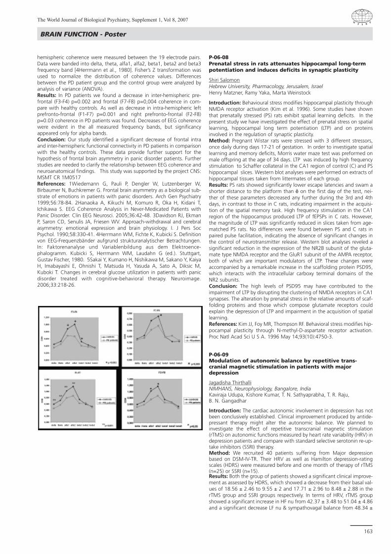
The World Journal of Biological Psychiatry, Supplement 1, Vol 8, 2007
163
BRAIN FUNCTION - Poster
hemispheric coherence were measured between the 19 electrode pairs.Data were banded into delta, theta, alfa1, alfa2, beta1, beta2 and beta3frequency band [4Herrmann et al., 1980]. Fisher’s Z transformation wasused to normalize the distribution of coherence values. Differencesbetween the PD patient group and the control group were analyzed byanalysis of variance (ANOVA).Results: In PD patients we found a decrease in inter-hemispheric pre-frontal (F3-F4) p=0.002 and frontal (F7-F8) p=0,004 coherence in com-pare with healthy controls. As well as decrease in intra-hemispheric leftprefronto-frontal (F1-F7) p=0.001 and right prefronto-frontal (F2-F8)p=0.03 coherence in PD patients was found. Decreases of EEG coherencewere evident in the all measured frequency bands, but significancyappeared only for alpha bands.Conclusion: Our study identified a significant decrease of frontal intraand inter-hemispheric functional connectivity in PD patients in comparisonwith the healthy controls. These data provide further support for thehypothesis of frontal brain asymmetry in panic disorder patients. Furtherstudies are needed to clarify the relationship between EEG coherence andneuroanatomical findings. This study was supported by the project CNS:MSMT CR 1M0517References: 1Wiedemann G, Pauli P, Dengler W, Lutzenberger W,Birbaumer N, Buchkremer G. Frontal brain asymmetry as a biological sub-strate of emotions in patients with panic disorders. Arch Gen Psychiatry1999;56:78-84. 2Hanaoka A, Kikuchi M, Komuro R, Oka H, Kidani T,Ichikawa S. EEG Coherence Analysis in Never-Medicated Patients withPanic Disorder. Clin EEG Neurosci. 2005;36:42-48. 3Davidson RJ, EkmanP, Saron CD, Senulis JA, Friesen WV. Approach-withdrawal and cerebralasymmetry: emotional expression and brain physiology. I. J Pers SocPsychol. 1990;58:330-41. 4Herrmann WM, Fichte K, Kubicki S. Definitionvon EEG-Frequenzbänder aufgrund strukturanalytischer Betrachtungen.In: Faktorenanalyse und Variablenbildung aus dem Elektroence-phalogramm. Kubicki S, Herrmann WM, Laudahn G (ed.). Stuttgart,Gustav Fischer, 1980. 5Sakai Y, Kumano H, Nishikawa M, Sakano Y, KaiyaH, Imabayashi E, Ohnishi T, Matsuda H, Yasuda A, Sato A, Diksic M,Kuboki T. Changes in cerebral glucose utilization in patients with panicdisorder treated with cognitive-behavioral therapy. Neuroimage.2006;33:218-26.
P-06-08Prenatal stress in rats attenuates hippocampal long-termpotentiation and induces deficits in synaptic plasticity
Shiri SalomonHebrew University, Pharmacology, Jerusalem, IsraelHenry Matzner, Ramy Yaka, Marta Weinstock
Introduction: Behavioural stress modifies hippocampal plasticity throughNMDA receptor activation (Kim et al. 1996). Some studies have shownthat prenatally stressed (PS) rats exhibit spatial learning deficits. In thepresent study we have investigated the effect of prenatal stress on spatiallearning, hippocampal long term potentiation (LTP) and on proteinsinvolved in the regulation of synaptic plasticity. Method: Pregnant Wistar rats were stressed with 3 different stressors,once daily during days 17-21 of gestation. In order to investigate spatiallearning and memory deficits, Morris water maze test was preformed onmale offspring at the age of 34 days. LTP was induced by high frequencystimulation to Schaffer collateral in the CA1 region of control (C) and PShippocampal slices. Western blot analyses were performed on extracts ofhippocampal tissues taken from littermates of each group.Results: PS rats showed significantly lower escape latencies and swam ashorter distance to the platform than ë on the first day of the test, nei-ther of these parameters decreased any further during the 3rd and 4thdays, in contrast to those in C rats, indicating impairment in the acquisi-tion of the spatial memory task. High frequency stimulation in the CA1region of the hippocampus produced LTP of fEPSPs in C rats. However,the magnitude of LTP was significantly reduced in slices taken from age-matched PS rats. No differences were found between PS and C rats inpaired pulse facilitation, indicating the absence of significant changes inthe control of neurotransmitter release. Western blot analyses reveled asignificant reduction in the expression of the NR2B subunit of the gluta-mate type NMDA receptor and the GluR1 subunit of the AMPA receptor,both of which are important modulators of LTP. These changes wereaccompanied by a remarkable increase in the scaffolding protein PSD95,which interacts with the intracellular carboxy terminal domains of theNR2 subunits.Conclusion: The high levels of PSD95 may have contributed to theimpairment of LTP by disrupting the clustering of NMDA receptors in CA1synapses. The alteration by prenatal stress in the relative amounts of scaf-folding proteins and those which compose glutamate receptors couldexplain the depression of LTP and impairment in the acquisition of spatiallearning.References: Kim JJ, Foy MR, Thompson RF. Behavioral stress modifies hip-pocampal plasticity through N-methyl-D-aspartate receptor activation.Proc Natl Acad Sci U S A. 1996 May 14;93(10):4750-3.
P-06-09Modulation of autonomic balance by repetitive trans-cranial magnetic stimulation in patients with majordepression
Jagadisha ThirthalliNIMHANS, Neurophysiology, Bangalore, IndiaKaviraja Udupa, Kishore Kumar, T. N. Sathyaprabha, T. R. Raju, B. N. Gangadhar
Introduction: The cardiac autonomic involvement in depression has notbeen conclusively established. Clinical improvement produced by antide-pressant therapy might alter the autonomic balance. We planned toinvestigate the effect of repetitive transcranial magnetic stimulation(rTMS) on autonomic functions measured by heart rate variability (HRV) indepression patients and compare with standard selective serotonin re-up-take inhibitors (SSRI) therapy.Method: We recruited 40 patients suffering from Major depressionbased on DSM-IV-TR. Their HRV as well as Hamilton depression-ratingscales (HDRS) were measured before and one month of therapy of rTMS(n=25) or SSRI (n=15).Results: Both the group of patients showed a significant clinical improve-ment as assessed by HDRS, which showed a decrease from their basal val-ues of 18.56 ± 2.46 to 9.55 ± 2 and 17.71 ± 2.96 to 8.48 ± 2.88 in therTMS group and SSRI groups respectively. In terms of HRV, rTMS groupshowed a significant increase in HF nu from 42.37 ± 3.48 to 51.04 ± 4.86and a significant decrease LF nu & sympathovagal balance from 48.34 ±

The World Journal of Biological Psychiatry, Supplement 1, Vol 8, 2007
164
3.75 to 40.63 ± 4.04 & 1.32 ± 0.19 to 1.05 ± 0.25 respectively. There wasno statistically significant difference in SSRI group in HRV parameters. Conclusion: Though rTMS and SSRI are effective antidepressants, theireffects on HRV were different. rTMS modulated the cardiac autonomictone towards increasing the parasympathetic activity and decreasing thesympathetic activity whereas SSRI did not modulate the neurocardiac reg-ulation. Further studies with large sample size, biochemical and imagingstudies would throw more light into the mechanism of such modulation.
P-06-10EEG responses to prefrontal low-intensity TMS
Seppo KähkönenHelsinki Univ. Centr. Hosp., BioMag Laboratory, FinlandPetri Savolainen, Juha Heiskala, Soile Komssi
Introduction: Left prefrontal TMS has been shown to have antidepres-sant effects. However, little is known about the effects of prefrontal TMSon EEG activity. TMS combined with simultaneous EEG recording allowsone to investigate directly transient neuronal responses. Method: Seven healthy subjects participated in the study. The left gyrusmedialis frontalis (BA 46) was identified and selected as stimulation sitewith image-guided coil navigation system from individual MRI (eximiaNBS, Nexstim Ltd., Helsinki, Finland). Magnetic pulses were delivered witha figure-of-eight coil with a loop diameter of 70 mm at the inter-stimulusinterval 3.3 s. Ten stimulus intensities (20-120% of the motor threshold;MT) were used in a random order. At each intensity, 100 pulses weredelivered. The EEG was recorded with 60 scalp electrodes referred to theforehead. The global mean field amplitude (GMFA) was used as an indi-cator of the strength of the evoked field. Linear regression analysis wasused to investigate the intensity dependence function. Results: Excitation threshold of prefrontal cortex was 40% of MT as esti-mated with TMS-EEG. GMFAs at the peaks were linearly related to thestimulus intensity i.e., TMS at higher intensity produced stronger neuronalactivity measured with EEG.Conclusion: Prefrontal TMS combined with EEG may be potential a newresearch tool for investigation of neuronal function of the prefrontal cor-tex for psychiatric disorders.
P-06-11Repetitive transcranial magnetic stimulation (rTMS) inMajor Depressive Episode during pregnancy
Monika KlirovaPsychiatric Centre, 23, Prague 8, Czech RepublicMilan Kopecek, Tomas Novak, Vera Strunzova, Pavel Mohr
Introduction: For women diagnosed with reccurrent depressive disorder,pregnancy poses a major treatment challenge. Apart from antidepres-sants, the most commonly used biological therapeutical method is ECT.We believe that similar efficacy can be achieved using rTMS as a saferoption with substantially less side effects. Method: Two pregnant females, with history of Reccurrent depressivedisorder and current symptoms of depression requiring treatment. Thefirst patient has been treated with escitalopram 15 mg p.d. + venlafax-ine XR 225 mg p.d. She started rTMS at the 16th week of pregnancy.Parameter settings were: 15 sessions, 20 Hz, left DLPFC, MT 100%, (train2.5s, intertrain 30 s), 2000 pulses/session. Depressive symptoms wereassessed using MADRS. The second patient started stimulation at the 31stweek of pregnancy. She did not respond to the previous therapy with esc-italopram 40 mg for 5 weeks with increase up to 60 mg p.d. for the fol-lowing 2 weeks. She was switched to venlafaxine 225 mg p.d.at the timeof rTMS initiation. Parameter settings: 15 sessions, 1 Hz, right DLPFC, MT100% (five 60 s trains with 60 sec intertrains, 300 pulses/session). Thesettings were adopted from the Fitzgerald’s study. This protocol was chosendue to the low number of magnetic pulses during rTMS. Depressive symp-toms were assessed using BDI, BAI.
Results: In both patients, a significant treatment response to rTMS treat-ment was observed. Baseline MADRS total score of the first patient was33. The patient achieved remission after 3 weeks of rTMS (MADRS scorewas 2). The remission was sustained for the remaining duration of preg-nancy. The delivery was uncomplicated, the baby was healthy, breastfed.Baseline BDI total score of the second patient was 29, BAI score 19. TheBDI and BAI scores decreased by 59 % and 58%, respectively after 3 weeks of rTMS treatment. The patient continued with venlafaxine XR225 mg p.d. for the remaining duration of pregnancy. The delivery waspremature (36 week), the newborn being more irritated during the firstweek; however, the baby fully recovered and is considered healthy.Conclusion: No side or adverse effects associated with rTMS wereobserved. Although no general recommendations can be drawn based onour results, the case reports suggest that rTMS should be considered asan effective and safe treatment option for depression in pregnancy. TheProject was supported by the CNPS and by the WFSBP. References: Fitzgerald P.B., rTMS in the Treatment of Depression, ArchGen Psychiatry. 2003;60:1002-1008.
P-06-12Deep brain stimulation in Huntington’s disease
Etienne HollGraz, SwitzerlandA.K. Hödl, R.M. Bonelli
Introduction: Huntington’s Disease (HD) is a neurodegenerative disorderleading to psychiatric and neurologic malfunction and progressivedementia. Prominent neurologic symptoms are chorea, dystonia, andparkinsonism in advanced stages. As shown in literature altered activity ofthe globus pallidus externus (GPe) is responsible for these signs [1,2]. Onlytransient improvement of choreatic symptoms are known with neurolep-tic medication until now.Method: To improve chorea and dystonia not enhancable with neurolep-tic medication or benzodiazepines in patients with HD different parts ofthe basal ganglia can be treated with deep brain stimulation (DBS).Results: In 2006 Temel et al stimulated the GPe with motor and cogni-tive improvement in a transgenic rat model [3]. In case reports alleviationof cognition differs after stimulation of the globus pallidus internus (GPi).Hebb et al stimulated the GPi in one patient and described an alleviationof HD-associated choreathetosis [4]. In 2004 Moro et al described adecrease of chorea and dystonia by DBS of GPi in one patient. Outcomeof cognition was not mentioned [5].Conclusion: GPe seems to be the better position for DBS due to the bettercognition performance but we need to demonstrate this in patients withHD. DBS in the basal ganglia will be an exclusive treatment option forpatients without effect of medication in advanced HD.References: [1] L. Ayalon, R. Doron, I. Weiner and D. Joel, Ameliorationof behavioral deficits in a rat model of Huntington’s disease by an excito-toxic lesion to the globus pallidus, Exp. Neurol. 186 (2004), pp. 46-58. [2 ] A. Reiner, Can lesions of GPe correct HD deficits?, Exp. Neurol. 186(2004), pp. 1-5. [3] Temel Y, Cao C, Vlamings R, et al. Motor and cogni-tive improvement by deep brain stimulation in a transgenic rat model ofHuntington’s disease. Neurosci Lett.2006;406(1-2):138-41 [4] Hebb MO,Garcia R, Gaudet P, Mendez IM. Bilateral stimulation of the globus pal-lidus internus to treat choreathetosis in Huntington’s disease: technicalcase report. Neurosurgery. 2006 Feb;58(2) [5] Moro E, Lang AE, StrafellaAP, Poon YY, Arango PM, Dagher A, Hutchison WD, Lozano AM. Bilateralglobus pallidus stimulation for Huntington’s disease. Ann Neurol. 2005Jul;58(1):168
BRAIN FUNCTION - Poster

The World Journal of Biological Psychiatry, Supplement 1, Vol 8, 2007
165
BRAIN FUNCTION - Poster
P-06-13Electroconvulsive stimulations prevent stress-induced morphological changes in the hippocampus
Ida HagemanRighospitalet, Lab of Neuropsychiatry, Copenhagen, DenmarkMarianne Nielsen, Gitta Wörtwein, Nils Diemer, Martin Balslev Jorgensen
Introduction: The first episode of depression is often precipitated bystress. Sustained stress can have numerous adverse effects on the brain.Animal models of chronic stress, such as 21 days of restraint stress, showthat chronic stress alters neuronal morphology in the hippocampus.Stressful conditions can induce retraction of apical dendrites and decreaseneurogenesis. These changes find their analogue in humans with depres-sive disorder and hippocampal shrinkage. In animals, stress-induced mor-phological changes can be reverted by antidepressant drugs.Electroconvulsive treatment is one of the most effective antidepressanttreatments. Thus, the aim of the present study was to investigate whetherECS could influence the length and branching of the hippocampal den-dritic tree in normal as well as stressed rats. Method: 24 male Sprague-Dawley rats were randomly assigned to thefollowing 4 groups: 1) 6 rats were subjected to 6 hours of restraint stressdaily for 21 days, 2) 6 rats underwent the same stress paradigm plus ECSthree times a week, 3) 6 rats received 3 weekly ECSs and 4) 6 rats wereleft undisturbed in their cages except for daily handling. At the end of thestudy brains were fixed and the dorsal hippocampus was cut into 100 µmsections and accordingly Golgi-impregnated. After processing well-impregnated pyramidal CA3 neurons were drawn using a camera lucidadevice. The number of dendritic branchpoints and the total length of thedendritic tree were measured. Data was subjected to one-way analysis ofvariance followed by Bonferonis post-hoc comparisons. Results: Stress caused atrophy of the dendritic tree by significantly reduc-ing the number of branchpoints and total length of the apical dendritictree. Concomitant treatment with ECS reverted this effect. ECS had noeffect on normal (non-stressed) rats.Conclusion: The stress-induced structural remodelling of the dendritictree was prevented by ECS. The mechanisms underlying the antidepres-sant effects of electroconvulsive treatment is not fully elucidated. As sug-gested by this study ECS might act by reverting atrophic processes. References: Watanabe Y, Gould E, McEwen BS (1992): Stress inducesatrophy of apical dendrites of hippocampal CA3 pyramidal neurons. BrainRes 588: 341-344. Post RM (1992). Transduction of psychosocial stressinto the neurobiology of recurrent affective disorder. Am J Psychiatry 149:999-1010. Videbech P, Ravnkilde B (2004). Hippocampal volume anddepression: A meta-analysis of MRI studies. Am J Psychiatry 161: 1957-1966.

The World Journal of Biological Psychiatry, Supplement 1, Vol 8, 2007
166
P-07Genetics
T9 Genetics
P-07-01Genetic variants of SERT and DRD2 and executive functionsin impulsive/aggressive borderline personality disorder
Maria Leonor BustamanteSantiago, ChileM. Jimenez, F. Galleguillos, L. Pumarino, N. Roa, P. Iturra, A. Solari, J.Villarroel, S. Jerez, H. Silva
Introduction: Variation on genes involved in monoaminergic pathwayscould influence individual differences on executive functioning. Indeed,association between the performance on different neuropsychologicaltasks and some alleles of COMT, DRD2 and DRD3 has been demonstratedin individuals with and without psychopathology. Our study is focused onindividuals with Borderline Personality Disorder (BPD) who had significantlevels of impulsivity/aggressiveness. We attempted to establish an associ-ation between their executive planning ability and variants of two genesthat have been associated with impulsive/aggressive behavior, the sero-tonin transporter (SERT) gene and the dopamine D2 receptor (DRD2)gene. Method: 37 BPD patients, classified as markedly impulsive/aggressiveaccording to the OAS-M interview were evaluated using the Tower ofLondon Test. They were genotyped using PCR and digestion with restric-tion enzymes. The polymorphisms considered in this study were the5HTTLPR polymorphism of the SERT gene and the TaqI polymorphism ofthe DRD2 gene.Results: There were no significant differences between the differentgenotype groups on their performance on the Tower of London Test. Conclusion: The studied polymorphisms do not influence the executiveplanning ability in impulsive/aggressive individuals. Although other meas-ures of executive functioning are needed, the present results suggest thatthe association between these variants and impulsivity/aggressivenessmay be mediated by factors other than alterations of the executive func-tions. Acknowledgments: This work was supported by FONDECYT(Project 1030305) and by the University of Chile Clinical Hospital.References: 1. Rosa et al., Am J Psychiatry (2004) 161:1110-2 2. Rybakowski et al., J Neural Transm (2005) 112:1575-82 3. Szekeres etal., Am J Med Genet B (2004) 124B :1-5 4. Sakado et al., Am J MedGenet B (2003) 121B:71-5
P-07-02Combined genetic effect of the serotonin transporter andHTR1A genes in clinical outcome of mirtazapine
Min-Soo LeeKorea University, Psychiatry, Seoul, Republic of KoreaRhee-Hun KANG, Myoung-Jin CHOI, Yoo-Jong JEONG
Introduction: This study was to analyze genetic variation at the 5-HTR1Agene and its possible combined effect with the 5-HTTLPR polymorphismat the serotonin transporter gene on clinical outcome in a major depres-sive disorder treated with mirtazapine.Method: Mirtazapine was administered for 8 weeks to the 101 patientswho completed this study. All subjects were examined using theStructured Clinical Interview for DSM-IV. The severity of depression wasassessed using the 21-item Hamilton Depression Rating (HAM-D-21)scale. Only subjects with a minimum score of 18 on the HAM-D-21 scaleentered the study. Prior to study entry, a 2-weeks-wash-out was per-formed. Their clinical symptoms were evaluated with the HAM-D-21scales at baseline and after 1, 2, 4 and 8 weeks of treatment.Results: A combined genetic effect of serotonin transporter and 5-HTR1A genes was found to affect the clinical outcome of MDD.Homozyous patients carrying the risk genotype combination (SS/GG)present a lower decreased HAMD score over 8 weeks (Time effectF=33.674, p<0.000; Genotype effect F=10.228, p=0.001; Genotype-time interaction F=0.139, p=0.937). There is no significant interactionbetween the genotype combination and time. These results may suggest
that the risk genotype combination(SS/GG) is affecting the clinical out-come, but not the pattern of clinical response to mirtazapine over time.Conclusion: Homozyous patients carrying the risk genotype (SS/GG)present a lower decreased HAMD score over 8 weeks. It means that therisk genotype combination is affecting the clinical outcome to mirtazapine.References: Arias et al., Evidence for a combined genetic effect of the 5-HT1A receptor and serotonin transporter genes in the clinical outcomeof major depressive patients treated with citalopram. Journal ofPsychopharmacology 19(2) (2005) 166-172
P-07-03Abnormalities in behaviour and reduction in expression ofspecific hippocampal genes by prenatal stress in femaleoffspring are reversed by neonatal handling
Yaarit Nachum-BialaHebrew University, Pharmacy School, Room 609, Jerusalem, IsraelYoel Bogush, Shiri Salomon, Michal Linial, Marta Weinstock
Introduction: Prenatally stressed (PS) rats exhibit hyperanxiety, depres-sive-like behaviour and learning deficits(1,2). The present study investigatedthe effect of neonatal handling (H)(1) from day 1-10 of age, on the devel-opment of the above mentioned abnormalities in behaviour and on hip-pocampal gene expression in female offspring.Method: Pregnant Wistar rats were stressed daily during days 17-22 ofgestation with three different types of stressors, one in each day. Half ofthe PS and control (C) offspring were handled daily for 3 minutes.Behavioural tests consisted of evaluation of spatial memory in the Morriswater maze, hyperanxiety and depressive-like behaviour in the elevatedplus maze and the forced swimming tests, respectively. Gene analysis wasperformed on the “whole rat” DNA AffyChip on 4 groups of 3 femaleseach. RT-PCR and Western-blot analysis verified the results in the genearray.Results: In the behavioral tests we found that in female offspring Hreversed the hyperanxiety and depressive-like behaviour in PS rats but hasno effect on memory. Furthermore, we found in the gene analysis thatout of 9000 valid genes, 600 were differently expressed between PS and C.out of those, 278 were up-regulated and 301 were down-regulated in PScompared with C. From those that were down-regulated 93 genes wererestored to normal by H. These 93 genes can be classified into 3 main bio-logical processes, all presynaptic nerve terminal-related that are active insynaptogenesis, synaptic vesicle transport, and neurotransmitter release.RT-PCR validated the results obtain from the genearray analysis, howevernot all the changes in the proteins match the profile of those seen at themRNA level.Conclusion: The study demonstrates that the alterations in gene expres-sion by prenatal stress are related to impairment of the formation of newsynapses and neurotransmitter release. Neonatal handling enables recov-ery of new synaptic sites and the building of more effective synapse con-nectivity that is probably very local specific. The synaptogenesis and neu-rogenesis may contribute to the normalization of behaviour seen after H. References: 1. Weinstock M. Alternation induced by gestational stress inbrain morphology and behaviour of the offspring. 2001. Prog Neurobiol.65:427-451 2. Wakshlak A, Weinstock M. Neonatal handling reversesbehavioral abnormalities induced in rats by prenatal stress. 1990. PhysiolBehav. 48:289-92.
P-07-04Trend for association of homozygous variants of DAT1 withnovelty seeking in healthy individuals
Alexandra SchosserUniv. Hospital for Psychiatry, Dep. of General Psychiatry, Vienna, AustriaKaroline Fuchs, Theresa Scharl, Monika Schloegelhofer, Jochen Kindler,Friedrich Leisch, Siegfried Kasper, Werner Sieghart, Harald N. Aschauer
Introduction: Cloninger (1993) hypothesized that the 4 heritable dimen-sions of personality are harm avoidance (HA), novelty seeking (NS),reward dependence (RD) and persistence (PS), and NS is theoreticallyrelated to dopaminergic, HA to serotonergic and RD to noradrenergicactivity. After not finding an association of DRD4 and DRD2 receptorgenes with personality in healthy individuals (Gebhardt et al. 2000), wenow focused on DRD3 and DAT1.
GENETICS - Poster

The World Journal of Biological Psychiatry, Supplement 1, Vol 8, 2007
167
GENETICS - Poster
Method: We examined 89 (59 female and 30 male, mean age of 32.57)normal volunteers using Cloninger’s “Temperament and CharacterInventory” (TCI). The normal volunteers as well as their family historywere free of any mental illness or psychiatric disease in first- and second-degree relatives. All individual donated 20 cmÑ blood by venipuncture.Genomic DNA was extracted from lymphocytes using standard protocols.Genotyping of the Ser9Gly polymorphism in exon 1 of the DRD3 genewas performed using PCR-RFLP, whereas genotyping of the DAT1 repeatpolymorphisms was performed using PCR amplification following elec-trophoresis in an 8% acrylamide gel with a set of size markers.Associations between TCI scores and DRD3 and DAT1 genotypes, and sexwere tested by Kruskal-Wallis Rank-Sum tests. Results: We found significant association between sex and HA(p=0.017), with women showing higher scores of HA. No association wasfound between sex and any other personality dimension. Testing dimen-sions of the TCI for association with homozygous or heterozygous genevariants, we found a trend for association of the homozygous variants(5,5 and 6,6) of the DAT1 with NS (p=0.054). No such association wasfound for the other personality dimensions, and for both homozygousand heterozygous variants of DRD3.Conclusion: In conclusion, we found a trend for association of thehomozygous variants of the DAT1 repeat polymorphism and personalitydimension of novelty seeking. However, correction for multiple testingwas not performed.References: Cloninger CR, Svrakic DM, Przybeck TR (1993) A psychobio-logical model of temperament and character. Arch Gen Psychiatry50:975-990. Gebhardt Ch, Leisch F, Schüssler P, Fuchs K, Stompe T,Sieghart W, Hornik K, Kasper S, Aschauer HN (2000) Non-association ofdopamine D4 and D2 receptor genes with personality in healthy individ-uals. Psychiatr Genet 10:131-137.
P-07-05Enviromental and genetic factors: A study on Slovenianpopulation of suicide victims
Peter PregeljUn.Psychiatric Hosp.Ljubljana, KOKP, SloveniaMartina Tomori, Galina Pungercic, Anja Videtic, Robert Dolnicar,Radovan Komel, Tomaz Zupanc, Joze Balazic
Introduction: Several studies have been carried out to investigate howgenetic variants in one side and environmental factors on the other sitemay influence susceptibility to suicide.Method: In our study dual approach was used, we analyzed 5-HTTLPRand VNTR polymorphisms in 35 suicide victims and 233 controls in aSlovenian population to find a possible association of the polymorphismsand suicidal behavior. On the other hand psychological autopsy was usedto evaluate environmental factors preceding suicide.Results: No statistically important differences between genotypes of con-trols and suicide group, as well as no differences in allele distribution werefound. Haplotype frequency analysis showed no excess of particular hap-lotypes between the two groups. Our study showed no association ofserotonin transporter polymorphisms and suicide. We observed on theother hand that more than 80% of all suicide victims had major stressorsreported in a month before suicide. Conclusion: We have not observed differences in genetic variationsbetween subgroups in the observed genes on the contrary environmentalfactors were observed.
P-07-06Association between the serotonin transporter gene andpersonality traits in borderline personality disorderpatients evaluated with zuckerman-kuhlman personalityquestionnaire (zkpq)
Juan Carlos PascualSta. Creu i Sant Pau Hospital, Psychiatry, Barcelona, SpainJoaquim Soler, Thais Tiana, Montserrat Baiget, Anna Cortes, MontserratGoma, Enrique Alvarez, Victor Perez
Introduction: Although there is general consensus that BorderlinePersonality Disorder (BPD) has a multifactorial etiology including genetic
and environmental etiology, the specific genetic influence have not beenextensively investigated (1). Some studies suggest that the serotonintransporter gene (5-HTT) plays an important role in suicide, impulsivity oraffective instability suggesting it as a candidate gene for BPD (2). Shortallele (S) of the 5-HTTLPR polymorphism in the promoter region hasshown to be associated with impulsivity, aggressive behaviour, anxietyand neuroticism. Of the VNTR polymorphism in intron 2, BPD patientsshowed higher frequencies of the allele with the 10 repeat (3). The aimof this study was to determine the association between 5-HTTLPR andVNTR polymorphism of 5-HTT and personality traits in borderline person-ality disorder.Method: A total of 65 BPD patients diagnosed by means of semi-struc-tured interviews SCID-II and DIB-R were included. Two common polymor-phisms of 5-HTT were genotyped: the 5-HTTLPR in the promoter regionand VNTR in intron 2. Personality traits were assessed by the Zuckerman-Kuhlman Personality Questionnaire (ZKPQ). Results: Patients with L allele (L/S or L/L) in the 5-HTTLPR polymorphismshowed lower scores in the subscale of Liking Parties and Friends. Patientswith the allele with 10 repeat of the VNTR polymorphism, showed lowerscores in Impulsivity, Sensation Seeking and in the subscale Liking ofParties and Friends.Conclusion: The results suggest an association between the serotonintransporter gene and some personality traits in BPD. This gene may playa role in the aetiology of borderline personality disorder. References: 1.- Skodol A, Siever L, Livesley W, Gunderson J. Pfohl Bruce,Widiger T. The Borderline Diagnosis II. Biology, Genetics, and ClinicalCourse. Society of Biological Psychiatry. 2002; 51: 951-963. 2.- Lesch KP,Bengel D, Jeils A, Sabol SZ, Greenberg BD, Petri y cols. Association of anx-iety- related traits with a polymorphism in the serotonin transporter generegulatory region. Science,1996; 27:1527-31. 3.- Ni X, Chan K, Bulgin N,Sicard T, Bismil R, McMain S, Kennedy J. Association between serotonintransporter gene and borderline personality disorder. Journal ofPsychiatric Research, 2006; 40: 448-453.
P-07-07Attention deficit disorder: Genetic and possible attach-ment style contributions
Maria-Eugenia MonetaUniversity of Chile, Faculty of Medicine, Santiago, ChileFrancisco Rothhammer, Daniela Camponovo, Hugo Henriquez
Introduction: The present paper adds evidence to the discussion of thehypothesis that genetic predisposition and environmental factors influ-ence psychological development. We studied a group of children of highsocioeconomic status in Santiago de Chile diagnosed with ADHD from agenetic and an attachment style point of view to evaluate to whichextend both factors may contribute to ADHD disorder.Method: 20 children between 6 and 12 years old where selected froma group with learning dishabilities from an upper class community inSantiago. The presence or absence of attentional deficit with hyperactiv-ity disorder (ADHD) was assessed through DSMIV scale and ChildBehavioural Check List (Achenbach, 1991). Attachment security level wasobtained applying the Security Scale from Kerns, Keplan and Cole (1996)Results: Preliminary data show that a majority (82%) of the childrenexperimented a secure attachment. Nevertheless ADHD was diagnosedin 40 % of children with learning dishabilities. Genetic studies revealed atendency of ADHD to be associated with DAT 1.10 and DRD4.7 polymor-phisms.Conclusion: Evidences obtained from this relatively small sample indi-cates the necessity of a multidisciplinary approach to study the interrela-tion between environmental factors, genetic contribution and learningdishability problems during development leading to ADHD. With regardto research on child health we suggest that knowledge about specificgenetic risks factors may help substantially to clarify environmental con-tributions such as attachment style.

The World Journal of Biological Psychiatry, Supplement 1, Vol 8, 2007
168
P-07-08A suggested endophenotype for suicide described in suicide attempters
Dolores SaizGregorio Maranon Hospital, Psychiatry, Madrid, SpainJeronimo Saiz-Ruiz
Introduction: Suicide is one of the most relevant issues in modern psy-chiatry. It is one of the first causes of death and years of life lost in youngadults. The description of endophenotypes opens a new line of researchwith good results in other mental disorders like schizophrenia.Method: The aim of this study was to analyze different features amongsuicide attempters with or without family history of suicide (includingattempts and or completed suicide), in order to define a phenotype thatcould be related with the biological substrate of suicide. 534 suicideattempters were recruited at the Emergency Room of a general hospital.They were interviewed within 24 hours of the attempted. We performeda univariate analysis with contingency tables and calculated statistic sig-nificance with Chi-square tests and Fisher exact tests. We analyzed eachvariable separately and compare the values in patients with family historyof suicide (attempts and/or completed suicide) and in those without theseantecedents. Results: The patients with family history of suicidal behaviour shows thefollowing features (p<0,001): method is overdose or cuts, patients are nottreated with mood stabilizers and they donãt have family history of schiz-ophrenia, affective disorders or other mental disorders (including unspec-ified mental diagnosis).Conclusion: One endophenotype is suggested, in relation with familyhistory of suicide. This phenotype is mainly defined by variables relatedinversely to treatment and mental disease in family. This work support,the current hypothesis of suicide genetic transmission independently fromthe heredity of mental diseases associated with this conduct. References: Gottesman II, Gould TD. The endophenotype concept inpsychiatry: etymology and strategic intentions. Am J Psychiatry 2003;160(4):636-645. Courtet P, Jollant F, Castelnau D, Buresi C, Malafosse A.Suicidal behavior: relationship between phenotype and serotonergicgenotype. Am J Med Genet C Semin Med Genet 2005; 133(1):25-33.Turecki G. Dissecting the suicide phenotype: the role of impulsive-aggres-sive behaviours. J Psychiatry Neurosci 2005; 30(6):398-408.
P-07-09Cytogenetic Regions of Interest (CROIs) analyzed withSingle Nucleotide Polymorphisms (SNP) microarrays inChilean children with autism spectrum disorders
Mario ValdiviaUniversidad de Concepcion, Psiquiatria y Salud Mental, ChileGabriela Zegers, Gian Carlo De Ferrari, Luis San Martin, AndrewSingleton, Mar Matarin, John Hardy
Introduction: Twin and family studies have provided compelling evidencefor a strong genetic component among autistic individuals. Multiplegenome-wide screens have found evidence for linkage to autism in sev-eral chromosomes. Likewise, cytogenetic abnormalities related to theautistic phenotype have been reported in chromosomal regions overlap-ping known loci of significant linkage. Method: We have begun to explore a sample of Chilean autistic childrenfor cytogenetical abnormalities with the aid of beadchips microarrays(Illumina’s Infinium-II Sentrix BeadChips) containing > 300.000 singlenucleotide polymorphisms (SNPs), which are increasingly used for whole-genome genotyping.Results: Our results reveal several cytogenetical abnormalities in five outof eight studied autistic children, including small duplications (15,000 -140,000 bp) in chromosomes 1, 2, 10 and 12, as well a deletion in chro-mosome 9 (281,000 bp).Conclusion: These preliminary results support the polygenic nature ofthis neurodevelopmental disorder, and stress the need for further researchwith larger samples of autistic children and their families.
P-07-10Psychiatric features associated with Fragile X MentalRetardation 1 (FMR1) gene premutation condition
Cary KoganUniversity of Ottawa, School of Psychology, CanadaKim Cornish
Introduction: Fragile X Syndrome (FXS) is a pervasive developmental dis-order arising from the silencing of the FMR1 gene due to trinucleotiderepeat expansion. Affected, full mutation (FM) individuals, display a spe-cific pattern of cognitive deficit concurrent with psychiatric symptomsthat include mood and anxiety disorders. In contrast to the FM condition,little is known about psychiatric features associated with premutation(PM) status. The paucity of data stems from a longstanding notion thatPM individuals are free of phenotypic effects because FMR1 gene expres-sion is relatively intact. More recent studies are beginning to characterizethe cognitive phenotype of PM individuals. The goal of the present studyis to extend this knowledge base to investigate potential psychiatric fea-tures associated with PM status. Method: As part of a larger study of psychiatric features of PM individuals,we administered numerous measures of psychiatric functioning to agroup of thirty-one PM males aged 18 to 69 years as well as to 56 un-affected comparison males matched on age and IQ. A subset of thesemeasures focusing on mood and anxiety were included in the presentanalysis. Specifically, self-report measures included the Liebowitz SocialAnxiety Scale (LSAS), the Rosenberg Self-Esteem scale, and the HospitalAnxiety and Depression Scale (HADS). T tests for independent meanswere conducted to compare mean scores on measures and their subscaleswhere appropriate.Results: PM males demonstrated significantly elevated scores on theLSAS - Fear/Anxiety subscale but neither the Avoidance subscale nor thetotal score for this measure were significantly elevated as compared tocomparison males. Further analysis revealed that 24% of the PM malesexceeded the cut-off score on the LSAS for meeting diagnostic criteria forSocial Phobia as compared to 7.5% of the comparison group.Interestingly, on a more general measure of anxiety, the HADS - Anxietysubscale, no significant differences were observed. Similarly, the HADS -Depression subscale revealed no significant differences between the twogroups. On a measure of self-esteem, PM males generally scored lowerthan comparison males, indicating lower self-esteem in this group.Conclusion: Measures of mood and anxiety revealed that PM status con-fers greater risk of experiencing fear in social contexts. Interestingly, PMmales were not more likely to avoid social situations despite their elevatedfear of social situations. These results are consistent with other studiesthat describe social phobia as a central psychiatric condition common tofemales with full mutation FXS.
P-07-11Personality characteristics and 5-HTTLPR in patients withchronic daily headache
Chung Tai LeeUijongbu St. Mary’s Hospital, Department of Psychiatry, UijongbuGyeonggido, Republic of KoreaHae Kook Lee, Jeong Wook Park, Yong Sil Kweon
Introduction: Chronic daily headache refers to a group of non-paroxys-mal daily or near-daily headaches with peculiar characteristics, which arehighly prevalent in population of psychiatric clinics and primary clinics.Serotonergic drug such as SSRI(paroxetin etc.) has been implicated to beeffective on chronic daily headache. Also, the relationship between sero-tonergic dysregulation and personality chraracteristics has beendescribed. The authors carried out this study to investigate the associatonbetween chronic daily headache and serotonergic dysregulation by com-parison of 5-HTTLPR & Tridimensional Personality Questionaire(TPQ).Method: Eighty one patients with chronic daily headache by criteria(pro-posed by silberstein et al.,1994) and 100 healthy individuals who did nothave history of psychiatric illness were selected for comparison of 5-HTTL-PR (assessed by PCR based method). Also, 45 patients group and 75 nor-mal controls answered TPQ to evaluated the association between specificcomponent of personality characteristics and chronic daily headache.
GENETICS - Poster

The World Journal of Biological Psychiatry, Supplement 1, Vol 8, 2007
169
GENETICS - Poster
Results: Although statistically significant differences in frequencies ofserotonin transporter gene genotype and allele between patients andcontrols were not found, the analysis showed a trend that frequencies ofshort alleles were higher in patients group. In analysis of TPQ, the noveltyseeking scores were lower and harm avoidance scores were higher inpatients group than normal controls. Conclusion: Although there was limitation in generalization of our result,this result suggested that chronic daily headache was associated withserotonergic abnormalities. Further study with larger sample are need tobetter understand of serotonergic abnormality in patients with chronicdaily headache. References: (1) Stoltenberg SF, et al. (2002) Serotonin transporter pro-moter polymorphism, peripheral indexes of serotonin function, and per-sonality measures in families with alcoholism. Am J Med Genet 114:230-234 ; (2) Park JW, et al. (2004). Serotonin transporter polymorphism andharm avoidance personality in chronic tension-type headache. Headache44:1005-1009
P-07-12Response to methylphenidate according to the 9- and 10-repeat allele of the DAT 1 and DRD 4 gene alleles in adultswith attention deficit hyperactivity disorder
Johanna KrauseOutpatient Clinic Psychiatry, Ottobrunn, Germany
Introduction: Relationships between methylphenidate (MPH) responseand polymorphisms of the dopamine transporter gene (DAT 1) in patientswith ADHD are described, but not with conclusive results (1); for DRD 4 ablunted response of the 7-repeat to MPH has been reported (2). In thisstudy response to MPH was related to the DAT 1 and DRD 4 genotype inadults with ADHD.Method: In 27 adults with ADHD the genotype of the SLC6A3 promotorVNTR polymorphism as well as polymorphisms of DRD 4 were assessed.Patients were categorized as responder or non responder, according toClinical Global Impressions Scale.Results: The two patients with 9-9 genotype of DAT 1 were non respon-ders, no difference in response could be found between 9-10 and 10-10carriers. Two patients were homozygote for the 7 allele of the DRD 4 gene, both were responders. No patients were heterozygote for the 7 allele.Conclusion: The non response to MPH in patients with 9-9 alleles of DAT1 gene is in accordance with earlier results in children (3). We observedno influence of the 7 allele of DRD 4 gene on response, but studies withmore patients are needed.References: 1. Krause J et al. World J Biol Psychiatry 2006; 7: 152-7 2.Hamarman S et al. J Child Adolesc Psychopharmacol 2004; 14: 564-74 3.Stein MA et al. Neuropsychopharmacology 2005; 49: 1874-82
P-07-13The DAT 1 association with ADHD behavior is mediated byreading ability in a general population sample
Kim CornishMcGill University, Neurology/Neurosurgery, Montreal, Canada
Introduction: Attention deficit hyperactivity disorder (ADHD) and read-ing disability (RD) frequently co-occur in the child population and there-fore raise the possibility of shared genetic aetiology. Method: We used a Quantitative Loci Trait (QLT) approach to assess theinvolvement of the dopamine transporter (DAT1) gene polymorphism inmediating reading disability and poor attention in a general populationsample of primary school children aged 6 - 11 years in the U.K. Thepotential confounding effects of IQ and chronological age were alsoinvestigated.Results: We found an independent association between the homozy-gous DAT1 10/10 repeat genotype and RD that was not accounted for bythe level of ADHD symptoms. Conclusion: This findings suggest that the DAT1 gene polymorphismmay influence a common neural mechanism underlying both readingacquisition and ADHD symptoms.
References: Cornish, K., Manly, T., Savage, R., Swanson, J., Morisano, D.,Butler, N., Grant, C., Cross, G., Bentley, L., & Hollis, C.P. (2005).Association of the dopamine transporter (DAT1) 10/10-repeat genotypewith ADHD symptoms and response inhibition in a general populationsample. Molecular Psychiatry, 10, 686-698. Willcutt, E. G., Pennington,B.F., Olson, R.K., Chhabildas, N., Hulslander, J. (2005). Neuropsycho-logical analyses of comorbidity between reading disability and attentiondeficit hyperactivity disorder: in search of the common deficit. DevNeuropsychol., 27(1), 35-78.
P-07-14Association of genetic factors with presence psychiatricdisorder in Malaysian population- (the MMHS study)
Tishya IndranRoyal Adelaide Hospital, AustraliaSaroja Krishnaswamy, Kavitha Subramaniam, Abdul Aziz Jemain, AbdulHamid Abdul Rahman, Vikram Patel
Introduction: This aim of this survey is to determine the association ofgenetic factors with presence of Common Mental Disorders and alsooverall mental illnesses.Method: This is a sub study form a larger survey entitled The MalaysianMental Health Survey (MMHS). Two genetic factors included in the MMHsurvey were presence of family history of mental illness among the sub-jects and delayed parenting or being born for old parents. Presence ofcommon mental disorders (CMD) refers to presence of CIS-R diagnosisalone whereas overall mental disorder refers to presence of CMD and allother diagnoses given by psychiatrists, which includes psychosis.Influences of the genetic factors on presence of Mental illnesses and CMDwere studied using logistic regression modelsResults: Presence of family history of mental illness increased the risk ofCMD and also mental illnesses after adjusting for demographic factors.The subjects born for old fathers also had elevated risk for mental illnessand CMD. When both the genetic factors were placed into one model,family history was found to be a stronger factor. Delayed parenting wasfound to be significant for CMD only and not for overall mental illness.Conclusion: Presence of family history of mental illness in the family andbeing born for aged parents were two important factors associated withpresence of CMD and mental illnesses. The final paper will discuss ondeveloped models and precise findings from the models.
P-07-15Polymorphisms of Dysbindin Gene (DTNBP1) and schizo-phrenia in the korean population
Tae-Youn JunCatholic University of Korea, Dept. of Psychiatry, Seoul, Republic ofKoreaWon-Myong Bahk, Jeong-Ho Chae, Chi-Un Pae, Young-Eun Jung,Byung-Wook Lee, Seung-Kyu Bang, Young Sup Woo
Introduction: The gene encoding the dystrobrevin binding protein(DTNBP1) has been implicated in the pathogenesis of schizophrenia byseveral association studies. The dysbindin gene is located at chromosome6p22.3, one of the most promising susceptibility loci in linkage studies ofschizophrenia. The aim of this study is to determine whether DTNBP1 isassociated with schizophrenia in the Korean population.Method: 452 Korean patients with schizophrenia in accordance withDSM-IV criteria and 442 Korean healthy individuals matched to age andsex participated in this study. We examined two single nucleotide poly-morphisms (p1635, p1325) using Pyrosequencing system, and examinedallele, genotype and haplotype association with schizophrenia. The resultswere analyzed by chi-square test and Fisher’s exact test. Results: There were no significant differences in genotype and allelic fre-quencies of SNP p1635 between cases and controls (x2 = 1.21, df=2,p=0.655, x2 = 0.575, df=1, p=0.448). However, SNP p1325 reached sig-nificance. The frequency of the rare allele was significantly higher in casescompared with controls (x2 = 6.35, df=2, p=0.042, x2 =4.41, df=1,p=0.036). Two SNP haplotype AC, GC were not associated with schizo-phrenia, but SNP haplotype AT was significantly in excess in cases com-pared with controls. (x2 = 4.41, df=1, p=0.036)
The World Journal of Biological Psychiatry, Supplement 1, Vol 8, 2007
170
Conclusion: These results provide the support for DTNBP1 as a suscepti-bility gene for schizophrenia in the Korean population. In the future, fur-ther studies should be needed to confirm the relationship between geneticvariation in DTNBP1 and the phenotype of schizophrenia.References: Williams NM, Preece A, Morris DW, Spurlock G, Bray NJ,Stephens M, Norton N, Williams H, Clement M, Dwyer S, Curran C,Wilkinson J, Moskvina V, Waddington JL, Gill M, Corvin AP, Zammit S,Kirov G, Owen MJ, O’Donovan MC. Identification in 2 independent sam-ples of a novel schizophrenia risk haplotype of the dystrobrevin bindingprotein gene (DTNBP1). Arch Gen Psychiatry. 2004 Apr;61(4):336-44.Kirov G, Ivanov D, Williams NM, Preece A, Nikolov I, Milev R, Koleva S,Dimitrova A, Toncheva D, O’Donovan MC, Owen MJ. Strong evidence forassociation between the dystrobrevin binding protein 1 gene (DTNBP1)and schizophrenia in 488 parent-offspring trios from Bulgaria. BiolPsychiatry. 2004 May 15;55(10):971-5.
P-07-16Study on differences in tumor suppressor adenomatouspolyposis coli gene polymorphisms between schizophreniaand colon cancer patients
Seong Hwan KimDong-a University Hospital, Psychiatry, BUSAN, Republic of KoreaH. Kim, H. Jung
Introduction: Low incidence of cancer in patients with schizophrenia isepidemiological puzzle in psychiatric field over decades. It has been pro-posed that genetic predisposition toward schizophrenia may be associa-ted with reduced vulnerability to cancer. Adenomatous polyposiscoli(APC), a tumor suppressor gene, has been studied for its role in can-cer development. APC is also known to be involved in cell adhesion andneuronal function. Methods: In order to analyze the genetic difference between schizophre-nia and colon cancer patients, polymorphisms of APC gene was studiedfrom 248 schizophrenia patients, 248 colon cancer patients, and 248control subjects in Korean population. Results: Two SNPs(rs2229992, rs456899) on the APC gene were investi-gated. The genotype, allele, and haplotype of the APC polymorphisms inpatients with schizophrenia were not significant different from those ofcontrols. When compared patients with colon cancer, the schizophrenicpatients showed significantly more frequent in rs2229992 CT/TT genoty-pe(odds ratio 2.179, 95% confidence interval(CI)=1.20-3.959) and themale schizophrenic patients revealed significant differences in T-G haplo-type(odds ratio 2.5915, 95% CI=1.4677-4.5759), after adjusting for ageand sex. Conclusion: The present results suggest that the APC polymorphismsfound in schizophrenic patients may be associated with reduced vulnera-bility to colon cancer. References: Catts VS, Catts SV. Apoptosis and schizophrenia: is thetumour suppressor gene, p53, a candidate susceptibility gene? SchizophrRes 2000; 41:405-415.Mortensen PB. The incidence of cancer in schizo-phrenic patients. J Epidemiol Community Health 1989; 43:43-47.Park JK,Lee HJ, Kim JW, Park YH, Lee SS, Chang HI, et al. Differences in p53 genepolymorphisms between Korean schizophrenia and lung cancer patients.Schizophr Res 2004; 67:71-74.Cui DH, Jiang KD, Jiang SD, Xu YF, Yoa H.The tumor suppressor adenomatous coli gene is associated with suscep-tibility to schizophrenia. Mol Psychiatry 2005; 10:669-677.
GENETICS - Poster

The World Journal of Biological Psychiatry, Supplement 1, Vol 8, 2007
171
NEUROIMAGING - Poster
P-08Neuroimaging
P-08-01Determination of NADH by laser-induced fluorescence spec-troscopy: Applications in preclinical neuroscience
Andre RexFreie Universität Berlin, Institute of Pharmacology, GermanyMelanie Hamann, Angelika Richter, Frank Fink, Heidrun Fink
Introduction: There is an increasing need for continuously monitoringchanges in brain metabolism and neuronal activity, respectively. Themajority of the energy, which is produced in neurons, is necessary for themaintenance of the physiological neuronal activity. The extent of theenergy production is thus closely coupled to the neural activity. Method: Measurement of NAD(P)H fluorescence by laser-induced fluo-rescence spectroscopy and with small glass fibre probes allows the deter-mination of the mitochondrial activity, and therefore neuronal activityindirectly, in the brain with high temporal and spatial resolution (Rex &Fink, 2006).Results: In vitro, dependence between the NADH concentration andNADH fluorescence was proven. Ex vivo investigations showed that underthe selected spectroscopic conditions predominantly NADH fluorescencecontributes to the fluorescence of the tissue and that the fluorescenceintensity differs between brain regions. We could show in anaesthetizedrats that the fluorescence intensity of NADH in the cortex is inversely pro-portional to the metabolic activity and that changes in the NADH fluores-cence due to haemodynamical effects altering the optical properties ofthe tissue can be excluded. In further in vivo experiments administrationof a serotonin 1A receptor agonist with known anxiolytic activity andinhibitory effects on neuronal activity in the hippocampus causes areversible increase in the intensity of hippocampal NADH fluorescence inpharmacologically relevant doses. In awake and freely moving mutantdtsz hamsters, a model of paroxysmal dystonia, we could measurereversible changes of the NADH fluorescence during a dystonic episode.The magnitude of the change corresponds to the severity of the dystonicepisode.Conclusion: The laser-induced fluorescence spectroscopy of the intrinsicand mainly mitochondrial bound NADH is a versatile applicable and reli-able method which allows the spatial and temporal characterization ofthe metabolic state and thus the neuronal activity in the brain in vitro andin vivo.References: A. Rex, F. Fink (2006)Las.Phys.Lett.,3:452-9.
P-08-02Early cerebral grey matter excess in basal ganglia after earlytreatment in first-onset, never-medicated schizophrenia
Yi DengRm 1206, Block B, Healthy Gard, North Point, HK Island, China, People’sRepublic of : Hong Kong SARCheung Vinci, Charlton Cheung, Eric Y. H. Chen, Jack T. K. Tsang, JasonC. H. Wong, Lawrance Yip, Kin S. Tai, John Suckling, Grainne M.McAlonan, Siew Chua
Introduction: In the early weeks following neuroleptic treatment,patients never previously been exposed to antipsychotic medication andpresenting with first-episode of schizophrenia may already demonstratebrain volumetric differences.Method: We used a comprehensive computational morphometry analy-sis of the brain. 38 individuals with first-episode psychosis were balancedfor age, sex, handedness, ethnicity, height, education in years, paternalsocio-economic status and PANSS score. They were a consecutive seriespresenting to their local hospital for treatment. BAMM (Brain activationand morphological mapping) software was used to measure grey matter,white matter and CSF volumes. 12 were treated with neuroleptics (NT)and 26 were neuroleptic-naìve (NN). The NT group had been scanned inthe earlier stage of the project as a pilot group and did not differ in MRIscanning parameters. Groups did not differ in age, sex, socioeconomicclass, handedness, ethnicity. In the NT group, significant clusters of volumeexcess in grey matter was detected bilaterally in the caudate, putamen, cin-
gulate, cerebellum, and brainstem after around 18 days of neuroleptictreatment. Region-of-interest measurement of the caudate using T1 scans, 1.2mm, contiguous, 128 slices was done by a single operatorblind to group membership. Results: It showed caudate volume significantly larger by 16% (on theleft, p 0.012, 2 tailed) and 13% (on the right, p 0.028, 2 tailed) in the NTgroup. Conclusion: This is the first study to demonstrate that basal ganglia andextrastriatal grey matter excess is likely to occur early on within threeweeks of initiating neuroleptic treatment.
P-08-03Delayed neurological syndrome secondary to carbonmonoxide intoxication. Case report
Cristina HidalgoMutual de Seguridad Hospital, Neurology and Neurosurgery, Santiago,ChileCarlos Flores, Ciro Pereira
Introduction: Carbon Monoxide (CO) intoxication is an important causeof morbimortality worldwide. The Late Neurological Syndrome is a rarecomplication (prevalence less than 1%). This syndrome presents 2 to 40 days after CO exposure, and after apparent symptom improvement.Diagnosis is clinical and neuropsychometric.The syndrome presents mem-ory loss, confusion, ataxia, seizures, sphincter incontinence, emotionaldistress, disorientation, hallucinations, Parkinsonism, mutism, corticalblindness, psychosis, gait and motor disturbances. CT in severe casesshows bilateral hypodensity of Globus Pallidus. MRI shows symmetric anddiffuse white substance lesions, predominantly periventricular.There is noevidence in relation to treatment. 50 to 70% of the patients recover with-in a year Method: Case ReportResults: N.P.Male, 54 years old, Security guard. No previous medical his-tory. On 10/14/06 he suffers carbon monoxide (CO) intoxication trying toget warm in a closed space. He is admitted to the Emergency Ward withquantitative and qualitative consciousness involvement. Saturation 93%.Laboratory Exams: Carboxyhemoglobin (HbCO) 3.7 %, CreatinePhosphokinase (CPK) 4679, Normal (N) blood gases, Lactic Acid (N), CTBrain imaging showed a slight hypodense area in the lentiform nuclei,EKG (N), Neurological exam (N). He was discharged in good general con-ditions. Twenty days after discharge he visits again due to memory distur-bances, disorientation and progressive psychomotor agitation. Neurologyevaluation shows an apathetic patient with bizarre behavior, who doesnot obey simple orders, changes in memory, and loss of sphincter control.An MRI is performed; showing symmetric lesions in both globus pallidus,diffuse changes of white substance in both hemispheres, with involve-ment of both semioval centers. The decision to start antioxidant use ismade, together with polyunsaturated long chain fatty acids, Memantineand hyperbaric chamber as therapeutic measures.Conclusion: In spite of the low prevalence of the Late NeurologicalSyndrome, we must be alert to suspect it, and use all the availableresources to prevent and manage it. Even if no evidence exists in treat-ment of this pathology, based on physiopathological knowledge weshould try to attempt management by means of hyperbaric chamber,Memantine, antioxidants, and longchain poli-insaturated fatty acids. References: 1. Kao L, Nanagas K. Toxicity Associated with CarbonMonoxide. Clinics in Laboratory Medicine 2006; 26:99-125. 2. Eicher T,Avery L. Toxic Encephalopathies. Neurologic Clinics 2005; 23:353-376. 3. Vila J, Meli F, Serqueira O. Revista de Neurologica Argentina. 2005;30:118-123. 4. Judge B, Brown M. To Dive or Not to Dive? Use ofHyperbaric Oxygen Therapy to Prevent Neurologic Sequelae in PatientsAcutely Poisoned with Carbon Monoxide. Annals of Emergency Medicine.2005; 46:462-464.

The World Journal of Biological Psychiatry, Supplement 1, Vol 8, 2007
172
P-08-04Von Hippel Lindau disease: Case history and review of theliterature
Jorge Ibanez DominguezIVSS, Psychiatry, Caracas, VenezuelaYamilet I., Romero L., Adolfo Rolando Alvarez, Pastor Oropeza Herrera,Alexis Chirinos, Maria Cristina Ortiz
Introduction: Von Hippel-Landau Disease is rare. Its origin is a geneticflaw that originates tumors in several organs, e.g. cerebellum(Hemangioblastomas), kidneys, pancreas, adrenal glands, and others, ofcyst, solid, or mixed characteristics.Method: The case history involves a female patient, 29 years old, whowas admitted to the Unidad Nacional de Psiquiatría with weight loss,weeping, diagnosed with anorexia nervosa 4 years before, weight loss of22 kg, nausea, vomit, feeling of difficulty swallowing and gastric repletion;for the past 2 years symptoms are associated to feelings of depression, fitsof weeping, feelings of despair, social and labor deterioration, asthenia,dry cough, and amenorrhea.Results: While in this center, a lab test showed hyponatremia; abdominalmagnetic resonance revealed numerous images of liquid behavior of var-ious sizes in cortex of both kidneys, heterogeneous behavior on the rightside. Retroperitoneal cyst images in pancreatic body and tail, also in psoasmuscle. Cranial magnetic resonance. showed space-occupying lesion(SOL) beneath the fourth ventricle and behind the medulla oblongata,heterogeneous solid anteroposterior component causing moderateobstructive hydrocephalia.Conclusion: Surgery to remove SOC. Died hours later.References: 1- Richard E. Behrman, M.D, Robert m. Kliegman, M.D.,Ann M. Arvin, M.D. Nelson Tratado de Pediatria 15a Ediccion Volumen II.Junio 1997: 2124. 2- Van Der Hout, A.H: et al; The region of common alelic losses in sporadic renal cell carcinoma is bordered by the foci D3s2and thrb; Genomics – Nov 1991. 11(3): 537-542. 3- Neumann HPH,Wiestler OD. Clustering of features of von Hippel Lindau syndrome: evi-dence for a complex genetic locus. Lancet 1991:337:1052-1054. 4.- Maher ER, Yates JRW, Harries R, Benjamin C, Harris R, Moore AT,Ferguson- Smith MA. Temporal Sequence. Q J Med 1990;77:51-63. 5.- PYKE, C.M. et al; The spectrum de Serous Cytoadeoma of the pan-creas; clinical, pathologic and surgical aspects; ann. Surg. Feb1992;215(5); 132-139. 6.- Glenn, E.M, et al; Screening for VHDL. By DNA poly-morphism analisys; Jama – March 1992. – 4; 267 7.- Choyke, PL; Glenn,G,; Walther, Me; Patronas, N; Von Hippel Lindau: Genetic, clinical andimagin features. Radiology (march) 1995: 146 : 126-642RA. 8.- MelmonKL, Rosen SW. Lindau’s disease. Am J Med 1964;36:595-617.
P-08-05QEEG cordance and LORETA changes as a predictor ofresponse to antidepressive treatment in patients withresistant depressive disorder
Martin BrunovskyPrague Psychiatric Center, Prague 8, Czech RepublicMartin Bares, Miloslav Kopecek, Tomas Novak, Pavla Stopkova, Vladimir Krajca
Introduction: A new quantitative EEG method - EEG cordance [1,2],which combines complementary information from absolute and relativepower of EEG spectra, have shown sufficient capability to predict clinicalresponse in patients with unipolar depression. We examined whether thisnew indicator represents a phenomenon associated with response to dif-ferent antidepressive treatment in patients with resistant depressive disorder.Method: The subjects were 25 inpatients (10 men, 15 women) withtreatment resistant depression. EEG data and response to treatment weremonitored at baseline and after 1 and 4 weeks on new antidepressanttreatment. QEEG cordance was computed at 3 frontal electrodes in thetafrequency band. The nonparametric statistical tests were used to performthe within group (Wilcoxon Sign Rank Test) and between group(Mann?Whitney U Test and Fisher Exact Test) analyses. Additionally weperformed brain electromagnetic tomography analysis (LORETA) [3] toreveal which brain regions differ in their activity with respect to time-spe-cific and response-specific changes.Results: All 25 patients completed the 4-week study. All eight respondersshowed decreases in prefrontal cordance after the first week of treat-
ment. The decrease of prefrontal QEEG cordance after week 1 in respon-ders as well as the increase in nonresponders were both statistically sig-nificant and the changes of prefrontal cordance values were differentbetween both groups. LORETA analysis revealed an increase of excitatoryprocesses in the posterior cingulate and insula in the responder group,whereas a decrease in the posterior cingulate and increase in the subgen-ual part of anterior cingulate was observed in non-responders after 1week of treatment. An increase of inhibition in the rostral anterior cingu-late in responders was seen after 4 week of treatment. The excitatoryprocesses were increased in widespread neocortical regions in responders,whereas decrease in right parietal lobe was found in nonresponders.Conclusion: Our results showed that significantly different time-specificand response-specific changes in neuronal electrical activities exist afterweek 1 and 4 of an antidepressant therapy. The decrease in prefrontalEEG cordance may indicate early changes of prefrontal activity and canbecome a useful tool in the prediction of response to antidepressants.Work was supported by the project of the IGA MZCR No.1A8600.References: 1.Leuchter AF, Cook IA, Lufkin RB et al. Neuroimage1994;1:208-219. 2.Cook IA, Leuchter AF, Morgan M et al.Neuropsychopharmacology 2002;27:120-131. 3.Pascual-Marqui RD,Michel CM, Lehmann D. Int J Psychophysiol 1994;18:49-65.
P-08-06The effects of emotion in facial cognition evaluated byP300 and LORETA analyses compared between patientswith schizophrenia and healthy subjects
Yoshifumi WasedaNeuropsychiatry, Kurume, JapanKiichro Morita, Toshimasa Matsuoka, Keiichiro Mori
Introduction: In this study, we analyzed the P300 component of the visu-al ERPs to evaluate the effects of an emotion in facial cognition betweenschizophrenic patients and healthy controls, and also performed LORETAanalysis.Method: Twelve schizophrenic patients diagnosed by ICD-10 (12 Paranoid and 6 non-paranoid) and age-sex matched 18 healthy sub-jects participated in this study. The visual induction-related potential wasmeasured using NeuroPack (Nihon Koden), and oddball tasks (target stim-ulation: crying or smiling baby face, non target stimulation: neutral babyface) were used. EEG was recorded with 18 sites (F3, F4, C3, C4, P3, P4,O1, O2, F7, F8, T3, T4, T5, T6, Fz, Cz, Pz, and Oz) according to the inter-national 10/20 System. LORETA analysis was performed using the LORE-TA Explorer by the paired test between stimuli and the non-paired testbetween groups (p<0.01). Positive and negative syndrome score (PANSS)was used to evaluate the symptoms in patients. Ethics Committee ofKurume University approved the present study. Written informed consentwas obtained from all subjects prior to study.Results: The P300 amplitude for the crying baby face was significantlylarger than that for smiling baby face in healthy controls but there was nosignificant differece in patients. When viewing the crying baby face, theP300 amplitude in healthy controls was significantly larger than that inpatients, but no difference was observed in the P300 amplitude for thesmiling baby face between groups. LORETA analysis demonstrated thatthere were significant differences of the activity in the Left 37, 39 areasbetween the crying and smiling baby faces in healthy controls.(Crying>smiling). LORETA analysis demonstrated that there were signifi-cant differences of the activity in the Right 4, 40 areas between the cry-ing and smiling baby faces in patients. (Smiling > Crying) Conclusion: Event-related potential and LORETA analyses can be used forthe psycho-physiological evaluation of cognitive dysfunction in schizo-phrenia, and it is important to diagnose such cognitive disorder second-arily and to determine the therapeutic strategy.References: 1)Y. Shoji, K. Morita, M. Shigemori et al: Characteristics ofcognitive function in patients with higher brain dysfunction after braininjury: an event-related potential study International Congress Series1278 (2005): 352-355 2)K. Morita, H. Shoji, H. Yamamoto et al:Characteristics of cognitive function in patients with Parkinson’s disease:a comparison with healthy subjects International Congress Series 1278(2005): 344-347
NEUROIMAGING - Poster

The World Journal of Biological Psychiatry, Supplement 1, Vol 8, 2007
173
NEUROIMAGING - Poster
P-08-07Neuropsychiatric aspects in Huntington’s disease focusedon aggression
Markus Alexander LindingerUniv. Clinic of Psychiatry, Medical University Graz, AustriaAnna Hoedl, Helmut Schoeggl, Hans Peter Kapfhammer, Paphael MariaBonelli
Introduction: Aggression is one of the most frequent psychiatric symp-toms in Huntington’s disease. The clinical observations showed anincreased aggressive behavior which comes with the progression of the ill-ness. The increased aggression presents a big problem for the patients aswell as for their social environment. In this study we wanted to figure outthe occurrence of aggression in patients with Huntington’s disease. Method: 41 patients (18 female and 23 male) in different stages (1 to 4)with genetically diagnosed HD were examined for this study at the Dept.of Psychiatry at the Medical University Graz. For the assessment of aggres-sion, 4 standardized tests ( FAF, BDF , STAXI and AF SE) which describe atotal of 25 qualities of aggression were used. The period of illness wasoperationalized by the illness stage, illness time, CAG-Repeats and theUHDRS.Results: High correlations between the aggression questionnaires andthe UHDRS subscale “cognitive function” were found. Especially for thesubscale “reactive aggression” (p=0.002), “debt” (p = 0.001), “aggression”(p=0,004), “anger state” (p= 0.004) and the subscale “sum of aggression”(p=0.019). Furthermore the aggression properties correlate significantlywith the subscale “ functional capacity”, “independence” and “psy-chosocial function” of the UHDRS, with age, gender, illness stage, illnessduration and the CAG-Repeats .Conclusion: Our results prove the clinical observation that increasedaggressive behavior depends on the period of illness. We can say thatUHDRS and its subscales show a good predictor for the development ofaggressive behavior in HD.
P-08-08Efficacy of a program of mental health implemented dur-ing eight years on a 60 patients group with diagnosis ofschizophrenia
Martinez PsiquisArgentinaDaniel Cosacow, Stella Maris di Cio, Fabianna Cabezaz, Jorge Galvan, Dorotea Agote
Introduction: It is a follow-up during eight years of a 60 patients`s groupwith diagnosis of Schizophrenia chosen at random, between those whowere under treatment in this Institution; making evaluations through theevolution of the disease implementing therapeutic strategies fromProgram of Mental Health. In order to measure the evolution and gradeof disease many indicators were chosen. This are: quality of life, psychi-atric symptomatology, psychiatric hospitalization needs, treatment contin-uation, and suicides prevalence. This Mental Health`s Program was instru-mented from 1.982 and allowed us: to reduce the rate of psychiatrichospitalization, not only emergency but chronic, to reduce hospitalizationperiod, to reduce the incidence of suicides, to reduce general symptoma-tology of patient, to improve patients quality of life and to reduce theincidence of all-cause discontinuations. Method: A-Inclusion Criteria Schizophrenia, as defined in the DSM IV inits different subtypes. B-Exclusion Criteria To have another associated psy-chiatric diagnosis C-Mental Health`s Program Criteria. 1. single psychiatriccontrols. 2. single, groups and relative psychotherapies. 3. medicationgroup. 4. day hospitalization. 5. 24 hs. Psychiatric-psychological help.6. 24 hs. domiciliary and institutional psychiatric emergency. 7. psyquiatrichospitalization in crisis, emergency/chronic. 8. night hospitalization. 9. weekend hospitalization 10. different programs for patients underobservation and/ or in crisis. 11. Protected house. D- Psychiatric sympto-matology`s evaluation.Positive and Negative Symptoms Scale from Drs.Fiszbein, Kay and Opler. E-Quality of life`s evaluation. Quality of Life Scalefrom Dres. Heinrichs, Carpenter and Nalón, from the PsychiatricInvestigational Center (Marryland) was used; it contains 21 items in orderto examine functionality at the moment of scale`s application in non-hos-pitalized patients, independently of psychotic symptoms presence or
absence. It submits reliable information about symptomatology and func-tionality within the 4 previous weeks. Each item contains 7 values (low-est:0-1; highest: 5-6). Maximun score of normal function is 132. Items in this scale can be divided in four categories: Interpersonalrelationships. Instrumental role. Intrapsyquical bases Bases and activities.Results: Will be exposed by using the statistical method showing eachexamined item and exposing a comparison between them andInternational bibliography used in this study.Conclusion: Mental Health`s Program implemented in this group ofpatients: to reduce the rate of psychiatric hospitalization, to reduce rateof chronic hospitalization, to shorten hospitalization`s period, to reducerate suicides, to reduce patient`s general symptomatology, to improvepatient’s quality of life and to reduce the amount of treatment discontin-uation.
P-08-09Polyunsaturated fatty acids omega 3 and prostaglandins indepression and schizophrenia
Iris LieberArgentinaAaron Epelbaum
Introduction: Purpose: We consider that the intake of polyunsaturatedfatty acids, Pufa ,omega 3 can be useful as a coadjuvant to other neu-ropsychomolecules, for the prevention and treatment of depression andschizophrenia. Considerations: It was found that there is an alteration inthe concentration of Pufa in the phospholipidic membrane and in the bal-ance in between the different series of prostaglandins. Essential fattyacids are fundamental constituents of the phospholipidic membrane, theyare precursors of prostaglandins, which belong to the family ofeicosanoids. Prostaglandins are tissue hormones, intracellular, local auta-coids, autocrine and paracrine, they act as neuromodulators. They con-trol the hormonal systems, vital functions like cardiovascular, inmunitary,reproductive, and central nervous system. We point out that they alsoparticipate in the psyconeuroinmunoendocrinologic system PNIE. An ade-quate equilibrium of the micro and macronutrients and of the ratio ofessential polyusaturated fatty acids Pufa omega 3 and omega 6, areimportant to successful eicosanoid modulation. We describe the differentfactors that can facilitate or interfere in the metabolic routes of the fattyacids omega 6 and omega 3, until its transformation in prostaglandins.Pufa are vulnerable to the presence of oxygen, they can form free radi-cals, which produce oxidative stress, accelerates the process of aging anddegenerative diseases. Rare fatty acids are produced in refined oil when itis heated ,or partially hydrogenated they transform the fatty acids to thetrans series and they act as toxic and blocking agents in the normal for-mation of prostaglandins. Hibbeln, Pett, Maes, Stoll, Kalina, have found adeficit of Pufa omega 3 in mayor depression and bipolar disorders, beinguseful its administration as stabilizer of mood. Horrobin presented thehypothesis of the membranes and neurodevelopment ,and sustains thatin schizophrenia there could be an unbalance of the different series ofprostaglandins, and that there is a reduction in the level of Pufa in theserum and in the membranes of the red corpuscles in the schizophrenics.We also treated patients with depression and schizophrenia with nutri-tional supplements with fatty acid omega 3.Method: Group A: Patients with Depression: mild to moderate. 24 patients, ranging 30 to 65 years.18 female, 6 male. Group A l, 12 patients were given fatty acid omega 3, ( A1a and A1 b.) Group A 2,12 were given placebo. From the group A l,a) six were given capsules thatcontained EPA acid eicosapentaenoic acid, 300 mg ,in 1000 ml of fish oil,2 capsules in each meal. A1 b) six patients received fatty acids omega 3,600 mg in 1000 ml of Chia oil, l capsule in each meal. Group B: Patientswith Schizophrenia: 16 patients: 10 male, 6 female. Ages: 25-50 B1 8 patients were treated with essential fatty acid omega 3, 600 mg in eachmeal, B2 8 patients with placebo.Results: Evaluation was done with the Beck Scale for depression. Theresponse rates calculated in the Beck total score was found a mean 50 %decrease from the baseline. We observed a reduction in the symptoms ofdepression and a better quality of life without side effects.,compared iththe group placebo. In the clinical assessments of patients with schizophre-nia using the positive and negative syndrome scale PANSS.They were car-ried at baseline and after 2 and 4 months. There was a remission in the
The World Journal of Biological Psychiatry, Supplement 1, Vol 8, 2007
174
symptoms, according to the mean Panss total score scale, that was notfound in the group placebo.Conclusion: In our clinical investigations we obtained improvement inthe symptoms in patients with depression and schizophrenia with a nutri-tional supplement containing omega 3 polyunsaturated fatty acid givenalone or associated to the usual psychiatric medication, as adjuvant, with-out side effects and good tolerance. The purpose is to compensate thedeficiency of essential Pufa in the organism to obtain a balance of essen-tial fatty acids and in the series of prostaglandins for a better health andquality of life according to the principles of orthomolecular psychiatry.References: Hibbeln J.R. “Fish consumption and mayor depression.“Lancet :33 51:1213,1998 Horrobin:D.F. “The membrane phospholipidshypothesis as a biochemical basis for the neurodevelopmental hypothesisof schizophrenia.” Schizophr.Res. 3 0:193-208,1998 Lieber I.I. EpelbaumA, Epelbaum C.A., Epelbaum D.M. “Orthomolecular Psychiatry ,essentialfatty acids and prostaglandins: Argentine Journal of BiologicalPsychiatry,” Buenos Aires, Vol IX, 75, 19-31-2002. Lieber I.I. Epelbaum A,Epelbaum C.A., Epelbaum D.M. “Polyunsaturated fatty acids omega:Deficit of fatty acids omega 3 in psychiatric disorders. “Journal of ClinicalNeuropsychopharmacologoy: Vol X,Nro 72,12-31-2005. Lieber I.I.Epelbaum A., Epelbaum C.A., Epelbaum D.M. “Influence of polyunsatu-rated fatty acids and prostaglandins.” Argentine Jounal of B iologicalPsychiatry. Vol X,Nro 77,pag 1 7-21, 2003. Stoll A. Severus W.-E.,Freeman M.P.,Rueter S. :”Omega 3 fatty acids in bipolar disorder”Arch.Gen.Psychiatric, 56:407-412,1999.
P-08-10Low resolution brain electromagnetic tomography (LORETA) in patients with Huntington’s disease
Annamaria PainoldMedical University Graz, Psychiatry, AustriaRaphael Maria Bonelli, Peter Anderer, Anna Katharina Hoedl, Josef Diez, Franz Reisecker, Hans Peter Kapfhammer, Bernd Saletu
Introduction: Huntington’s disease (HD), a progressive neurodegenerativedisorder, is characterized by a clinical triad of psychiatric, cognitive andmotor disturbances. Its core pathology involves degeneration of the basalganglia, in particular, the caudate and putamen. Patients with HD showEEG maps that differ statistically from each other and from normal con-trols. This study is going to be performed in order to address the questionwhether the newly introduced technique of 3-dimensional low-resolutionelectromagnetic tomography (LORETA) is able to detect abnormalities inpatients with HD. Method: We investigated vigilance-controlled resting EEGs of 95 patientswith HD of 20-69 years of age compared to 95 healthy age- and sex-matched controls. Furthermore the EEG results were correlated with theresults of the Hamilton Rating Scale for Depression, the Hamilton AnxietyScale, the PANSS und the Unified Huntington’s Disease Rating Scale(UHDRS). Imaging of the regional brain electrical activity compared to thegroup of healthy subjects by means of EEG tomography such as low-reso-lution electromagnetic tomography (LORETA) is going to be used for identifying brain areas predominantly involved in our patients. Results: The new method supposes that neighboring neuronal populationsare simultaneously and synchronously activated. The basic assumption isbased on evidence from single cell recordings in the brain that demonstratesstrong synchronization of adjacent neurons. Conclusion: This is the largest EEG study on HD, and the first with LORETA.
P-08-11The clock drawing test and its neuroanatomical correlatesin mild cognitive impairment and Alzheimer’s disease asrevealed by optimized voxel-based morphometry
Philipp ThomannUniversity of Heidelberg, Department of Psychiatry, GermanyVasco dos Santos, Pablo Toro, Johannes Schröder, Marco Essig
Introduction: The Clock Drawing Test (CDT) is a widely used instrumentin the neuropsychological assessment of Alzheimer’s disease (AD). As CDTperformance necessitates several cognitive functions (e.g. visuospatialand constructional abilities, executive functioning), an interaction of mul-tiple brain regions is likely.
Method: 51 subjects with mild cognitive impairment, 23 with AD and 15 healthy controls underwent high-resolution magnetic resonance imag-ing. Optimized voxel-based morphometry (VBM) was performed to inves-tigate the putative association between CDT performance and gray matter(GM) density throughout the entire brain. Results: In the first step of analysis (p<0.001, uncorrected), VBM revealeda reduced GM density in numerous cortical (temporal lobe, frontal lobe,cerebellum) and subcortical (thalamus, basal ganglia) brain regions to beassociated with poorer CDT performance. When corrected for multiplecomparisons over the whole brain (p<0.01), the associations remainedsignificant in the left temporal and - less pronounced - the right temporallobe.Conclusion: VBM demonstrated an impaired CDT performance to be significantly correlated with atrophic processes in multiple brain regions,reflecting the fact that CDT performance requires an interaction of sever-al cognitive domains. The observed structural alterations were pro-nounced in the temporal lobe, a region which is known to be affectedearly in the course of AD.
P-08-12Visual event related potentials in patients with autonomicdominant spinocerebellar ataxia type 2
Ales UrbanUniversity Medical School, Dept. of Psychiatry, Stara Boleslav, CzechRepublicJan Kremlaek, J. Masopust, Marin Valis
Introduction: The aim of the present study was to determine how cere-bellar pathology can impact visual information processing. The primaryinterest of our study was to explore whether there is a differential contri-bution of parvocellular and magnocellular system to the deficit of visualprocessing in patients with spinocerebellar ataxia type 2 (SCA2).According to literature data we expected normal function of parvocellu-lar pathway and impairment in the magnocellular system function. Method: We examined 8 patients (ages 25 - 72 years, mean 44.4 years; fourmale) with genotypically confirmed SCA2. For the electrophysiological evalu-ation of the visual system function we used three types of stimuli: pattern-reversal (activating primary visual cortex), motion-onset(activating dorsal stream and extrastriate areas) and direction discrimination task in odd-ballparadigm (complex fronto-central activation). For statistical evaluation weused dominant peak latencies and amplitudes for each stimulus and com-pared them with normal values.Results: There was no statistically significant difference in P100.Interestingly the late negative peak (N145) in the pattern reversalresponse had prolonged latency and reduced amplitude. The motion-onset dominant-peak (N160) had prolonged latency and reduced ampli-tude in SCA2 group. Similar changes likewise in N145 and N160 werepresent in N2 wave of the odd-ball response. In our patient group weobserved also the P300 latency shift together with amplitude reduction.The reaction time was slower as well. The comparison between group ofSCA patients and normal data exhibited significant differences in dorsalstream processing. Conclusion: These preliminary data support the notion that cerebellumcan play a role in visual information processing. Our results show a deficitparticularly in motion perception. It might be possible that processing inthe dorsal pathway is affected by cerebellar dysfunction. It is unclearwhether the deficit in magnocellular system is specific for cerebellum orif it is marker of general vulnerability. The work was supported by Czechfund VZ MSM 0021620816. References: Abele M, Burk K, Andres F, Topka H, Laccone F, Bosch T,Brice A, Cancel G, Dichgans J, Klockgether T. Autosomal dominant cere-bellar ataxia type I Nerve conduction and evoked potential studies in fam-ilies with SCA1, SCA2 and SCA3. Brain 1997; 120: 2141-2148. Jokisch D,Troje NF, Koch B, Schwarz M, Daum I. Differential involvement of the cere-bellum in biological and coherent motion perception. European Journal ofNeuroscience 2005; 21: 3439-3446.
NEUROIMAGING - Poster

The World Journal of Biological Psychiatry, Supplement 1, Vol 8, 2007
175
NEUROIMAGING - Poster
P-08-13How genes modulate reward processing and the relation-ship to sensation seeking
Juliana YacubianHamburg, GermanyTobias Sommer, Katrin Schroeder, Jan Glaescher, Raffael Kalische, DieterF. Braus, Boris Leuenberger, Christian Büchel
Introduction: Motivational and reward related functions depend on phasicdopaminergic neurotransmission from the midbrain to the ventral stria-tum which is modulated by reuptake through the dopamine transporter(DAT) and degradation through Catechol-O-Methyltransferase (COMT).Genetic variations of both proteins have repeatedly been linked to noveltyseeking and neuropsychiatric diseases. Method: To understand how genetic variation of these genes influencesthe dopaminergic system, we evaluated 105 volunteers using fMRI anda guessing task that is sensitive to ventral striatal activation during rewardanticipation. Each trial began with the presentation of the backside ofeight playing cards. Volunteers had to place a given amount of money(EUR 1.00 or EUR 5.00) on individual playing cards, allowing for the con-trol of reward magnitude. In some trials, the bet had to be placed on thecorners of four adjacent cards and in others only on single cards, whichallowed for the control of reward probability. Sensation seeking wasassessed using Zuckerman’s Sensation-Seeking Scale (Form V). For thedopamine transporter 44bp VNTR and COMT polymorphisms the sampleswere amplified by PCR and analysed using forward and reverse primers.Seven volunteers were excluded from the sample due to a positive urinedrug screen. Results: We show that ventral striatal function was modulated by anepistatic gene-gene interaction between COMT and DAT: Although mostgenotype combinations exhibited the expected activity increase in theventral striatum with more likely and larger rewards, two COMT-DATgenotype combinations predicting either extremely low or high phasicdopamine neurotransmission showed a decrease of ventral striatal activityand thus rendered the relationship between neural activity and predictedphasic DA non-linear (i.e. inverted u-shape). Our data shows that thisinverted u-shape can simply emerge from an epistatic gene-gene interac-tion between two dopamine regulating genes. Importantly, this responsepattern was behaviourally relevant as it was paralleled by increased sen-sation seeking.Conclusion: Our approach shows the strengths of combining imagingwith genetics in explaining more complex behavioural traits with a gene-gene interactions. We expect this to be a starting point for revisiting thegenetic basis of polygenic personality traits and psychiatric disorders withemphasis on gene-gene interactions. In particular, the observed linkbetween DA regulating genes, sensation seeking and ventral striatalresponsiveness might be relevant for addiction research, because higherlevels of sensation seeking and decreased responsiveness of the mesolim-bic reward system are a hallmark of addiction.
P-08-14Gender differences in pharmacotherapy response to SSRI’samong a group of Latino children
Nikaury Rivera-AntongiorgiUPR - School of Medicine, Dept. of Psychiatry, San Juan, Puerto Rico
Introduction: Anxiety disorders affect as many as 20% of children andadolescents (American Academy of Child and Adolescent Psychiatry,1997). Childhood onset anxiety and depressive disorders cause significantpsychosocial and work impairment during adulthood (Otto et al, 2001;Pine et al, 1998), indicating the need for early and efficacious treatment.Selective serotonin reuptake inhibitors (SSRIs) are part of the pharma-cotherapy used to treat anxiety disorders but few, if any, studies havebeen done to correlate response to treatment and gender differencesbetween children taking SSRIs or combined therapies. Of children withanxiety disorders, at least 60% suffer from 2 anxiety disorders and 30%suffer from 3 disorders (Clark et al 1994, RUPP Anxiety Study Group,2001).Method: The goal is to establish a population profile by reviewing themedical history on record for children age 11 and under (to eliminate theeffect of hormonal changes during puberty), treated with SSRI alone or in
combination with other medications, at the Child & Adolescent MentalHealth Clinic in the Antonio Ortiz Pediatric Hospital. The variables perti-nent to our study are the following:<BR>- Age<BR>-Gender<BR>-Diagnosis (DSM-IV including comorbid diagnosis)<BR>-Severity of illness(# of symptoms present) and GAF (global assessment of functioning)<BR>- Age at time of diagnosis<BR>-Pharmacotherapy (simple or com-bined, fixed or flexible dosage, frequency and duration)<BR>-Psychosocial Therapy Modalities (ex. Cognitive Behavioral Therapy)<BR>-Response to treatment (based on # symptoms, GAF, and parent/teacherobservations)<BR>-Family history of mental illness (including pre-natal orpost-natal exposure through breastfeeding to SSRI)<BR>-Socio-economiclevel<BR>Working Hypothesis: We hypothesized that there are gender-specific differences in diagnosis of anxiety disorders and subtype combi-nations present (ex. generalized anxiety disorder with co-existing separa-tion anxiety disorder being more common in females and social phobiawith OCD being more common in males). We also expect gender-specificdifferences in response to pharmacotherapy (SSRI versus SSRI’s + benzo-diazepine)
P-15Neuroimaging II
T10 Neuroimaging
P-15-01123-i-adam-spect imaging of serotonin transporters indepressed patients-impact of gender to the relationshipbetween sert and psychopathological symptoms
Konrad UebelhackCharite University Medicine, Department of Psychiatry, Berlin, GermanyLeonora Franke, Natalie Herold, Michail Plotkin, Holger Amthauer, RalfUebelhack
Introduction: In patients suffering from major depression, the availabilityof serotonin transporters (SERT) for the binding of SPECT and PET radioli-gands has been found to be either decreased, increased or unchangeddepending on the brain region investigated, the radiotracer and the studymethodology. The objective of this study was to determine whether char-acteristics of [123I]-ADAM binding in the midbrain and frontal lobe arerelated to different aspects of major depression in the whole group ofpatients as well as in males and females separately.Method: SPECT scans were performed on 32 non-medicated patientssuffering from major depression (14 females and 18 males) after admin-istration of 165-200 MBq [123I]-ADAM. The [123I]-ADAM binding wasassessed 4h after injection using MR-guided regions-of-interest placed inthe midbrain, frontal lobe and cerebellum. The ratios of activities measured in the midbrain or frontal lobe to those in the reference region,the cerebellum, (Quomid and Quofro) were calculated and correlatedwith the severity of depression as expressed in the HAMD-17 total scoresor with the items of the scale. The effect on these correlations of con-founding factors such as age, gender, and activity measured in the cere-bellum was analyzed.Results: There were no significant correlations between HAMD totalscore and the ratios Quomid or Quofro, nether in the whole group norin males or females separately. However, gender differences in significantrelations between the HAMD items (item 5,6,9,12, and 17) and theindices of [123I]-ADAM binding were observed. The findings were influ-enced by the activity measured in the cerebellum, a region which repre-sents per definition the nonspecific binding of the tracer.Conclusion: The SERT in the midbrain and frontal lobe seems to con-tribute differently to the severity of several symptoms in depressed malesand females. In the interpretation of these findings, methodical difficul-ties with the SPECT data should be take into account.

The World Journal of Biological Psychiatry, Supplement 1, Vol 8, 2007
176
P-15-02Influence of gender on serotonin-1a receptor distributionin the hypothalamus
Ulrike MoserMedical University of Vienna, Dep. of General Psychiatry, AustriaR. Lanzenberger, C. Spindelegger, W. Wadsak, M. Mitterhauser, P. Stein,A. Holik, L. Mien, K. Kletter, S. Kasper
Introduction: Studies in animals have shown that serotonin-1A receptor(5-HT1A) agonists stimulate the hypothalamic-pituitary-adrenal (HPA) andthe hypothalamic-pituitary-gonadal axis (HPG). Alterations in these hor-monal systems as well as decreased 5-HT1A receptor levels were reportedin affective disorders [1]. Depression and some anxiety disorders are muchmore common in women. The aim of this PET study was to investigategender differences in the hypothalamic 5-HT1A receptor binding poten-tial (RBP). Methods: 18 healthy males and 18 matched healthy females participatedin this PET study. Dynamic scans were acquired after bolus injection of[carbonyl-11C]WAY-100635. A series of 30 time frames during 90 minu-tes was collected. 35 contiguous slices (FWHM = 4.36mm) were recon-structed. Two regions of interest (ROI) were drawn on co-registeredstructural MR images (hypothalamus, cerebellum). For quantification of 5-HT1A RBP we used the Simplified Reference Tissue Model (SRTM) imple-mented in PMOD 2.7. An univariate ANOVA with hypothalamic 5-HT1ARBP as dependent variable, group (females versus males) as fixed factor,and age, ROI-size and radiochemical variables as covariates was performed. Results: The hypothalamic 5-HT1A RBP was 0.81±0.33 (mean ± SD) infemales and 1.09±0.38 in males. The 5-HT1A BP was significantly lowerin females compared to males (-24,8%, p=0.035, ANOVA). Conclusion: 1) Our results show a sexual dimorphism of the 5-HT1A dis-tribution in the hypothalamus which indicates that sex hormones mighthave a regulatory effect on the 5-HT1A distribution consistent to findingsin animals [2]. 2) Gender-related differences in the HPA- and HPG-axismight be modulated by the 5-HT1A receptor. 3) The 5-HT1A receptormight be involved in gender-specific differences in the prevalence ofdepression, as well as some anxiety disorders. References: 1. Lanzenberger R, et al. Reduced serotonin-1A receptor bin-ding in social anxiety disorder. Biol Psychiatry, 2006. in press2. Bethea CL,et al. Diverse actions of ovarian steroids in the serotonin neural system.Front Neuroendocrinol, 2002. 23(1): p. 41-100
P-15-03Neuroanatomical basis of behavioural disturbances inpa-tients with prefrontal lesions
Andrea SlachevskyU. de Chile, Facultad de Medicina, Santiago, ChileCarolina Perez, Gonzalo Rojas, Patricia Alegria, Eduardo Bravo, MarcelaPena
Introduction: The study of patients with prefrontal lesions suggestedthat neuropsychological and behavioural evaluations reflect differentaspects of the organization of mind in prefrontal patients. The functionalrelationship between cognitive and behavioural abilities and the anatom-ical correlates subserving them still remain unclear, partly because of thelack of standard behavioural tests and the scarce number of multiple-casereports. The recent development of structured questionnaires has alloweda more reliable assessment of patients with prefrontal lobe lesions, thusimproving the identification and quantification of dysexecutive symptomsin every day life. However, to the best of our knowledge, no previous mul-tiple-case studies using structured questionnaires to explore deficienciesin daily life in patients with lesions restricted to the prefrontal cortex havebeen reported. In addition, significant advances in neuroimaging tech-niques allow a more reliable characterization of focal cerebral lesions. Method: We studied 34 patients with prefrontal lesions and 40 healthycontrols. The neuropsychological assessment included the: a) FrontalAssessment Battery; b) Modified Version of the Wisconsin Card Sortingtest; c) Verbal fluency; d) Graphic series and e) Frontal behavior.Behavioural abilities of patients were assessed with the: i) Dysexecutivequestionnaire and ii) Inventaire du Syndrome Dysexecutif
Comportemental. All patients received a 3D-MRIResults: Our results showed that patients with simultaneous lesions insupero and infero medial areas of the prefrontal cortex exhibited higherbehavioral disturbances. Bilateral lesions were also associated to greaterbehavioral troubles. On the contrary, cognitive abilities were globallyimpaired in prefrontal patientsConclusion: Our results suggested that troubles in behaviour were notrelated to cognitive troubles. Results were discussed in relation to currentmodels of the organization of the prefrontal cortex and its role on behav-ior control.
P-15-04BDNF Val66Met polymorphism and hippocampal volume inelderly bipolar patients
Stevin ZungUniversity of Sao Paulo, School of Medicine, BrazilQuirino Cordeiro, Fabio Duran, Geraldo Busatto, Homero Vallada
Introduction: The Val66Met polymorphism of the BDNF gene is associ-ated with susceptibility to bipolar disorder and also with variations of hip-pocampal volume in healthy subjects. The present study investigatedwhether this polymorphism was associated with hippocampal volume inelderly bipolar patients. Method: Magnetic resonance imaging data were obtained in 49 bipolarpatients aged 60 years or older. Patients were divided in two groups: Valhomozygotes (n=28) and Met homozygotes or heterozygotes (n=21). Thevolume of hippocampus was measured and compared between thegroups using voxel-based morphometry (VBM) based upon the StatisticalParametric Mapping (SPM) technique. The voxels mapped to hippocam-pus were circumscribed using the “small volume correction” tool avail-able in the SPM and differences were reported as significant if survivingfamily-wise error (FWE) correction for multiple comparisons (p < 0.05).Results: There was no difference on hippocampal volume between eld-erly bipolar patients with Val/Val genotype and those with Met alleles.Conclusion: The volume of hippocampus was not associated with theBDNF Val66Met polymorphism in our sample of bipolar patients. Studieswith larger samples are required to investigate the possible associationbetween BDNF Val66Met polymorphism and hippocampal volume.References: 1- LU B, GOTTSCHALK W. Modulation of hippocampalsynaptic transmission and plasticity by neurotrophins. Prog. Brain Res.200; 128: 231-241. 2- HARIRI AR et al. Brain-derived neurotrophic factorval66met polymorphism affects human memory-related hippocampalactivity and predicts memory performance. J Neurosci. 2003; 23: 6690-6694. 3- SZESZKO PR et al. Brain-derived neurotrophic factor val66metpolymorphism and volume of the hippocampal formation. Mol Psychiatry2005; 10: 631-636. 4- BUELLER JA et al. val66met allele is associated withreduced hippoacampal volume in healthy subjects. Biol Psychiatry 2006;59: 812-815. 5- RASMUSSON AM, SHI L, DUMAN R. Downregulation ofBDNF mRNA in the hippocampal dentate gyrus after re-exposure to cuespreviously associated with footshock. Neuropsychopharmacology 2002;27: 133-142. 6- DUMAN RS. Novel therapeutic approach beyond theserotonin receptor. Biol Psychiatry 1998; 44: 324-335.
P-15-05Effects of diazepam on the neuronal response to facialexpressions: A fMRI study
Cristina Del-BenFMRP-USP, Division of Psychiatry, Ribeirao Preto, BrazilCesar Ferreira, Tiago Sanches, Wolme Alves-Neto, Vinicius Guapo,Draulio Araujo, Frederico Graeff
Introduction: Abnormalities in fundamental processes underlying thebrain’s response to environmental threats have been implicated in theneurobiology of anxiety disorders (Deakin, 1998). The anxiolytic activity ofthe benzodiazepines is well established. The aim of this study was to verify the effects of the diazepam on the modulation of facial emotionprocessing using blood oxygen level dependent (BOLD) fMRI.Method: Nine healthy males volunteers completed two fMRI scanningsessions each, separated by at least 2 weeks, in a randomised, balancedorder, double-blind design. Images were acquired on a Siemens
NEUROIMAGING - Poster

The World Journal of Biological Psychiatry, Supplement 1, Vol 8, 2007
177
NEUROIMAGING - Poster
Magnetom Vision 1.5 T scanner using single shot echo planar sequences.An oral dose of diazepam 10 mg or placebo was given 60 minutes beforethe scanning. In a blocked design task, subjects were presented with pic-tures of neutral (A) and aversive (B) (angry, disgusted and fearful) facesfrom the Pictures of Facial Affect Series (Ekman and Friesen 1976). Theblocks lasted 30 seconds in an ABABABABA design for each aversiveemotion. Whole-brain images were acquired over 4.5 minutes for eachemotion, in a randomized order. Subjects were asked to identify the gen-der of the faces not to describe or focus on the emotion pictured. Datawere analysed using BrainVoyager tm (version QX) with a general linearmodel and normalized to Talairach space. Group comparisons (multy-study) have been done using a fixed effects analyses.Results: Preliminary results have shown that, in comparison to neutralfaces, aversive faces enhanced haemodynamic activation in bilateralamygdala, inferior frontal gyrus (Brodmann area = 46), superior temporalgyrus (Brodmann area = 22) and fusiform gyrus. Diazepam increased theactivation in amygdala, thalamus and inferior frontal gyrus, bilaterally,whereas attenuated the neuronal response in the right caudate and rightglobus pallidus to aversive faces.Conclusion: These results suggest that diazepam critically modulates inci-dental responses to emotional stimuli at a neural level. This could haveimplications for our understanding of the neurocircuitary involved in nor-mal and pathological anxietyReferences: Deakin JF (1998) The role of serotonin in panic, anxiety anddepression. Int Clin Psychopharmacol;13 Suppl 4:S1-5. Ekman P, FriesenWV (1976) Pictures of facial affect. Palo Alto: Consulting Psychologists.
P-15-06White matter and ultrastructural changes in attentiondeficit hyperactivity disorder: A Diffusion Tensor Imaging(DTI) study in adult patients
Thomas F. DielentheisPsychiatrische Universitätsklinik Mainz, GermanyD. El Masri, G. Vucurevic, M. Mazanek, M. Bayerl, T. Gesierich, P. Stoeter, L.G. Schmidt
Introduction: Structural and functional neuroimaging studies of subjectswith Attention Deficit Hyperactivity Disorder (ADHD) demonstratedabnormalities of the frontal-striatal circuitry as well as of other corticalregions and the cerebellum. Most structural studies have been conductedin children. The aim of this study was to investigate possible changes ofwhite matter in adult subjects with ADHD.Method: 20 (11male, 9 female) ADHD patients and 20 age- and gender-matched healthy controls (age 19-51 years) underwent comprehensiveneuropsychological investigations as well as conventional and DTI scans.MRI was performed with a 1,5 Tesla MR imager (Vision, Siemens,Germany). Mean diffusivity (MD) and fractional anisotropy (FA) weremeasured in 24 regions of interest (ROIs), e.g. in frontal, parietal, temporal,cerebellar regions of the brain. For group comparisons, we computednonparametric Mann-Whitney U tests. For bivariate correlations, we usedSpearman’s rank correlation test. Results: ADHD patients showed higher diffusivity in the left frontobasalregion; in addition, there was a trend for a higher diffusivity in the rightcaudate. Patients showed reduced anisotropy in the left temporal and -as trend - in the left frontobasal white matter. Diffusivity and anisotropyin these regions were correlated with ADHD parameters (WURS, TOVA),but not with other neuropsychological parameters (e.g. intelligence).
Conclusion: Frontal and temporal ultrastructural and white matterchanges were found in adult ADHD patients and were associated withtest- and neuropsychological parameters of ADHD. These results confirmfrontal dysfunction found in many structural and functional studies inADHD patients.
P-15-07Association between working memory dysfunction andstructural atrophy in schizophrenia
Paik In-HoKangnam St. Mary’s Hosp, Psychiatry, Seoul, Republic of KoreaHyun-Kook Lim, Seung-Kyu Bang, Chul Lee, Chang-Uk Lee
Introduction: The inferior frontal cortex and parietal cortex are impor-tant in performing working memory tasks. In this study we performedfMRI studies to compare the activation regions between the schizophrenicpatients and normal controls. Gray matter (GM) loss between these twogroups was compared using the voxel based morphometry (VBM) analysis.The brain regions showing differences in fMRI and VBM studies werecompared to investigate the consequence of structural atrophy on func-tional activation. Method: This study included 14 schizophrenic patients (27.8 ± 7.3y, M/F:8/6) and 10 normal controls (22.0 ± 6.6y, M/F: 8/2). The fMRI employedan echo planar imaging (EPI) sequence using a 2-back WM test withKorean Alphabet (KA). The VBM analysis was performed using theAnCova model with sex, age, disease onset age, duration and MMSE. Results: The working memory of schizophrenic patients was significantlyimpaired comp! ared to controls. In fMRI schizophrenic patients showeddecreased activity in middle frontal and superior temporal, insula, cau-date, hippocampus, parahippocampal gyrus, and fusiform gyrus com-pared to controls. On the other hand, the patient group showedincreased activity in the inferior frontal and inferior parietal brain. TheVBM results showed GM reductions in Schizophrenic patients in the mid-dle frontal, medial frontal, inferior frontal, inferior parietal, middle tempo-ral, superior temporal brain, and parahippocampal gyrus, lingual,fusiform gyrus. Conclusion: The WM impairment was related to the differences in func-tional activation in fMRI studies, and was associated with structural differ-ences in the VBM analysis. The fMRI reveals that the parietal lobe andinferior frontal show elevated activation in patients, and interestinglythese regions show corresponding GM loss in VBM analysis. The resultssuggest that in order to perform the working memory task, a higher neu-ronal activity is needed to compensate for the volumetric loss References: Petrides, M., Alivisatos, B., Meyer, E., Evans, A.C., 1993.Functional activation of the human prefrontal cortex during the perform-ance of verbal working memory tasks. Proc. Natl. Acad. Sci. 90, 878-882.Shenton, M.E., Dickey, C.C., Frumin, M., McCarley, R.W., 2001. A reviewof MRI findings in schizophrenia. Schizophr. Res. 49 (12), 1-52. Suzuki,M., Nohara, S., Hagino, H., Kurokawa, K., et al., 2002. Regional changesin brain gray and white matter in patients with schizophrenia demonstrat-ed with voxel-based analysis of MRI. Schizophr. Res. 55 (12),41- 54.
P-15-08Cortisole modulates task performance in emotion discrimi-nation during fMRI scanning
Alexander HolikMedical University Vienna, Dept. of General Psychiatry, AustriaRupert Lanzenberger, Christian Windischberger, Christoph Spindelegger,Susanne Friedreich, Patrycja Stein, Ulrike Moser, Siegfried Kasper
Introduction: The application of functional magnetic resonance imaging(fMRI) in pharmacological drug research is limited by the fact that theBOLD signal is an indirect haemodynamic measure of neural activity. Assuch, test-retest reproducibility is rather low compared to other brainmapping methods [1]. Here we investigated the relationship between cor-tisole and the performance in a frequently used emotion paradigm [2]during fMRI scanning. Cortisole and its metabolites strongly modulate theactivation in limbic areas by activation of glucocorticoid receptors andneurosteroidal effects such as GABA antagonism [3].

The World Journal of Biological Psychiatry, Supplement 1, Vol 8, 2007
178
Method: 15 male and 18 matched female healthy subjects (28.1±7.0and 23.7±2.4 yrs) performed both an emotion discrimination task (EDT)and a cognitive task (“Tower of London”) during a single fMRI scanningsession at 3T. Blood samples were obtained during pre-examination atleast one week prior to fMRI-scanning at 8 am ± 1h to quantify basal cor-tisole levels.Results: Error rates during EDT and TOL tasks were significantly higher infemales (0.10±0.05 vs. 0.05±0.04, p=0.004 and 0.23±0.14 vs.0.13±0.10, p=0.018, ANOVA). Both error-rate (r=-0.75, p=0.001,Pearsons` correlation) and performance (r=0.72, p=0.003) correlated sig-nificantly with cortisole plasma levels in males but not in females. No sig-nificant correlations were found in the cognitive paradigm.Conclusion: The task-specific effects of cortisole do not indicate a signif-icant inhibitory neurosteroidal effect of cortisole. Given the several timeslower frontal level of the mineralocorticoid receptor (MR) in men [4], thegender-differences in performance might be modulated by MR bindingcortisole with high-affinity. The highly significant correlation between cor-tisole and performance supports the inclusion of the cortisole plasma levelas covariate in fMRI group analyses using emotion paradigms.References: 1. McGonigle DJ et al. (2000). Neuroimaging 11: 708-34 2.Fisher PM et al. (2006). Nat Neurosci 9: 1362-3. 3. Birzniece V et al.(2006). Brain Res Rev 51: 212-39. 4. Watzka M et al. (2000). Steroids 65:895-901.
P-15-09Decreased brain activation in healthy subjects afterclomipramine treatment: An fMRI study during presenta-tion of fear-provoking and neutral images
Jorge R. C. AlmeidaUniversity of Pittsburgh, Department of Psychiatry, USACarlos T. Cerqueira, Mary L. Phillips, Hermano Tavares, Edson Amaro Jr.,Clarice Gorenstein, Valentim Gentil Filho, Geraldo Busatto
Introduction: Pharmacological functional magnetic resonance imaging(fMRI) allows in vivo and non invasive mapping of the effects of centrallyacting compounds in the human brain. Antidepressant agents incrementserotonin and norepinephrine transmission in the brain, but there is notyet a clear understanding about the pharmacodynamic effects of thesedrugs in specific brain systems relevant to emotional processing. In orderto investigate this without the confounding influence of features of psy-chiatric disorders, we have treated healthy volunteers with an antidepres-sant agent and acquired fMRI data during presentation of emotion-pro-voking and neutral stimuli.Method: Fifteen healthy volunteers were evaluated with fMRI afterreceiving low doses of clomipramine for four weeks (time 1) and onceagain after four weeks of washout (time 2). Fearful and neutral picturesfrom the International Affective Picture System were used as stimuli.Statistical parametric mapping was employed to analyze the fMRI data(p<0.05, corrected for multiple comparisons).Results: Relative to the unmedicated state, performance of the paradigmin the medicated state was associated with decreased activation of theright inferior frontal cortex during presentation of fearful stimuli. Also,there was reduced activation of the left anterior cingulate gyrus duringpresentation of neutral stimuli in the medicated state. Conclusion: The above results are consistent with previous studiesreporting decreased activation of fronto-limbic/paralimbic regions impli-cated in emotion processing after use of antidepressants. Our findingsalso suggest that this may occur not only during presentation of emotion-provoking stimuli, but also in response to neutral stimuli (which, whencontrasted with fear-provoking images, may be seen as positive stimuli).
P-15-10Dopamine transporter availability of putamen and caudatenucleus in adult patients with combined and with inatten-tive type of ADHD
Klaus-Henning KrauseUniversität Munich, Friedrich Baur Institut, München, GermanyStefan Dresel, Christian LaFougere, Johanna Krause
Introduction: Adult patients with the inattentive and the combined typeof ADHD differed in the left striatum in a short echo time (1)H-magnetic
resonance spectroscopy study (1). With SPECT, an increased striataldopamine transporter (DAT) availability has been described in ADHD (2,3);in this study we looked for differences in the ratio of DAT availability ofthe right and left caudate nucleus and the putamen of adult patients withcombined type of ADHD and with the purely inattentive type. Method: DAT availability was measured by [Tc-99m]TRODAT-1 SPECT incaudate nucleus and putamen in 20 adult patients with ADHD of inatten-tive type and in 21 adults with the combined type of ADHD, according tothe DSM IV criteria. For each patient the ratio of pixel per count in thecaudate nucleus and the putamen (CP ratio) was calculated for the rightand the left side. The two groups of patients were compared for possibledifferences using t-test. Results: Mean ± s.d. for CP ratio was 1.06 ± 0.05 for the right side in theinattentive group and 1.07 ± 0.05 in the combined group (p = 0.30 in t test, not significant); for the left side CP ratio was 1.04 ± 0.06 in the inat-tentive and 1.05 ± 0.05 in the combined group (p = 0.17, not significant). Conclusion: No differences in the ratio of DAT availability in the caudatenucleus and putamen could be found between adult patients with ADHDof combined and inattentive type. Looking at the dopaminergic system,both parts of striatum seem to contribute in a similar extent to symptomsof attention deficit and hyperactivity/impulsivity in patients with ADHD. References: 1. Hesslinger B et al. Neurosci Lett 2001; 304: 117-119 2.Dougherty DD et al. Lancet 1999; 354: 2132-2133 3. Krause KH et al.Neurosci Lett 2000; 285: 107-110
P-15-11Decreased activation on cingulate gyrus to neutral stimuliin health controls after four weeks of clomipramine treat-ment
Jorge AlmeidaSao Paulo, BrazilCarlos Cerqueira, Mary Phillips, Hermano Tavares, Edson Amaro, ClariceGorenstein, Valentim Gentil, Geraldo Busatto
Introduction: Pharmacological functional magnetic resonance (fMRI)allows in vivo and non invasive mapping effects of centrally compoundsin human brain. Antidepressants increase serotonin and norepenephrinein the brain are effective in depression and anxiety treatment, but there isno clear understanding so far in the pharmacodynamic effects in specificbrain systems without abnormalities of psychiatric disorders. The aim ofthis study is identify the brain activity in healthy volunteers during emo-tional task performance and the relation with antidepressant treatment.Method: Fifteen health volunteers were evaluated with fMRI after receiv-ing low dose clomipramine for four weeks (time 1) and after four weeksof washout (time 2). Fearful and neutral pictures were used as stimuli dur-ing the fMRI task. Statistical parametric mapping was used to examinethe fMRI data (p<0.05, corrected).Results: Medicated subjects showed decreased activation in left anteriorcingulate gyri (t=4.69) to neutral stimuli ( see figure 1). Decreased activa-tion in right inferior frontal (t=5.56) was found to fearful stimuli in themedicated state.
Conclusion: Our findings are consistent with previous literature reportingdecrease activation in subcortical limbic regions implicated in emotionprocessing after use of antidepressants, and suggest that this may alsooccur to neutral stimuli rather then fearful stimuli. Contrasted with thefear stimuli, the neutral condition might be seen as a positive stimulus.
NEUROIMAGING - Poster

The World Journal of Biological Psychiatry, Supplement 1, Vol 8, 2007
179
NEUROIMAGING - Poster
The serotonin role in the process of fearful and happy stimuli has beenshown an increased recognition of these conditions.References: GORENSTEIN, C., GENTIL, V., MELO, M., LOTUFO-NETO, F. &LAURIANO, V. (1998) Mood improvement in ‘normal’ volunteers. JPsychopharmacol, 12, 246-51. HARMER, C. J., BHAGWAGAR, Z., PER-RETT, D. I., VOLLM, B. A., COWEN, P. J. & GOODWIN, G. M. (2003) AcuteSSRI administration affects the processing of social cues in healthy volun-teers. Neuropsychopharmacology, 28, 148-52. HARMER, C. J., SHELLEY,N. C., COWEN, P. J. & GOODWIN, G. M. (2004) Increased positive versusnegative affective perception and memory in healthy volunteers followingselective serotonin and norepinephrine reuptake inhibition. Am JPsychiatry, 161, 1256-63.
P-15-12Volume preservation of limbic and paralimbic structures inaging during adult life: A voxel-based morphometry study
Debora TerribilliClinics Hospital, Psychiatry Institute, Sao Paulo, Brazil
Introduction: Previous morphometric MRI studies of elderly individualshave shown accelerated gray matter (GM) loss with aging involving neo-cortical regions, but preservation of limbic/paralimbic structures1. Wewished to investigate the relationship between GM volumes and age in anon-elderly adult population.Method: A total of 91 healthy individuals (aged 18-50 years, 41 females/50 males), recruited in a circumscribed region of Sao Paulo,Brazil, were investigated with morphometric MRI. The presence of signif-icant linear correlations between GM volumes and age were investigatedusing voxel-based morphometry. Results: There were significant negative correlations between GM vol-umes and age across the entire brain. However, when regional GM analy-ses were performed using the total amount of GM in the brain as a con-founding covariate, clusters of significant regional GM preservation werefound in limbic and paralimbic structures, involving the entire cingulategyrus and areas of the hippocampus, insula and amygdala (Z>3.09,p<0.001). Conclusion: Our results reinforce the notion that non-limbic structuresare more vulnerable to age-related morphometric changes in the humanbrain, and indicate that divergent patterns of GM loss betweenlimbic/paralimbic and neocortical regions are already evident during non-elderly adult life. Such findings may be relevant to the understanding ofadult-onset neuropsychiatric disorders that supposedly involve abnormal-ities of cortico-limbic connections. Our results also warrant caution in theinterpretation of findings of case-control morphometric MRI studies inwhich longitudinal designs are used, with repeated measures separatedby years. References: Grieve SM, Clark CR, Williams LM, Peduto AJ, Gordon E.Preservation of limbic and paralimbic structures in aging. Hum BrainMapp. 2005 Aug;25(4):391-401. Good CD, Johnsrude IS, Ashburner J,Henson RN, Friston KJ, Frackowiak RS. A voxel-based morphometricstudy of ageing in 465 normal adult human brains. Neuroimage. 2001Jul;14(1 Pt 1):21-36.
P-15-13Reduced glutamatergic signals in adult patients withADHD - a controlled magnet resonance spectroscopic study
Evgeniy PerlovUniversity Clinic Freiburg, Psychiatry and Psychotherapy, GermanyAlexandra Philipsen, Bernd Hesslinger, Martin Buechert, JohannesAhrendts, Bernd Feige, Emanuel Bubl, Jurgen Henning, Dieter Ebert,Ludger Tebartz van Elst
Introduction: The dopaminergic system is thought to be essentiallyinvolved in the pathogenesis of attention deficit/hyperactivity disorder(ADHD). However, there is also evidence for abnormalities in the gluta-matergic system and recent theories focus on a disturbed interactionbetween the two systems as the essential pathogenetic mechanism ofADHD. In the present study we wanted to test the hypothesis that pre-frontal glutamate signals indirectly indicate dopaminergic dysfunction inadult patients with ADHD.Method: Twenty eight adult patients with ADHD and 28 group-matchedcontrols were studied using chemical-shift MR spectroscopy (TR = 1670 ms,
TE = 3.9 ms) of the prefrontal cortex covering the anterior cingulate gyrusat 1,5 T hole body system (Magnetom Sonata, Siemens). The acquireddata was analyzed using Lc-Model software. Results: A significant reduction of the combined glutamate /glutamine-signal in the right anterior cingulate cortex in patients with ADHD wasfound (1.68±0.5 vs 1.93±0.5; t=0.8, df=61, p=0.05). Conclusion: Glutamatergic alterations as measured with MRS might playa role in the pathogenesis of adult patients with ADHD. References: Carlsson, A., Hansson, L. O., Waters, N., & Carlsson, M. L.1999, “A glutamatergic deficiency model of schizophrenia”, Br. J.Psychiatry Suppl no. 37, pp. 2-6. Carrey, N., MacMaster, F. P., Sparkes, S.J., Khan, S. C., & Kusumakar, V. 2002, “Glutamatergic changes withtreatment in attention deficit hyperactivity disorder: a preliminary caseseries”, J.Child Adolesc.Psychopharmacol., vol. 12, no. 4, pp. 331-336.
P-15-14Compensatory activity of cerebellum in cognitive impair-ment? An emission tomography study
Dario SafersteinImat, Neuroimaging, Buenos Aires, ArgentinaJ. Butman, M. Guirao
Introduction: Until recently, the cerebellum was held to play its role inmotor control. The pourpose of this study is to characterize cerebrocere-bellar participation in non vascular cognitive decline. We investigatedasymmetry differences of right and left cerebellum in non vascular cog-nitive decline patients with left or right frontal hypoactivity.Method: Twenty brain perfusion studies using 99Tc-Etilen Cistein Dimerand 21 Fluor Desoxi Glucose studies were performed in 41 non vasculardecline cognitive patients with left or right frontal compromise.Results: Cerebellum reveals consistently a perfusion asymmetry whenfrontal hypoactivity asymmetry is present, compared with a normal data-base, by statistical parametric mapping.Conclusion: This results show a trend towards a compensatory role ofcerebellum.References: 1- Claeys KG, Orban GA, Dupont P, Sunaert S, Van Hecke P,De Schutter E.Involvement of multiple functionally distinct cerebellarregions in visual discrimination: a human functional imaging study ñNeuroimage. 2003Oct;20(2):840-54. Pages 840-854 2- Chen SH,Desmond JE. Temporal dynamics of cerebro-cerebellar network recruit-ment during a cognitive task.Neuropsychologia. 2005;43 (9):1227-37. 3-Mathiak K, Hertrich I, Grodd W, Ackermann H. Discrimination of tem-poral information at the cerebellum: functional magnetic resonanceimaging of nonverbal auditory memory. Neuroimage. 2004 Jan;21(1):154-62. 4-Woodward TS, Cairo TA, Ruff CC, Takane Y, Hunter MA,Ngan ET. Functional connectivity reveals load dependent neural systemsunderlying encoding and maintenance in verbal working memory.Neuroscience. 2006 Apr 28;139(1):317-25. 5-Loose R, Kaufmann C,Tucha O, Auer DP, Lange KW. Neural networks of response shifting: influ-ence of task speed and stimulus material. Brain Res. 2006 May 23;1090(1):146-55. 6-Karlsgodt KH, Shirinyan D, van Erp TG, Cohen MS, CannonTD. Hippocampal activations during encoding and retrieval in a verbalworking memory paradigm. Neuroimage. 2005 May 1;25(4):1224-31. 7- Hester R, Garavan H. Executive Dysfunction in Cocaine Addiction:Evidence for Discordant Frontal, Cingulate, and Cerebellar Activity TheJournal of Neuroscience, December 8, 2004, 24(49):11017-11022 8-WISER A. K. ; ANDREASEN N. C. ; O’LEARY D. S. ; WATKINS G. L.;BOLES PONTO L. L.; HICHWA R. D. Dysfunctional cortico-cerebellar cir-cuits cause ‘cognitive dysmetria’ in schizophrenia 9-Patterson II JC.,Cerebellar Perfusion Abnormalities Correlated With Change in Cognitiveand Affective State in a 78-Year-Old Man. Am J Geriatr Psychiatry 9:309-314, August 2001 Ullsperger M, von Cramon DY (2001) Subprocesses ofperformance monitoring: a dissociation of error processing and responsecompetition revealed by event-related fMRI and ERPs. NeuroImage 14:1387-1401.

The World Journal of Biological Psychiatry, Supplement 1, Vol 8, 2007
180
P-15-15Panic disorder and social phobia: Nosological entities orpreponderant symptoms?
Raul Fernando IserhardPorto Alegre, Brazil
Introduction: Panic Disorder and Social Phobia do not constitute noso-logical entities per se. Neurophysiological techniques as the QEEG withNeurometric analysis seem not support this proposition.Method: Presentation of the results obtained by means of QEEG withNeurometric analysis in three patients with diagnoses of Panic Disorderand/or Social Phobia (DSM-IV and ICD-10 criteria). Demographic andmedications data will be presented in the Poster Results: The QEEG with Neurometric analysis data obtained from threepatients with the above mentioned diagnoses show a concordant con-centration on Major Affective Disorder subtype Unipolar ( Depression).Images and discriminant analyses will be provided in the PosterConclusion: The clinical implications directly derived from this featurepoint, on one side, to the use of antidepressants as first choice medica-tion, and perhaps, on the other, to the necessity of psychopathological -and further nosological - revision of the basis of these two diagnoses.
NEUROIMAGING - Poster

The World Journal of Biological Psychiatry, Supplement 1, Vol 8, 2007
181
PSYCHOPHARMACOLOGY - Poster
P-09Psychopharmacology/Antidepressants
T11 Psychopharmacology
P-09-01Short-term citalopram augmentation in partial/non respon-der major depressives: A randomized placebo-controlledstudy
Carlo AltamuraUniversity of Milan, Dept. of Psychiatry, ItalyBernardo Dell’Osso, Massimiliano Buoli, Monica Francesca Bosi,Emanuela Mundo
Introduction: Approximately 30%-45% of patients with a MajorDepressive Episode (MDE) do not fully respond to standard recommend-ed treatments1and further strategies of intervention, including pharma-cological augmentation, have been proposed for these patients2. Thepresent study was aimed to evaluate the efficacy of short-term, low-dose(10 mg/day) iv. Citalopram augmentation vs Placebo in a sample ofpatients with MDE and partial or no response to Selective SerotoninReuptake Inhibitors (SSRIs).Method: Thirty-eight patients with a DSM-IV-TR MDE and partial or noresponse to SSRIs were selected and randomly assigned to Citalopram(n=16) or to Placebo (n=22) iv. augmentation. The augmentation regimenlasted 5 consecutive days during which the patients were maintained ontheir current treatment with oral SSRIs. ANOVA with repeated measureson Hamilton Depression Rating Scale (HAM-D) and Montgomery-AsbergDepression Rating Scale (MADRS) total scores administered daily weredone.Results: With regard to the HAM-D total scores, a significant time effect(F=48.834, p<0.0001), treatment effect (F=5.967, p=0.02), and timex-treatment effect (F=25.097, p<0.0001) were found in favour of citalo-pram. Similar results were obtained from the analyses on the MADRStotal scores: time effect (F=46.532, p<0.0001) and timextreatment effect(F=21.290, p<0.0001). Conclusion: Our findings seem to indicate that short-term, low-dose,intravenous citalopram augmentation may be efficacious in depressedpatients with partial/no response to SSRIs. Further controlled studies arewarranted to confirm these preliminary data.References: 1. Fava M., Davidson KG. Definition and epidemiology oftreatment-resistant depression. Psychiatric Clinics of North America,1996; 19 (2): 179-200. 2. Nemeroff CB. Augmentation strategies inpatients with refractory depression. Depress. Anxiety, 1996; 4 (4): 169-181.
P-09-02Atypical antipsychotics association to serotonin reuptakeinhibitors in patients with generalized anxiety disorder
Carlo AltamuraUniversity of Milan, Dept. of of Psychiatry, ItalyBernardo Dell’Osso, Massimiliano Buoli, Nazario D’Urso, EmanuelaMundo
Introduction: Recommended treatments for GAD include SelectiveSerotonin Reuptake Inhibitors (SSRIs) along with Venlafaxine, Buspironeand Benzos. Nonetheless, a consistent percentage of patients do not fullyrespond to these therapies and specific augmentation strategies includingatypical antipsychotics have been proposed for individuals with partial orno response. Recent studies1-3 have shown positive results in patientswith refractory GAD treated with augmentative antipsychotics(Risperidone and Olanzapine). The present study was performed to assessthe efficacy of SSRIs vs SSRIs plus atypical antipsychotics in a sample ofGAD patients.Method: The sample consisted of 26 patients (8 males and 18 females)with DSM-IV-TR GAD, who were divided in 2 groups: one treated withSSRIs in monotherapy for 8 weeks (n=13) and the other with SSRIs plusatypical antipsychotics at flexible doses for 8 weeks (n=13). At baselineand endpoint, patients were evalued with the Hamilton Anxiety RatingScale (HARS), Hamilton Depression Rating Scale (HDRS) and the Clinical
Global Impression Scale (CGI) . ANOVA with repeated measures weredone on HARS, HDRS and CGI scores. In addition, t-tests for paired datawere applied on pre and post-treatment scores in the two treatmentgroups.Results: The two groups of patients did not show any difference at base-line with respect to demographic and clinical variables including theseverity of illness. All patients were maintained on the same dosage ofSSRIs for all the duration of the study. In the group treated with atypicalantipsychotics plus SSRIs, the mean dose of Olanzapine at study end-point was 7.08+6.97 mg/day, and 67.86+74.60 mg/day for Quetiapine.Both groups showed a statistically significant improvement in anxietysymptoms over time on the HARS (time effect F=89.348, p<0.001), HRDS(time effect F=64.071, p<0.001) and CGI (time effect F=50.361,p<0.001) scores. Differences between treatment groups were found infavour of the combined treatment only in CGI scores (timextreatmenteffect F=4.918, p<0.04; t-test for paired data t=5.579, p<0.001). Allpatients reported mild to moderate side-effects without showing signifi-cant differences between the two groups of treatment.Conclusion: These preliminary data would indicate a better globalimprovement for patients treated with the association of SSRIs plus atyp-ical antipsychotics than for patients treated with SSRIs monotherapy.References: 1. Brawman-Mintzer O, Knapp RG, Nietert PJ. Adjunctiverisperidone in generalized anxiety disorder: a double-blind, placebo-con-trolled study. J Clin Psychiatry. 2005;66(10):1321-5. 2. Simon NM, HogeEA, Fischmann D, Worthington JJ, Christian KM, Kinrys G, Pollack MH. Anopen-label trial of risperidone augmentation for refractory anxiety disor-ders. J Clin Psychiatry. 2006;67(3):381-5. 3. Pollack MH, Simon NM, ZaltaAK, Worthington JJ, Hoge EA, Mick E, Kinrys G, Oppenheimer J.Olanzapine augmentation of fluoxetine for refractory generalized anxietydisorder: a placebo controlled study. Biol Psychiatry. 2006;59(3):211-5.
P-09-03Clinical characteristics and drug response to mood stabilizers in late onset bipolar disorders
Carlo AltamuraUniversity of Milan, Dept. of of Psychiatry, ItalyBernardo Dell’Osso, Silvia Zanoni, Nazario D’Urso, Bassetti Roberta,Emanuela Mundo
Introduction: The present study was aimed to investigate for differencesin the basic clinical characteristics and drug response to mood stabilizersbetween patients with earlier (< 45 years) or later (>45 years) onsetBipolar Disorder (BD)1Method: 103 patients with DSM-IV-TR BD I or II diagnosis, were select-ed. All patients gave their informed consent to participate into the study.Patients were subdivided in two groups according to the age at onset (> 45 years or < 45 years). The main demographic and clinical variableswere collected by clinical interviews (SCID-I) or review of the clinical chartsby expert psychiatrists, and then compared between the two groups(Student’s t-test and chi-square tests). Response to mood stabilizers wasassessed by computing the number of major mood episodes occurringduring the 24 months before and after the start of Mood Stabilizers (t-tests for paired data). Results: 13.8% of patients had an age at onset > 45 years. Patients withlater onset BD had a shorter duration of untreated illness (DUI) (t=4.652,p<0.04), and a lower number of mixed episodes occurring before moodstabilizer treatment (t=3.498, p<0.001) than patients with earlier onset.Patients were treated with lithium, Valproate, or Atypical Antipsychoticsas Mood Stabilizers and followed up for 24 months. In earlier onset BDmood stabilizers were effective in reducing manic (t=3.749, p<0.001) anddepressive (t=3.323, p<0.001) recurrences, while in later onset BD moodstabilizers were effective only in reducing manic recurrences (t=2.876,p=0.01) but not depressive ones (t=0.001, p>0.9).Conclusion: Patients with later onset BD may have significant clinical dif-ferences from patients with earlier onset BD. In addition, the lack of effi-cacy of Mood Stabilizers in preventing depressive recurrences in lateronset BD suggests that depressive episodes in this sub-group of BDpatients may need a specific clinical and pharmacological management2. References: 1. Angst J, Gamma A, Sellaro R et al. Recurrence of bipolardisorders and major depression. A life-long perspective. Eur ArchPsychiatry Clin Neurosci, 2003; 253(5):236-40. 2. Altamura AC, Bassetti

The World Journal of Biological Psychiatry, Supplement 1, Vol 8, 2007
182
R, Santini A, Frisoni GB, Mundo E. Emotional withdrawal, CT abnormali-ties and drug response in late life depression. ProgrNeuropsychopharmacol Biol Psychiatry, 2004, 28(2): 349-354.
P-09-04Open study with escitalopram in depressed outpatientswith seasonal affective disorder
Dietmar WinklerMedical University of Vienna, Dept. of General Psychiatry, Vienna,Austria
Introduction: The dual serotonin reuptake inhibitor escitalopram(Cipralex®, Lexapro®) has been approved for the treatment of majordepressive disorder, generalized anxiety disorder, panic disorder and socialanxiety disorder [1]. This study evaluated the clinical usefulness and safetyof escitalopram in the treatment of seasonal affective disorder (SAD).Method: 20 SAD patients (14 females and 6 males) were included in an8-week drug surveillance. Patients received treatment open-label escitalo-pram as a daily dosage of 10 to 20 mg. Study visits after baseline werescheduled at week 1, week 2, week 4, week 6 and week 8. At each ofthese visits patients were rated with the Structured Interview Guide forthe Hamilton Depression Rating Scale (SAD version; SIGH-SAD), theClinical Global Impression of Severity (CGI-S), the Clinical GlobalImpression of Improvement (CGI-I), the CGI efficacy index and the SocialAdaptation Self-Evaluation Scale (SASS) [2]. Side effects were monitoredwith the Udvalg for Kliniske Unders„gelser (UKU) Side Effect Rating Scale.Results: Escitalopram significantly reduced the SIGH-SAD score and CGIseverity score from week 2 on (p < 0.001). SASS score significantlyimproved from week 4 on wards (p < 0.05). The response rate (SIGH-SAD< 50% of baseline value) after 8 weeks of treatment was 95%, the rateof remission (SIGH-SAD = 7) was 85%. Side effects were mild to moder-ate and did not lead to cessation of therapy.Conclusion: The results of this open study show that escitalopram is aneffective and relatively well-tolerated psychopharmacological treatmentoption for SAD. Further double-blind, randomized, placebo-controlled tri-als investigating escitalopram in SAD would be useful to confirm thesefindings.References: 1. Winkler D, Kasper S. Escitalopram. Arzneimitteltherapie2004; 22: 97-102 2. Bosc M, Dubini A, Polin V. Development and valida-tion of a social functioning scale, the Social Adaptation Self-evaluationScale. Eur Neuropsychopharmacol 1997; 7(Suppl 1): 57-70; discussion71-73
P-09-05Agomelatine in the treatment of patients with fall-winterdepression: Results of an open study
Dietmar WinklerMedical University of Vienna, Dept. of General Psychiatry, Vienna,Austria
Introduction: Agomelatine (S-20098, Valdoxan®) is a novel antidepres-sant, which acts as a melatonin (MT1 and MT2) receptor agonist and asa serotonin (5-HT2C) receptor antagonist [1]. Prior studies have suggestedthat agomelatine is apt to restore disrupted circadian rhythms [2], whichhave been implicated in the pathophysiology of seasonal affective disorder(SAD, fall-winter depression). This study was aimed to examine the efficacyand tolerability of agomelatine in the treatment of SAD.Method: 37 subjects with a moderate or severe episode of recurrentmajor depressive disorder, who fulfilled the criteria for the DSM-IV-TR sea-sonal pattern specifier, were treated with open-label agomelatine (fixeddose: 25 mg daily in the evening) over 14 weeks. Efficacy assessmentsincluded the Structured Interview Guide for the Hamilton DepressionRating Scale (SAD version; SIGH-SAD), the Clinical Global Impression ofSeverity (CGI-S) and Improvement (CGI-I) and the Circscreen [3].Results: SIGH-SAD, CGI-S and CGI-I scores decreased significantly fromweek 2 on (p < 0.001). Moreover, scores on the Circscreen (sum of items1-5) improved significantly during the study (p < 0.001). Treatment withagomelatine over 14 weeks resulted in a response rate of 75.7% (SIGH-SAD < 50% of baseline value) and a remission rate (SIGH-SAD < 8) of70.3%. Agomelatine was overally very well tolerated: only one adverseevent (fatigue during days 1 to 5) was related to the study drug.
Conclusion: Our results demonstrate that agomelatine is an effective andsafe treatment for SAD. Further randomized, placebo-controlled trials arenecessary to ascertain the results of this open study.References: 1. Den Boer JA, Bosker FJ, Meesters Y. Clinical efficacy ofagomelatine in depression: the evidence. Int Clin Psychopharmacol 2006;21 (Suppl 1): S21-24 2. Weibel L, Turek FW, Mocaer E, Van Reeth O. Amelatonin agonist facilitates circadian resynchronization in old hamstersafter abrupt shifts in the light-dark cycle. Brain Res 2000; 880: 207-2113. Laredo J, Quera Salva MA, Falissard B, de Bodinat C. Screening of sleepand circadian rhythms in major depression. J Sleep Res 2002; 11 (Suppl.1): 132-133
P-09-06Early response to venlafaxine antidepressant and remis-sion are correlated with lower ACTH levels previous to thepharmacological treatment
Jenny FiedlerUniversity of Chile, Biochemistry and Molecular Bi, Santiago, ChilePaulina Rojas, Veronica Araya, Rosemarie Fritsch, Romina Rojas, LuisaHerrera, Graciela Rojas, Hernan Silva
Introduction: Evidences suggest an alteration in the regulation of thehypothalamic-pituitary-adrenal (HPA) axis associated with an increase incortisol secretion in depressive disorder. The goal of this study was toinvestigate whether treatment response to venlafaxine antidepressant ispredicted by HPA axis parameters. As arginine-vasopressin (AVP) plays animportant role in the activation of HPA axis during stress, the presentstudy investigated the ACTH and cortisol secretion induced by AVP ana-logue desmopressine (ddAVP) in depressive patient and control subjects.Also endocrinological parameters were correlated with venlafaxine anti-depressant response. Method: 18 depressive patients (with no other psychiatric pathologies)completed 6 weeks of venlafaxine antidepressant treatment. The 17-itemHamilton Depression Scale (HAM-D) was used to follow-up the responseto venlafaxine. Early response to venlafaxine was defined as a 25%decrease in HAM-D during the first week of treatment. Response wasdefined as a 50% decrease in HAM-D at week 6 and remission was con-sidered with a HAM-D score = 7. At baseline, 24h urinary cortisol excre-tion, and cortisol and ACTH concentrations in ddAVP test were measured.Non-parametric analysis was used to evaluate differences betweengroups. In order to assess change over time of the variables betweengroups of patients during treatment, repeated measurement analysis ofvariance (ANOVA) was used. Results: After one week of treatment, a 60% of patients behave as earlyresponder in contrast to late responder group (40%) that started toreduce their HAM-D after 3 weeks. Both groups respond to treatmentafter 6 weeks of treatment. Prior to the pharmacological treatment, lateresponder group showed higher basal ACTH than both early responderand control subjects. The ddAVP promoted ACTH secretion only in lateresponder group. Remitters showed lower basal ACTH and cortisol secre-tion than non-remitters. Conclusion: The results suggest that ACTH levels could be related withthe rate and treatment outcome. FONDECYT1040937
P-09-07Relationship between antidepressants and the IGF-1 sys-tem in the brain: Possible role in cognition
Nurit Grunbaum-NovakFelsenst. Inst. Tel Aviv Univ., Lab of Psychobiology, Petach Tiqva, IsraelMichal Taler, Irit Gil-Ad, Hagit Cohen, Avraham Weizman, Ronit Weizman
Introduction: Antidepressants were found to facilitate neuroplasticityand cognition by stimulating trophic factors. Insulin-like-growth-factor-1(IGF-1) is a potent neurotrophic factor in the brain. Previous studies havedemonstrated that IGF-1 accelerates brain growth and neuroplasticity.IGF-1 is regulated by different neurotransmitters such as norepinephrine(NE) and serotonin (5-HT). We aimed to evaluate the effect of selectedantidepressants, which act differently on 5-HT and on NE neurotransmitters,on the IGF-1 system in different regions of the rat brain and to assess theeffect of IGF-1 on cognition.
PSYCHOPHARMACOLOGY - Poster

The World Journal of Biological Psychiatry, Supplement 1, Vol 8, 2007
183
PSYCHOPHARMACOLOGY - Poster
Method: Frontal cortex and hippocampus were dissected from maleWistar rats treated with reboxetine and fluoxetine given orally (15mg/kgdaily for 3 or 21 days). IGF-1 receptor (IGF-1R) expression was determinedby Western blot analysis. IGF-1 mRNA levels were assessed by semiquan-titive PCR reaction. To determine the role of IGF-1 in cognition, rats wereinjected with IGF-1 (5mcg/rat icv) and subjected to the Morris WaterMaze (MWM) and to the object recognition task (OR). Results: In the frontal cortex, both drugs decreased IGF-1 mRNA levelsafter 3d, and increased IGF-1 mRNA and receptor levels after 21d. In thehippocampus, reboxetine increased the receptor expression after 3d. Bothdrugs decreased IGF-1 mRNA levels, and reboxetine decreased also thereceptor levels after 21d. In the MWM, acute IGF-1 decreased the laten-cies to locate the platform. In the OR, the IGF-1 group tended to spendmore time with the new object and showed significantly higher explo-ration activity in the arena compared to controls. Conclusion: Our results suggest that IGF-1 possesses a short-term cogni-tive enhancing effect. Selected antidepressants affect IGF-1 system in thecortex and the hippocampus differently. After chronic treatment, the sys-tem is up-regulated in the frontal cortex, and tends to be down-regulat-ed in the hippocampus. It is possible that antidepressants affect neuro-plasticity through the IGF-1 system, and the frontal cortex might be oneof the main regions involved.References: 1.D’Sa C., Duman RS. 2002, Antidepressant and neuroplas-ticity, Bipolar Disord, 4:183-194 2.Brian A. Hoshawa, Jessica E. Malbergb,Irwin Luckia,c,T (2005) Central administration of IGF-I and BDNF leads tolong-lasting antidepressant-like effects, Brain Research 1037:204-208
P-09-08Antidepressive treatment of major depression inParkinson’s disease with venlafaxine
Anna HoedlDepartment of Psychiatry, Graz, AustriaHelmut Schoeggl, Karin M. Reisinger, Raphael M. Bonelli
Introduction: Major depression is a common sign in Parkinson’s disease(PD), but mostly underdiagnosed and undertreated. Although patientssuffer a lot there are little studies on antidepressive treatment in PD. Method: To assess the efficacy of venlafaxine, a new serotonine andnoradrenaline reuptake-inhibitor, in major depression in PD, 22 in-patients at the Hospital BHB Eggenberg were examined. All patients suf-fered from idiopathic PD and major depression (mean age 70.11 a). Otherantidepressants were withdrawn at least 7 days before beginning of thestudy, if required patients were prescribed a benzodiazepine for insomnia.11 patients received the extended release preparation of venlafaxine, 11 received the non-retard preparation. Patients were assessed at week 0and after 3 weeks of treatment with the Beck’s Depressions Inventar (BDI)assessing major depression.Results: Three patients in the non-retard group were withdrawn from thestudy because of nausea and emesis. Patients receiving the extendedrelease preparation tolerated a higher dose of venlafaxine than the non-retard group (156.4 mg [SD 76] vs. 84.4 mg [SD 30]). The mean BDI atweek 0 for both groups was 31.0 (SD 7.92), at week three the mean BDIwas 12.42 points (SD 6.78). In the non-retard group one patient met thecriteria for remission (minimal symptomatic depression or no depression)and in the extended release group 6 patients.Conclusion: Venlafaxine is an effective antidepressant in major depres-sion in PD, especially the extended release preparation shows good toler-ability. To objectify results a larger, double-blind, placebo-controlled studyis needed.
P-09-09Organic depressive disorder in Huntington’s disease:Effective treatment with venlafaxine
Anna HoedlDepartment of Psychiatry, Graz, AustriaDaniela V. Otti, Markus M. Magnet, Raphael M. Bonelli
Introduction: A prominent sign in Huntington’s disease (HD), a heredi-tary, devastating disorder, is major depression, often caused by organicmalfunction of the brain. Organic major depression is frequently seen in
a prodromal stage of HD, when neurological and cognitive decline is notpresent yet. As suicide rate in patients with HD and asymptomatic genecarriers is much higher than in the general population effective treatmentof major depression in HD is crucial. In advanced stages of HD nearlyevery patient suffers from depression, nevertheless only 16 studies ondepression in HD, assessing a total of 40 patients, have been publisheduntil now (Bonelli and Wenning 2006).Method: 25 in-patients of the Department of Psychiatry of the GrazMedical University suffering from HD (9 women, 16 men) were treatedwith a mean daily dose of 147 mg of venlafaxine (SD 50.7). All patientswere diagnosed as organic depressive after the criteria of ICD-10. AHamilton Rating Scale for Depression (HAM-D), a Hamilton Anxiety Scale(HAM-A) and a Beck-Depressions-Inventar (BDI) were done before treat-ment and after 4 weeks (±1 week) on antidepressive treatment. 3 HD-patients were asymptomatic (Shoulson’s clinical stages 1), 9 in stage 3, 6in stage 4, and 7 of all patients belonged to the most severely handi-capped patients in stage 5 (Shoulson 1979).Results: In one patient venlafaxine had to be withdrawn after five daysdue to increasing restlessness. All other patients continued the treatment.At week 4 ± 1 week HAM-D, HAM-A and BDI showed significant betterresults than at week 0 (see table). Clinical signs and symptoms of depres-sion decreased remarkable. Venlafaxine was as effective in those 52% ofHD-patients in stage 4 and 5 who were already severely handicapped asin the group of asymptomatic patients. m (SD) week 0 m (SD) week 4±1p HAM-D 18.04 (6.64) 8.96 (4.40) 0.000* HAM-A 18.60 (9.95) 7.04(5.15) 0.000* BDI 15.12 (5.50) 7.56 (3.28) 0.000* 0.000*= highly signif-icant Conclusion: As organic depression in HD is not easy to treat and patientstake mostly numerous drugs, Venlafaxine is an effective treatment in HDand is well tolerated in those patients. Still suicide and depression are acommon cause of death in HD, therefore right treatment for thesepatients and more studies on antidepressive treatment in HD are urgentlyneeded.
P-09-10Influence of the antidepressant therapy on apoptosis oflymphocytes in patients with depressive disorders
Nataliya RakitinaMental Health Institute, Biological Laboratory, Tomsk, RussiaSvetlana Ivanova, German Simutkin
Introduction: In last years interdisciplinary investigations of psychoneu-roimmunomodulation in clinic and therapy of stress-related mental dis-eases are topical (Ader R., 1995). Apoptosis can be enhanced by a varietyof external stimuli, such as stress, viral infections, medications and patho-logical conditions. Psychotropic (antidepressant) medications can beeffect on various immune functions, including normal physiological pro-grammed cell death of lymphocytes.Method: We measured apoptosis in the MNLs (mononuclears lympho-cytes of peripheral blood) of 18 patients with depression and in 20 age-and sex-matched controls. The investigation was carried out in dynamics:before the beginning the pharmacotherapy against the background ofdepressive symptomatology and after the course of medication withselective serotonin re-uptake inhibitors (fluoxetine). We counted numberof leukocytes, percent CD95+lymphocytes and morphological changescharacteristic of apoptosis in lymphocytes. Results: We observed significantly increased apoptosis in the MNLs ofdepressive patients: the percentage of lymphocytes with expression FAS-receptors was 19,47±1,02% ( 12,00±0,77% in control, p<0,05). Also,we demonstrated a significant increase of cells with morphologicalchanges characteristic of apoptosis (nuclear condensation, vacuolation,and blebbing) in the depressive patients (2,03±0,72% and 0,97±0,35%in control, p<0,05). The normalisation of CD95+lymphocytes wasobserved after the conducted treatment with selective serotonin re-uptake inhibitors. Also, we demonstrated a significant decrease lympho-cytes with fragmented nucleus in schizophrenic patients (0,23±0,08 %and 0,97±0,35% in control, p<0,05). Conclusion: Antidepressant therapy have potent immunomodulatoryproperties, resulting in decreased levels of expressing receptors to Fas-dependent death and apoptosis realization of immunocompetent cells.Our results can explain findings obtained by others that showed reduced
The World Journal of Biological Psychiatry, Supplement 1, Vol 8, 2007
184
NK activity and lower mitogen stimulation in depressed patients. Some ofthese observations can be attributed to the increased apoptosis in thesecells. It is not yet clear whether this tendency could be attributed to a cer-tain subpopulation of the MNLs or to the MNLs in general. References: 1. Ader R., Cohen N., Felten D. (1995) Psychoneuroimmuno-logy: interactions between the nervous system and the immune system //Lancet. - Vol. 345 (8942). - P. 99-103.
P-09-11Venlafaxine eficacy and tolerability in treatment ofpatients with posttraumatic stress disorder
Alma Bravo MehmedbasicCentre for Torture Victims, Sarajevo, Bosnia and HerzegovinaAbdulah Kucukalic, Dubravka Salcic, Sabina Popovic
Introduction: Venlafaxine is an antidepressant with dual reuptake mech-anism of action; inhibiting both the reuptake of serotonin and noradren-aline. It exhibits significant antidepressant action and it is a structurallynovel antidepressant. Venlafaxine is a powerful inhibitor of serotonin andnoradrenaline reuptake and a weak dopamine reuptake inhibitor. Theadvantage of dual mechanism of action is in increased efficiency of sero-tonin and noradrenaline reuptake inhibition; this leading to faster andstronger clinical response in the majority of subjects. Venlafaxine is a firstline treatment option for depression and depression with anxiety symp-toms. It is safe and well tolerated in long-term treatment. Its pharmaco-kinetic and pharmacodynamic properties make venlafaxine an attractivechoice for the treatment of PTSD patients. The efficacy is maintained dur-ing long-term treatment. Compared with the SSRIs venlafaxine exhibitsfaster clinical response and a higher percentage of clinical remissions. Itsside-effects are rare and time limited. Treatment with higher dozes of ven-lafaxine can be beneficial in reducing physical (including pain) and anxi-ety symptoms in PTSD subjects. Method: The sample consisted of 30 patients with symptoms ofPosttraumatic stress disorder. All subjects received treatment with ven-lafaxine in therapeutic dose in the period of six months. All subjects wereassessed prior to therapy and in 3 month-follow-up and 6 months follow-up using of following instruments: Mississippi Questionnaire for PTSD,The Clinical Global Impressions scale (CGI), and the Hamilton Depressionrating scale (HAM-D-21). Results: The difference between three assessments with MississippiQuestionnaire for PTSD was statistically significant. PTSD rate in our sam-ple was reduced from 100% prior to treatment to 53% subsequent totreatment with venlafaxine. The difference between three assessmentswith The Clinical Global Impressions scale was statistically significant. Theresults indicate statistically significant reduction of depression on theHamilton Depression rating scale (HAM-D-21), following six monthstreatment with Venlafaxine. Venlafaxine was administered in daily doze of112,5 mg in 80% of the subjects, and in the daily doze of 150 mg in theremaining 20% of subjects. Unwanted effects were registered in three ofthe subjects (increased blood pressure) and they were of mild intensity. Conclusion: Venlafaxine proved to be very efficient, well tolerated andsafe in treatment of patients with Posttraumatic stress disorder.References: Richard L. Rudolph, MD, Richard Entsuah, PhD, A Meta-Analysis of the Effects of Venlafaxine on Anxiety Associated WithDepression. Journal of Clinical Psychopharmacology;1998;18:212-221
P-09-12Tianeptine and its neural plasticy effect in patients withboth severe depression and severe somatic i.e. organicdisorders and with cognitive and mnemonic disturbances
Eduard PavlovicUniversity Hospital Center, Psychiatric Clinic, Rijeka, CroatiaSuzana Jonovska, Nikola Jonovski
Introduction: Depression can be accompanied by other mental andphysical disorders. The tretment of depression in comorbid patientsrequires therapeutic adaption to their overall helth condition. The aim ofthis study was to establish the neural plasticy effect of tianeptine inpatients with both severe depression and severe somatic i.e. organic dis-orders and with cognitive and mnemonic disturbances.
Method: This pilot study included 51 participants (17 male and 34female), 40-61 years of age, suffered from severe depresion (ICD-10:F09)and one or several severe organic contolled deseases (hepatic, cardiovas-cular, neurological, metabolic diseases, cancer or allergy) who were con-trolled during July 2006 at Psychiatric Clinic in Rijeka. Their depressionhave been treated with tianeptine (Coaxil) in effective dose of 25 or 37,5 mg\\day within last 6 months. The evalutions included the followinginstruments: 1. sociodemographic questionnaire, 2. The Montgomery andAsberg Depression Rating Scale (MADRS), 3. physical i.e. organic disorderquestionnaire, 4. cognitive and mnemonic questionnaire and 5. compli-ance questionnaire. In statistic analysis of data were used Student t testand Fisher exact test.Results: Most female patients were treated with tianeptine in 25 mg/dayand most male patients were treated with tianeptine in 37.5 mg/day. Bothgender groups have had respondent treatment against physical i.e.organic disturbances. The most of them are cardivascular patients butsome more male patients have had severe surgical operations. All partic-ipants had very severe cognitive and mnemonic disturbances before treat-ment with tianeptine. After treatment male patients showed betterimprovement than female patients. The compliance were good in twogender groups but female patients collaborated better than male ones.Conclusion: This study shows that tianeptine (Coaxil) is the medicamentof choise for patients with both severe depression and severe physical i.e.organic deseases and with cognitive and mnemonic disturbances.References: Folnegovic-Smalc V., ed. Depressive disorders. Medicus2004;23(2); Macher PJ,ed. Nauroplasticy. Dialogues in clinical neuro-science 2004;6(2); Novotny V, Faltus F. Tianeptine and fluxotine in majordepression: a 6-week randomised double-blind study. Hum Psycho-pharmacol Clin Exp 2002:17:299-303.
P-09-13Duloxetine as an add-on treatment in resistant depression
Patricia WillisInstitute of biological, psychiatry, Buenos Aires, ArgentinaPaula Oyhamburu, Andrea Marquez Lopez Mato
Introduction: According to the concept that depression is a systemis dis-ease, duloxetine is considered an excellent antidepressant with a bal-anced and mixed profile, useful in many types of depression, not only inthat associated with pain or somatic symptoms.Method: A 6 months open experience with duloxetine after total or par-tial failure with other treatments, in a population of 28 depressivepatients (20 women, 8 men between 26 and 76 years old), treated with30 mgs- 120 mgs/day, during 4-6 weeks is presented. population includ-ed:unipolar, bipolar, anxiuos, somatic and non anxious patients.Exclussion criteria were: associated neurologic pathology, involution signs,pregnancy and lactation period. Clinical evaluation, Hamilton (Ham-D)and Beck scales were assessed. Results were evaluated as: 1) excellentresponse: non symptomatic; 2) very good response: more than 50%improvement in Ham-D and beck scales: 3) partial response: less than50% improvement; 4) no response; 5) dsicontinuation.Results: 24 patients finished the study. 4 patients discontinued treatmentbecause of intolerable side effects. None of them discontinued treatmentbecause lack of efficiency. There was a 70% of response to treatmentwith no significant differences between gender. Those with depressionassociated with pain an d somatic symptoms revealed an excellentresponse (21,4 %). Very good response (39,3%) was observed in othertypes of depression. Partial response was observed in 5 patients (17,9%),and only 2 had no response (7,1%). Adverse effects were observed onlyin the beginning of treatment and did not last after two-three weekstreatment. Most frequent were nausea (39,3%), dry mouth (25%), andsleep changes (25%). Many can be avoided by changing the hour ofadministration. Recurrency was not evaluated due to the short period oftime.Conclusion: Duloxetine is useful in many types of depression (anxious,inhibited, unipolar, comorbid diseases, and mixed depressions). Yhe effec-tiveness is observed between 60-120 mgs/day.References: reference information will be further explained and werebased on the patients clinical evaluation in the Institute of BiologicalPsychiatry.
PSYCHOPHARMACOLOGY - Poster

The World Journal of Biological Psychiatry, Supplement 1, Vol 8, 2007
185
PSYCHOPHARMACOLOGY - Poster
P-16Psychopharmacology/Antipsychotics
T11 Psychopharmacology
P-16-01Physicochemical disparities between commercially avail-able olanzapines
Daniel FloresEli Lilly Interamerica, Buenos Aires, ArgentinaLivio Centanni, Jorge Rovner
Introduction: Olanzapine (2-methyl-4-(4-methyl-1-piperazinyl)-10H-thieno[2,3-b][1,5] benzodiazepine) is a proved antipsychotic agent indi-cated for the treatment of Schizophrenia and Bipolar I Disorder of thethienobenzodiazepine class developed by Eli Lilly & Co and commerciallyoffered for over 10 years worldwide. Recent availability of different kindsof olanzapines claiming similarity with the original product has been seenin some countries. We have compared one of such copies regarding itssolubility at different pH and at different points in time within itsapproved shelf life.Method: An in vitro dissolution test was carried out using the USP 29Apparatus II (paddle) method. Hanson Research equipment has beenused. Dissolution tests were performed in 900 ml of HCl 0.1 N for pH 1and phosphate buffer of pH 4.5 at 37.0+/-0.5 °C at 50 rpm for 30 min. Every point represents 3 to 6 measurements. Percentage of olan-zapine dissolved at 15 and 30 minutes was reported. Results: Dissolution tests performed after 1 year of manufactured at pH1 have shown statistically significant differences between the originalproduct (Zyp) and the claimed similar product (Oli). At 15 minutes 93.3%of Zyp and 76.8% of Oli dissolved (p<0.001). At 30 minutes 96.6% ofZyp and 89.9% of Oli dissolved (p<0.05) After 18 months of manufac-tured at pH 1 after 15 minutes 68.8% of Zyp and 54.7% of Oli dissolved(p=0.063) and after 30 minutes 104.5% of Zyp and 76.1% of Oli dis-solved (p<0.001). When analyzed at pH 4.5 at 15 minutes 66.4% of Zypand 51.9 of Oli dissolved (p<0.01) and at 30 minutes 102.3% of Zyp and72.1% of Oli dissolved (p<0.001). Values below 80% after 30 minutes are considered unacceptable for olanzapine. Conclusion: In products soluble in water, dissolution rates correlates pos-itively with bioavailability of such medications and significant disparitiescould account for variations in the clinical effectiveness of the product.Polymorphism of olanzapine is altered by humidity and in consequence itssolubility. We speculate that found differences in dissolution rates couldbe due by differences in the crystallization form of the molecule anddiversity in the primary packaging (Aluminum-Aluminum for Zyp andAluminum-PVC for Oli).
P-16-02Does weight gain affect treatment response in patientswith schizophrenia on olanzapine and haloperidol treat-ment?
Murad AtmacaFirat University School of, Medicine, Dept. Psychiatry, Elazig, TurkeyB. Ustundag
Introduction: The relationship between body weight gain and clinicalefficacy of antipsychotics has not been enough analysed (1, 2). Therefore,we aimed to investigate the relationship betwwen weight gain and anti-psychotic efficacy. Methods: The study comprised 24 patients (range 18-47 years). Thepatients were divided into two groups randomizely: olanzapine (n=12)and haloperidol (n=12) monotherapy. All participants were free of allmedications at least in the previous two weeks. All patients received aroutine hospital diet. The patients were evaluated at the baseline andeighth week with respect to the Positive and Negative Syndrome Scale(PANSS), weight, and serum leptinlevels. The leptin levels were measuredusing enzyme-linked immunoassay (ELISA) method. All patients comple-ted the 8-week study period as inpatients. Results: The mean age was 28.3±5.3 years in the olanzapine group and30.3±6.1 years in haloperidol treatment group (p=0.28). At the week 8,
the mean changes in weight for olanzapine and haloperidol groups were4.8±3.1 and 1.5±0.8 kg, respectively (p=0.0023). There was statisticallysignificant difference regarding decrease in the mean PANSS scores inolanzapine group compared to haloperidol group (from 95.2±6.1 to72.6±5.2 in olanzapine group and 91.1±3.9 to 79.7±4.4; p=0.03). Themean changes in leptin levels for the olanzapine and haloperidol groupswere 4.1±2.3, and 1.5±0.7 mg/dL, respectively (p=0.02). The change intotal PANSS scores correlated with change in leptin levels in olanzapinegroup (r=0.58, p<0.05) but not in the haloperidol group (r=0.22,p>0.05), and also with the change in weight in only olanzapinegroup(r=0.75, p<0.01). Conclusion: Our present study suggests that there may be a link bet-ween weight gain and leptin increase, and clinical efficacy of olanzapine.On the other hand, it has been suggested that leptin could exert centralnervous system effects involved in the beneficial effect of antipsychotics(3). References: 1. Jalenques I, Tauveron I, Albuisson E, et al. Weight gain asa predictor of long term clozapine efficiency. Clin Drug Invest1996;12:16-25.2. Baptista T. Body weight gain induced by antipsychoticdrugs : mechanisms and management. Acta Psychiatr Scand 1999;100:3-16.3. Kraus T, Haack M, Schuld A, et al. Body weight and leptin plasmalevels during treatment with antipsychotic drugs. Am J Psychiatry1999;156:312-314
P-16-03Olanzapine induces early hyperplasia of subcutaneousadipocytes in the absence of changes of cytokines infemale Wistar rats
Peter YuNeuropsychiatry Research Unit, Psychiatry, Saskatoon, CanadaWei Tan, Hui Tan
Introduction: Olanzapine, an atypical antipsychotic, does not induceextra-pyramidal side effects, but often causes weight gain in somepatients. Increase in body weight may lead to more serious health com-plications, i.e. diabetes and heart diseases. Therefore, it becomes a majorconcern in compliance of health. The mechanism how atypical antipsy-chotics affect weight gain remains unclear. Method: The present investigation employs female Wistar rats studyingthe mechanism of Olanzapine-induced weight gain. Blood levels of glu-cose, triglyceride, 16 different cytokines, as well as the protein profiles,morphological and histochemical alteration of the adipose in theOlanzapine-treated animals were assessed. Results: Olanzapine increased both feeding and body weight in theserats, but data vary widely among individual rats and methods of handlingthe animals. Data of the increase in adipose mass following chronic treat-ment was relatively consistent. Olanzapine also induces a dramatic andhighly reproducible morphological changes, i.e. pinkish color with anappearance “fish egg” texture of the subcutaneous adipocytes.Histological examination reveals an increase in hyperplasia of adipocytes.It was detected as early as third days after treatment and become severein a time and dose dependent manner. The protein profiles in the adiposeare significant altered following chronic treatment of the drug. Using acytokine antibody arrays Olanzapine fails to show any affect of 16 cytokines in blood and adipose tested. Conclusion: The occurrence of hyperplasia in the adipose of theOlanzapine-treated rats in the absence of significant increase in feedingand alteration of cytokines suggest that Olanzapine may directly affectthe adipose neurotransmitter function, since innervation has been shownto affect adipocytes proliferation. [Supported by Canadian Institute ofHealth Research and Saskatchewan Health Research Foundation]
The World Journal of Biological Psychiatry, Supplement 1, Vol 8, 2007
186
P-16-04Effect of dopamine D1 agonist A77636 on active allotheticplace avoidance, a spatial cognition task, in an animalmodel of schizophrenia
Karel ValesInstitute of Physiology, Dep. Neurophysiology of Memory, Prague 4,Czech RepublicAles Stuchlik, Vera Bubenikova-Valesova
Introduction: Administration of noncompetitive NMDA receptor antag-onists is used as animal model of schizophrenia-like behaviour. Blockadeof NMDA receptors with MK-801 causes specific behavioural changes,including deficit in cognitive function. Results of recent experimentalstudies have shown that D1-like receptors in prefrontal cortex (PFC) playa key role in cognitive functions, preferentially for working memory, exec-utive functions and attention. Aim of the present study was investigaterole of dopamine D1 receptors in spatial cognition using systemic admin-istration of D1-specific agonist A77636 (0,1 and 0,5 mg/kg).Subsequently, animals were tested on cognitive deficit induced by MK-801 (0,1 mg/kg, s.c.) in the Active Allothetic Place Avoidance (AAPA) task.Method: Active Allothetic Place Avoidance (AAPA) task, recently intro-duced behavioural paradigm, requires animals to actively avoid a room-frame-defined sector on a continuously rotation arena. A unique featureof this task is that the rats have to solve a conflict between two discor-dant subsets of spatial stimuli. Information from the room frame isbrought into conflict with information from the arena frame by rotationof the arena. This requirement that the subject differentiate between rel-evant and irrelevant stimuli, is similar to the concept that schizophrenicpatients are often unable to differentiate between relevant and irrelevantstimuli because their information processing is impaired. Data was statis-ticaly evaluated by two-way ANOVA with MK-801 treatment as one fac-tor and A77636 treatment as the second factor. Comparisons betweentreatment groups were conducted using the Student-Newman-KeulsMethod post-hoc test.Results: The results demonstrate that application of MK-801 increaseslocomotor activity and decrease spatial efficiency in AAPA. D1 agonistA77636 alone at doses 0,1 and 0,5 mg/kg improved the task solution.But application of higher dose (1 mg/kg) was without effect on spatialefficiency. Similarly, A77636 delimits cognitive deficit induced by applica-tion of MK-801 without effect on locomotor activity.Conclusion: Our results with active allothetic place avoidance indicatethat activation of D1 receptors improve performance of cognitive func-tions only in specific concentration range. We conclude that in AAPA taskapplication of D1 agonist A77636 could improved spatial cognition anddelimit cognitive deficit. These findings support the notion that braindopaminergic D1 neurotransmitter system modulates neural processesunderlying spatial cognition in schizophrenia-like behavior. This researchwas supported by grant MEYS 1M0517, LC554, GACR 309/06/1231 andMHCR NR 9178-7.
P-16-05A study of comparision of risperidone and conventionalneuroleptics in patients admitted to a privet psychiatryhospital
Arivind VaithiyamRam Psychiatry Hospital, Psychiatry, Madurai, India
Introduction: To compare the safety and efficacy of risperidone with thatof conventional neuroleptics in patients admitted to a private psychiatryhospital for the treatment of psychotic symptoms and behavior.Method: Of the 42 patients included in this reterospective chart reviewstudy, 15 were admitted to the hospital for the first time and were treatedwith risperidone on admission. The remaining 27 patients had beenadmitted previously and had been treated with conventional neuroleptics.During the current admission, these patients were switched from conven-tional neuroleptics to risperidoneResults: According to a 5-point clinical global impression scale, the cur-rent study found that 9 out of 15 patients (60%) admitted for the firsttime showed marked to moderate improvement with risperidone com-pared with only 6 of the 27 patients (22%) treated with conventionalneuroleptics. When patients receiving conventional neuroleptics were
switched to risperidone, 19 (70%) improved. Patients treated with risperi-done also showed significant improvement in several psychotic symp-toms, including hallucinations, delusions and bizarre behavior. Adverseeffects were more common in patients treated with conventional neu-roleptics.Conclusion: Based on this finding the study concludes that risperidone issafe and effective for treating psychotic symptoms in patients admitted toa private psychiatry hospital.References: Chouinard G,et al. A Canadian Multicenter placebo-con-trolled study of fixed doses of risperidone and haloperidol in treatment ofchronic schizophrenic patients. journal of clincal Psychopharmacaology1993:13;25-40
P-16-06Weight development in patients treated with risperidone:A five year naturalistic study
Martin NeoviusKarolinska University Hospital, Institution Dept. of Medicine, Stockholm,SwedenJonas Eberhard, Eva Lindström, Sten Levander
Introduction: To examine annual weight-development in a sample of158 psychotic patients treated with risperidone over 5 years. Method: Naturalistic longitudinal study.Results: In patients with complete weight data (n=87), approximately97% of the observed 5y weight gain (4.7±11.6kg) had been accumulatedafter 2y. The dispersion was large in 5y weight and BMI changes(6.7±12.9 vs. 3.0±10.3kg; 2.4±4.7 vs. 1.0±3.2kg/m2 for females andmales, respectively) and the gender differences did not reach statisticalsignificance. Patients becoming drug-free maintained their weight andthe average BMI gain in the study population was twice and 24 timeshigher for males and females, respectively, compared to estimates fromthe general population (1995-2002). Excessive weight gain (>7%) wasexperienced by 40.2% of the patients and was weakly associated withweight at baseline (β=-0.2%; p=0.02), while independent of gender,symptoms, years of illness, prolactine and nicotine. Conclusion: Antipsychotic drug treatment resulted in significant weightgain, which levelled off over time.
P-16-07Erotomania treated with risperidone
Elvira KoicGeneral Hospital, Dept. of Psychiatry, Virovitica, Croatia
Introduction: In the case presented the female patient showed a clinicalpicture of a pure, primary form of erotomania, i.e., de Clerambault’s syn-drome. Method: The condition has been developing for ten years prior to thefirst treatment attempt, which was unsuccessful until the psychotherapywas combined with antipsychotic risperidone at the maintenance dose of4 mg a day. In addition to risperidone the antidepressant fluoxetine wasused at the dose of 20 mg a day. The patient was treated in both in-patient and outpatient settings.Results: We used antipsychotic risperidone (4 mg a day), and antidepres-sant fluoxetine (20 mg a day). After six months of treatment the patient’smental condition improved significantly.Conclusion: Patient from the case presented suffered from the syndromeof erotomania. Is a delusional disorder in which patients have delusionsthat another person, usually more prominent or of a higher status, is inlove with them. The course of illness may be chronic, recurrent and brief.The interview with the patient has to be conducted diplomatically. In theassessment of potentially dangerous patients it is recommended to inves-tigate if there are previous records of antisocial conduct, stalking, abuse,jealousy, violence, aggression, or the existence of multiple objects of pur-suit. Separation from the love object may be the only satisfactory meansof intervention. The combination of psychotherapy and pharmacotherapyis most effective. References: 1.de Clerambault GG. Les Psychoses Passionelles (1921), inOeuvre Psychiatrique. Paris, Presses Universitaires de France, 1942.2.Signer SF. “Les psychoses passionnelles” reconsidered: a review of de
PSYCHOPHARMACOLOGY - Poster

The World Journal of Biological Psychiatry, Supplement 1, Vol 8, 2007
187
PSYCHOPHARMACOLOGY - Poster
Clerambault’s cases and syndrome with respect to mood disorders. JPsychiatry Neurosci 1991; 16(2):81-90. 3. Muzinic L, Goreta M, Jukic V,Dordevic V, Koic E, Herceg M. Forensic importance of jealousy. CollAntropol 2003; 27(1): 293-300. 4.Barkic J. Filakovic P, Radanovic-GrguricL, Koic O, Laufer D, Pozgain I, Koic E, Hotujac L. The influence of risperi-done on cognitive functions in schizophrenia. Coll Antropol 2003; 27Suppl 1: 111-8.
P-16-08Positive correlation between clinical side effects andhyperprolactinemia among patients on risperidone
Ya Mei BaiVGH, Taipei, Taiwan, Department of Psychiatry, GermanyJen-Yeu Chen, Tzu Ting Chen, Wen-Ho Chang
Introduction: Hyperprolactinemia is a frequent side effect of risperidone,associated with both acute (galactorrhea, amenorrhea, decreased libidoetc.) and chronic (osteoporosis and cardiovascular disease) emergenteffects. The dopamine system theory suggested that dopaminergic anta-gonism of antipsychotics within the tuberoinfundibular and nigrostriatalsystems were associated with elevated prolactin level and extrapyramidalside effects, but no reports investigated the correlation of these two sideeffects. The present study explored the correlation between porlactinlevel, serum concentration of risperidone metabolites, and clinical sideeffects, to test the possibility of clinical side effects as an index of hyper-prolactinemia. Method: The study sample was 50 schizophrenic patients who main-tained on risperidone for more than three months. Clinical assessmentincluded Positive and Negative Syndrome Scale, Abnormal InvoluntaryMovement Scale, Simpson Angus Scale (SAS), Barnes Akathisia Scale, andUdvalg for Kliniske Undersogelser (UKU) Side Effect Rating Scale. Serumconcentration of metabolites of risperidone and 9-OH risperidone andprolactin were measured.Results: The study sample consisted of 25 male and 25 female patients,with average age of 46.4±11.9, and risperidoen dose of 4.3 ± 1.7. Theserum concentration of prolactin, risperidone, 9-OH risperidone, and totalmetabolites were 56.3±35.7, 5.7±3.6, 8.7±5.3, and 14.3±7.2 ng/ml,respectively. Significantly positive correlation between serum concentra-tion of prolactin and 9-OH risperidoen (r=0.42, p=0.003) and totalmetabolites (r=0.33, p=0.022) were noted. The prolactin level were alsopositively correlated with side effects profiles: SAS total score (r=0.291,p=0.062) [gait (r=0.288, p=0.043), wrist rigidity (r=0.263,p=0.065),tremor(r=0.263,p=0.065), and salivation(r=0.349,p=0.013)] and UKUtotal score (r=0.378, p=0.007), [Lassitude (r=0.305,p=0.031), emotionalindifference (r=0.315,p=0.026), hypokinesia (r=0.385,p=0.006), blurredvision (r=0.346,p=0.014), and increased salivation (r=0.264,p=0.064)],but without significant association with risperidone dose. Conclusion: Positive correlations between serum concentration of pro-lactin, risperidone metabolites and side effects profiles supported thehypothesis that simultaneous dopaminergic antagonism on the tuberoin-fundibular and nigrostriatal systems. The prolactin level was not associatedwith risperidne dose suggested inter-individual variation of metabolismactivity. However, the positive correlation of prolactin level and clinicalside effects indicated that when patients experienced more clinical sideeffects, the clinicians should consider that the patients might have higherconcentration of risperidone metabolites and monitor the possibility ofhyperprolactinemia.
P-16-09Long-term efficacy of ziprasidone in treatment-resistantschizophrenia: Results from the 1-year, open-label mozartextension study
Emilio SacchettiBrescia University School of M, University Psychiatric Unit, ItalyFabio Romeo, Anil Jina
Introduction: The purpose of the present study was to evaluate the effi-cacy and safety of ziprasidone over a 1-year, follow-up period, in a sam-ple of treatment-resistant and/or intolerant patients with schizophrenia.
Method: Subjects who completed a randomized, double-blind, 18-weektrial (MOZART) comparing clozapine and ziprasidone in refractory ortreatment-intolerant patients with schizophrenia and who responded totreatment with ziprasidone (= 20% reduction in Positive and NegativeSyndrome Scale (PANSS) total score) were enrolled in a 1-year, open-label,flexible-dose study. Subjects continued to receive the same dose ofziprasidone (80-160 mg/day) as they were receiving when they completedthe double-blind study. Dose changes were permitted based on clinicalimpression of efficacy or adverse events. The change in PANSS total scorefrom baseline to study end point and the proportion of patients maintain-ing = 20% PANSS improvement at study end point were recorded. Safetymeasures included adverse events, laboratory tests, body weight, vitalsigns, and electrocardiograms.Results: Of 45 patients who completed the initial study, 42 were enrolledin the extension study and 40 were included in the intent-to-treat analysis.The mean change from core study baseline in PANSS total score was–37.0 (95% CI, –41.8 to –2.2; P < 0.001) on entry to the extension study.Following 1 year of oral ziprasidone, the mean change in PANSS totalscore from core study baseline was –32.2 (95% CI, –39.1 to –25.3; P <0.001), a change from extension study baseline of 5.1 ± 16.7 (P = 0.061).Of the 40 patients, 28 (70%) maintained = 20% reduction in PANSS totalscore (vs core study baseline) at the extension study end point. The safetyevaluation showed no detrimental effects. Conclusion: These findings show that the efficacy and safety of ziprasi-done observed in refractory or treatment-intolerant patients with schizo-phrenia are maintained in a long follow-up period.
P-16-10Ziprasidone in hospitalized patients with schizophrenia:Evidence for rapid dose titration
Anil JinaPfizer Inc., New York, USALewis Warrington, Doug Vanderburg, Ruoyong Yang
Introduction: Optimal dosing of psychotherapeutic agents has implica-tions for both symptom control and patient compliance. Trials of ziprasi-done in bipolar mania and schizophrenia suggest a target dose of 120-160 mg/d and that rapid titration to this level provides maximum symp-tom improvement.Method: In this report, data from 2 similarly designed fixed-dose, place-bo-controlled studies of ziprasidone (rapidly titrated to a target dose of40, 80, 120, or 160 mg/d) in patients with acute schizophrenia werepooled. A total of 369 patients received ziprasidone and 171 patientsreceived placebo. Efficacy was assessed using Positive and NegativeSyndrome Scale (PANSS) at Weeks 1 and 6 (last observation carried for-ward end point) of treatment. Tolerability was assessed by discontinua-tions (all-cause and due to adverse events) at the relevant visits.Results: There was a significant linear dose-response relationshipbetween ziprasidone dose and PANSS total score (F = 12.32, P = 0.001).All ziprasidone doses produced statistically significant improvement inPANSS total score; the largest effect size (0.52) was observed for the 160 mg/d group. At Week 6, least-squares mean PANSS total scoredecreases from baseline were 9.98, 9.54, 11.71, and 14.87 in 40, 80,120, and 160 mg/d groups, respectively. The corresponding placebodecrease was 2.79. At Week 1, least-squares mean PANSS total scoredecreases from baseline were 6.18, 5.70, 7.80, and 8.96 in 40, 80, 120,and 160 mg/d groups, respectively. The corresponding placebo decreasewas 0.84. Tolerability of ziprasidone 160 mg/d was comparable with thatof lower doses. Conclusion: Rapid titration of ziprasidone to 160 mg/d was associatedwith greater efficacy compared with lower doses and was well toleratedin these studies.
The World Journal of Biological Psychiatry, Supplement 1, Vol 8, 2007
188
P-16-11The impact of food on absorption of ziprasidone
Anil JinaPfizer Inc., New York, USAJeffrey Alderman, Jeffrey Miceli, Keith Wilner
Introduction: Oral ziprasidone shows increased bioavailability whentaken with food. Here we describe 2 pharmacokinetic studies to quantifythe impact of food on ziprasidone absorption.Method: The first study, an open-label, 6-way crossover design, investi-gated ziprasidone absorption in 8 healthy males. Subjects received oralziprasidone (20, 40, and 80 mg) after an 8-hour fast or immediately fol-lowing an FDA standard meal (60% fat). The second, an open-label, ran-domized, 3-way crossover study, explored the impact of dietary fat onziprasidone absorption in 14 healthy subjects. Subjects received ziprasi-done (40 mg) under 3 conditions: fasting, with an FDA standard meal(60% fat), and with a 30%-fat meal.Results: In the first study, AUC was greater in fed than fasting states ateach dose (20 mg, +48%; 40 mg, +87%; 80 mg, +101%). Increases inAUC and Cmax with dose were only linear in the fed state. In the secondstudy, decreasing the fat content had a modest impact on ziprasidoneabsorption. AUC increased by 100% (60%-fat meal) and 80% (30%-fatmeal) relative to the fasting state. These increases can be attributed toenhanced ziprasidone solubilization, leading to greater intestinal absorp-tion. Less pharmacokinetic variability was observed in the fed state, sug-gesting more consistent absorption of ziprasidone when taken with food. Conclusion: These results demonstrate that administration of ziprasidonewith food is crucial to ensure optimal absorption and necessary for linearpharmacokinetics. Food will also provide greater consistency in daily sys-temic exposure to ziprasidone and, thus, better symptom control and tol-erability.
P-16-12Association between plasmatic levels of clozapine and hismetabolite: Risk factor study
Cesar JaraInstituto Psiquiatrico, Psiquiatria, Santiago, ChileRoberto Gallardo, Juan Soncini, Gustavo Murillo, Diana Penna, AlbertoSalas, Juan Sanchez, Miguel Morales, Maritza Alderete, Eugenio Olea
Introduction: It has been considered in the literature as a important riskfactor when rate is bigger than 0.70 between nor-demethyl-clozapine/clozapine. It is known that to establish this association canavoid the intoxication of the patients underwent to a clozapine treat-ment. The subject of this work is to realized a prospective study in adults,both sex schizophrenic patientsMethod: The sample of 911 schizophrenic patients hospitalised wereanalysed. A total of 4442 of plasmatic levels of clozapine and nor-demethyl clozapine were studied by HPLC method during ten years(1996-2006).Results: The plasmatic levels of Clozapine and nor-demethyl-clozapineshowed a significant correlation with a R of 0.797, p < 0.01 (n = 4440).Fig1. Global rate between plasmatic level of nor-demethyl-clozapine/clozapine was 0.53.Conclusion: Internationally is accepted as risk factor the rate of 0.7between nor-demethyl-clozapine/clozapine. However, our chronic sampleof patients treated the global rate was 0.53. The variability that presentthe relation of nor-demethyl-clozapine/clozapine was increase in greatplasmatic levels.
References: Oyewumi L. et al.. Relation of blood counts duringClozapine Treatment to serum concentrations of Clozapine and nor cloza-pina. Can J. Psychiatry , 47 (3), 2002. Olesen O.V. et al.. Clozapine serumlevels and side effects during steady state treatment of schizophrenicpatients: a cross sectional study. Psychopharmacology 117 (3): 371-378,1995.
P-16-13Clozapine oral dose and plasmatic levels in refractoryschizophrenic patients
Cesar JaraInstituto Psiquiatrico, Psiquiatria, Santiago, ChileRoberto Gallardo, Alejandra Armijo, Nour Benito, Mariana Monarde,Sandra Araya, Patricio Galvez, Ruben Nachar, Diana Penna
Introduction: Clozapine plasmatic levels are relevant to know the patientadherence to treatment. The interaction with other drugs or concomitantillnesses can modify these values. The aim of this research was to estimateclozapine mean and maximun doses. There are scanty data in scientificliterature about these values estimated for chilean population.Method: Clozapine doses were measured in 911 schizophrenic refractorypatients and their corresponding plasmatic levels were assessed from4442 blood samples obtained and analysed during ten years (1996-2006). The group of patients considered only adults and the statisticanalysis was accomplished without considering variables like weight, ageor sex.Results: The mean oral dose of clozapine was 412±158 mg with a medi-an value of 400 mg. The plasmatic mean level of clozapine was 632 ±389 ng/ml and the median value was 548 ng/ml. Of the entire group, only8 cases were treated with the maximum dose of 900 mg and their respec-tive 31 blood samples allowed to determine a mean of 1.083 ng/ml anda median of 911 ng/ml. Conclusion: It was determined that an exceptionally low number ofpatients reached 900 mg of clozapine, the maximum dose internationallyrecommended. It is also remarkable that plasmatic levels of clozapineallowed to know with precision the nature of treatment of each patientan even to detect patients that were not using it.References: Greenwood-Smith C. et al. Serum clozapine levels:a reviewof their clinical utility.Journal of Psychopharmacology Vol.17,(2).234-238.2003 Ulrich et al Therapeutic drug monitoring of clozapine andrelapse a retrospective study of routine clinical data. Int J. ClinicalPharmacol Ther 41(1):3-13.2003
P-16-14Clozapine induced anticholinergic effects in geriatricpatients: Plasmatic levels importance. A case report
Cesar JaraInstituto Psiquiatrico, Psiquiatria, Santiago, ChileRoberto Gallardo, Diana Penna, Natalia Clavijo, Juan Sanchez, Maria ICovarrubias, Margarita Faunes, Carlos Cid
Introduction: The efficacy and safety of clozapine is still in evaluation ingeriatric patients who are more sensitive than young patients. They pres-ent orthostatic hypotension, anticholinergic symptoms and central nerv-ous system side effects. The aim of this study was to analyze a case of afemale patient that showed an anticholinergic syndrome, in which plas-matic levels were used as guides for diagnosis and treatmentMethod: It is presented a 78 years old female patient treated for 10 yearswith clozapine. She showed signs of postration and sensorium compro-mise (delirium), alteration of intestinal transit (ileum and constipation),with no digestive organic pathology.Results: Plasmatic levels of clozapine were suddenly doubled (1330 ng/dl) as compared to previous levels. After drug withdrawal andreplacement with another atypical antipsychotic agent (risperidone), thepatient improved the sensorial and intestinal symptoms until completerecuperation was achieved Conclusion: Older schizophrenic patients under pharmacological treat-ment with clozapine require special controls and permanent plasma mon-itoring. Plasmatic levels association to physical morbidity can be used effi-ciently as an alternative.
PSYCHOPHARMACOLOGY - Poster

The World Journal of Biological Psychiatry, Supplement 1, Vol 8, 2007
189
PSYCHOPHARMACOLOGY - Poster
References: Schneider L et al. Efficacy and adverse effects of atypicalantipsychotics for dementia: meta-analysis of randomised placebo-con-trolled trials. Am. J. Geriatr. Psychiatry 14 (3): 191-210, 2006 SzymanskiD. O et al. Anticholinergic delirium caused by retreatment with clozapine.Am. J. Psychiatry 148:1486,1991. Levin et al. Death from Clozapine-induced constipation. Case report. Psychosomatics 43: 71-73, 2002.
P-17Psychopharmacology/Antidepressants II
T11 Psychopharmacology
P-17-01Recurrence prevention with 12 months of Venlafaxine XRtreatment in patients with recurrent depression
Jeff MusgnungWyeth Pharmaceuticals, Global Medical Affairs, Collegeville,Pennsylvania, USAMartin Keller, Bing Yan, David Dunner, James Ferguson, EdwardFriedman, Alan Gelenberg, Robert Hirschfeld, James Kocsis, SusanKornstein
Introduction: Introduction: We report results from the first 12 months ofa 2-year maintenance study evaluating long-term efficacy of venlafaxineextended-release (XR) in preventing recurrence of MDD.Method: Patients with recurrent unipolar MDD (N=1096) were random-ized in a 3:1 ratio to receive 10-week acute phase treatment with ven-lafaxine XR (75 mg/d to 300 mg/d) or fluoxetine (20 mg/d to 60 mg/d),followed by a 6-month continuation phase for responders. Subsequently,at the start of 2 consecutive, double-blind 12-month maintenance phases,venlafaxine XR responders were randomized to venlafaxine XR or placebo.Time to recurrence was evaluated using Kaplan-Meier methods and com-pared between groups using log-rank tests.Results: In this phase, 129 patients in each group were evaluated for effi-cacy. The probabilities of recurrence through 12 months were 23.1%(95% CI: 15.3, 30.9) and 42.0% (95% CI: 31.8, 52.2) for venlafaxine XRand placebo, respectively (P=0.005).Conclusion: Twelve months of venlafaxine XR maintenance therapy waseffective in preventing recurrence in depressed patients who respondedto acute and continuation therapy with venlafaxine XR.
P-17-02Two years of maintenance treatment in patients withrecurrent unipolar major depression: Efficacy ofVenlafaxine xr 75 mg/d to 225 mg/d
Jeff MusgnungWyeth Pharmaceuticals, Global Medical Affairs, Collegeville,Pennsylvania, USAJames Kocsis, Susan Kornstein, Saeed Ahmed, Tahmina Ferdousi,Michael Thase, Edward Friedman, Boadie Dunlop, Bing Yan, RonPedersen
Introduction: The efficacy of venlafaxine extended-release (XR) at dosesbetween 75 mg/d and 300 mg/d has been demonstrated in patients withrecurrent major depressive disorder (MDD) over 2.5 years. As 300 mg isabove the maximum approved dose for venlafaxine XR in most countries,a reanalysis was undertaken focusing on the patients taking =225 mg/day.Method: In the primary multicenter, double-blind trial, outpatients withrecurrent MDD (n=1096) were randomized to 10-week acute phase treat-ment with venlafaxine XR (75 mg/d to 300 mg/d) or fluoxetine (20 mg/dto 60 mg/d), followed by a 6-month continuation phase. Subsequently, atthe start of 2 consecutive, double-blind, 12-month maintenance phases,venlafaxine XR responders were randomized to venlafaxine XR or placebo.Data from 24 months of maintenance treatment were analyzed for thecombined end point of maintenance of response (ie, no recurrence ofdepression and no dose increase >225 mg/d), and each component indi-vidually. Time to each outcome was evaluated with Kaplan-Meier meth-ods and compared using log-rank tests.Results: The analysis population included 114 venlafaxine XR patientswho had received doses =225 mg/d prior to maintenance phase baseline(venlafaxine XR: n=55; placebo: n=59). During the 24-month mainten-ance phase, 7 venlafaxine XR patients required a dose increase above 225mg/d, 4 had a recurrence; 16 placebo patients required a dose increase,7 had a recurrence. Probability estimates for maintaining response were70% for venlafaxine XR and 38% for placebo (P=0.007), for no doseincrease above 225 mg/d were 76% and 58%, respectively (P=0.019),and for no recurrence were 87% and 65%, respectively (P=.099).Conclusion: These data confirm venlafaxine XR is effective maintainingresponse at doses =225 mg/d for up to 2.5 years in patients with MDD.
P-17-03Brain-derived neurotrophic factor gene polymorphisms andmirtazapine response in Korean with major depression
Min-Soo LeeKorea University, Psychiatry, Seoul, Republic of KoreaMyoung-Jin Choi, Mi-Young Ji, Sang-Woo Hahn
Introduction: This study was to determine the relationship between theVal66Met polymorphism in the BNDF gene and the response to mirtaza-pine in a Korean population with major depressive disorder (MDD). Method: Mirtazapine was administered for 8 weeks to the 101 patientswho completed this study. All subjects were examined using theStructured Clinical Interview for DSM-IV. The severity of depression wasassessed using the 21-item Hamilton Depression Rating (HAM-D-21)scale. Only subjects with a minimum score of 18 on the HAM-D-21 scaleentered the study. Their clinical symptoms were evaluated with the HAM-D-21 scales at baseline and after 1, 2, 4 and 8 weeks of treatment. Results: Main effect of genotype or effect of genotype- time interactionson the decrease of HAMD score over the 8 weeks follow-up was notfound. However, significant effect of genotype or allele carrier on thedecrease of psychic anxiety score over the 8 weeks was also found(Genotype: F=5.593, p=0.004; Allele carrier: F=11.074, p=0.001). Buteffect of genotype - time interactions on the decrease of HAMD scoreover the 8 weeks follow-up was not found. T-test was used the evalua-tion the effect of the Val66Met polymorphism on the psychic anxietyimprovement at each of time period. There were significant differences inthe anxiety scores after 1th week and 2nd week of mirtazapine adminis-tration. The anxiety scores were lower for with the Met allele group thanfor without the Met allele group after 1th week (p=0.007) and 2nd week(p=0.028) of mirtazapine administration.
The World Journal of Biological Psychiatry, Supplement 1, Vol 8, 2007
190
Conclusion: However Val66Met polymorphism is affecting the psychicanxiety improvement, our results do not support the hypothesis that theVal66Met polymorphism is involved in the therapeutic response to mir-tazapine in Korean patients with major depressive disorder. References: S.J. Tsai, C.Y. Cheng, Y.W. Yu, T.J. Chen, C.J. Hong,Association study of a brain-derived neurotrophic-factor genetic polymor-phism and major depressive disorders, symptomatology, and antidepres-sant response, Am. J. Med. Genet. 123B (2003) 19-22.
P-17-04Two-year placebo-controlled maintenance study withVenlafaxine XR in patients with recurrent major depression
Jeff MusgnungWyeth Pharmaceuticals, Global Medical Affairs, Collegeville,Pennsylvania, USAMartin Keller, Bing Yan, David Dunner, James Ferguson, EdwardFriedman, Alan Gelenberg, Robert Hirschfeld, James Kocsis, SusanKornstein
Introduction: To evaluate efficacy and safety of venlafaxine extended-release (XR) in preventing recurrence of depression.Method: Patients with recurrent unipolar MDD (n=1096) were random-ized in a 3:1 ratio to 10-week acute phase treatment with venlafaxine XR (75 mg/d to 300 mg/d) or fluoxetine (20 mg/d to 60 mg/d), followedby a 6-month continuation phase for responders. Subsequently, at thestart of 2 consecutive, double-blind, 12-month maintenance phases, ven-lafaxine XR responders were randomized to venlafaxine XR or placebo.Time to recurrence was evaluated using Kaplan-Meier methods and com-pared between groups using log-rank tests.Results: In this phase, the probabilities of recurrence through 12 monthsin the venlafaxine XR (n=43) and placebo (n=40) groups were 8.0% (95%CI: 0.0, 16.8) and 44.8% (95% CI: 27.6, 62.0), respectively (P<0.001).Conclusion: An additional 12 months of venlafaxine XR maintenancetherapy was effective in preventing recurrence in depressed patients whohad responded to venlafaxine XR after acute, continuation, and 12 months of initial maintenance therapy.
P-17-05G-protein Beta 3 Subunit C825T polymorphisms and mir-tazapine response in Korean with major depression
Sang Woo HanSoon Chun Hyang University, Psychiatry, Seoul, Republic of KoreaMin-Soo Lee, Rhee-Hun Kang
Introduction: G-protein ‚3 Subunit (GNB3) gene is a candidate gene forinfluencing the clinical response to treatment with antidepressants. Thisstudy was to determine the relationship between the C825T polymor-phism in the G-protein ‚3 Subunit gene and the response to mirtazapinein a Korean population with major depressive disorder (MDD)Method: Mirtazapine was administered for 8 weeks to the 101 patientswho completed this study. All subjects were examined using theStructured Clinical Interview for DSM-IV. The severity of depression wasassessed using the 21-item Hamilton Depression Rating (HAM-D-21)scale. Only subjects with a minimum score of 18 on the HAM-D-21 scaleentered the study. Their clinical symptoms were evaluated with the HAM-D-21 scales at baseline and after 1, 2, 4 and 8 weeks of treatment. DNAwas extracted from the peripheral blood and all polymorphisms wereexamined using a polymerase chain reaction (PCR) method.Results: There was a significant main effect of time (p<0.0001) and, asexpected, a significant effect on the decrease in the HAMD score over the8-week follow-up. But a main effect of or an interaction of genotype withtime on the decrease in the HAMD score over the 8-week follow-up wasnot found. ANOVA test-based evaluation of the effect of the GNB3T825C polymorphism on the decrease in the HAMD score at each timeperiod did not reveal significant differences.Conclusion: However C825T polymorphism of the G-protein b3 subunitgene(GNB3) is affecting the pathogenesis of major depressive disorder,our results do not support the hypothesis that the C825T polymorphismis involved in the therapeutic response to mirtazapine in Korean patientswith major depressive disorder.
P-17-06Clinical efficacy of combined application of hypericumextract and light therapy in depressive disorders
German SimutkinMental Health Institute, Affective States Department, Tomsk, RussiaS. Vasilyeva, L. Gorshkova
Introduction: Comparative assessment of efficacy of monotherapy with“Negrustin” (preparation on the base of hypericum extract made by firm“Hexal AG”, Germany; 1 capsule “Negrustin” corresponds to 1300 mcgof Hypericini) and combination of “Negrustin” with light therapy indepressive disorders.Method: in the course of 4 weeks of an open controlled randomizedinvestigation of 20 inpatients (2 men and 18 women) aged 44,2±12,7years, suffering from depressive disorders (depressive episode - 3 persons;recurrent depressive disorder - 2 persons, dysthymia - 10 persons anddepressive reaction - 9 persons; severity of depression in 12 cases wasregarded as mild, and in 8 cases as moderate), were distributed into twomatched groups – basic (“Negrustin” and light therapy) and control(“Negrustin”). Dose of “Negrustin” constituted 2 capsules a day. Lighttherapy was conducted with lamps of daylight (2500 luxes, morning andevening sessions, 90 minutes each). In dynamic of treatment total scoreaccording to SIGH-SAD (Structured Interview Guide for the HamiltonDepression Rating Scale, Seasonal Affective Disorders Version; Williams J.et al., 1991) and level of severity of disease according to CGI were iden-tified.Results: In basic group total score according to SIGH-SAD at baselineconstituted 23,3±3,1, at day 14 of the investigations 11,9±2,0, at day 28of the investigation 3,4±1,3 scores. In control group respective indices:20,2±1,5; 11,8±1,7; 6,3±1,4 scores. In basic group trend to more severereduction of degree of severity of depression was noticed to week 4 ofthe therapy (p=0,1). Severity of disease according to scale CGI reliably(p<0,01) reduced from 3,9±0,2 to 1,6±0,2 scores to the end of the treat-ment for basic group and statistically insignificantly (p>0,05) from3,4±0,2 to 1,4±0,2 scores for control group. In the course of conductedtherapy in no case phototoxic reactions were observed. Conclusion: Trend to higher clinical efficacy of combined application of“Negrustin” and light therapy as compared with monotherapy with“Negrustin» in relation to depressive disorders. Better clinical effect ofcombination of light therapy and Negrustin in treatment of depressivedisorders may be associated with their mutual potentiation as well as withpossible decrease of the threshold for biological action of bright lightagainst the background of intake of hypericum.
P-17-07Efficacy of duloxetine in the treatment of unspecific painassociated with depression
Stefan BrechtBoehringer Ingelheim GmbH, Dept Medical Affairs, Ingelheim, GermanyChristine Courtecuisse, Catherine Debieuvre, Jens Croenlein, DurisalaDesaiah, Joel Raskin, Koen Demyttenaere
Introduction: Painful physical symptoms (PPS) in major depressive disor-der (MDD) can obscure the diagnosis and impair treatment outcome.Antidepressants inhibiting serotonin and norepinephrine reuptake (SNRI)can be effective in the treatment of both emotional and PPS in MDD. Thisstudy evaluated efficacy and safety of duloxetine, an SNRI, in the treat-ment of patients with moderate pain associated with depression.Method: In this double-blind, placebo-controlled, European, 8-weekstudy, outpatients _18 years of age, presenting with major depression(Montgomery-Asberg Depression Rating Scale [MADRS] _20 and ClinicalGlobal Impression-Severity [CGI-S] _4) and moderate pain (brief paininventory [BPI] average pain score _3) not attributable to a diagnosed painsyndrome were randomized to either placebo (N=165) or duloxetine 60 mg (N=162) once daily. Primary outcome measure was the BPI avera-ge pain score at endpoint. Secondary measures were MADRS total score,CGI-S, PGI-I, SCL-90 R, response and remission in MDD, safety, and tole-rability.
PSYCHOPHARMACOLOGY - Poster

The World Journal of Biological Psychiatry, Supplement 1, Vol 8, 2007
191
PSYCHOPHARMACOLOGY - Poster
Results: Duloxetine compared with placebo significantly (P <.001) impro-ved the mean change of both BPI average pain (-2.57 vs. -1.64) andMADRS total scores (-16.69 vs. -11.31) with significant separation after 1 or 2 weeks. Remission in MDD (53% vs. 29%) and response rates inpain (60% vs. 44%) and MDD (55% vs. 35%) were significantly higherin duloxetine-treated patients as compared with placebo. Duloxetineseparated on most secondary outcome measures from placebo.Treatment-emergent adverse events (_10%) observed in duloxetine- trea-ted patients were nausea, hyperhydrosis, and dry mouth.Conclusion: These results support duloxetine’s efficacy and tolerability inthe treatment of PPS and emotional symptoms in patients with modera-te pain associated with depression.
P-17-08 Switching to duloxetine from other antidepressants: A regional multicenter trial comparing two switching techniques
David PerahiaEli Lilly & Company, Surrey, United KongdomDeborah Quail, Durisala Desaiah, Kurt Rasmussen
Introduction: To compare 2 methods of switching selective serotoninreuptake inhibitor (SSRI) non- or partial responders to duloxetine.Method: Adult outpatients with major depressive disorder (MDD), aHamilton Depression Rating Scale (HAMD17) total score of ≥15, and aClinical Global Impression of Severity (CGI-S) score of ≥3 despite at least6 weeks of SSRI treatment, were randomized to either abrupt disconti-nuation of SSRI immediately followed by initiation of duloxetine (directswitch; DS) or tapered discontinuation of SSRI over 2 weeks and simulta-neous administration of duloxetine (start-taper switch; STS). The primaryoutcome was non-inferiority of DS compared with STS as measured bycomparison of mean change in HAMD17 total score after 10 weeks ofduloxetine treatment. Other efficacy measures included response rates(50% decrease in HAMD17 total score), remission rates (HAMD17 totalscore ≤7 at endpoint), and visual analogue scales for pain. Safety andtolerability were assessed via adverse events (AEs), vital signs, and labs.Results: There was a significant improvement in depressive symptom seve-rity in both switch groups as measured by mean change in HAMD17 totalscore, but no difference between the groups (-10.23 DS vs -10.49 STS; P= .698). Criteria for non-inferiority of the DS group were met. The switchgroups also were similar with respect to response (54.4% DS vs 59.6%STS; P = .360), remission (35.7% DS vs 37.2% STS; P = .849), and othersecondary measures. There was a significant within-group improvementfor all efficacy measures (P <.01). Few patients experienced a seriousadverse event, and there was a low rate of discontinuation due to AEs(6.6% DS vs 3.8% STS). Headache, dry mouth, and nausea were the mostfrequently reported AEs. Subgroup analysis according to SSRI at studyentry did not suggest differential efficacy, safety or tolerability associatedwith any particular SSRI following duloxetine switch.Conclusion: Switch to duloxetine was associated with significant improve-ments in both emotional and painful physical symptoms of depression,and was well tolerated and safe, regardless of which switch method wasused and which SSRI the patient was taking at study entry.
P-17-09The efficacy of sertraline in the treatment of psychotic andnonpsychotic depression
Valentina Calovska SamardziskaClinic of Psychiatry, Biological Psychiatry, Skopje, Macedonia, Republic ofthe former Yugoslav Elizabet Miceva Velickoska, Nensi Manuseva, Branko Stefanovski,Mirjana Polazarevska, Vilma Videnova
Introduction: Sertraline is a antidepresant used in the treatment ofdepression.Our investigation is a commparative study of the efficacy ofSetraline in both psychotic and nonpsychotic depressed patients.Method: In our study we analysed 80 patients treated with Sertralinewithin a eight week period on the Clinic of Psychiatry. For diagnostic cri-teria we used MKB 10 and Hamilton Rating Scale that was repeated in atwo week period.The patients were divided in two droups. N=40 patients
with psychotic features and N=40 patients with nonpsychotic depression.Medication dosage was started at 50 mg daily raised up to 100 mg dailyafter one week period.If patients had not remitted we raised 50 mg everytwo weeks.Results: Response rate was significantly higher in nonpsychotic depres-sive patients 75% than in psychotic depressive patients 32%.Nonpsychotic depressed patients responded earlier than patients withpsychotic features.Conclusion: Response rate in patients with psychotic depression toSertraline therapy is lower than in nonpsychotic depressed patinets.Thepsychotic features were a predictor of response independant on degreeof depression.
P-17-10The optimal dose of sertraline in the treatment of depression
Elizabet Miceva VelickoskaClinic of Psychiatry, Biological Psychiatry, Skopje, Macedonia, Republic ofthe former Yugoslav Valentina Calovska Samardziska, Viktorija Vujovic, Antoni Novotni, NensiManuseva, Mirjana Polazarevska
Introduction: Sertralin is a SSRi antidepressant commonly used inMacedonia in the treatment of depressive patients.Our study was in orderto determine the optimal dose and suitable duration of Sertraline in thetreatment of depression.Method: In our retrospective study we treated 120 depressed outpatientsat the Clinic of Psychiatry during a 3 month period, 2006.The compara-tion during the study was between the dose responce and the initial clin-ical action of Sertraline.Results: Our results improved that high daily doses (100 mg) Sertralinewere more effective than lower doses (50-75 mg).The percentage ofimproved patients was more than 75% in a five week period.Conclusion: Sertraline is a effective antidepresant in the treatment ofdepression in a recommended daily dose of 100 mg.If improvement hasnot been seen within five weeks the daily doses should be increased in anext 3-4 week period. After this period Sertraline should be altered.
P-17-11A case of seizure activity after use of low dose bupropion
Sehoon ShimSoonchunhyang Univ. Hospital, Psychiatry, Cheonan, Republic of KoreaYoung-Joon Kwon, Hee-Yeon Jung, Seo-Young Kim, Ho-Joon Jang
Introduction: To report a case of seizure development in a patient withmajor depressive disorder treated with NDRI (norepinephrine & dopaminreuptake inhibitor) bupropion.Method: A 19-year-old female with Depressed mood, feeling of worth-lessness, recurrent suicidal ideation, diminished interest, and insomniawas diagnosised for major depressive disorder, based on DSM-IV diagnos-tic criteria and hospitalized for the treatment of depression symptoms inclosed wards. Her Neurologic examination, Brain Computed Tomography,and electroencephalogram(EEG) routinely taken at the time of admissionwas normal. First she was prescribed with Mirtazapine 15mg orally daily.then increased to 45mg. but symptoms improved partially, so bupropion150mg a day was added. This combination therapy produced a significantimprovement in her symptoms. However, after 3 days of treatment, sheexperienced a seizure without preictal event (auras) that started from thelegs and arms in a generalized tonic-clonic seizure feature. It lasted for aminute and 40 seconds with only salivation but without tongue biting,eyeball deviation, and urination. Postictal confusion was observed butphysical, neurologic, and EEG findings were normal. Considering manycase reports of bupropion induced seizures, we immediately discontiuedthe usage of bupropion and started with a combination therapy of mir-tazapine 45mg and venlafaxine 37.5mg. And then depressed symptomsimproved without seizure activity. Results: This report is an example of the many case reports of bupropioninduced seizures, more interesting because of the low dosage of bupro-pion which induced seizures. Bupropion blocks reuptakes of dopaminemainly also with blockage of norepinephrine and serotonin which is calleda NDRI that is effective for depression. Combimation therapy of SSRI and

The World Journal of Biological Psychiatry, Supplement 1, Vol 8, 2007
192
bupropion seems to increase the antidepressant effect but is contraindi-cated in patients with a history of a head trauma, brain tumor, organicmental syndrome and abnormalities in EEG and also in patients with alco-hol withdrawal and sedatives withdrawal symptoms. Conclusion: Low doses of bupropion can cause seizures even in patientswith no head trauma and normal EEG.References: 1. Johnston JA, Lineberry CG, Ascher JA, et al. A 102-centerprospective study of seizure in association with bupropion. J ClinPsychiatry. 1991; 52:450-456. 2. Van Wyck Fleet J, Manberg PJ, Miller LL,et al. Overview of clinically significant adverse reactions to bupropion. JClin Psychiatry. 1983;44: 191-196.
P-17-12Randomized trial of pharmacotherapy with telephonemonitoring to improve treatment of depression in primarycare in Santiago, Chile
Rosemarie FritschUniversidad de Chile, Psiquiatria, Santiago, ChileRicardo Araya, Jaime Solis, Elena Montt, Daniel Pilowsky, Graciela Rojas,Manuel Fuentes
Introduction: Depression constitutes a public health problem due to itshigh prevalence and its associated disability. Ministry of Health has madeimportant efforts to aboard this pathology. In order to improve the assis-tance of this disorder, a randomized clinical trial that compared a pharma-coterapeutic intervention controlled by phone from a central level (TM)and the usual treatment (TH) was performedMethod: Sample: 345 women, average age of 37,4 (IC95%:36,6-38,1).Age range was between 22 and 59 years. Women were blind evaluatedat 3 and 6 months with the HDRS and the SF-36 for rating depressivesimpthomathology and quality of life respectively.Results: It both evaluations, improvement was significantly greater in theTM group than the TH group. At 3 months evaluation improvement washigher in TM group in the subscales of physical function, pain, generalhealth, energy, emotional role, mental health and standardized physicaland psychic scales of SF-36. At 6 months of treatment, this significant dif-ference in favor of TM was maintained for energy, mental health and thestandardized psychic scale. Conclusion: This clinical trial gives us evidence for simplifying andimproving interventions that are carried out in primary care.References: 1. ARAYA R, ROJAS G, FRITSCH R, ACUNA J, LEWIS G.Common mental disorders in Santiago, Chile: prevalence and socio –demographic correlates. Br J Psychiatry. 2001; 178:228-33 2. MURRAYC, LOPEZ A. Alternative projections of mortality and disability by cause1900 – 2020: Global Burden of Disease Study. Lancet. 1997;349:1498–504. 3. WHO. The WORLD HEALTH REPORT. Mental Health:New Understanding, New Hope. © World Health Organization 2001.Switzerland. 2001. 4. BADAMGARAV E, WEINGARTEN S, HENNING J,KNIGHT K, HASSELBLAD V, GANO A, et al. Effectiveness of DiseaseManagement Programs in depression: a systematic review. Am JPsychiatry. 2003; 160:2080-2090. 5. SIMON G, VON KORFF M, RUTTERC, WAGNER E. Randomised trial of monitoring, feedback, and manage-ment of care by telephone to improve treatment of depression in primarycare. BMJ. 2000; 320:550-4. 6. SIMON G, REVICKI D, VONKORFF M.Telephone assessment of depression severity. J Psychiatr Res 1993;27:247- 7. Hamilton M. A rating scale for depression. J Neurol NeurosurgPsychiatry 1960; 23:56-62.
P-17-13Assessment compliance to antidepressant drug by measur-ing plasma drug level
Michael WongQueen Mary Hospital, Psychiatry, Hong Kong, People’s Republic ofChina: Hong Kong SARSiu W. Tang, T Lee, Emily Chu
Introduction: Poor drug compliance may lead to relapse of depressionand may even be the real cause of “treatment-resistance”. The measure-ment of plasma antidepressant level may be more accurate than thepatient’s self report or the physician’s own judgment of drug compliance.
Method: Patients age 18-65 on Citalopram/Escitaloproam were recruitedat public specialist psychiatric outpatient clinic and private clinic at com-munity. At the day of study recruitment, 15 ml of blood sample is collectedby vacutainer. Plasma drug concentration assay is performed using highperformance liquid chromatography (HPLC) method. The treating physi-cian completes a data collection form which capturing the subject’s reportand the physician’s estimate of drug compliance, clinical status, globalimprovement of the psychiatric condition and the treatment regimen. Thesubject’s report and the physician’s estimate on drug compliance are com-pared against the plasma drug level.Results: Results show total unreliability of patients’ own report of drugcompliance and physicians’ judgment of patients’ compliance. Conclusion: Patients’ report of compliance and treating physicians’ judg-ment of compliance are both unreliable. This might result in the erro-neous impression of drug resistance resulting in unnecessary switching oraugmentation and false positive and false negative data in drug trials.Measuring the plasma level of drug is not only of value in telling drugcompliance but also of great value before drug switching or augmenta-tion. Measuring of drug level should be mandatory in clinical trials toavoid inclusion of false response or non-response data.References: Maddox, J. C., Levi, M., Thompson, C. (1994). The compli-ance with antidepressants in general practice. Journal ofPsychopharmacology 8, 48-53. Baumann, P., Hiemke, C., Ulrich, S.,Gaertner, I., Gaertner, M. L., Rao, M. L., Eckermann, G., Gerlach, M.,Kuss, H. J., Laux, G., Muller-Oerlingausen, B., Riederer, P., & Zernig, G.(2004). Therapeutic monitoring of psychotropic drugs: An outline of theAGNP-TDM Expert Group Consensus Guideline. Therapeutic DrugMonitoring 26, 167-170.
P-17-14Effect of stress during brain development on the forcedswimming behavior of barakol-treated rats
Noppamars WongwitdechaWalailak University, School of Medicine, Nakhon Si Thammarat, ThailandSompop Sooampon
Introduction: Stress during brain development such as social isolationrearing from weaning has been reported to alter the behavior of adultanimals and modify the responsibility to many psychotropic agents (1-3).Barakol, 3a,4-dihydro-3a,8-dihydroxy-2,5-dimethyl-1,4-dioxaphenalene,was isolated from the fresh young leaves of Cassia siamea, a plant usedin Thai traditional medicine. This compound has been reported to havethe anxiolytic and antidepressant properties (4). The aim of the presentexperiment was to investigate the effect of social isolation rearing fromweaning on the forced swimming behavior in barakol-treated rats.Method: Male Wistar rats were obtained from weaning, and housedeither alone (isolation rearing) or in groups of five-six rats/cage (socialrearing). Six weeks later, these rats were tested for their sensitivity tobarakol using the forced swimming test (5). Results: The results demonstrated that the forced swimming behavior ofthe saline-treated isolation reared rats was not significantly differencefrom the socially reared controls. Sub-chronic administration of barakol (5and 10 mg/kg i.p.) 24, 5 and 1 h to both isolation and socially reared rats,significantly reduced the immobility time (antidepressant-like effect) andincreased struggling (P<0.05) compare with the saline treated isolationreared rats. However, the antidepressant-like effect of barakol (5 and 10 mg/kg i.p.) was not observed in the socially reared rats. Conclusion: These results indicate that stress during brain developmentsuch as social isolation rearing alters the forced swimming behavior,enhances the antidepressant-like effect in barakol-treated rats. Futureexperiments will need to determine whether there are alterations of neu-rotransmitters in the central nervous system of the isolation stress rats. References: 1. Wongwitdecha, N., Kasemsook, C., Plasen, S. (2006)Behav. Brain Res. 167: 232-236. 2. Wongwitdecha, N., Ngamnawakul,B., Thaidee, H. and Soo-ampon, S. (2005) World J. Biol. Psychiat. 6(Suppl.): 321. 3. Soo-ampon, S. and Wongwitdecha, N. (2002)Pharmacologist 44(Suppl 1): A250. 4. Wongwitdecha, N., Soo-ampon, S.,Ritilert, P. and Verawatanapakul, V. (2006) The International Journal ofNeuropsychopharmacology 9(Suppl 1): S67. 5. Porsolt et al., (1978) Eur JPharmacol 47, 379-391
PSYCHOPHARMACOLOGY - Poster

The World Journal of Biological Psychiatry, Supplement 1, Vol 8, 2007
193
PSYCHOPHARMACOLOGY - Poster
P-17-15Paroxetine in the treatment of depression and anxiety inprimary care
Rok TavcarUniv. Psychiatric Hospital, Ljubljana-Polje, Slovenia
Introduction: The first aim of this research was to increase the awarenessof primary care physicians to the whole spectrum of signs and symptomsof depression which is often combined with anxiety. The second aim ofthe study was to increase the knowledge of primary care physicians toparticular symptoms that need adjusted drug regime and appropriatelength of treatment. The third aim was to analyse treatment in the firstweek of therapy.Method: During the first visit the primary care physician recorded basicinformation about a patient (gender, age), years of education (in ourcountry this index still reflects socio-economic status) and full descriptionof depression and anxiety symptoms using Hamilton depression scale(HAM-D) and Hamilton anxiety scale (HAM-A). At the second visit ques-tions about compliance, patients well-being, difficulties they have had inthe first week, additional questions they have about mental disorder orantidepressants, and their willingness to continue the treatment. This visitcould be done also as a phone contact. The third visit included repeatedassessment of symptoms (HAM-D, HAM-A), assessment of treatment effi-cacy and further plans for treatment. The HAM-D and HAM-A total scoreswere calculated. The therapy with the antidepressant paroxetine was doc-umented since this drug is effective in anxiety and depressive symptoms.The research was carried out in 60 centres across Slovenia. Results: A total of 422 patients (mean age 49,8 ± 14.23) with depres-sion were included. HAM-D score at first visit was 23.2 and at the end ofthe study it decreased to 10.2 (p<0.0001). The same was true for HAM-A scores (18.9 vs. 8.3, p<0.0001).Conclusion: Patients with higher rates of depression and anxiety symp-toms at the beginning have had more additional questions and have moreoften misinterpreted symptoms of mental disorder with side effects ofprescribed drug. Therefore we must provide them more information onillness and medication side effects.References: Kupfer DJ. Long term treatment of depression. J ClinPsychiatry 1991; 5: Suppl 7: 28-34. Filteau MJ. Optimizing therapeuticoutcomes in the management of depression. Can J Diagnosis 2001; suppl1: 1-8.
P-18Psychopharmacology
T11 Psychopharmacology
P-18-01Demystification of the fear of taking methylphenidate inchildren with ADHD
Eunice Yuk Chun WongLane Crawford House, Room 1408, Hong Kong, People’s Republic ofChina: Hong Kong SAR
Introduction: Although methylphenidate has been known over fiftyyears in medicine, its usage in ADHD is still alarming not only to parents,but to educators and even to physicians. It is striking that in Hong Kongover 99% of ADHD children and adults have not been diagnosed or treated.It is important to assess commonly worried side effects of the stimulant inHong Kong and to review positive outcome of treatment in order todemystify the fear of the medication here. Method: Out of the total number of clinic patients, 94 patients wereselected for the study. The inclusion criteria included age range from 6 to18 years old; minimum five visits; treatment duration minimum of threemonths. The exclusion criteria included patients who received other med-ications for co-morbidities. The retrieval of clinic data included socioeco-nomic data, the entry of data of each patient’s height, weight measuredin each visit; the complete sleep history, verbal report from the child, theparent(s), and /or the teacher(s) about his/ her academic performance,mood and socialization. For example, in review of academic performance,the most recent test or examination results were documented and com-pared with previous results.
Results: The height, weight and sleep were found to be affected in theinitial period, but the adverse effects were found to be transient and theybecame normalized after the initial period of time. On the other hand, thepositive outcome was found remarkable in academic performance,improvement of mood, and socialization.Conclusion: From the review of the treatment outcome ofmethylphenidate on 94 ADHD clinic patients in Hong Kong, the widelybelieved adverse side effects on growth and sleep were only transient. Inaddition, the remarkably positive outcome of methylphenidate made themedical treatment worthwhile as a starter of the comprehensive manage-ment plan, which included behavioral modification and training of learn-ing skills.
P-18-02The safety of topiramate in pregnancy
Salvatore GentileAsl salerno 1 operative, Unit n.4, Mental Health, Cava de’ Tirreni, Italy
Introduction: The reproductive safety of psychotropic drugs is becominga focus of growing interest, as several neuropsychiatric disorders are com-mon in women in childbearing age. However, a paucity of studies havebeen focused on topiramate, a new antiepileptic drug also effective in theprophylactic treatment of migraine. Hence, aim of the study is to investi-gate the safety of topiramate in pregnancy and breastfeeding. Methods: Computerized search via MEDLINE/Pubmed databases (1996-2006). Results: Pregnancy. Treatment of pregnant mice, rats, and rabbits withtopiramate increases the rate of skeletal-craniofacial anomalies in the off-spring.(1) Human data are summarized in Table 1.Breastfeeding. Limitedinformation suggests a free transfer of TPM into breast milk. TheMilk/Plasma ratio and the maternal weight-adjusted relative infant doseranges between 0.67 and 1.1 and from 3% to 23%, respectively. Theapproximate absolute dose for infants ranges between 0.1 and 0.7 mg/kg/day. No adverse events were observed in 5 infants.(2) Table:TABLE 1. Topiramate-induced fetal malformations and perinatal complica-tions Study/Sample size (n)Malformations/ Peri-natal complicationsMorrow, 2006 (35) 2 cases of fetal malformationsMorrell, 1996 (3)NoVajda, 2003 (8)NoOhman, 2002 (5)NoCissoko, 2002 (1)1 case of neona-tal distressVila Ceren, 2005 (1)1 case of fetal malformationsHoyme,1999(1)1 case of fetal malformationsSee the Poster for the description of fetalmalformations Conclusion: The teratogenic potential of the majority of the other oldand new antiepileptic drug is well-known.(3) Moreover, no conclusionscan be drawn about the reproductive safety of topiramate. Hence, thereis an urgent need for further data to be collected to estimate the safetyof the drug in pregnancy and lactation. Meanwhile, the use of topirama-te in these female conditions should be taken into consideration only ifthe expected benefits for the mother largely overweigh the unknownpotential risks for the fetus and newborn. References: 1.Physician’s Desk Reference, 2001. Montvale, NJ. MedicalEconomics.2.Ohman I, Vitols S, Luef G, et al. Topiramate kinetics duringdelivery, lactation and in the neonate: preliminary observations. Epilepsia2002; 43 (10): 1157-60.3.Gentile S. Prophylactic treatment of bipolar dis-order in pregnancy and breastfeeding: focus on emerging mood stabili-zers. Bipolar Disord 2006; 8: 207-220.
P-18-03Cognitive behavioural therapy for antipsychotic inducedweight gain: A preliminary randomized controlled trial
Yasser KhazaalCHUV, Dept. of Psychiatry, Lausanne, SwitzerlandEmmanuelle Fresard, Anne Chatton
Introduction: Antipsychotic (AP) drugs frequently induce weight gain (1).This phenomenon lacks current management, and, no previous controlledstudies seem to use cognitive therapy in order to modify eating andweight related cognitions. Moreover, none of these studies consider out-come in terms of binge eating or eating and weight relatedcognitions.The main aim of this study is to assess the effectiveness of acognitive and behavioural treatment (CBT) on eating and weight related

The World Journal of Biological Psychiatry, Supplement 1, Vol 8, 2007
194
cognitions, binge eating symptomatology and weight loss in patients whoreported weight gain during an AP treatment. Method: A randomized controlled study ( 12 weeks CBT vs. Brief nutri-tional education) was carried out on 61 patients treated by an antipsy-chotic and having reported weight gain following treatment. Binge eat-ing symptomatology, eating and weight related cognitions, weight andbody mass index were assessed before treatment, at week 12 and 24.Results: The CBT group improved on binge eating symptomatology, andweight related cognitions whereas the control group did not. Weight lossoccured more progressively and was higher in the CBT group at week 24.Conclusion: The proposed CBT treatment seems to be of particular inter-est for patients suffering from weight gain associated to an antipsychotictreatment.References: 1.Baptista,T., Kin,N.M., Beaulieu,S., and de Baptista, E.A.,2002a. Obesity and related metabolic abnormalities during antipsychoticdrug administration: mechanisms, management and research perspec-tives. Pharmacopsychiatry. 35, 205-219.
P-18-04Use of high doses of quetiapine in bipolar disorderepisodes are not linked to high activity of cytochromeP4503A4 and/or cytochrome P4502D6
Yasser KhazaalCHUV, Dept. of Psychiatry, Lausanne, SwitzerlandChin Eap
Introduction: The use of quetiapine for treatment of bipolar disorders ata higher dosage than the licensed range is not unusual in clinical practice.Quetiapine is predominantly metabolised by cytochrome P450 3A4(CYP3A4) and to a lesser extent by CYP2D6. The large interindividual vari-ability of those isozyme activities could contribute to the variabilityobserved in quetiapine dosage.Method: CYP3A4 activities were determined using the midazolam meta-bolic ratio (1,2) in 21 bipolar and schizoaffective bipolar patients geno-typed for CYP2D6. 9 patients were treated with a high quetiapine dosageand 11 with a normal quetiapine dosage.Results: One patient in the high dose and one patient in the normal dosegroups were genotyped as CYP2D6 ultrarapid metabolizers. CYP3A4activities were not significantly different between the two groups, witheven a trend for a higher CYP3A4 activity in the normal dose group ascompared to the high dose group (midazolam metabolic ratio: 9.4 ± 8.2;6.2; 1.7-26.8 versus 3.9 ± 2.3 ; 3.8; 1.5-7.6, respectively, p = 0.06). Conclusion: The use of high quetiapine dosage for the patients includedin the present study cannot be explained by variations in pharmacokinet-ics parameters such as a high activity of CYP3A4 and/or of CYP2D6. References: 1.Eap CB, Buclin T, Cucchia G, et al. Oral administration ofa low dose of midazolam (75 microg) as an in vivo probe for CYP3A activ-ity. Eur J Clin Pharmacol 2004;60:237-246 2.Eap CB, Bouchoux G, PowellGK, Baumann P. Determination of picogram levels of midazolam, and 1- and 4-hydroxymidazolam in human plasma by gas chromatography-negative chemical ionization-mass spectrometry. J Chromatogr B AnalytTechnol Biomed Life Sci 2004;802:339-345
P-18-05Switching to olanzapine among adolescents with schizo-phrenia nonresponsive to risperidone: An open trail
Evgeny KorenMoscow Research Institute of Psychiatry, Moscow, Russia
Introduction: Based on chemical structure differences, it was hypo-thesized that the two compounds would show distinct efficacy in treat-ment of adolescents with schizophrenia. Published experience of switch-ing with newer atypical antipsychotics is still lacking in this age range Useof olanzapine in adolescent age not yet officially permitted in Russia andrisperidone allowed only from 15 years. Method: To test this hypothesis, inpatient and outpatient adolescents(mean age 16,2 years) with ICD-10 schizophrenia (N=12) currently unsuc-cessfully treated with risperidone were switched to olanzapine (initialdose 5 mg/day, mean dose 13,4 mg/day) for 8 week. The primary efficacyvariable was baseline to end point change in PANSS and CGI score.
Results: Dropped out 2 patients due with intolerability and adverseevents (somnolence and weight gain) and 10 patients (83,3%) completedthe full trial. Responder rate (at least 20% decrease in PANSS total scoreand final CGI of 3 or less) was 66,7%. On average, the total scores ofPANSS were reduced from baseline to endpoint by 36.9% (p<0,05) after8 weeks of olanzapine therapy. These clinical improvements occurredacross a broad range of symptom domains and especially included reduc-tions in positive, negative and general psychopathology subscores.Weight gain occurred in significantly greater proportion of patients andno incidents of extrapyramidal side effects were reported.Conclusion: These findings support the efficacy of the switching to olan-zapine due with unsuccessful treatment of risperidone and could be oneof the treatment strategy in this population. However, differential preclin-ical profiles of this two drugs need to be also evident in a controlled clin-ical investigations.
P-18-06Taurine: Stabilizer of membranes and hydrotropic sub-stance. Its application in neuropsychiatric disorders andaging
Iris LieberArgentinaAaron Epelbaum
Introduction: Purpose We suggest the implementation of Taurine,amino2 - ethan sulphonic acid, for the prevention and treatment in patients thatare with aging and for neuro - psychiatric disorders. It increases the vital-ity, immunity, acts as osmotic regulator ,as an inhibitory neurotransmitterand modulator, hydrotropic, tension active substance and as stabilizer ofmembranes. Considerations: Taurine is a conditional natural essentialamino acid .It is found in abundance in the liver, heart muscle, retina, thebrain, pituitary gland,, hypothalamus, pineal gland, olfactory bulb, in thecentral nervous system, platelets and leukocytes We consider that it par-ticipates in the psychoneuroinmunoendocrinological PNIE system.Taurine which is found naturally in bile salts, as taurocholate, it is ahydrotropic and tensioactive substance which facilitates the emulsifica-tion of fats. Hydrotropy refers to the significant increase in the solubilityof poorly soluble substances, with the presence of an hydrotropic sub-stances ,maintaining in solution all the elements ,even in a reduced vol-ume of water making the blood less viscose, more fluid. In this way wouldprevent the deposit of insoluble substances such cholesterol esters, lipidsin atherosclerosis and beta amyloid in Alzheimer disease and in aging.Taurine is an inhibitory neurotransmitter and stabilizer of membranes,therefore could be used where there is an excess of excitability or alter-ation in the membranes, to counteract and balance the excitatory aminoacids such as glutamate and aspartate. It would be useful in aging wherethere is less synthesis of taurine, in hypomania, anxiety, bipolar disordersand in epilepsy. Clinical Experiences: We realized a preliminary clinicalexperience administering as adjuvant taurine in patients suffering frombipolar disorder, comparing with patients taking placebo. According to adaily diary and with the evaluation with Hamilton Scale, we saw a reduc-tion in their symptom,with a stabilization of mood without adverse reactions. Conclusion: We consider that the supplementation of taurine could beuseful for the elderly, and for prevention and treatment of neuro -psy-chiatric disorders, because of its multiple functions.References: Lieber I.I. “Hydrotropy in Medicine”? Semana M dica 123,18l0-18l6,1963 Lieber I.I.,Epelbaum A, Epelbaum C.A.,EpelbaumD.M. “ Hydrotropy as a factor of prevention and treatment in brain ath-erosclerosis and Alzheimer disease“ Argentine Journal of PsychiatricBiology XI,81,12-24, 2004-
PSYCHOPHARMACOLOGY - Poster

The World Journal of Biological Psychiatry, Supplement 1, Vol 8, 2007
195
PSYCHOPHARMACOLOGY - Poster
P-18-07Omeprazole-induced delirium
Dimos DimellisMaxima SA, Kalamaria Thessalonikis, GreeceDimitris Mpalaskas, Nikos Kofidis
Introduction: Omeprazole is a commonly used proton pump inhibitorwith rarely reported psychiatric adverse effects. Current literature containsonly one other case-report of omeprazole-induced delirium. Method: We present the case of a 33-year old (actively) schizoprenicpatient who while on an antipsychotic regimen (amisulpride), receivedomeprazole.Results: The day after, the patient presented with confusion, daytimesleepiness, disorientation to time and place (especially near bedtime) anddisorganised behaviour. Laboratory tests and neurological clinical evalu-ation were normal. Two days later we discontinued omeprazole and onthe third day Mr A was alert and orientated with good attention and con-centration.Conclusion: Omeprazole-induced delirium has been previously reportedby Heckmann et al (Heckmann et al, 2000). As far as we know, our caseis the first report of delirium that occurred to a psychotic patient duringactive schizophrenic phase, while receiving omeprazole. There is no bibli-ographical support for possible pharmacokinetic or pharmakodynamicinteractions between haloperidol, amisulpride, biperiden and omepra-zole. Other organic or functional causes contributing to mental statusalterations were excluded. Moreover, the delirium started and subsided inrelation to omeprazole’s introduction and discontinuation implicating atemporal and possible etiological relation between omeprazole and delir-ium. Although we are not able to propose the underlining mechanism wesuggest extra cau-tion when administering omeprazole to actively psy-chotic patients already receiving antipsychotic treatment. References: Heckmann JC, Birklein F, Neundorfer B (2000) Omeprazole-induced delirium. J Neu-rol. Jan; 247 (1):56-7
P-18-08Pregabalin in Geriatric Psychiatry
Wolfgang WittgensVKKD, Dep. of Geriatric Psychiatry, Meerbusch, GermanyM. Schreiber
Introduction: The goal of our katamnestic study was to examine efficacy,tolerability and dose range of Pregabalin given as mood stabilizer to agedpsychiatric patients.Method: During one year we treated 38 aged patients with Pregabalinbecause of affective symptoms in depression, anxiety disorders or dementia.Nearly all patients showed a high comorbidity with internal diseases andhad a complex comedication. The age ranged between 66 and 94 years(mean: 78,6 years), male: 8, female: 30. The dose ranged from 25 to 300 mg (mean: 121,05 mg). Excepting a mild dizziness we didn’t noticeany side effects. Especially with regard to the renal elimination ofPregabalin no interaction of drugs could be found. Pregabalin was effec-tive even in low dose to the leading affective symptoms.Conclusion: As a result of study we think that Pregabalin is also effectivein affective symptoms of geriatric psychiatric patients, well tolerable andunproblematic with regard to the interaction of drugs which is all-impor-tant in the treatment of aged patients.
P-18-09Trazodone as prophylactic treatment of classic migraine ina patient with bipolar disorder and epilepsy: Systematicreview and a case report
Vitor Hugo Sambati OlivaFederal University of Parana, Dept. of Psychiatry, Curitiba, BrazilCristiano Alvarez, Osmar Ratzke
Introduction: Some scientific data have shown a high prevalence ofmigraine in patients with bipolar disorder. Considering the severity ofthese diseases, the current study aims are: 1) accomplishing a systematicreview about Trazodone (TZD) for prophylaxis of classic migraine in adultswith bipolar disorder and epilepsy; 2) illustrating its effect with a casereport.
Method: Methods and results: We searched MEDLINE and the main termhas been Trazodone in combination with bipolar disorder, migraine andepilepsy. From 28 selected articles, none has approached TZD for prophy-laxis of migraine in adults and only 7 studies have considered this actionin children and adolescents. Among the latter, there was one study infavor of TZD, two against it and two with inconclusive results. However,TZD showed efficacy in the following case: a 43-year-old woman hasreceived the diagnosis of recurrent depressive disorder since her adoles-cence. She has been in a current depressive episode, severe without psy-chotic symptoms for 4 years. She has had 3 suicide attempts, besides clas-sic migraine, corioretinome cicatrix and hypothyroidism. AlthoughFluoxetine and Clonazepam use, there was no improvement. Moreover,syncope, increased generalized irritability and persistence of headacheappeared. After important electroencephalogram changes were noticed,preliminary diagnosis of epilepsy was made and Valproic Acid was initiat-ed. Depressive symptoms have persisted and the patient became very irri-table and agitated. Therefore, bipolar spectrum was considered and con-firmed by no suicide self agression crisis throughout the follow-up. Thus,the treatment has aimed at 4 effects: anticonvulsivant, antimigrainous,mood stabiliser and antidepressant. In that case, Lithium, Carba-mazepine, Topiramate, Propranolol and Sertraline were unsuccessfullytried in therapeutic doses. Anticonvulsivant and mood stabiliser effectswere achieved with Valproic Acid. In order to have antimigrainous andantidepressant actions, TZD was prescribed at therapeutic dose. Conclusion: Despite the missing data from the literature about thiseffect, we noticed that TZD was effective in the current case not only inthe treatment of a depressive episode of bipolar disorder but also in pro-phylaxis of classic migraine. Finally, we suggest further studies to sustainor not these findings.References: Headache. 1993 Jan;33(1):36-9. Cephalalgia. 2006May;26(5):497-505. Cochrane Database Syst Rev. 2003;(4):CD002761.Neurology. 2004 Dec 28;63(12):2215-24. Paediatr Drugs. 1999 Jan-Mar;1(1):7-18. Br J Psychiatry. 1991 Feb;158:275-8. J Affect Disord. 1990Apr;18(4):253-7.
P-18-10Trends in sedative-hypnotic drugs prescription in Croatiafrom 2001 to 2005
Dinko VitezicUniv. of Rijeka Medical School, Clinical Pharmacology, CroatiaV. Matkovic, T. Buble, D. Ljubicic, V. Dordevic, J. Mrsic Pelcic, G. Zupan,A. Simonic
Introduction: The aim of this study is to analyze the trends in prescrip-tion of sedative-hypnotic drugs in Croatia during the five-year period. Method: The information data on sedative-hypnotic drugs utilization forthe period 2001-2005 were obtained from the Croatian National HealthInsurance Company. According to WHO Collaborating Centre for DrugStatistics Methodology, the drug utilization data are presented in defineddaily doses/1000 inhabitants/day (DDD/1000/day). Results: During the investigated period sedative-hypnotic drugs utiliza-tion increases from 29.76 DDD/1000/day in the year 2001 to 50.33 (69%increase) in the year 2005. The most widely used sedative-hypnotic drugin Croatia was diazepam (five-years average 12.99), followed byoxazepam (9.14), alprazolam (8.47), lorazepam (4.62), zolpidem (2.86)and nitrazepam (2.64). There is significant increase for the diazepam(from 8.56 in 2001 to 15.85 in 2005), alprazolam (from 4.48 to 12.19),and zolpidem (from 0.6 to 5.67), while there was decrease in utilizationof oxazepam (9.53 to 8.34). The usage of nitrazepam, meprobamate andclonazepam was low and remains steady during the investigated period. Conclusion: During the five-year period there was a considerableincrease in prescription of all sedative-hypnotic drugs. According to theseresults the educational efforts, information campaigns and therapeuticrationalization should be implemented in Croatia in the following years inorder to slow down sedative-hypnotic drugs usage. There is also obviousneed for more complex research in order to investigate reasons for thisdrug utilization increase.

The World Journal of Biological Psychiatry, Supplement 1, Vol 8, 2007
196
P-18-11Atomoxetin as a treatment option for attention deficit/hyperactivity disorder (adhd) and comorbid addiction
Martin OhlmeierHannover Medical School, Psychiatry and Psychotherap., GermanyB. T. e. Wildt, M. Ziegenbein, H. M. Emrich, N. Buddensiek
Introduction: Attention deficit/hyperactivity disorder (ADHD) is a com-mon affliction in adults with a 2-4% prevalency. Several studies reporteda 3-4 fold increased prevalency for alcohol and drug addiction amongADHD patients. Due to the supposed addictive potential of the ampheta-mine derivative methylphenidate, there are controversial discussions con-cerning the treatment of ADHD using drugs, particularly in the therapy ofcomorbid addiction. Just recently, a possible alternative has been madeavailable for the treatment of ADHD in infancy in the form of a noradren-ergic receptive inhibitor, atomoxetin.Method: Four young adult male ADHD patients presenting with comor-bid addiction (alcohol, cannabis and/or cocaine) underwent a detoxifica-tion programme. Atomoxetin was then administered according to theindividual’s requirement. The dosage was gradually increased. Clinical fol-low-ups and examinations were carried out using the Brown ADD Scales.Results: Even with the initial dosage of 18mg/die the patients reportedimprovement of their ADHD symptoms. This was also confirmed by theBrown ADD Scales. An increase in dosage of 40-80mg/die resulted in fur-ther clinical improvement. The patients complained initially of side effectssuch as dizziness, nausea and increased sweating but these symptomsregressed after a while. Three patients demonstrated considerableimprovement and one patient only moderate change in the intrinsicsymptoms of ADHD.Conclusion: The results of various studies on the effect of atomoxetin onadult ADHD patients were in accordance with our own study. Severalpatients were found to benefit from this treatment. No alcohol or drugswere consumed whilst under therapy with atomoxetin and this, in turn,had a positive effect on the addiction itself. It also demonstrates that sev-eral ADHD patients turn to drugs and/or alcohol as a form of “self therapy”.Atomoxetin is now recognized as an effective therapy option in the treat-ment of ADHD and comorbid addiction. It is essential to administer thecorrect dosage for each individual patient in order to avoid any unwantedside effects.References: Heil SH, Holmes HW, Bickel WK, Higgins ST, Badger GJ, LawsHF, Faries DE. Comparison of the subjective, physiological, and psychomotoreffects of atomoxetine and methylphenidate in light drug users. Drug andAlcohol Dependence 67, 2002, 149-156. Michelson D,Adler L, Spencer T,Reimherr FW, West SA, Allen AJ, Kelsey D, Wernicke J, Dietrich A,MiltonD. Atomoxetine in adults with ADHS: two randomized, Placebo-con-trolled studies. Biological Psychiatr. 53, 2003, 112-120. Ohlmeier M,Peters K, Buddensiek N, Seifert J,teWildt B, Emrich HM, Schneider U.ADHS und Sucht. Psychoneuro 31, 2005, 554-562.
P-18-12The evaluation of Analgesics use (and abuse) in patientswith haemophilia
Bojana AvgustinUniversity Psyhiatric Hospital, Ljubljana, SloveniaZdenka Cebasek Travnik, Brigita Novak Sarotar
Introduction: Haemophilia is a life threatening, life long conditioncaused by absence of or defective coagulation factors. People withhaemophilia tend to bleed internally into joints and muscles, which canlead to pain. Pain is a distressing symptom that can affect people withhaemophilia in a number of ways. A bleed into a joint can cause acute,severe pain whereas the long-term effects of recurrent bleeds can lead tochronic and disabling symptoms. People with haemophilia use differenttypes of analgesics for pain relief. On the other hand, patients withhaemophilia need to manage psychological pain, too. A high percentageof hemophiliac patients suffer from different psychological problems,most common anxiety, depression and somatization disorders. Physicaland psychological pain need to be differentiated and assessed correctly inorder to be managed properly.
Method: According to our clinical experiences, several patients withhaemophilia were abusing analgesics to reduce and control co-morbidanxiety or depression. Results: Aim of our study is to evaluate the use of analgesics in patientswith haemophilia. We will evaluate the medical records of 180 patientswith haemophilia from Slovenian Haemophilia society and search for typeof analgesics, indications for their prescription and potential complica-tions: physical and psychiatric side effects (overdoses, analgetic abuse ormisuse according to the DSM - IV criteria. The pattern of the analgetic usewill be compared to the results of the quality of life questionnaire andfunctional parameters of the large joints. Conclusion: Will be presented at the conference.References: Canclini M, Saviolo-Negrin N, Zanon E, Bertoletti R, GirolamiA, Pagnan A. Psychological aspects and coping in haemophilic patients: acase-control study. Haemophilia 2003; 9:619-624. Elander J, Barry T.Analgesic use and pain coping among patients with haemophilia.Haemophilia 2003; 9: 202-213. Fakhari A, Dolatkhah R. Psychiatric disor-ders in Haemophilic patients. Haematol 2004; 26:243-51. AmericanPsychiatric Association. Diagnostic and statistical manual of mental disor-ders, 4 rd Ed, Washington, DC: American Psychiatric Press, 1994. DershJ, Polatin PB, Gatcel RJ. Chronic pain and psychopathology: research find-ings and theoretical considerations. Psychosom Med. 2002; 64: 773-786.Patt S, McDiarmid T. Tramadol addiction. J Fam Pract 2005;54:356.
P-18-13Double blind trial of amantadine and haloperidol for treat-ment of delirium
Hanyong JungSoonchunhyang University, Neuropsychiatry, Bucheon-Si, Gyeonggi-Do,Republic of KoreaKim Yangrae, Lee Soyoung
Introduction: The purpose of this study is to compare the clinical efficacyof amantadine with haloperidol in the treatment of delirium.Method: 19 patients with delirium were randomly assigned to receive aflexible-dose regimen over 7days. The 10 subjects received amantadinewhile the other 9 received haloperidol. The delirium rating scale (DRS) wasused as a specific tool to rate the severity of delirium. The neurobehav-ioral rating scale (NRS) was used for psychiatric and behavioral symptoms.MMSE-K (mini-mental status examination Korea version) was used to ratethe cognitive functions. One psychiatrist, blind to the status of treatment,measured the changes of symptoms at the baseline, and on the third andseventh day after starting medication. Results: There were no significant differences in the mean baseline scoresof DRS, NRS and MMSE-K between the haloperidol group and the aman-tadine group. Compared with the baseline (mean=9.83, SD=5.49),MMSE-K score of the amantadine group significantly improved (t=-2.69,p=.043) on the seventh day (mean=15.33, SD=4.84). In addition, the NRSscore of the amantadine group also exhibited a tendency (t=2.17,p=.082) to improve on the seventh day (mean=26.67, SD=8.89), com-pared with the baseline (mean=47.00, SD=22.30). In the haloperidolgroup, the DRS scores were decreased significantly (t=2.90, p=.027;t=3.69, p=.010) on the third (mean=14.29, SD=7.74) and seventh day(mean=12.29, SD=8.28), compared with the baseline (mean=17.14,SD=6.15). Likewise, the MMSE-K scores were significantly improved (t=-2.99, p=.024; t=-2.46, p=.049) on the third (mean=12.71, SD=10.14)and seventh day (mean=15.29, SD=11.53), compared with the baseline(mean=10.29, SD=8.81). In addition, The NRS scores showed a tendencyto improve (t=2.41, p=.053; t=2.24, p=.066) on the third (mean=43.29,SD=23.73) and seventh (mean=41.29, SD=26.98) day, compared with thebaseline (mean=51.14, SD=17.91).Conclusion: Haloperidol group showed significant difference in DRS andMMSE-K scores and amantadine group showed improvement in NRS andMMSE-K scores. But there was no significant difference between thosetwo groups in comparation of the effects. Likewise, it can be suggestedthat each medication has its own property and different effects on symp-toms.
PSYCHOPHARMACOLOGY - Poster

The World Journal of Biological Psychiatry, Supplement 1, Vol 8, 2007
197
PSYCHOPHARMACOLOGY - Poster
P-28Psychopharmacology II
T11 Psychopharmacology
P-28-01Serotonin transporter polymorphism and fluoxetine effecton impulsivity and aggression in borderline personalitydisorder (*).(*) Proyecto FONDECYT N° 1030307
Hernan SilvaUniversidad de Chile, Psiquiatria, Santiago, ChilePatricia Iturra, Aldo Solari, Juana Villarroel, Sonia Jerez, Wilson Vielma,Cristian Montes, Lorena Pumarino, Natalia Roa
Introduction: In depressive patients short (S) allele of the serotonin trans-porter promoter gene (5-HTTLPR) predicts a poorer response to selectiveserotonin reuptake inhibitors (SSRI). The purpose of this work is to studythose polymorphisms in order to predict SSRI anti-aggressive and anti-impulsive effect in borderline personality disorder patients.Method: 59 borderline personality disordered patients according to theInternational Personality Disorder (IPDE) for DSM-IV and without axis I dis-orders were treated for 12 weeks with flexible doses of fluoxetine. Allsubjects were evaluated with Overt Aggression Scale Modified (OAS-M)at the entry and 2, 4, 8 and 12 weeks of treatment. L and S polymor-phisms of the serotonin transporter promoter were determined.Fluoxetine response was compared for LL versus S carriers (LS+SS).Statistical analysis was performed using the Statistical Package for theSocial Sciences (SPSS). Results: The reduction of OAS-M total scores and of the Aggression andIrritability components scores shows a better response to fluoxetine in LL carriers than in S carriers. Conclusion: Similarly to depressive patients L carriers have betterresponse to fluoxetine than S carriers. S allele may be a common factorof poor response to SSRI in disorders related to a serotonergic dysfunction.References: 1- Lesch KP, Bengel D, Heils A, Sabol SZ et al. Association ofanxiety-related traits with a polymorphism in the serotonin transportergene regulatory region. Science 1996;274:1527-31 2- Lesch KP,Gutknecht L. Pharmacogenetics of the serotonin transporter. ProgNeuropsychopharmacol Biol Psychiatry 2005;29(6):1062-1073. 3- KimDK, Lim SW, Lee S, Sohn SE, Kim S, Hahn CG, Carroll BJ. Serotonin trans-porter gene polymorphism and antidepressant response. Neuroreport2000;11(1):215-219. 4- Serretti A, Benedetti F, Zanardi R, Smeraldi E. Theinfluence of Serotonin Transporter Promoter Polymorphism (SERTPR) andother polymorphisms of the serotonin pathway on the efficacy of antide-pressant treatments. Prog Neuropsychopharmacol Biol Psychiatry2005;29(6):1074-1084
P-28-02A randomized, double-blind, comparison of sertraline withearly alprazolam xr co-administration vs sertraline/placebofor panic disorder: Preliminary data
Andrew GoddardIndiana University, Psychiatry, Indianapolis, USAElizabeth Hay, Anantha Shekhar, Karl Rickels, David Sheehan
Introduction: Several studies have suggested the early clinical benefitand tolerability of brief benzodiazepine (BZD) co-administration with theSSRI therapy for the stabilization treatment of acute panic disorder (PD)(Goddard et al., 2001)(Pollack et al., 2003). In order to retest these initialresults, and enhance their generalizability, we are conducting a SSRI/BZDtreatment study, involving 3 sites, with the aim of enrolling 150 PDpatients with multiple psychiatric and medical co-morbidities.Method: Patients have a current principal DSM-IV diagnosis of PD con-firmed by MINI interview. They receive open-label, flexible-dose sertralinefor 12 weeks (target dose=150 mg/d; range 25-200 mg/d), and, in addi-tion, are randomized to either active alprazolam XR for 4 weeks (flexible-dosing; target dose 3 mg/d, range 1-4 mg/d) or matching placebo (PLAC)tabs. Alprazolam XR/PLAC is then tapered over 3 weeks and discontin-ued. The PDSS scale (Shear et al., 1997)(scoring range 0-28) is one of theprimary efficacy measures. Study data are captured in real time usingdirect data entry into a tablet PC. A follow-up visit is performed 3 months
after completion of the acute 12-week trial. Results: 63 patients have been randomized to study treatment thus far(mean±SD total PDSS score at baseline=14.0±4.3). 23/63 patients (36%)were early withdrawals from the acute trial, with 8 of these dropping outdue to adverse events. 40 patients completed week 12 and were signif-icantly improved from baseline (mean±SD total PDSS score at wk12=4.5±4.6)(paired t-test t=9.3, df,39, P<0.0001-blinded pooled data).27/63 patients (43%) came back for the 3-month follow-up visit(mean±SD total PDSS score=3.5±3.7). Inspection of aggregate PWCmean scores by visit did not reveal dramatic increases in scores during BZDtapering. Conclusion: Patients are generally making expectable clinical improve-ments during the course of the study with the regimens used. The 3-weekBZD tapering schedule appears to be well tolerated. References: Goddard A W; Brouette T; Almai A; Jetty P; Woods S W;Charney D. Early co-administration of clonazepam with sertraline forpanic disorder. Archives of General Psychiatry, 58, 681-686, 2001.Pollack MH, Simon NM, Worthington JJ, Doyle AL, Peters P, Toshkov F,Otto MW. Combined paroxetine and clonazepam treatment strategiescompared to paroxetine monotherapy for panic disorder. JPsychopharmacology, 17(3) 276-282, 2003.
P-28-03Acute neuroendocrine response to IV citalopram and treat-ment response in OCD
Fabio CorregiariInstituto de Psiquiatria, Amban, Sao Paulo, BrazilWagner Gattaz, Marcio Bernik
Introduction: Serotonergic pharmacological challenges have failed toproduce consensual results in patients with OCD diagnosis, suggesting aheterogeneous 5-HT activity in this disorder.The aim of the present studywas to compare the neuroendocrine response to a serotonergic challengein serotonin reuptake inhibitors treatment resistant and responsive OCDpatients and healthy volunteers.Method: Thirty OCD treatment resistant patients, thirty responsive and30 controls paired for sex and age were included. Each subject received20 mg of IV citalopram. Prolactin, cortisol and GH plasma concentrationwere measured at times - 20, 0, 20, 40, 60, 80, 100, 120, 140 and 160minutes after the onset of citalopram infusion.Results: Citalopram did not induce OCD symptoms in patients. The druginduced a larger peek of prolactin in control group (max%=65.76 ±105.1) than in resistant (max%=17.41 ± 31.06) and responsive groups(max%=15.87 ± 31.71; p=0.032; Friedman qui2=6.87; df=2). However,the cortisol response did not differ between Control and Responsivegroups and was attenuated in the treatment resistant group (RTmax%=20.98 ±58.14 versus RP max%= 47.69 ± 66.94; CN max%=63.58 ±88.4; p=0.015; Friedman qui2=8.60; df=2).Conclusion: We concluded that either treatment resistant or responsivepatients have blunted prolactin response to citalopram, but only resistantpatients also show an attenuated cortisol response, suggesting a moredisrupted central serotonergic transmission in this group.
P-28-04Platelet serotonin concentration and behavioral therapyresponse in obsessive-compulsive disorder
Thiago SampaioInstituto de Psiquiatria, Amban, São Paulo, BrazilCristiane Pinheiro, Fabio Corregiari, Márcio Bernik
Introduction: Serotonin reuptake inhibitors and behavioral therapy withexposure and response prevention [ERP]) are similarly effective for OCD.Studies have demonstrated similar neurobiological changes eliciated bythese different treatments in OCD patients, suggesting that ERP couldinvolve serotonergic mechanisms. Platelet 5HT concentration is a repre-sentative measure of central serotonergic system used as a biologicalmarker of the synaptic transmition.The aim of this study was to comparethe platelet 5HT concentration (basal and variation in 8 weeks of ERP)among responders and non-responders, as well as among adherent andnon-adherent patients.
The World Journal of Biological Psychiatry, Supplement 1, Vol 8, 2007
198
Method: 27 OCD patients were included and submitted to a 16 weeksERP standard protocol. The basal and final (after 8 weeks) platelet 5HTconcentrations were dosed and the clinical response measured by YBOCS.Results: There were higher basal concentration and reduction in 8 weeksof platelet 5HT concentration in the responders group compared withnon-responders and higher basal platelet 5HT concentration in adherentscompared with non-adherents. Nevertheles, any one of this differenceswas significant (p>0,05).Conclusion: In the studied sample the platelet 5HT concentration did notpredict the clinical response or adherence to ERP, and there wasn’t rela-tionship between the platelet 5HT variation and the clinical response toERP.
P-28-05Treatment resistant obsessive-compulsive disorder: An investigation of co morbid personality disorders
Cristina RosenthalInstituto de Psiquiatria, Amban, São Paulo, BrazilMárcio Bernik, Fabio Corregiari
Introduction: There is limited experience reported in the literature aboutthe influence of personality disorders on the treatment response of sub-jects with obsessive-compulsive disorder (OCD). Jenike et al(1986) verifiedthat a subgroup of patients resistant to conventional treatment met thediagnostic criteria for schizotypal personality disorder. Baer & Jenike(1992) suggested that the subjects who are comorbid for any of the per-sonality disorders in Cluster A - which comprise Paranoid, Schizoid, andSchizotypal Personality PDs - have a worse prognostic. In previousresearch, applying the Structured Interview for DSM-III Axis II PersonalityDisorders (SIPD-R). Subsequent findings have shown that the totality ofthe cases met the criteria for at least one PD, predominantly from cluster C,which includes obsessive-compulsive personality disorder (OCPD),dependent and avoiding PDs. Method: This study presents findings from a sample of ten adult subjectswith treatment-resistant OCD. The inclusion criteria were: at least 6 yearsof diagnostic and treatment of the disease; a Yale-Brown Obsessive-Compulsive Scale (Y-BOCS) score of >= 20; and a (1994) DSM-IV Axis VGlobal Assessment of Functioning (GAF) score >=50. To investigate thepersonality disorders, the Structured Interview for Personality Disorders asdefined by DSM III-R was used.Results: The final results showed the most prevalent PDs were from clusterC (84, 22% of the subjects), followed by cluster A (19,52% of the subjects)and to a lesser degree, cluster B (5,26% dos cases), which includes the diag-noses of anti-social, borderline, histrionic, and narcissistic PDs.Conclusion: The most prevalent PDs among patients with treatmentresistant OCD were from cluster C, followed by cluster A personality dis-orders.References: Baer L, Jenike MA. Personality disorders in obsessive com-pulsive disorder. Psychiatr Clin North Am. 1992 Dec;15(4):803-12. JenikeMA, Baer L, Minichiello WE, Schwartz CE, Carey RJ Jr. Coexistent obses-sive-compulsive disorder and schizotypal personality disorder: a poorprognostic indicator. Arch Gen Psychiatry. 1986;43(3):296.
P-28-06Transition from immediate-release methylphenidate (ir-mph) to extended-release methylphenidate improves qual-ity of life of patients with adhd - results from a naturalis-tic study (42603-att-4001)
Ludger HargarterJanssen-Cilag GmbH, Medical & Scientific Affairs, Neuss, GermanyMartin Gerwe, Joerg Czekalla, Fritz Mattejat
Introduction: To investigate the effectiveness, tolerability, functionalityand quality of life (QoL) under naturalistic conditions of once daily extendedrelease methylphenidate (OROS®-MPH - CONCERTA®) in children andadolescents with attention-deficit/hyperactivity-disorder (ADHD), whohad previously been treated with IR-MPH.Method: Interim analysis of an open-label, prospective, multicenterobservational study (42603-ATT-4001)in children and adolescents aged 6-18 years with ADHD (DSM-VI). After transition patients were treated
with OROS®-MPH in flexible doses for 3 months. Primary documentationparameters were change in IOWA Conners’ parent rating scale, C-GAS,and inventory for the assessment of quality of life (ILK). Statistical analy-ses based on ITT population (LOCF, Wilcoxon-test for dependent samples).Results: Data from 296 patients (mean age 10.4±2.5 years; 85% male)were documented with 85% completing the study. There was a markedreduction in symptomatology from 29±11 to 19±11 points at endpointon the IOWA Connor’s parents rating scale (p<0.0001). QoL significantlyimproved from 17±4 to 20±4 points on the ILK parent rating scale(p<0.0001). Functionality showed a significant improvement of 12±14points in C-GAS (p<0.0001). 19.3% of the patients had at least oneadverse event (AE). In 2 patients serious AE were documented and wererated as unrelated to OROS®-MPH. Most frequent AEs were insomnia(5.7%) and nervousness (2.7%). Quality of sleep and appetite of thepatients did not change after transition to OROS®-MPH. Tolerability wasrated as “good” or “very good” by 85% of the parents.Conclusion: In this naturalistic study the transition to OROS®-MPH led toa significant improvement in clinical symptomatology, functionality andquality of life in patients with ADHD pretreated with IR-MPH. Treatmentwith OROS®-MPH showed to be safe and well tolerated.
P-28-07Early life stress increases the effect of valproic acid on therat elevated plus maze
Noppamars WongwitdechaWalailak University, School of Medicine, Nakhon Si Thammarat, Thailand
Introduction: Early life stress e.g. social isolation rearing from weaningmay alter the behaviors and the neurochemical properties of the adultanimals and modifies the responsibility to many psychotropic agents (1-3).The aims of the present experiments were to investigate the effect ofearly life stress on anxiety using the rat elevated plus-maze, and to com-pare the effect of valproic acid on the plus-maze behaviors in rats rearedwith different stress conditions. Method: Male Wistar rats obtained at 21 days of age and housed eitherin groups of five rats/cage (social rearing) or singly (isolation rearing) forfive weeks. These rats were placed individually onto the elevated plus-maze following intraperitoneal injection of saline or valproic acid (100and 400 mg/kg) 30 min before a 5 min test. Results: The results demonstrated that social isolation rearing had nomarked effect on the rat elevated plus-maze behavior. Pretreatment withvalproic acid (100 and 400 mg/kg i.p.) produced a dose related anxiolyticeffect in both socially and isolation reared rats. This effect was indicatedby increasing in the percentage of open:total arm entries and time, andsignificantly increase in time spent and the number of the end of openarm entries (P< 0.05) on the elevated plus-maze. These anxiolytic profilesof valproic acid were more pronounced in isolation reared rats. Conclusion: The present results show that early life stress such as socialisolation rearing from weaning enhances the anxiolytic effect of valproicacid in the adult rats. This abnormality may reflect alterations in centralneurotransmitters such as GABA and glutamate etc. References: 1. Wongwitdecha, N., Kasemsook, C., Plasen, S. (2006)Behav. Brain Res. 167: 232-236. 2. Wongwitdecha, N. and Marsden, C.A.(1996) Brain Res., 715, 119-124. 3. Wongwitdecha, N., and Marsden,C.A. (1996) Behav. Brain Res., 75, 27-32.
P-28-08Influence of rearing conditions on the anxiolytic profile ofnitric oxide synthase inhibitor, l-name
Noppamars WongwitdechaWalailak University, School of Medicine, Nakhon Si Thammarat, Thailand
Introduction: Rearing conditions during the early stage of life have beenknown to exert effects on animals’ emotionality, behavioral differenceand responsively to several psychoactive drugs (1-5). The purpose of thepresent experiments was to investigate the influence of rearing conditionson the anxiolytic profile of nitric oxide synthase inhibitor, NG-nitro-L-argi-nine methyl ester (L-NAME) in the adult rats.Method: Male Wistar rats were obtained at 21 days of age and rearedeither alone (isolation rearing) or in groups of five rats/cage (social rear-
PSYCHOPHARMACOLOGY - Poster

The World Journal of Biological Psychiatry, Supplement 1, Vol 8, 2007
199
PSYCHOPHARMACOLOGY - Poster
ing) for five weeks. These rats were placed into a circular open field arenafollowing intraperitonean injection of either saline or L-NAME 30 minbefore a 5 min test.Results: Under high light condition, untreated isolation reared rats exhib-ited hypolocomotor activity (indicated by reduction of total zone transi-tions) and reared less than socially reared rats. Both socially and isolationreared rats spent more time in the outer zone (P<0.05), however, isola-tion reared rats spent longer time in the inner zone than the social rearedrats. Systemic administration of L-NAME (10 and 25 mg/kg i.p.) decreasedlocomotor activities (total zone transitions) in socially reared rats, in adose-related manner, but had no effect on the isolation reared rats. L-NAME significantly reduced the number of reared and time spent onthe inner zone in both socially and isolation reared rats, however, theseeffects were greater in the isolation reared rats. Conclusion: The present results indicate that rearing rats in social isola-tion alter the open field behavior and increase the anxiolytic-like effect ofL-NAME in the mature rats. This abnormality may reflect alterations ofnitric oxide and other central neurotransmitters such as GABA, 5-HT andNA in the central nervous system of isolation reared rats.References: 1. Nikolaev, E., Kaczmarek, L., Zhu, S.W., Winblad, B.,Mohammed, A.H. (2002) Brain Res. 957:91-98. 2. Wongwitdecha, N.,Kasemsook, C., Plasen, S. (2006) Behav. Brain Res. 167: 232-236. 3. Wongwitdecha, N., and Thaidee, H. (2006) The International Journal ofNeuropsychopharmacology 9(Suppl 1): S208. 4. Wongwitdecha, N.and Marsden, C.A. (1996) Brain Res., 715, 119-124. 5.Wongwitdecha, N. and Marsden, C.A. (1996) Behav. Brain Res., 75, 27-32.
P-28-09Selective antiaggressive effect of partial alpha-2 agonistnaphtylmedetomidine in mice
Martin VotavaCharles University, 3rd Faculty of Medicine, Prague 10, Czech RepublicLadislav Hess, Miloslav Krsiak
Introduction: The aim of the present study was to ascertain behavioralprofile of naphtylmedetomidine. Naphtylmedetomidine is a partial alpha-2 adrenoreceptor agonist. Full alpha-2 adrenoreceptor agonists are clini-cally used e.g. to induce sedation in patients in the intensive are units.Behavioural effects of partial alpha-2 adrenoreceptor agonists have beenlittle studied so far. Method: Behavioral effects of naphtylmedetomidine were studied in theactivity cage and in the social conflict test in mice which provides a widespectrum of behavioural activities in two types of animals (aggressive andsociable mice) and in the activity cage. Results: Naphtylmedetomidine (150-1200 µg/kg i.p.) dose-dependentlydecreased locomotion in the activity cage, but the lower doses of thedrug did not reduce locomotion during social conflict. The significanteffect during social conflict was a selective and dose dependent antiag-gressive effect in aggressive mice observed already after the lowest dose,while the sociability and locomotion were attenuated only after the high-est dose of naphtylmedetomidine. In the sociable mice, the reduction ofsocial investigation (‘sociability’) was observed after the lowest dose,while the locomotion was reduced after the higher doses ofnaphtylmedetomidine. Conclusion: Naphtylmedetomidine showed a very selective antiaggres-sive effect, observed already after the lowest dose used in the study. Thiseffect is more selective than that observed with dexmedetomidine, sug-gesting promising role of partial alpha-2 adrenoreceptor agonists in thetreatment of aggressive states. References: Supported by the grants GACR 305/05/P003, GACR305/06/0283 and the VZ MSMT 0021620816
P-28-10Sertindole in Huntington’s disease: The first case report
Markus MagnetUniversityclinic Graz Austria, Psychiatry, AustriaAnna Hödl, Raphael Maria Bonelli, Hans Peter Kapfhammer
Introduction: HD is a neurodegenerative disorder which is autosomaldominantly inherted. The genetic defect is located on the short arm of thefourth chromosome and leads to an increased number of CytosineAdenine Guanine (CAG) base triplets. HD clinically shows a trias of symp-
toms. Hyperkinesia, psychiatric symptoms like depression and psychosis,and ultimately dementia. It is wellknown and documented that neurolep-tic drugs reduce hyperkinetic movement in HD. Method: We describe the case of a 36 year old woman with geneticallyproven HD. When she was referred to our department she showed severehyperkinetic movements which were evaluated with the UHDRS. Choreascored to 25 points which is severe. Sertindole 16 mg/d was given for aone week periode. The effects of the treatment were evaluated by anotherUHDRS after five days. Results: . The score for chorea improved significantly to 3 points but onlydue to highly increasing rigidity (parkinsonism from 6 to 9 points) whichworsened life-quality for our patient. She was no longer able to get outof bed without assistance and activities of daily life such as cutting foodby herself were no longer possible. Choreatic hyperkinesias are commonlytreated with atypical neuroleptics. There are some reports on olanzapine,quetiapine, clozapine and zotepine, but, to our knowledge, sertindole hasnot been described before.Conclusion: As an atypical neuroleptic drug sertindole has good antipsy-chotic effects and very few EPS-side-effects, but, as we could see in ourcase, it can induce rigidity. This is only an anecdotal case report and fur-ther study is needed. Nevertheless the authors suggest that sertindoleshould not be the treatment of choice for choreatic movement in HD.
P-28-11Anxiolytic and panicolytic effects of escitalopram in theelevated t-maze
Frederico GraeffUniversity of Sao Paulo, Psychiatry, Ribeirao Preto, BrazilS. Pinheiro, C. Del-Ben, H. Zangrossi
Introduction: Escitalopram is a highly selective inhibitor of serotoninreuptake that is used to treat anxiety disorders. In the present study, weinvestigated the effects of escitalopram in the elevated T-maze (ETM), ananimal model of anxiety and panic. Methods: The ETM has one arm enclosed by walls that is perpendicularto two opposed open arms, all elevated from the floor. Inhibitory avoidan-ce of the open arms, trained in the enclosed arm, has been related togeneralised anxiety disorder, while one-way escape from one open arm,to panic disorder. After the ETM test, locomotion was measured in asquare arena. Rats were randomly allocated to five groups, respectivelyadministered with 15 mg/kg imipramine, 2, 4 and 8 mg/kg escitalopramor saline, orally. Three regimens of drug administration were used: acute(single), subchronic (once daily for 14 days) and chronic (21 days). ANOVAstatistics was used. Results: After acute administration, the three doses of escitalopramimpaired avoidance (anxiolytic effect), while imipramine was ineffective.Escape was unaffected by either drug. With subchronic administration,both drugs were ineffective on either avoidance or escape. After chronictreatment, avoidance was impaired by imipramine and by the two highestdoses of escitalopram. In addition, escape was impaired (panicolyticeffect) by imipramine and by the highest dose of escitalopram.Locomotion measured in a square arena was increased by the three dosesof escitalopram, given chronically. Conclusion: Both imipramine and escitalopram had anxiolytic and pani-colytic effects in the ETM after chronic administration. Acutely, only esci-talopram decreased anxiety. Since no such effect was observed followingsubchronic administration, the mechanisms of the early and late anxioly-tic actions of escitalopram are likely to be different. Chronic escitalopramhad a psychostimulant-like effect in the arena.
P-28-12Ziprasidone in the treatment of borderline personality dis-order: A double-blind, placebo-controlled study
Juan Carlos PascualSta. Creu i Sant Pau Hospital, Psychiatry, Barcelona, SpainJoaquim Soler, Thais Tiana, Judith Barrachina, Dolors Puigdemont,Rosario Pérez-Egea, Enrique Alvarez, Víctor Perez
Introduction: Ziprasidone is an atypical antipsychotic with a favorableefficacy and safety profile due to its lower risk of inducing extrapyramidalsymptoms. Only one open-label, uncontrolled study have been published
The World Journal of Biological Psychiatry, Supplement 1, Vol 8, 2007
200
on the use of ziprasidone to treat Borderline Personality Disorder (BPD)patients attending the emergency psychiatric units for a crisis. Resultsfrom this uncontrolled study appear to support its efficacy and safety fortreating acute BPD patients (1). The aim of this study was to determinethe efficacy and safety of ziprasidone versus placebo in the treatment ofadult patients with BPD.Method: A total of 60 BPD patients were included in a single-center,double-blind, placebo-controlled study. The subjects were randomlyassigned to ziprasidone or placebo in a 1:1 ratio following a two weeksbaseline period. The study duration was 14 weeks and all subjectsreceived psychotherapy. Measures included scales and self-reports relatedto affective, behavioral, psychosis and general psychopathologydomains.This report presents data from clinical measures of safety andtreatment response. Results: The sample consisted in mild-severe BPD patients (7.38 DIB-R)with moderate affective and psychiatric sintomatology. At the baseline,those clinical and sociodemographic variables are comparable and no sta-tistically significance is observed between ziprasidone and placebo samples. At the end of the study, the global retention rate after 3 monthsof treatment was 48,3 %. Further analysis comparing both conditions oftreatment will be included in the poster.Conclusion: This is the first double-blind, placebo-controlled study onthe effect of ziprasidone in BPD patients. Conclusions will be commentedin the poster.References: 1 Pascual JC, Oller S, Soler J, et al. Ziprasidone in the acutetreatment of borderline personality disorder in psychiatric emergencyservices. J Clin Psychiatry. 2004 Sep;65(9):1281-2.
P-28-13Gender differences in consumption of benzodiazepines byhuman population and in their effects on mice and rats
Viktor GoudochnikovUNIJUI, DCSa, Ijui - RS, Brazil
Introduction: At present, only scarce literature data exist on gender dif-ferences in psychotropic drug consumption in some countries. Besides,possible causes and consequences of such differences are not clear yet. Method: In order to resolve partially a problem, we have evaluated con-sumption of benzodiazepines (BZD), according to prescription data ofcontrolled medications in commercial pharmacies. As a whole, a numberof pharmacies in many small towns were sampled during the years 2004and 2005 in North-Western region of the state of Rio Grande do Sul (RS),Brazil. Anxiolytic effects of alprazolam were evaluated in adult male andfemale mice, using elevated plus maze test. Effects of diazepam (aloneand in combination with dexamethasone or methylprednisolone) on bodygrowth, organ weights and other indices were evaluated in male andfemale rats of different age groups. Data were treated by means ofStudent t, Mann-Whitney and exact Fisher tests. Results: The results collected in human population have demonstratedhigher consumption of diazepam, bromazepam, alprazolam andlorazepam, as well as feminine predominance in the consumption of themajority of BZD. In general, our results confirmed literature data on gen-der differences in psychotropic drug use. Female mice have shown signifi-cantly higher response to anxiolytic action of alprazolam, as compared tomales. Several gender differences were observed also in the effects ofdiazepam on various parameters in rats. For example, diazepam was ableto diminish some glucocorticoid effects only in female rats. Conclusion: Since females consume more BZD, then, perhaps, theyshould demonstrate higher and/or more specific responses to their phar-macologic action. Indeed, it may be the case, according to our data onexperimental models employed. In conclusion, both pharmacoepidemio-logical data on psychotropic drug consumption and drug surveillanceresults in experimental models on laboratory animals are equally impor-tant, in order to justify and promote rational use of BZD and other psy-chotropic drugs, especially in women.References: Kapczinski, F. et al. Use and misuse of benzodiazepines inBrazil: a review. Substance Use & Misuse, v.36, p.1053-1069, 2001. Lima,M.S. et al. Gender differences in the use of alcohol and psychotropics ina Brazilian population. Substance Use & Misuse, v.38, p.51-65, 2003.
P-28-14Using pattern and potential inappropriate use of benzodi-azepines in Taiwan
Erin Chia-Hsuan WuSun Yat-Sen Cancer Center, Department of Pschiatry, Taipei, TaiwanI-Shou Chang, Fang-Yu Tsai, Kehming Lin
Introduction: Benzodiazepines(BZDs) are the most frequently prescribedpsychotropic drugs in Taiwan, but inappropriate prescribing is not uncom-mon. Therefore, the aims of the study were to investigate (1) the preva-lence of BZD use; (2) the pattern of use, and (3) the potentially inappro-priate prescriptions.Method: We applied a computerized dataset named LongitudinalNational Health Insurance Database 2003 to investigate the utilization ofBZDs in 2003. The samples were representative of the insured, as well asthe general population in Taiwan. According to the WHO anatomicaltherapeutic chemical classification system, we analyzed the prevalence ofBZDs use, the usage level, expressed in defined daily dose (DDD) per 1000inhabitants per day, and the prescription patterns. We identified thepotential inappropriate use of BZDs in terms of the simultaneous use ofmultiple BZDs and the users with large annual amount (>360DDD/person-year).Results: The one-year prevalent rate of BZD use was 18.63% for thewhole sample and 41.8% in those 65 years and older. Female consumedmore BZDs than male in all of the age groups. The DDD per 1000 inhab-itants per day was 35.74. The top three prescribers in terms of frequencywere internists (29.41%), psychiatrists (18.11%), and general practition-ers (15.91%). Regarding to potential inappropriate use, though short-act-ing BZDs were prescribed more often than long-acting ones, 25.53% ofthe elderly still took long-acting BZDs. 20.34% of the users were onceprescribed more than one BZD in a medical visit. In the visits with any BZDprescription, 18.87% were with more than one BZD prescription, 2.35%were written 3 BZDs or more. 5.11% of users consumed more than 360 DDD/person-year; and there were more female users than males.People taking BZDs had higher medical utilization than the general pop-ulation (26.4 v.s.14.3 medical visits per year) Conclusion: BZDs use was prevalent and increasing in Taiwan. Femaleand elderly were prone to use more BZDs. Though there still was no con-sensus on the operating criteria for potentially inappropriate use, accord-ing our definitions, it was not uncommon. This issue deserves furtherexploration especially in regard to the associated sociodemographic andclinical variables, as well as the users’ medical cost and health outcomes.References: 1. Su TP, et al. Utilization of psychotropic drugs in Taiwan:An overview of outpatient sector in 2000. Chin Med J 2002; 65: 378-391
P-29Psychopharmacology/Antipsychotics II
T11 Psychopharmacology
P-29-01Randomised, double-blind clinical trial of clonazepam ver-sus carbamazepine as add-on to lithium in acute mania
Amresh ShrivastavaUniversity of Western Ontario, London, Ontario, CanadaJ. K. Trivedi, Nimesh G. Desai, K. Krishna Murthy, Ram Avatar Singh,Rajul Tandon, Virendra Singh Yadav
Introduction: Several limitations in management of acute mania contin-ue to confront clinicians, in spite of a range of new molecules being avail-able. g. Rapid resolution of manic symptoms. Benzodiazepines have beenreported to be effective in tranquilising the manic state. Clonazepan rep-resents unique benzodiazepines, which have been evaluated in mania,and requires further investigation. The present mutlicentric, study hasbeen undertaken to evaluate efficacy and safety of clonazepam as anadditional pharmacological agent to Lithium carbonate as compared tocarbamezapine.
PSYCHOPHARMACOLOGY - Poster

The World Journal of Biological Psychiatry, Supplement 1, Vol 8, 2007
201
PSYCHOPHARMACOLOGY - Poster
Method: In a multicentric study Randomized, Double-blind, ParallelGroup Trial was conducted using Lithium and Clonazepam verses Lithiumand Carbamazepine in a 33 weeks trial. Haloperidol for Excitability andPromethazine for EPS was used whenever required only. Efficacy and out-come Evaluation was done Using BPRS, BMRS, CGI, and SADS-C. Sideeffects were recorded using a checklist. Data was pooled and analysed.The study was carried out in three centers in India Results: In a comparable study groups it was observed that 52 patientsin Carbamazapine (CBZ) group and 53 in clonazepam group (CNP) com-pleted the trial, The average duration of therapy in the carbamazepinegroups was 19.25 days as compared to 19.41 days in the clonazepamgroup. The average dose of lithium per day in the carbamazepine and theclonazepam group was 1184.83 (±53.44) mg and 1193.14 (±42.73) mgrespectively. he average dose of carbamazepine per day was 439.27(±150.05), and that of clonazepam was 2.70 (±1.52) mg. on Total BriefPsychiatric Rating Scale scores a reduction of 32.20% (± 13.85%) and34.58% (±16.40%) was found in the clonazepam and carbamazepinegroup respectively. The difference was statistically non-significant in boththe intent-to-treat and in the completed-patients populations. In eachgroup there was a significant decrease in the total scores of median CGISscores, which declined from 4 (Moderately ill) to 3 (Mildly ill) and from 5(Markedly ill) to 2 (Borderline mentally ill) in the carbamazepine and clon-azepam group respectively. On total Beech Refuels Mania Rating Scale(BRMS) scores, there was a 67.49% (±26.57%) and 65.16% (±27.29%)decrease in the symptoms of acute mania in carbamazepine and clon-azepam group respectively. On total SADS-C Mania Component scalescores, there was a 67.23% (25.82%) and 64.62% (26.33%) decreaseacross both the groups. Each group showed significant improvement,however there was no significant difference in the extent of improvementbetween the two groups on any measure. Less than 5% of the patientshad the any signs of depression or abnormal involuntary movements. 36events in adverse event profile were noted in the carbamazepine group ascompared to 25 events in the clonazepam group. In none of the patients,the dose of carbamazepine or clonazepam had to be reduced due to theobserved adverse events.Conclusion: The present study highlights that efficacy and outcome arefavourable to clonazepam as on no parameter any statistically significantdifference was observed between clonazepam and carbamezapine groupwhen added to lithium carbonate. Clonazepam appears a promising mol-ecule because of variety of mechanism of action to be effective in treat-ing acute mania. Acknowledgement: The study was sponsored byresearch grant from Nicholas-Piramal India limited
P-29-02Effectiveness of quetiapine treatment in patients withmanic episode who didn’t respond well to previous treat-ment
Oliver KozumplikVrapce Psychiatric Hospital, University Department, Zagreb, CroatiaSuzana Uzun, Ninoslav Mimica, Vera Folnegovic-Smalc
Introduction: Bipolar disorder is a serious public health problem with sig-nificant morbidity and mortality, and typically recurrent with lifelong vul-nerability. Previous investigations showed that quetiapine has efficacyagainst manic symptoms in patients with bipolar I disorder. Objective: toassess the effectiveness of quetiapine treatment in the open-label, non-comparative, 12-week study with flexible doses of quetiapine in patientswith manic episode who had inadequate response to or intolerance oftheir previous therapy.Method: Thirty patients with manic episode according to DSM-IV TRdiagnostic criteria were switched to quetiapine during hospitalization inVrapce Psychiatric Hospital in the period from April to July 2006. Patientswere switched due to inadequate response to or intolerance of their pre-vious therapy. The Clinical Global Impression (CGI) and Young ManiaRating Scale (YMRS) were used in order to assess effectiveness of treat-ment with quetiapine. Quetiapine was introduced at dose of 50 mg onday one, followed by 100 mg, 200 mg and 300 mg on days 2, 3, and 4.After day 4, the daily dosage was between 300 mg and 900 mg. Thepatients were assessed prior to initiation of quetiapine treatment, onweekly basis during first four weeks and later every four weeks of contin-uous therapy with quetiapine. Statistical significance was set to p<0.05.
Results: After twelve weeks of continuous therapy with quetiapine,YMRS total score significantly decreased in the group of patients whowere switched to quetiapine because of inadequate response to previoustherapy, but not in the group of patients with intolerance of their previ-ous therapy. The mean CGI severity score for all patients improved from3.9 (“moderately ill”) to 2.4 (“mildly ill”).Conclusion: Administration of quetiapine was clinically beneficial for thepatients with manic episode who had inadequate response to or intoler-ance of their previous antipsychotic therapy. References: 1. Jarema M. Atypical antipsychotics in the treatment ofmood disorders. Curr Opin Psychiatry 2007;20(1):23-29. 2. GUY W1976 Clinical global impressions. In: ECDEU Assessment Manual forPsychopharmacology Revised, US Department of Health, Education andWelfare, Rockville, Maryland, p 217. 3. Young RC, Biggs JT, Ziegler VE,Meyer DA. A rating scale for mania: reliability, validity and sensitivity. Br JPsychiatry 1978;133:429-435. 4. Kozumplik O, Folnegovic-Smalc V, JukicV, Mimica N, Uzun S. Quality of life in patients with paranoid schizophre-nia treated with typical and atypical antipsychotics. Sch Research2004;67(1):228.
P-29-03Amisulpride versus risperidone treatment for behavioraland psychological symptoms in patients with dementia ofthe Alzheimer’s type: A randomized, open prospectivestudy
Paik In-HoKangnam St. Mary’s Hosp, Psychiatry, Seoul, Republic of KoreaSeung-Kyu Bang, Hyun-Kook Lim, Chul Lee, Chang-Uk Lee
Introduction: The aim of this study was to evaluate the effectiveness andtolerability of both amisulpride and risperidone for treating the behavioraland psychological symptoms of dementia (BPSD) in patients with dementiaof the Alzheimer’s type (DAT). Method: Twenty-eight patients with DAT were randomly assigned totreatment with either amisulpride or risperidone for 8 weeks. The effec-tiveness of the treatments was assessed with the NeuropsychiatryInventory (NPI) and the Clinical Global Impression-Severity of Illness (CGI-S)scale. The Simpson-Angus Rating Scale, the Barnes Akathisia Rating Scaleand the Abnormal Involuntary Movement Scale were used for the assess-ment of side effects. Results: The NPI and CGI-S scores were significantly decreased over timein both treatment groups without any significant group difference andtime by the group interaction effect (F=71.85, P<0.0001). There were noserious adverse events in both groups. Conclusion: This study showed that either amisulpride or risperidonewould be effective and tolerable for treating patients with DAT.Adequately powered studies with a head-to-head comparison design willbe mandatory to draw any definite conclusion. References: Kindermann SS, Dolder CR, Bailey A, Katz IR, Jeste DV, et al.(2002). Pharmacological treatment of psychosis and agitation in elderlypatients with dementia: four decades of experience. Drugs Aging 19:257-276. McKeage K, Plosker, GL (2004 ). Amisulpride A Review of its Use inthe Management of Schizophrenia. CNS Drugs 18: 933-956.
P-29-04Involvement of adenosinergic receptor system in an ani-mal model of tardive dyskinesia and associated behaviou-ral, biochemical and neurochemical changes
Shrinivas KulkarniUniversity Institute of, Pharmaceutical Sciences, Chandigarh, IndiaM. Bishnoi, K. Chopra
Introduction: Tardive dyskinesia is a syndrome characterized by repetiti-ve involuntary movements usually involving mouth, face and tongue. It isconsidered as the late onset adverse effect of prolonged administration oftypical neuroleptic drugs. Adenosine is now widely accepted as the majorinhibitory neuromodulators in the central nervous system besides GABA.Both, agonists of adenosine A1 and A2 receptors and the antagonists ofA2A receptors are known to protect against neuronal damage caused bytoxins as well as they can also protect against the cell damage inflicted byreactive oxygen species.

The World Journal of Biological Psychiatry, Supplement 1, Vol 8, 2007
202
Methods: The present study investigated the effect of adenosine andA2A receptor antagonist, caffeine in an animal model of tardive dyskine-sia by using different behavioural (orofacial dyskinetic movements, stereo-typic rearing, locomotor activity, % retention), biochemical (lipid peroxi-dation, reduced glutathione levels, antioxidant enzyme levels (superoxidedismutase and catalase) and neurochemical (neurotransmitter levels)parameters. Results: Chronic administration of haloperidol (1 mg/kg i.p. for 21 days)significantly increased vacuous chewing movements (VCMs), tongue pro-trusions, facial jerking in rats which was dose dependently inhibited byadenosine and caffeine. Chronic administration of haloperidol also resul-ted in an increased dopamine receptor sensitivity as evident by increasedlocomotor activity and stereotypic rearing after day 14. Chronic adminis-tration of haloperidol also decreased % retention time on elevated plusmaze paradigm. Treatment with adenosine or caffeine reversed thesebehavioural changes. Besides, haloperidol also induced oxidative damagein all regions of brain which was prevented by caffeine and adenosine,especially in striatum. On chronic administration of haloperidol there wasa decrease in dopamine and norepinephrine turnover which was dose-dependently reversed by treatment with adenosine or caffeine. When caf-feine and adenosine were co-administered, there was no synergisticeffect, possibly due to mutual antagonistic effects. Conclusion: The findings of the present study suggested the involvementof adenosinergic receptor system in the genesis of neuroleptic- inducedtardive dyskinesia.
P-29-05The opposite effect of a low and a high dose of serotonin-1A agonist on deficit in sensorimotor gating induced byNMDA antagonist
Vera Bubenikova-ValesovaPrague Psychiatric Center, Prague 8, Czech RepublicJiri Horacek, Martin Votava, Tomas Palenicek
Introduction: Several antipsychotic drugs have partial agonistic actionson serotonin 5-HT1A receptors, and it is believed that a 5-HT1A receptoragonist might improve the negative symptoms and cognitive functiondeficits in schizophrenia [1]. We investigated the effect of different doseof 5-HT1A agonist on deficit in information processing in animal modelof schizophrenia-like behavior (induced by administration of NMDAantagonist; MK-801).Method: Male Wistar rats received two doses (0.025 and 1 mg/kg) of 5-HT1A receptor agonist 8-OH-DPAT and/or MK-801 in two differentdoses, 0.1 mg/kg or 0.3 mg/kg. We measured sensorimotor gating bytesting prepulse inhibition of acoustic startle response (PPI).Data wasevaluated bt two-ways ANOVA with 8-OH-DPAT treatment as one factorand MK-801 treatment as the second factor. Comparisons between treat-ment groups were conducted using the Student-Newman-Keuls Methodpost-hoc test.Results: We found an opposite effect of the low and high 5-HT1A recep-tor agonist dose on MK-801 induced deficit in PPI. The low dose of 8-OH-DPAT, which preferentially acts on presynaptic 5-HT1A receptors, restoredthe deficit in PPI in MK-801 (0.1 mg/kg) treated rats. However, the highdose of 8-OH-DPAT, which activates both pre- and postsynaptic 5-HT1Areceptors, decreased PPI after administration of MK-801. Conclusion: Based on our results we suggest that the low dose of 8-OH-DPAT, which activates predominantly presynaptic 5-HT1A receptors [2]with subsequent decrease of serotonin release in synaptic terminals, hasan antipsychotic-like profile in our model of schizophrenia-like behavior.These results are interesting in relation to its possible utilization to restoredeficits in information processing in patients with schizophrenia.Thisresearch was supported by grant GACR 305/06/0283, MEYS 1M0517and MHCR MZ0PCP2005 .References: [1] Roth BL, Hanizavareh SM, Blum AE (2004),Psychopharmacology 174: 17-24. [2] Yocca FD, Iben L, Meller E (1992),Mol Pharmacol 41:1066-1072.
P-29-06Long-term antipsychotic monotherapy: Clinical outcomesfrom the 3-year Intercontinental Schizophrenia OutpatientsHealth Outcomes Study (IC-SOHO)
J. RovnerEli Lilly, Interamerica, Capital Federal, ArgentinaH. I. Silva, J. Zarra, S. Zamora, S. Battaglia, T. West, Amanda Lowry, M. Dossenbach
Introduction: Schizophrenia commonly follows a remitting course requir-ing life-long management. The observational design of IC-SOHO allowsus to explore treatment strategies and outcomes in a diverse ‘real-life’population over a 3-year period. Here we describe the comparative effec-tiveness of antipsychotic monotherapies. Method: Clinical and functional assessment of outpatients initiating orswitching to olanzapine (olz, n=2641), risperidone (ris, n=863), quetia-pine (quet, n=142) and haloperidol (hal, n=189) monotherapy for schizo-phrenia was made at 0, 3, 6, 12, 18, 24, 30, and 36 months. Kaplan-Meier estimates of time to discontinuation, response, relapse, and remis-sion were calculated while patients remained on their baseline (B)monotherapy. Response=Clinical Global Impressions (CGI) total scoredecreased by >2 points lower than B, if the B CGI total score was =4, or>1 point lower if the B CGI total score =3. Remission=CGI total, positive,negative, and cognition scores =2 for 2 consecutive visits > 6 months postbaseline and no inpatient admissions.Results: Patients on olanzapine were more likely (p<.001) to maintaintheir monotherapy than patients on risperidone, quetiapine, or haloperi-dol; and were also more likely (p<.001) to respond than risperidone, que-tiapine or haloperidol patients, with median response times (months) of:olz 5, ris 6, quet 11, and hal 12. Median times to remission (months) were- olz 25, ris 36, and quet 36; patients on olz were more likely (p<.001) toexperience remission than ris or hal patients. For haloperidol, the risk ofrelapse was 2.8 times that (p<.001) of olanzapine, and 2.1 times that(p=.006) of risperidone.Conclusion: Antipsychotic monotherapy is a viable treatment strategy forschizophrenia, however there appears to be variable effectiveness acrosscompounds.
P-29-07Case report: Late respiratory dyskinesia, an unusualadverse effect
Erika QuintanillaUniversidad de Chile, Residente de Psquiatria, Santiago, ChileMaritza Castiglioni
Introduction: An older woman who has schizophrenia was diagnosedwith late respiratory dyskinesia (LRD). Before this diagnosis was made, thispatient was considered to have a chronic respiratory insufficiency duringat least 5 years, but with no a clear illness that explain that condition. LRDis a very rare adverse effect that occurs usually in older people exposed toclassic antipsychotics for a long time, which usually have other types ofdyskinesia. Here we exposed a bibliographic review of this uncommoncomplication of antipsychotics. Method: The medical history of the patient with LRD was analyzed andcompared with the information found in the bibliographic review. Results: There are scarce publications about LRD. In a few reports isdescribed a prevalence of 1.2 to 2.3% between patients who use chron-ically classics antipsychotics. Because of the lack of suspicion of this ill-ness, it is infrequently reported. In most cases patients are considered tohave a malfunction of the respiratory system but with a no anatomicalsubstrate, receiving inappropiate treatments.Conclusion: Symptoms associated with the respiratory system amongchronic users of classical antipsychotics make necessary the differentialdiagnosis between respiratory syndromes and LRD. It is very important thesuspicion and the earlier diagnosis of LRD to offer an effective treatmentto a serious adverse effect.
PSYCHOPHARMACOLOGY - Poster
The World Journal of Biological Psychiatry, Supplement 1, Vol 8, 2007
203
PSYCHOPHARMACOLOGY - Poster
P-29-08Aripiprazole in treatment of schizophrenia with comorbidheroin dependence
Vyacheslav SushkoOdessa State Medical Univ., Chair of Psychiatry, Ukraine
Introduction: Comorbid heroin abuse adversely affects clinical outcomesin schizophrenia. The debilitation of schizophrenia worsens markedly withcomorbid heroin dependence. To date, no medications have conclusivelydemonstrated effects against both schizophrenia and comorbid heroindependence simultaneously. People with schizophrenia and comorbidheroin abuse may require considerable support from health care profes-sionals, in most cases over a long period. The aims of this study were toassess the efficacy, safety, and tolerability of aripiprazole, 30 mg, for alle-viate heroin craving in patients with schizophrenia and comorbid heroindependence. Secondary objectives were to assess heroin craving longitu-dinally in comorbid schizophrenia patients and to describe its time courseby means of the Heroin Craving Questionnaire (HCQ). Method: Twenty-three heroin-dependent males with comorbid schizo-phrenia who met IDC-10 criterias for schizophrenia and heroin depend-ence were seeking treatment. Patients remained hospitalized for at least4 of the weeks. They were interviewed by psychiatrists and tested usingHeroin Craving Questionnaire, Brief Psychiatric Rating Scale, Positive andNegative Syndrome Scale (PANSS) scores in the beginning and at the endof treatment. Each received aripiprazole as their sole neuroleptic agent ata maximum dose of 30 mg once daily. Maintenance doses were individ-ual for each patient according to their response to treatment after a satisfactory stabilization. Results: The results suggest that aripiprazole is well tolerate in schizo-phrenia and comorbid heroin-dependent individuals and may reduceheroin-related withdrawal symptoms and schizophrenia symptoms simul-taneously. Mean heroin craving scores declined significantly (P = 0.028).Declining psychosis scores were associated with declining heroin craving(r = 0.84, P < 0.01). Patients with schizophrenia treated with aripiprazolehave reduced heroin craving. Conclusion: Aripiprazole, may provide new avenues for pharmacologicaltreatment of heroin dependence in comorbid schizophrenia subjects.Aripiprazole is effective, safe, and well tolerated for the positive and neg-ative symptoms in schizophrenia and heroin-related withdrawal symp-toms. Aripiprazole, 15 mg once daily, is an effective, well-tolerated treat-ment for prevention of relapse in patients with chronic, stable schizophre-nia and comorbid heroin dependence. Aripiprazole represents a promis-ing new option for the long-term treatment of schizophrenia and comor-bid heroin dependence. Aripiprazole may further decrease the potentialfor abuse, especially among heroin-dependent injection-drug users.
P-29-09Antimanic efficacy of amisulpiride
Lorenzo Livianos-AldanaHospital Universitario La FE, Psiquiatria, Valencia, SpainSergio Arques Egea, Javier Castello Gasco, Pilar Sierra SanMiguel, TeresaRubio Granero, Alejandro Povo Canut, Luis Rojo Moreno
Introduction: The clinical experience acquired in every day practice atour Bipolar Disorder Unity, show us that the second generation antipsy-chotic Amisulpride has an important antimanic action. The aim of thisstudy is to test this clinical impression.Method: The retrospective analysis was performed on the clinical recordsof 54 patients from our Bipolar Disorder Unity. The measures include therating from the Chinese Polarity Inventory (CPI) and Numeric EvaluationScale (NES) questionnaires and the clinical evaluations. Whenever possi-ble, each patient was compared with himself at different manic episodes(treatment with Amisulpride or other antipsychotics). The statistical analy-sis were performed with the SSPS v12.0 Results: Patients who received Amisulpride for the manic episode shownfaster normalisation of the CPI and NES questionnaires scores thanpatients who received other antipsychotic. We observed a faster clinicalimprovement of the Amisulpride treatment’s group than the otherantipsychotic group.
Conclusion: The antipsychotic Amisulpiride seems to have an antimaniceffect superior to those of other antipsychotics. Double-bind clinical tri-als are needed to test it.References: Carta MG, Zairo F, Mellino G, Hardoy MC, Vieta E. An openlabel follow-up study on amisulpride in the add-on treatment of bipolar Ipatients. Clin Pract Epidemol Ment Health. 2006 Aug 24;2:19. Vieta E,Ros S, Goikolea JM, Benabarre A, Popova E, Comes M, Capapey J,Sanchez-Moreno J. An open-label study of amisulpride in the treatmentof mania. J Clin Psychiatry. 2005 May;66(5):575-8.
P-29-10Amisulpride in the treatment of depression with psychotic,melancholic and catatonic features
Maria LadeaClinical Hospital, Prof. Dr. Al. Obregia, Bucharest, RomaniaMihaela Ruxandra Dumitrescu
Introduction: Amisulpride is a unique atypical antipsychotic which at lowdoses, selectively blocks D2/D3 dopamine receptors presynaptically in thefrontal cortex, possibly enhancing dopaminergic transmission. Thedopamine deficit in cortical prefrontal areas can explain both some symp-toms of depression and negative symptoms of schizophrenia. In the pres-ent study we investigated the effects of amisulpride in depression withpsychotic, melancholic and catatonic features and especially its efficacy inpsychomotor retardation.Method: In an open-label, non-blinded study, 17 patients with DSM-IVmajor depressive disorder (MDD) with psychotic features, melancholicfeatures and catatonic features were treated with amisulpride and anantidepressant for 6 weeks. All patients were evaluated regarding theirclinical improvement using the Hamilton Rating Scale for Depression(HAM-D-17 items), and Positive and Negative Syndrome Scale (PANSS).Secondary efficacy measures included scores on the Clinical GlobalImpressions-Improvement (CGI-I) and -Severity (CGI-S) scales. Clinicalresponse was defined as a 50% or greater decrease in depressive symp-toms during the course of the trial (baseline to endpoint), as measured bythe HAM-D-17 total score. Decrease in PANSS score was also registeredat the end of the period. Results: Fourteen of 17 patients completed the trial. All 14 completersachieved remission 6 weeks after the initiation of the treatment (HAM-Dscore = 7). Common adverse effects were mild akathisia and nausea. Conclusion: These results indicate that the combination of amisulpridewith antidepressants is a good therapeutic option in obtaining a rapidamelioration of severe depression with marked psychomotor retardation.Based on this clinical signal, randomized controlled trials are necessary toevaluate this role of amisulpride.References: 1. Montgomery SA: Dopaminergic deficit and the role ofamisulpride in the treatment of mood disorders. Imperial College,University of London, UK. Int Clin Psychopharmacol. 2002 Dec;17 Suppl4:S9-15. . Quintin P, Thomas P: Efficacy of atypical antipsychotics indepressive syndromes. Encephale. 2004, Nov-Dec, 30(6):583-9.
P-29-11Age-related susceptibility to chronic haloperidol-inducedorofacial dyskinesia: Biochemical and neurochemical evidence
Mahendra BishnoiPanjab University, Cpebs, Chandigarh, IndiaS. K. Kulkarni, K. Chopra
Introduction: Aging is a continuous and intrinsic process of systemsdeterioration with time. Although mean prevalence of tardive dyskinesia(TD), a neurological disorder associated with chronic haloperidol adminis-tration is 20-25 % but cumulative rates of this disorder increases signifi-cantly in patients aged 55 or more. Methods: The present study investigated the effect of aging on sponta-neous development of orofacial dyskinesia and tried to find out the inter-active substrate which is activated by chronic neuroleptic treatment andresult into premature emergence of tardive dyskinesia in adult animals.Various behavioural (orofacial dyskinetic movements, stereotypy, locomo-tor activity, % retention), biochemical (lipid peroxidation, reduced glutat-hione levels, antioxidant enzyme levels (SOD and catalase) and neuroche-mical (neurotransmitter levels) parameters were assessed in young(60-80 g), mature adult(180-200 g) and aged (380-400 g) rats.

The World Journal of Biological Psychiatry, Supplement 1, Vol 8, 2007
204
Results: Aging resulted in significant increase in hyperkinetic motor acti-vities, vacuous chewing movements (VCM’s), tongue protrusions, facialjerking and development of dopamine supersensitivity (increased locomo-tor activity and stereotypy) associated with oxidative damage in all regi-ons of brain. The extracellular dopamine levels were also significantlydecreased (45 %) in forebrain of aged animals. Chronic administration ofhaloperidol to aged animals further significantly increased all the parame-ters as compared to age matched control animals. Chronic administrationof haloperidol to matured adult animals showed similar changes especi-ally hyperkinetic movements and oxidative damage in different parts ofbrain. There was no significant change in young animals on chronic admi-nistration of haloperidol. Conclusion: The findings of the present study suggested that free radi-cal generation and development of dopamine supersensitivity are theprime interactive substrates which are activated by chronic neuroleptictreatment in matured animals and causing the development of tardivedyskinesia where as these paradigms are increased with aging and resultinto spontaneous orofacial hyperkinetic movements.
P-29-12Assessment of a risk of venous thromboembolism and itspossible prevention in psychiatric patients
R. MalyUniversity Hospital, Psychiatry, Hradec Kralove, Czech RepublicJ. Masopust, L. Hosak, K. Konupcikova
Introduction: Venous thromboembolism (VTE) is a multifactorial diseasewhich belongs to the main causes of morbidity and mortality in hospita-lized non-surgical patients. The risk of a pathological blood coagulationin psychiatric patients, especially those hospitalized and treated with anti-psychotics, is important particularly in the first months of the treatment. Methods: Based on the literary evidence on effective VTE prevention innon-psychiatric patients, and our knowledge of specific VTE risk factors inpsychiatric patients (physical restraint, catatonia, hypohydration, obesity,antipsychotic treatment), we constructed the algorithm of VTE preventi-on applicable exclusively in psychiatry. Results: We suggest a five-step algorithm of prevention of venous throm-boembolism in mentally ill people. Based on the magnitude of risk ofvenous thromboembolism, we recommend regime (physical exercises),mechanic (compressive tights), metabolic (hydratation) or pharmacologi-cal (low molecular weight heparin, unfractionated heparin) measures. Conclusion: According to our knowledge, this is the first attempt todesign specific guidelines for thromboprophylaxis in psychiatry. We assu-me the suggested prevention to be safe, cheap, and effective.
P-29-13Efficacy of quetiapin and combination of flufenazin andone SSRI (paroxetin or sertralin) in treatment of PTSDpatients with psychotic symptoms
Vladimir GrosicPB, Zagreb, CroatiaAnamarija Bogovi, Mate Mihanovi, Vera Folnegovsmalc, Petra FolnegoviGrosi
Introduction: Aim of this paper is to evaluate efficacy of treatment ofexamined patients treated with quetiapin as monotherapy and patientstreated with both flufenazin and one of SSRIs (paroxetin or sertralin).Method: The patients have been diagnosed by criteria of DSM-IV classi-fication. PANSS has been used to evaluate psychotic symptoms. Risk ofsuicide has been evaluated too. Efficacy has been evaluated by PANSS andCGI-I and CGI-S scales. Evaluation is conducted at the beginning of thestudy and after that each two weeks during the 6 months period.In addi-tion to the efficacy estimation, safety, tolerability and compliance havebeen also estimated.
Results: Final results will be summerized at the end of March, 2007,when the study will be completed.Conclusion: The safety and tolerability profile of quetiapin monotherapiis better then in combination therapy. Effective dose range in the major-ity of patients of 400 to 800 mg/day. Quetiapin is effective, fast actingand well tolerated for the treatment of PTSD with psychotic simptoms.References: 1. Naber D (1995) A self-rating to measure subjective effectsof neuroleptic drugs, relationship to objective psychopathology, quality oflife, compliance and other clinical variables. 2. Breznik D. Demografija:Analiza, metodi i modeli. Beograd: Naucna knjiga, 1980 3. Bedenic B,Kesic B., Korbar M, et al. Differential rates of psychoses in Croatia. FinalReport PL 480 02-276-2. Zagreb: Andrija ·tampar School of Public Health,Medical faculty University of Zagreb, 1972. 4. DSM-IV, Naklada Slap,Jastrebarsko, 1996.
P-29-14Effects of clozapine in inmunological system. A clinicalstudy in schizophrenic patients
Alejandra ArmijoSantiago, ChileHernan Silva, Miguel Prieto, Alberto Bienna
Introduction: Clozapine is associated to inmunological adverse effects,specially agranulocytosis in 0,8% of patients, potentially lethal, reasonwhy it requires strict hematological control. The mechanism is not com-pletely elucidated. Althoughthe frequency of agranulocytosis has beenwidely explored, there are not studies showing clinical inmunologicaleffects (recurrent infections) in schizophrenic patients in treatment withclozapine, even being the hematological parameters within normal limits.Objetives: to determine the frequency of pathologies secondary toinmunological alterations in schizophrenic patients in treatment withclozapine.Method: A sample of 400 schizophrenic patients in treatment with cloza-pine controled in the Ambulatory Program of Atypical Antipsychotics.Results: In processConclusion: In processReferences: Multiples studies of clozapine and inmunological system.
PSYCHOPHARMACOLOGY - Poster

The World Journal of Biological Psychiatry, Supplement 1, Vol 8, 2007
205
OTHER - Poster
P-10Other
T12 Other
P-10-01Comorbidity between mood and substance dependencedisorders: Behavioral and neurochemical phenotyping inlaboratory rats
Francesco LeriUniversity of Guelph, Psychology, CanadaCraig Allen, Martin Sticht, Shabdis Djazayeri
Introduction: These experiments were designed to test the “self-medica-tion” hypothesis of comorbid mood disorders and substance depend-ence, and to explore the involvement of the mesocorticolimbic dopaminesystem in linking these disorders. Method: Using the forced-swim test, 27 male Sprague-Dawley rats wereassessed for their individual predisposition to actively struggle to escapefrom a cylinder filled with water. Five days following this test, rats under-went two consecutive tests of locomotion; the first following a salineinjection, and the second following a 15 mg/kg cocaine (IP) injection.Three days following this test, the hot-plate test was used to assess theiranalgesic threshold. All rats were then implanted with intravenouscatheters and, following recovery from surgery, tested in operant cham-bers for responding to: 1) a novel audio-visual cue; 2) the same cue afterits association with intravenous infusions of cocaine (150 in total, 0.05 mg/kg/inf); and 3) the same cue after a priming injection of cocaine(15 mg/kg, IP) and after exposure to foot-shock stress. Following thesebehavioral tests, the animals were sacrificed and the expression of mRNAfor the D1, D2 and D3 receptors, as well as for the dopamine transporter,was measured within the ventral tegmental area, the dorsal and ventralstriatum, the amygdala, and the frontal cortex.Results: Our behavioral data are partially consistent with the predictionsof the “self-medication” hypothesis of comorbidity. In fact, those ratsshowing the least amount of struggling in the forced-swim test (i.e.,depressive-like behavior; p<0.001), showed the highest responding fornovel stimulation (p<0.001), as well as the highest level of spontaneousand precipitated cocaine-seeking behavior (p<0.05). Performance on theforced-swim test, however, was not predictive of exploratory behavior,sensitivity to the stimulatory action of cocaine, or analgesic threshold.The data from the neurochemical essays will be available in March of2007.Conclusion: It is therefore possible to demonstrate the existence of a par-ticular group of individuals predisposed both to behavioral depression andto novelty- and drug-seeking behaviors using out-bred laboratory rats.An understanding of the specific neurobiological profile of this group isnecessary to identify specific pharmacological interventions to effectivelymanage substance abuse and relapse in individuals suffering from comor-bid mood disorders.
P-10-02Disorders of attention in dual diagnosis patients
Maciej MatuszczykDepartment of Psychiatry, Katowice, PolandKrzysztof Krysta, Irena Krupka-Matuszczyk, Adam Klasik, Jacek Przybylo,Zdzislawa Pilarz
Introduction: Most patients suffering from this disorder achieve worseresults in neuropsychological tests measuring attention than healthy con-trols. These results are also worse in comparison with patients sufferingfrom affective disorders. According to the literature these deficits remainrelatively stable over the course of the disease, however they may period-ically increase and decrease according to the current clinical state. Thestudies show that among the patients suffering from schizophrenia,about 50% abused psychoactive substances during their lives. The dataconcerning the impact of substance abuse on attention in schizophreniaare inconsistent.
Method: The objective of this study was to examine continuous attentiondifferences between subjects with and without a dual diagnosis. A groupof 80 patients with schizophrenia were examined. 40 of them never usedillicit drugs, the other 40 also received a diagnose of addiction to psy-choactive substances. The group with a comorbid addiction was exam-ined after 6 weeks of detoxification and treatment in a therapeutic com-munity. Continuous Performance Test was applied to for the neuropsy-chological assessment. The CPT-IP version of this test was used. Thepatients were presented 450 stimuli in three groups.Results: No statistically significant differences were found between twogroups when they had to omit the identical pair stimuli (finger-up). Thesame happened in case of false alarms stimuli. However statistical signif-icance appeared when the patients had to react to random stimuli. Thispart of the test was performed better by the group of schizophrenicpatients without addiction.Conclusion: The above inconsistence of the results may be due to thecomplexity of attention deficits. It is possible that the impacts of psy-choactive substances may be different on the mechanism responsible forreaction to the sequence of experimentally important stimuli than to forignoring those stimuli, which originally were defined as unimportant.References: 1. Pencer A., Addington J., 2003 Substance use and cogni-tion in early psychosis. J Psychiatry Neurosci. 28, 48-54. Serper M.R.,Bergman A., Copersino M.L., Chou J.C., Richarme D., Cancro R., 2000Learning and memory impairment in cocaine-dependent and comorbidschizophrenic patients. Psychiatry Res. 14, 21-32.Serper M.R., CopersinoM.L., Richarme D., Vadhan N., Cancro R., 2000 Neurocognitive function-ing in recently abstinent, cocaine-abusing schizophrenic patients. J SubstAbuse. 11, 205-213.
P-10-03The role of serotonin 5-HT2A/C receptors in the behavioralaction of the synthetic drug 2C-B
Tomas PalenieekPrague Psychiatric Center, Prague 8, Czech RepublicV. Valesova-Bubenikova, J. Hora
Introduction: 2C-B (4-bromo-2,5-dimethoxyphenethylamine) is a psy-chedelic drug with an abuse potential. Whether it is used recreationally byhumans, almost no data about it?s pharmacology are available to date.We have concentrated on the role of serotonin 5-HT2A/C receptors inthe behavioral action of 2C-B in two distinct behavioral testing proce-dures in rats - on the deficits in sensorimotor gating and on inducedchanges in the locomotor activity.Method: All experiments were carried out on male Wistar rats (200-250g). In behavioral experiments each animal was tested only once.Sensorimotor gating was tested in the test of Prepulse inhibition ofacoustic startle reaction (PPI)(SR-LAB, San Diego Instruments, USA) andthe locomotor activity in the open field test (Ethovision Color Pro. V3.1.1,Noldus, NL). Animals were injected single subcutaneous dose (10, 25 or50 mg/kg) of 2C-B hydrochloride salt 15 minutes before testing.Antagonists of serotonin 5-HT2A/C receptors MDL-100907 (0.5 mg/kg),SB-242084 (1.0 mg/kg) and ritanserin (2.5 mg/kg) or vehicle were inject-ed s.c. 15 min and 60 min prior testing. For statistical analyses Analysis ofvariance (ANOVA) was used (SigmaStat v.3.0) with p<0.05 considered assignificant.Results: 2C-B significantly disrupted acoustic startle as well as PPI andinhibited locomotion of animals in the open field. None of the threeantagonists restored the startle reaction; however selective antagonist atserotonin 5-HT2A receptors MDL-100907 and mixed antagonist at sero-tonin 5-TH2A/C receptors ritanserin prevented the deficits in PPI whileselective serotonin 5-HT2C receptor antagonist SB-242084 had no effecton PPI. In locomotor experiments MDL-100907 and ritanserin slightlypotentiated the hypolocomotor effect of 2C-B, on the contrary adminis-tration of SB-242084 antagonized the effects of 2C-B and had even pro-duced hyperlocomotion.Conclusion: Our results indicate that 2C-B influences both subtypes ofserotonin 5-HT2 receptors. While the disruption of PPI is associated pre-dominantly with the 5-HT2A stimulation, the locomotor effects of 2C-Bare mediated via action at 5-HT2C receptors. The effect on acoustic startleis suspiciously mediated by different serotonin receptors. This work hasbeen supported by the grant IGA MHCR NR-8785-3.

The World Journal of Biological Psychiatry, Supplement 1, Vol 8, 2007
206
P-10-04Mental and behavior disorders caused by the psychoactivesubstance consumption as psychopatologies most found inan psychiatric urgency and emergency service
Francisco Ribeiro JrUniversidade Federal, do Rio Grande do Norte, Natal, Brazil
Introduction: The objective of this work is to analyze the prevalence ofthe psychopathologies diagnosed in the patients assisted at the psychi-atric urgency and emergency service of the psychiatric hospital Dr. JoãoMachado, located in Natal-RN - Brazil. Method: Prospective work, according to International Classification ofDisease – ICD, with protocol application to 80 patients assisted in the psy-chiatric urgency and emergency service at the hospital Dr. João Machado,between the months of November and December of 2004. Results: F00-F09 Organic Mental Disorder, besides the symptomatic ones(6.25%); F10-F19 Mental and Behavior Disorders caused by thePsychoactive Substance Consumption (33.75%), of the which F10.2Mental and Behavior Disorders caused by the Alcohol Consumption -Dependence Syndrome (22.22%) was the most found; F20-F29Schizophrenia, Schizotypal and Delirious Disorders (26.25%); F30-F39Humor and Affective Disorder (15%), being F31.2 Bipolar AffectiveDisorder, Maniac Current Episode with Psychotic Symptoms (33.33%) themost prevalent; F40-F48 Neurotic Disorders, Disorders Related with Stress(6.25%), with 60% for F41.2 Anxious and Depressive Mixed Disorder.More than one diagnosis (11.25%), in that 66.67% were related F10Mental and Behavior Disorders caused by the Alcohol Consumption. Andpatients without diagnosis - 20%.Conclusion: A larger prevalence of patients could be observed with diag-nosis of Mental and Behavior Disorders caused by the PsychoactiveSubstance Consumption, being followed by Schizophrenia, Schizotypaland Delirious Disorders, and Humor and Affective Disorder, being, there-fore, the most found in this study. It is still valid to point out the impor-tant percentile value of patient with more than one diagnosis and theimportant amount of patients without diagnosis.
P-10-05Sweet related visual cue reactivity fades out until adulthood
Daniele Fabio ZullinoUniversity Hospitals of Geneva, Division of Substance Abuse, Geneva,SwitzerlandEmmanuelle Fresard, Riaz Khan, Djamel Benguettat, Fabian Clays, YvesMontagrin, Farfalla Ribordy, Sophie Taparel, Sonia Krenz, Yasser Khazaal
Introduction: Psychological and physiological responses associated withcraving has been shown in experimental studies to be elicited by cueexposure to addiction related stimuli, but also to natural rewards. Skinconductance activity is a sensitive measure of autonomic nervous systemactivity that correlates well with other physiologic measures and the sub-ject arousal associated with craving. This is due to ability of skin conduc-tance to abruptly rise and fall in response to stimuli (within two secondsdelayed peaking in five seconds after presentation of stimulus) and todelayed habituation of skin conductance response to arousal. The objec-tive of the present study was to explore possible factors modulating thecue related psychophysiological reactivity in response to images ofsweets.Method: 166 volunteers (12 children [age < 12], 48 adolescents [age 12-17], 99 adults [age 18-64], and 7 elderly subjects [age >64]) recruited dur-ing a scientific exposition for the general public, participated in a 3-minute laboratory session, during which they were submitted first to a60-second presentation of “neutral” pictures (landscapes), which was fol-lowed by a 120-sec presentation of slides depicting different types ofsweets (i.e. chocolate, soft-ice, cookies etc). Skin conductance data wascollected via a ProComp+/Biograph system (Thought Technology).Repeated measures GLM were computed. Initial cue reactivity (last 30 secof the neutral phase vs first 30 sec of the tobacco phase) and habituation(last 30 sec vs first 30 sec of tobacco phase) were defined through con-trast calculations.
Results: While BMI, current craving for sweets (VAS scale), hunger (VAS)and time since the last meal were without effect on the reactivity, theeffect of the factor “age class” was significant with regard to the initialcue reactivity (F(3)=5.52; p=0.001) and habituation (F(3)=5.25; p=0.002).The contrasts between the children and each of the 3 other age groupswere significant (for all contrasts p<0.005)Conclusion: While children’s psychophysiological activation seem not tohabituate in front of sweets, adolescents and adults reactions fade outwithin few minutes. As a possible explanation one can hypothesize amore developed cognitive control over the reactivity from adolescence on.
P-10-06COMT val158met polymorphism is associated with cold-pain response in Chinese heroin-dependent subjects
Alfreda StadlinGriffith University, School of Medical Science, Southport, AustraliaAda M. C. Ho, Ben K. L. Cheung
Introduction: Opioid users were particularly sensitive to cold-pressorpain than other forms of pain induction methods such as mechanicalpressor and electrical stimulation. It has been suggested that chronic opi-oid administration can induce cold-pain hyperalgesia (Doverty et al.2001). The present study aims to examine whether pain threshold andtolerance are different between heroin-dependent subjects and controlsin a Chinese population. The COMT val158met polymorphism has beenshown to be associated with substance abuse as well as pain response.This study further attempts to explore this correlate. Method: Thirty-seven male Chinese subjects who fulfilled DSM-IV crite-ria for heroin-dependence and 63 age-matched healthy male subjectswith no history of substance use were recruited as controls. Cold-pressortest was administered to all participants to ascertain both pain thresholdand tolerance. Subjects provided blood samples for DNA extraction.Results: Results showed that pain tolerance of controls was significantlyhigher than heroin-dependent subjects (P = 0.022) although no signifi-cant difference in pain threshold was observed. Pain tolerance/threshold(tol/thr) ratio of heroin-dependent subjects was significantly lower thancontrols (P < 0.0001). COMT val allele frequency of all subjects was>70%. The val allele is shown to be associated with a lower pain toler-ance and threshold in a gene dosage-dependent manner in all subjectsstudied with heroin-dependent subjects having a significantly lowertol/thr ratio when compared to controls (P=0.001). Conclusion: This study suggested that COMT val158met may be influen-tial in the temporal summation of pain, thus further suggests thatalthough the val allele is associated with a lower total pain tolerance (thetotal time exposed to the painful environment) and a lower pain thresh-old (i.e. higher pain sensitivity), it is actually associated with the mainte-nance of a high net pain tolerance in healthy controls, which may be dis-turbed by chronic opioid administration. This may in turn affect the painmaintenance of heroin-dependent subjects.References: Doverty M. et al. (2001) Pain 93, 155-163.
P-10-07Facets of impulsivity in club drug use: Genetic and person-ality correlates in a chinese population
Alfreda StadlinGriffith University, School of Medical Science, Southport, AustraliaNatalie Loxton, Venice LN Wan, Ben KL Cheung
Introduction: The present study investigated the relationship betweentwo biologically-based dimensions of impulsivity, “reward drive” and“rash impulsiveness”, and the COMT and MAOA gene variants in HongKong Chinese club drug users.Method: 360 ‘club drug’ users and 303 non-users completed measuresof rash impulsivity (Zuckerman’s Sensation Seeking Scale, Zuckerman,1994) and reward drive (BAS Scales, Carver & White, 1994). A relatedpersonality dimension, behavioural inhibition, was also assessed (BISscale, Carver & White). COMT and MAOA gene variants were studied toexplore their correlates with personality traits.
OTHER - Poster

The World Journal of Biological Psychiatry, Supplement 1, Vol 8, 2007
207
OTHER - Poster
Results: “Club drug” users were found to be more rash impulsive,reward-driven and less inhibited than non-users in that drug users scoredsignificantly higher on the total Sensation Seeking Scale (SSS) and the SSSdisinhibition scale than non-drug users, they also scored higher on BASFun seeking and BAS Drive. Drug users were also less behaviourally inhib-ited than non-drug users. Rash impulsivity (high SSS disinhibition score),but neither reward drive nor behavioural inhibition, was associated withpoly-drug use and drug use across the border from Hong Kong into main-land China. Club drug users were also found to have a higher frequencyof the high-activity val allele of the COMT val158met polymorphism(80.0%, p = 0.015) when compared to controls (73.6%). Presence of theCOMT val allele was also associated with lower BAS drive in drug users,In female club drug users, the frequency of the 941T allele of MAOA(50%, p = 0.01) was higher than in controls (37%); no difference wasfound in the male users. MAOA gene variant in the female is also associ-ated with lower behavioural inhibition. Conclusion: These findings suggest that while those who use club drugsare generally more impulsive, more fine-grained impulsivity facets areassociated with differing patterns of drug use behaviour. Further, thatindividuals with COMT and MAOA gene variants are associated withimpulsivity and inhibition, perhaps rendering these individuals more pre-disposed to experimenting with club drugs. References: Zuckerman M (1994) Behavioral expressions and biosoicalbases of sensation seeking. Cambridge, England: Cambridge. Carver SC& White TL (1994) Journal of Personality and Social Psychology 67:319-333.
P-10-08Suicidal thoughts among psychiatry residents with burnout
Frank van der HeijdenVincent van Gogh Institute, Psychiatry, Venray, NetherlandsGea Dillingh, Arnold Bakker, Jelle Prins
Introduction: Medical residents have a high risk for developing burnout,since they are confronted with high job and educational demands, andonly few resources. The syndrome has been shown to have enormousconsequences, particularly when it is severe, including serious mentalhealth problems such as depression. Moreover, recent overviews indicatean increased suicide rate in physicians and medical students. Therefore wehave performed a nationwide study to investigate the prevalence ofburnout and its relation with suicidal thoughts among medical residentsin The Netherlands.Method: All Dutch medical residents (n = 5126; 52% is female) whowere in training on October 1st 2005 received a self-report questionnaire.Burnout was measured with the Dutch version of the Maslach BurnoutInventory – Human Services Survey. Residents were asked about the fre-quency of suicidal thoughts with the question: “Did you experience suici-dal thoughts during your residency period?”.Results: Response rate was 41.3% (n = 2115). The age of the respon-dents varied between 23 and 58 years (mean= 31.5; SD=3.5). Highestnumbers came from internal medicine (14%, n = 292) and psychiatry(11.5%, n = 242). A total of 432 residents (20.6%) were classified as hav-ing burnout. Twelve percent (n = 240) of all the participants reported hav-ing suicidal thoughts ‘at least 1 time’ during their residency. Suicidalthoughts were significantly associated with burnout (p < .01).Furthermore, psychiatry residents most frequently reported suicidalthoughts (significant versus all 26 medical specialties; p < .001).Conclusion: The prevalence of suicidal thoughts among Dutch medicalresidents is high. Suicidal thoughts are associated with burnout.Particularly psychiatry residents are at risk.
P-10-09Suicide ability and nightmares in mayor depression
Isabel BarrosPsichiatry, Santiago de Chile, ChileFlorenzano Ramon, Jadresin William
Introduction: Insomnia and nightmares are associated to depressivesymptoms and suicide ability. The SEROTONI NERGIC mechanisms wouldbe involved in regulating sleep and in the production of depressive symp-toms and suicide.
Objective: Look for the association between nightmares and suicide abil-ity in patients with mayor depression and describe their experiences fromthe proper patients perspectiveMethod: We interviewed profoundly 12 woman from the Hospital delSalvador who are under treatment for mayor depression chosen by inten-tion. The database is analyzed using the method of Fundamated Theory. Results: Through codification in first place the interview was analyzed.Through codification in second place, the information was interpretatedand four themes were chosen for discussion: Depresive Simptoms, SuicideAbility, Trauma, Nightmares and their meanings to the patients. 42% ofthe interviewed present suicide intent (all describe nightmares). 60% ofthem had trauma backround and their dreams are related to this.References: 1. Bernert, RA, Joiner, TE, 1.Bernert, RA, Joiner, TE,Cukrowicz KC, Schmidt NB, Krakow B. Suicidality and sleep disturbances.Sleep 2005;28:1135-41 2.Sabo E, reynolds CF, Kupfer DJ, Berman SR.Sleep, depression, and suicide. Psychiatry Res 1991;36:265-77 3. BensonKL, FaullK, Zarcone VP. The effects of age and serotononergic activity onslow wave sleep in depressive illness. Biol Psychiatry 1993;33:842-4
P-10-10Epidemiology profile in suicide behaviour
Eugenio ChineaHospt Univ. de Canarias, Servicio de Psiquiatria, La Laguna, SpainR. Gracia, M. Henry, C. Morales, D. Díaz, I. Fernández
Introduction: Analysing descriptive sociodemographic features inpatients with suicide behaviour attended in a general hospital emergencyward, creating a data base for clinical study of suicide behaviour in ourcommunity. Methods: Suicide behaviour in a sample of 1000 suicide behaviour emer-gencies in 2003 through 2004 was studied. 18 “epidemiology” variablesand 2 clinical variables were considered. Results: Mean suicide behaviours 1.3 per day. Intersex differences, male/female 1/1.5. More frequent at age 20-40, mean age 34.2. Marital sta-tus, predominantly single. Primary education, with a job. Half of themwith a positive history of sucide behaviours, 35% had repeated in the lastyear. On tueday (most frequent week day), in the afternoon, 12-6 p.m.Mainly deliberate non-lethal attempts, not seeking “death aranged” withsecurity means. Most of them refered to a community mental health faci-lity. More frequent diagnoses, Adjustment disorder, depression, and per-sonality disorders. Main comorbility with substance use. Rather passivemethods, with psychotropic drugs. Conclusion: Profile, 34 y.o. women, single, with a job, previous suicideatempts, in the afternoon, on tueday, with pychotropic intoxication, low-medium suicide intention, refered to a mental health facility with depres-sive adjustment disorder as diagnosis
P-10-11Comparison between prevalence and severity of cognitivedysfunctions among tick borne encephalitis patients in anacute phase of the neuroinfection and three month later
Dariusz JuchnowicsMedical University Bialystok, Dept. of Psychiatry, Choroszcz, PolandAnna Agnieszka Tomczak
Introduction: The objective of the study was evaluation of the frequencyan severity of cognitive dysfunctions in an acute phase of tick-borneencephalitis (TBE) and three months later. Earlier studies indicated higheroccurrence of cognitive dysfunctions in TBE patients.Method: We examined TBE patients two times. The first examination wasperformed during their hospitalisation at Infectious Department ofMedical University in Bialystok and infectious wards in Hajnowka andBielsk Podlaski (North-Eastern Poland). The second examination was con-ducted three months after an acute phase of the neuroinfection- all ofsubjects during the second examination were outpatients. There were 51subjects - 31 men and 20 women; ages: 21 to 74 (average 44.8). TBE wasserologically confirmed in all examined patients. Subjects did not receiveany psychiatric care before the onset of the neuroinfection. They wereevaluated according to the Mini Mental State Examination (MMSE),Reitan’s Trial Making Test (TMT) and Choynowsky Memory Scale. 31 con-

The World Journal of Biological Psychiatry, Supplement 1, Vol 8, 2007
208
trol healthy subjects were also examined two times with three monthsinterval.Results: TBE patients had significantly more cognitive dysfunctions in thefirst and the second evaluation in comparison to healthy subjects. Mostof cognitive deficits manifest themselves as mild cognitive disorder ordementia. Among the most frequent cognitive dysfunctions were atten-tion deficits, short term memory deficits, long term memory deficits andvisual memory deficits. Conclusion: TBE patients should be examined by psychiatrist and oftenneed psychiatric treatment.
P-10-12Efficacy of using plasmopheresis in treatment of mentaldisorders
Marat AssimovKazakh National Medical Univ., Dept. of Psychiatry, Almaty, KazakhstanPavel Shein, Anastasiya Kurbanova
Introduction: Modern psychopharmacological drugs besides positivefeatures have negative ones: side effects and resistance. This factdemands searching for other methods. Method: In this research work we examined the efficacy of using plas-mopheresis (PP) among patients whose getting psychopharmacotherapy(PFT) during 6 weeks was not effective enough. Registration of psy-chopathological symptoms was realized with the helping of CGI, HAM-Dand BPRS. The group of patients that were getting plasmopheresis con-sisted of 50 people. According to ICD - 10: 13 patients with F20.0; 20 patients with F25; 5 patients with F31, 12 patients with F33.Results: Using combination PP with cPPT showed strong therapeuticeffect in 36% of cases and in 50% of cases therapeutic effect was medi-um (according to CGI) . Conducting PP caused strong therapeutic effectamong patients with apathic and melancholic depressions. Using thismethod in the groups of patients with hypohondriac depressions was lesseffective. Patients with anxiety depression showed increasing of symp-toms after PP. Factorial analysis according to HAM: PP decreased 29% ofaffective symptoms and 23% of somatic ones. Decreasing “slow reacti-on”- from 1,6 ± 0,2 till 0,2 ± 0,1 and “work and activity” - from 2,2 ±0,1 till 0,8 ± 0,1.Conclusion: Using PP among patients getting PPT can be used as amethod activating effect of modern psychopharmacological drugs.
P-10-13Description of a transsexual‘s voice characteristics: A case study
Andrea LottoIPQ HC FMUSP, GRUDA, Sao Paulo, BrazilMaria Gabriela Cunha, Luis Antonio P. de Figueiredo, Ricardo AlbertoMoreno
Introduction: The transsexual has a sexual identification disorder andeven taking estrogens, the genetic male patient results basically in physi-cal female appearance but still sustain a strong and not compatible voice,as it should be, a female voice to female body. The present study aimedto describe the voice characteristics of a transsexual patient from a voiceambulatory and tried to identify functional aspects which are involved inthis transformation. Method: The perceptive-auditory parameters were evaluated in onetranssexual patient: A.M.S., 25 years old, without any kind of surgery(sex, vocal) from the Voice Ambulatory at Hospital das ClínicasOtorhinolaryngology Department-University of São Paulo Medical School(HC ORL FMUSP). It was used the CAPE_ASHA 2004, SID 3 Protocol-Perceptive-Auditory Voice Evaluation Consensus (Behlau’s translation);Vocal Profile Evaluation’s Protocol (Behlau, 2000).Results: The voice complaint was basically about the very low in pitchand problems with social life. The patient’s vocal characteristics findingswere also: poor vocal inflections/ variations, speech in blocks, short phrases,propensity to nasality and oppressed larynx - pharynx resonance, intona-tion out of context. Conclusion: Despite of psychiatric and psychotherapy treatment be rec-ognized as essential, if the vocal qualities do not be compatible with the
physical appearance, the patient can create inappropriate way of leadingwith this problem. According to Coleman (1983), the voice is the mostresistant gender’s characteristics sensitive to changes. So, a proper voicetherapy can contribute to develop a better voice and life quality conditionto this patient. References: American Psychiatric Association. Manual Diagnóstico eEstatístico de Transtornos Mentais (DSM-IV TR). 4a Ed.Porto Alegre: ArtesMédicas, 2000. Behlau, M. Voz. O Livro do Especialista.Ed. Revinter, VOLII, Rio de Janeiro, 2005 Sataloff R. Professional Voice: The Science and artof Clinical Care. Raven Press, New York, 1991. Organização Pan-Americana da Saúde e Organização Mundial de Saúde. Relatório sobre aSaúde no Mundo 2001. Saúde Mental: Nova Concepção, NovaEsperança. Disponível na Internet em http://[email protected] Phillips,M.A. Developing a female voice. The transgender support site. Internet:http://heartcorps.com/journeys/, 17p. 1998.
P-10-14Formal caregivers of dementia patients: Global descriptiveprofile
Patricio FuentesPsychogeriatric Unit, Mental Health Service, Santiago, ChileP. Alegria, L. Olavarria, C. Olave, S. Vega
Introduction: Exists insufficient characterization of the global profile ofthe professional caregivers of patients with dementia. The objective ofthis study was to characterize in a multidimensional way to a group ofcaregivers of inpatients with dementia and related disorders in a mediumstay psychogeriatric clinic.Method: 20 professional caregivers were evaluated using a generalknowledge test about dementia, the Zung anxiety scale, the P.E.N.L.(Eysenck) questionnaire of personality, Dominoes Intelligence test and anindividual interview that included personal antecedents and motivation tothe work. The patients were diagnosed according to the DSM-IV criteria.80% of the patients corresponds properly to dementia, of which 52.9%is Alzheimer type. The care of the patients is distributed by work shifts,assigning 6 patients for day to each caregiver.Results: The 20 caregivers are women, age average 50,11 years (SD 5.9),education level 10,63 years (SD 3,62), previous experience in the care ofdependent patients of 6,89 years (SD 5,45). Of these caregivers 60% hadreceived some type of specific training. 78,9% of the caregivers knewthat the main disorder of the unit is the dementia, nevertheless 57.8%doesn’t understand the concept of the syndrome. 5,2% of the caregiversaccepts that the global stimulation of the patient should be includedamong their daily tasks. 47.3% of them points out that the interventionstimulation is only to have patience and to give affection, while 15.7% ofthem associates it with fomenting the patient’s autonomy.Complementarily data will be presented about level of anxiety, labor moti-vation, intellectual coefficient and personality traits of these caregiversConclusion: This study suggests that professional caregivers of dementiapatients, still when they have important previous labor experience, revealinsufficient knowledge about the illness that they attend and evidenceparticular personality traits and motivation toward its work
P-19Other II
T12 Other
P-19-01Plasma cytokine profiles in depressed patients who fail torespond to selective serotonin reuptake inhibitor therapy
Sinead O’BrienSt Annes Day Hospital, Psychiatry, Cork, IrelandPaul Scully, Peter Fitzgerald, Lucinda V. Scott, Timothy G. Dinan
Introduction: Approximately 30% of patients with depression fail torespond to a selective serotonin reuptake inhibitor (SSRI). Few studieshave attempted to define these patients from a biological perspective.
OTHER - Poster
The World Journal of Biological Psychiatry, Supplement 1, Vol 8, 2007
209
OTHER - Poster
Studies suggest that overall patients with depression show increased pro-duction of proinflammatory cytokine. We examined pro- and anti-inflam-matory cytokine levels in patients who were SSRI resistant.Method: Plasma concentrations of IL-6, IL-8, IL-10, TNF-a and sIL-6Rwere measured with enzyme linked immunosorbent assays (ELISA) inDSM-1V major depressives who were SSRI resistant, in formerly SSRIresistant patients currently euthymic and in healthy controls.Results: Patients with SSRI-resistant depression had significantly higherproduction of the pro-inflammatory cytokines IL-6 (p=0.01) and TNF-a(p=0.004) compared to normal controls. Euthymic patients who were for-merly SSRI resistant had proinflammatory cytokine levels which were sim-ilar to the healthy subject group. Anti-inflammatory cytokine levels didnot differ across the 3 groups.Conclusion: Suppression of proinflammatory cytokines does not occur indepressed patients who fail to respond to SSRIs and is necessary for clin-ical recovery.
P-19-02Impact of gender and menstrual cycle phase on plasmacytokine concentrations
Sinead O’BrienSt Annes Day Hospital, Psychiatry, Cork, IrelandPeter Fitzgerald, Paul Scully, Anne M.T. Landers, Lucinda V. Scott,Timothy G. Dinan
Introduction: The lifetime prevalence of major depression is twice ashigh in females as compared to males. Depression is known to increase atperiods where there are changes in gonadal hormones. We examinedpro- and anti-inflammatory cytokine levels during the normal menstrualcycle of healthy females as compared to similar time points in healthymales.Method: Plasma concentrations of IL-4, IL-6, IL-8, IL-10, TNF-a and sIL-6Rwere measured with enzyme linked immunosorbent assays (ELISA) inhealthy females during the normal ovulatory menstrual cycle and also inmales at similar time-points.Results: The luteal phase of the menstrual cycle is associated withincreased production of sIL-6R, IL-4 and TNF-a as compared to the earlyfollicular phase. No change was observed in IL-6, IL-8 and IL-10 concen-tration throughout the menstrual cycle. We found IL-4 positively correlatedwith oestrogen while TNF-a positively correlated with progesterone.Females were found to have significantly higher concentrations of TNF-aand sIL-6R across all phases of the menstrual cycle, as compared to malesacross similar time-points.Conclusion: The normal menstrual cycle is associated with increased pro-duction of sIL-6R, IL-4 and TNF-a in the luteal phase as compared to theearly follicular phase. Females have significantly higher concentrations ofsIL-6R and TNF-a at all time points across the menstrual cycle as comparedto males.
P-19-03PMS and its relationship with previous abuse experinceand dissociative symptoms
Shinyoung SuhCollege of Medicine, Cha Univ., Dept. of Psychiatry, Sungnam-si,Kyonggi-do, Republic of Korea
Introduction: It is postulated in this study that the severity of character-istic of PMS is more dependent on psychological factor than biologicalfactors. It can be further postulated that symptom severity of PMS is asso-ciated with dissociative symptoms. Since traumatic event are known to berelated to dissociative symptoms. The purpose of this study is to find outthe association of PMS symptom severity with previous abuse history anddissocitive symptom. Method: Subject for this study were 377 nurse, out of which 183 sub-jects submitted valid data. The presence and the symptom severity of PMSwere rated using Daily Record of Severity of Problem.(DRSP) andShortened Premenstural Assesment Form(SPAF). Dissociative symptomwere evaluated Dissociative Experience Scale Korean version(DES-K) Results: Subject were divided into 3group as no PMS, mild to moderatePMS and severe PMS group. There was a statistically significant difference
in DES-k total abuse scale among 3group. Pysical/verbal abuse scale, sex-ual abuse scale and parental spouse abuse scale also revealed a stastisti-cally significant differnence among the 3 group. Conclusion: The symptom severity of PMS using SPAF total score showeda positive correlation both with degree of dissociative experience and pre-vious abuse experience. There was a statistically significant difference inboth dissociative experience and previous abuse experience amongthe3groups. These results indicate that there can be a possible associationamong PMS symptoms, dissociative symptoms, and previous psychologi-cally traumatic experience.References: Richards M, Rubinow DR, Daly RC, Schmidt PJ. PremenstrualSymptoms and Perimeonpausal Depression. Am J Psychiatry 2006;163:133-137 Connolly M. Premenstrual syndrome: an update on defini-tions, diagnosis and management. Advance in Psychiatric Treatment2001; 7:469-477
P-19-04Premestrual syndrom and its relationship with previousabuse experience and dissociative symptoms
Shinyoung SuhCollege of Medicine, Cha Univ., Dept. of Psychiatry, Sungnam-si,Kyonggi-do,Republic of Korea
Introduction: It is postulated in this study that the severity of character-istic of PMS is more dependent on psychological factor than biologicalfactors.It can be further postulated that synptom severity of PMS is asso-ciated with dissociative symptom. In this study, we tried to find out theassociation of PMS symptom severity with prevous abuse history and dis-socitive symptom. Method: Subject for this study were 377 nurse,out of which 183 subjectssubmitted valid data.the presence and the symptom severity of PMSwererated using Daily Record of Severity of Problem.(DRSP) and ShortenedPremenatural AssesmentForm(SPAF).Dissociative symptom were evaluatedDissociative Experience Scale korean version(DES-K) Results: Subject were divied into 3group as NoPMS,mild to moderatePMS and Severe PMS.There was a statistically significant difference inDES-k total ause scaleConclusion: Symptom severity of PMS using SPAF total score showed apositive crrelation both with degree of dissociative exprience and previousabuse experience. There was a statistically significant difference in bothdissociative experience and previous abuse experience among 3 groups.These results show that there can be a possible association PMS symp-toms, dissociative symptoms, and previous psychologically traumaticexperience. References: Richards M, Rubinow DR, Daly RC, Schmidt PJ. PremenstrualSymptoms and Perimeonpausal Depression. Am J Psychiatry 2006;163:133-137 Connolly M. Premenstrual syndrome: an update on defini-tions, diagnosis and management. Advance in Psychiatric Treatment2001; 7:469-477
P-19-05A comparison of tridimensional personality questionnaire(TPQ) in premenstrual dysphoric disorder and majordepressive disorder
Chia-Yih LiuChang-Gung Hospital, Department of Psychiatry, Kweishang, Taoyuan,TaiwanMei-Chun Hsiao
Introduction: The unified biosocial model of the TridimensionalPersonality Questionnaire (TPQ) is composed of three independent herita-ble dimensions of temperament-novelty seeking (NS), harm avoidance(HA), and reward dependence (RD). Several studies have found that majordepressive disorder (MDD) is associated with high score on the HA dimen-sion. Using the TPQ in women with premenstrual dysphoric disorder(PMDD) is rare. The study was designed to examine the relationshipamong MDD, PMDD and normal subjects in women by the biosocialmodel.

The World Journal of Biological Psychiatry, Supplement 1, Vol 8, 2007
210
Method: The subjects were 149 women including patients in our clinicand our medical staffs. All people were assessed by means of the TPQ andDSM-IV to confirm the diagnosis.Results: The average of age was 33.3±7.3 years and educationwas14.3±2.7 years. Of the 149 subjects, 51(34.2%) had MDD, 45(30.2%) had PMDD, and 53(35.6%) were normal control group. Age andeducation in MDD group differed significantly than PMDD and normalsubjects (one way ANOVA age, p= .004; education, p= .000). Adjustedthe influences of age and education, PMDD and MDD groups had thehigher NS scores than normal group (ANCOVA p= .000).Conclusion: Elevations of HA dimension was associated with a tendencyfor the PMDD to present the same psychopathology as MDD. Furtherresearch might focus on the different symptoms to clarify the possibilityof the differential subtypes of PMDD.References: 1. Cloninger CR. A systematic method for clinical descriptionand classification of personality variants: a proposal. Arch Gen Psychiatry1987;44:573-588 2. Farabaugh A, Ongur D, Fava M, Hamill SK, BurnsAM, Alpert J. Personality disorders and the trimensional personality ques-tionnaire factors in major depressive disorder. J Nerv Ment Dis2005;193:747-750. 3. Hsiao MC, Liu CY, Chen KC, Hsieh TT.Characteristics of women seeking treatment for premenstrual syndromein Taiwan. Acta Psychiatr Scand 2002;106:150-155.
P-19-061h-MRS measurements of glutamate levels in premenstrualdysphoric disorder
Neha BatraUniversity of Alberta, Dept. of Psychiatry, Edmonton, CanadaJanette Seres, Chris Hanstock, Peter Seres, Peter Allen, Glen Baker,Janisse Khudabux, Jean-Michel Le Melledo
Introduction: Premenstrual dysphoric disorder (PMDD) is a clinical syn-drome characterized by moderate to severe alterations in mood, behav-iour and physical well-being that impairs the personal, professional and/orsocial functioning of 5 to 7% of premenopausal women. Classical symp-toms include anxiety, sadness, and irritability. Several studies illustrate theassociation between PMDD symptomatology and the normal fluctuationof female hormones and their metabolites in PMDD women. In this con-text it is worth noting that both progesterone and estrogen, and theirassociated metabolites influence glutamate and gamma-aminobutyricacid (GABA) system neurotransmission in the brain. Both glutamate andGABA play a major role in symptoms of anxiety and depression.Glutamate is the major excitatory neurotransmitter in the brain cortex andits action is counterbalanced by the inhibitory action of GABA. Advancesin proton magnetic resonance spectroscopy (1HMRS) allow for the direct,non-invasive and in vivo measurement of brain metabolite concentrationsin specific brain regions of interest. Glutamate was examined in vivo inthe medial prefrontal cortex (MPFC) of women who suffer from PMDDand healthy controls (HCs) during the follicular phase (FP) and the lutealphase (LP) of menstrual cycle (MC). Method: Diagnosis of PMDD was made according to DSM-IV criteria andfollowing prospective daily monitoring of premenstrual symptoms for 2consecutive menstrual cycles. 12 PMDD and 13 HCs were randomized totwo single-voxel 3T 1HMRS examinations of the medial prefrontal cortexin the FP and the LP. Results: In HCs, the Glx/Cr levels were 5.86±2.30 and 4.85±1.34 in FPand LP, respectively. In PMDD women, Glx/Cr levels were 6.14±1.49 and4.51±1.15 in FP and LP, respectively. There was a phase effect for Glx/Cr(F(1,46)=7.96, p=0.007). There was no diagnosis effect (F(1,46)=0,p=1.0) and no diagnosis by phase effect (F(1,46)=0.46, p=0.50). Conclusion: Our preliminary results suggest that the hormonal fluctua-tions associated with the MC alter the glutamate levels. Our currentresults suggest that despite undergoing a similar menstrual-related fluc-tuation of brain glutamate levels as HCs, PMDD women may display anincreased behavioural sensitivity to those phase related alterations.
P-19-07Assessing symptoms of depression in narcolepsy patientswith and without cataplexy
Cecilia Jara OpazoSleep Disorders Center, Psychiatry U. Regensburg, GermanyP. Geisler
Introduction: Depressive symptoms are commonly reported by literaturein patients with Narcolepsy. The objective was to assess depression inthese patients and furthermore to compare depression scores in patientswith and without cataplexy.Method: Sixty-two patients with narcolepsy were seen for psychologicaltesting. Age between 20 and 74 years, 30 female and 32 male. Theycompleted Beck Depression Inventory (BDI), Zung Self-rating DepressionScale (ZSDS), Numeric Subjective Depression Scale (NSDS) and EpworthSleepiness Scale (ESS). All the patients were in pharmacological treatmentfor Narcolepsy. Descriptive statistics were performed using SPSS 14.0 forWindows. The presence of depressive symptoms was based on the cut offpoints of BDI and ZSDS.Results: In BDI (score: 0-9 normal, 10-17 dysphoria, 18-20 depressivesymptoms, 20 or more depression) the percentage of patients withoutany depression symptoms was 58 % , with dysphoria 21%, depressivesymptoms 3,2% and depression 14,5%. In ZSDS (score: 20-39 normal,40-47 mild depression, 48-55 moderate depression and 56-80 severedepression) the percentage of patients in the normal range was 58.1%,mild 24%, moderate 9,7% and severe depression 6,5%. The percentageof narcolepsy patients with a pathological score in the ESS (=12 points)was 90%. Was no significant correlation between hypersomnia (accord-ing ESS) and depression (according ZSDS and BDI) using Kruskall-Wallistest (Chi square=0.524, df=2, p=0.770). In NSDS (score 0-3 no depressivefeelings, 4-7 feel depressed, 7-10 feel very depressed) the percentage ofpatients who do not report depressive feelings was 58%. The subgroupwith Narcolepsy-cataplexy in comparison with Narcolepsy without cata-plexy, using Mann-Whitney test, showed no significant difference in thedepressive symptoms. Conclusion: The prevalence of manifest depression was relatively low,compared to earlier results, but many patients experience some symp-toms of depression. This cannot be attributed exclusively to an overlapbetween typical symptoms of Narcolepsy and depressive symptoms in thescales of depression, because a similar proportion of depression is foundin the NSDS, where no specific symptoms are asked for.
P-19-08Comparison of narcolepsy with Cataplexy and withoutCataplexy - clinical variables, HLA-DQB1*0602 andHypocretin -
Sung-Pil LeeSt.Vincent Hosp, Catholic Univ, Psychiatry, Suwon, Republic of KoreaJong-Hyun Jeong, Seung-Chul Hong, Jin-Hee Han, Yoon-Kyung Shin,ChungTai Lee
Introduction: Narcolepsy is a sleep disorder, characterized by excessivedaytime sleepiness, cataplexy, sleep paralysis, and hypnagogic hallucina-tion. Among these symptoms, cataplexy is one of the most pathogno-monic symptoms in narcolepsy. This study was designed to investigate theclinical features, frequency of DQB1*0602 and CSF hypocretin levels inKorean narcoleptics with cataplexy to compare with those who have notcataplexy.Method: From August 2003 to July 2005, we selected 72 patients whohave narcolepsy confirmed by nocturnal polysomnography and multiplesleep latency test(MSLT) as well as their history and clinical symptoms atSleep Disorders Clinic of St. Vincent’s Hospital, the Catholic University ofKorea. The patients were divided into 56 cataplexy-positive group(nar-colepsy with cataplexy group) and 12 cataplexy-negative group(narcolepsywithout cataplexy group). All patients have done HLA typing for the pres-ence of DQB1*0602 and received spinal tapping for measuring the levelof CSF hypocretin. Clinical variables were examined by semi-structuredinterview for narcolepsy patients.Results: 1. In cataplexy-positive group, compared with cataplexy-nega-tive group, the frequency of HLA-DQB1*0602 was found to be signifi-cantly increased(50 subjects, 89.3% vs. 8 subjects, 50.0%)(P=0.000).
OTHER - Poster

The World Journal of Biological Psychiatry, Supplement 1, Vol 8, 2007
211
OTHER - Poster
2. In 48 out of 56 cataplexy-positive patients(85.7%), hypocretin levelswere decreased(=110 pg/ml) or below the detection limit of assay(<40pg/ml). However, only 6 out of 16 cataplexy-negative patients(37.5%)exhibited decreased hyopcretin level. And the difference between twogroups were statistically significant(P=0.000). 3. Cataplexy-positive group,compared with cataplexy-negative group, reported more frequent hypn-agogic hallucinations(36 subjects, 64.3% vs. 4 subjects, 25.0%)(P=0.005). However, there were no significant differences in frequency orseverity of daytime sleepiness, sleep paralysis and demographic data. 4.In nocturnal polysomnography and MSLT findings, there were no signifi-cant differences in all sleep parameters between two groups.Conclusion: Higher frequency of HLA-DQB1*0602, and lower hypocre-tin levels in cataplexy-positive groups, than catapelxy-negatives, suggestthat narcoleptics with cataplexy might be a etiologically different diseaseentity from narcoleptics without cataplexy. Additionally, Current criteriaprevail for the diagnosis of narcolepsy need to be reclassified according tothe presence of cataplexy or not.References: 1) Aldrich MS. Narcolepsy. N Eng J Med 1990;323:389-394.2) Bassetti C, Aldrich M. Narcolepsy. Neurol Clin 1996;14:545-571.
P-19-09Comorbid interrelations in depressions, alcoholism andsexual dysfunctions in men
Georgy DremovMental Health Institute, Sexological Service, Tomsk, RussiaEvgeny Schastnyy, Nikolay Bokhan
Introduction: Sexual disorders in depressions, comorbid with alcoholism,are determined by interaction of severity of depressive manifestations,frequency and duration of phases and remissions, severity of alcoholdependence, personality peculiarities as well as peculiarities of psychosex-ual development. Depression may evoke sexual disturbances and viceversa. Clinically outlined depressions requiring medical intervention areobserved in 50 % of persons, suffering from alcoholism.Objective: To distinguish totality of significant constitutional-biologicalfactors for identification of prognosis, course and development of meth-ods of treatment of comorbid depressive, addictive and sexual disorders. Method: In clinics of Mental Health Research Institute 42 men withdepressive, addictive and sexual disorders (25-55 years) have been exam-ined. Examination was conducted with psychopathological method,method of structural analysis of sexological disorders, statistic method.Three psychological variants of depression have been distinguished: “vitaldepression”, characterized by anguish and apathy (25%), “reactivedepression”, characterized by actual psychogenic experiences (30%),“depression of exhaustion”, for which asthenic, psychosomatic manifes-tations (45%) are typical. Depressive disorders were combined with alco-hol dependence in 38 % of cases.Results: In mild (ICD-10: F43.2, F32.0: HDRS - 10 scores; 50%) and mod-erate (ICD-10: F32.1; HDRS - 16 scores; 30%) depressive disorders, sexu-al dysfunctions may be regarded as a common clinical radical in depres-sions in men. Thereby, comorbidity of depressions with alcohol depend-ence increased up to 42 %., and prevalence of sexual dysfunctionsreached 75 %. They manifested themselves in decrease of libido (75%),erectile dysfunctions (68%), ejaculatory (22%) and orgasmic disturbances(15%). In severe depressive states (ICD-10: F.32.2: HDRS - 25 scores; 20%of patients) sexological disturbances digress from the foreground, sexuallife disactualizes. Conclusion: After restoration of mental state in depressive and addictivepatients, sexual disturbances often remain. In this association program oftreatment and rehabilitation of patients with comorbid depressions,addictive and sexual disorders with preparation LEVITRA (vardenafil) hasbeen developed allowing realization of the principle of complex biopsy-chosocial model, considering this pathology as multi-factorial disorder.
P-19-10A comparison of the mmpi -2 among transsexuals acrossgender and sex-reasignment stage
Esther Gomez GilHospital Clinic, Psychiatry, Barcelona, Spain
Angela Vidal Hagemeijer, Manuel Salamero Baro
Introduction: Transsexual patients show noticeable levels of psychologi-cal distress that may result from multiple stressors, including frequentlyfamiliar and social reject, employment problems and economical and legaldifficulties for sex reassignment. The purpose of the present study is toassess psychopathology using the MMPI-2 across Spanish transsexualsseeking for sex-reassignment. Method: Minnesota Multiphasic Personality Inventory-2 (MMPI-2) wasadministered to 161 sex change applicants who had been diagnosed astranssexualism (ICD-10 and DSM-IV-TR criteria). The sample was dividedin groups according gender (n=107 male to female (M-F) and n=54female-to-male transsexuals (F-M)), and each gender group according sexreassignment stage (transsexuals requesting sex reassignment hormonaltherapy (SRHT) (n=37 M-F and n=44 F-M) and transsexuals requesting sexreassignment surgery (SRS) (n=69 M-F and n=10 F-M)).Results: Mean T scores from the MMPI-2 were within the normal rangeexcept for the Masculinity-feminity (M-f) scale. M-F transsexuals did notdiffer significantly in mean T clinical scores nor in the percentage ofpatients with T=65 from the F-M transsexual group. In the M-F group,compared with patients seeking for SRS, individuals seeking for SRHTtrend to score higher in all scales, and score significantly higher in scalesDepression (D), Hysteria (Hy), Psychopathic Deviate (Pd), Paranoia (Pa),Psychasthenia (Pt), and Social Introversion (Sc). In the F-M group signifi-cant differences are found only in Pt scale. Nevertheless, both groups nei-ther demonstrated any significant elevations on all the clinical scales otherthan on Mf scale. Conclusion: Results support the view that 1) transsexuals candidates tosex reassignment were notably free of psychopathology, 2) M-F did notdiffer from F-M in degree of psychopathology, and 3) a large number ofM-F transsexuals in the first stages of sex reassignment may experiencemore psychological distress than patients in the last stages, but the resultsare unlikely to reflect clinically relevant differences.References: Miach P, Berah E, Butcher J, Rouse S. Utility of the MMPI-2in assessing gender dysphoric patients. Journal of Personality Assessment2000; 75: 268-279. Michel A, Ansseau M, Legros JJ, Pitchot W, Cornet JP,Mormont C. Comparisons of two groups of sex-change applicants basedon the MMPI. Psychol Rep 2002; 91 (1): 233-240.
P-19-11Personality traits among patients with chronic fatigue syn-drome: An evaluation using the Cloninger Temperamentand Character Inventory (TCI)
Esther Gomez GilHospital Clinic, Psychiatry, Barcelona, SpainFernando Gutierrez Ponce De Leon, Teresa Godas Sieso, JoaquinFernandez Sola, Jose Manuel Fernandez Huerta
Introduction: The cause of chronic fatigue syndrome (CFS) is poorlyunderstood. It has been hypothetized that personality may play a role inthe vulnerability to suffer this syndrome, however results are inconsistent(Henderson and Tannock, 2004). The aim of this study is to assess the per-sonality profiles of CFS patients,Method: The sample include 100 patients with CFS (Fukuda criteria,1994), remitted consecutively to a specialized Unit for this syndrome inthe Hospital Clinic of Barcelona. The personality profiles were examinedusing the Cloninger’s Temperament and Character Inventory (TCI). Traitswere compared with Cloninger’s normal Community Sample, using bothChi-squared and t-testing, with p < 0.05 taken as a level of significance. Results: SRC patients were not significantly different than the communitysample in the average values with respect to the four Temperament andthree Character trait scores. Conclusion: The TCI dimensions might not be considered as predictors offatigue chronic syndromeReferences: Kane RL, Gantz NM, DiPino RK. Neuropsychological and psy-chological functioning in chronic fatigue syndrome. NeuropsychiatryNeuropsychol Behav Neurol 1997; 10:25-31

The World Journal of Biological Psychiatry, Supplement 1, Vol 8, 2007
212
OTHER - Poster
P-19-12Common mental disorders and medically unexplainedsymptoms in family health units (primary care) in petropo-lis/Brasil
Sandra FortesFCM/UERJ, Psicologia Medica/DEM, Rio de Janeiro, BrazilJavier Garcia-Campayo, Monica Campos, Claudia Lopes, Luiz AugustroVillano
Introduction: Common Mental Disorder (CMD), mainly depressive andanxiety disorders, are very frequent in Primary Care, usually being presentedas somatic symptoms. Medically Unexplained Symptoms (MUS) are veryfrequent in Latin population, especially among those seen in generalhealth care. Brazil has created the Family Health Program (FHP) who isresponsible for primary care for 44,5% of the population, specially thepoorest ones, including mental heath interventions.AIMS: To detect CMDand MUS prevalence and the type of the commonest medically unex-plained complaints among the patients in FHP, and to study their associ-ation depending on the type of consultation.Method: A survey among 714 patients attending doctors consultation infive FHP units during one month was held in Petrópolis, Rio de Janeiro.GHQ was used for CMD detection and CIDI for obtaining the positivepatients nosological profile. The doctors rated all symptoms presented bythese patients as somatic justified, medically unexplained or psychologicalones. Results: CMD was present in 56% of the patients attending medical con-sultations in FHP, reaching 61% of those with new complaints but only49% of group ones. MUS were present in 43% of patients with newcomplaints, 52% in subsequent individual consultations and 40% ofthose attending psycho educational groups for hypertension and diabetespatients. They were the type of symptom most strongly associated withCMD in new consultations, but they are also strongly associated with psy-chological symptoms. The most prevalent MUS were pain, pseudoneuro-logic symptoms and sleep problems. The nosological profile of thepatients with CMD revealed that 19% of them had a life time diagnosisof Somatoform Pain Disorder and 20% of Dissociative Disorders, besideDepressive and Anxiety Disorders. Conclusion: MUS are very prevalent in primary care in Brazil and stronglyassociated with CMD, revealing the importance of somatoform and dis-sociative disorders among this population. Regular and stable follow-upwith the FHP teams, especially using group interventions, seems to helpdecreasing CMD prevalence.References: - Araya R., Rojas G., Fritsch R., Acuña J., Lewis G. CommonMental Disorders in Santiago, Chile: Prevalence and Socio-DemographicCorrelates. Br J Psychiatry 2001;178: 228-33. - Garcia- Campayo J., LoboA., Perez-Echeverria MJ., Campos R. Three Forms of SomatisationPresenting in Primary Care Settings in Spain. Journal of Nervous andMental Disorders 1998; 186:554-560. - Goldberg D., Huxley P. CommonMental Disorders: a Bio-Social Model. 1992; Tavistock/ RoutledgeLondon.
P-19-13Mania secondary to brainstem infarction
Nedim HavleBakirkoy Research and Training, Hospital for Psychiatry, Istanbul, TurkeySamuray Ozdemir
Introduction: An uncommon case of secondary mania due to brainstemstroke in a patient is presented. Method: A 50-year-old male patient was brought to the outpatient clinicwith complaints of aggressive behaviors existing for 2 weeks. He hadsleepness, excessive talking, increased religious activities, going on aspending spree and quickness of temper. These complaints developed fol-lowing a cerebrovasculary accident that affected ventral pons ten yearsago. There was neither past/family history of psychiatric disorder nor sub-stance abuse/dependence. The patient has been experiencing similarepisodes every year, approximately 2 months in duration which werebeing resolved spontaneously without any medication. Results: Psychiatric examination revealed increased psychomotor activity,irritable mood, grandiosity, pressured speech, flight of ideas andincreased religious thinking. There were no psychotic symptoms. Patient
had a mild left hemiparesis, left hemihypoesthesia and ataxic gait in neu-rologic examination. Routine blood tests and serological examinationswere within normal limits. Magnetic resonance studies demonstrated dif-fuse atrophy in brainstem and gliotic changes in ventral pons. The patientis diagnosed with mood disorder due to a general medical condition withmanic features depending on DSM-IV. His symptoms were recovered with400 mg/day carbamazepine treatment. Conclusion: Injuries in right hemisphere lead to increased serotonin bind-ing in undamaged regions which produce a “biochemical compensation”for damaged areas. Poststroke mania is associated with brain regionswhich have a connection to limbic cortex. Previous studies demonstratedthat frontal or basotemporal cortex injuries may induce compensatoryserotoninergic activity in limbic cortex. Furthermore, it has been shownthat brainstem also has subcortical connections, and injuries of it mayinduce manic symptoms by affecting limbic systems. Secondary maniawhich developed following ventral pontine infarction was reported previ-ously. This case emphasizes the relationship between secondary maniaand stroke. Therefore, we suggest that brainstem disturbances have influ-ences on mood.References: Drake ME Jr, Pakalnis A, Phillips B. Secondary mania afterventral pontine infarction. J Neuropsychiatry Clin Neurosci. 1990Summer;2(3):322-5. Goyal R, Sameer M, Chandrasekaran R. Mania sec-ondary to right-sided stroke-responsive to olanzapine. Gen HospPsychiatry. 2006 May-Jun;28(3):262-3.
P-19-14Psychiatric evaluation predictors of non-compliance inrenal transplantation: A six-year resaerch
Ximena TorregrosaViña del mar, ChileCaty Gonzales, Maybe Riveros, Hernan Borja, Rene Clavero, ChristianVidela, Beatriz Tapia
Introduction: A major cause of organ transplantation failure is the non-compliance with immunosupressive medication. The Renal TransplantProgram started including psychiatric evaluation of all patients with theobjetives in mind :1) Identify the risk factors associated to the futuretransplantation failure due to emotional or behavioral problems, and 2)Diagnose mental Disorders interfering with the patient´s evolution thatcould be treated.Method: 282 patients were evaluated by means of an instrument espe-cially designed for rating behavior, cognitive and affective status andtrough psychosocial screening, between May 2000 and May 2006. Of thisgroup, 11 patients (3.9%) evidenced severe psychiatric disorder. This wasdiscussed by the transplant staff and the patients were rejected from theprogram as they had a bad prognosis. 45 patients (15.9%) were found tobe affected by a disease that required treatment. The were put under psy-chiatric treatment and/or cognitive-behavioral psychotherapeutic treat-ment and then they were re-evaluated and entered the program later.Results: Of the total of 67 transplanted patients, in six years, only onepatients abandoned immunosupressive medication and had acute rejec-tion, losing her transplanted kidney. Conclusion: This research found that psychiatric and psychological inter-vention in a renal transplant program allows to reduce the post-transplantnon-compliance risk and favor a better general adaptation level in thepatients.

The World Journal of Biological Psychiatry, Supplement 1, Vol 8, 2007
213
P-20Other III
T12 Other
P-20-01Role of physiogenic factors in genesis of borderline disorders
Valentin SemkeMental Health Institute, Borderline States Department, Tomsk, Russia
Introduction: Psychogenia contributes to development of pathologicalshifts in nervous system and internal organs; to complication of personal-ity disorders. Method: 154 combatants with PTSD aged 20-45 years have been exam-ined. In clinical picture of initial period symptoms of general neurotic planprevail reflecting shifts in vegetative domain. Basic dysfunctional statesemerging in extreme conditions of combat setting, are mental disordersof borderline level: neurotic - 91 persons (59,1 %), pathocharacterologi-cal - 43 (27,9 %) and neurosis-, psychopath-like - 20 (13,0 %).Results: In the first group presence of cardiovascular pathology andpathology respiratory ways has been revealed equally (11 %); in 9,9 % ofcombatants ulcerous illness of the stomach and duodenal intestine hasbeen diagnosed. In the second – reliably more frequently “other illnessesof GIT” were found – in 53,5 % (p<0,05), on the second place in inci-dence rate cardiovascular diseases were – 16,3 %. In the third group in45 % of cases cardiovascular pathology was diagnosed, in 20 % – pathol-ogy of urino-genital organs. Confirmation of psychogenic origin of viscer-al-vegetative symptoms was severe therapeutic effect from individuallyselected schemes of psychotropic preparations in complex with psy-chotherapeutic methods. Experience of medical assistance rendering forcombatants during combat actions in Afghanistan and the other “hotpoints” of recent decades has shown that under conditions of local warsalong with mental disorders, peculiarities of course of visceral pathologyhave been revealed: lowered resistance toward infections, growth of are-active forms of pathology and diseases against the background of aller-gization of the organism, more frequent development of chronic andrelapsing processes. This is why, along with neurometabolic, dehydrotat-ing-absorbable, vascular, general-tonic therapy the special accent is puton use of immunocorrection Conclusion: As a whole, investigation of the role of somatic disorders inpathogenesis of PTSD has allowed identification of its chronifying andpathoplastic role in formation of clinical variants (to greater extent in thethird group, to lesser - in the first). Of great significance was correlationof contribution of psychogenic factor of combat stress, constitutional-bio-logical peculiarities of a combatant and influence of somato-organicpathology.
P-20-02Why immigrants visited at psychiatry emergency servicesare not diagnosed as a borderline personality disorder?Differences according to region of origin
Juan Carlos PascualSta. Creu i Sant Pau Hospital, Psychiatry, Barcelona, SpainAngeles Malagón, David Córcoles, Jose María Ginés, Andrea Gabilondo,Rosa Aceña, Carlos García-Ribera, Antoni Bulbena
Introduction: The association between immigration and borderline per-sonality disorder (BPD) has not been extensively investigated. Some stud-ies have suggested that immigration could be a risk factor for mentalhealth disorders such as psychosis and that immigrants have higher ratesof psychiatric emergency service (PES) utilization than natives (1,2).However, in some of these studies immigrant samples have been analyzedas a single group, therefore not taking into account major cultural differ-ences that depend on their region of origin. The aim of this study is toexamine the association between immigration and BPD diagnosis in thepsychiatric emergency unit and determine differences according to areaof origin.
Method: A total of 11578 consecutive visits at a tertiary hospital PES overa 4-year period were reviewed. Data collected included socio-demograph-ic, clinical, social and therapeutic information and the Severity ofPsychiatric Illness (SPI) score. The sample was divided in six groups: NorthAfrica, Subsaharian area, Southamerica, Asia, Western-origin (Europe,North America and Australia) and natives.Results: 1340 (11.6%) patients were immigrants: 35.8% Southamerican,29.6% North Africa, 23.1% Western-origin, 7.9% Asian and 3.7% fromSubsaharian regions. The subgroup of southamerican patients and thosefrom Western-origin presented similar rates of BPD diagnosis to thosefrom the native sample. Asian and Subsaharian subgroups presentedlower rates of BPD diagnosis than native group.Conclusion: These results indicate differences in the BPD diagnosisaccording on the region of origin. Cultural differences between sub-groups and level of cultural integration could explain some results.References: 1.-Baleydier B, Damsa C, Schutzbach C, Stauffer O, Glauser D.Comparison between Swiss and foreign patients characteristics at thepsychiatric emergencies department and the predictive factors of theirmanagement strategies. Encephale 2003;29:205-12. 2.-Perez-RodriguezMM, Baca-Garcia E, Quintero-Gutierrez FJ et al. Demand for psychiatricemergency services and immigration. Findings in a Spanish hospital dur-ing the year 2003. Eur J Public Health 2006 8.
P-20-03Attention Deficit Hyperactivity Disorder (ADHD) and narcis-sistic personality: A relationship?
Vanessa de Almeida SilvaInst. de Psiquiatria d Hosp., Fac. de Medic. d. Univ., Sao Paulo, BrazilMario Louza
Introduction: Recent studies of narcissism suggest the usefulness of dif-ferentiating between two ways (overt and covert) in which the underly-ing narcissistic personality is expressed in current behavior. Overtly andcovertly narcissistic individuals share characteristics such as self-absorp-tion, feeling of being special, and a basic vulnerability to slight. Overt nar-cissism shows sense of social poise, extraversion, and rebelliousness thatis reflected in the DSM-IV criteria for Narcissistic Personality Disorder(NPD). Among covert narcissism an underlying inflated sense of the self isfrequently obscured by an introverted interpersonal style, feelings ofdepression, anxiety, an absence of zest for work and a sense of insecurityand vulnerability (Wink,1991). Studies have previously reported substan-tial overlap between NPD (overt narcissism) and Borderline PersonalityDisorder (BPD) -affect dysregulation, impulsivity, unstable relationships(Kernberg, 1975). Other studies reported relationship between BPD andAttention-Deficit Hyperactivity Disorder (impulsivity, mood lability, feelingsof boredom). Differences are that BPD patients have suicidal preoccupa-tions, continued self-mutilation and fear of being abandoned (Wender,1998). We did not find in the literature any research that associatingADHD with the two forms of narcissism (covert/overt). Our purpose is toinvestigate, through a systematic bibliographic research, the possibility ofoverlap between narcissistic personality and ADHD. Method: Bibliographical research was completed using Lillacs andPubmed from 1975 to 2006.Results: We observed notable phenomenological similarities betweennarcissistic personality and ADHD in adults: anger to criticism, tendencyto withdraw into fantasy, difficulty in executate plans, intense feelings ofinsecurity and inadequacy, self-absorption, resentment of authority, lackof empathy, antisocial behavior, novelty seeking, substance abuse, affectdysregulation, impulsivity, and unstable relationships. The symptomsmanifest chronically and there are evidences of heritability (Heiser, 2006;Torgersen, 2000).Conclusion: There is a possible overlap between ADHD and narcissisticpersonality and more studies are needed in order to clarify this hypothesis. References: 1. Wink, P. Two faces of narcissism. Journal of Personalityand Social Psychology. 1991;61,590?597. 2. Kernberg, O.Borderline con-ditions and pathological narcissism. New York: Jason Aronson;1975. 3.Wender, P.H. Attention Deficit Hyperactivity Disorder in adults. PsychiatricClinics of North America. 1998;21(4):761-774. 4. Heiser, P.Twin study onheritability of activity, attention, and impulsivity as assessed by objectivemeasures. J Atten Disord. 2006;9(4): 575-581. 5. Torgersen, S. A twinstudy of personality disorders. Comprehensive Psychiatry.2000;41(6):416-425.
OTHER - Poster

The World Journal of Biological Psychiatry, Supplement 1, Vol 8, 2007
214
OTHER - Poster
P-20-04Panic disorders in adult celiac disease
Svetlana KopishinskayaNizhny Novgorod Med. Acad., Chair of Neurology + Psych., RussiaAlexander Gustov
Introduction: Celiac disease is an autoimmune gastrointestinal disordercharacterized by mucosal atrophy of the jejunum on exposure to gluten,a protein found in grains. An increased prevalence of celiac disease hasbeen reported in psychic symptoms and depression. We planned to evalu-ate the association between celiac disease and panic disorders and whatpart autoimmune impairment of thyroid may play in it. Method: We revealed 28 celiac patients with panic disorders, 21 femalesand 6 males, aged 16-61. The diagnosis of celiac disease was made onthe basis of clinical history, serological criteria and endoscopical duodenalbiopsy. Panic disorder was formulated using the International CompositeDiagnostic Interview, according to DSM-IV criteria. Thyroid was evaluatedwith palpation, echography and measurement of serum-free thyroid hor-mones (FT4, FT3), thyroid-stimulating hormone (TSH) and antithyroidautoantibodies (anti-TPO). Celiac patients were evaluated for the level ofknowledge about celiac disease and the compliance with gluten-free diet.Results: Anti-TPO were significantly high in 17 celiac patients. In 22 patients, the panic disorders improved quickly with a gluten-free diet.Conclusion: In conclusion, we think celiac disease should be taken intoconsideration in the presence of panic disorders, particularly if they arenot responsive to psychopharmacological therapy, because withdrawal ofgluten from the diet usually results in disappearance of symptoms.Screening for celiac disease in all cases of panic disorders with subclinicalthyroid disease is therefore recommended.
P-20-05Oxytocin plasma levels and romantic attachment
Donatella MarazzitiUniversity of Pisa, Psychiatry, ItalyStefano Baroni, Michela Picchietti, Mario Catena, Marina Carlini, GinoGiannaccini, Antonio Lucacchini, Giovanni Ciampa, Liliana Dell’Osso
Introduction: Recent data would suggest that oxytocin is one of themediators of important processes that are fundamental for the survival ofthe species, such as the initiation and maintenance of infant attachment,maternal behaviour and pair-bonding. In the present study we aimed toexplore the possible relationships between plasma oxytocin levels andromantic attachment in a group of healthy subjects.Method: Forty-five healthy subjects (12 male, 33 female, mean age 31.5± 6.2 years) with no family or personal history of any major psychiatricdisorder were enrolled in the study. All subjects were free of physical ill-ness, were neither heavy cigarette smokers, nor did any take regular med-ication. Thirty-three subjects had a current romantic relationship with amean duration of 80.5 months (ranging from a minimum of one monthto a maximum of 25 years); the remaining 12 had no current relationship.The romantic attachment was assessed using the Italian version of the“Experiences in Close Relationships” (ECR), a self-report questionnaire formeasuring this parameter in adults.Results: Plasma oxytocin levels were unrelated with age, gender, maritalstatus, or length of the relationship and ranged between 0.13 and 4.59 pg/ml (mean+SD: 1.53+1.18). A significant and positive correlationwas observed between the anxiety scale of the ECR and oxytocin levels(r= 0.30, p= 0.04). On the other hand, the correlation between the avoid-ance scale and oxytocin levels was not significant (r= 0.12, p=0.42). Thedistribution of attachment styles was twenty-six (57.8 %) subjectsshowed a secure attachment, 12 (26.7 %) a preoccupied, 5 (11.1 %) afearful/avoidant and 2 (4.4 %) a dismissing style. Although no statisticalcorrelation was observed between these styles and oxytocin levels, thepreoccupied style of attachment was related to higher peptide concentra-tions.Conclusion: Anxiety and oxytocin seem to be positively linked in romanticattachment: that is, the higher or lower the oxytocin levels, the higher orlower the score on the anxiety scale of the ECR. It could be speculatedthat this link represents the biological basis of those processes resulting inpositive emotions related to romantic attachment and, possibly, to allsocial bondings.
References: Aron A, Fisher H, Mashek DJ, Strong G, Li H, Brown LL 2005Reward, motivation, and emotion systems associated with early-stageintense romantic love. J Neurophysiol 94(1), 327-37
P-20-06Validation of a new motor subtype scheme for delirium
David MeagherSt Annes Day Hospital, Psychiatry, Limerick, IrelandMaeve Leonard, Paula Trzepacz
Introduction: We sought to validate a new approach to motor subtyp-ing based on analysis of data from a controlled comparison of items fromthree existing psychomotor schema to identify a subgroup of items thatcorrelated substantially with an independent severity rating of motorpresentation and were relatively specific for delirium. Method: Consecutive cases (n=100) of DSM IV delirium identified in apalliative care setting were assessed by a research physician using theDelirium Rating Scale-Revised-98 (DRS-R98) and the Cognitive Test forDelirium (CTD). Motor symptoms were rated by nurses using the DeliriumMotor Checklist (DMC). The DMC consists of 30 nonredundant itemsfrom among three previously published psychomotor subtyping schema.Nondelirious controls (n=52) in the same setting were compared on DMCratings. 24 hour Accelerometry (motion analysis) was used to explore dif-ferences between subtypes.Results: Principal components analysis of the DMC identified nine fac-tors. Only two factors correlated significantly with either the DRS-R98motor agitation (#7) or retardation (# 8) items. Symptoms loading at > 0.65 were extracted to form subtype criteria composed of 4 hyperac-tive items and 7 hypoactive items. Application of these criteria to thedelirious population suggested a cutoff of 2 items for subtypes with 30 hypoactive, 28 hyperactive, 27 mixed, and 15 no motor subtypepatients. Patients who did not meet criteria for a motor subtype had lesssevere delirium as measured by the DRS-R98 and CTD. Subtypes substan-tially differed in relation to overall volume of movement as well as numberof changes in posture.Conclusion: We validated a new scale for rating motor subtypes in delir-ium that, while derived from existing approaches, is more concise,focused on motor disturbances, and validated against nondelirious con-trols and independent rating of pure motor disturbance by the DRS-R98and accelerometry readings. References: Meagher DJ, Trzepacz PT (2000). Motor subtypes of deliri-um. Sem Clin Neuropsychiatry 5:75-85. Trzepacz PT, Meagher DJ (2004).Delirium. American Psychiatric Association Textbook of Consultation-Liaison Psychiatry. American Psychiatric Association, Washington, USA
P-20-07Lamotrigin in the epilepsy therapy at the SarajevoNeurology Clinic
Azra AlajbegovicClinical Center, Neurology Clinic, Sarajevo, Bosnia and HerzegovinaS. Alajbegovic, E. Suljic, A. Kulenovic-Dzubur, A. Bravo-Mehmedbasic, A.Kucukalic
Introduction: New antiepileptic drugs have opened a new era of therapyin the epilepsy treatment. The aim of this paper is to establish a therapeu-tic effect of the new antiepileptics (AE) as a supplement therapy in thetreatment of those types of epilepsy which proved to be resistant to pre-viously used therapy, with a special emphasis on lamotrigin.Method: The research was undertaken at the Sarajevo Neurology Clinic.It comprised 60 epileptic patients (30 patients with the old antiepileptictherapy, 30 with the new antiepileptics as add on). All patients had par-tial epileptic fits with secondary generalizations.Results: The male female ratio in both groups was the same 16M:14F.33% of patients were under 20, mean age was 27.2 years. The most fre-quent antiepileptic therapy was the combination of carbamazepin andlamotrigin in 67% of patients in the group with the new AE, while 90%of patients in the group with the old AE received carbamazepin.Conclusion: New antiepileptics have proven to be efficient as a supple-ment therapy in the treatment of previously resistant epileptic fits. Thefrequency of fits was reduced in 66.7% of patients, and in 16.7% of

The World Journal of Biological Psychiatry, Supplement 1, Vol 8, 2007
215
OTHER - Poster
patients the fits were alleviated. The new AE did not significantly influ-ence the psychological traits of patients; in all patients an improved qual-ity of life was noted after introducing the new lamotrigin to the therapy.
P-20-08Malondialdehyde levels in adult attention deficit/hyperac-tivity disorder
Haluk SavasGaziantep Universitesi, Psikiyatri AD, TurkeyMahmut Bulut, Salih Selek, H. Serdar Gergerlioglu, H. Ramazan Yilmaz,Murat Yuce, Giyasettin Ekici
Introduction: Recent evidence supports 4 % prevalence rate of adultattention deficit hyperactivity disorder (A-ADHD) (1). Malondialdehyde(MDA) is the breakdown product of the major chain reactions leading tooxidation of polyunsaturated fatty acids and thus serves as a reliablemarker of oxidative stress (2). Recent studies reported elevated MDAactivity in various psychiatric diseases (3) as well. In the present study, weaimed to examine the association between MDA and A-ADHD.Method: 20 A-ADHD patients diagnosed according to The Turkish ver-sion of Adult ADD/ADHD DSM IV- Based Diagnostic Screening and RatingScale and 21 healthy volunteer controls were included to the study(4).The serum MDA levels were determined by the method of Draper andHadley (5). Results: The mean serum MDA level in patients with A-ADHD (2.44±0.84 nmol/ml) was higher than those of the controls (0.36±0.20nmol/ml) (t= 11.01, df= 39, p< 0.05). MDA levels were correlated withhyperactivity fulfilled number of criteria (n= 20, p= 0.01, Ro= 0.56) andtotal score in hyperactivity/impulsivity subtitle (n= 20, p= 0.02, Ro= 0.51). Conclusion: This is the first study evaluating the oxidative metabolism inA-ADHD. The present study shows that the mean serum MDA levels ofpatients A-ADHD were significantly higher than control group. As in otherpsychiatric disorders, higher MDAs may play a role in the pathophysiolo-gy of A-ADHD. In our study MDA levels were correlated with hyperactiv-ity fulfilled number of criteria and total score in hyperactivity/ impulsivitysubtitle. Recent studies demonstrated evidence of increased oxidativebreakdown of fatty acids which leads to low levels of omega-3 com-pounds in the cell membrane of ADHD subjects (6). Some studies havealso shown that of fatty acid deficiency were significantly associated withthe severity of reported behavior problems (7). Our present study hasshown that the increase of lipid peroxidation in A-ADHD patients and thismight be the reason of fatty acid deficiency in ADHD subjects. In ourstudy MDA levels were correlated with hyperactivity fulfilled number ofcriteria and total score in hyperactivity/impulsivity subtitle in A-ADHD sub-jects. Thus there may be an association between reliable measures ofMDA levels and particular clinical symptoms and aspects of hyperactivitybehavior. Our results suggest that A-ADHD may be associated with fattyacid oxidation, a finding which may bear further treatment designs. MDAmay predict hyperactivity among subjects.
P-20-09Level of the contents magnesium and some mineral sub-stances in blood of the patients with obsessive - compul-sive disorders
Marat AssimovKazakh National Medical Univ., Dept. of Psychiatry, Almaty, KazakhstanGuram Pichkhadze, Anastasiya Kurbanova
Introduction: The interrelation between a level of the contents of min-eral substances and weight by display at the patients OCD for revealingrole magnesium and other mineral substances in development of thegiven pathology is investigated. Method: In job for research 27 out-patient patients with various weightof display of symptoms of illness, from them 22 women and 5 men in theage of from 20 till 80 years undertook. The control was served by thehealthy persons about similar of age (of a floor) - 12 men. The contentsof mineral substances in blood healthy and patients of the persons deter-mined with the help of the semi-automatic biochemical analyzer (ScreenMaster).
Results: The researches have shown that the contents of mineral sub-stances in blood of the patients OCD considerably differs from healthy.And, the contents last in blood of the patients appreciably depend onweight of display of illness.Conclusion: The received preliminary results testify to role magnesium inpathology of development OCD that requires the further research.
P-20-10How comorbid body dismorphic disorder can modify clinicalfeatures and psychiatric comorbidities in obsessive-com-pulsive disorder patients
Ygor FerraoCentro Univ Metodista IPA, Post-Graduation, Porto Alegre, BrazilLuciano Guterres, Maria Alice de Mathis, Ana Cristina Nakata, JulianaDiniz, Albina Torres, Euripedes Miguel
Introduction: Obsessive-compulsive disorder (OCD) is an heterogenousdisorder. If it centers the Obsessive-compulsive Spectrum it may sharesome features with other disorders, where one of them is BodyDismorphic Disorder (BDD). We intended to verify how BDD may interferewith clinical presentation and psychiatric comorbidities of OCD patients.Method: This cross-sectional study compared 282 OCD patients with 48 OCD+BDD patients, focusing on some intrinsic and some extrinsic clin-ical characteristics.Results: At the logistic regression model, OCD + BDD group more fre-quently showed social phobia, skin picking, and Tourette Syndrome. Theseverity of anxiety and depression according to Beck scales were higherfor OCD + BDD group, that also presented more frequently somaticobsessions and compulsions of ordering and hoarding. YBOCS did notshowed differences between groups.Conclusion: BDD associated to OCD may alter the clinical presentation ofthe disorder, stressing the heterogenous presentation of OCD.References: 1- Philips,K et al. OCD versus BDD: a comparision study oftwo possible related disorder. Depression and Anxiety, 2006. 2- Frare E etal. OCD and BDD: a comparision of clinical features. Eur. Psychiatry, 2004.
P-20-11Intrinsic obsessive-compulsive disorder phenomena andtemperament and character features in obsessive-compul-sive patients
Ygor FerraoCentro Univ. Metodista IPA, Post-Graduation, Porto Alegre, BrazilHelena Bins, Maria Eugenia de Mathis, Maria Conceicao do Rosario,Hermano Tavares, Euripedes Miguel
Introduction: Obsessive-Compulsive Disorder (OCD), as a heterogeneousdisease, may influence individual personality features. Dimensional strate-gies may help to identify more homogeneous groups of OCD. Method: This cross-sectional study intended to evaluate how intrinsicOCD phenomena could influence on aspects of temperament and char-acter. Temperament and Character Inventory and Dimensional Yale-Brown Obsessive-Compulsive Scale were applied to 50 OCD patientsdiagnosed according to DSM-IV. Results: Patients with aggressive and sexual/religious obsessive-compul-sive dimensions showed higher scores on persistence; simetry/ordershowed higher scores on harm avoidance and lower on self-directnessand cooperativeness; contamination/washing showed higher scores onharm avoidance. The sample also showed that as higher the scores ofDYBOCS, lower the scores on self-directness. Anxiety and depression maybe confounding variables on novelty seeking, harm avoidance and self-directness. Conclusion: Obsessive-compulsive symptoms content, the intensity ofOCD and of the anxiety and depressive symptoms seems to have someinfluence on temperament and character of OCD patients. Prospectivestudies on temperament and character of OCD patients, with response totreatment evaluation, may be conducted.References: 1- Cruz-Fuentes et al. Severity of Obsessive-Compulsivesymptoms is related to self-directedness character trait in Obsessive-Compulsive Disorder. CNS Spectrum, 2004:9(8):607-12. 2- Clonninger, R.A practical way to diagnosis personality disorder: aproposal. J Pers Dis,

The World Journal of Biological Psychiatry, Supplement 1, Vol 8, 2007
216
2000:14(2):99-108. 3- Ros·rio-Campos et al. The dimensional Yale -Brown Obsessive-Compulsive Scale (DYBOCS): an instrument for assess-ing obsessive-compulisve symptoms dimensions. Molecular Psychiatry,2006: 11(5):495-504.
P-20-12The psychobiology of task completion: Nest buildingbehaviour as a possible model for understanding thepathophysiology of obsessive compulsive disorder (OCD)
Kurt HoffmanUniv. Autonoma Tlaxcala, CIRA, Mexico
Introduction: An understanding of neural mechanisms underlying theperception of task completion, and the subsequent inhibition of goal-directed behavior, could provide an improved understanding of thepathophysiology of OCD. The pregnant female laboratory rabbit con-structs a nest by repeatedly collecting straw in her mouth and carrying itinto a wooden nest box inside her cage (straw carrying). I am studyingthis behavioral pattern, with the goal of defining neural mechanismsunderlying the perception of a completed nest and the subsequent inhi-bition of further nest building behavior. Method: Female rabbits (4 d pre-partum) were given straw and theirbehavior was observed for 3 hr while they were subjected to the follow-ing treatments: 1) allowed to construct their nest normally (control); 2)prevented from completing their nest by removing the straw from thenest box after she deposited it inside; 3) provided with a nest box thatalready contained a completed nest. At 3 hr, all straw was removed fromthe nest box and weighed, and the empty nest box was returned to thecage. More straw was placed inside the cage, and at 24 hr the strawinside the nest box was again weighed. Results: Placing straw inside the cage stimulated straw carrying in mostfemales within 15 min. At 3 hr, the females that were provided with apre-made nest had carried significantly less straw into their nest box com-pared to the other 2 groups. However, during the subsequent 21 hr, thefemales that had been prevented from completing their nest continued tocarry straw into their nest box, while straw carrying was inhibited in thecontrol group and in the group that had been given a pre-made nest. Conclusion: These results indicate that the perception of straw stimulatesthe onset of straw carrying behavior in pre-partum female rabbits. Theperception of a completed nest terminates and subsequently inhibits thisbehavior for at least 21 hr. An understanding of neural mechanismsunderlying the perception of task completion and the subsequent inhibi-tion of the corresponding goal-directed behavior could provide insightinto the pathophysiology of OCD, in which both of these processes aredysregulated.
P-30Other IV
T12 Other
P-30-01Clinical features and main causes of ospitalisation inrepeat users of psychiatric emergency service in a generalhospital
Erika QuintanillaUniversidad de Chile, Residente de Psquiatria, Santiago, ChileFanny Layton, Carlos Barrantes, Juan Pablo Osorio, Marcela Rojas, JorgeMonardes
Introduction: Determine the percentage of repeat users of psychiatricemergency service in a general hospital. Compare the distribution ofpathologies (ICD-10) and percentage of ospitalisation of this populationin three consecutives years.Method: It was made a descriptive study of transversal cut. The informa-tion was obtained through the medical registry of the Psychiatric UrgencyService of BLH and the discharge registry from the Hospitalization Unitbetween 2003 and 2005.A repeat user was defined as patients who
made at least 3 consult in one year. The diagnosis was made according toICD-10. Statistical analysis was made with Microsoft Excel and Stata 8.0.Results: The number of psychiatrist users from 2003 to 2005 were8053,8735 and 7687, each year. The predominant disorders among allpsychiatric users were in women the mood and neurotic disorders, andamong men were disorders due to psychoactive substance use. The percentage of consults to repeated users was 16.84%, 16.53% and14.99% respectively. The principal causes were Schizophrenia,Schizotypal and delusional disorders, Mental and behavioural disordersdue to psychoactive substance use, Neurotic disorders and Mood disor-ders. From the total of repeated users, 32% were ospitalisat in 2003,26.1% in 2004, and 27.9% in 2005. The principal causes of ospitalisati-on were Schizophrenia and Schizotypal and delusional disorders, beingMood disorders and disorders due to psychoactive substance use thesecond and third mayor cause. Conclusion: On the period observed in the Urgency Service of BLH thepercentage of repeated users did not differ to the reported in otherPsychiatrist Urgency Services. On the other hand the causes of consulta-tion of repeated users differ from the non repeated users, where the principal cause was Neurotic disorders.It is of interest that the mean cause of seeking attention and ospitalisationamong repeated users was Schizophrenia, being most of the patientsdecompensate. This observation makes us to reflect about the importanceof the efficiency of welfare network on psychiatrist and mental health.
P-30-02“Download of Psychiatry” - a free student text
Saxby PridmoreUniversity of Tasmania, RHH, Dept. of Psychiatry, Hobart, Australia
Introduction: Medical textbooks are expensive and rapidly outdated.Student psychiatry texts are not readily available to some students. Theaim was to write a psychiatric text in English at about medical studentlevel to be placed on the web, where it would be freely available to allstudents (teachers and the general public). This text would be updatedregularly. It would be easy to read. It would be interesting, and includepictures and case historiesMethod: A 30 chapter, 350 page document, “Download of Psychiatry”(DOP) was written. It was placed on the University of Tasmania webpageon July 28, 2006. It is available via Google, or the link: http://eprints.comp.utas.edu.au:81/archive/00000287/. Notice of the availabilityof DOP was emailed to all known medical schools, many nursing, socialwork and some psychology schools.Results: 1. In the first 10 weeks, 12 500 chapters were downloaded. 2. Downloads went to the following countries : Australia, 42%; USA,40%; New Zealand, 5%; South Africa, 4%; United Kingdom, 3%,Canada, 2%, China, 2%, Hong Kong, 1%. The rest of the world accessedless than 3% of the total. Conclusion: It is possible to create a free psychiatry text in English whichis consulted by students. However, the uptake is skewed to those coun-tries with advanced technology in which English is widely spoken. It maybe possible to reduce reliance on technology by distributing the text onCD (which would over come the need for internet connection). It may bepossible to reduce the language problem by having the text translatedinto other languages.References: http://eprints.comp.utas.edu.au:81/archive/00000287/
P-30-03Neurosyphilis revisited - a challenging case report withpsychiatric manifestations
Margarida LoboHospital N. Senhora do Rosario, Psychiatry, Barreiro, PortugalSusana Fernandes, Susana Mendes, Zaida Pires, António Paiva, JulietaChainho, José Graça, Manuela Pereira
Introduction: The incidence of neurosyphilis, the infection of the centralnervous system by the spirochete Treponema pallidum, is rising in spite ofwidely available curative treatment. The authors present a case of a 43-year-old patient that showed a multitude of psychiatric signs andsymptoms - personality changes, bizarre behaviour, verbal aggressions,
OTHER - Poster

The World Journal of Biological Psychiatry, Supplement 1, Vol 8, 2007
217
OTHER - Poster
agitation, insomnia, auditory hallucinations, persecutory and megaloma-nia delusions, spatial and temporal disorientation, leading to a directadmission to a psychiatric unit. Method: Literature review derived from the MEDLINE and PUBMED data-base. Results: This disease may present as virtually any psychiatric disorder,including personality disorder, depression, mania, psychosis, delirium anddementia. The authors suggest criteria for screening and stress that a spe-cific treponemal test should be used because of their higher sensitivity.Treatment should consist of penicillin plus psychotropics for any psych-iatric symptoms secondary to or concurrent with neurosyphilis.Conclusion: The authors recommend that clinicians have a high index ofsuspicion of neurosyphilis, which may have an exclusively psychiatric pres-entation rather than medical or neurological symptoms. References: Sobhan, T. (2004), Three Cases of psychiatric Manifestationsof Neurosyphilis, Psychiatric Services, 55:830-832; Brown, D. (2003),Diagnosis and Management of Neurosyphilis, Am Fam Physician, 68:283-90.; Gastal, F. (1999), Tratamento Etiológico em Neurosífilis: o modelo daneurosífilis, Rev Bras Psiquiatr, 21(1), Ritchie, M (1998), Neurosyphilis:Considerations for a Psychiatrist, Priory Lodge Education.
P-30-04Assessing the new graduate, physician, job stressful factorsduring transition period in teaching centers and affiliatedhospital of Ardabil, Medical Sciences University, 2005-2006
Zahra TazakoriArdabil, IranZ. Hashempour, P. Molavi
Introduction: In process of transition or transfer from student role toqualified physician in the medical university is one of the high lightedstressful periods. Pressure of work, especially in terms of medical educa-tion that student have preparing for beginning the work & doing profes-sional knowledge & skills were have the most stressful. In the other man-ner, the impending graduation and the transition to increased levels ofresponsibility were high lighted as particular stressful periods. Medicalstudent have passed many of transition periods in there graduation time.Transition between physiopathology and preclinical, preclinical and clini-cal training , and clinical training to beginning work. Most of new grad-uate physician because of exposure to new and unknown situation andposition be involved in stress at the six month of beginning the work. Aperceived lack of support from the medical school and responsible alsoappeared to add the new graduates physician stress level. Therefore it isnecessary to recognize effects of transition process on the new graduatephysician and the stressful job experiences in the transition period fromstudent role to employment to decrease new graduates stress. Method: This survey is a descriptive study in which job stress full factorsof new graduate physician during transition period has been assessed thedate were collected by using a questionnaire including 4 section. Targetpopulation consisted of all of new graduates placed at the end of first sixmonth of work, the sample size was as population and consists of 62peoples and census was used in sampling method. Data analysis wasdone using descriptive statistic (frequency, percent, mean, and std-error).Results: The finding of this study indicated that many of new graduatephysician in the transit period had undesirable supportive stressful factors(61.2%) as well, high level of individual(58%) and caring stressful fac-tors(53.2%). From the supportive stressful factors paint of view, notimplementation of orientation programs in the first job from the respon-sible with (14.5%) and the chance of partnership in the determines fromthe responsible with (29 %) were the least of supportive stressful factors.From the individual stressful factors need to new skills with (40.3%),exposure to the new situation & position and responsibilities with(22.6%) reported as the most stressful factors. From the caring Stressfulfactors, caring the patients in the critical position and during patients with(29%) and awarding the patients and their family with (22.6%) were themost important caring stressful factors. Also in spite of the most of physi-cian (54.8%) had a little management stressful factors , the heavy bulk ofwork with(51.6%) and incongruity between condition of work settingand what learned in education course with(22.6%) identified as higheststressful events.
Conclusion: The finding of this study indicated that supportive , individ-ual & management factors was the stressful factors of new graduatesphysician in the first six month of work. Therefore explaining and orien-tation the new educate physician with real situations of work setting isnecessary.
P-30-05Mental health research capacity in Latin America and theCaribbean: Focus on basic sciences
Carla GalloUniversidad Cayetano Heredia, Lab Investigacion y Desarrollo, Lima, PeruFabian Fiestas, Giovanni Poletti, Denise Razzouk, Jair Mari, InesBustamante, Silvana Sarabia, Abel Sagastegui, Guido Mazzotti
Introduction: Basic sciences (BSci) - along with epidemiological and clin-ical research- are a key element in efforts to improve mental health con-ditions across the world. This study aims to evaluate the BSci researchcapacity for mental health and related disciplines, currently existing inLatin American and the Caribbean (LAC) countries. This is part of a largerinitiative of the Global Forum for Health Research aimed at mapping aroster of actors and research agendas in low- and middle income coun-tries, traditionally unable to efficiently respond to health challenges withlocal research data based- policies and interventionsMethod: A questionnaire/survey was sent to 2664 researchers and stake-holders identified by a mapping process through publication databases(1993-2003), internet searches of institutions, and snowball sampling.We received response of 463 researchers and 119 stakeholders, from 15 of 30 countries in the region.Results: Two hundred sixty-two out of 2653 PsycInfo or PubMed indexedmental health publications were related to BSci. Only 9 LAC countries hadmental health publications in this area. One out of four researchers inmental health in the region êmostly psychiatrists (54%) and neurologists(14%) - had formal training in BSci research methods. About 40% ofthem declared to have technical support in neurosciences and/or BSciresearch in their institutions. Barely 10% of the research projects in thepast 5 years had a BSci approach. Most (45%) were local, but 35% weredone in collaboration with developed countries, being funded mainly byNGOs or foundations (28%). The main motivations for research in BSciwere personal interest (28%), and burden of disease (26%). BSci researchwas not considered a priority by most (60%) researchers or stakeholders.Conclusion: BSci research in mental health is not a priority in the region.There is a need to redefine the role of BSci in mental health research,establishing agendas and setting priorities based on local necessities.References: 1. World Health Organization. Mental Health Atlas 2005.WHO, 2005. 2. Global Forum for Health Research. Ivo Nuyens. NoDevelopment without Research. 2005. 3. Zenteno-Savin T, Oliveira R,Hermes-Lima M. The cost of Latin American science Introduction for thesecond issue of CBP-Latin America. Comparative Biochemistry andPhysiology, Part A (2006). In Press. 4. Global Forum Update in Researchfor Health 2005. 5. Promoting Mental Health: Concepts, EmergingEvidence, Practice : Report of the World Health Organization, / [editors: HHerrman, S Saxena, R Moodie]. WHO 2005.
P-30-06Latin-American psychiatrist profile
Rodrigo Nel CórdobaCISNE, Psychiatry, Bogot· D.C., ColombiaIsmael Salazar, Juan Fernando Cano, Ricardo Cendales, Marcela Alzate,Ana Olarte, Liliana Gonzalez, Claudia Vanegas
Introduction: The Professionals in charge of mental health are identifiedas a high risk group in terms of laboral stress, Chronic Fatigue Syndrome,and psychiatric disorders. The psychiatrist’s Professional satisfaction caninfluence the quality of the psychiatric services. Objective: To describe theprofessional satisfaction of the psychiatrist affiliated to the PsychiatricNational Societies in 19 Latin-American countries (Argentina, Bolivia,Brasil, Colombia, Costa Rica, Cuba, Republica Dominicana, Ecuador,Chile, El Salvador, Guatemala, Honduras, Mexico, Nicaragua, Peru,Panama, Paraguay, Uruguay, Venezuela).
OTHER - Poster

The World Journal of Biological Psychiatry, Supplement 1, Vol 8, 2007
218
Method: A probabilistic sample (Stratificated random sample) of theassociated psychiatrists in every one of the 19 participants countries; Tothe psychiatrist selected in the sample was requested to fill up an elec-tronic questionnaire which look-for some aspects related with personaland laboral issues.Results: In the eleven countries with answers rate above 70%, 887 psy-chiatrist were polled; in the others countries 405 psychiatrist were polled.the average age was 48,2 years; the average experience was 18,2 years,63,8% were male and 97,4% of the polled were exercising its practice atpresent. Most of polled declared themselves satisfied with their quality oflife level (70,8%), a slight bigger percentaje declared themselves satisfiedwith the daily task of the psychiatrist practice (86,4%); nevertheless,35,3% declared being not satisfied with the incomes obtained from itspractice. Conclusion: The factors which associated more with dissatisfaction in thepsychiatrists quality of life were: The sensation of shortage of the incomesobtained from its practice in order to satisfy their needs (OR 8,2), to havean income below two thousand dollars (O.R. 2,26), to have short holidaysperiod, to have prolonged working days, and attending a high number ofpatients in a daily working day. References: Haas JS. Physician discontent: a barometer of change andneed for intervention. Journal of General Internal Medicine 2001;16:496-497. Thomsen S, Soares J, Nolan P, Dallender J, Arnetz B. Feelings of pro-fessional fulfilment and exhaustion in mental health personnel: theimportance of organizational and individual factors. Psychotherapy andPsychosomatics 1999; 68:157ê164. Looney JG, Harding RK, Blotcky MJ,Bamhart FD. Psychiatrists’ transition from training to career: stress andmastery. Atii J Psychiatry 1980;l37:32-6. Prosser D, Johnson S, Kuipers E,Szmukler G, Bebbington P, Thomicroft G.Mental health, “bumout” andjob satisfaction among hospital and comunity-based mental health staff.Br J Psychiatry 1996; 169:334-7.
P-30-07Migraine and psychopathology
Kittie DhoineVincent van Gogh Institute, RCG Venray, NetherlandsW. Mulleners, W. M. A. Verhoeven, S. Tuinier
Introduction: Migraine is known for many centuries and can be highlyinvalidating. The prevalence of this condition for female and male is esti-mated to be 15% and 5% respectively. The pathophysiology implies theso called “cortical spreading depression” which consists of a wave of neu-ronal and glial depolarization moving slowly over the cortex. This phe-nomenon is accompanied by a brief and dramatic increase of regionalcerebral blood perfusion, followed by hypoperfusion. These events leadto a stimulation of the ipsilateral trigeminal nerve. Substantial evidenceshows an association between migraine and psychopathology, especiallyanxiety- and mood disorders, including panic disorder, depressive statesand bipolar affective disorder.Method: In a first study the prevalence of migraine in psychiatric outpa-tients was investigated. Migraine was diagnosed according to the guide-lines of the International Headache Society. A group of consecutivelyrecruited outpatients (n=95), 27 (female:8; male:9) who met the diagnosticcriteria was collected. Results: From the 27 patients who met the criteria for migraine, 16 weregiven a diagnosis of a mood disorder and 4 of an anxiety disorder. In theremaining 7 patients, various psychiatric diagnoses were established. Conclusion: These findings corroborate the comorbidity of migraine inpsychiatric disorders. Moreover, they stress the importance of completeneuropsychiatric examination. Finally, the comorbidity of psychiatric disor-ders and migraine warrants a well tailored pharmacological strategybecause of possible interactions. Subsequently a second study wasdesigned aimed to investigate both the relevance of a structured collabo-ration between neurologists and psychiatrists in neurological outpatientswith migraine and the psychiatric comorbidity in patients referred pri-marely to the neurologist.
P-30-08Liaison psychiatry: Achieving quality through education
Gordana RubesaClinical Hospital Center Rijek, Psychiatry Clinic, Rijeka, CroatiaHelena Svesko-Visentin, Jasna Grkovic, Tamara Tic-Bacic, SandraBlazevic-Zelic
Introduction: Interventions in liaison psychiatry are predominantlypatient directed, and also can be directed towards crisis (coping strate-gies), or situations themselves, and aimed at helping medical staff. Ourintention was the education of medical staff trough experience in Balintgroups and expanding their emotional capacities, performing detectionand improving their conscious and unconscious attitudes towardspatients.Method: In our research 98 nurses and 317 severely ill patients from fourClinics in Clinical Hospital Centre Rijeka were included. Experimentalgroup consisted of nurses with one year experience in Balintgroup(N=62), and control group consisted of nurses with no similar expe-rience (N=36). They were all tested before the research, and retestedthree months after the termination of one year Balint group. We per-formed the following tests: Questionnaire of conscious attitudes, MANSA - Quality of life test, STAI- Spielberg test for anxiety, Semantic dif-ferential, Beck’s depression scale, Coping strategies style questionnaire.Results: We found statistically relevant changes in attitudes at medicalstaff (nurses) included in education and with experience in Balint groups.We found changes in unconscious attitudes: evaluation (p <0.02) andpotention (p<0.04). We also found lower levels of anxiety (p <0.005) anddepression (p<0.01), and higher satisfaction in quality of life(p<0.009).Among the nurses from control group we did not find statistically signif-icant differences between first and second testing. Education of nursesresulted in significant changes in unconscious attitudes towards thepatients: activation ( p<0.001) and potention (p<0.001); and statisticallysignificant increase in development of emotionally directed coping strate-gies in stress (p<0.001).Conclusion: Participation of nurses in Balint groups created significantchanges in understanding severely ill patient. It also increased workingperformance in medical staff and their quality of life. Indirectly, itchanged unconscious attitudes in patients towards their illness, and italso changed their use of coping strategies.
P-30-09A study on morbidity pattern of patients attending psychi-tary opd, medical college, Vadodara, India
Mohsin ShaikhM.s.Univ. Medic. Colleg, Baroda Prevent. & Social Med., Kapadwanj,IndiaDr. Mohsin G. Shaikh, Dr. Jayshree K. Rathod
Introduction: WHO data: On global burden of disease Mental illnessesaccounts for over 15 percent of thetotal burden of disease. Disabilitycaused by major depression ranked second only to ischemic heart diseasein magnitude of disease burden.INDIA - Urban morbidity is 3.5 % higherthan the Rural India has a high rate of suicides - 89,000 persons commit-ted suicide in 1995, increasing to 96,000 in 1997 and 104,000 in 1998,which is a 25% increase over the previous year(WHO 2001b). To find outthe common psychiatric disorders among patients attending psychiatryOPD and also to study the role of factors like age, sex, education andoccupation, marital status and various precipitating factors on mentalhealth status of the patients. Study Design: Cross Sectional Study.Methods: A total of 200 patients were studied in a period of two months(Aug-Sept ’05). Patients attending psychiatry OPD were examined withthe help of a trained psychiatrist. Information regarding the patients wastaken using pre-designed structured proforma. Diagnosis was made bytrained psychiatrist based on ICD-10 classification in regard to DSM IV criteria. Settings: Psychiatry OPD of SSG Hospital, Vadodara.
OTHER - Poster
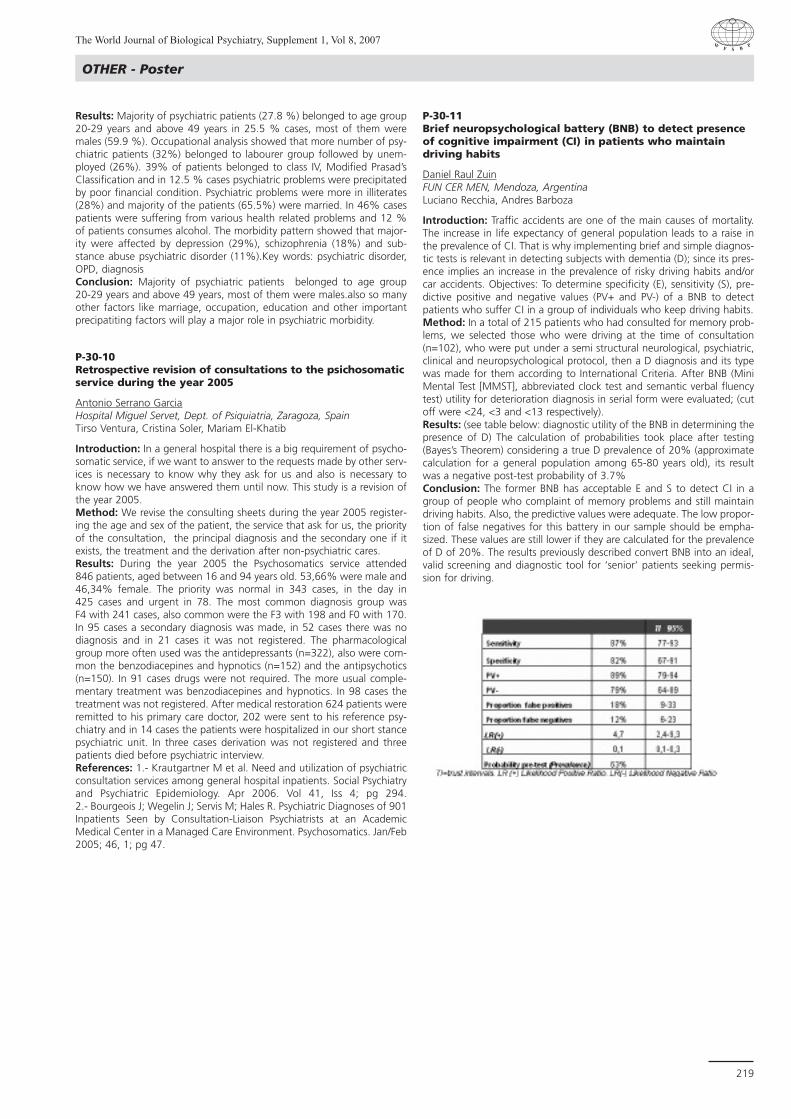
The World Journal of Biological Psychiatry, Supplement 1, Vol 8, 2007
219
OTHER - Poster
Results: Majority of psychiatric patients (27.8 %) belonged to age group20-29 years and above 49 years in 25.5 % cases, most of them weremales (59.9 %). Occupational analysis showed that more number of psy-chiatric patients (32%) belonged to labourer group followed by unem-ployed (26%). 39% of patients belonged to class IV, Modified Prasad’sClassification and in 12.5 % cases psychiatric problems were precipitatedby poor financial condition. Psychiatric problems were more in illiterates(28%) and majority of the patients (65.5%) were married. In 46% casespatients were suffering from various health related problems and 12 %of patients consumes alcohol. The morbidity pattern showed that major-ity were affected by depression (29%), schizophrenia (18%) and sub-stance abuse psychiatric disorder (11%).Key words: psychiatric disorder,OPD, diagnosisConclusion: Majority of psychiatric patients belonged to age group 20-29 years and above 49 years, most of them were males.also so manyother factors like marriage, occupation, education and other importantprecipatiting factors will play a major role in psychiatric morbidity.
P-30-10Retrospective revision of consultations to the psichosomaticservice during the year 2005
Antonio Serrano GarciaHospital Miguel Servet, Dept. of Psiquiatria, Zaragoza, SpainTirso Ventura, Cristina Soler, Mariam El-Khatib
Introduction: In a general hospital there is a big requirement of psycho-somatic service, if we want to answer to the requests made by other serv-ices is necessary to know why they ask for us and also is necessary toknow how we have answered them until now. This study is a revision ofthe year 2005. Method: We revise the consulting sheets during the year 2005 register-ing the age and sex of the patient, the service that ask for us, the priorityof the consultation, the principal diagnosis and the secondary one if itexists, the treatment and the derivation after non-psychiatric cares. Results: During the year 2005 the Psychosomatics service attended 846 patients, aged between 16 and 94 years old. 53,66% were male and46,34% female. The priority was normal in 343 cases, in the day in 425 cases and urgent in 78. The most common diagnosis group was F4 with 241 cases, also common were the F3 with 198 and F0 with 170.In 95 cases a secondary diagnosis was made, in 52 cases there was nodiagnosis and in 21 cases it was not registered. The pharmacologicalgroup more often used was the antidepressants (n=322), also were com-mon the benzodiacepines and hypnotics (n=152) and the antipsychotics(n=150). In 91 cases drugs were not required. The more usual comple-mentary treatment was benzodiacepines and hypnotics. In 98 cases thetreatment was not registered. After medical restoration 624 patients wereremitted to his primary care doctor, 202 were sent to his reference psy-chiatry and in 14 cases the patients were hospitalized in our short stancepsychiatric unit. In three cases derivation was not registered and threepatients died before psychiatric interview. References: 1.- Krautgartner M et al. Need and utilization of psychiatricconsultation services among general hospital inpatients. Social Psychiatryand Psychiatric Epidemiology. Apr 2006. Vol 41, Iss 4; pg 294. 2.- Bourgeois J; Wegelin J; Servis M; Hales R. Psychiatric Diagnoses of 901Inpatients Seen by Consultation-Liaison Psychiatrists at an AcademicMedical Center in a Managed Care Environment. Psychosomatics. Jan/Feb2005; 46, 1; pg 47.
P-30-11Brief neuropsychological battery (BNB) to detect presenceof cognitive impairment (CI) in patients who maintaindriving habits
Daniel Raul ZuinFUN CER MEN, Mendoza, ArgentinaLuciano Recchia, Andres Barboza
Introduction: Traffic accidents are one of the main causes of mortality.The increase in life expectancy of general population leads to a raise inthe prevalence of CI. That is why implementing brief and simple diagnos-tic tests is relevant in detecting subjects with dementia (D); since its pres-ence implies an increase in the prevalence of risky driving habits and/orcar accidents. Objectives: To determine specificity (E), sensitivity (S), pre-dictive positive and negative values (PV+ and PV-) of a BNB to detectpatients who suffer CI in a group of individuals who keep driving habits.Method: In a total of 215 patients who had consulted for memory prob-lems, we selected those who were driving at the time of consultation(n=102), who were put under a semi structural neurological, psychiatric,clinical and neuropsychological protocol, then a D diagnosis and its typewas made for them according to International Criteria. After BNB (MiniMental Test [MMST], abbreviated clock test and semantic verbal fluencytest) utility for deterioration diagnosis in serial form were evaluated; (cutoff were <24, <3 and <13 respectively).Results: (see table below: diagnostic utility of the BNB in determining thepresence of D) The calculation of probabilities took place after testing(Bayes’s Theorem) considering a true D prevalence of 20% (approximatecalculation for a general population among 65-80 years old), its resultwas a negative post-test probability of 3.7% Conclusion: The former BNB has acceptable E and S to detect CI in agroup of people who complaint of memory problems and still maintaindriving habits. Also, the predictive values were adequate. The low propor-tion of false negatives for this battery in our sample should be empha-sized. These values are still lower if they are calculated for the prevalenceof D of 20%. The results previously described convert BNB into an ideal,valid screening and diagnostic tool for ‘senior’ patients seeking permis-sion for driving.

The World Journal of Biological Psychiatry, Supplement 1, Vol 8, 2007
220
P-30-12Neurobiology of ethical behavior: Some comments basedon Francisco Varela’s neurophenomenology
Maria ValdiviaIquique, Chile
Introduction: The purpose of this work is to present a critical analysis ofone aspect of Francisco Varela’s work on the study of ethical behavior andits neurobiological foundations in light of current knowledge on neu-rocognition. Said author poses basic questions about the way of betterunderstanding behavior and how this is developed and made manifest inhumans. Psychic phenomenology was one of the main focuses of interestfor this scientist, who fueled a new research paradigm called neurophe-nomenology in which he tried to conciliate neurobiological research andoriental philosophies, such as Buddhism and others. Ethical humanbehavior is analyzed, posing the problem of how we carry out ethicalactions facing immediate confrontation with perceived facts. Method: Critical analysis of bibliographic review of related texts.Results: An approach to understand behavior and how it is acquired.From Varela’s point of view, the proximity between perception and actionexists, opposing the normal approach of examining ethical behavior,which begins by analyzing the intention of an action and ends evaluatingthe rationality of specific moral judgments. A paradigm change is present-ed, where knowledge units are mainly understood as concrete or corpo-real and where the cognitive being behaves structurally linked to the con-text.Conclusion: Ethical action begins in immediate confrontation with thefacts that we perceive. It is important not to confuse ethical behavior withmoral judgment, a rational process that is not necessarily linked to behav-ior. Cognition is based on concrete activities of the organism as a whole,in the sensor-motor area. Cognitive properties are distant from the “arti-ficial intelligence” model; the cognitive being is made up of his/her historyand actions. Neuronal networks do not work hierarchically; global effectsare achieved where context matters. We have a disposition for an actioncoherent with each specific situation and we constantly move from oneto the next.References: Varela Francisco, Un Know-How per l’Etica, 1ra ed., Roma-Bari, 1992, Gius Laterza & Figli Spa, (Spanish translation by Javier OrtizGarcÌa, La Habilidad âtica, Barcelona ê Spain, Debate, 2002, 143 pp.)
OTHER - Poster

The World Journal of Biological Psychiatry, Supplement 1, Vol 8, 2007
221
SATELLITE SYMPOSIA
SA-02Depression: Mechanisms to Medicine
SA-02-01Neurotransmitters, pain, and depression
Duncan MitchellUniversity of the, Witwatersrand, South Africa
Pain is the most common comorbid physical symptom in depressedpatients and successful treatment of depression in these patients alsorelieves the painful physical symptoms. In addition to their effect onmood, data show antidepressants can relieve pain in patients withoutdepressive symptoms. It is probable that antidepressants relieve painthrough their effect on monoaminergic neural pathways. A likely mecha-nism for at least part of their antinociceptive action is an increase inmonoamine activity in the descending neural pathways that regulatenociceptive input. These descending pathways hyperpolarise both pri-mary afferent fibres, arising from nociceptors, and interneurones in theascending nociceptive pathways of the spinal cord. Antidepressants mayalso act on catecholaminergic antinociceptive pathways in the brain itself.Serotonin and norepinephrine reuptake inhibitors (SNRIs) are more effec-tive antinociceptives than selective serotonin reuptake inhibitors (SSRIs).Antidepressants are the most widely used antinociceptive as first-linetherapy for neuropathic pain in patients with normal mood. Neuropathicpain does not respond to COX inhibitors (NSAIDs, COX-2 inhibitors,paracetamol) that are the most-common first-line therapies for hyperal-gesic nociceptive pain. In recent years, there has been growing under-standing of how antidepressants relieve pain, which is likely to lead totheir increasing use as antinociceptives, especially with the availability ofagents with a more tolerable side-effect profile.
SA-02-02Depression and Pain: A Common Pathophysiology
Amado Nieto-CaraveoUniversity of Autonoma de, San Luis Potosi, Mexico
The association between pain and depressive symptoms is clear and it hasbeen documented by many studies, both in the clinical setting and in thegeneral population1. Near 70% of patients with depression report somedegree of painful symptoms2. This association implies a worse prognosisof depressive disease. Taking into account this fact is relevant to get a bet-ter treatment in a disorder has shown a lower rate of therapeuticresponse that we expected3. The homeostatic model of pain states thatpain is both a sensation and a motivation (such as appetite, itching, thirst,and temperature), a “homeostatic” emotion between interoception andexteroception4. This homeostasis is maintained by regulatory pathwaysbetween brain and spinal cord, with the purpose to achieve a balancebetween analgesic and hiperalgesic actions. There are several sites ofaction of analgesia. The dorsal horn of the spinal cord is the regulationsite of neural transmission through ascending and descending fibers frommesencephalic nucleus5. At this place, monoaminergic, gabaergic andopioid neurons interact between pronociceptive and antinociceptiveactions. Serotoninergic fibers from the rafÈ nucleus have both inhibitoryand facilitatory actions. Noradrenergic descending pathways from locusceruleus have analgesic effects by activation of alfa-1 (by way of gabaer-gic interneuron) and alfa-2 receptors in the Wide Dynamic Range (WDR)neurons. Animal and human studies have shown that drugs that enhancenoradrenergic and serotoninergic activity have analgesic properties6.Some antidepressants with “dual” action on these monoamine systems,like amytriptiline, venlafaxine and duloxetine, improve acute and chronicpainful conditions (like diabetic neuropathy and fibromyalgia)7.Duloxetine has a more balanced affinity to SE and NE receptors than ven-lafaxine and a most secure profile than amitriptiline8. Due to these prop-erties, duloxetine may provide some advantages in the treatment ofdepression.References: 1. Ohayon, M.M., et al. Using chronic pain to predict depres-sive morbidity in the general population. Arch Gen Psychiatry, 2003.60(1): p. 39-47. 2. Kroenke, K., et al. Common symptoms in ambulatorycare: incidence, evaluation, therapy, and outcome. Am J Med, 1989.86(3): p. 262-6. 3. Rush A.J. et al. Acute and Longer-Term Outcomes in
Depressed Outpatients Requiring One or Several Treatment Steps: ASTAR*D Report. Am J Psychiatry 2006; 163:1905-1917. 4. Craig, A.D. Anew view of pain as a homeostatic emotion. Trends Neurosci, 2003.26(6): p. 303-7. 5. Millan, M.J. Descending control of pain. ProgNeurobiol, 2002. 66(6): p. 355-474. 6. Bomholt, S.F., et al.Antinociceptive effects of the antidepressants amitriptyline, duloxetine,mirtazapine and citalopram in animal models of acute, persistent andneuropathic pain. Neuropharmacology, 2005. 48(2): p. 252-63. 7.Sindrup, S.H., et al., Antidepressants in the treatment of neuropathicpain. Basic Clin Pharmacol Toxicol, 2005. 96(6): p. 399-409. 8. Wong,D.T., et al. Dual serotonin and noradrenaline uptake inhibitor class of anti-depressants potential for greater efficacy or just hype? Prog Drug Res,2002. 58: p. 169-222.
SA-02-03Optimizing Sustained Remission
Andrea Marquez Lopez MatoInstitute of Biological, Psychiatry, Argentina
Depression is a common, recurrent, often debilitating and potentiallylethal disorder which needs treatment. Response means 50% improve-ment on a variety of rating scales. To assess treatment “remission not onlyresponse” is a goal of therapy, assuring wellness beyond improvement.Remission means return to full functioning. Recovery is a sustained andtotal remission for an extended period of time and an absence of diseasestate. Failure to achieve full remission results in many negative conse-quences including increased risk of relapse (76%) , lifelong recurrentdepression (80%), greater impairment in psychosocial functioning,increased medical morbidity and mortality, as well as increased rates ofsuicide and substance abuse. For depressed patients, the measure ofremission is based on their feeling that they have returned to their nor-mal selves. Our clinical conception of depression must be expandedbeyond the affective realm, with somatic symptoms, such as PainfulPhysical Symptoms (PPS), included in the definition. Both support a com-mon pathophysiological neurobiological mechanism. Furthermore, 90%of residual symptoms are physical, which play an important part in notbeing able to return to previous functional level and can cause relapse. Itis important to regularly monitor and treat all emotional, including anxi-ety, and PPS for full remission to occur.
supported by an unrestricted educational grant from Elli Lilly andCompany/Boehringer Ingelheim
SA-03In search of superior antidepressants
SA-03-01The place of SNRIs in treatment of depression
Mike BrileyNeuroBiz Consulting, Castres, France
Introduction: Selective serotonin reuptake inhibitors (SSRI) have provento be a considerable improvement over tricyclic antidepressants (TCA) interms of side-effects especially anticholinergic and cardiovascular effects.The SSRIs are not, however, devoid of problems and widespread clinicaluse has brought a number of other problems to light some of them quiteserious. In terms of antidepressant efficacy there is a general consensusthat SSRIs are less effective than TCAs especially in more severe depression.

The World Journal of Biological Psychiatry, Supplement 1, Vol 8, 2007
222
Method: A new class of antidepressants, the serotonin and noradrena-line reuptake inhibitors (SNRI), has been developed with the aim of pro-viding potent antidepressant activity combined with improved tolerance.The SNRI class currently comprises three compounds, venlafaxine, mil-nacipran and duloxetine, which all block the reuptake of both serotonin(5-HT) and noradrenaline (NA) but with differing selectivity. Whereas mil-nacipran blocks 5-HT and NA reuptake with equal affinity, duloxetine hasa 10-fold selectivity for 5-HT and venlafaxine a 30-fold selectivity for 5-HT.In contrast to TCAs all three SNRIs are devoid of interactions at the post-synaptic receptors responsible for many of the side-effects of the olderdrugs. As with other dual acting antidepressants, there is evidence foreach of the SNRIs that they may produce superior antidepressant efficacy,at least in more severely depressed patients. Results: The different SNRIs vary in their tolerability at therapeutic doses.Although no direct comparative data are available, venlafaxine seems tobe the least well-tolerated, with serotonergic adverse effects (nausea, sex-ual dysfunction and withdrawal problems) as well as cardiovasculareffects, principally hypertension, at high doses. Duloxetine appears to bebetter tolerated although problems of hepatotoxicity have led to warn-ings and limitations on its use by regulatory authorities. Conclusion: The available data suggest that milnacipran is probably thebest tolerated of the SNRIs. In addition because of its lack of hepaticmetabolism there is no risk of interaction with other drugs metabolisedby the hepatic cytochrome P450 system.
SA-03-02SNRIs in depression and in chronic pain
Siegfried KasperMedical University, General Psychiatry, Vienna, Austria
Introduction: The common goal of antidepressant therapy sought by adepressed patient and his physician is remission from all symptoms and areturn to normal functioning. Remission is increasingly seen as the crite-rion by which all antidepressants should be judged. Early studies showedthat tricyclic antidepressants (TCA), such as clomipramine, producedgreater rates of remission than selective serotonin reuptake inhibitors(SSRI), such as paroxetine or citalopram. More recently, evidence has beenpublished that all three serotonin and noradrenaline reuptake inhibitors(SNRI) also produce significantly greater rates of remission than the SSRIsstudied.Method: A symptom which is present in a very large number ofdepressed patients is chronic pain, commonly backache or painful joints.In view of the very high comorbidity of chronic pain and depression, manyexperts believe that chronic pain is an integral part of the symptomatol-ogy of depression. TCAs are effective in reducing chronic pain whereas,in general, SSRIs are not helpful. SNRIs have been extensively studied in avariety of pain syndromes. Venlafaxine, for example, has been shown tobe effective in a number of chronic pain conditions both associated withdepression and independent of it, including peripheral diabetic neuropa-thy. Duloxetine and milnacipran have also been shown to be active in var-ious chronic pain conditions and duloxetine has been approved by theFDA for the treatment of peripheral diabetic neuropathy.Results: Fibromyalgia syndrome is a chronic disease of widespread anddebilitating pain, estimated to affect about 3% of the general population.It is characterised by both physical and psychiatric symptoms suggestingthat it may result from abnormal pain processing within the central nerv-ous system. Currently there is no approved treatment in any country. Bothduloxetine and milnacipran have been shown in double-blind placebo-controlled trials to be effective in reducing pain and other symptoms offibromyalgia.Conclusion: As indicated above, painful physical symptoms are increas-ingly being seen as an integral part of the symptomatology of majordepression. It may not be a coincidence that the antidepressant drugscapable of relieving chronic pain such as TCA and SNRI are those that pro-duce remission in the largest number of patients.
SA-03-03Optimising antidepressant response to SNRIs
Marcio Antonini BernikUniversidade de Sao Paolo, Psychiatry, Sao Paulo, Brazil
Introduction: As with all antidepressants, some patients respond betterto SNRIs, such as milnacipran, than others. Polymorphism of genes cod-ing for the noradrenaline transporter influence response to milnacipran.The presence of the T allele of the NET-182C polymorphism is correlatedwith a greater response to milnacipran, whereas the A/A genotype of theNAT G1287A polymorphism is associated with a slower onset ofresponse. Method: Dopamine transporter VNTR polymorphisms also influence therate of response to antidepressant therapy but this effect has been foundin all classes of antidepressants. Similarly, the G/A genotype polymor-phism of BDNF G196A is associated with a better antidepressant responseto milnacipran but also to the SSRI, fluvoxamine. These polymorphismswould therefore appear to involve common downstream pathways ofantidepressant action rather than pathways specific to certain antidepres-sants. Tolerability can also be influenced by polymorphic differences.Serotonin transporter VNTR polymorphism and the serotonin transporter-gene-linked polymorphic region (HTTLPR) polymorphism influence tolera-bility of drugs acting primarily through the inhibition of 5-HT reuptake,such as SSRIs and venlafaxine, which has a 100 fold selectivity for theserotonin transporter. In contrast, these gene polymorphisms have noeffect on the antidepressant response or tolerability of milnacipran.Results: Differences in metabolism determined by genetic variables inCYP2D6 activity are a major determinant of venlafaxine levels to such anextent that genetically determined decreases in CYP2D6 activity havebeen associated with cardiovascular toxicity. Milnacipran, which is notmetabolised by the enzymes of the CYP450 system is not influenced bypolymorphism of these enzymes. Low pretreatment levels of plasma 3-methoxy-4-hydroxyphenylglycol (pMHPG) have also been found to pre-dict a better antidepressant response to milnacipran. In addition, improve-ment in depressive symptoms over 4 weeks has accompanied by increasesin pMHPG levels. Conclusion: Taken together these recent results confirm that milnacipranhas a major impact on the noradrenergic system in contrast to SSRIs andvenlafaxine whose main target is the serotonin system. Clearly pharmaco-genetic data will soon become a significant factor in the choice of themost appropriate antidepressant medication for a particular patient.
supported by an unrestricted educational grant from Pierre Fabre
SA-05Do we need another atypical antipsychotic?
S-05-01Do we need another atypical antipsychotic?
Siegfried KasperMedical University, General Psychiatry, Vienna, Austria
Atypical antipsychotics were a great advance in the treatment of schizo-phrenia. However, thus far, there is no antipsychotic with exceptional effi-cacy and safety in all patients. The atypical antipsychotics have differingpatterns of treatment-emergent side effects. Clinical skills and knowled-ge are required to find a suitable option for treatment of individualpatients. Following its suspension in 1998, the safety and efficacy of ser-tindole has been investigated in several post-marketing studies based inclinical settings. As well as providing the safety data needed to supportthe reintroduction of sertindole, these studies have provided specificexamples demonstrating that certain patients, in particular, may benefitfrom a switch from other atypical antipsychotics to sertindole.Sertindole is an effective atypical antipsychotic agent and, as with all themedications in this group, it is well tolerated with a lack of extrapyrami-dal symptom side effects. Its individual and mostly favourable profile oftreatment-emergent effects and safety allows for flexibility in treatingpatients since they differ in their response and susceptibility to treatment-
SATELLITE SYMPOSIA

The World Journal of Biological Psychiatry, Supplement 1, Vol 8, 2007
223
SATELLITE SYMPOSIA
emergent effects. Sedation and anticholinergic effects are particularlytroublesome side effects which may be common to some of the atypicalantipsychotics. Sedation may lead to non-compliance during the initialperiod of treatment. Sedation may also have consequences for patients'quality-of-life. Anticholinergic effects may adversely influence cognitiveperformance, and they pose a number of risks especially for olderpatients. The propensity of sertindole to cause anticholinergic effects issmall and, more recently, there have been suggestions that sertindole mayhave beneficial effects on cognition.
supported by an unrestricted educational grant from H. LundbeckA/S
SA-06Closing the Global Performance Gap inManaging Major Depressive Disorder and Co-morbid Anxiety
Overall Statement of NeedMajor depressive disorder (MDD) is now the fourth-leading cause of theglobal disease burden and the leading cause of disability worldwide;WHO expects that depression will be the second leading cause of disabi-lity after heart disease by 2020.1,2 Cost-effective interventions are avai-lable, but do not often reach those who need them because of a numberof overwhelming challenges in low-resource settings--including lack offacilities and trained mental health personnel, questions about effectivepopulation-based screening, and the general stigma surrounding mentaldisorders. Generalized anxiety disorder (GAD) is a chronic, disabling disor-der associated with substantial personal, societal, and economic costs. 3 Co-existing GAD in depressed patients may worsen the outcome byincreasing the rates of suicide, worsening overall symptoms, conferring apoorer response to treatment, increasing the number of medically unex-plained symptoms, and increasing functional disability. The comorbidity ofGAD with MDE has been of special interest because of the high level ofcomorbidity and the status of MDE as one of the most burdensome dis-orders worldwide.4 In this live activity, the faculty experts will review theimportance of early recognition of MDD and comorbid anxiety and focuson innovative strategies to manage these challenging comorbidities inpatients from both developed and developing countries.
1. Hyman S, et al. Mental Disorders. In: Disease Control Priorities inDeveloping Countries, 2d ed., ed. Dean T. Jamison et al. New York:Oxford University Press, 2006. 2. The WHO World Mental Health SurveyConsortium. Prevalence, severity, and unmet need for treatment of men-tal disorders in the World Health Organization world mental health sur-veys. JAMA 2004;291:2581-2590 3. Kessler RC, Chiu WT, Demler O,Walters EE. Prevalence, severity, and comorbidity of 12-month DSM-IVdisorders in the National Comorbidity Survey Replication. Arch GenPsychiatry 2005;62:616-627. 4. World Health Organization (WHO),Gender and Women's Mental Health (1997), accessed online atwww.who.int. Accessed 10/23/2006 Learning Objectives
At the end of this CE activity, participants should be able to:
n Identify three patient barriers that contribute to the performancegap noted by the World Health Organization
n Provide clinicians with tools and strategies to improve the deliveryof costeffective interventions for patients with co-morbid anxietyand depression
n Implement innovative and novel teaching approaches to improve theglobal performance gap in the diagnosis and management ofdepression
n and co-morbid anxiety
Activity Goal: To provide evidence-based educational information regar-ding the global prevalence, recognition, and management of depressionwith co-morbid anxiety.Target Audience: This global initiative will target physicians worldwidethrough affiliations with associations and other medical centers of excel-lence.Determination of Need: Need for this activity was determined by sur-vey of the target audience, review of the literature and current trends andsuggestions from past program participants.
SA-06-01
Pedro DelgadoUniversity of Texas, Dept. of Psychiatry, San Antonio, TX, USA
SA-06-02
Julio LicinioUniversity of Miami, Dept. of Psychiatry, Miami, FL, USA
supported by an unrestricted educational grant from WyethPharmaceuticals
SA-08Bipolar Illness: Beyond major depression andeuphoric mania
The diagnosis and treatment of bipolar disorder (BD) has been inconsis-tent and frequently misunderstood in recent years. Both the underdiag-nosis of BD and its frequent misdiagnosis as unipolar major depressive dis-order (MDD) appear to be problems in patients with BD. Underdiagnosisresults from clinicians' inadequate understanding of manic symptoms,from patients' impaired insight into mania, and especially from failure toinvolve family members or third parties in the diagnostic process. Some ofthe underdiagnosis problem may also result from lack of agreementabout the breadth of the bipolar spectrum, beyond classic type I manic-depressive illness. The diagnosis would give greater weight to family his-tory and antidepressant-induced manic symptoms and would apply tonon-type I or II bipolar illness, in which depressive symptom, course, andtreatment response characteristics are more typical of bipolar than unipo-lar illness. Furthermore, some specials forms of bipolar disorder - mixedstates, rapid-cycling and atypical bipolar disorder- are of major clinical andresearch relevance.
SA-08-01
Frederick GoodwinGeorge Washington Univ. Medical Center, Bethesda, MD, USA
supported by an unrestricted educational grant fromGlaxoSmithKline Chile

AAbdeldayem, H. ..................................................69Abdollahian, E. ..................................................144Abdul Rahman, A. H. ..............71, 88, 92, 121, 169Abreu, P. ............................................................146Abushayeva, M. ..................................................94Aceña, R. ..........................................................213Acuña, A. ..........................................................105Adamcova, K. ....................................................161Adelson, M. ........................................................96Ader, M. ..............................................................47Adewuya, A. ........................................................76Agius, M. ..........................................................138Agote, D. ..........................................................173Agranat-Meged, A. ............................................123Ahmed, S. ..........................................................189Ahrendts, J. ........................................................179Aigner, M. ............................................................45Ais, E. ........................................................101, 102Akahane, A. ......................................................151Alajbegovic, A. ..........................................109, 214Alajbegovic, S. ..........................................109, 214Alda, M. ..................................................37, 38, 39Alderete, M. ......................................................188Alderman, J. ......................................................188Alegria, P. ..................................................176, 208Alex, J. ................................................................58Alexander, H. ......................................................82Alfonso, Y. ........................................................122Allen, C. ............................................................205Allen, P. ..............................................................210Allgulander, C. ..................................................113Almeida, J. ........................................................178Almeida Silva, V. de ............................123, 213, 224Almonte, J. C. ....................................................103Alphs, L. ............................................135, 144, 159Altamura, C. ..........................................27, 52, 181Alvarado, M. T. ..................................................140Alvarez, A.R. ......................................................172Alvarez, C. ........................................................195Alvarez, E. ..................................................167, 199Alves-Neto, W. ..................................................176Alzate, M. ..........................................................217Amaro Jr., E. ................................................42, 178Amaro, E. ..........................................................178Aminzadeh, F. ....................................................126Amminger, G. P. ................................................135Amminger, P. ......................................................138Amthauer, H. ....................................................175Anderer, P. ....................................................45, 174Androsiuk, W. ....................................................132Andruskevicius, S. ..............................................104Anghelescu, I.-G. ................................................110Anilkumar, A. ....................................................139Anker, J. ..............................................................97Araujo, D. ..................................................143, 176Araya, R. ............................................................192Araya, S. ............................................................188Araya, V. ............................................................182Arizaga, R. ..........................................................20Armijo, A. ..................................................188, 204Armony, J. ............................................................35Arosio, B. ............................................................27Arques Egea, S. ..................................................203Arzt, E. ................................................................18Aschauer, H. ..............................................135, 166Asenbaum, S. ......................................................42Assimov, M. ......................................156, 208, 215Assion, H.-J. ......................................................147Atmaca, M. ........................................................185Attarbaschi, T. ......................................................42Aukes, M. ............................................................76Avgustin, B. ......................................138, 140, 196Ayuso Gutierrez, J.-L. ..........................................17Aziz Jemain, A. ........................71, 88, 92, 121, 169
BBabakhanyan, A. ..........................................92, 106Babic, D. ......................................................67, 116Babinkostova, Z. ................................................128Bachmann, S. ....................................................156
Bader, M. ..........................................................117Baeken, C. ..........................................................79Baewert, A. ..........................................................60Bagby, R.M. ........................................................111Baghai, T. C. ......................................................104Bahk, W.-M. ......................................................169Bai, P. ................................................................145Bai, Y. M. ..........................................................187Bai, Y.-M. ..........................................................158Baiget, M. ..........................................................167Baji, I. ..................................................................65Baker, G. ............................................................210Bakker, A. ..........................................................207Balazic, J. ..........................................................167Ball, S. ................................................................113Ballesteros, S. ....................................108, 119, 120Balslev Jorgensen, M. ........................................165Bang, S.-K. ........................................169, 177, 201Baptista, T. ..........................................................76Barbosa, C. ........................................................138Barboza, A. ........................................................219Barcikowska, M. ................................................132Barczak, A. ........................................................132Bares, M. ..................................................161, 172Baroni, S. ..........................................................214Barr, C. ....................................................12, 65, 70Barra, F. de la ................................................53,121Barrachina, J. ......................................................199Barrantes, C. ......................................................216Barros, I. ............................................................207Bartenstein, P. ......................................................83Bartholomew, J. ................................................122Batra, N. ............................................................210Battaglia, S. ........................................................202Bayerl, M. ..........................................................177Bayle, F. ................................................................33Becker, A. ............................................................45Becker, J. ............................................................138Bedarida, G. ......................................................104Bellina, V. ............................................................52Bellodi, L. ............................................................12Belohlavek, O. ......................................................78Bender, M. ........................................................118Benegal, V. ..........................................................58Benguettat, D. ....................................................206Benites, F. ..................................................132, 138Benitez-Estevez, A. ............................................146Benito, A. ............................................................86Benito, N. ..........................................................188Beretta, M. ........................................................161Bergemann, N. ....................................................75Berger, G. ..........................................................125Berger, M. ......................................................11, 12Bernik, M. ..........................113, 117, 197, 198, 222Bernstein, H.-G. ................................................150Bert, B. ..............................................................133Besche-Richard, C. ............................................136Bienna, A. ..........................................................204Binneman, B. ......................................................144Bins, H. ..............................................................215Bio, D. ................................................................100Bishnoi, M. ........................................113, 201, 203Bjerkenstedt, L. ............................................25, 151Blanco, C. ..........................................................114Blazevic-Zelic, S. ................................................218Blednov, Y. ..........................................................94Blier, P. ................................................................84Blinc, M. ............................................................138Blom, H. ..............................................................76Bobek - Billewicz, B. ..........................................154Bobes, J. ..............................................................10Bodnar, B. ............................................................93Boer, J. A. den ......................................................13Bogerts, B. ..................................................78, 150Bogovi, A. ..........................................................204Bogush, Y. ..........................................................166Bokhan, N. ............................................92, 94, 211Bonati, M. T. ........................................................ 80Bonci, A. ..............................................................98Bondy, B. ............................................................104Bonelli, R. M. ....101, 128, 129, 133, 164, 173, 174..................................................................183, 199
Borduqui, T. ........................................................143Borges-Gargano, C. ............................................146Borja, H. ............................................................212Borkowska, A. ..............................................38, 130Born, C. ............................................................104Bosi, M. F. E. ................................................52, 181Bourdieu, M. ......................................................150Bourhis, E. ..........................................................142Bourin, M. ............................................................85Boyajyan, A.S. ....................................................148Bozina, N. ..........................................................141Bozkurt, A. ..........................................................65Brand, S. ........................................................18, 45Brathwaite, J. ........................................................9Braus, D.F. ..........................................................175Bravo, A. ............................................................109Bravo, E. ............................................................176Bravo-Mehmedbasic, A. ....................117, 184, 214Brecht, S. ..........................................................190Bressan, R. A. ....................................................138Breznoscakova, D. ................................................93Briley, M. ............................................................221Brunovsky, M. ....................................149, 162, 172Bubenikova-Valesova, V. ............................186, 202Bubl, E. ..............................................................179Buble, T. ............................................................195Büchel, C. ..........................................................175Buchholz, H.-G. ....................................................83Buddensiek, N. ..................................................196Buechert, M. ......................................................179Bukowska, A. ....................................................150Bulbena, A. ........................................................213Bulut, M. ............................................................215Bunevicius, R. ......................................................34Buoli, M. ......................................................52, 181Burns, J. .............................................................. 77Busatto, G. ..........................................42, 176, 178Bustamante, F. ......................................................95Bustamante, I. ....................................................217Bustamante, M. L. ..............................................166Busto, U. ............................................................7, 8Butman, J. ..........................................................179Byszewski, A. ....................................................126
CCabanes, L. ..........................................................86Cabezaz, F. ........................................................173Cabrera, J. ..........................................................155Caffo, E. ..............................................................80Cahn, W. ..............................................................29Calderon, A. ......................................104, 119, 120Caldirola, D. ........................................................12Callicott, J. ..........................................................83Calovska Samardziska, V. ....................................191Calvo Prandi, M. S. ............................................100Calvó, M. ..................................................102, 150Calvo-Malvar, M.M. ............................................146Calzada Reyes, A. A. ..........................................115Camacho Lopez, P. A. ................................122, 124Camacho, A. ......................................................100Camponovo, D. ..................................................167Campos, M. ......................................................212Canisius, S. ..........................................................73Canitano, R. ........................................................69Cannon, D. M. ....................................................11Cano, J. F. 136, ..................................................217Carey, P. ............................................................118Carlini, M. ..........................................................214Carr, V. ..............................................................157Carrasco, J. ..........................................................15Carrizo, E. ............................................................76Carroll, M.E. ........................................................97Cassel, W. ............................................................45Cassin, B. ............................................................60Castelli, P. ..........................................................158Castello Gasco, J. ..............................................203Castiglioni, M. ....................................................202Castilla-Puentes, R. .......................... 62, 64, 70, 140Castilla-Puentes, S. ............................................140Castilla-Puentes, W. ............................................140Castillo, C. ........................................................135Castro, J. ..............................................................62
AUTHOR INDEX
The World Journal of Biological Psychiatry, Supplement 1, Vol 8, 2007
224

AUTHOR INDEX
The World Journal of Biological Psychiatry, Supplement 1, Vol 8, 2007
225
Catena, M. ........................................110, 119, 214Cattaneo, E. ........................................................27Cavalleri, F. ..........................................................80Cazorla, P. ..........................................................135Cazzullo, C. L. ......................................................27Cebasek Travnik, Z. ............................................196Celis, A. ............................................................105Cendales, R. ..............................................136, 217Centanni, L. ......................................................185Cerqueira, C. ......................................................178Ceskova, E. ..........................................................30Cetkovich-Bakmas, M. ..........................................34Chae, J.-H. ........................................................169Chainho, J. ................................................105, 216Chaki, S. ..............................................................51Chang, H.-A. ......................................................149Chang, I.-S. ........................................................200Chang, W.-H. ....................................................187Change, C.-C. ....................................................149Chatton, A. ................................................102, 193Chaudhry, H. R. ............................................75, 159Chazeron, I. de ..................................................126Chee, I.-S. ..................................................137, 139Chee, K. Y. ..........................................................75Chei Tung, T. ......................................................103Chele, G. ................................................96, 97, 99Chen, C.-C. ........................................................143Chen, C.-L. ........................................................149Chen, E..Y.H. ......................................................171Chen, J.-Y. ........................................................187Chen, T. T. ..........................................................187Cheng, S.-T. ......................................................111Chereau-Boudet, I. ............................................126Cheung, B. K. L. ................................................206Cheung, C. ........................................................171Chieri, P. ..............................................................40Chinea, E. ..................................................102, 207Chirinos, A. ........................................................172Chirita, R. ................................................96, 97, 99Chirita, V. ................................................96, 97, 99Chittiprol, S. ..............................................109, 146Chiu, F.-Y. ..........................................................158Chivite, S...............................................................22Chodakowska-Zebrowska, M. ............................132Choi, M.-J. ........................................110, 166, 189Choi, S. ................................................................93Choi, Y. ..............................................................131Chopra, K. ................................................201, 203Chou, K.-H. ........................................................158Chou, Y.-H. ..................................................86, 158Chour, W.-Y. ......................................................143Christodoulou, C. ..............................................145Chu, E. ..............................................................192Chua, S. ............................................................171Chung, J. H. ........................................................23Ciampa, G. ................................................119, 214Cid, C. ..............................................................188Ciobanu, A. M. ..................................................100Cizmovic, B. ......................................................160Clavero, R. ........................................................212Clavijo, N. ..........................................................188Clays, F. ..............................................................206Clerici, M. ............................................................27Clivio, A. ..............................................................27Cogliati, F. ............................................................80Cohen, H. ..........................................................182Cohen, M. ..........................................................157Cohen, S. ............................................................ 65Conesa, H.A. ......................................................150Consoli, G. ................................................110, 119Corchs, F. ..........................................................117Córcoles, D. ......................................................213Cordeiro, Q. ..............................................103, 176Cordes, J. ..........................................................145Cordoba, R. N. ..................................................136Córdoba, R. N. ..................................................217Cornish, K. ................................................168, 169Cornutiu, G. ......................................................147Cornutiu, O. ......................................................147Corrales, A. ..........................................................40Correa, E. ..............................................10, 17, 112Corregiari, F. ..............................113, 117, 197, 198
Cortes, A. ..........................................................167Cosacow, D. ......................................................173Costa Leite, C. ....................................................42Costa, P.T. ..........................................................111Cosyns, P. ............................................................53Courtecuisse, C. ................................................190Covarrubias, M. I. ..............................................188Craddock, N. ........................................................65Craig, I. ................................................................65Crawford, T. ......................................................139Crayton, J.W. ......................................................67Crippa, J. A. ................................................42, 143Croenlein, J. ......................................................190Cuervo, C.-V. ......................................................136Cuesta, M. J. ........................................................22Cumming, P. ........................................................83Cunha, M. G. ............................................109, 208Curran, H. V. ............................................94, 95, 96Czekalla, J. ........................................134, 146, 198
DD'haenen, H. ........................................................79Dachesky, D. ......................................................128Damjanovic, A. ..................................................152Daróczy, G. ..........................................................65Dashniani, M. ....................................................162Dasi, C. ..............................................................148Davidson, J. ........................................................114Debieuvre, C. ....................................................190Deitcher, C. ........................................................123Del Debbio, A. ..................................................110Del-Ben, C. ................................................176, 199Delgado, P. ........................................................223DeLisi, L. ..............................................................28Dell'Osso, B. ................................................52, 181Dell'Osso, L. ..............................................110, 214Deluca, V. ............................................................12Demetrio, F. N. ..................................................103Dempster, E. ........................................................70Demyttenaere, K. ..............................................190Deng, Y. ............................................................171DeRaedt, R. ..........................................................79Derost, P. ............................................................126Desai, N. G. ........................................................200Desaiah, D. ................................................190, 191Detke, M. ..........................................................113Dezeljin, M. ..................................................67, 116Dhoine, K. ..........................................................218Di Nicola, M. ........................................................93Diamond, I. ..........................................................98Diaz Martinez, C. ................................................93Diaz Martinez, L. A. ............................................124Díaz, D. ..............................................................207Diaz, E. ..............................................................137Diaz-Mesa, E. ....................................................146Diehl-Schmid, J. ....................................................72Dielentheis, T. F. ................................................177Diemer, N. ..........................................................165Diez, J. ..............................................................174Dillingh, G. ........................................................207Dimellis, D. ........................................................195Dinan, T. G. ................................................208, 209Diniz, B. O. ........................................................103Diniz, J. ..............................................................215Divos, H. ..............................................................45Divosevic, S. ......................................................151Djazayeri, S. ......................................................205Djedovic, J. ........................................................160Dmitrzak-Weglarz, M. ..........................................38Dobrowolny, H. ..................................................150Dodd, P. ................................................................8Dodel, R. ..............................................................73Doknic, M. ........................................................152Dolnicar, R. ..................................................72, 167Dominguez, J.I. ..................................................172Donoso, E. ..........................................................95Dordevic, V. ........................................................195Dossenbach, M. ................................................202Douzenis, A. ......................................................145Dragasek, J........................................................... 93Dremencov, E. ......................................................84Dremov, G. ........................................................211
Dresel, S. ............................................................178Drimalova, M. ......................................................93Dro, W. ..............................................................130Drzezga, A. ..........................................................72Dudczak, R. ..........................................................42Duenas, H. ........................................................105Duffy, A. ........................................................37, 38Duletic, N. ..........................................................160Dumitrescu, M. R. ..............................................203Dunlop, B. ..........................................................189Dunner, D. ................................................189, 190Duran, F. ............................................................176Durif, F. ..............................................................126D’Urso, N. ..........................................................181Dwivedi, Y. ........................................................161Dyachenko, L. ......................................................98Dyck, R. van ........................................................80Dzubur Kulenovic, A. ........................................117
EEap, C. ..............................................................194Ebeling, H. ..........................................................75Eberhard, J. ..................................96, 149, 153, 186Ebert, D. ............................................................179Ebner, N. ..............................................................60Ebner, S. ..............................................................88Eckert, A. ............................................................18Edman, G. ............................................................25Eguiluz, I. ..........................................................135Ekici, G. ..............................................................215El Mansari, M. ......................................................84El Masri, D. ........................................................177El-Khatib, M. ......................................................219ElFakih, Y. ............................................................76Elliott, M. ............................................................28Ellis, J. ..................................................................37ElNahas, G. M. 161Emrich, H.M. ......................................................196Emsley, R. ............................................................29Encina, C. ..........................................................155Eng, S. ..............................................................141Enterman, J. ......................................................137Entsuah, R. ........................................................114Epelbaum, A. ....................................................194Erausquin, G.A. de ............................................150Escobar Sanchez, M. ..................................122, 124Escobar, J.I. ........................................................150Eser, D. ..............................................................104Espinosa, C. ......................................................155Essig, M. ............................................127, 156, 174Ethesdam, S. .......................................................... 9Ettinger, U. ........................................................139Eyck, D. van ........................................................47
FFaich, G. ............................................................141Fallon, J. ..............................................................83Faradoni, F. ........................................................136Faravelli, C. ........................................................119Faravelli, L. ........................................................ 119Farmer, A. ............................................................65Faunes, M. ........................................................188Fedorenko, O. ..............................................92, 150Feige, B. ................................................11, 12, 179Feldon, J. ....................................................24, 153Fellmann, H. ........................................................78Feng, Y. ................................................................65Ferdousi, T. ........................................................189Ferguson, J. ................................................189, 190Fernandes, S. ............................................105, 216Fernandez Huerta, J. M. ....................................211Fernandez Sola, J. ..............................................211Fernández, I. ......................................................207Fernandez, L. ....................................................137Fernandez, V. ......................................................76Ferrao, Y. ..........................................................215Ferrari, G. C. de ................................................168Ferreira, C. ........................................................176Ferreira, J. ............................................................95Ferris, C. ..............................................................19Fian, R. ..............................................................101Fiedler, J. ....................................................106, 182

Fierro, M. ..........................................................136Fiestas, F. ............................................................217Figueiredo, L. A. P. de ........................................208Figueroa, E. ..........................................................59Fink, H. ..............................................117, 133, 171Fink-Jensen, A. ....................................................97Fischer, G. ............................................................60Fitzgerald, P. ..............................................208, 209Fleming, K. ........................................................144Florenzano, N. ....................................................150Florenzano, R. ............................104, 108, 119, 120Flores, C. ............................................................171Flores, D. ............................................................185Flyckt, L. ..............................................................25Folnegovi Grosi, P. ..............................................204Folnegovic-Smalc, V. ..................................127, 201Folnegovsmalc, V. ..............................................204Ford, J. ................................................................44Forlenza, O. ........................................................126Fortes, S. ............................................................ 212Fossati, P. ............................................................35Franciskovic, T. ..................................................147Franco Lopez, J. A. ....................................122, 124Franke, L. ..........................................................175Freedman, R. ........................................................37Freitas, C. ..........................................................132Fresard, E. ..................................................193, 206Friedman, E. ..............................................189, 190Friedreich, S. ................................................84, 177Fritsch, R. ..........................................106, 182, 192Fritz, R. ..............................................................106Fuchs, K. ............................................................166Fuentes, M. ........................................................192Fuentes, P. ..........................................................208Fullerton, C. ......................................................103Furey, M. ..............................................................11
GGaag, van der ....................................................137Gabilondo, A. ....................................................213Gabryelewicz, T. ................................................132Gado, M. ..........................................................141Gadoros, J. ....................................................65, 70Gaebel, W. ....................................................26, 28Galeano, E. ..........................................................36Galeazzi, T. ..........................................................76Galeno, R. B. ..................................................29, 54Galili-Wiesstub, E. ..............................................123Gallardo, R. ........................................................188Galleguillos, F. ....................................................166Galli, E. ................................................................64Gallo, C. ............................................................217Galvan, J. ..........................................................173Galvez, P. ..........................................................188Gangadhar, B. N. ........................86, 109, 146, 163Garcia, R. ..........................................................155Garcia-Amador, M. .............................................. 62Garcia-Aquirre, J. L. ..............................................49García-Barriga, C. ................................................49Garcia-Campayo, J. ............................................ 212Garcia-Hernandez, A. ........................................137García-Ribera, C. ................................................213Gardner, M. ........................................................158Garver, K. ............................................................82Gattaz, W. ............................................24, 57, 197Gau, S.-F. ............................................................50Gaviria, S. ............................................................10Gaysina, D. ..........................................................65Geier, C. ..............................................................82Geisler, P. ............................................................210Gelazonia, L. ......................................................162Gelenberg, A. ............................................189, 190Gentil Filho, V. ....................................................178Gentil Savoia, M. ................................................113Gentil, V. ............................................................178Gentile, S. ..........................................................193George, M. ..........................................................36Gergerlioglu, H. S. ..............................................215Geri, S. ................................................................93Gerstl, F. ..............................................................84Gerwe, M. ................................131, 134, 146, 198Gesierich, T. ........................................................177
Gheorghe, M.-D. ..................................................76Giannaccini, G. ..................................................214Gierski, F. ..........................................................136Giesel, F. ....................................................127, 156Giesler, M. ............................................45, 73, 118Gil-Ad, I. ............................................................182Ginés, J. M. ........................................................213Glaescher, J. ......................................................175Glasinovic, A. ......................................................95Gliddon, L.A. ........................................................97Godas Sieso, T. ..................................................211Goddard, A. ......................................................197Gold, L. H. ........................................................ 101Goldzweig, G. ....................................................123Golia, A. ............................................................110Goma, M. ..........................................................167Gomez Gil, E. ....................................................211Gomez-Restrepo, C. ..........................................100Gonzales, C. ......................................................212Gonzalez Alemán, G. ........................................150Gonzalez, L. ......................................................217Goodwin, F. ..................................................5, 223Gordon, H. ..........................................................53Gorelick, D. A. ..................................................5, 8Gorenstein, C. ....................................................178Gorini Amedei, S. ..............................................119Gorshkova, L. ....................................................190Goudochnikov, V. ..............................................200Gournellis, R. ....................................................145Graça, J. ....................................................105, 216Gracia, R. ..........................................102, 137, 207Graeff, F. ....................................................176, 199Graovac, M. ......................................................147Grassi, M. ............................................................12Graulich, A. ........................................................158Green, M. ............................................................62Grkovic, J. ..........................................................218Grof, P. ................................................................37Groleger, U. ........................................................106Grosic, V. ............................................................204Grosjean, B. ........................................................59Gruber, G. ............................................................45Grunbaum-Novak, N. ........................................182Gründer, G. ....................................................41, 83Grzyzwa, A. ..............................................154, 159Gschaider, S. ................................................75, 159Guapo, V. ..........................................................176Gubka, U. ............................................................ 78Guduri, S. ............................................................59Guerra, M. ..........................................................19Guerreiro Anastacio, M. ............................107, 152Guerrero, G. ......................................................150Guffanti, G. ..........................................................80Guillermo, L. ........................................................86Guirao, M. ........................................................179Gur, C. ............................................................5, 28Gur, R. ............................................................5, 28Gustov, A. ..........................................................214Guterres, L. ........................................................215Gutierrez Ponce De Leon, F. ................................211Gutierrez, M. ......................................................135Gutierrez, R. S. ..................................................135Gutt, E. ..............................................................100
HHaag, A. ........................................................45, 73Haberkorn, U. ....................................................127Haefner, S. ........................................................104Hageman, I. ......................................................165Hahn, S.-W. ........................................................189Hahshemizadeh, H. ..............................................88Hajek, T. ........................................................ 37, 38Hallak, J. ............................................................143Hamann, M. ......................................................171Han, B. ..............................................................145Han, D. H. ............................................................93Han, J.-H. ..........................................................210Han, S. I. ............................................................106Han, S. W. ....................................................46, 190Hanlon, M.-C. ....................................................123Hanssens, L. ........................................................47Hanstock, C. ......................................................210
Hardy, J. ............................................................168Hargarter, L. ..............................................146, 198Harris, A. ..............................................................94Hartford, J. ........................................................113Hartman, C. ........................................................13Hascoët, M. ..........................................................85Hashempour, Z. ..................................................217Hashimoto, R. ......................................................21Hasna, A. ............................................................90Hata, T. ..............................................................151Hatzinger, M. ................................................18, 45Haupt, D. ............................................................48Hauser, J. ............................................................38Havle, N. ............................................................212Hay, E. ................................................................197Heffernan, T. ................................................60, 122Heijden, F. van der ..............................................207Heijer, M. den ......................................................76Heinimaa, M. ......................................................75Heiskala, J. ........................................................164Hemmeter, U. ..................................18, 45, 73, 118Henning, J. ........................................................179Henriquez, H. ....................................................167Henry, B. ............................................................143Henry, J. ..............................................................96Henry, M. ..................................102, 137, 146, 207Henze, M. ..........................................................127Hernandez- Garcia, D. ........................................146Herold, N. ..........................................................175Herpertz, S. ..........................................................15Herpertz-Dahlmann, B. ........................................ 55Herranhof, B. ....................................128, 129, 133Herrera, F. ............................................................10Herrera, L. ..................................................106, 182Hess, L. ..............................................................199Hesslinger, B. ......................................................179Hettema, J. M. ....................................................14Hert, M. de ....................................................33, 47Hietala, J. ............................................................41Higuchi, Y. ..........................................................31Hill, C. D. ..........................................................135Hirschfeld, R. ..............................................189, 190Ho, A. M. C. ......................................................206Hoda, F. ................................................................65Hodjat, S. K. ......................................................144Hödl, A. ............101, 128, 129, 133, 164, 173, 174..................................................................183, 199
Hodler, C. ..........................................................142Hoencamp, E. ....................................................137Hoffman, K. ......................................................216Hofmann, P. ......................................................101Hohagen, F. ..........................................................85Hojaij, C. ..............................................................29Holik, A. ................................42, 84, 117, 176, 177Holl, E. ..............................................................164Hollis, J. ................................................................37Holsboer-Trachsler, E. ....................................18, 45Holston, E. ..........................................................72Holzer, M. M. ......................................................87Hong, K. S. ................................................140, 141Hong, S.-C. ........................................................210Hoof, J. van ..........................................................89Hoogendijk, W. ....................................................80Hoogendoorn, M. ................................................76Hopf, W. F. ..........................................................98Hora, J. ..............................................................205Horacek, J. ............................ 78, 88, 149, 161, 202Hosak, L. ............................................................204Höschl, C. ....................................................78, 149Hossain, I. ............................................................90Hotsenpiller, G. ....................................................67Hotujac, L. ............................................................63Hsiao, C.-C. ........................................................107Hsiao, M.-C. ......................................................209Huang, S.-Y. ......................................................149Humberto, J. ......................................................152Hungria, J. O. Soares ..........................................132Hunt, A. ......................................................72, 127Hunter, M. ........................................................157
IIbach, B. ............................................131, 134, 146
AUTHOR INDEX
The World Journal of Biological Psychiatry, Supplement 1, Vol 8, 2007
226

AUTHOR INDEX
The World Journal of Biological Psychiatry, Supplement 1, Vol 8, 2007
227
Idemudia, E. E. ..................................................159Idris, N. F. ..........................................................143Igimi, H. ............................................................157Igoa, A. ......................................................101, 102Ikeda, M. ..............................................................21Ilinca, M. ..................................................96, 97, 99Ille, R. ................................................................128In-Ho, P. ....................................................177, 201Indran, T. ..................................71, 88, 92, 121, 169Inoue, M. ....................................................98, 157Inoue, S. ..............................................................98Inoue, Y. ..............................................................98Intzausti, A. ........................................................146Iserhard, R. F. ......................................................180Ising, M. ..............................................................18Islam, N. ..............................................................90Iturra, P. ....................................................166, 197Iturria, I. ....................................................119, 120Ivanova, S. ....................................92, 94, 150, 183Ivanovic-Zuvic, F. ..........................................10, 112Ivanovic-Zuvic, N. ..............................................112Iwata, N. ..............................................................21
JJabbi, M. ..............................................................13Jabs, B. ................................................................23Jagsch, R. ..............................................................60Jain, S. ..............................................................141Jakovljevic, M. ..................63, 67, 89, 91, 116, 132Janas-Kozik, M. ....................................................87Jandl, M. ..............................................................64Janelle, E. ..........................................................113Jang, H.-J. ..........................................................191Janiri, L. ........................................................56, 93Jankovic, P. ..........................................................93Japaridze, N. ......................................................162Jara Opazo, C. ..................................................210Jara, C. ..............................................................188Järvelin, M.-R. ......................................................75Jasovic Gasic, M. ................................................152Jeon Lan, K. ......................................................139Jeon, Y. W. ........................................................106Jeong, J.-H. ........................................................210Jeong, Y.-J. ........................................................166Jerez, S. ....................................................166, 197Ji, M.-Y. ..............................................................189Jiang, Q. ............................................................114Jimenez, M. ........................................................166Jina, A. ......................................................187, 188John, J. P. ..........................................................141Johnson, L. L. ...................................................... 37Johnston, P. ........................................................157Jones, P. B. ..........................................................75Jong, S. de ..........................................................80Jonovska, S. ......................................................184Jonovski, N. ........................................................184Jordaan, I. ..........................................................118Joukamaa, M. ......................................................75Jovanovic, N. ..............................................141, 151Juchnowics, D. ..........................................111, 207Jun, T.-Y. ..............................................22, 152, 169Jung, H. ....................................................170, 196Jung, H.-Y. ........................................................191Jung, Y.-E. ..........................................................169
KKader, L. ............................................................148Kähkönen, S. ......................................................164Kahn, R. ..............................................................76Kaibuchi, K. ..........................................................21Kaiser, E. ......................................................72, 127Kalische, R. ........................................................175Kane, J. ..............................................................141Kang, R.-H. ..........................................46, 166, 190Kaparnai, K. ........................................................70Kapfhammer, H. P. ............101, 129, 173, 174, 199Kapornay, K. ........................................................65Karakula, H. ..............................................154, 159Karlidere, T. .......................................................... 65Karwautz, A. ......................................................125Kaschka, W. ........................................................64Kasparek, T. ..........................................................30
Kasper, S. ........6, 42, 52, 57, 82, 84, 117, 166, 176.................................................................. 177, 222Kaufmann, J. ........................................................78Kaufmann, R. ....................................................135Kawas, O. ..........................................................105Kawasaki, Y. ........................................................ 31Ke, C.-L. ............................................111, 114, 143Kean, M. ............................................................139Keller, D. ............................................................144Keller, M. ..................................................189, 190Kema, I. ..............................................................13Kennedy, A. ........................................................97Kennedy, J. ..........................................7, 12, 65, 70Kennedy, J. L. ....................................................7, 9Kennedy, S.H. ....................................................111Kesebir, S. ..........................................................108Keshavan, M. ......................................................31Khalaf, M. ............................................................70Khan, R. ............................................................206Khazaal, Y. ................................102, 193, 194, 206Khoyetsyan, A. ..................................................148Khudabux, J. ......................................................210Kienbacher, C. ....................................................125Kim, H. ..............................................................170Kim, J. J. ............................................................152Kim, S. H. ..........................................................170Kim, S.-Y. ..........................................................191Kim, Y.-K. ..........................................................141Kindler, J. ..........................................................166King, N................................................................... 9Kirakosyan, A. ..............................................92, 106Kishimoto, T. ........................................................98Kiss, E. ..........................................................65, 70Kitamura, S. ........................................................98Kitchener, N. ..................................................69, 70Klasik, A. ....................................................118, 205Klein, E. ................................................................16Klein, F. ................................................................34Klein, N. ..............................................................42Kletter, K. ..................................................117, 176Klirova, M. ........................................................164Klitzing, K. von....................................................227Knezevic, J. .......................................................... 67Koch, N. ..............................................................85Kochetkov, Y. ....................................................110Kocmur, M. ..................................................90, 138Kocsis, J. ....................................................189, 190Koenig, B. ............................................................87Kofidis, N. ..........................................................195Kogan, C. ..........................................................168Kohler, C. ............................................................28Koic, E. ..............................................................186Koldolbsky, N. ......................................................59Kombian, S. ........................................................86Komel, R. ..........................................................167Komssi, S. ..........................................................164Konrad, A. ........................................................125Konstantinidis, A. ................................................52Konupcikova, K. ................................................204Kopecek, M. ........................78, 149, 161, 164, 172Kopishinskaya, S. ................................................214Koponen, H. ........................................................77Koppitz, M. ................................................101, 129Kordon, A. ..........................................................85Koren, D. ............................................................16Koren, E. ............................................................ 194Kornetova, E. ....................................................150Kornstein, S. ..............................................189, 190Korszun, A. ..........................................................65Kovacs, M. .................................................... 65, 70Kozaric-Kovacic, D. ......................................67, 116Kozumplik, O. ....................................................201Krajca, V. ............................................................172Krause, J. ..................................................169, 178Krause, K.-H. ......................................................178Krelling, R. ........................................................100Kremlacek, J. ..............................................156, 174Krenz, S. ............................................................206Krieg, J. C. ............................................45, 73, 118Krishnaswamy, S. ..............49, 71, 88, 92, 121, 169Krsiak, M. ..........................................................199Krupka-Matuszczyk, I. ..........................87, 118, 205
Kruse, A. ..............................................................73Krysta, K. ..........................................................205Kuba, M. ............................................................156Kucukalic, A. ..............................109, 117, 184, 214Kühmayer, D. ...................................................... 88Kulenovic-Dzubur, A. ..................................109, 214Kulkarni, S. ................................................113, 201Kulkarni, S.K. ....................................................203Kumakura, Y. ......................................................83Kumar, K. ..................................................109, 163Kumari, V. ..........................................................139Kurachi, M. ..........................................................31Kurbanova, A. ............................................208, 215Kurt, K. ................................................................82Kurz, A. ................................................................72Kushnir, V. ..............................................................8Kweon, Y. S. ......................................................168Kwon, O. J. ........................................................152Kwon, Y.-J. ........................................................191
LLadea, M. ..........................................................203LaFougere, C. ....................................................178Lahera, G. ............................................................86Laitinen, J. ............................................................77Lamy, C. ............................................................158Lancaster, S. ..............................................135, 159Landers, A. M. T. ................................................209Lanzenberger, R. ..............42, 82, 84, 117, 176, 177Lara, M. d. Carmen ............................................105Larach-Walters, V. ..........................................27, 54Laranjeira, L. ......................................................107Larizza, L. ............................................................80Larson, E.B. ..........................................................97Latorre Latorre, J. F. ............................................122Lauronen, E. ........................................................75Lavekar, G. S. ......................................................86Lay-Son, G. ........................................................106Layton, F............................................................. 216Le Melledo, J.-M. ..............................................210Lee, B.-W. ..........................................................169Lee, C. ......................................152, 177, 201, 210Lee, C. T. ............................................................168Lee, C.-U. ..........................................152, 177, 201Lee, H. K. ..........................................................168Lee, M.-S. ..................................110, 166, 189, 190Lee, S.-J. ............................................................140Lee, S.-P. ............................................................210Lee, S.-Y. ....................................................140, 141Lee, T. ................................................................ 192Lee, Y.-S. ..............................................93, 140, 141LeFoll, B. ................................................................7Leibenson, L. ......................................................123Leisch, F. ............................................................166Leiva, F. ..............................................................155Lemaire, J.-J. ......................................................126Lemanski, S. ......................................................147Lena, F. ..............................................................151Lendeckel, U. ....................................................150Leonard, M. ......................................................214Leonhard-Key, M. ................................................82Lepage, M. ....................................................31, 35Leri, F. ................................................................205Levander, S. ................................96, 149, 153, 186Levesque, D. ......................................................142Levitt, A. ..............................................................10Levy Neto, M. ....................................................103Lewin, T. ............................................................157Leyman, L. ............................................................79Leyton, M. ..............................................................7Li, Y. .................................................................... 72Libiger, J. ............................................................156Licinio, J. ............................................................223Lieber, I. ....................................................173, 194Liebowitz, M. ....................................................114Liegeois, J.-F. ......................................................158Lim, H.-K. ..........................................152, 177, 201Lim, S.-W. ....................................................47, 113Limosin, F. ..........................................................136Lin, K. ................................................................200Lin, K.-M. ............................................................50Lin, W. ......................................................111, 114

Lin, W.-W. ..........................................................149Lindinger, M.A. ..................................................173Lindström, E. ................................96, 149, 153, 186Linial, M. ............................................................166Linne, A. B. ........................................................153Linsky, I. ..............................................................98Lippi, J. ................................................................89Little, K. ..............................................................60Liu, C.-Y. ............................................................209Liu, R. ..................................................................82Livianos-Aldana, L. ............................................203Ljubicic, D. ................................................154, 195Llibre Rodriguez, J. ..............................................19Llorca, P. M. ................................................33, 126Lobacheva, O. ....................................................142Lobo, D. S. S. ........................................................9Lobo, M. ....................................................105, 216Loginov, V. ........................................................150Loh, E.-W. ............................................................50Lolas Stepke, F. ......................................................5Loncarevic, N. ....................................................109Lopes, C. ............................................................212Lopez de Lara, C. ................................................37Lopez Mato, A. M. ............................................221Lopez, S. ............................................................140Lotto, A. ............................................................208Loughead, J. ........................................................28Loughland, C. ....................................................157Louza, M. ..........................................123, 159, 213Love, T. ................................................................59Lowry, A. ............................................................202Loxton, N. ..........................................................206Lucacchini, A. .................................................... 214Luchtman, D. ....................................................132Luczywek, E. ......................................................132Luna, B. ................................................................82Lyford-Pike, A. G. ................................................36Lykouras, L. ........................................................145
MMacciardi, F. ..................................................80, 83Machado Vianna, A. ..........................................113MacQueen, G....................................................... 38Madrigal, E. ........................................................105Magnet, M. ........................................101, 129, 199Magnet, M. M. ..................................................183Magroob, . ..........................................................15Maheux, J. ........................................................142Maki, P. ..........................................................75, 77Malagón, A. ......................................................213Malhi, G............................................................... 62Malla, A. ..............................................................30Manasyan, N. ..............................................92, 106Mandl, M. ............................................................45Manes, F. ......................................................34, 35Mangano, R. ......................................................114Manuseva, N. ....................................................191Marazziti, D. ..............................................110, 214Marchi, M. ..........................................................80Marcinko, D. ..................................................89, 91Marcolin, M. A. ....................................................52Mardi, A. ............................................................105Marengo, E. ..............................................101, 102Margaryan, S. ........................................90, 92, 106Mari, J. ..............................................................217Maric, N. ............................................................152Marieiro, A. ........................................................152Mariotti, M. ..........................................................52Maris di Cio, S. ..................................................173Markus, M. ..........................................................82Maron, E. ............................................................14Marques-Teixeira, J. ....................................152, 157Marquez Lopez Mato, A. ....................................184Marston, H. ................................................101, 143Martin, F. D. C. ..................................................101Martin, M. ..........................................................137Martinac, M. ..........................................67, 89, 116Martinez, J. C. ......................................................10Martino, D. ................................................101, 102Martinotti, G. ................................................57, 93Martinove, M. ......................................................93Maslov, B. ....................................................67, 116
Masopust, J. ..............................133, 156, 174, 204Massimini, M. ......................................................52Matarin, M. ........................................................168Mathalon, D......................................................... 44Mathis, M. A. de ................................................215Mathis, M. E. de ................................................215Matkovic, V. ......................................................195Matsui, M. ..........................................................31Matsuoka, T. ..............................................157, 172Mattay, V. ............................................................83Mattejat, F. ........................................................198Matuszczyk, M. ..........................................118, 205Matzner, H. ........................................................163Mavinakayinahalli Neelakantach, N. ..................146Mayilyan, K. ......................................................148Mazanek, M. ......................................................177Mazzotti, G. ......................................................217McAlonan, G.M. ................................................171McCabe, K. ........................................................157McCarley, R. W. ....................................................43McGuffin, P. ........................................................ 65McGuire, P. K. ......................................................42McKenna, P. ........................................................26Meagher, D. ......................................................214Medina, P. ..........................................................130Medina, S. ..........................................................155Medved, V. ................................................141, 151Meehan, O........................................................... 80Mendes, S. ................................................105, 216Meyer, U. ..........................................................153Meyer-Lindenberg, A. ....................................41, 83Miceli, J. ............................................................188Miceva Velickoska, E. ........................................191Micheli, F. ..........................................................150Michopoulos, I. ..................................................145Middleton, T. ......................................................118Mien, L. ..............................................................176Mien, L. K. ........................................................117Miettunen, J. ........................................................75Miguel, E. ..........................................................215Mihaljevic-Peles, A. ........................................63, 89Mihanovi, M. ......................................................204Mikova, O. .......................................................... 45Miller-Reiter, E. ....................................................88Mimica, N. ................................................127, 201Minko, A. ............................................................98Minshew, N. ........................................................82Miranda, E. ........................................................155Miranda-Camacho, R. ..........................................49Mitchell, D. ........................................................221Mittal, M. ............................................................16Mittal, S. ..............................................................16Mitterhauser, M. ........................................117, 176Moeller, H. ........................................................ 134Mohandas, E. ................................................15, 49Mohr, P. ..............................................................164Moilanen, I. ....................................................75, 77Molavi, P. ..........................................................217Molina, V. A. ........................................................55Möller, H.J. ..........................................................84Molnar, F. ..........................................................126Monarde, M. ......................................................188Monardes, J. ......................................................216Monchablon, A. ..................................................57Moneta, M.-E. ....................................................167Montagrin, Y. ....................................................206Monteiro, L. ......................................................159Montes, C. ........................................................197Montes, J. M. ......................................................86Montt, E. ............................................................192Monzon, J. ........................................................137Morales, C. ........................................................207Morales, M. ........................................................188Moran-Gates, T. ................................................ 158Morena, A. L. ....................................................137Moreno, D. H. ....................................................103Moreno, R. A. ....................................100, 109, 208Morera, A. ................................................107, 146Morgan, C. ..............................................94, 95, 96Mori, K............................................................... 172Morita, K. ..........................................................157Morlock, R. ........................................................135
Morokutti, W.N. ................................................112Morshed, N. M. ....................................................90Moser, E. ..............................................................84Moser, U. ..............................82, 84, 117, 176, 177Mossaheb, N. ......................................42, 135, 138Moya, C. ............................................................106Mpalaskas, D. ....................................................195Mrsic Pelcic, J. ....................................................195Muck-Seler, D. ..........................63, 67, 89, 116, 127Mulleners, W. ....................................................218Müller, N. ........................................23, 32, 33, 129Mundo, E. ....................................................27, 181Muntjewerff, J.-W. ..............................................76Murillo, G. ..........................................................188Murray, G........................................................... 103Murray, R. M. ..............................................42, 155Murthy, K. K. ......................................................200Musalek, M. ........................................................55Musgnung, J. ....................................114, 189, 190Mustapic, M. ........................................67, 116, 127Mut, F. ..............................................................161Myeung-Soo, K. ................................................139
NNa, C. ..................................................................93Nachar, R. ..........................................................188Nachum-Biala, Y. ................................................166Nagpal, R. ............................................................58Nagy, N. .............................................................. 70Nakamura, L. ......................................................109Nakata, A. C. ....................................................215Nambi, S. ............................................................16Nanko, S. ..........................................................151Nascimento, A. P. ..............................................103Navarro Mancilla, A. A. ..............................122, 124Naveen, M. N. ....................................................146Neafsey, E.J. ........................................................ 67Neill, J. C. ..........................................................143Nekwasil, B. ......................................................118Neovius, M. ................................................153, 186Nibuya, M. ..........................................................16Nielsen, M. ........................................................165Nieto-Caraveo, A. ..............................................221Nissen, C. ......................................................11, 12Northoff, G. ........................................................78Novak Sarotar, B. ..............................138, 140, 196Novak, T. ....................78, 149, 161, 162, 164, 172Novotni, A. ........................................................191Nowak, G. ............................................................50Noya-Tapia, N. ........................................................8Nunes, P. ............................................................126Nunez, C. ..........................................................119
OO'Brien, S. ..................................................208, 209O'Neill, T.S. ..................................................98, 122Obermeyer-Pietsch, B. ........................................129Obmann, S. ........................................................129Oh, K.-S. ..............................................47, 113, 141Ohlmeier, M. ......................................................196Okribelashvili, N. ................................................159Olarte, A. ..................................................136, 217Olavarria, L. ........................................................208Olave, C. ............................................................208Olea, E. ..............................................................188Olincy, A. ............................................................37Olivera, S. ............................................................36Olmos, M. ..........................................................136Oosthuizen, P. ......................................................29Opolska, A. ........................................................154Ordyan, M. ........................................................106Orellana, G. ........................................................157Ormel, J. ..............................................................13Oropeza Herrera, P. ............................................172Orozco, A. ................................................137, 146Ortega, A. ............................................................10Ortiz, M.C. ........................................................172Osorio, J.P. ........................................................216Ospina-Duque, J. ..................................................39Otti, D. V. ..........................................128, 129, 183Owen, M. ............................................................65Oyhamburu, P. ..................................................184
AUTHOR INDEX
The World Journal of Biological Psychiatry, Supplement 1, Vol 8, 2007
228

AUTHOR INDEX
The World Journal of Biological Psychiatry, Supplement 1, Vol 8, 2007
229
Ozaki, N. ..............................................................21Ozdemir, S. ........................................................212Ozluk, K. ............................................................108Ozmenler, K. N. ....................................................65
PPae, C. U. ..................................................152, 169Paes de Barros Neto, T. ......................................113Paganelli, P. ........................................................126Paggini, R. ..........................................................119Paik, I. H. ....................................................24, 152Paik, J.-W. ..........................................................110Paina, G. ............................................................147Painold, A. ........................................................174Paiva, A. ....................................................105, 216Palenicek, T. ..............................................202, 205Palla, A. ....................................................110, 119Palova, E. ............................................................93Panagides, J. ......................................................144Pandey, G. ..........................................................161Panico, R. ............................................................80Pantel, J. ........................................................72, 73Papageorgiou, K. ................................................138Papp, M. ............................................................101Paranhos Jr., A. ..................................................138Park, C.-W. ................................................140, 141Park, E. J. ..........................................................106Park, J. W. ..........................................................168Parnas, J. ............................................................154Parzer, P. ..............................................................75Pascual, J. C. ......................................167, 199, 213Paskova, B. ............................................78, 88, 161Patel, V. 71, ..................................88, 92, 121, 169Patrycja, S. ..........................................................82Pavelic, J. ..............................................................67Pavlovic, E. ........................................................184Pawezka, J. ........................................................154Pedersen, R. ......................................................189Pedrera, A. ..........................................................86Peitl, V. ..............................................................154Pekic, S. ............................................................152Peles, E. ................................................................96Pena, M. ....................................................157, 176Penna, D. ..........................................................188Penninx, B.W.J. .................................................... 80Peplonska, B. ......................................................132Perahia, D. ........................................................191Peralta, V. ............................................................22Pereira, A. ..........................................................152Pereira, C. ..........................................................171Pereira, J. ..........................................................107Pereira, M. ................................................105, 216Pereira, P. ..........................................................106Perez, C. ............................................................176Perez, V. ....................................................167, 199Pérez-Egea, R. ....................................................199Perinot, L. ..................................................101, 102Perlov, E. ............................................................179Perna, G............................................................... 12Perneczky, R. ........................................................72Perren, S. ............................................................ 45Pervomaysky, E. ....................................................98Peternell, A. ........................................................60Peterson, D. ........................................................67Peterson, L. ..........................................................67Petit-Demoulière, B. ............................................85Petresco, S. ........................................................100Petreski, D. ........................................................128Petric, D. ............................................................147Peuskens, J. ....................................................33, 47Pezawas, L. ..........................................................83Pfeffer, A. ..........................................................132Pfuhlmann, B. ......................................................22Philipsen, A. ......................................................179Phillips, M. ........................................................178Picchietti, M. ..............................................119, 214Piccinni, A. ........................................................ 110Pichkhadze, G. ..................................................215Pilarz, Z. ....................................................118, 205Pilc, A. ..................................................................50Pilowsky, D. ........................................................192Pinder, R. ..............................................................74
Pinheiro, C. ........................................................197Pinheiro, S. ........................................................199Pires, Z. ......................................................105, 216Piszczek, R. ........................................................154Pivac, N. ..................................63, 67, 89, 116, 127Plotkin, M. ........................................................175Pogosyan, A................................................. 92, 106Pokan, R. ..............................................................88Polazarevska, M. ................................................191Poletti, G. ..........................................................217Pollack, M. ........................................................114Popovic, S. ........................................................184Popovic, V. ........................................................152Portala, K. ..........................................................116Porto da Nobrega, L. ..........................................138Potkin, S. G. ................................................83, 144Povo Canut, A. ..................................................203Powell, J. ............................................................155Power, A. E. ........................................................12Pozzoli, S. ......................................................27, 52Prasko, J. ......................................78, 88, 161, 162Prause, W. ............................................................45Pregelj, P. ..............................................72, 90, 167Presecki, P. ..........................................................127Pridmore, S. ......................................................216Prieto, M. ..........................................................204Prikryl, R. ..............................................................30Primus, G. ..........................................................129Prince, M. ............................................................19Prins, J. ..............................................................207Przybylo, J. ................................................118, 205Psiquis, M. ..........................................................173Puigdemont, D. ..................................................199Pumarino, L. ..............................................166, 197Pungercic, G. ......................................................167
QQuadrelli, B. ........................................................36Quail, D. ............................................................191Quevedo, Y. ......................................................103Quijada, M. ........................................................155Quilty, L.C. ........................................................111Quintanilla, E. ............................................202, 216Qusar, S. ..............................................................90
RRadut, C. ............................................................115Rainer, M. ............................................................85Raju, T. R. ....................................................86, 163Raju, T.R. ............................................................109Rakitina, N. ..................................................92, 183Ramakrishna, K. K. ..............................................86Rambu, A. ............................................................76Ramesh, D. J. ....................................................141Ramirez, L. ..........................................................54Ramon, F. ..........................................................207Rangel, N. ............................................................76Rao, G. P. ............................................................58Rao, R. .............................................................. 140Raouf, A............................................................... 70Rappard, F. ........................................................141Raskin, J. ............................................................190Rasmussen, K. ....................................................191Ratzke, O. ..........................................................195Ravishankar, S. ..................................................141Razzouk, D. ........................................................217Recchia, L. ..........................................................219Rees, H. ........................................................94, 95Reisecker, F. ........................................................174Reisinger, K. ......................................128, 133, 183Renou, J. ................................................................7Resta, F. ................................................................52Retamal, P. ........................................................103Rex, A. ..............................................117, 133, 171Reynolds, R. ......................................................141Ribeiro Jr, F. ................................................125, 206Ribordy, F. ..........................................................206Rickels, K. ..........................................................197Riedel, M. ............................................................32Riemann, D. ..................................................11, 12Riepe, M. ..........................................................131Rihova, Z. ..........................................................133
Risco, L. ................................................................10Ritvo, A. ..............................................................37Rivera, G. ....................................................60, 114Rivera-Antongiorgi, N. ........................................175Riveros, M. ........................................................212Rizos, E. ............................................................145Roa, N. ......................................................166, 197Robert, P. ..........................................................128Roberta, B. ........................................................181Rocamora, R. ................................................45, 73Rocca, C. ............................................................100Rodriguez Garin, E. ..............................................40Rodríguez, A. ....................................................135Rodriguez-Martos, T. ..........................................137Rohrmeister, K. ....................................................60Rojas, G. ....................................106, 176, 182, 192Rojas, M. ............................................................216Rojas, P. ......................................................106, 182Rojas, R. ....................................................106, 182Rojnic Kuzman, M. ............................................141Rojo Moreno, L. ................................................203Roland, C. ............................................................ 60Romanelli, R. ........................................................93Romeo, F. ..........................................................187Romero, L. ........................................................172Roncaglia, I. ......................................................110Rosa, P. ..............................................................117Rosanova, M. ......................................................52Rosário, M. C. do ..............................................215Rosenthal, C. ......................................................198Rossi Menezes, P. ................................................ 42Rotella, F. ..........................................................119Rothe, J. ............................................................133Rothhammer, F. ..................................................167Rouillard, C. ......................................................142Rouleau, G. A. ....................................................37Rovner, J. ..................................................185, 202Rozanov, V. .......................................................... 81Rubesa, G. ........................................................218Rubio Granero, T. ..............................................203Rudalevicienne, P. ..............................................159Rueda Jaimes, G. E. ....................................122, 124Ruiz, A. ..............................................................155Ruiz, I. ................................................................135Ruiz, J. C. ..........................................................148Rupprecht, R. ....................................................104Russel, J. ............................................................113Russo, S. ..............................................................80Rybakowski, J. ..............................................38, 130Rynn, M. ............................................................113
SSacchetti, E. ......................................................187Sacher, J. ..............................................................42Saferstein, D. ......................................................179Safiullina, V. ........................................................94Sagar, R. ............................................................148Sagastegui, A. ....................................................217Sager, T. .............................................................. 97Saghafi, M. ..........................................................72Sagud, M. ............................................................63Sain, I. ................................................................151Saito, H. ............................................................151Saiz, D. ..............................................................168Saiz-Ruiz, J. ..................................................86, 168Sajatovic, M. ........................................................53Salamero Baro, M. ..............................................211Salas, A. ......................................................19, 188Salazar, I. ............................................................217Salcic, D. ............................................................184Saletu, B. ....................................................45, 174Saletu-Zyhlarz, G. ................................................45Salomon, S. ................................................163, 166Sambati Oliva, V. H. ............................................195Sampaio, T. ........................................................197San Martin, L. ....................................................168Sanches, T. ........................................................176Sanchez Pedraza, R. ..........................................100Sanchez, J. ........................................................188Sanchez-Russi, C. ..............................................140Sander, K. ............................................................82Santelia, S. ........................................................153

Santos, A. C. ......................................................143Santos, V. dos ............................................127, 156Sarabia, S. .......................................................... 217Sari, M. ................................................................89Saric, M. ......................................................91, 116Sathyaprabha, T. N. ..............................86, 109, 163Sato, M. ..............................................................26Savas, H. ............................................................215Savolainen, P. ....................................................164Saxon, A. ............................................................97Scandurra, V......................................................... 69Scapola, M. ................................................101, 102Scarpato, A. ......................................................119Scazufca, M. ........................................................42Schacky, C. von ..................................................104Schäfer, M. ........................................................138Schall, U. ............................................................123Scharl, T. ............................................................166Schastnyy, E. ......................................................211Schaufelberger, M. ..............................................42Scheen, A. ............................................................47Schewe, H.-J. ....................................................131Schiavi, E. ..........................................................110Schiltz, K. ............................................................ 78Schloegelhofer, M. ............................135, 138, 166Schmahl, C. ..........................................................54Schmidt, L. G. ....................................................177Schmidt, L.C. ......................................................130Schneider, F. ........................................................43Schnellenkamp, K. ................................................95Schneyer, T. ........................................................118Schoeggl, H. ..............................................173, 183Schoeman, R. ....................................................118Schoenknecht, P. ....................................72, 73, 127Schosser, A. ........................................................166Schreiber, M. ......................................................195Schreiber, S. ........................................................96Schroeder, J. ..........................72, 73, 127, 156, 174Schuele, C. ........................................................104Schwalen, S. ......................................................131Schwartz, D. ..............................................108, 119Schwarz, M. J. ................................................32, 33Scott, L. V. ..................................................208, 209Scully, P. ....................................................208, 209Seedat, S. ....................................................49, 118Segal, Z.V. ..........................................................111Seidl, U. ............................................................127Selek, S. ............................................................215Selim, O. ..............................................................69Semke, A. ..........................................................150Semke, V. ..........................................................213Seres, J. ..............................................................210Seres, P. ..............................................................210Serfaty, E. ............................................................17Serrano Garcia, A. ..............................................219Serrano, A. ..........................................................76Seung-Keun, W. ................................................139Sevaljevic, N. ......................................................160Shah, N. ............................................................136Shahid, M. ........................................101, 143, 144Shaikh, M. ..........................................................218Sham, P. ............................................................155Sharma, T. ............................................................37Sheehan, D. ......................................................197Shein, P. ............................................................208Shekhar, A. ........................................................197Shih-Tsung, C. ....................................................114Shim, S. ..............................................................191Shin, C. K. ..........................................................137Shin, Y.-K. ..........................................................210Shlik, J. ................................................................14Shoji, Y. ..............................................................157Shou, S.-F. ..........................................................143Shrivastava, A. ..........................................136, 200Shy, M.-J. ..........................................................149Si, T. ....................................................................48Sieghart, W. ......................................................166Sierra SanMiguel, P. ............................................203Siessmeier, T. ........................................................83Siever, L. ........................................................15, 55Silk, K. ..................................................................59Silva, A. ..............................................................152
Silva, H. ..............................56, 166, 182, 197, 204Silva, H. I. ..........................................................202Silva, V. ..............................................................159Simal, P. ..............................................................86Simhandl, C. ........................................................87Simon, N. ..........................................................114Simonic, A. ........................................................195Simsek, E. ..........................................................108Simsek, Y. ..........................................................108Simutkin, G. ..............................................183, 190Singh, K. ............................................................ 113Singh, R. A. ........................................................200Singleton, A. ......................................................168Sinke, R. ..............................................................76Skibinska, M. ........................................................38Skolnick, P. ..........................................................50Skrdlantova, L. ....................................................78Slachevsky, A. ............................................157, 176Smekal, G. ..........................................................88Smit, G. ................................................................80Smit, J. ................................................................80Soares, O.T. ........................................................109Sokolowski, M. .................................................... 81Sokolska, E. ........................................................154Solari, A. ....................................................166, 197Soler, C. ............................................................219Soler, J. ......................................................167, 199Soler, M. J. ........................................................148Solis, J. ..............................................................192Sommer, T. ........................................................175Soncini, J. ..........................................................188Song, C. ......................................................31, 132Sooampon, S. ....................................................192Sorensen, G. ........................................................97Sos, P. ................................................................162Sosa Ortiz, A. L. ..................................................19Souza, C. ............................................................69Souza Duran, F. L. de ..........................................42Soyoung, L. ........................................................196Spahic-Mihajlovic, A. ............................................67Spaniel, F. ..........................................................149Sperner-Unterweger, B. ........................................23Spijker, S. ............................................................80Spindelegger, C. ..............42, 82, 84, 117, 176, 177Stachowicz, M. ....................................................87Stadelmann, S. ....................................................45Stadlin, A. ..........................................................206Starcevic, V. ..........................................................87Stefanescu, C. ..........................................96, 97, 99Stefanovski, B. ....................................................191Stein, D. ............................................................114Stein, M. ............................................................123Stein, P. ........................................84, 117, 176, 177Stern, H. ..............................................................83Sticht, M. ..........................................................205Stöber, G. ............................................................23Stoeter, P. ..........................................................177Stravogiannis, A. ................................................103Strejilevich, S. ............................................101, 102Strele, A. ............................................................129Strik Lievers, L. ....................................................80Strik, W. ..............................................................44Strnad, A. ....................................................75, 159Strom, B. ............................................................141Strukelj, K. B. ....................................................106Strunzova, V. ......................................................164Stuchlik, A. ........................................................186Styczynska, M. ..................................................132Su, T.-P. ........................................................86, 158Subhani, A. U. Haq ..............................................73Subhani, F. U. Haq ................................................73Subramaniam, K. ....................71, 88, 92, 121, 169Suckling, J. ........................................................171Suh, S. ..............................................................209Suhara, T. ............................................................41Sukiasyan, S. ................................................ 92, 106Suljic, E. ............................................................214Sumiyoshi, T. ........................................................31Sumner, B. E. H. ................................................143Sun, H. .............................................................. 145Sun, X. ................................................................37Sun-Woo, L. ......................................................139
Sushko, V. ..........................................................203Suzuki, M. ............................................................31Svesko-Visentin, H. ............................................218Sweeney, J. ..........................................................82Szajer, K. ............................................................154Szmycinska, E. ....................................................154Szymona, K. ......................................................154
TTaanila, A. ......................................................75, 77Tai, K.S. ..............................................................171Taler, M. ............................................................182Tamas, Z. ..............................................................70Tammelin, T. ........................................................77Tan, H. ..............................................................185Tan, W. ..............................................................185Tandon, R. ..........................................................200Tang, S. W. ..................................................64, 192Taparel, S. ..........................................................206Tapia, B. ............................................................212Tapia, G. ............................................................105Tapp, A. ..............................................................97Taranath Shetty, K. ............................................146Tarazi, F. ............................................................158Taso, C.-H. ........................................................143Tauscher, J. ..........................................................42Tavares, H. ................................................178, 215Tavcar, R. ....................................................140, 193Taya, S...................................................................21Tazakori, Z. ........................................................217Tebartz van Elst, L. ..............................................179Tedeschi, D. ..........................................................93Tempelmann, C. ..................................................78Terovsky, S. ..........................................................94Terribilli, D. ........................................................179Thakar, M. ..........................................................136Thase, M. ..........................................................189Thau, K. ..............................................................60Thibaut, F. ......................................................39, 53Thirthalli, J. ..................................86, 109, 146, 163Thomann, P. ......................................127, 156, 174Thomas, R. ..........................................................67Thum, A. ................................................45, 73, 118Tiana, T. ....................................................167, 199Tic-Bacic, T. ........................................................218Tislerova, B. ........................................................161Tisseron, A. ........................................................136Tiugan, A. ..........................................................115Toledo, V. ..........................................................121Toloza, P. ............................................................104Tomas, P. ............................................................148Tomcuk, A. ........................................................160Tomczak, A. A. ..........................................111, 207Tomori, M. ..................................................90, 167Tondo, L. ..............................................................17Topic, R. ................................................67, 91, 116Topkova, P. ........................................................172Toro, P. ............................65, 72, 73, 127, 156, 174Torregrosa, X. ....................................................212Torres, A. ............................................................215Tottrup Brennum, L. ............................................97Trabattoni, D. ......................................................27Trabulo, C. ........................................................107Trimble, M. ..........................................................36Tripathi, B. M. ....................................................148Trivedi, J. ............................................................104Trivedi, J. K. ........................................................200Trujillo, F. ............................................................137Trzepacz, P. ........................................................214Tsai, F.-Y. ............................................................200Tsakanova, G.V. ..................................................148Tsang, J.T.K. ........................................................171Tschacher, W. ......................................................82Tuinier, S. ..........................................................218Tummala, R. ......................................................114Turecki, G. ......................................................37, 38Turner, J. ..............................................................83Turner, N. ..............................................................9Turyanov, T. ........................................................142
UUbilla, J. ............................................................119
AUTHOR INDEX
The World Journal of Biological Psychiatry, Supplement 1, Vol 8, 2007
230

AUTHOR INDEX
The World Journal of Biological Psychiatry, Supplement 1, Vol 8, 2007
231
Udupa, K. ............................................86, 109, 163Uebelhack, K. ....................................................175Uebelhack, R. ............................................131, 175Ueno, M. ............................................................151Ulla, M. ..............................................................126Urban, A. ..........................................133, 156, 174Urbanova, E. ......................................................133Urbanska, A. ......................................................154Ustundag, B. ......................................................185Uzcategui, E. ........................................................76Uzcategui, M. C. ..................................................76Uzun, S. ............................................................201
VVacarezza, A. ............................................104, 119Vaithiyam, A. ......................................................186Valdebenito, M. ....................................................95Valdivia, M. ................................................168, 220Valentí, M. ..........................................................62Vales, K. ............................................................186Valesova-Bubenikova, V. ....................................205Valim, A. C. ........................................................103Valinurov, R. ......................................................142Valis, M. ....................................................133, 174Vallada, H. ................................................103, 176Vanderburg, D. ..................................................187Vanderhasselt, M.-A. ......................................78, 79Vanegas, C. ................................................136, 217Vargas, A. ..........................................................105Vargas, D. ............................................................76Vasilyeva, S. ........................................................190Vaswani, M. ......................................................140Vavrusova, L. ........................................................30Vaz de Lima, M. ................................................132Vazquez, J. L. ....................................................105Vega, R. de la ....................................................112Vega, S. ..............................................................208Veijola, J. ........................................................75, 77Velligan, D. ........................................................159Venizelos, N. ................................................25, 151Venkatasubramanian, G. ....................................146Ventura, T. ..........................................................219Verhoeven, W. M. A. ..........................................218Vernaleken, I. ......................................................83Vetlugina, T. ......................................................142Vetro, A. ........................................................65, 70Vidal Hagemeijer, A. ..........................................211Videla, C. ..........................................................212Videnova, V. ......................................................191Videtic, A. ..........................................................167Vielma, W. ........................................................197Vieta, E. ..............................................................62Villano, L. A. ......................................................212Villarroel, J. ................................................166, 197Vinci, C. ............................................................171Vitezic, D. ..........................................................195Vitriol, V. ............................................108, 119, 120Vladut, V. ..........................................................147Voderholzer, U. ..............................................12, 85Vöhringer, P. ......................................................155Vohs, K. ............................................................131Voigt, J.-P. ..................................................117, 133Votava, M. ................................................199, 202Vucic Peitl, M. ....................................................154Vucurevic, G. ......................................................177Vue, C. ..............................................................142Vujovic, V. ..........................................................191Vuksan, B. ............................................................89Vuksan-Cusa, B. ................................................132Vumma, R. ........................................................151Vyssoki, D. ........................................................116
WWada, Y. ..............................................................98Wadsak, W. ................................................117, 176Wagner, G. ........................................................125Wahl, K. ..............................................................85Wampers, M. ......................................................47Wan, V. L. ..........................................................206Wang, J.-F. ..........................................................37Wang, L. ............................................................141Wang, S.-J. ..........................................................86
Wanner, C. ........................................................125Warrington, L. ....................................................187Wasiak, B. ..........................................................132Wasserman, D. ....................................................81Wasserman, J. ......................................................81Weickert, C. S. ....................................................21Weil, K. ..............................................................120Weinberger, D. ............................................83, 148Weiner, I. ..............................................................24Weinstock, M. ............................................163, 166Weizman, A. ......................................................182Weizman, R. ......................................................182Werneck-Rohrer, S. ............................................135West, T. ..............................................................202Wheeler, A. ........................................................139Whitty, A. ............................................................96Wiebking, C. ........................................................78Wiesel, F.-A. ................................................25, 151Wigg, K. ........................................................65, 70Wildt, B. T. e. ....................................................196Willeit, M. ............................................................42William, J. ..........................................................207Williams, S. ........................................................139Willis, P. ..............................................................184Wilner, K. ..........................................................188Wilson, J. ..........................................................135Windischberger, C. ................................82, 84, 177Winklbaur, B. ......................................................60Winkler, D. ........................................................182Wistel, A. ..........................................................133Witkin, J. ..............................................................50Wittgens, W. ......................................................195Witzel, J. ..............................................................78Wöber, C. ..........................................................125Wöber-Bingöl, C. ................................................125Woldbye, D. ........................................................97Wollenberg, J. ......................................................91Wölwer, W. ..........................................................28Wong, E. H. F. ....................................101, 143, 144Wong, E. Y. Chun ..............................................193Wong, J.C.H. ......................................................171Wong, M. ....................................................49, 192Wong, M.-L. ........................................................46Wongwitdecha, N. ......................84, 115, 192, 198Woo, Y. S. ..........................................................169Woo, Y.-J. ..........................................................137Wood, A. ............................................................97Woodruff, P. W. ....................................................43Wörtwein, G. ..............................................97, 165Wu, E. C.-Hsuan ................................................200Wu, Y.-l. ............................................................148Wübbena, T. ......................................................118Wyl, A. von ........................................................45
YYacubian, J. ........................................................175Yadav, V. S. ........................................................200Yaka, R. ..............................................................163Yamamoto, H. ....................................................157Yamilet, I. ..........................................................172Yan, B. ......................................................189, 190Yang, B.-H. ..........................................................86Yang, M.-J. ........................................................114Yang, R. ............................................................187Yangrae, K. ........................................................196Yao, J. ..................................................................24Yao, L. ..................................................................98Yashavantha, B. S. ..............................................141Yasseen, B. ............................................................7Yee, B. K. ..........................................................153Yehuda, R. ..........................................................16Yetkin, S. ..............................................................65Yilmaz, H. R. ......................................................215Yip, L. ................................................................171Ylstra, B. ..............................................................80Yolken, R.H. ........................................................32Young, L. T. ..........................................................37Young, T. ..............................................................38Yu, C. Y. ............................................................148Yu, P. ..................................................................185Yuce, M. ............................................................215Yunez, R. ............................................................140
ZZalesky, R. ....................................................78, 161Zamora Cabral, R. T. ..........................................161Zamora, S. ................................................108, 202Zamorano, C. ....................................................106Zanarini, M. ..........................................................54Zangrossi, H. ......................................................199Zanoni, S. ..........................................................181Zanten, J. van ......................................................80Zarra, J. ......................................................130, 202Zawertailo, L. ....................................................7, 8Zegers, G. ..........................................................168Zesch, H.E. ........................................................125Zhang, X. ............................................................27Zhou, B. ............................................................148Zhvania, M. ........................................................162Ziegenbein, M. ..................................................196Zierhut, K. ............................................................78Ziolkowska-Kochan, M. ......................................130Zivin, M. ..............................................................72Zohar, J. ..............................................................14Zormann, A. ......................................................125Zuardi, A. ..........................................................143Zubieta, J. ............................................................59Zuin, D. R. ..........................................................219Zullino, D. F. ......................................................206Zumrova, A. ......................................................133Zung, S. ....................................................103, 176Zuniga, C. ..........................................................104Zupan, G. ..........................................................195Zupanc, T. ..........................................................167Zurowski, B. ........................................................85

Advert SWBP