western blotting brochure
DESCRIPTION
The Western Blotting Brochure provides technical and product information about KPL's blotting reagents and kits.TRANSCRIPT

Western Blotting
Some protein detection kitsmake you wonder if your eyesare playing tricks on you.
SEE MORE with KPL!

Western Blotting
KPL’s blotting substrates and Protein
Detector™ Western Blot Kits pro-
vide consistent, high quality results
when analyzing proteins immobi-
lized on membrane. Choose from a
line of substrates and kits for either
chemiluminescent or colorimetric
methods of detecting specific anti-
gens in a complex protein mixture.
The cornerstone of KPL’s Western
blotting systems is our line of sensi-
tive liquid substrates. You have a
choice among multiple chemilumi-
nescent and chromogenic substrates
for peroxidase and phosphatase that
provide varied levels of sensitivity.
The chemiluminescent substrates
provide a reliable alternative to con-
ventional colorimetric ELISA with
the advantage of increased sensitivity.
Our chemiluminescent substrates
for alkaline phosphatase detection,
PhosphaGLO™ and PhosphaGLO
Reserve™ AP Chemiluminescent
Substrates, offer sensitivity and
duration of signal in a one-compo-
nent system.
KPL’s substrates are offered as stand-
alone reagents or combined with
unique blocking and wash solutions
and highly specific secondary anti-
bodies to create comprehensive,
fully optimized kits for Western
blotting. Each of these kits is
designed to deliver the maximum
signal-to-noise ratio available.
Choose the detection system that
best meets your needs and achieve
clean, clear results blot after blot.
No matter which KPL Western Blotting system you use,you’ll get a strong, clean signal every time.
Substrates for Western Blotting
KPL’s Western blotting substrates for use in peroxidase and phosphatase reporter systems offer high quality, reproduciblechemiluminescent and colorimetric detection. It’s your choice!
comp = component • chemi. = chemiluminescent • color. = colorimetric • HRP = peroxidase • AP = phosphatase • NA = not applicable
LumiGLO LumiGLO PhosphaGLO PhosphaGLO TMB HRP 4 CN BCIP/NBT FirePhosReserve HRP HRP Reserve AP AP Substrate Substrate Substrate AP Substrate MembraneSubstrate Substrate Substrate AP Substrate
Type chemi. chemi. chemi. chemi. color. color. color. color.
Format 2-comp 2-comp 1-comp 1-comp 1- or 3- 2-comp 1- or 3- 1-compcomp comp
Detection film or image film or image film or image film or image visual-dark blue visual - purple visual - purple visual - redMethod analysis analysis analysis analysis precipitate precipitate precipitate
Detection Sub-picogram; 0.6 picogram femtogram picogram 50 picogram 500 picogram low nanogram low nanogramLimit femtogram
Stability of 8 hours 24 hours 2 years 2 years 1-comp- 1 hour 1-comp- NAWorking Solution at 4o C at 4o C 1 year 1 year
at 4o C. at 4o C.3-comp- 3-comp- 24 hours 1 hour
Duration of 4 - 8 hours 1 - 2 hours 5 days 5 days NA NA NA NASignal
Enzyme HRP HRP AP AP HRP HRP AP APCatalyst
Kinetics 5 minutes 5 minutes 15 minutes 15 minutes 10 minutes 10 minutes 10 minutes 10 minutes
Recommended nitrocellulose nitrocellulose nitrocellulose nitrocellulose nitrocellulose nitrocellulose nitrocellulose nitrocelluloseMembrane or PVDF or PVDF or PVDF or PVDF or PVDF or PVDF or PVDF or PVDF
www.kpl.com
With KPL’s Protein Detector™
Western Blotting Systems, theresults jump right out at you.

Rapid, Sensitive Detection withLuminol-based SubstratesChemiluminescent detection involves
the creation of light through the catalysis
of an enzyme substrate. Use of this
method for protein detection allows an
increase in sensitivity by orders of mag-
nitude compared to traditional colori-
metric Western or dot blotting. KPL
offers two unique luminol-based chemi-
luminescent substrates —LumiGLO®
and LumiGLO Reserve™, for the rapid
and sensitive detection of horseradish
peroxidase (HRP)-labeled conjugates.
Maximum Sensitivity withLumiGLO ReserveTM
LumiGLO Reserve HRP Chemilum-
inescent Substrate offers an option for
those assays where enhanced sensitivity
is critical to success. This proprietary
two-component substrate formulation
provides greater than 20 times the sensi-
tivity of standard LumiGLO with detec-
tion levels as low as the femtogram
range (Figure 1).
Long Signal DurationLumiGLO Reserve emits light over the
course of 4-8 hours with the most
intense emission occurring within the
first two hours. Because of its extreme
light intensity, most images may be cap-
tured well under 10 minutes; multiple
exposures are easy to obtain. High sig-
nal intensity facilitates the detection of
both high and low abundance proteins
and makes it an ideal system for use
with chemiluminescent imagers.
Sample and AntibodyConservationLumiGLO Reserve provides the added
benefit of strong signal with the use of
reduced amounts of precious target and
antibodies. Therefore, material of limit-
ed supply or higher expense can be con-
served while maintaining your current
level of sensitivity.
Superior Signal to NoiseLumiGLO Reserve delivers lower non-
specific signal than competitor sub-
strates in its class (Figures 2 and 3).
ConvenienceLumiGLO Reserve Chemiluminescent
Substrate Kits are supplied in several kit
formats to meet diverse user needs.
This substrate can replace your current
chemiluminescent substrate in existing
assays where greater sensitivity is desired.
LumiGLO ReserveWestern Blot KitLumiGLO Reserve is also offered as part
of a fully optimized kit, the Protein
Detector LumiGLO Reserve Western Blot
Kit, providing femtogram detection of
proteins immobilized on membranes.
For assurance against background inter-
ference, this kit contains our unique
Detector™ Block for optimal signal-to-
noise ratio; very low background can be
achieved without compromise to signal
intensity (Figures 2 and 3). Detector
Block is also offered separately.
Reliable and EconomicalLumiGLO®
KPL’s original LumiGLO Chemilum-
inescent Substrate provides high quality
results in a variety of immuoassays.
Chemiluminescent Substrates and Western Blot Kits
Figure 1: Relative expression of transcription factor,c-myc, using different chemiluminescent substrates. Fivetwo-fold serial dilutions of purified c-myc (25 ng–1.56ng, lanes 1–5) were compared to a 64 µg total proteinHeLa nuclear lysate (lane 6). Following separation on a4-20% PAGE gel and transfer to PVDF, protein wasdetected using a rabbit anti-c-myc antibody (1:200)and anti-rabbit HRP conjugate (1:10,000). Detectionconditions were identical with the exception of substrate.
While the c-myc lysate sample was not detectable withA) LumiGLO or C) ECL Plus™ after 10 minutes, thesample was easily detected with B) LumiGLO Reserveafter just a 2-minute film exposure.
1 2 3 4 5 6
Figure 2: Comparison of low-end sensitivity using LumiGLO Reserve and ECLDetection Kits. Two-fold serial dilutions of Mouse IgG (1 ng – 31 pg) wereseparated by SDS-PAGE and transferred to PVDF. Under manufacturer’s recommend-ed conditions, protein was detected using HRP-labeled anti-mouse antibody (varieddilutions according to recommended optimization) and each respective substrate:A) LumiGLO Reserve, B) ECL Plus™, C) ECL Advance™. Film was exposed for 10minutes and analyzed for sensitivity and signal to noise.
A B C
Western Blotting
LumiGLO Reserve Signal-to-Noise Comparison
LumiGLO Reserve ECL Plus
LumiGLO
LumiGLO Reserve
ECL Plus
ECL Advance
A
B
C
www.kpl.com

Figure 5: Detection of ß-galactosidase in Western blots using alternativechemiluminescent substrates. A two-fold dilution series of purifiedß-galactosidase (from 25 was electrophoresed on a polyacrylamide geland transferred to KODAK BIOMAX Multi-Blot Kit for Proteins. Each blotwas detected under the same conditions using Protein Detector LumiGLOWestern Blotting Kit, substituting 5 mL LumiGLO on 3 of the4 blots with 5 mL of leading competitive chemiluminescent substrates,respectively. Following a 10 minute film exposure, results were evaluatedfor sensitivity and signal:noise ratio.
ECL™
Supersignal® West Pico
ECL Plus™
LumiGLO ChemiluminescentPeroxidase Substrate
Figure 4: Signal response comparison of LumiGLO vs. ECL™chemiluminescent substrates in Western blotting. ß-galactosidase waselectrophoresed, transferred to PVDF membrane and subsequentlydetected using rabbit anti-ß-gal followed by HRP conjugated goatanti-rabbit IgG (H+L). Each blot was treated with 5 mL of LumiGLO orECL substrate and exposed to film for 10 minutes. The density of eachband was analyzed on a Syngene GeneGenius™ image analyzer, usingautomatic background subtract.
LumiGLO vs. Leading Traditional Chemiluminescent Substrates
Figure 3: Signal linearity and signal-to-noise ratio comparison of advanced chemiluminescent substrates.Mouse IgG (250 pg to 3.91 pg) was transferred to nitrocellulose. Blots were detected under identical condi-tions with a 1:10,000 dilution of HRP-Goat-anti-Mouse IgG (except ECL Advance, 1:100,000) and exposed tofilm for 10 minutes. Densitometry was then performed using the Syngene GeneGenius. LumiGLO Reservedemonstrates superior signal linearity over a larger dynamic range and the greatest comparative signal-to-noise.
Superior Signal to Noise with Detector™ Block
Researchers continue to select
LumiGLO for reliable performance in
Western blot detection.
Ideal for Routine Western Blotting
LumiGLO detects low picogram quanti-
ties of target protein on blots. After
reaction with membrane-bound HRP
conjugates, light emission begins imme-
diately and continues for approximately
1–2 hours. Detection is possible within
minutes of blot exposure.
Greater Linearity
LumiGLO produces a quantitatively lin-
ear signal on film that ensures a broad-
er dynamic range of detection. While
the light output from other chemilumi-
nescent substrates tends to reduce
sharply as the concentration of protein
is titrated, a proportional reduction in
sensitivity is achieved with LumiGLO
(Figure 4).
Low Background
LumiGLO delivers superior signal-to-
noise by design. When used with KPL’s
Detector™ Block, nonspecific binding is
further reduced and ensures low back-
ground without loss of signal intensity.
Figure 5 compares the superior signal-
to-noise from blots blocked with
Detector Block and subsequently
detected with LumiGLO to leading
competitor substrates.
Cost Effective
LumiGLO is more economical blot for
blot compared to competitive products.
Enjoy the benefits of a sensitive,
reliable substrate while staying within
budget.
LumiGLO Western Blot Kit
The LumiGLO Western Blot Kit is
designed for low picogram detection of
proteins immobilized on membranes.
Researchers choose this kit for its reli-
able, consistent performance. The com-
bination of a stable, liquid conjugate
and a sensitive chemiluminescent
substrate allows rapid and accurate
identification of samples. For assurance
against background interference, this
kit contains our unique Detector Block
for optimal signal-to- noise ratio
(Figure 5).
SEE MORE with KPL!
0
Densitometry for LumiGLO vs. ECL
Rel
ativ
eIm
age
Den
sity
Concentration (ng)
45000
50000
40000
35000
30000
25000
20000
15000
10000
5000
0 50 100 150 200 250
LumiGLO
ECL
Sensitivity and Convenience withPhosphaGLOTM AP Substrates
KPL offers two chemiluminescent sub-
strates for use with alkaline phosphatase,
PhosphaGLO ReserveTM and PhosphaGLOTM
AP Substrates. PhosphaGLO Reserve
Substrate overcomes the limitations
posed by conventional chemiluminescent
substrates for AP. With sensitivity in the
femtogram range, PhosphaGLO Reserve
enables detection of target protein in low
concentrations. PhosphaGLO AP
Substrate is recommended for routine
detection of proteins in the picogram
range (Figure 6).
Low Background without
Special Blockers
PhosphaGLO Substrates produce superi-
or signal and low background, providing
a better signal to noise ratio and a cleaner
image than other chemiluminescent sub-
strates for AP (Figure 6). No special
blockers are needed with either nitrocel-
lulose or PVDF membrane.
Ultimate Convenience
Both PhosphaGLO AP Substrates offer
exceptional convenience as one-compo-
nent solutions ready to use. They are
stable for up to two years when stored at
4o C. Their long glow times of 5 days
facilitate assay development, enabling
repeat exposures.
KPL also offers colorimetric Western blot
systems when chemiluminescent detec-
tion is not preferred. Results can be inter-
preted by direct visualization of the blot.
Everything you need to detect your anti-
gen and primary antibody is supplied for
convenience and optimal performance.
Protein DetectorTM TMBWestern Blot Kit
The Protein DetectorTM TMB Western Blot
Kit utilizes TMB Peroxidase Membrane
Substrate (3,3’,5,5’-tetramethylbenzi-
dine), the most sensitive chromogenic
peroxidase substrate for Western and dot
blotting applications. Detection limits are
significantly increased as compared to
other chromogenic membrane
substrates. TMB produces a dark blue
precipitate upon reaction with HRP.
Protein DetectorTM BCIP/NBTWestern Blot Kit
For detection of phosphatase-labeled
conjugates, the Protein Detector
BCIP/NBT Western Blot Kit is ideal.
When reacted with alkaline phosphatase,
BCIP/NBT produces clean, intense bands
of purple precipitate. It also provides
stable, more permanent results than
other chromogenic substrates.
Western Blotting
In addition to our line of substrates and kits,
KPL offers a broad line of secondary antibodies,
conjugates and support reagents in various
packaging formats to suit your needs.
Secondary Antibodies
• Highly purified and specific.
• Large offering of species-specific polyclonal
antibodies
• Enzyme, biotin and fluorochrome-labeled
antibodies
Support Reagents
• One source for block solutions, wash
buffers, diluents and stabilizers.
• Consistently produced for reliable results.
LumiGLO is a registered trademark and LumiGLOReserve, Protein Detector, PhosphaGLO, PhosphaGLOReserve, FirePhos and Detector are trademarksof KPL, Inc.ECL, ECL Plus and ECL Advance are trademarks of GEHealthcare.SuperSignal West is a registered trademark of PierceBiotechnology, Inc.BIOMAX is a trademark of KODAK.GeneGenius is a trademark of Syngene.Biodyne is a registered trademark of Pall/GelmanCorporation.CDP-Star is a registered trademark of AppliedBiosystems.
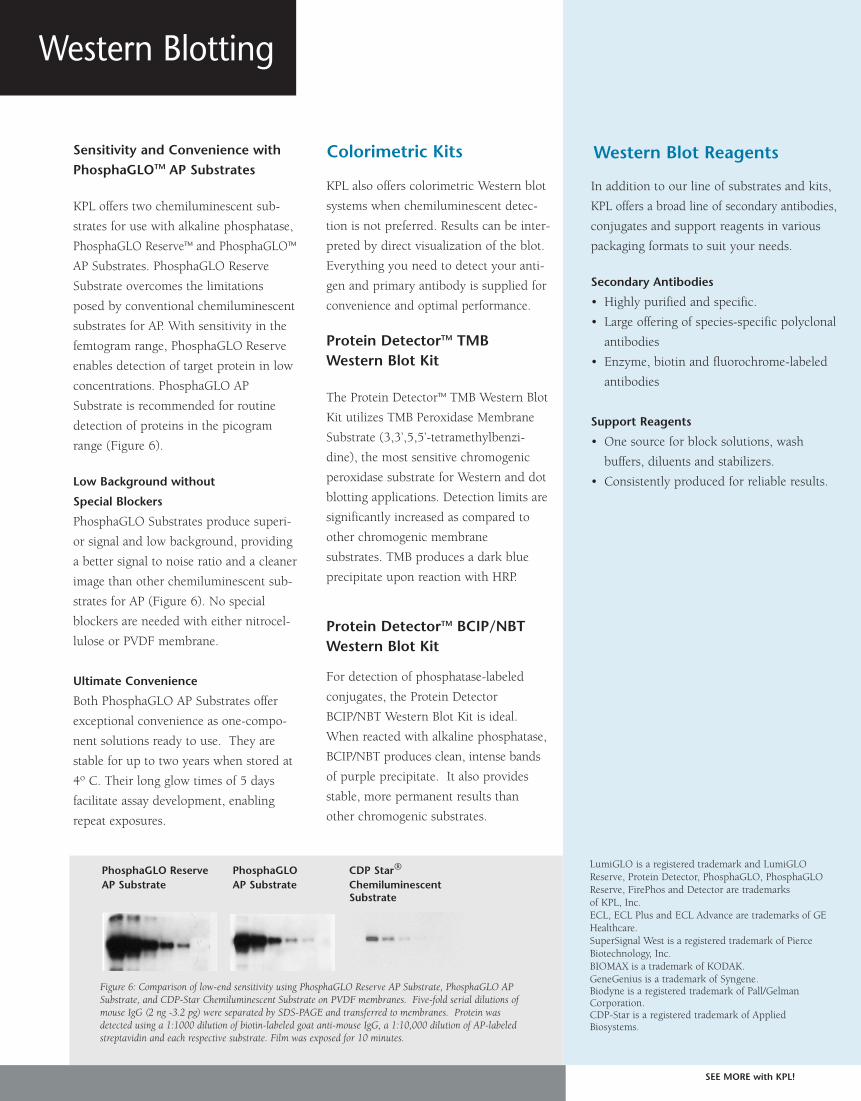
Figure 6: Comparison of low-end sensitivity using PhosphaGLO Reserve AP Substrate, PhosphaGLO APSubstrate, and CDP-Star Chemiluminescent Substrate on PVDF membranes. Five-fold serial dilutions ofmouse IgG (2 ng -3.2 pg) were separated by SDS-PAGE and transferred to membranes. Protein wasdetected using a 1:1000 dilution of biotin-labeled goat anti-mouse IgG, a 1:10,000 dilution of AP-labeledstreptavidin and each respective substrate. Film was exposed for 10 minutes.
Colorimetric Kits
PhosphaGLO Reserve PhosphaGLO CDP Star®
AP Substrate AP Substrate ChemiluminescentSubstrate
Western Blot Reagents
SEE MORE with KPL!

BCIP/NBT (5-Bromo-4-chloro-3-indolyl phosphate/
nitroblue tetrazolium) is the most commonly used
substrate for alkaline phosphatase (AP) detection in
Western blotting due to its high sensitivity and low
background staining. Traditional formulations of
BCIP/NBT produce a deep purple color when reacted
with AP conjugates.
Now KPL’s new BCIP/INT formulation, FirePhosTM
AP Membrane Substrate, forms a red precipitate, pro-
viding a choice of red or purple precipitating AP sub-
strates to better optimize your individual assays.
FirePhosTM AP Membrane Substrate
BCIP/NBT Phosphatase Substrate
Sensitive and reproducible
No matter which substrate you choose, you’ll achieve
sensitive and reproducible detection of proteins on
Western blots. Both substrates provide sensitivity in
the low nanogram range with minimal background
(Figure 1) , sharp band resolution and a clear image.
The color produced is stable and linear over a wide
dynamic range.
See RED and PURPLE in your Western blots!
SEE MORE with KPL!
AP Membrane Color Substrates
FirePhos AP BCIP/NBT Substrate BCIP/NBT SubstrateMembrane Substrate (1-Component) (3-Component)
Figure 1: KPL’s substrates for alkaline phosphatasedetection in Western blotting demonstrate high sen-sitivity and low background. A two-fold dilutionseries from 250 ng to 3.9 ng of mouse IgG was loadedonto a Tris-HCl gradient gel, then blotted onto nitro-cellulose. The antigen was detected with ReserveAPTM
Goat Anti-mouse Conjugate (KPL) followed byFirePhos AP Membrane and BCIP/NBT Substrates (1-component and 3-component).

Ordering Information
To order or for more information, contact us at 800.638.3167 / 301.948.7755,
fax 301.948.0169 or visit us at www.kpl.com.
FirePhos and ReserveAP are trademarks of KPL.
Catalog# Description Size
FirePhosTM AP Membrane Substrate
50-81-30 FirePhos Membrane Phosphatase Substrate (1-Component) 100 mL
50-81-40 FirePhos Membrane Phosphatase Substrate (1-Component) 400 mL
50-81-34 FirePhos Membrane Phosphatase Substratee (1-Component) 1 L
BCIP/NBT Phosphatase Membrane Substrate
50-81-18 BCIP/NBT Membrane Phosphatase Substrate (1-Component) 100 mL
50-81-07 BCIP/NBT Membrane Phosphatase Substrate (1-Component) 600 mL
50-81-10 BCIP/NBT Membrane Phosphatase Substrate (1-Component) 1 L
50-81-00 BCIP/NBT Membrane Phosphatase Substrate (3-Component) 300 mL
Membrane Blocking
71-83-00 Detector Block (5X) 240 mL
KPL’s colorimetric membrane substrates are part of our comprehensive line of Western blottng
kits and reagents. Ask about our AP chemiluminescent substrates that can significantly lower
the detection limit of an assay.
ReserveAPTM
Conjugates
Gaithersburg, MD 20878
Phone: 800.638.3167
Fax: 301.948.0169
www.kpl.com
ISO 9001:2000 Registered
ML348-02
For research use only.© 2007 KPL, Inc. All rights reserved.
Improve your assay performance even furtherwith ReserveAPTM-labeledAntibodies!
KPL’s new ReserveAPTM Alkaline
Phosphatase-labeled Conjugates
exhibit high potency and consistent
performance in immunoassays.
These conjugates are the result of
advances in our conjugation chem-
istry and offer high signal and low
background while meeting KPL’s
standards for stability and repro-
ducibility. They are ideal for
demanding immunoassays that
require high detection sensitivity,
including ELISA, Western blotting
and immunohistology. Visit
www.kpl.com for more information
on KPL’s ReserveAP conjugates.
SEE MORE with KPL!
SEE MORE with KPL!
Non-fading
FirePhos and BCIP/NBT produce a bright red or purple precipitate that can be
deposited on nitrocellulose and PVDF membranes. They resist fading when
exposed to light.
Convenient
These substrates are supplied as convenient one-component solutions that are
ready to use. KPL’s BCIP/NBT is also available as a three-component solution.
Both substrates are provided in several sizes to meet your needs.
AP Membrane Color Substrates
KPL offers two substrates commonly used to detect
horseradish peroxidase (HRP) conjugated antibodies
that bring complementary benefits to membrane
detection; TMB (3, 3´, 5, 5´-tetramethylbenzidene)
Membrane Substrate and 4 CN (4-chloro-1-naphthol)
Membrane Substrate. TMB substrate offers the most
sensitive colorimetric detection for blotting applica-
tions and is recommended when high signal and low
background are required. 4 CN substrate is highly
reliable and is notable for its characteristically low
background.
• TMB Membrane Substrates (1- and (3-Component)
In the presence of peroxidase conjugate, KPL’s TMB
Membrane Substrates produce a stable, dark blue pre-
cipitate at the site of a positive reaction, enabling
detection of as little as 50 pg of peroxidase. Our TMB
substrates are available as 1- and 3- component solu-
tions. 1-component TMB Peroxidase Substrate is ready
to use and contains hydrogen peroxide and buffer.
Our 3-component TMB contains TMB in an organic
base, H2O2 in a citric acid buffer and a third compo-
nent, TMB Membrane Enhancer. It provides sensitivi-
ty equivalent to 1-component TMB at an economic
price. The substrate may be adapted as a soluble sub-
strate for ELISA by omitting TMB Membrane
Enhancer. Both substrates perform equally well on
nitrocellulose and PVDF membranes.
• 4 CN Peroxidase Substrate (2-Component)
4 CN (4-chloro-1-napthol) Peroxidase Membrane
Substrate produces a purple precipitate at the reaction
site in the presence of peroxidase conjugates. Due to
its low background, 4 CN presents a desirable alter-
native to other, more sensitive substrates when back-
ground poses a problem.
Sensitivity, reliability and affordability!
SEE MORE with KPL!
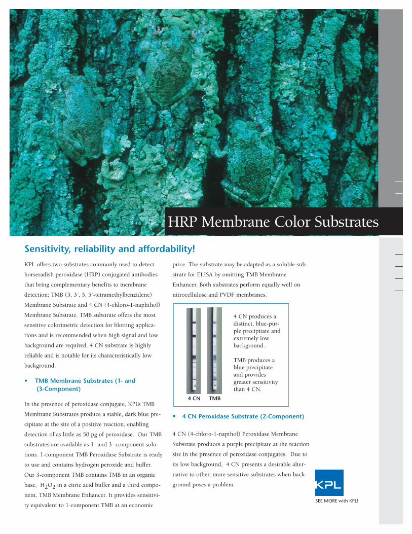
HRP Membrane Color Substrates
4 CN produces adistinct, blue-pur-ple precipitate andextremely lowbackground.
TMB produces ablue precipitateand providesgreater sensitivitythan 4 CN.
4 CN TMB

•
•
Benefits of KPL’s HRP Membrane Substrates
Ordering Information
To order or for more information on KPL’s line of protein detection products, contact
us at 800.638.3167 / 301.948.7755, fax 301.948.0169 or visit us at www.kpl.com.
Catalog# Description Size
50-77-18 TMB Membrane Substrate (1-Component) 100 mL
50-77-03 TMB Membrane Substrate (1-Component) 200 mL
50-77-04 TMB Membrane Substrate (1-Component) 1 L
50-77-00 TMB Membrane Substrate (3-Component) 440 mL
50-77-01 TMB Membrane Enhancer 40 mL
50-73-00 4 CN Substrate System (2-Component) 600 mL
50-73-04 4 CN Substrate System (2-Component) 2700 mL
Protein Detector TMB Western Blot Kit
54-11-50 Protein Detector TMB Western Blot Kit 2500 cm2
Detector Block
71-83-00 Detector Block 240 mL
KPL provides a broad range ofassay support reagents forWestern blot detection.Conjugate stabilizers, diluents,block and wash solutions areoffered in convenient stable liq-uid form to ensure accuratereproducible results, blot afterblot. Our Detector Block isideal for use as a diluent/blocksolution for Western blots,Southern blots and Northernblots on a variety of mem-branes.
• DetectorTM Block
Detector Block is recommend-ed as a general blocking agentin membrane-based detectionassays. It is especially usefulwhen traditional blocking solu-tions (i.e., milk, BSA, casein)reduce sensitivity or do not ade-quately block background.Detector Block can be used asboth a blocking solution and aconjugate diluent in membraneapplications.
Support Reagents
Gaithersburg, MD 20878
Phone: 800.638.3167
Fax: 301.948.0169
www.kpl.com
Protein Detector is a trademark of KPL.©2008 KPL, Inc. All rights reserved.ML353-01
SEE MORE with KPL!
ISO 9001:2000 Registered
4 CN Peroxidase Substrate
TMB Peroxidase Membrane Substrate
• Most sensitive colorimetric substrate for blotting• Delivers results at the picogram level• Recommended for both kinetic and endpoint ELISA • Contains no harmful organic solvents; safer and more sensitive
than DAB or AEC.• 1-Component TMB-no preparation required.
Ensures more consistent results• 3-Component TMB-same performance as 1-Component TMB
at an economic price. • Blots remain stable with minimal fading
• Easy mix-and-use, two-component solutions• Very low background, moderate sensitivity• High contrast image ideal for photography and publication

Lighten your work load with KPL’s new line of quality
buffers. Offered as convenient liquid concentrates, they
eliminate the need for time-consuming buffer preparation.
KPL buffers are manufactured and carefully controlled for
quality and consistent performance with our ISO 9001:2008-
registered quality management system. All buffers are con-
ductivity controlled. KPL offers a variety of buffers designed
for use in general immunoassay applications such as Western
blotting, gel electrophoresis, ELISA and sample preparation.
Save preparation time with KPL’s new line of quality buffers!
Immunoassay Buffers
SEE MORE with KPL!
Description 1X Composition Applications
10X Tris-Glycine 25 mM Tris, 192 mM glycine. For preparing a standard Western blot transfer buffer Transfer Buffer pH 8.3 (Towbin) and as a gel electrophoresis buffer for native
Tris-glycine gels without SDS. .
10X Tris-Glycine-SDS 25 mM Tris, 192 mM glycine, Running buffer for sodium dodecyl sulfate – polyacrylamide0.1% SDS. pH 8.3 gel electrophoresis (SDS-PAGE) of proteins.
10X Phosphate-Buffered 10 mM Phosphate,150 mM NaCl. Wash buffer for general immunoassay applicationsSaline (PBS) pH 7.4 and sample preparation. Where applicable, Tween 20
is a nonionic detergent that reduces nonspecificPhosphate-Buffered 10 mM Phosphate, 150 mM NaCl, binding and protein-protein interaction during wash steps.Tween 20, (PBST) 0.05% Tween 20. pH 7.4
10X Tris-Buffered Saline 50 mM Tris, 150 mM NaCl. (TBS) pH 7.6
10% Tween 20 10% Tween 20 Nonionic detergent additive for PBS and Western blotting wash solutions. Reduces nonspecific binding and protein- protein interactions.
See reverse side.

Catalog# Description Size
51-10-01 10X Tris-Glycine Transfer Buffer 1 L
51-10-02 5 L
51-11-01 10X Tris-Glycine SDS Buffer 1 L
51-11-02 5 L
51-13-01 10X Phosphate-Buffered Saline (PBS) 1 L
51-13-02 5 L
51-14-01 10X Phosphate-Buffered Saline with 200 mL
51-14-02 Tween 20 (PBST) 1 L
51-17-01 10X Tris-Buffered Saline (TBS) 1 L
51-17-02 5 L
51-12-01 10% Tween 20 200 mL
51-12-02 1 L
51-20-01 5% SDS 200 mL
50-86-05 20X SSC 1 L
51-15-01 10X Dulbecco's PBS (D-PBS) 1 L
51-15-02 5 L
51-16-01 10X Dulbecco's PBS with Tween 20 (D-PBST) 200 mL
51-16-02 1 L
51-18-01 10X Tris Buffered Saline with 0.5% Tween 20 200 mL
51-18-02 (TBST) 1 L
51-19-01 10X Tris Buffered Saline with 10% Tween 20 200 mL
51-19-02 (TBST) 1 L
Ordering Information
To order or for more information on KPL’s full line of protein detection products, contact us at800.638.3167 / 301.948.7755, fax 301.948.0169 or visit us at www.kpl.com.
Other Immunoassay Support
Reagents Available from KPL
ELISA and Western Blotting
Applications:
• AP and HRP Conjugate Stabilizers
• 10% BSA Diluent/Blocking Solution
Concentrate
• Milk/Diluent Blocking Solution
Concentrate
• 20X Wash Solution Concentrate
Western Blotting Applications:
• Detector Block
• Biotin Wash Kit
ELISA Applications:
• Coating Solution Concentrate
• Stop Solutions
Immunoassay Buffers
Gaithersburg, MD 20878
Phone: 800.638.3167
Fax: 301.948.0169
www.kpl.com
ML363-03
©2009 KPL, Inc. All rights reserved.SEE MORE with KPL!
ISO 9001:2008 Registered
KPL Quality Buffers (cont’d)
Take the time and effort out of your buffer preparation. Choose from our line of popular buffers andenjoy the benefits - convenience, reliability and confidence - that KPL’s pretested, quality buffersbring to your research.
Description 1X Composition Applications
5% SDS 5% Sodium Detergent surfactant used in preparing proteins for Dodecyl Sulfate sodium dodecyl sulfate – polyacrylamide gel electro-
phoresis (SDS-PAGE) and as an additive to transfer buffers in Western blotting. SDS increases the elution rate of proteins from the gel.
20X SSC 15 mM sodium citrate, Used in a variety of molecular biology applications. 150 mM NaCl Facilitates transfer of nucleic acids to membranes. in DEPC water. pH 7.0
10X Dulbecco's 8.5 mM sodium phosphate, For sample preparation and as a wash buffer in general PBS (D-PBS) 1.5 mM potassium phosphate, immunoassay, tissue, and cell culture applications.
137 mM NaCl. pH 7.4 Not formulated with magnesium or calcium salts. Where applicable, Tween 20 is a non-ionic detergent that reducesnonspecific binding.
10X Dulbecco's 8.5 mM sodium phosphate,with Tween 20. 1.5 mM potassium phosphate,(D-PBST) 137 mM NaCl, 2.7 mM KCl.
0.05% Tween 20. pH 7.4
10X Tris-Buffered 50 mM Tris, 150 mM NaCl, As a general wash buffer in immunoassay applications. Saline with 0.5% 0.05% Tween 20. pH 7.6 Tween 20 is a non-ionic detergent that reduces nonspecific Tween 20 (TBST) binding. The 10% Tween 20 formulation provides a more
stringent wash than standard TBST formulations. 10X Tris-Buffered 50 mM Tris, 150 mM NaCl,Saline with 10% 1.0% Tween 20. pH 7.6 Tween 20 (TBST)

KPL’s new ReserveAPTM Alkaline Phosphatase
(AP)-labeled antibody conjugates exhibit high
potency and consistent performance in
immunoassays. These conjugates are the result
of advances in our conjugation technology and
offer higher signal than our current line of AP
conjugates while meeting the same standards
for low background, stability and reproducibili-
ty. They are intended for demanding
immunoassays that require high detection sen-
sitivity, including ELISA, Western blotting and
immunohistology.
Higher Potency
ReserveAP Conjugates are affinity purified and
conjugated to the highest grade of alkaline
phosphatase. In our studies, they generate two-
to-three fold higher values than our current
line of AP conjugates in ELISA and outperform
AP conjugates offered by other manufacturers.
Higher conjugate dilutability is also observed
without loss of linearity, enabling precious
antigen or primary antibody conservation.
Consistent Performance
Reproducible antibody conjugation and consis-
tent performance are verified according to our
ISO 9001:2000-registered quality assurance
system. Lot consistency studies in which three
lots were studied by ELISA indicated minimal
variability.
Excellent Value
ReserveAP Conjugates provide high perform-
ance at an economical price.
Spice up your assay with our red hot high potency ReserveAPTM Conjugates!
ReserveAPTM Conjugates
SEE MORE with KPL!

ReserveAPTM Conjugates Ordering Information
Catalog# Description Size
0751-1001 Goat Anti-Human IgA (α) 1.0 mg
0751-1002 Goat Anti-Human IgG (γ) 1.0 mg
0751-1003 Goat Anti-Human IgM (µ) 1.0 mg
0751-1004 Goat Anti-Human IgE (ε) 1.0 mg
0751-1006 Goat Anti-Human IgG (H+L) 1.0 mg
0751-1007 Goat Anti-Human IgA+IgG+IgM (H+L) 1.0 mg
2151-1002 F(ab’)2 Anti-Human IgG (γ) 0.5 mg
4751-1002 Goat Anti-Human IgG (γ) liquid 1.0 mg
4751-1003 Goat Anti-Human IgM (µ) liquid 1.0 mg
4751-1006 Goat Anti-Human IgG (H+L) liquid 1.0 mg
0751-1802 Goat Anti-Mouse IgG (γ) HSA 1.0 mg
0751-1803 Goat Anti-Mouse IgM (µ) HSA 1.0 mg
0751-1806 Goat Anti-Mouse IgG (H+L) HSA 1.0 mg
0751-1809 Goat Anti-Mouse IgG +IgM (H+L) HSA 1.0 mg
4751-1802 Goat Anti-Mouse IgG (γ) HSA, liquid 1.0 mg
4751-1806 Goat Anti-Mouse IgG (H+L) HSA, liquid 1.0 mg
151-18-01 Goat Anti-Mouse IgA (α) HSA 0.5 mg
0751-1807 Goat Anti-Mouse IgA+IgG+IgM (H+L) HSA 1.0 mg
0751-1506 Goat Anti-Rabbit IgG (H+L) 1.0 mg
0751-1516 Goat Anti-Rabbit IgG (H+L) HSA 1.0 mg
4751-1506 Goat Anti-Rabbit IgG (H+L), liquid 1.0 mg
4751-1516 Goat Anti-Rabbit IgG (H+L) HSA, liquid 1.0 mg
HSA=Human Serum Adsorbed
AP Substrates
Gaithersburg, MD 20878
Phone: 800.638.3167
Fax: 301.948.0169
www.kpl.com
ISO 9001:2000 Registered
ML349-04
For research use only.©2007 KPL, Inc. All rights reserved.
SEE MORE with KPL!
Visit our website at www.kpl.com for a complete listing of ReserveAP conjugates. To
order or for more information on KPL’s protein research products, contact us at
800.638.3167 / 301.948.7755, FAX 301.948.0169 or visit us at www.kpl.com.
KPL offers a range of sensitive sub-
strates for the detection and quantifi-
cation of phosphatase (AP) activity.
They provide a choice of intense col-
ors for ELISA and blotting applica-
tions.
ELISA
• FirePhosTM AP Microwell
Substrate
• BluePhos® AP Microwell
Substrate
• pNPP Phosphatase Substrate
Blotting
• FirePhos AP Membrane
Substrate
• BCIP/NBT Phosphatase Substrate
• PhosphaGLO AP Substrate
• PhosphaGLOTM Reserve AP
Substrate
Whichever substrate you choose,
enjoy the benefits of excellent signal-
to-noise with higher sensitivity and
lower background than that of other
AP substrates. Visit www.kpl.com
for more information.
Chemiluminescent ELISA: A con-stant amount of antigen (mouse IgG)was coated onto a microtiter plate.The plate was probed with varyingconcentrations of goat anti-mouseIgG (H+L) AP conjugate, includingKPL’s standard AP conjugate andReserveAP™ conjugate, followed byKPL’s PhosphaGLO™ Chemilum-inescent AP Substrate.
Conclusion: The results indicate that ReserveAP™ conjugate offers superior performance to our stan-dard AP conjugate with a ~3-fold lower amount of conjugate required for detection.

KPL’s DyLightTM conjugates offer a brilliant
choice in a variety of multicolor detection ap-
plications, including fluorescence microscopy,
flow cytometry, Western blotting, ELISA and
array platforms. Our affinity purified antibod-
ies combined with a series of outstanding
DyLight dyes provide superior performance
over conventional CyDyeTM fluors, fluorescein
and rhodamine, with performance comparable
to that of Alexa Fluor® dyes (Figure 1). Enjoy
these advantages when you switch to KPL’s
DyLight Conjugates:
KPL offers eight DyLight dyes, including 405,
488, 549, 594, 633, 649, 680 and 800 with
well-differentiated excitation and emission
spectra. Our extensive line of over 170 DyLight
conjugates is available across a range of animal
species immunoglobulin, including human,
mouse, rabbit, rat, other species and strepta-
vidin. See back cover to find out what sets KPL’s
antibodies apart.
SEE MORE with KPL!
KPL’s DyLightTM Conjugates - A Brilliant Choice!
DyLightTM Fluorescent Conjugates
Image above: HMVEC-L primary endothelial cells. F-actin detected with DY554-Phalloidin (rendered green). Microtubules detected with anti-tubulinantibody and DyLight 649 conjugated goat anti-mouse IgG (red). Nuclei detected with DAPI (blue)
Figure 1. KPL DyLight 649 conjugates demonstrate comparablefluorescense intensity and photostability to Alexa 647 conjugatesin a FLISA.
•• High sensitivity, bright signal enable conjugate
conservation
•• More photostable than Cy, FITC; comparable to Alexa
•• High signal-to-noise ratios; low background
•• Wide selection of specific antibodies
•• Consistent, lot-to-lot performance
•• Narrow emission spectra enable specific, multicolor analysis
•• Compatible with a variety of buffers.
Antigen Concentration (ng/mL)
Comparison of DyLight 649 and Alexa 647Conjugates in a Performance Assay

Emission DyLight Dye Ex/Em(nm) Replaces
DyLight 405 400/420 Alexa 405 and Cascade Blue
DyLight 488 493/518 Alexa 488, Fluorescein and FITC
DyLight 549 550/568 Alexa 546, Alexa 555, Cy3, TRITC
DyLight 594 593/618 Alexa 594, Texas Red
DyLight 633 638/658 Alexa 633
DyLight 649 646/674 Alexa 647, Cy5
DyLight 680 682/715 Alexa 680, Cy5.5
DyLight 800 770/794 IRDye 800
KPL DyLight conjugates provide a choice of outstanding flu-
orescent conjugates with absorption spectra ranging from
405 nm to 800 nm. Their emission profiles correspond to
those of other commonly used fluorophores such as Alexa,
FITC and the Cy dyes. Narrow, defined emission spectra en-
able the use of multiple fluorescence labeling and simultane-
ous identification of several target molecules in the same
sample. Light output is comparable to IRDye and Alexa and
more intense than Cy Dyes, FITC or TRITC. DyLight dyes
demonstrate resistance to photobleaching, resulting in excel-
lent photostability as well as high solubility in aqueous solu-
tions across a range of pH values.
Anti-Mouse IgG (γ), HSA 072-03-18-02 072-04-18-02 072-05-18-02
Anti-Mouse IgG (H+L), HSA 072-08-18-06 072-03-18-06 072-04-18-06 072-09-18-06 072-10-18-06 072-05-18-06 072-06-18-06 072-07-18-06
Anti-Mouse IgG (H+L), HSA, 0.1 mg 042-08-18-06 042-03-18-06 042-04-18-06 042-09-18-06 042-10-18-06 042-05-18-06 042-06-18-06 042-07-18-06
Anti-Mouse IgG (H+L), RbSA, HSA 072-03-18-18 072-04-18-18 072-05-18-18 072-06-18-18 072-07-18-18
Anti-Mouse IgM (µ), HSA 072-03-18-03 072-04-18-03 072-05-18-03
Anti-Mouse IgG+IgM (H+L), HSA 072-03-18-09 072-04-18-09 072-05-18-09
F(ab’)2 Anti-Mouse IgG (γ), HSA 202-03-18-02 202-04-18-02 202-05-18-02
F(ab’)2 Anti-Mouse IgG (H+L), HSA 202-08-18-06 202-03-18-06 202-04-18-06 202-09-18-06 202-10-18-06 202-05-18-06 202-06-18-06 202-07-18-06
Anti-Human IgG (H+L) 072-08-10-06 072-03-10-06 072-04-10-06 072-09-10-06 072-10-10-06 072-05-10-06 072-06-10-06 072-07-10-06
Anti-Human IgG (H+L), 0.1 mg 042-08-10-06 042-03-10-06 042-04-10-06 042-09-10-06 042-10-10-06 042-05-10-06 042-06-10-06 042-07-10-06
Anti-Human IgG (γ) 072-03-10-02 072-04-10-02 072-05-10-02
Anti-Human IgM (µ) 072-03-10-03 072-04-10-03 072-05-10-03
F(ab’)2 Anti-Human IgG (γ) 202-03-10-02 202-04-10-02 202-05-10-02
F(ab’)2 Anti-Human IgM (µ) 202-03-10-03 202-04-10-03 202-05-10-03
F(ab’)2 Anti-Human IgG (H+L) 202-08-10-06 202-03-10-06 202-04-10-06 202-09-10-06 202-10-10-06 202-05-10-06 202-06-10-06 202-07-10-06
Anti-Rabbit IgG (H+L) 072-08-15-06 072-03-15-06 072-04-15-06 072-09-15-06 072-10-15-06 072-05-15-06 072-06-15-06 072-07-15-06
Anti-Rabbit IgG (H+L), 0.1 mg 042-08-15-06 042-03-15-06 042-04-15-06 042-09-15-06 042-10-15-06 042-05-15-06 042-06-15-06 042-07-15-06
Anti-Rabbit IgG (H+L), HSA 072-03-15-16 072-04-15-16 072-05-15-16 072-06-15-16 072-07-15-16
F(ab’)2 Anti-Rabbit IgG (H+L), HSA 202-08-15-16 202-03-15-16 202-04-15-16 202-09-15-16 202-10-15-16 202-05-15-16 202-06-15-16 202-07-15-16
Anti-Rat IgG (H+L) 072-08-16-06 072-03-16-06 072-04-16-06 072-09-16-06 072-10-16-06 072-05-16-06 072-06-16-06 072-07-16-06
Anti-Rat IgG (H+L), 0.1 mg 042-03-16-06
Anti-Guinea Pig IgG (H+L) 072-03-17-06 072-04-17-06 072-05-17-06 072-06-17-06 072-07-17-06
Anti-Chicken IgG (H+L) 072-08-24-06 072-03-24-06 072-04-24-06 072-09-24-06 072-10-24-06 072-05-24-06 072-06-24-06 072-07-24-06
Anti-Dog IgG (H+L) 072-03-19-06 072-04-19-06 072-05-19-06 072-06-19-06 072-07-19-06
Anti-Goat IgG (H+L) 072-08-13-06 072-03-13-06 072-04-13-06 072-09-13-06 072-10-13-06 072-05-13-06 072-06-13-06 072-07-13-06
Anti-Horse IgG (H+L) 072-03-21-06 072-04-21-06 072-05-21-06 072-06-21-06 072-07-21-06
Rabbit Anti-Sheep IgG (H+L) 072-03-23-06 072-04-23-06 072-05-23-06 072-06-23-06 072-07-23-06
Anti-Swine IgG (H+L) 072-03-14-06 072-04-14-06 072-05-14-06 072-06-14-06 072-07-14-06
Streptavidin 072-08-30-00 072-03-30-00 072-04-30-00 072-09-30-00 072-10-30-00 072-05-30-00 072-06-30-00 072-07-30-00
Streptavidin, 0.1 mg 042-08-30-00 042-03-30-00 042-04-30-00 042-09-30-00 042-10-30-00 042-05-30-00 042-06-30-00 042-07-30-00
DyLightTM FluorescentConjugates
DyLight Conjugated Affinity PurifiedAntibodies and Streptavidin
nm = nanometersIR = infrared
Description DyLight 405* DyLight 488 DyLight 549 DyLight 594* DyLight 633* DyLight 649 DyLight 680 DyLight 800
SEE MORE with KPL!
*Coming soon!
Blue
Green
Yellow
Orange
Red
Red
Near IR
Infrared
Ordering Information
HSA = human serum adsorbedRbSA = rabbit serum adsorbedDyLight antibody conjugates are made in goat exceptanti-goat and anti-sheep antibodies are made in rabbit.Supplied in 1.0 mg lyophilized form except select 0.1mg sizes.
DyLight Dyes Emission Spectra
Near Infrared and Infrared Fluorophores DyLight680 and 800 Conjugates Offer Sensitive MulticolorImaging in Western Blotting
DyLight 680 and 800 dyes emit in the near infrared and
infrared ranges of the light spectrum respectively and are
ideal for multicolor protein detection in Western blot-
ting. Unlike fluorescent conjugates that emit in the visi-
ble range, DyLight 680 and 800 conjugates provide a
unique set of advantages:
•• Secondary antibodies with defined specificity and
sensitivity
•• Brighter signal than visible fluorescence
•• Virtually no background autofluorescence from
membranes or most biological specimens
Effective Alternative to Chemiluminescence
•• Broader dynamic range than chemiluminescence
•• Quantitation accuracy superior to traditional
methods
•• Easy to assay multiple proteins simultaneously on
one Western blot
•• Cost-effective - eliminates need for chemilumin-
escence substrates, film and darkroom
•• Clean results - no bleeding from consecutive lanes
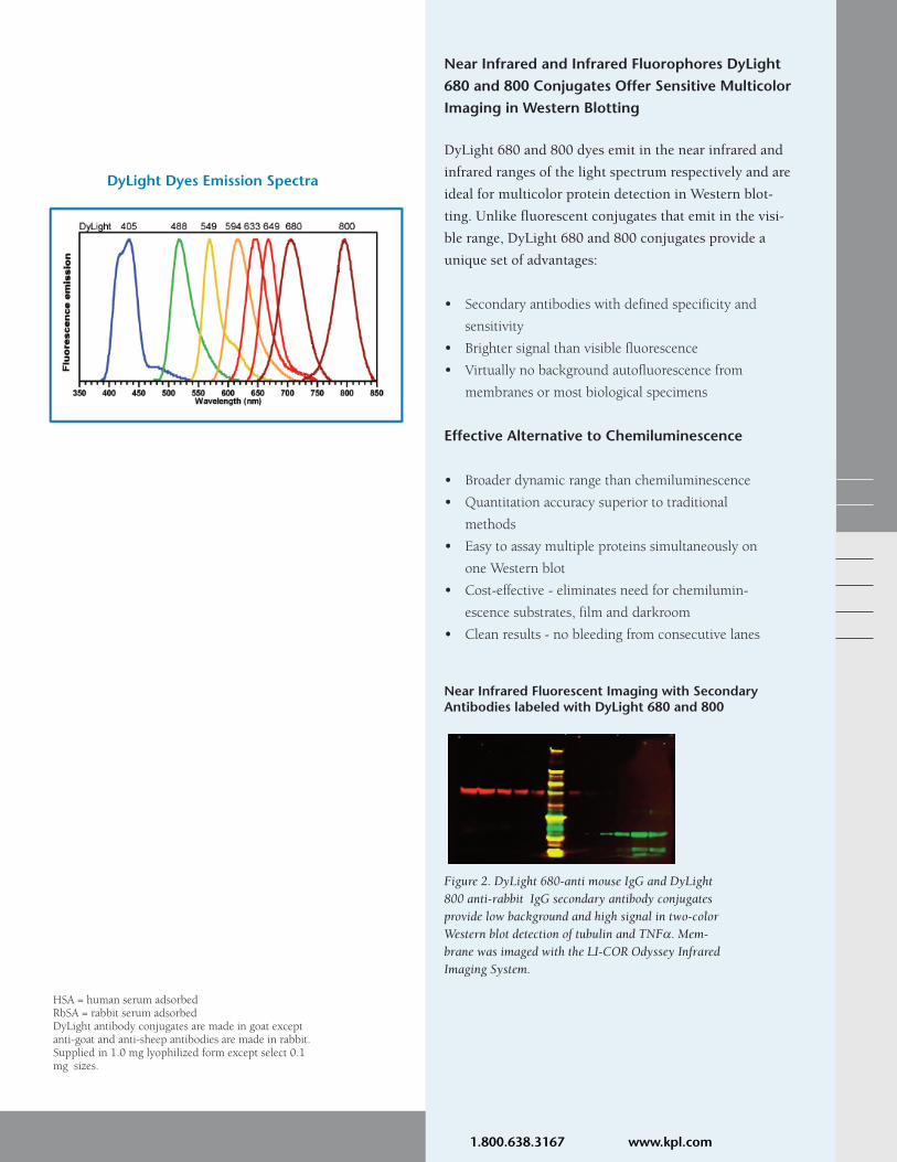
Figure 2. DyLight 680-anti mouse IgG and DyLight800 anti-rabbit IgG secondary antibody conjugatesprovide low background and high signal in two-colorWestern blot detection of tubulin and TNFα. Mem-brane was imaged with the LI-COR Odyssey InfraredImaging System.
Near Infrared Fluorescent Imaging with SecondaryAntibodies labeled with DyLight 680 and 800
SEE MORE with KPL! 1.800.638.3167 www.kpl.com
Gaithersburg, MD
Phone: 800.638.3167/301.948.7755
Fax: 301.948.0169
www.kpl.com
SEE MORE with KPL!
ISO 9001:2008 Registered
DyLightTM FluorescentConjugates
KPL Antibodies-What Sets Them Apart
ML356-02For research use only.DyLight is a trademark of Thermo Fisher Scientific Inc. and its subsidiaries. Cy and Cy Dye are trademarks of GE Healthcare.Alexa Fluor is a registered trademark of Invitrogen.2009 KPL, Inc. All rights reserved.
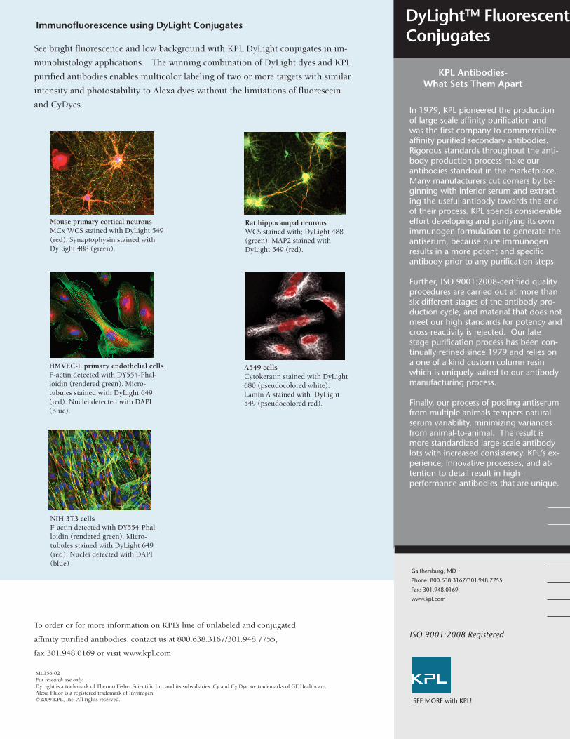
Rat hippocampal neuronsWCS stained with; DyLight 488(green). MAP2 stained withDyLight 549 (red).
Mouse primary cortical neuronsMCx WCS stained with DyLight 549(red). Synaptophysin stained withDyLight 488 (green).
HMVEC-L primary endothelial cellsF-actin detected with DY554-Phal-loidin (rendered green). Micro-tubules stained with DyLight 649(red). Nuclei detected with DAPI(blue).
NIH 3T3 cells F-actin detected with DY554-Phal-loidin (rendered green). Micro-tubules stained with DyLight 649(red). Nuclei detected with DAPI(blue)
A549 cells Cytokeratin stained with DyLight680 (pseudocolored white).Lamin A stained with DyLight549 (pseudocolored red).
Immunofluorescence using DyLight Conjugates
See bright fluorescence and low background with KPL DyLight conjugates in im-
munohistology applications. The winning combination of DyLight dyes and KPL
purified antibodies enables multicolor labeling of two or more targets with similar
intensity and photostability to Alexa dyes without the limitations of fluorescein
and CyDyes.
To order or for more information on KPL’s line of unlabeled and conjugated
affinity purified antibodies, contact us at 800.638.3167/301.948.7755,
fax 301.948.0169 or visit www.kpl.com.
In 1979, KPL pioneered the productionof large-scale affinity purification andwas the first company to commercializeaffinity purified secondary antibodies.Rigorous standards throughout the anti-body production process make ourantibodies standout in the marketplace.Many manufacturers cut corners by be-ginning with inferior serum and extract-ing the useful antibody towards the endof their process. KPL spends considerableeffort developing and purifying its ownimmunogen formulation to generate theantiserum, because pure immunogenresults in a more potent and specificantibody prior to any purification steps.
Further, ISO 9001:2008-certified qualityprocedures are carried out at more thansix different stages of the antibody pro-duction cycle, and material that does notmeet our high standards for potency andcross-reactivity is rejected. Our latestage purification process has been con-tinually refined since 1979 and relies ona one of a kind custom column resinwhich is uniquely suited to our antibodymanufacturing process.
Finally, our process of pooling antiserumfrom multiple animals tempers naturalserum variability, minimizing variancesfrom animal-to-animal. The result ismore standardized large-scale antibodylots with increased consistency. KPL’s ex-perience, innovative processes, and at-tention to detail result in high-performance antibodies that are unique.

Cy Dyes offer intense fluorescence when coupled
with KPL’s affinity purified antibodies and strepta-
vidin. The sensitivity and reproducibility of KPL
antibodies combined with the brightness of Cy3 and
Cy5 dyes produce an exceptional set of conjugates
ideal for multiple labeling experiments. Dye/protein
ratios have been established to ensure optimal fluo-
rescence with minimal background. They present
maximum excitation/emission spectra at 550/570
nm (Cy3) and 650/670 nm (Cy5).
Cy dyes are excellent alternatives to most other fluor-
rescein dyes as they are brighter and offer greater
photostability. Cy Dye conjugates are used in both
visual and image analysis fluorescent microscopy
and in situ hybridization. Cy3 conjugates are ideally
used in visual color applications, whereas Cy5 con-
jugates emit in the far red spectrum and are not easi-
ly visualized.
Benefits of KPL Cy Dye Conjugates
High performance conjugates – optimized dye-protein ratios ensure high signal-to-noise.
Intense fluorescence – offers greater sensitivity than TRITC conjugates.
Narrow emission spectra – enables sensitive, multi-color analysis.
Excellent photostability – more photostable than TRITC conjugates.
Consistent performance – minimal lot-to-lot vari-ation reduces need for assay optimization.
Buffer stability – after rehydration, conjugates are stable at pH 4-9.
Instrument compatibility – excitation and emis-sion spectra correspond with standard filter sets and laser settings.
KPL CyTM Dye Conjugates- Improve your assay and brighten your day!
SEE MORE with KPL!
Cy3 and Cy5 Conjugates
Fluo
resc
ence
Wavelength (nm)
Cy3 Cy5ExEm
Image above: Confocal fluorescence micrograph of HeLa cells stained with monoclonal antibody against mitochondria enzyme and Cy3-
conjugated anti-mouse antibody (red); rabbit polyclonal antibody to histones in DNA and Cy5-conjugated anti-rabbit antibody (blue).

Ordering Information
To order or for more information on KPL’s line of unlabeled and conjugated
affinity purified antibodies, contact us at 800.638.3167 / 301.948.7755, fax
301.948.0169 or visit us at www.kpl.com.
Cy3 and Cy5 Conjugates
ML368-01For research use only.Cy Dye is a trademark of GE Healthcare.©2009 KPL, Inc. All rights reserved.
Cytomegalovirus-infected cellsdetected with a biotinylated CMVprobe and the DNADetectorTM
Fluorescent in situ Hybridization Kitusing Cy3-Strept-avidin and DAPI.
Signal DetectionCy3 is excited maximally at 550 nmand fluoresces maximally at 570 nm.It is excited to about 50% of maxi-mum with an argon laser (514 nm or528 nm lines), or to about 75% ofmaximum with a helium/neon laser(543 nm line) or mercury lamp (546nm line).
Cy5 is excited maximally at 650 nmand fluoresces maximally at 670 nm.It is excited to about 98% of maxi-mum with a krypton/argon laser (647nm line) or to about 63% of maxi-mum with a helium/neon laser (633nm line). Cy5 produces minimal auto-fluorescence of biological specimensin this region of the spectrum.
A confocal microscope equipped withthe appropriate laser for excitationand a far-red detector enable doublelabeling with Cy3 and Cy5.
Gaithersburg, MD 20878
Phone: 800.638.3167
Fax: 301.948.0169
www.kpl.com
ISO 9001:2000 Registered
SEE MORE with KPL!
Anti-Mouse IgG (γ), HSA 072-01-18-02 072-02-18-02
F(ab’)2 Anti-Mouse IgG (γ), HSA 202-01-18-02 202-02-18-02
Anti-Mouse IgG (H+L), HSA 072-01-18-06 072-02-18-06
F(ab’)2 Anti-Mouse IgG (H+L), HSA 202-01-18-06 202-02-18-06
Anti-Mouse IgG (H+L), RbSA, HSA 072-01-18-18 072-02-18-18
Anti-Mouse IgM (µ), HSA 072-01-18-03 072-02-18-03
Anti-Mouse IgG+IgM (H+L), HSA 072-01-18-09 072-02-18-09
Anti-Rabbit IgG (H+L) 072-01-15-06 072-02-15-06
F(ab’)2 Anti-Rabbit IgG (H+L), HSA 202-01-15-16 202-02-15-16
Anti-Rabbit IgG (H+L), HSA 072-01-15-16 072-02-15-16
Anti-Rat IgG (H+L) 072-01-16-06 072-02-16-06
F(ab’)2 Anti-Human IgG (H+L) 202-01-10-06 202-02-10-06
Anti-Human IgG (γ) 072-01-10-02 072-02-10-02
F(ab’)2 Anti-Human IgG (γ) 202-01-10-02 202-02-10-02
Anti-Human IgM (µ) 072-01-10-03 072-02-10-03
F(ab’)2 Anti-Human IgM (µ) 202-01-10-03 202-02-10-03
Anti-Guinea Pig IgG (H+L) 072-01-17-06 072-02-17-06
Anti-Chicken IgG (H+L) 072-01-24-06 072-02-24-06
Anti-Horse IgG (H+L) 072-01-21-06 072-02-21-06
Anti-Swine IgG (H+L) 072-01-14-06 072-02-14-06
Anti-Dog IgG (H+L) 072-01-19-06 072-02-19-06
Anti-Sheep IgG (H+L) 072-01-23-06 072-02-23-06
Anti-Goat IgG (H+L) 072-01-13-06 072-02-13-06
Streptavidin 072-01-30-00 072-02-30-00
Description Cy3 Cy5
HSA=human serum adsorbed RbSA=rabbit serum adsorbedCy Dye antibody conjugates are made in goat except anti-goat and anti-sheep antibodiesmade in rabbit. Supplied in 1 mg lyophilized form.
Microplates were coated with serially diluted mouse IgG at the indicated concentrations. Conjugates at aconcentration of 0.01 mg/mL were applied and incubated for 30 minutes. Fluorescence was measured witha Perkin Elmer VICTOR 3 Multilabel Plate Reader.
Excellent PerformanceAs demonstrated below KPL Cy3- and Cy5-labeled conjugates produce brighter fluorescence thanthose of other suppliers.
Since 1979 KPL has provided quality affinity
purified antibodies to researchers worldwide.
Over the years we have refined our production
process to provide antibodies with high potency
and consistent performance in immunoassays.
From the start KPL gives careful consideration
to immunogen preparation, using a highly
purifed formulation to generate antiserum. KPL
pools antiserum from multiple animals to
reduce natural animal to animal serum variabili-
ty. During the purification process our ISO
9001:2008-certified quality procedures impose
rigorous standards for potency and cross-reac-
tivity. The result is standardized antibody lots
with excellent reproducibility.
Our extensive line of peroxidase (HRP) conju-
gates is available across a range of animal
species, including human, mouse, rabbit and rat
antibodies, as well as other animal species and
streptavidin. They are affinity purified and in
some cases further adsorbed to minimize cross-
reactivity between animal species or shared
reactivity with other immunoglobulin classes.
HRP-labeled F(ab’)2 fragment antibodies are
offered for assays requiring extremely low back-
ground and absence of F(c)-mediated binding.
KPL reacts HRP of the highest quality with
affinity purified antibodies and streptavidin
using the periodate method of Nakane and
Kawaoi. Special features of HRP include:
• faster catalytic rate than alkaline phosphatase
• generates more product in shorter incubation
times
• provides maximum sensitivity, low nonspe-
cific binding
• ideal for ELISA, Western blotting and
immunohistology applications.
KPL Peroxidase Conjugates: Time-tested, Sensitive and Reliable
Peroxidase Conjugates
SEE MORE with KPL!

Peroxidase Conjugates Ordering Information(Partial listing)
Peroxidase Conjugates
Gaithersburg, MD 20878
Phone: 800.638.3167
Fax: 301.948.0169
www.kpl.com
ISO 9001:2008 Registered
For research use only.©2009 KPL, Inc. All rights reserved.ABTS is a registered trademark of Roche Biochemicals.ML371-01
SEE MORE with KPL!
Visit our website at www.kpl.com for a complete listing of HRP-labeled antibodies.
To order or for more information on KPL’s protein research products, contact us at
800.638.3167 / 301.948.7755, FAX 301.948.0169 or visit us at www.kpl.com.
KPL offers a range of sensitive
substrates for use with HRP conju-
gates. They provide a choice of
intense colors for ELISA, blotting
and cell staining applications.
ELISA
• ABTS® 1- and 2-Component
Microwell Peroxidase Substrates
• SureBlueTM TMB Peroxidase
Substrate
• SureBlue ReserveTM TMB
Peroxidase Substrate
• TMB Peroxidase Substrate
Blotting
• 4 CN Peroxidase Substrate
• TMB Membrane Peroxidase
Substrate
• LumiGLO®Chemiluminescent
Substrate
• LumiGLO ReserveTM
Chemiluminescent Substrate
Whichever substrate you choose,
enjoy the benefits of excellent signal-
to-noise and reproducibility.
Catalog# Description Size
04-10-06 HRP-labeled Goat Anti-Human IgG (H+L) 0.1 mg
04-10-17 HRP-labeled Goat Anti-Human IgA+IgG+IgM (H+L), MSA 0.1 mg
04-10-20 HRP-labeled Goat Anti-Human IgG (Fc) 0.1 mg
074-1002 HRP-labeled Goat Anti-Human IgG (γ) 1.0 mg
074-1003 HRP-labeled Goat Anti-Human IgM (μ) 1.0 mg
074-1004 HRP-labeled Goat Anti-Human IgE (ε) 1.0 mg
074-1006 HRP-labeled Goat Anti-Human IgG (H+L) 1.0 mg
074-1007 HRP-labeled Goat Anti-Human IgA+IgG+IgM (H+L) 1.0 mg
14-10-01 HRP-labeled Goat Anti-Human IgA (α) 0.5 mg
214-1002 HRP-labeled F(ab’)2 Goat Anti-Human IgG (γ) 0.5 mg
214-1003 HRP-labeled F(ab’)2 Goat Anti-Human IgM (μ) 0.5 mg
214-1006 HRP-labeled F(ab’)2 Goat Anti-Human IgG (H+L) 0.5 mg
474-1002 HRP-labeled Goat Anti-Human IgG (γ), Liquid 1.0 mL
474-1003 HRP-labeled Goat Anti-Human IgM (μ), Liquid 1.0 mL
474-1006 HRP-labeled Goat Anti-Human IgG (H+L), Liquid 1.0 mL
04-18-06 HRP-labeled Goat Anti-Mouse IgG (H+L), HSA 0.1 mg
04-18-15 HRP-labeled Goat Anti-Mouse IgG (H+L), RtSA, HSA 0.1 mg
04-18-18 HRP-labeled Goat Anti-Mouse IgG (H+L), RbSA, HSA 0.1 mg
074-1802 HRP-labeled Goat Anti-Mouse IgG (γ), HSA 1.0 mg
074-1803 HRP-labeled Goat Anti-Mouse IgM (μ), HSA 1.0 mg
074-1806 HRP-labeled Goat Anti-Mouse IgG (H+L), HSA 1.0 mg
074-18-061 HRP-labeled Goat Anti-Mouse IgG (H+L), XSA 1.0 mg
074-1807 HRP-labeled Goat Anti-Mouse IgA+IgG+IgM (H+L), HSA 1.0 mg
074-1809 HRP-labeled Goat Anti-Mouse IgG+IgM (H+L), HSA 1.0 mg
14-18-01 HRP-labeled Goat Anti-Mouse IgA (α), HSA 0.5 mg
214-1802 HRP-labeled F(ab’)2 Goat Anti-Mouse IgG (γ), HSA 0.5 mg
214-1806 HRP-labeled F(ab’)2 Goat Anti-Mouse IgG (H+L), HSA 0.5 mg
474-1802 HRP-labeled Goat Anti-Mouse IgG (γ), HSA, Liquid 1.0 mL
474-1806 HRP-labeled Goat Anti-Mouse IgG (H+L), HSA, Liquid 1.0 mL
074-1506 HRP-labeled Goat Anti-Rabbit IgG (H+L) 1.0 mg
074-15-061 HRP-labeled Goat Anti-Rabbit IgG (H+L), XSA 1.0 mg
074-1516 HRP-labeled Goat Anti-Rabbit IgG (H+L), HSA 1.0 mg
214-1516 HRP-labeled F(ab’)2 Goat Anti-Rabbit IgG (H+L), HSA 0.5 mg
474-1506 HRP-labeled Goat Anti-Rabbit IgG (H+L), Liquid 1.0 mL
474-1516 HRP-labeled Goat Anti-Rabbit IgG (H+L), HSA, Liquid 1.0 mL
14-30-00 HRP-labeled Streptavidin 0.5 mg
474-3000 HRP-labeled Streptavidin, Liquid, Molecular Grade 1.0 mL

Ordering Information
Catalog # Description Size
Protein Detector™ Western Blotting KitsEach kit includes anti-mouse and anti-rabbit conjugates, DetectorBlock, Wash Solution Concentrate and Substrate.
Phosphatase Chromogenic
55-11-50 BCIP/NBT Western Blot Kit 2500 cm2
Peroxidase Chromogenic
54-11-50 TMB Western Blot Kit 2500 cm2
Peroxidase Chemiluminescent54-12-50 LumiGLO® Western Blot Kit 2500 cm2
54-13-50 LumiGLO ReserveTM Western 2400 cm2
Blot Kit
Related Reagents and Kits
Antibody ConjugatesAll antibodies listed below are produced in goat.For a complete antibody listing, refer to KPL’s Product Catalog.
Phosphatase-labeled475-1006 Anti-Human IgG (H+L) 1.0 mL, liquid
475-1806 Anti-Mouse IgG (H+L), HSA 1.0 mL, liquid
475-1506 Anti-Rabbit IgG (H+L) 1.0 mL, liquid
Peroxidase-labeled474-1006 Anti-Human IgG (H+L) 1.0 mL, liquid
474-1806 Anti-Mouse IgG (H+L) HSA 1.0 mL, liquid
474-1506 Anti-Rabbit IgG (H+L) 1.0 mL, liquid
Biotin-labeled16-10-06 Anti-Human IgG (H+L) 0.5 mg
176-1006 Anti-Human IgG (H+L) 2.0 mg
Labeled Streptavidin474-3000 HRP-labeled 1.0 mL, liquid
475-3000 AP-labeled 1.0 mL, liquid
Catalog # Description Size
Substrates for Western Blotting
Phosphatase Colorimetric Substrates50-81-18 BCIP/NBT Substrate 100 mL
50-81-07 BCIP/NBT Substrate 600 mL
50-81-30 FirePhosTM Membrane AP Substrate 100 mL
50-81-40 FirePhos Membrane AP Substrate 400 mL
50-81-34 FirePhos Membrane AP Substrate 1000 mL
Phosphatase Chemiluminescent Substrates55-60-03 PhosphaGLOTM AP Substrate 30 mL
55-60-04 PhosphaGLO AP Substrate 100 mL
55-60-01 PhosphaGLO Reserve AP Substrate 30 mL
55-60-02 PhosphaGLO Reserve AP Substrate 100 mL
Peroxidase Chromogenic Substrates50-77-18 TMB Membrane Substrate 100 mL
50-77-03 TMB Membrane Substrate 200 mL
50-73-00 4 CN Substrate 600 mL
50-73-04 4 CN Substrate 2700 mL
Peroxidase Chemiluminescent Substrates54-61-02 LumiGLO Chemiluminescent Substrate 60 mL
54-61-00 LumiGLO Chemiluminescent Substrate 240 mL
54-61-01 LumiGLO Chemiluminescent Substrate 720 mL
54-71-00 LumiGLO Reserve Substrate Kit 2400 cm2
54-71-01 LumiGLO Reserve Substrate Kit 600 cm2
Assay Support Reagents50-84-00 Coating Solution Concentrate 50 mL
54-15-01 HRPStabilizer 200 mL
55-15-00 APStabilizer 200 mL
50-61-00 10% BSA Diluent/Blocking Solution 200 mLConcentrate
50-82-01 Milk Diluent/Blocking Solution 200 mLConcentrate
71-83-00 DetectorTM Block (5X) 240 mL
50-63-00 Wash Solution Concentrate (20X) 800 mL
50-63-06 Biotin Wash Solution Concentrate (10X) 200 mL
60-00-50 Biodyne® B Nylon Membrane 1 roll
HSA=human serum adsorbed.
To order or for more information,call KPL at 800.638.3167, 301.948.7755,Fax: 301.948.0169. www.kpl.comor contact your local sales partner.
ML282-04SEE MORE with KPL!