week 1: chapter 18 the endocrine system - collin...
TRANSCRIPT
1/13/15
1
BIOL. 2402 Anatomy & Physiology
WEEK 1: Chapter 18
The Endocrine System
Collin College
Coordinate of the functions of the body occurs via different systems.
• releases messenger molecules called hormones into the bloodstream • bloodstream transports this to virtual all cells to see • action of the messenger depends on the presence of specific receptors
for these hormones • Hormones act slower over a period of seconds to several hours • Lingering effect of the Endocrine system is longer lasting
• operates via electrical impulses and neurotransmitters • excites or inhibits neurons, muscles and glands • nerve impulses produce their effect quite rapidly, within millisecond • the lingering effect of the N.S. stimulus is very short
Nervous system :
Endocrine System :
Coordination of Body Functions
1/13/15
2
Additional mechanisms are present
Coordination of Body Functions
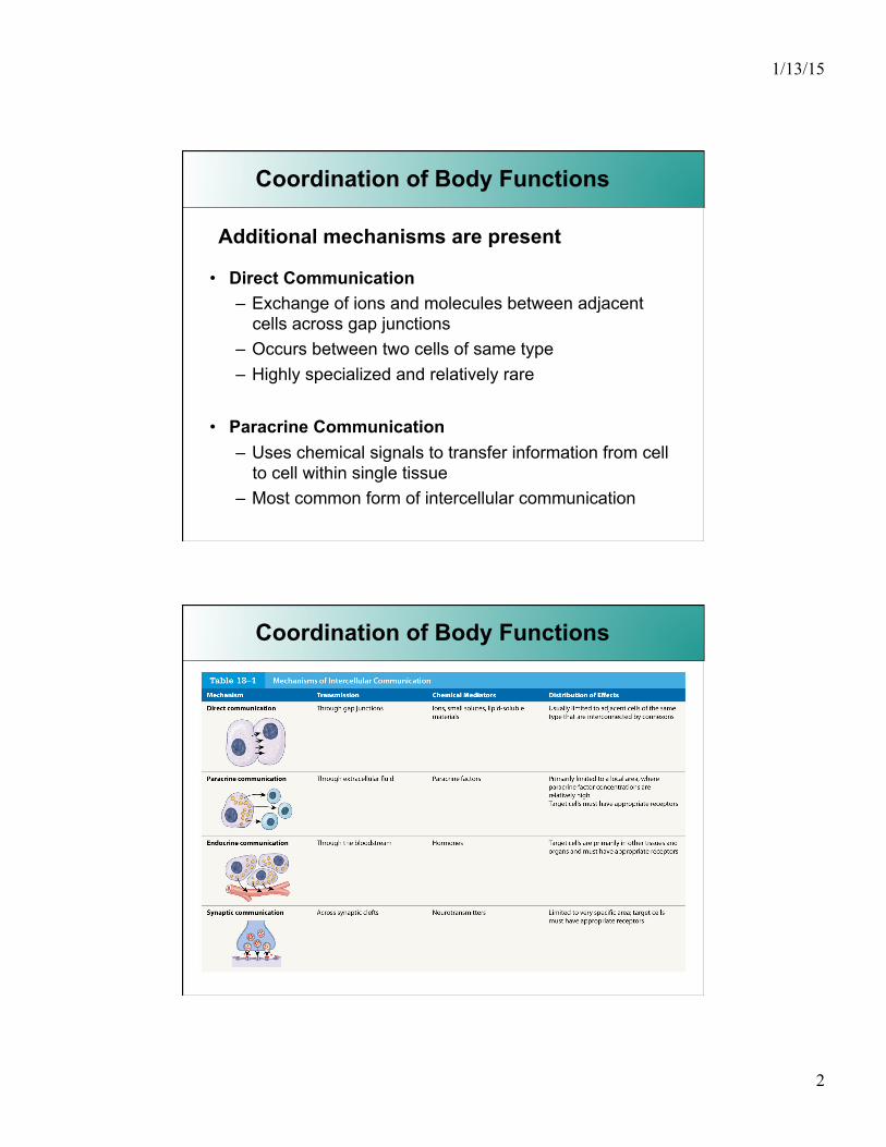
• Direct Communication – Exchange of ions and molecules between adjacent
cells across gap junctions – Occurs between two cells of same type – Highly specialized and relatively rare
• Paracrine Communication – Uses chemical signals to transfer information from cell
to cell within single tissue – Most common form of intercellular communication
Coordination of Body Functions
1/13/15
3
Endocrine System
• The Endocrine System Regulates long-term processes
such as Growth …. Development ……Reproduction
• The Endocrine system includes all cells and tissues which produce and secrete hormones or agents that have an effect beyond their tissue of origin.
• Hormones are produced in the cells of Endocrine Glands. The product is released into the extracellular space where it diffuses into a rich network of capillaries and lymph vessels.
Endocrine System
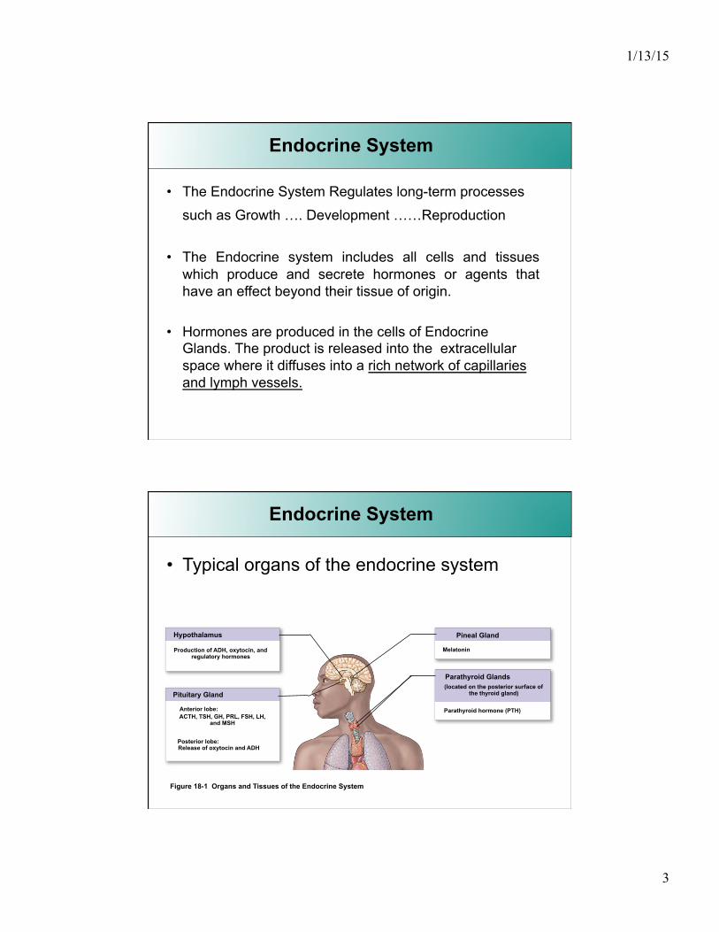
• Typical organs of the endocrine system
Figure 18-1 Organs and Tissues of the Endocrine System
Hypothalamus
Production of ADH, oxytocin, and regulatory hormones
Pituitary Gland
Anterior lobe: ACTH, TSH, GH, PRL, FSH, LH,
and MSH
Posterior lobe: Release of oxytocin and ADH
Parathyroid Glands (located on the posterior surface of
the thyroid gland)
Parathyroid hormone (PTH)
Pineal Gland
Melatonin
1/13/15
4
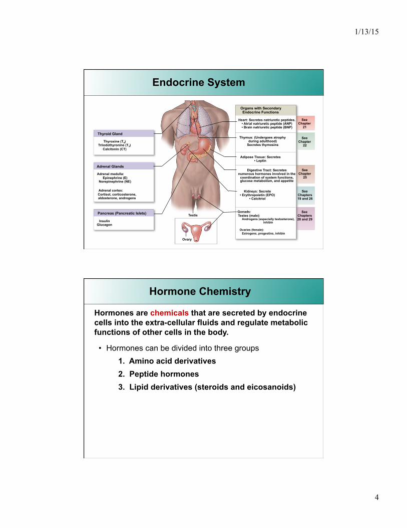
Figure 18-1 Organs and Tissues of the Endocrine System
Thyroid Gland
Thyroxine (T4) Triiodothyronine (T3)
Calcitonin (CT)
Adrenal Glands
Adrenal medulla: Epinephrine (E)
Norepinephrine (NE)
Adrenal cortex: Cortisol, corticosterone, aldosterone, androgens
Insulin Glucagon
Pancreas (Pancreatic Islets) Testis
Ovary
Thymus: (Undergoes atrophy during adulthood)
Secretes thymosins
Adipose Tissue: Secretes • Leptin
Digestive Tract: Secretes numerous hormones involved in the
coordination of system functions, glucose metabolism, and appetite
Kidneys: Secrete • Erythropoietin (EPO)
• Calcitriol
Gonads: Testes (male):
Androgens (especially testosterone), inhibin
Ovaries (female): Estrogens, progestins, inhibin
Organs with Secondary Endocrine Functions
Heart: Secretes natriuretic peptides. • Atrial natriuretic peptide (ANP) • Brain natriuretic peptide (BNP)
See Chapter
21
See Chapter
22
See Chapter
25
See Chapters 19 and 26
See Chapters 28 and 29
Endocrine System
Hormone Chemistry
• Hormones can be divided into three groups 1. Amino acid derivatives 2. Peptide hormones 3. Lipid derivatives (steroids and eicosanoids)
Hormones are chemicals that are secreted by endocrine cells into the extra-cellular fluids and regulate metabolic functions of other cells in the body.
1/13/15
5
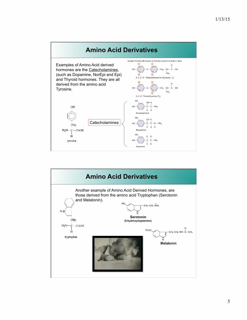
Amino Acid Derivatives
Examples of Amino Acid derived hormones are the Catecholamines, (such as Dopamine, NorEpi and Epi) and Thyroid hormones. They are all derived from the amino acid Tyrosine.
Catecholamines
Amino Acid Derivatives
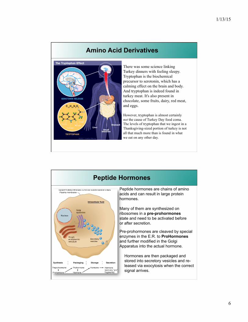
Another example of Amino Acid Derived Hormones, are those derived from the amino acid Tryptophan (Serotonin and Melatonin).
1/13/15
6
Amino Acid Derivatives
There was some science linking Turkey dinners with feeling sleepy. Tryptophan is the biochemical precursor to serotonin, which has a calming effect on the brain and body. And tryptophan is indeed found in turkey meat. It's also present in chocolate, some fruits, dairy, red meat, and eggs.
However, tryptophan is almost certainly not the cause of Turkey Day food coma. The levels of tryptophan that we ingest in a Thanksgiving-sized portion of turkey is not all that much more than is found in what we eat on any other day.
Peptide Hormones Peptide hormones are chains of amino acids and can result in large protein hormones.
Many of them are synthesized on ribosomes in a pre-prohormones state and need to be activated before or after secretion.
Pre-prohormones are cleaved by special enzymes in the E.R. to ProHormones and further modified in the Golgi Apparatus into the actual hormone.
Hormones are then packaged and stored into secretory vesicles and re-leased via exocytosis when the correct signal arrives.
1/13/15
7
Peptide Hormones
• Peptide Hormones include all hormones secreted by: – Hypothalamus, heart, thymus, digestive tract,
pancreas, and posterior lobe of the pituitary gland, as well as several hormones produced in other organs
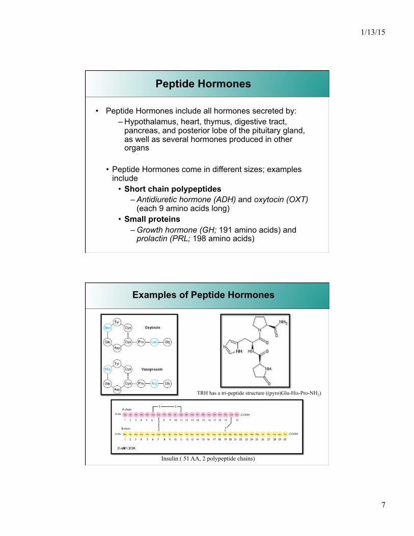
• Peptide Hormones come in different sizes; examples include
• Short chain polypeptides – Antidiuretic hormone (ADH) and oxytocin (OXT)
(each 9 amino acids long) • Small proteins
– Growth hormone (GH; 191 amino acids) and prolactin (PRL; 198 amino acids)
Examples of Peptide Hormones
TRH has a tri-peptide structure ((pyro)Glu-His-Pro-NH2)
Insulin ( 51 AA, 2 polypeptide chains)
1/13/15
8
Lipid Derived Hormones
• Lipid derived hormones fall into two main categories
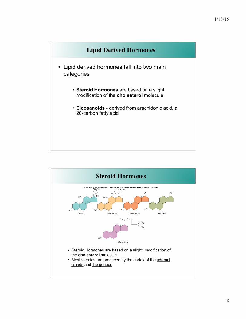
• Steroid Hormones are based on a slight modification of the cholesterol molecule.
• Eicosanoids - derived from arachidonic acid, a 20-carbon fatty acid
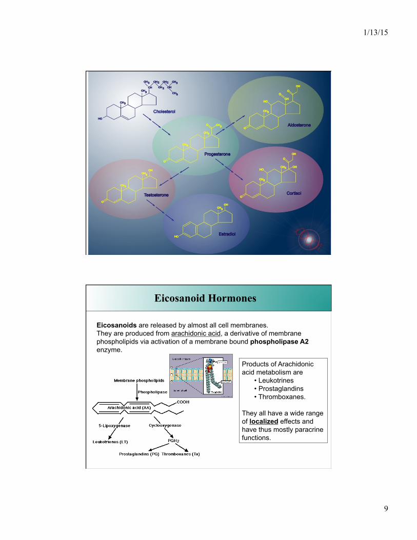
Steroid Hormones
• Steroid Hormones are based on a slight modification of the cholesterol molecule.
• Most steroids are produced by the cortex of the adrenal glands and the gonads.
1/13/15
9
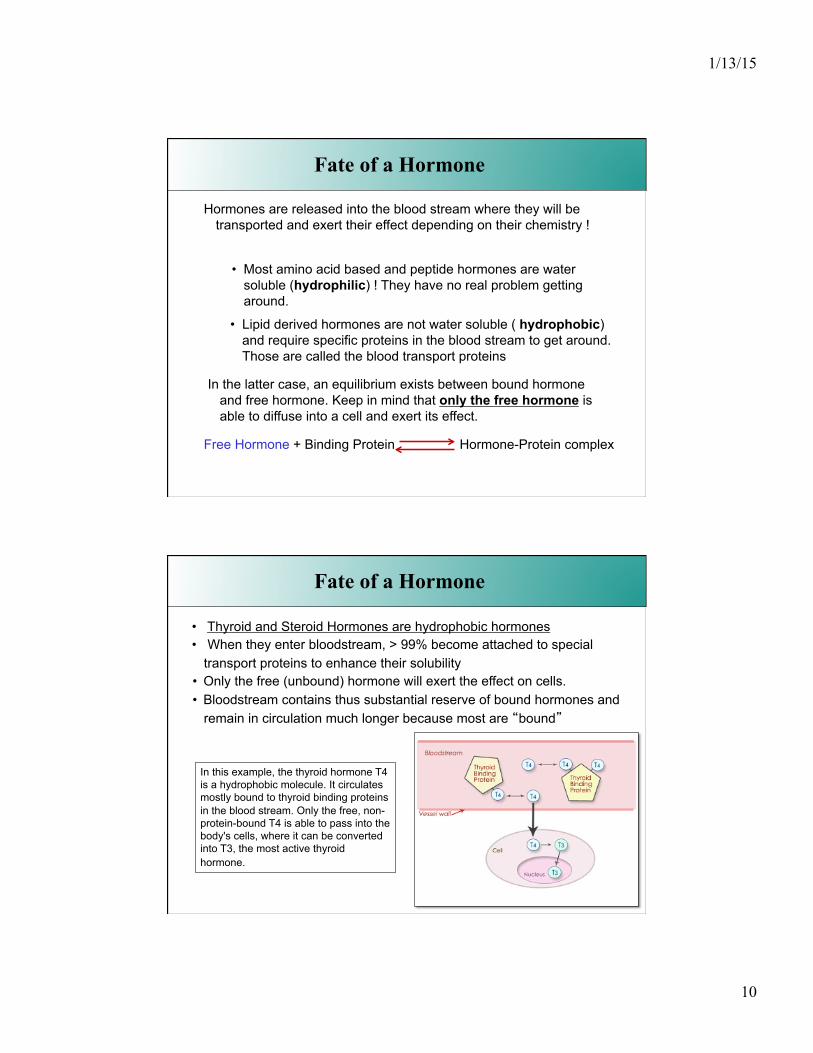
Eicosanoid Hormones
Eicosanoids are released by almost all cell membranes. They are produced from arachidonic acid, a derivative of membrane phospholipids via activation of a membrane bound phospholipase A2 enzyme.
Products of Arachidonic acid metabolism are
• Leukotrines • Prostaglandins • Thromboxanes.
They all have a wide range of localized effects and have thus mostly paracrine functions.
1/13/15
10
• Lipid derived hormones are not water soluble ( hydrophobic) and require specific proteins in the blood stream to get around. Those are called the blood transport proteins
Hormones are released into the blood stream where they will be transported and exert their effect depending on their chemistry !
• Most amino acid based and peptide hormones are water soluble (hydrophilic) ! They have no real problem getting around.
In the latter case, an equilibrium exists between bound hormone and free hormone. Keep in mind that only the free hormone is able to diffuse into a cell and exert its effect.
Fate of a Hormone
Free Hormone + Binding Protein Hormone-Protein complex
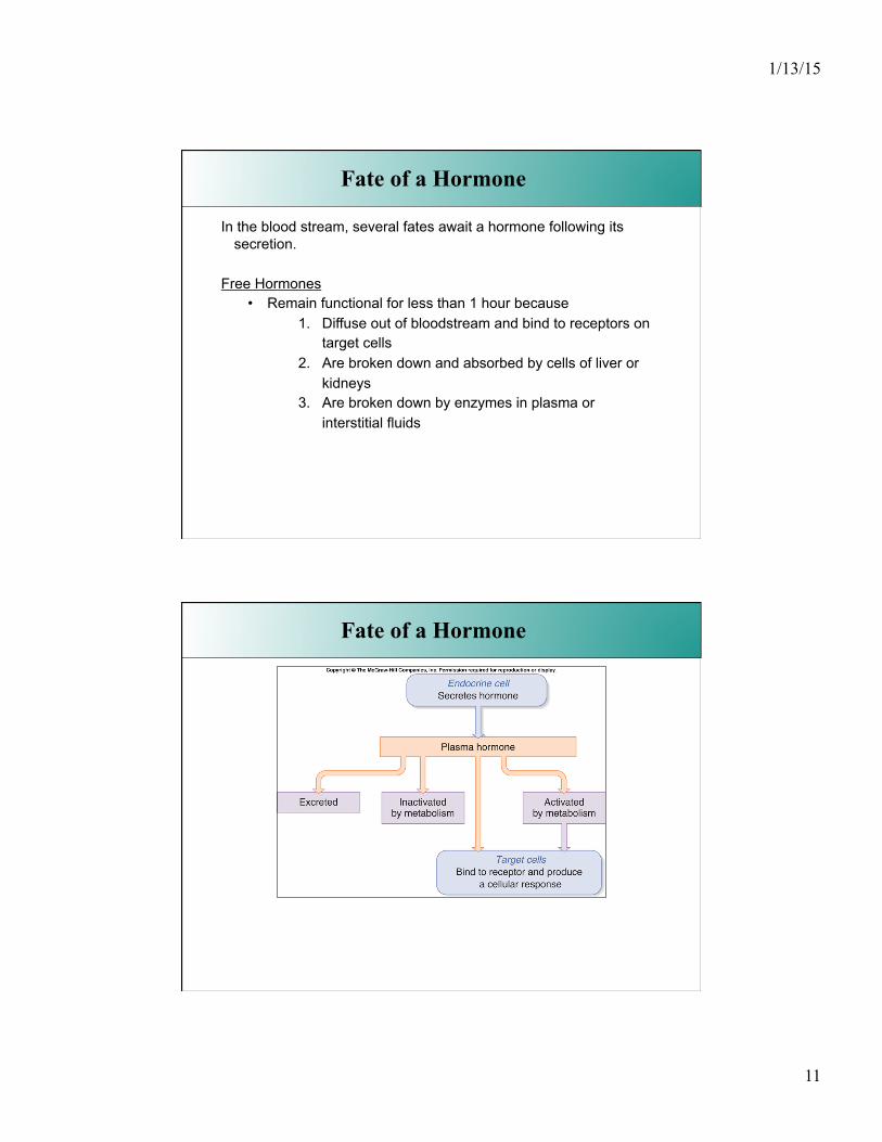
• Thyroid and Steroid Hormones are hydrophobic hormones • When they enter bloodstream, > 99% become attached to special
transport proteins to enhance their solubility • Only the free (unbound) hormone will exert the effect on cells. • Bloodstream contains thus substantial reserve of bound hormones and
remain in circulation much longer because most are “bound”
Fate of a Hormone
In this example, the thyroid hormone T4 is a hydrophobic molecule. It circulates mostly bound to thyroid binding proteins in the blood stream. Only the free, non-protein-bound T4 is able to pass into the body's cells, where it can be converted into T3, the most active thyroid hormone.
1/13/15
11
In the blood stream, several fates await a hormone following its secretion.
Free Hormones • Remain functional for less than 1 hour because
1. Diffuse out of bloodstream and bind to receptors on target cells
2. Are broken down and absorbed by cells of liver or kidneys
3. Are broken down by enzymes in plasma or interstitial fluids
Fate of a Hormone
Fate of a Hormone
1/13/15
12
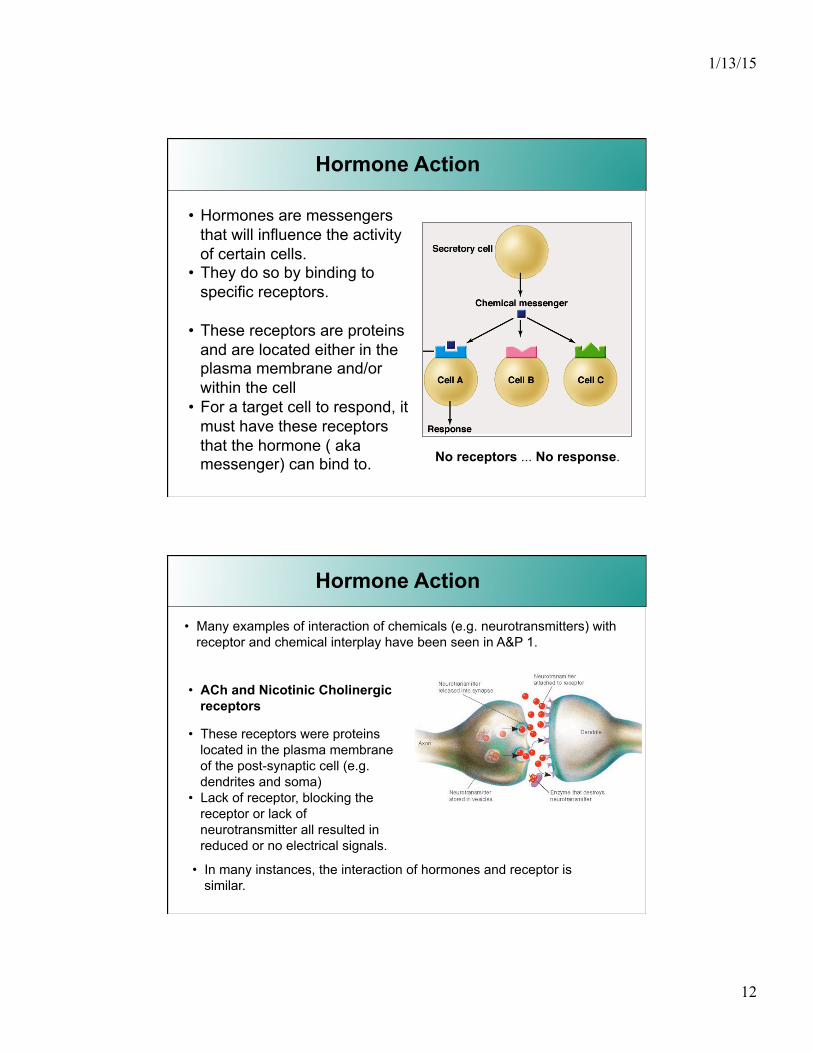
• Hormones are messengers that will influence the activity of certain cells.
• They do so by binding to specific receptors.
• These receptors are proteins and are located either in the plasma membrane and/or within the cell
• For a target cell to respond, it must have these receptors that the hormone ( aka messenger) can bind to.
Hormone Action
No receptors ... No response.
• ACh and Nicotinic Cholinergic receptors
• These receptors were proteins located in the plasma membrane of the post-synaptic cell (e.g. dendrites and soma)
• Lack of receptor, blocking the receptor or lack of neurotransmitter all resulted in reduced or no electrical signals.
• Many examples of interaction of chemicals (e.g. neurotransmitters) with receptor and chemical interplay have been seen in A&P 1.
Hormone Action
• In many instances, the interaction of hormones and receptor is similar.

1/13/15
13
Hormones • Rely on diffusion into bloodstream • Concentrations thus become quite diluted • Hormones are usually found in the order of ~ 10-8 M or lower • Receptors therefore have high affinity for the hormones
Hormones / Neurotransmitter
Neurotransmitters • Effect is faster, more concentrated within synapse • diffusion distance is very short • concentrations are ~ 10-4 M • affinity of receptor in synapse is relatively low
Hormone Action
• Another example was that of NorEpinephrine (neurotransmitter from Symp. N.S.)
• Those receptors were located in tissue activated by the S.N.S.
• If you recall, epinephrine (a hormone) also binds to these receptors and mediate similar effects.
1/13/15
14
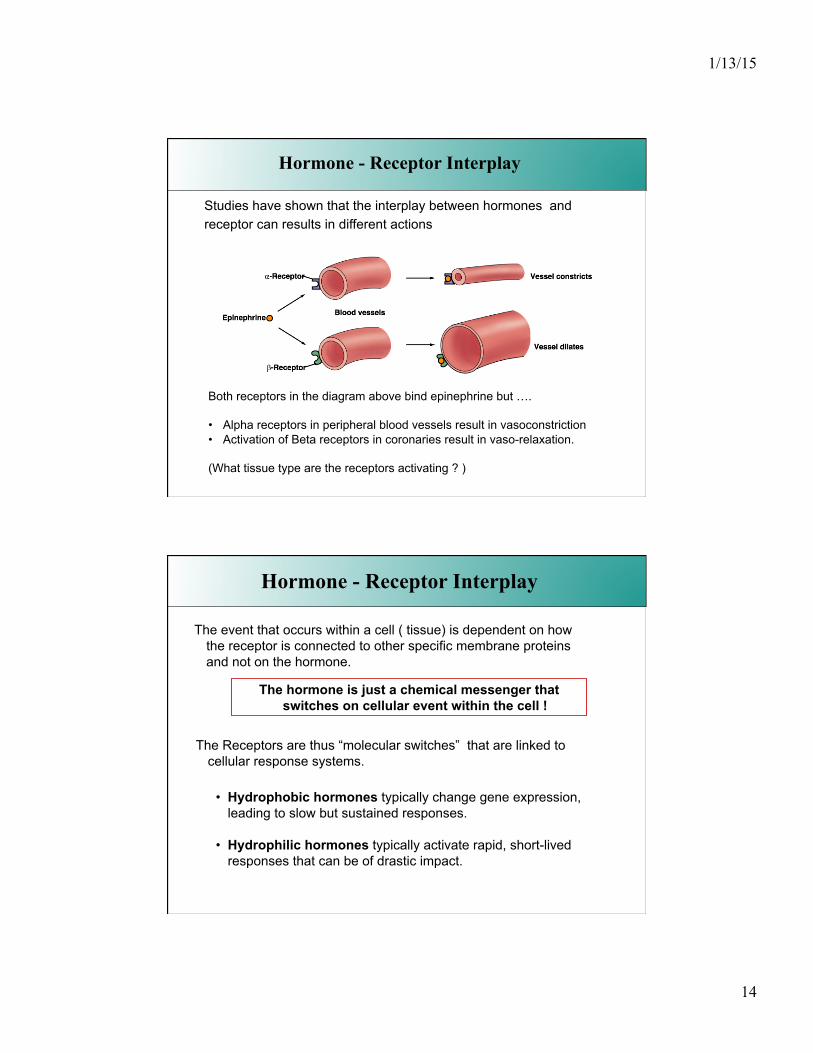
Studies have shown that the interplay between hormones and receptor can results in different actions
Hormone - Receptor Interplay
Both receptors in the diagram above bind epinephrine but ….
• Alpha receptors in peripheral blood vessels result in vasoconstriction • Activation of Beta receptors in coronaries result in vaso-relaxation.
(What tissue type are the receptors activating ? )
Hormone - Receptor Interplay
The Receptors are thus “molecular switches” that are linked to cellular response systems.
The event that occurs within a cell ( tissue) is dependent on how the receptor is connected to other specific membrane proteins and not on the hormone.
The hormone is just a chemical messenger that switches on cellular event within the cell !
• Hydrophobic hormones typically change gene expression, leading to slow but sustained responses.
• Hydrophilic hormones typically activate rapid, short-lived responses that can be of drastic impact.
1/13/15
15
From our previous discussion about hormone chemistry, we can make a distinction between two general kinds of hormones.
• Those that are lipid soluble (hydrophobic) : these will zip right into the cell without the need for a membrane bound receptor. The receptor for this messenger is located inside the cell.
• Those that are lipid insoluble (hydrophilic) : these hormones need to bind to a plasma membrane receptor before they can exert and effect.
The binding of the hormone to the receptor results in an action by the receptor. This is referred to as receptor activation.
This combination between {hormone -> receptor -> cellular events } is called a signal transduction pathway.
Hormone - Receptor Interplay
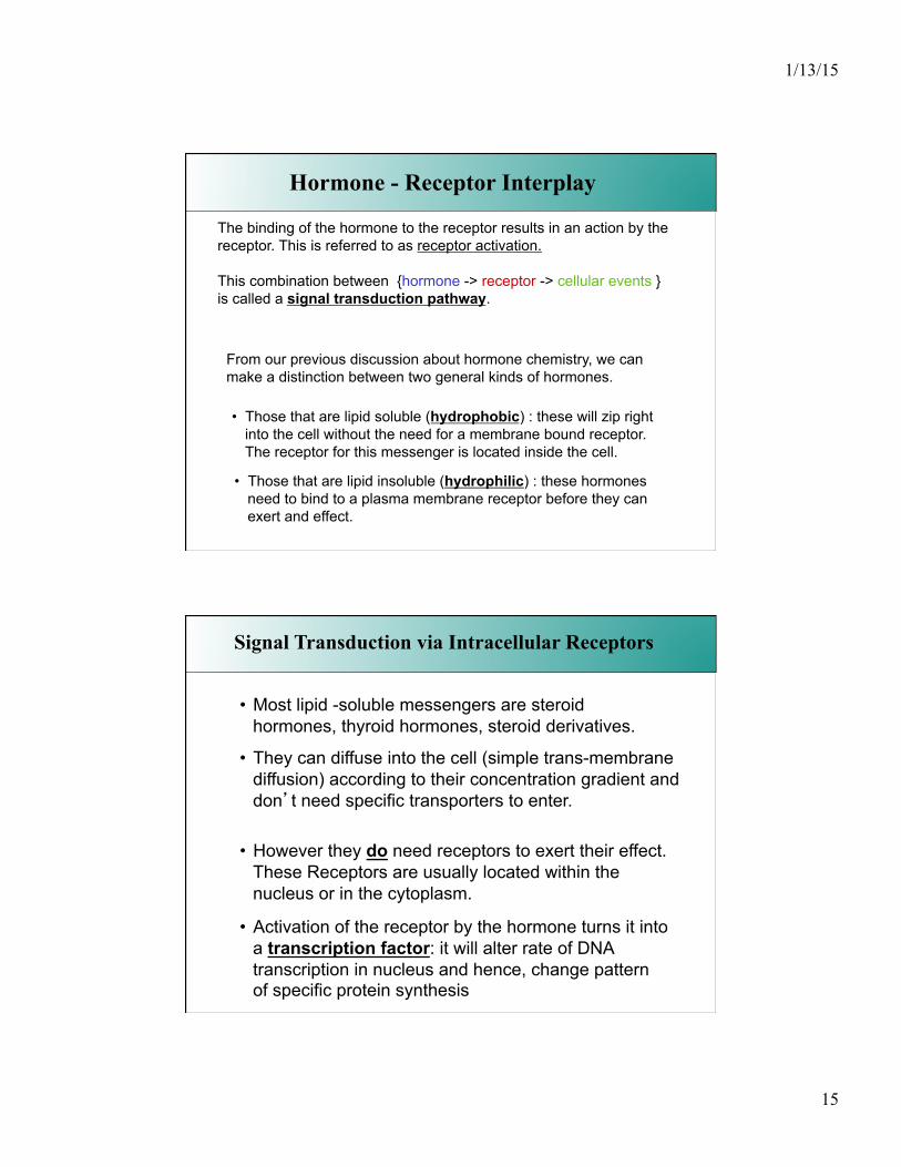
• Most lipid -soluble messengers are steroid hormones, thyroid hormones, steroid derivatives.
• They can diffuse into the cell (simple trans-membrane diffusion) according to their concentration gradient and don’t need specific transporters to enter.
• However they do need receptors to exert their effect. These Receptors are usually located within the nucleus or in the cytoplasm.
• Activation of the receptor by the hormone turns it into a transcription factor: it will alter rate of DNA transcription in nucleus and hence, change pattern of specific protein synthesis
Signal Transduction via Intracellular Receptors

1/13/15
16
Binding to the carrier protein is highly reversible, and at the target cell the free hormones moves easily through the membrane systems and binds to its receptor, found mostly the nucleus ( but some hormones have receptors in cytoplasm).
Lipid based hormones are hydrophobic and thus requires a carrier protein while in the plasma.
Signal Transduction via Intracellular Receptors
Binding to the receptor kicks off a chaperone molecule that keeps the receptor in an inactive state.
The “receptor-steroid” complex can now bind to a specific sequence of the DNA and start the process of translation.
The hormone-receptor complex thus acts as a transcription factor to alter gene expression.
Signal Transduction via Intracellular Receptors
1/13/15
17
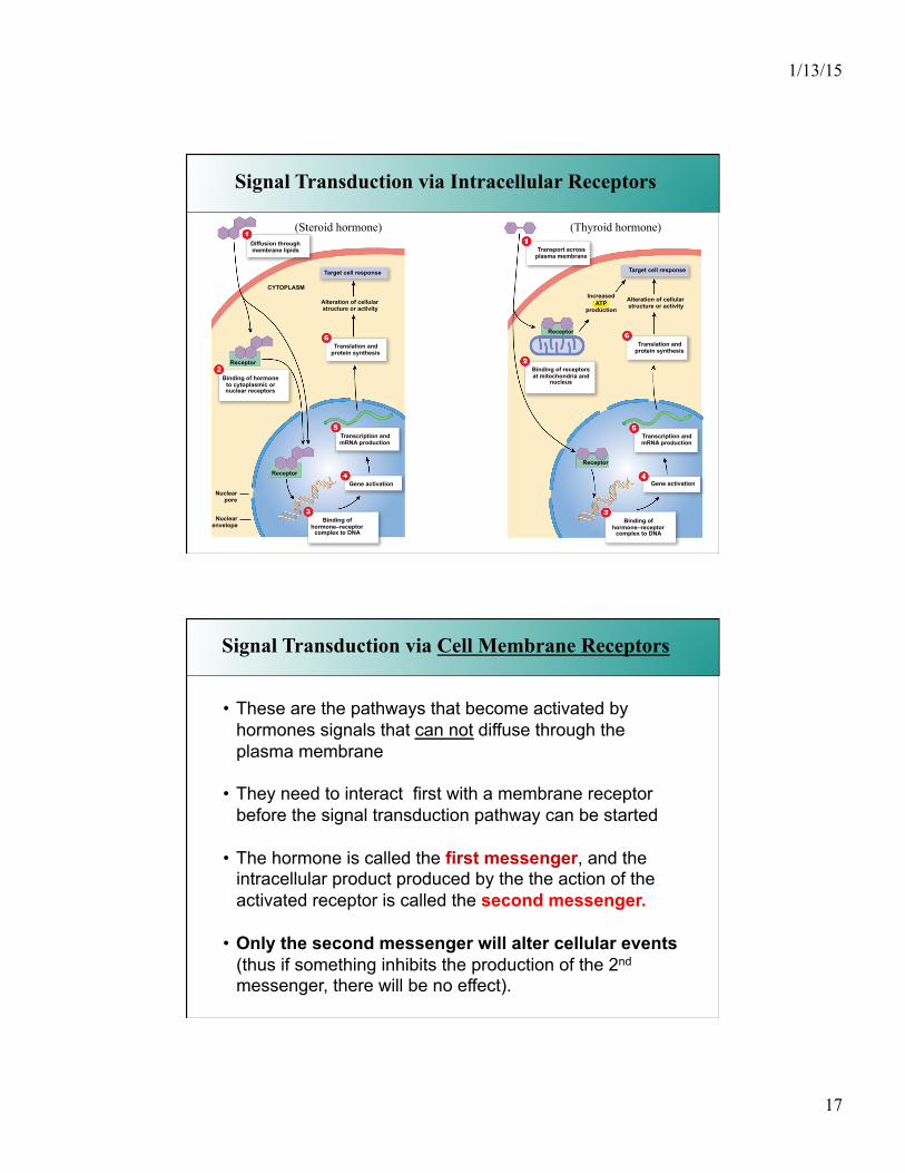
Signal Transduction via Intracellular Receptors
Receptor
Diffusion through membrane lipids
CYTOPLASM
Target cell response
Alteration of cellular structure or activity
Translation and protein synthesis
Binding of hormone to cytoplasmic or nuclear receptors
Transcription and mRNA production
Gene activation
Binding of hormone–receptor
complex to DNA
Nuclear pore
Nuclear envelope
Receptor
Receptor
Receptor
Target cell response
Alteration of cellular structure or activity
Translation and protein synthesis
Transcription and mRNA production
Gene activation
Binding of hormone–receptor
complex to DNA
Binding of receptors at mitochondria and
nucleus
Transport across plasma membrane
Increased ATP
production
(Steroid hormone) (Thyroid hormone)
Signal Transduction via Cell Membrane Receptors
• These are the pathways that become activated by hormones signals that can not diffuse through the plasma membrane
• They need to interact first with a membrane receptor before the signal transduction pathway can be started
• The hormone is called the first messenger, and the intracellular product produced by the the action of the activated receptor is called the second messenger.
• Only the second messenger will alter cellular events (thus if something inhibits the production of the 2nd messenger, there will be no effect).
1/13/15
18
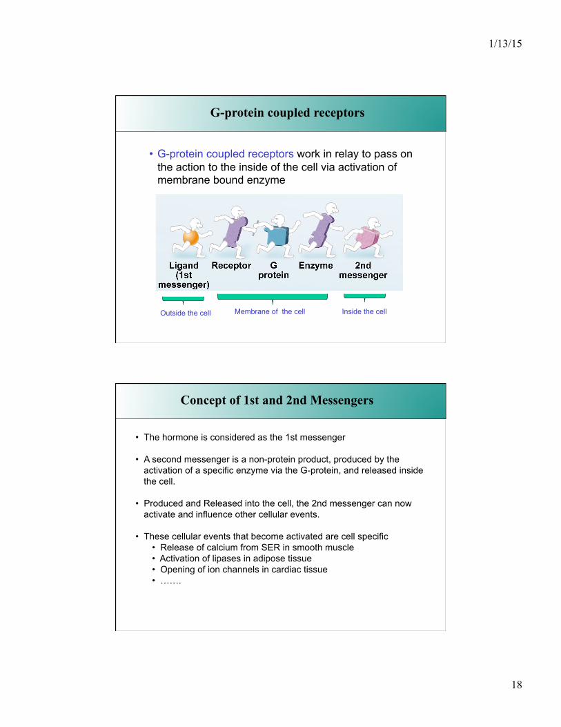
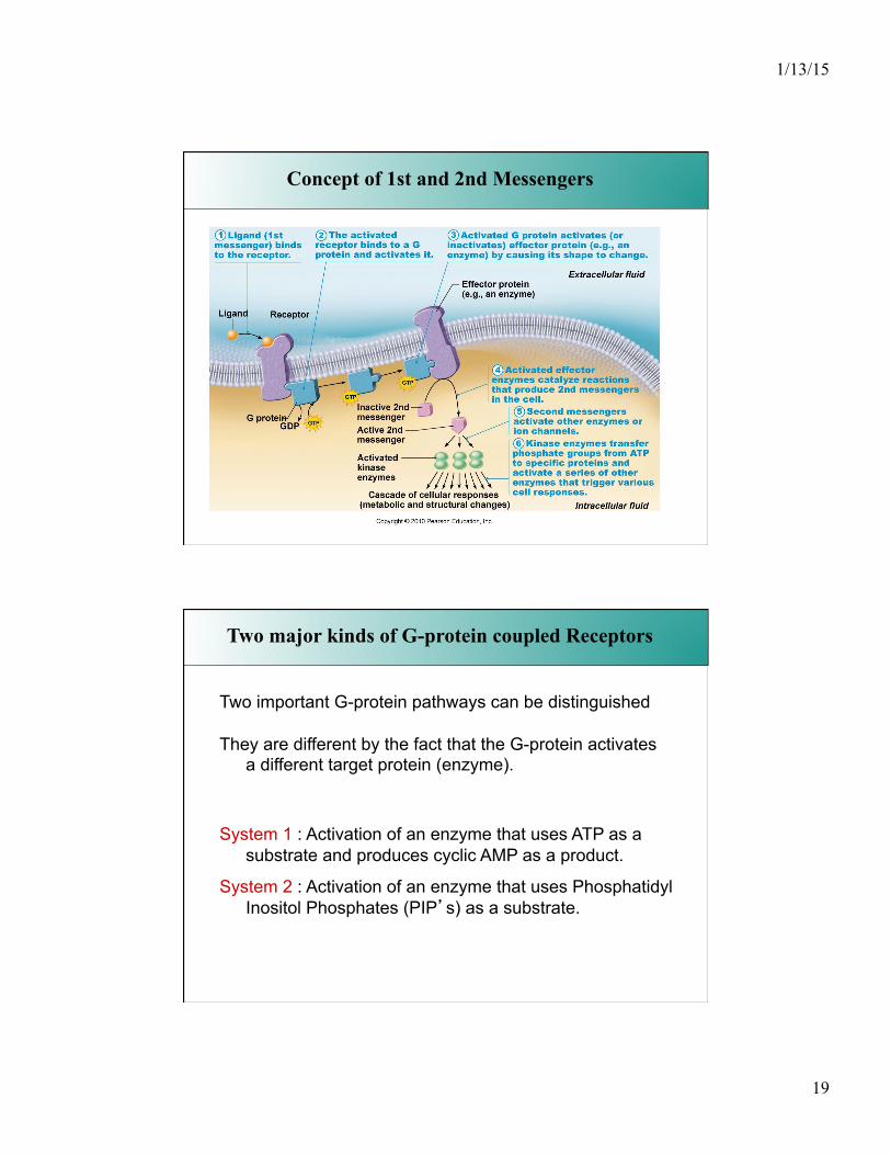
G-protein coupled receptors
• G-protein coupled receptors work in relay to pass on the action to the inside of the cell via activation of membrane bound enzyme
Outside the cell Membrane of the cell Inside the cell
• The hormone is considered as the 1st messenger
• A second messenger is a non-protein product, produced by the activation of a specific enzyme via the G-protein, and released inside the cell.
• Produced and Released into the cell, the 2nd messenger can now activate and influence other cellular events.
• These cellular events that become activated are cell specific • Release of calcium from SER in smooth muscle • Activation of lipases in adipose tissue • Opening of ion channels in cardiac tissue • …….
Concept of 1st and 2nd Messengers
1/13/15
19
• G-protein coupled receptors work in relay to pass on the action to the inside of the cell.
Concept of 1st and 2nd Messengers
Two major kinds of G-protein coupled Receptors
Two important G-protein pathways can be distinguished
They are different by the fact that the G-protein activates a different target protein (enzyme).
System 1 : Activation of an enzyme that uses ATP as a substrate and produces cyclic AMP as a product.
System 2 : Activation of an enzyme that uses Phosphatidyl Inositol Phosphates (PIP’s) as a substrate.
1/13/15
20
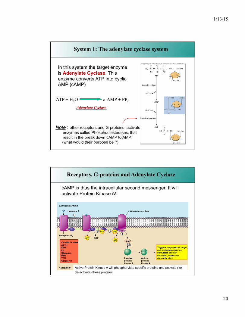
System 1: The adenylate cyclase system
In this system the target enzyme is Adenylate Cyclase. This enzyme converts ATP into cyclic AMP (cAMP)
ATP + H2O c-AMP + PPi Adenylate Cyclase
Note : other receptors and G-proteins activate enzymes called Phosphodiesterases, that result in the break down cAMP to AMP.
(what would their purpose be ?)
Hormone A
ReceptorGTP
GTP GTP
ATP cAMP
Inactive protein kinase A
Active protein kinase A
Catecholamines ACTH FSH LH Glucagon PTH TSH Calcitonin
Triggers responses of target cell (activates enzymes, stimulates cellular secretion, opens ion channels, etc.)
Adenylate cyclase
GDP
Extracellular fluid
Cytoplasm
Gs
1
2 3 4
5
Receptors, G-proteins and Adenylate Cyclase
cAMP is thus the intracellular second messenger. It will activate Protein Kinase A!
Active Protein Kinase A will phosphorylate specific proteins and activate ( or de-activate) these proteins.
1/13/15
21
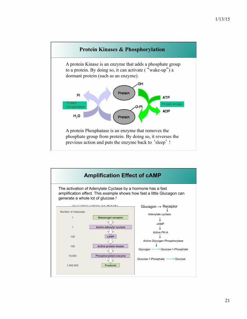
Protein Kinases & Phosphorylation
A protein Kinase is an enzyme that adds a phosphate group to a protein. By doing so, it can activate ( “wake-up”) a dormant protein (such as an enzyme).
A protein Phosphatase is an enzyme that removes the phosphate group from protein. By doing so, it reverses the previous action and puts the enzyme back to ‘sleep’ !
Amplification Effect of cAMP
The activation of Adenylate Cyclase by a hormone has a fast amplification effect. This example shows how fast a little Glucagon can generate a whole lot of glucose !
Glucagon Adenylate cyclase
Receptor
cAMP
Active PK-A
Active Glycogen Phosphorylase
Glycogen Glucose-1-Phosphate
Glucose-1-Phosphate Glucose

1/13/15
22
Terminating cAMP effects
Because of the fast amplification effect of cAMP, it needs to be terminated in order to prevent it from going out of control.
This is done by : • Activation of Phosphodiesterase enzymes (remember, PDE break down cAMP)
• Activation of Phosphatase enzyme that de-phosphorylate phosphorylated proteins.
These enzymes are controlled via activation or inhibition by means via of different hormones-receptor interactions
• Activation of a inhibitory G protein that turns off the adenylate cyclse.
Hormone
Protein receptor
G protein activated
Hormone
Protein receptor
G protein activated
Effects on cAMP Levels
Many G proteins, once activated, exert their effects by changing the concentration of cyclic-AMP, which acts as the second messenger within the cell.
Increased production of cAMP
adenylate cyclase
Acts as second
messenger
kinase
Activates enzymes
Opens ion channels
If levels of cAMP increase, enzymes may be activated
or ion channels may be opened, accelerating the
metabolic activity of the cell.
Examples:
• Epinephrine and norepinephrine (β receptors) • Calcitonin
• Parathyroid hormone • ADh, ACTH, FSH, LH, TSH
• Glucagon
Examples:
• Epinephrine and norepineph- rine (α2 receptors)
In some instances, G protein activation results in decreased
levels of cAMP in the cytoplasm. This decrease has
an inhibitory effect on the cell.
Enhanced breakdown of cAMP PDE
Reduced enzyme activity
Activation and Inactivation of Adenylate Cyclase
Whereas some Specific receptors (thus with specific hormones) are coupled to stimulatory G proteins (and thus activate), some receptors ( binding other hormones) are coupled to inhibitory G proteins. These turn off the AC enzyme and/or activate PDE. This ends the previous activation of cellular events and is a way to “switch off” a turned on event.
1/13/15
23
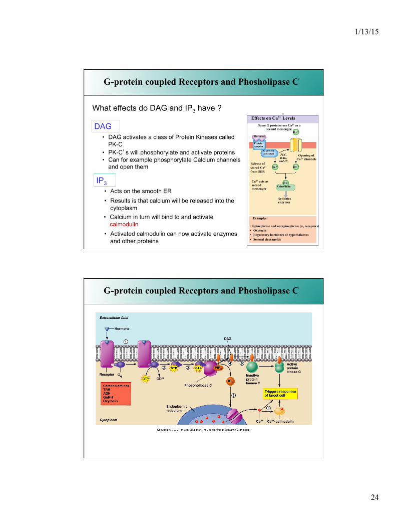
G-protein coupled Receptors and Phosholipase C
System 1 : Activation of an enzyme that makes cyclic AMP
System 2 : Activation of an enzyme that makes Phosphatidyl Inositol Phosphates (PIP’s).
Whereas the target enzyme in System 1 is Adenylate Cyclase, the target enzyme in System 2 is Phospholipase C !
G-protein coupled Receptors and Phosholipase C
What reaction does Phospholipase C catalyze ?
PIP2 Diacylglycerol + Inositol TriPhosphate (DAG) (IP3)
It acts on phosholipid membrane components called Phosphatidyl Inositol Bi Phosphates (PIP2).
DAG and IP3 are thus your second messengers !
1/13/15
24
Hormone
Protein receptor
G protein activated
Effects on Ca2+ Levels
Some G proteins use Ca2+ as a second messenger.
Examples:
• Epinephrine and norepinephrine (α1 receptors) • Oxytocin • Regulatory hormones of hypothalamus • Several eicosanoids
Activates enzymes
Calmodulin
PLC, DAG,
and IP3
Opening of Ca2+ channels
Release of stored Ca2+ from SER
Ca2+ acts as second messenger
G-protein coupled Receptors and Phosholipase C
What effects do DAG and IP3 have ?
DAG • DAG activates a class of Protein Kinases called
PK-C • PK-C’s will phosphorylate and activate proteins • Can for example phosphorylate Calcium channels
and open them
IP3 • Acts on the smooth ER • Results is that calcium will be released into the
cytoplasm • Calcium in turn will bind to and activate
calmodulin • Activated calmodulin can now activate enzymes
and other proteins
G-protein coupled Receptors and Phosholipase C
1/13/15
25
The interaction of a hormone with a receptor is like a hit and run mechanism.
• The more hormones present, the more hit and run effects • If a receptor has a higher affinity, it causes a hormone to
stay on the receptor longer and thus keep the triggering action going.
• The more receptors on a cell, the easier it is for a hormone to find a receptor
When a hormone dissociates (stops interacting) from a receptor it can jump on a new receptor and start the trigger again.
This lasts until the hormone (messenger) is destroyed or removed.
Hormones and Receptors
Once the desired effect is obtained, the hormone concentration is reduced via
• inhibition or attenuation of production of the hormone through negative feedback
• breakdown via degrading enzymes or removal via diffusion
• when located in the blood stream, the hormone can be removed by the action of kidney and/or liver
Concentration of a molecule can be adjusted quickly only if the lifetime of the molecule is short. Lifetime of most hormones is very short !
Regulation Of Receptors
Because of their importance and their interaction with cell receptors, hormones are present in very small quantities and only released when needed or when induced by specific stimuli. via stringent feedback cycles.
1/13/15
26
Abundance of Receptor are regulated as well by physiological feedback systems.
Up-regulation.
When a cell is not being targeted by a lot of hormones due to a shortage of messengers (or when the presence of antagonists block the action of the receptors), the receptor numbers tend to increase.
Down-regulation.
When there is an abundance of a certain hormone (resulting in over-stimulation), the number of cellular receptors for that hormone tend to decrease over time.
Regulation Of Receptors
Control of Hormone Release
• Endocrine Activity is controlled by Endocrine Reflexes • Endocrine Reflexes are the Functional counterparts of
neural reflexes • In most cases, controlled by negative feedback
mechanisms – Stimulus triggers production of hormone, the direct or
indirect effects of the hormone reduce intensity of the stimulus ( and reduces hormone production)
• This keeps the hormone within a certain functional range and variations in concentration are kept within a narrow desirable range
1/13/15
27
Control of Hormone Release
• Hormones are synthesized and released in response to:
• Humoral stimuli
• Neural stimuli
• Hormonal stimuli
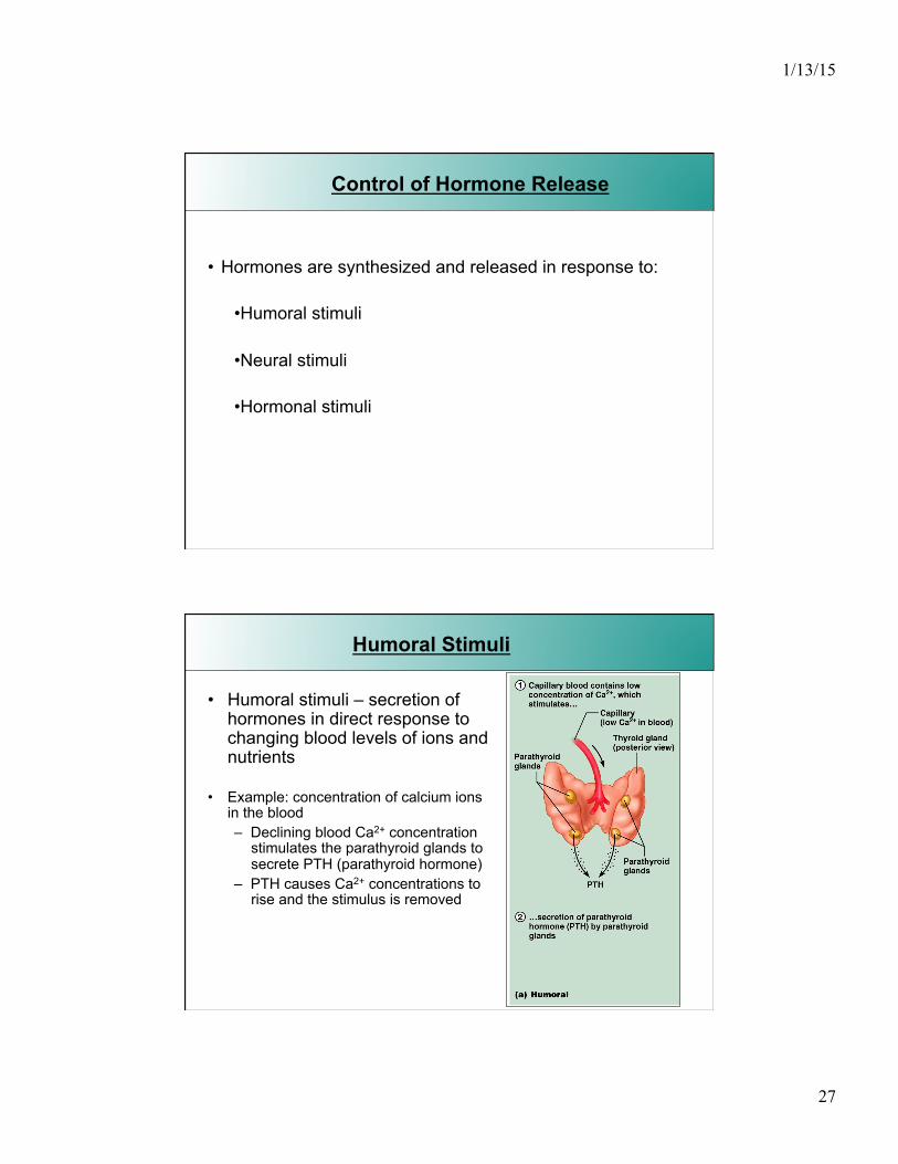
Humoral Stimuli
• Humoral stimuli – secretion of hormones in direct response to changing blood levels of ions and nutrients
• Example: concentration of calcium ions in the blood – Declining blood Ca2+ concentration
stimulates the parathyroid glands to secrete PTH (parathyroid hormone)
– PTH causes Ca2+ concentrations to rise and the stimulus is removed
1/13/15
28
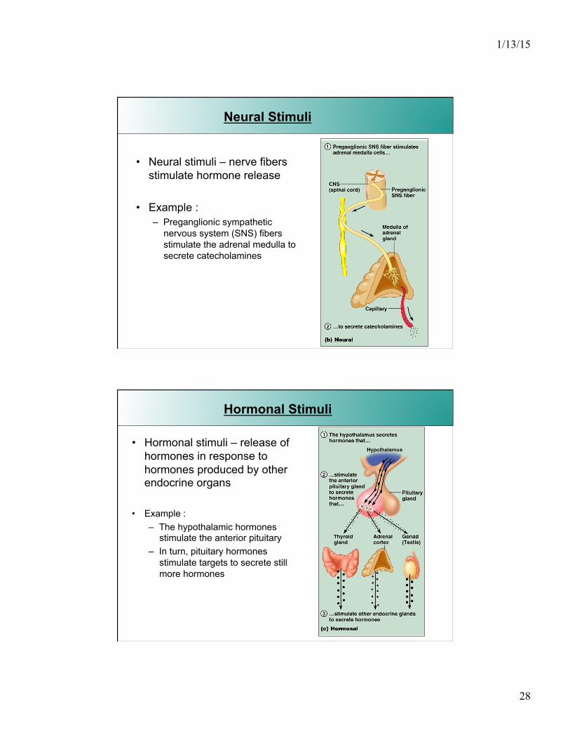
Neural Stimuli
• Neural stimuli – nerve fibers stimulate hormone release
• Example : – Preganglionic sympathetic
nervous system (SNS) fibers stimulate the adrenal medulla to secrete catecholamines
Hormonal Stimuli
• Hormonal stimuli – release of hormones in response to hormones produced by other endocrine organs
• Example : – The hypothalamic hormones
stimulate the anterior pituitary – In turn, pituitary hormones
stimulate targets to secrete still more hormones

1/13/15
29
Nervous System Modulation
• The nervous system modifies the stimulation of endocrine glands and their negative feedback mechanisms
• For example, control of blood glucose levels • Normally the endocrine system maintains blood
glucose • Under stress, the body needs more glucose • The hypothalamus and the sympathetic nervous
system are activated to supply ample glucose
Hormone-Target Cell Interaction
There are three types of hormone interaction :
Permissiveness – one hormone cannot exert its effects without another hormone being present Synergism – more than one hormone produces the same effects on a target cell Antagonism – one or more hormones opposes the action of another hormone
1/13/15
30
General Endocrine disorders
Many of the endocrine disorders are due to malfunctioning glands or malfunctioning feedback systems.
In simple endocrine reflexes, only one hormone is involved
We will cover some complex feedback systems where more than one hormone is involved.
In the latter case, the definition of a trop(h)ic hormone becomes important : it is a hormone whose only function is to regulate the release of another hormone !
General Endocrine disorders
1. Hypo-secretion
• Gland secretes too little hormone • If the gland itself is not functioning properly
= primary hypo-secretion • If the gland is normal but there is a problem with the
tropic hormone that stimulates the gland = secondary hypo-secretion
• If the tropic-hormone releasing gland is normal but something is missing that results in secretion of the tropic hormone = tertiary hypo-secretion

1/13/15
31
General Endocrine disorders
Primary
Secondary
Tertiary
Tropic Hormone.
Is a hormone that affects the release of another hormone
2. Hyper-secretion
• Primary hyper-secretion gland is secreting too much hormone on its own
• Secondary hyper-secretion over-stimulation of a gland by the tropic hormone
( thus too much tropic hormone is made/present)
Most hyper-secretions result from endocrine-cell tumors
General Endocrine disorders
1/13/15
32
General Endocrine disorders
In both conditions, nothing is actually wrong with the secretion of the hormones but the response is abnormal
• Hypo-responsiveness can be due to • Deficient receptors or lack of receptors • Deficient membrane proteins that interact with an
otherwise normal receptor ( e.g. G-proteins or the target enzymes of the alpha subunits)
• Lack or deficiency of enzymes that turn pro-hormones into active hormones
3. Hypo- and hyper- responsiveness
• Hyper-responsiveness is most often due to an abnormal up-regulation of receptors for the hormone.