eprints.soton.ac.uk€¦ · web viewtransfer and metabolism of cortisol by the isolated perfused...
TRANSCRIPT

Transfer and Metabolism of Cortisol by the Isolated Perfused Human Placenta
*Laura I Stirrat1, *Bram G Sengers2,3, Jane E Norman1, Natalie Z.M Homer4,5, Ruth Andrew4,5, **Rohan M Lewis3 and **Rebecca M Reynolds1,5
1. Tommy’s Centre for Maternal and Fetal Health, MRC Centre for Reproductive Health, University of Edinburgh
2. Bioengineering Science Research Group, Faculty of Engineering and the Environment, University of Southampton, UK
3. Institute for Life Sciences, University of Southampton, UK.
4. Mass Spectrometry Core, Edinburgh Clinical Research Facility, University of Edinburgh, UK
5. University/BHF Centre for Cardiovascular Science, University of Edinburgh, UK
*joint first author
**joint last author
Precis: Placental cortisol metabolism and transfer was studied using tracers and computational modelling. This indicated that the placenta
presents both metabolic and physical barriers to cortisol transfer.
Stirrat LI et al
Corresponding author and person to whom reprint requests should be addressed:
1
2
3
4
5
6
7
8
9
10
11
12
13

Professor Rebecca Reynolds
University/BHF Centre for Cardiovascular Science
Queen’s Medical Research Institute
47 Little France Crescent
Edinburgh EH16 4TJ
Phone: +44 131 2426762
Fax: +44 131 2426779
Email: [email protected]
Funding: This work was supported by funding from Tommy’s and the Medical Research Council (MR/N022556/1). We also acknowledge the
support of the British Heart Foundation and the Mass Spectrometry Core of the Edinburgh Clinical Research Facility.
Disclosure statement: The author reports no conflicts of interest in this work.
14
15
16
17
18
19
20
21
22
23
24
25
26
27
28
29
ABSTRACT
30
31
32
33
34
35
36
37
38
39
40
41

Context: Fetal overexposure to glucocorticoids in utero is associated with fetal growth restriction and is postulated to be a key mechanism
linking suboptimal fetal growth with cardiovascular disease in later life.
Objective: To develop a model to predict maternal-fetal glucocorticoid transfer. We hypothesised placental 11β-HSD2 would be the major rate-
limiting step in maternal cortisol transfer to the fetus.
Design: We used a deuterated cortisol tracer in the ex vivo placental perfusion model, in combination with computational modelling, to
investigate the role of interconversion of cortisol and its inactive metabolite cortisone on transfer of cortisol from mother to fetus.
Participants: Term placentas were collected from five women with uncomplicated pregnancies, at elective caesarean delivery.
Intervention: Maternal artery of the isolated perfused placenta was perfused with D4-cortisol.
Main Outcome Measures: D4-cortisol, D3-cortisone and D3-cortisol were measured in maternal and fetal venous outflows.
Results: D4-cortisol, D3-cortisone and D3-cortisol were detected and increased in maternal and fetal veins as the concentration of D4-cortisol
perfusion increased. D3-cortisone synthesis was inhibited when 11β-HSD activity was inhibited. At the highest inlet concentration only 3.0% of
the maternal cortisol was transferred to the fetal circulation, while 26.5% was metabolised and 70.5% exited via the maternal vein. Inhibiting
11β-HSD activity increased the transfer to the fetus to 7.3% of the maternal input, while 92.7% exited via the maternal vein.
42
43
44
45
46
47
48
49
50
51
52
53
54

Conclusions: Our findings challenge the concept that maternal cortisol diffuses freely across the placenta and confirm that 11β-HSD2 acts as
a major ‘barrier’ to cortisol transfer to the fetus.
Keywords: Cortisol; placenta; 11β-HSD2; cortisone; tracer
Word count: 3564
55
56
57
58
1. Introduction
Cortisol, the principal circulating glucocorticoid hormone in humans, is essential for normal fetal development and tissue maturation. Fetal
overexposure to glucocorticoids in utero is associated with intrauterine growth restriction, [1] and is postulated to be a key mechanism linking
suboptimal fetal growth with increased risk of cardiovascular disease in later life. [2] Better knowledge of the factors regulating cortisol transfer
to the fetus is essential to understand the pathophysiology of fetal growth restriction and is also relevant for prescribing of antenatal steroids
which are widely used in clinical management of women at threat of pre-term birth.
Maternal circulating cortisol levels rise exponentially during pregnancy. [3] Although glucocorticoids are lipophilic and thus are believed to freely
cross the placenta, fetal cortisol levels are 5 to 10-fold lower than maternal levels [4] due to the activity of the placental enzyme 11-beta-
hydroxysteroid dehydrogenase-type 2 (11β-HSD2) [5-7] which catalyses the conversion of active cortisol into inactive cortisone. In human
placenta 11β-HSD2 is localized to the syncytiotrophoblast, [7] which is the primary barrier between the mother and the fetus and thus prevents
glucocorticoids accessing placental cells and the fetal compartment. [8] Indeed placental 11β-HSD2 has been suggested to inactivate the
majority of maternal glucocorticoids passing to the fetus in rodents [9] and in humans. [10] 11-beta-hydroxysteroid dehydrogenase-type 1 (11β-
HSD1), which regenerates cortisol from inactive cortisone, is undetectable in the syncytiotrophoblast, but is localized in the extravillous
trophoblasts (situated near maternal circulation) and endothelial cells lining fetal capillaries in terminal villi. [11] Whether or not the activity of
placental 11β-HSD1 regenerates a substantial amount of cortisol or contributes significantly to maternal or fetal circulations is not well
59
60
61
62
63
64
65
66
67
68
69
70
71
72
73

understood. With a number of studies demonstrating links between placental glucocorticoid transfer, sensitivity and metabolism and adverse
outcomes in infancy, childhood and adolescence, [12,13] understanding of the regulatory mechanisms and rate-limiting steps of maternal-fetal
cortisol transfer is essential in order to identify whether there are any options for targeted intervention to improve pregnancy outcomes.
Studies using the ex vivo dual perfused placental perfusion model together with computational modelling have generated new mechanistic
insights into placental amino acid and lipid transfer from mother to fetus. [14-16] In the current study we used this combined experimental and
computational modelling approach to develop a model to explore placental cortisol metabolism and transfer and its regulation. We hypothesised
that activity of placental 11β-HSD2 would be the major rate limiting step in maternal cortisol transfer to the fetus.
74
75
76
77
78
79
80
2. Methods
Five term placentas from women with uncomplicated pregnancies were collected on ice immediately after delivery by elective caesarean section
at the Royal Infirmary of Edinburgh, with ethical approval (REC09/S0704/3) and written informed consent. Elective caesarean sections were
performed between 39-40 weeks of gestation.
A. Placental Perfusions
Placentas were perfused using the methodology of Schneider [17] as adapted in a previous study. [18] Non-recirculating maternal and fetal
circulations were established in an isolated cotyledon within 30 minutes of delivery. The fetal circulation and maternal intervillous space were
perfused with a modified Earle’s bicarbonate buffer (EBB: 5 mmol L-1 glucose, 1.8 mmol L-1 CaCl2, 0.4 mmol L-1 MgSO4, 116.4 mmol L-1 NaCl,
5.4 mmol L-1 KCl, 26.2 mmol L-1, NaHCO3, 0.9 mmol L-1 NaH2PO4), with Heparin (25,000 units/L; Fannin, Northamptonshire, UK) and bovine
serum albumin (BSA [Fraction V; 98 %], 2 g/L, Sigma, UK) added. Maternal perfusate was equilibrated with 95% air and 5% CO 2, and fetal
perfusate with 95% N2 and 5% CO2 (BOC, UK). Maternal circulation was at 14 mL/min and fetal circulation at 6 mL/min using a peristaltic pump
(Watson-Marlow, UK).
Approximately 2 mL of venous perfusate was collected from the maternal and fetal venous outflows, at 5-minute intervals. Fetal artery pressure
was maintained between 40 – 70 mmHg and fetal venous return was > 95%. At the end of the experiments, the perfused mass was identified
81
82
83
84
85
86
87
88
89
90
91
92
93
94
on the ‘maternal side’ by slight blanching. The perfused placental cotyledon was weighed. Cotyledon volume was calculated on the basis of 1
mL per g tissue. Samples of maternal and fetal perfusate fluid, un-perfused tissue and perfused tissue were stored at -80 oC until analysis.
B. Use of deuterated tracers to investigate cortisol metabolism
Cortisol metabolism by 11β-HSD enzymes and transport between the maternal and fetal circulations was investigated using the stable isotope
deuterium (D)-labelled tracer, [9,11,12,12 2H4]-cortisol “D4-cortisol” [19] which is converted to [9,12,12 2H3]-cortisone “D3-cortisone” by 11β-
HSD2. Measurement of [9,12,12 2H3]-cortisol “D3-cortisol”, which is regenerated from D3-cortisone can be used to assess activity of 11β-HSD1
(Figure 1). After an initial ‘washout’ period of 30 minutes, D4-cortisol (Steraloids, USA) was perfused into the maternal circulation with stepped
increases in concentrations of 20 nM, 200 nM and 800 nM every 30 minutes. The 800 nM D4-cortisol concentration was considered to be
representative of circulating maternal cortisol levels in the third trimester [20]. The HSD inhibitor carbenoxolone (Sigma, UK) was added to the
perfusion solution in addition to 800 nM D4-cortisol in the final 30 minutes at a concentration of 1000 nM, as informed by a previous study. [10]
C. LC-MS/MS quantification
Endogenous (cortisol, cortisone) and deuterated (D4-cortisol, D3-cortisone and D3-cortisol) glucocorticoids were measured simultaneously by
liquid chromatography tandem mass spectrometry (LC-MS/MS) using a Waters Acquity™ UPLC (Manchester, UK) liquid chromatography
system followed by mass spectral detection on an ABSciex QTRAP® 5500 (Warrington, UK) operated in positive electrospray ionization mode.
95
96
97
98
99
100
101
102
103
104
105
106
107
108
Mass spectral conditions are described in Supplementary Table 1 in conjunction with ion spray voltage (5500 V) and source temperature (700
oC).
D. Perfusate fluid extraction
Following enrichment of perfusate (500 µL) with the internal standard epi-cortisol (10 ng; Steraloids, USA) and dilution with water (500 µL)
analytes were extracted using a Sep-Pak C18 40 mg 96-well plate (Waters, Manchester, UK). Plates were primed with methanol (1 mL), then
EBB (1 mL) then samples (500 µL) were loaded and plates washed with water (1 mL). Analytes were eluted from the plate using acetonitrile (1
mL) directly into a 2 mL deep well collection plate (Waters, UK). Eluants were dried under oxygen-free nitrogen (60 oC) using a 96-well Dry
down apparatus, and reconstituted in mobile phase (30:70 methanol: water; 100 µL).
E. Tissue Extraction
Placental tissue (200 mg) was homogenized in 3 mL 7:2 methanol: water and enriched with internal standard epi-cortisol (10 ng, as above)
before being centrifuged at 3200 g for 45 minutes at 4 oC. Supernatant was transferred to a clean glass vial and dried under oxygen-free
nitrogen (60 oC) and reconstituted in water (5 mL). Analytes were extracted using Sep-Pak C18 360 mg Classic Cartridges (Waters). Cartridges
were primed with 100% methanol (5 mL) followed by water (5 mL). Samples were added to cartridges and allowed to flow through with gravity.
Cartridges were washed with water (5 mL), and analytes were eluted with 100% methanol (2 mL) into a 3.5 mL glass vial. Eluants were dried
109
110
111
112
113
114
115
116
117
118
119
120
121
122

down under oxygen-free nitrogen (60 oC) and reconstituted in 100 µL mobile phase.
F. Liquid chromatography tandem mass spectrometry (LC-MS/MS)
Samples in the auto-sampler were maintained at 10 oC. Analytes were separated at 40 oC on an ACE Excel C18-AR column (100 x 2.1 mm, 1.7
um; Hichrom Limited®, Berkshire, UK) at a flow rate of 0.5 mL/min. Samples in the auto-sampler and sample manager were maintained at 10
oC. Starting with 70% water with 0.1% formic acid (FA) (solution A) and 30% acetonitrile with 0.1% FA (solution B), maintained for 4 minutes
followed by a 1-minute linear rise to 60% solution B, a subsequent rise to 90% solution B, before restoring to 30% solution B at 6.1 minutes.
This condition was sustained for 1-minute to re-equilibrate.
The inter-assay precision of D4-Cortisol in perfusate fluid was 3.6% - 11.6%, and inter-assay accuracy was 93% - 103%. Inter-assay precision
of D3-Cortisol in perfusate fluid was 8.8% - 17.3% and inter-assay accuracy was 98% - 101%. For placental tissue samples (which were all
analysed on the same day), intra-assay precision was 7.0% for D4-Cortisol, and 6.4% for D3-Cortisol.
The peak areas of deuterated steroids were corrected for the abundances of naturally occurring isotopomers at baseline. In addition, the peak
area of D4-cortisol was corrected for interference from the M+4 isotopologue of cortisol and the M+1 isotopologue of D3-cortisol. There was no
available standard for D3-cortisone, so concentrations were estimated using the calibration curve for cortisone and the ‘fold-change’ or ‘units /
mL’ rather than concentration calculated. The peak area of D3-cortisol was corrected for interference from the M+3 isotopologue of cortisol.
123
124
125
126
127
128
129
130
131
132
133
134
135
136

G. Data analysis
Deuterated hormone levels were adjusted for flow rate and were normalised to tissue weight of the perfused cotyledon. D4-cortisol and D3-
cortisol were reported in ng, and in the absence of a standard for accurate quantification, D3-cortisone was measured in arbitrary units.
H. Computational model for placental transfer
A compartmental modelling framework was adopted to model the placental transfer of cortisol and cortisone in the ex-vivo perfusion
experiments, based on our previous work. [14,15,21] The model distinguishes three separate physiological compartments associated with the
maternal, syncytiotrophoblast and fetal capillary volumes (Figure 1a). Each compartment is described as well mixed. Transfer between
compartments is determined by the fluxes across the apical and basal membranes and assumed to occur by simple diffusion for both cortisol
and cortisone. Metabolic conversion from cortisol to cortisone within the syncytiotrophoblast is described as unidirectional using Michaelis-
Menten kinetics. Model equations were implemented in Matlab (R2016a) as outlined previously [14,15,21]. Details of the equations that resulted
and model parameters are described in Supplementary Methods
A sensitivity analysis was carried out in which the model parameters were varied with respect to the values for the reference fit. The reported
changes in placental transfer predicted by the model were based on the steady state results at the highest maternal input concentration.
137
138
139
140
141
142
143
144
145
146
147
148
149
3. Results
Characterisation of subjects
The mean (sd) maternal age was 36.4 ± 6.3 years, mean gestational length was 277 ± 2 days (39+4 weeks ± 2 days), and mean birthweight
was 3721 ± 223 g.
D4-cortisol, D3-cortisone and D3-cortisol levels
Figure 2 shows the levels of D4-cortisol, D3-cortisone and D3-cortisol (plotted data with error bars) in maternal and fetal veins increased as the
concentration of D4-cortisol in the maternal artery perfusion increased. D4-cortisol (Figure 2a-b) and D3-cortisone (Figure 2c-d) were detected
in maternal and fetal vein 5-minutes after commencement of D4-cortisol perfusion (20 nM) in the maternal artery. D3-cortisol (Figure 2e-f) was
detected at 95-minutes into the experiment in the maternal vein (perfusion phase: 800 nM D4-cortisol), and at 75-minutes in the fetal vein
(perfusion phase: 200 nM D4-cortisol). Variation in the D3-cortisol levels reflects both the fact that D3-cortisol levels were near the limit of
detection, and technical considerations when collecting maternal side samples in the perfusion system where variation tends to be higher. The
biggest increase in D4-cortisol and D3-cortisone levels occurred when maternal artery D4-cortisol perfusion increased from 200 nM to 800 nM.
Levels of D3-cortisone in the maternal circulation were approximately 5-fold higher than in the fetal circulation. When carbenoxolone was added
150
151
152
153
154
155
156
157
158
159
160
161
162
163
to the maternal artery perfusion, D4-cortisol levels further increased in maternal and fetal veins, and D3-cortisone synthesis was completely
inhibited. D3-cortisol levels were around 300-fold lower than D4-cortisol in both maternal and fetal circulations, and were close to the assay limit
of detection. Levels of D3-cortisol in the maternal circulation were approximately 2-3-fold higher than levels in the fetal circulation.
Proportionately more of the produced D3-cortisol was released into the fetal circulation than maternal circulation, when compared with the
proportion of D3-cortisol released into maternal and fetal circulations. Samples of buffer obtained on completion of the ‘washout’ phase of the
experiment confirmed that there were no remaining endogenous or labelled glucocorticoids within the tubing used for the circuit.
Placental model results
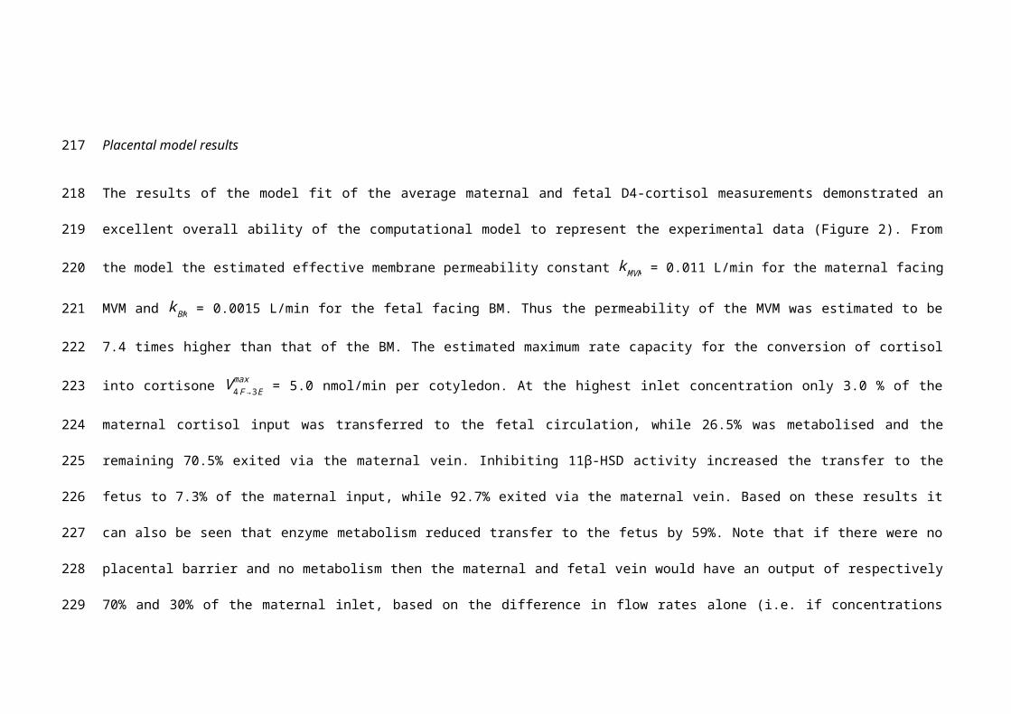
The results of the model fit of the average maternal and fetal D4-cortisol measurements demonstrated an excellent overall ability of the
computational model to represent the experimental data (Figure 2). From the model the estimated effective membrane permeability constant
kMVM = 0.011 L/min for the maternal facing MVM and k BM = 0.0015 L/min for the fetal facing BM. Thus the permeability of the MVM was
estimated to be 7.4 times higher than that of the BM. The estimated maximum rate capacity for the conversion of cortisol into cortisone V 4 F→3Emax
= 5.0 nmol/min per cotyledon. At the highest inlet concentration only 3.0 % of the maternal cortisol input was transferred to the fetal circulation,
while 26.5% was metabolised and the remaining 70.5% exited via the maternal vein. Inhibiting 11β-HSD activity increased the transfer to the
fetus to 7.3% of the maternal input, while 92.7% exited via the maternal vein. Based on these results it can also be seen that enzyme
metabolism reduced transfer to the fetus by 59%. Note that if there were no placental barrier and no metabolism then the maternal and fetal
164
165
166
167
168
169
170
171
172
173
174
175
176
177
178
vein would have an output of respectively 70% and 30% of the maternal inlet, based on the difference in flow rates alone (i.e. if concentrations
within the placenta were perfectly mixed). The comparison between the predicted D3-cortisone and the scaled experimental data is shown in
figure 2. It can be observed that the relative steady state levels correspond well for the fetal D3-cortisone, while the maternal D3-cortisone
shows some larger discrepancies. In addition, the model responds much more rapidly to changes in input conditions. In this respect, the sharp
peak at t = 150 min predicted by the model is due to the absence of blocker in the washout buffer, which is assumed to take immediate effect in
the model.
The results of the sensitivity analysis in figure 3 show that when varying single parameters the placental transfer of cortisol was affected
most by changes in k BM, the membrane permeability of the BM, and the metabolic conversion rate of cortisol into cortisone V 4 F→ 3Emax . In addition,
placental transfer was predicted to be moderately sensitive to kMVM , the permeability of the MVM, and the maternal flow rate used in the
experiment Qm. Variations in Km only had a small impact as the metabolism continued to operate in the saturated regime, while increasing the
fetal flow rate Qf used in the experiment was predicted to only have a minor effect on transfer. Steady state transfer was not sensitive to any of
the compartment volumes, as expected. To evaluate the impact of the overall membrane permeability, an additional study was done in which
k BM and kMVM were both varied simultaneously, demonstrating a considerably larger effect than for the permeability of each membrane
separately (figure 3).
179
180
181
182
183
184
185
186
187
188
189
190
191
192
193

4. Discussion
The experiments performed in this study using a deuterated cortisol tracer in the ex vivo placental perfusion model allowed investigation of the
role of interconversion of cortisol and its inactive metabolite cortisone on transfer of cortisol from mother to fetus at term. The application of
computational modelling enabled interpretation of the transfer mechanisms that underlie these processes. Our findings challenge the concept
that maternal cortisol diffuses freely across the placenta, confirm that 11β-HSD2 acts as a major ‘barrier’ to cortisol transfer to the fetus and
show preliminary evidence of local cortisol production within the placenta.
Addition of carbenoxolone (a potent HSD inhibitor) to the maternal artery perfusion, resulted in no further production of D3-cortisone. This
supports the role of 11β-HSD2 as a key player in the maternal barrier to fetal glucocorticoid exposure. The activity (but not mRNA) of 11β-HSD2
has been shown to decrease in the last two weeks before parturition. [22] The placentas used in the experiments were obtained from elective
caesarean sections at between 39-40 weeks gestation, so it is not known when parturition would have occurred in these pregnancies. The
model allowed an estimation of the maximum capacity of 11β-HSD2 for conversion of cortisol to cortisone as 5.0 nmol/min per cotyledon. It is
not known what the capacity of 11β-HSD2 would be if exposed to high levels of maternal glucocorticoids for more prolonged periods, but
studies have demonstrated that 11β-HSD2 mRNA and activity is down-regulated by maternal stress [23] and inflammatory diseases. [22]
Further, inhibition of 11β-HSD2 by maternal liquorice consumption has adverse consequences on child development [24,25]. Our study
194
195
196
197
198
199
200
201
202
203
204
205
206
207
supports the premise that the adverse offspring outcomes are due to increased fetal glucocorticoid exposure as when 11β-HSD2 was inhibited
by carbenoxolone, transplacental passage of maternal cortisol to the fetal circulation was more than doubled.
Yet, even when 11β-HSD activity was inhibited using carbenoxolone, less than 10% of maternal D4-cortisol crossed the placenta in our
experiments. This observation challenges the concept that cortisol freely diffuses across the placenta, and suggests alternate mechanisms to
protect the fetus from high maternal cortisol levels in addition to the well described inactivation of cortisol by 11β-HSD2. Three ABC-
transporters; multidrug-resistant protein (MRP1, encoded by ABCC1), p-glycoprotein (P-gp, encoded by ABCB1) and breast-cancer-resistant
protein (BCRP, encoded by ABCG2) are localised to placental syncytiotrophoblast, and the fetal vessel endothelium [26,27] consistent with the
potential for active transport of cortisol in and out of the placenta. Further studies are needed to investigate the contribution of ABC transporters,
levels of which are known to alter across gestation, [28-31] in regulating maternal cortisol transfer to the fetus and in particular to understand the
kinetics of efflux transporters, which our preliminary observations suggest may also protect the fetus.
Further we observed approximately a 5-fold higher D3-cortisone release to the maternal circulation compared with the fetal circulation. It also
needs to be considered that the physical process of cortisol diffusion across tissues may be more challenging than has been thought previously.
In particular, in the placenta diffusion across the water filled villous stroma may prove a barrier to cortisol diffusion. This is consistent with the
observation that cortisone was preferentially released into the maternal circulation (2:1 maternal:fetal circulation), and the lower placental to
fetal permeability calculated within the model.
208
209
210
211
212
213
214
215
216
217
218
219
220
221
222

A novel finding is the observation of de novo placental cortisol synthesis, as evidenced by the detection of D3-cortisol in both maternal and fetal
circulations. Though the absolute levels of D3-cortisol were low, this regeneration of cortisol may have local paracrine roles and increased
placental 11β-HSD1 mRNA levels have been associated with maternal depression and with altered infant regulatory behaviours. [12,13]
Further, proportionately more D3-cortisol was transferred to the fetus than D3-cortisone, which is in line with localisation of 11β-HSD1 to the
endothelium. [11] The computational model provided a good overall representation of the experimental data under different experimental
conditions. In general, the compartmental model showed a faster response due to the well-mixed assumption, but this did not affect the steady
state levels. The model predicted that changing membrane permeability of the BM would affect placental transfer of cortisol. Placental transfer
of lipids has been reported to be increased in pre-eclampsia. [32] Further studies are required to investigate whether inflammatory conditions
such as pre-eclampsia and preterm labour alter the permeability of the BM, and thus alter placental cortisol transfer.
Our study has several limitations. Our experiments were conducted using EBB buffer and albumin. The findings may be altered in vivo with the
presence of corticosteroid binding globulin (CBG), the primary binding protein for cortisol [33] and this should be considered in future studies.
Including such binding effects would not affect the overall modelling results if the unbound fraction is constant in the concentration range used,
but would become important if binding differs between compartments. We were also unable to accurately quantify D3-cortisone concentrations,
as there are no available standards. Nevertheless, we were able to estimate fold-changes in D3-cortisone concentrations so this should not limit
interpretation of the results. A caveat of the model is that it does not account for further interconversion of D3-cortisol to D3-cortisone, although
the net values of D3-cortisol quantified were very low. We did not study other pathways of cortisol metabolism such as the A-ring reductase
223
224
225
226
227
228
229
230
231
232
233
234
235
236
237
238
enzymes, although Benediktsson et al., 1997 found that the products of 5β-reductase or 20α/β-hydroxysteroid dehydrogenase did not co-elute
with cortisol or cortisone in placental perfusion studies, suggesting that these pathways may not metabolise cortisol or cortisone in the placenta.
The contribution of other potential metabolism pathway, such as via carbonyl reductase 1 [34] which is located in placenta, is also unknown.
Direct measurement of arterial input concentrations would also have provided additional confidence to this analysis.
Further studies using this model could investigate in more detail the contribution of the fetal circulation to maternal cortisol levels. Regeneration
of cortisol from cortisone could be studied by perfusing the fetal circuit with D2-cortisone [35], and measuring the regenerated cortisol in the
maternal or fetal circuits. The potential for free placental passage of cortisol from the fetal to maternal circuit could be studied by perfusing the
fetal circuit with D4-cortisol and measuring D4-cortisol, D3-cortisone and D3-cortisol in the maternal circulation. Future studies utilising inhibitors
of ABC transporters are also needed to assess their contribution to placental cortisol transport. While technically challenging, functional studies
using early and mid-gestation tissue would be of value as cortisol exposure at earlier gestations is thought to influence fetal growth. [36,37] Our
model may also be a helpful tool in predicting fetal effects of synthetic glucocorticoids such as dexamethasone and betamethasone, used
clinically to promote fetal lung maturation when preterm delivery is anticipated.
To conclude, we have developed a model to predict maternal-fetal cortisol transfer, which can now be used in future experimental design.
Further studies are now needed to refine and develop the model in order to improve understanding of the mechanisms underlying maternal-fetal
cortisol transfer and the pathways to normal fetal growth.
239
240
241
242
243
244
245
246
247
248
249
250
251
252
253
5. Acknowledgements
Author Contributions
LS designed the study, conducted the placental perfusion experiments and laboratory analysis, interpreted data and wrote the manuscript. BG
designed the study, conducted the computational modelling, interpreted data and wrote the manuscript. JN interpreted data. NH and RA
advised with laboratory assay development and data interpretation. RL designed the study, conducted computational modelling, interpreted
data and wrote the manuscript. RR designed the study, interpreted data and wrote the manuscript. All authors reviewed the manuscript.
254
255
256
257
258
259
260
6. References
1. Stewart PM, Rogerson FM, Mason JI. Type 2 11 beta-hydroxysteroid dehydrogenase messenger ribonucleic acid and activity in human placenta and
fetal membranes: its relationship to birth weight and putative role in fetal adrenal steroidogenesis. J Clin Endocrinol Metab 1995; 80:885-890
2. Reynolds RM. Glucocorticoid excess and the developmental origins of disease: two decades of testing the hypothesis--2012 Curt Richter Award
Winner. Psychoneuroendocrinology 2013; 38:1-11
3. Jung C, Ho JT, Torpy DJ, Rogers A, Doogue M, Lewis JG, Czajko RJ, Inder WJ. A longitudinal study of plasma and urinary cortisol in pregnancy and
postpartum. J Clin Endocrinol Metab 2011; 96:1533-1540
4. Beitins IZ, Bayard F, Ances IG, Kowarski A, Migeon CJ. The metabolic clearance rate, blood production, interconversion and transplacental passage of
cortisol and cortisone in pregnancy near term. Pediatr Res 1973; 7:509-519
5. Brown RW, Chapman KE, Edwards CR, Seckl JR. Human placental 11 beta-hydroxysteroid dehydrogenase: evidence for and partial purification of a
distinct NAD-dependent isoform. Endocrinology 1993; 132:2614-2621
6. Brown RW, Diaz R, Robson AC, Kotelevtsev YV, Mullins JJ, Kaufman MH, Seckl JR. The ontogeny of 11 beta-hydroxysteroid dehydrogenase type 2 and
mineralocorticoid receptor gene expression reveal intricate control of glucocorticoid action in development. Endocrinology 1996; 137:794-797
7. Krozowski Z, MaGuire JA, Stein-Oakley AN, Dowling J, Smith RE, Andrews RK. Immunohistochemical localization of the 11 beta-hydroxysteroid
dehydrogenase type II enzyme in human kidney and placenta. J Clin Endocrinol Metab 1995; 80:2203-2209
261
262
263
264
265
266
267
268
269
270
271
272
273
274
275
8. Chapman K, Holmes M, Seckl J. 11beta-hydroxysteroid dehydrogenases: intracellular gate-keepers of tissue glucocorticoid action. Physiol Rev 2013;
93:1139-1206
9. Cottrell EC, Holmes MC, Livingstone DE, Kenyon CJ, Seckl JR. Reconciling the nutritional and glucocorticoid hypotheses of fetal programming. FASEB J
2012; 26:1866-1874
10. Benediktsson R, Calder AA, Edwards CR, Seckl JR. Placental 11 beta-hydroxysteroid dehydrogenase: a key regulator of fetal glucocorticoid exposure.
Clin Endocrinol (Oxf) 1997; 46:161-166
11. Sun K, Yang K, Challis JR. Differential expression of 11 beta-hydroxysteroid dehydrogenase types 1 and 2 in human placenta and fetal membranes. J
Clin Endocrinol Metab 1997; 82:300-305
12. Reynolds RM, Pesonen AK, O'Reilly JR, Tuovinen S, Lahti M, Kajantie E, Villa PM, Laivuori H, Hamalainen E, Seckl JR, Raikkonen K. Maternal
depressive symptoms throughout pregnancy are associated with increased placental glucocorticoid sensitivity. Psychol Med 2015:1-8
13. Raikkonen K, Pesonen AK, O'Reilly JR, Tuovinen S, Lahti M, Kajantie E, Villa P, Laivuori H, Hamalainen E, Seckl JR, Reynolds RM. Maternal depressive
symptoms during pregnancy, placental expression of genes regulating glucocorticoid and serotonin function and infant regulatory behaviors. Psychol
Med 2015; 45:3217-3226
14. Sengers BG, Please CP, Lewis RM. Computational modelling of amino acid transfer interactions in the placenta. Exp Physiol 2010; 95:829-840
276
277
278
279
280
281
282
283
284
285
286
287
288
289
15. Panitchob N, Widdows KL, Crocker IP, Hanson MA, Johnstone ED, Please CP, Sibley CP, Glazier JD, Lewis RM, Sengers BG. Computational modelling
of amino acid exchange and facilitated transport in placental membrane vesicles. J Theor Biol 2015; 365:352-364
16. Perazzolo S, Hirschmugl B, Wadsack C, Desoye G, Lewis RM, Sengers BG. The influence of placental metabolism on fatty acid transfer to the fetus. J
Lipid Res 2017; 58:443-454
17. Schneider H, Panigel M, Dancis J. Transfer across the perfused human placenta of antipyrine, sodium and leucine. Am J Obstet Gynecol 1972;
114:822-828
18. Cleal JK, Brownbill P, Godfrey KM, Jackson JM, Jackson AA, Sibley CP, Hanson MA, Lewis RM. Modification of fetal plasma amino acid composition by
placental amino acid exchangers in vitro. J Physiol 2007; 582:871-882
19. Andrew R, Smith K, Jones GC, Walker BR. Distinguishing the activities of 11beta-hydroxysteroid dehydrogenases in vivo using isotopically labeled
cortisol. J Clin Endocrinol Metab 2002; 87:277-285
20. Stirrat LI, O'Reilly JR, Barr SM, Andrew R, Riley SC, Howie AF, Bowman M, Smith R, Lewis JG, Denison FC, Forbes S, Seckl JR, Walker BR, Norman JE,
Reynolds RM. Decreased maternal hypothalamic-pituitary-adrenal axis activity in very severely obese pregnancy: Associations with birthweight and
gestation at delivery. Psychoneuroendocrinology 2016; 63:135-143
21. Panitchob N, Widdows KL, Crocker IP, Johnstone ED, Please CP, Sibley CP, Glazier JD, Lewis RM, Sengers BG. Computational modelling of placental
amino acid transfer as an integrated system. Biochim Biophys Acta 2016; 1858:1451-1461
290
291
292
293
294
295
296
297
298
299
300
301
302
303
304

22. Murphy VE, Clifton VL. Alterations in human placental 11beta-hydroxysteroid dehydrogenase type 1 and 2 with gestational age and labour. Placenta
2003; 24:739-744
23. O'Donnell KJ, Bugge Jensen A, Freeman L, Khalife N, O'Connor TG, Glover V. Maternal prenatal anxiety and downregulation of placental 11beta-
HSD2. Psychoneuroendocrinology 2012; 37:818-826
24. Raikkonen K, Pesonen AK, Heinonen K, Lahti J, Komsi N, Eriksson JG, Seckl JR, Jarvenpaa AL, Strandberg TE. Maternal licorice consumption and
detrimental cognitive and psychiatric outcomes in children. Am J Epidemiol 2009; 170:1137-1146
25. Raikkonen K, Martikainen S, Pesonen AK, Lahti J, Heinonen K, Pyhala R, Lahti M, Tuovinen S, Wehkalampi K, Sammallahti S, Kuula L, Andersson S,
Eriksson JG, Ortega-Alonso A, Reynolds RM, Strandberg TE, Seckl JR, Kajantie E. Maternal Licorice Consumption During Pregnancy and Pubertal,
Cognitive, and Psychiatric Outcomes in Children. Am J Epidemiol 2017:1-12
26. St-Pierre MV, Serrano MA, Macias RI, Dubs U, Hoechli M, Lauper U, Meier PJ, Marin JJ. Expression of members of the multidrug resistance protein
family in human term placenta. Am J Physiol Regul Integr Comp Physiol 2000; 279:R1495-1503
27. Yeboah D, Sun M, Kingdom J, Baczyk D, Lye SJ, Matthews SG, Gibb W. Expression of breast cancer resistance protein (BCRP/ABCG2) in human
placenta throughout gestation and at term before and after labor. Can J Physiol Pharmacol 2006; 84:1251-1258
28. Iqbal M, Audette MC, Petropoulos S, Gibb W, Matthews SG. Placental drug transporters and their role in fetal protection. Placenta 2012; 33:137-142
305
306
307
308
309
310
311
312
313
314
315
316
317
318
29. Kalabis GM, Kostaki A, Andrews MH, Petropoulos S, Gibb W, Matthews SG. Multidrug resistance phosphoglycoprotein (ABCB1) in the mouse
placenta: fetal protection. Biol Reprod 2005; 73:591-597
30. Pascolo L, Fernetti C, Pirulli D, Crovella S, Amoroso A, Tiribelli C. Effects of maturation on RNA transcription and protein expression of four MRP
genes in human placenta and in BeWo cells. Biochem Biophys Res Commun 2003; 303:259-265
31. Sun M, Kingdom J, Baczyk D, Lye SJ, Matthews SG, Gibb W. Expression of the multidrug resistance P-glycoprotein, (ABCB1 glycoprotein) in the
human placenta decreases with advancing gestation. Placenta 2006; 27:602-609
32. Huang X, Jain A, Baumann M, Korner M, Surbek D, Butikofer P, Albrecht C. Increased placental phospholipid levels in pre-eclamptic pregnancies. Int J
Mol Sci 2013; 14:3487-3499
33. Hammond GL. Plasma steroid-binding proteins: primary gatekeepers of steroid hormone action. J Endocrinol 2016; 230:R13-25
34. Phillips RJ, Fortier MA, Lopez Bernal A. Prostaglandin pathway gene expression in human placenta, amnion and choriodecidua is differentially
affected by preterm and term labour and by uterine inflammation. BMC Pregnancy Childbirth 2014; 14:241
35. Hughes KA, Manolopoulos KN, Iqbal J, Cruden NL, Stimson RH, Reynolds RM, Newby DE, Andrew R, Karpe F, Walker BR. Recycling between cortisol
and cortisone in human splanchnic, subcutaneous adipose, and skeletal muscle tissues in vivo. Diabetes 2012; 61:1357-1364
36. Goedhart G, Vrijkotte TG, Roseboom TJ, van der Wal MF, Cuijpers P, Bonsel GJ. Maternal cortisol and offspring birthweight: results from a large
prospective cohort study. Psychoneuroendocrinology 2010; 35:644-652
319
320
321
322
323
324
325
326
327
328
329
330
331
332
333

37. Baibazarova E, van de Beek C, Cohen-Kettenis PT, Buitelaar J, Shelton KH, van Goozen SH. Influence of prenatal maternal stress, maternal plasma
cortisol and cortisol in the amniotic fluid on birth outcomes and child temperament at 3 months. Psychoneuroendocrinology 2013; 38:907-915
334
335
336
337
338
339
340
341
342
343
344
345
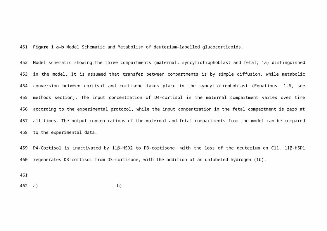
Figure 1 a-b Model Schematic and Metabolism of deuterium-labelled glucocorticoids.
Model schematic showing the three compartments (maternal, syncytiotrophoblast and fetal; 1a) distinguished in the model. It is assumed that
transfer between compartments is by simple diffusion, while metabolic conversion between cortisol and cortisone takes place in the
syncytiotrophoblast (Equations. 1-6, see methods section). The input concentration of D4-cortisol in the maternal compartment varies over time
according to the experimental protocol, while the input concentration in the fetal compartment is zero at all times. The output concentrations of
the maternal and fetal compartments from the model can be compared to the experimental data.
D4-Cortisol is inactivated by 11β-HSD2 to D3-cortisone, with the loss of the deuterium on C11. 11β-HSD1 regenerates D3-cortisol from D3-
cortisone, with the addition of an unlabeled hydrogen (1b).
a) b)
346
347
348
349
350
351
352
353
354
355
356
357
358
359
360
361
362

Figure 2 a-f Model fit of experimental data.
In maternal circulation was 0-30 minutes EBB alone, 30-60 minutes EBB + 20nM D4-Cortisol, 60-90 minutes EBB + 200nM D4-Cortisol, 90-120
minutes EBB + 800nM D4-Cortisol, 120-150 minutes EBB + 800nM D4F + 0.001M Carbenoxolone, 150-170 minutes EBB alone. The
appearance of D4-cortisol in the fetal circulation is consistent with free transplacental passage of D4-cortisol. Inactivation of D4-cortisol by 11β-
HSD2 is indicated by the appearance of D3-cortisone in the maternal or fetal circulations, and cortisol regeneration from D3-cortisone is
indicated by the appearance of D3-cortisol.
Model fit of the experimental data for D4-cortisol in the maternal (2a) and fetal (2b) compartments, with a single set of parameters. Results show
an excellent correspondence between model (straight line) and experiments (plotted data and error bars) (R2 = 0.99). Model prediction of D3-
cortisone in comparison with the scaled experimental data (2c-d). Note the experimental units for D3-cortisone could not be directly related to
concentration and have been scaled here to allow comparison of the relative changes predicted by the model. The same conversion factor was
applied to both maternal and fetal D3-cortisone based on the average ratio between experimental units and computed concentrations at the
highest input level (time points t = 110, 115 and 120 min). Experimental data for D3-cortisol (2e-f). Values were comparatively low and were not
modelled as they do not contribute significantly to the overall mass balance. All experimental results are the average of 5 placentas, expressed
as mean and SEM (n = 5).
Key: D4F (D4-Cortisol), EBB (Earle’s Bicarbonate Buffer), CBX (carbenoxolone).
363
364
365
366
367
368
369
370
371
372
373
374
375
376
377
378
379

0 30 60 90 120 150Time [min]
0
50
100
150
200
250
Feta
l Vei
n D
4-C
ortis
ol (n
M)
0 30 60 90 120 150Time (min)
0
50
100
150
200
250
300
350
Mat
erna
l Vei
n D
3-C
ortis
one
(nM
)
0 30 60 90 120 150Time [min]
0
50
100
150
Feta
l Vei
n D
3-C
ortis
one
(nM
)
a) b)
c) d)
0 30 60 90 120 150Time (min)
0
500
1000
1500
Mat
erna
l Vei
n D
4-C
ortis
ol (n
M)
MaternalPerfusion
800 nMD4F + CBX
800 nMD4F
200 nMD4F
20 nMD4FWashout Maternal
Perfusion800 nM
D4F + CBX800 nM
D4F200 nM
D4F20 nM
D4FWashout
380
381
382
383
384
385
386
387
388
389
390
391
392
393
394
395

e) f)
Figure 3 Sensitivity analysis for D4-Cortisol transfer to the fetus as a function of variations in the model parameters.
The model parameters were varied with respect to the values for the reference fit. The reported changes in placental transfer predicted by the
model were based on the steady state results at the highest maternal input concentration.
MaternalPerfusion
800 nMD4F + CBX
800 nMD4F
200 nMD4F
20 nMD4FWashout
MaternalPerfusion
800 nMD4F + CBX
800 nMD4F
200 nMD4F
20 nMD4FWashout
0 30 60 90 120 150Time (min)
0
2
4
6
8
Mat
erna
l Vei
n D
3-C
ortis
ol (n
M)
0 30 60 90 120 150Time [min]
0
1
2
3
4
5
Feta
l Vei
n D
3-C
ortis
ol (n
M)
MaternalPerfusion
800 nMD4F + CBX
800 nMD4F
200 nMD4F
20 nMD4FWashout Maternal
Perfusion800 nM
D4F + CBX800 nM
D4F200 nM
D4F20 nM
D4FWashout
396
397
398
399
400
401
402
403
404
405
406
407
408

Key: kMVM (MVM permeability constant), kBM (BM permeability constant), Vmax (maximum rate of reaction), Km (Michaelis-Menton constant), Vm
(maternal compartment volume), Vs (syncytiotrophoblast compartment volume), Vf (fetal compartment volume), Qm (maternal flow rate, L/min), Qf
(fetal flow rate, L/min).
409
410
411
412

413
414
415
416

Supplementary Tables417
418
Supplementary Table 1 Mass spectral conditions for analysis of analytes and internal standards by positive ion electrospray ionisation
Abbreviations : Atomic mass units (amu) Quan (quantifier ion), Qual (qualifier ion), V (volts)
Molecular Weight (amu)
Precursor ion (m/z)
Product ion
(m/z)
Quan; Qual
Declustering Potential
(V)
Collision energy
(V)
Quan; Qual
Cell exit potential
(V)
Quan; Qual
ANALYTES
D4-cortisol 367.0 367.0 121; only one 121 25 20
D3-cortisol 366.0 366.0 121.1; only one
121 25 20
D3-cortisone 363.2 364.2 164.0; only one
166 31 14
INTERNAL STANDARDS
Epi-cortisol 363.2 363.2 121.0; 77.0 131 29; 101 14; 14
419
420
421
422
423
424
425
426
427
Supplementary Table 2 Inter-assay precision and accuracy
Concentrations of cortisol, cortisone, D4-Cortisol and D3-Cortisol were determined using
calibration curves. Fourteen standards were prepared in 500 µL EBB (range of
concentrations 0.1 ng – 400 ng) enriched with internal standards (10 ng) along with blank
samples were diluted in 500 µL of water and processed using the same extraction method
and analysis conditions as perfusate samples. Standard curves were plotted by calculating
the peak area (analyte peak area / internal standard peak area). Weighting of 1/x and was
applied to form standard curves of best fit with a regression coefficient above 0.99. The ion
ratio (quantitative ion/qualitative ion) of the analytes was calculated using MultiQuant
software and results were not considered acceptable if the ratio was greater than 20% of the
ratio of the standards. Inter-assay fourteen point standard curve validation (n=6 different day
respectively) was used to assess the limits of quantification of accuracy and precision for
each analyte. Precision was based on the percentage relative standard deviation (%RSD),
which was calculated using peak area ratios. Tissue sample* is intra-assay (amount, ng for
tissue replicates (n=6). Inter-assay was not performed for tissue samples, as all tissue
samples were analysed on the same day. Low values are the limit of quantification for each
analyte.
Concentration (ng/200 μL perfusate or mg tissue*):
mean (SD)
Precision
(% RSD)
Accuracy (%)
D4-Cortisol Low (0.2) 0.21 (0.02) 10.5 103
Mid (50) 46.7 (1.7) 3.6 93
High (400) 396.6 (43.4) 11.6 93
Tissue Sample* 8.3 (0.6) 7.0
D3-Cortisol Low (0.1) 0.1 (0.02) 17.3 98
Mid (10) 10.4 (0.9) 8.8 104
High (20) 20.2 (2.0) 10.1 101
Tissue Sample* 0.6 (0.04) 6.4
37
428
429
430
431
432
433
434
435
436
437
438
439
440
441
442
443
444
445
446
Abbreviations: EBB (Earle’s Bicarbonate Buffer), SD (Standard Deviation), RSD (Relative
Standard Deviation)
38
447
448

Supplementary Method
Details of the model equations and model parameters are described below.
1. Model equations
dC Am
dt= 1vm
(Qm (C A¿ , m−C A
m )−kMVM (CAm−C As )) [1]
dC As
dt= 1v s
(kMVM (C Am−C A
s )−kBM (CAs−C Af )+J Ametab) [2]
dC Af
dt= 1v f
(k BM (C As −CA
f )−Q f CAf ) [3] (Harris,
#687)
where C Am,C A
s and C Af are the concentrations (mol/L) of solute A which can be either D4-
cortisol (D4F), D3-cortisone (D3E) or D3-cortisol (D3F) in the maternal “m”,
syncytiotrophoblast “s” and fetal “f” compartment respectively. Similarly, the volumes v (L) of
the different compartments are indicated with subscripts using the same notation. Qm and Qf
(L/min) are the fluid flow rates in the maternal and fetal circulation. C A¿ , m is the maternal inlet
concentration, which is zero for all solute species except D4-cortisol. Note that the fetal inlet
concentration is zero for all species and therefore has not been included. kMVM and k BM
denote the effective overall permeability constants (L/min) for the microvillous membrane
(MVM) and basal membrane (BM) including surface area. These diffusive permeability
39
449
450
451
452
453
454
455
456
457
458
459
460
461
462
463
464
465
466
467
468
469
constants were assumed to be the same for all solute species. The metabolic conversion
rate J Ametab (mol/min) depends on the solute species as follows:
JD4 Fmetab=
−V 4 F→3Emax CD 4F
s
Km+CD 4Fs [4]
JD3Eme tab=
V 4F→3Emax CD 4F
s
Km+C D4 Fs −
V 3E→ 3Fmax CD3 E
s
Km+CD 3Es [5]
JD3Fmetab=
V 3E→ 3Fmax CD 3E
s
K m+CD3Es [6]
where V max (mol/min) is the maximum overall metabolic conversion rate and Km (mol/L) is
the Michaelis-Menten constant, i.e. the concentration at which half the maximum rate occurs.
2. Model parameters
The total cotyledon volume was based on the average cotyledon weight from the
experiments (30.8 × 10⁻³ kg, n = 5), which was directly equated to the volume in L. The
volume fractions of the maternal, syncytiotrophoblast and fetal compartments distinguished
in the model were set to 34%, 15% and 7.4% respectively, as in our previous work. [14,22]
The flow rates in the maternal and fetal circulations Qm= 14 × 10-3 L/min and Qf= 6 × 10⁻³
L/min were directly based on the experimental settings. To account for any discrepancies
between nominal and actual values, the D4-cortisol input concentrations C A¿ used in the
40
470
471
472
473
474
475
476
477
478
479
480
481
482
483
484
485
486
487
488
489

model were calculated based on the combined maternal and fetal steady state output during
the blocking phase. The Michaelis-Menten constant Km was set to 44 × 10-9 mol/L, based on
the value for the enzyme 11β-HSD2 for cortisol. [23] In first instance the same value was
adopted for both metabolic conversion steps in Equations 4-6.
2. Parameter estimation
The remaining parameters in the model were determined by fitting the experimental data.
The following error criterion was defined for a certain species A and compartment j in
general:
RAj = 1
(CAexp , j )2∑i∈T
(CA ,ij −C A, iexp , j )2 [7]
where C A,ij and C A,i
exp , j are the computed and experimental concentrations at time pointi,
respectively, while C Aexp , j is the mean of the experimental time points considered. The model
was fitted to the steady state values after each change in maternal input concentration,
including the blocking phase, therefore the set of time points T consisted of the last 4 time
points for each different input phase (16 time points in total).
The D3-cortisol concentrations measured experimentally were 300 times smaller
compared to D4-cortisol and did not contribute significantly to the overall mass balance.
Therefore the conversion to D3-cortisol was neglected in the parameter estimation by setting
V 3 E→3 Fmax to zero. In addition, the measured D3-cortisone values could not be directly related
to concentration. Therefore D3-cortisone was not fitted, but instead the experimental values
for D3-cortisone were scaled to allow comparison of the relative changes predicted by the
41
490
491
492
493
494
495
496
497
498
499
500
501
502
503
504
505
506
507
508
509
510
511
512

model. Thus, only the D4-cortisol values in the maternal and fetal compartments (averaged
over 5 placentas) were fitted according to the following overall error criterion:
Rtot=RD 4Fm +RD 4F
f [8]
In total 3 parameters were fitted, the membrane permeability constants kMVM and k BM and
the maximum rate of conversion from cortisol to cortisone V 4 F→3Emax . Time integration of
Equations 1-3 was performed in Matlab (R2016a) using the ode45 function (Runge-Kutta (4,
5) method). Parameter estimation by minimising Eq. 8 was implemented using the
fminsearch function (Nelder-Mead method). Initial parameter estimates were varied to verify
that the algorithm converged to a unique solution.
42
513
514
515
516
517
518
519
520
521
522
523
524